29
Diseases of the Nervous System Neal G. Simon, Ph.D. Professor, Dept of Biological Sciences Lehigh University
Diseases of the Nervous System
Neal G. Simon, Ph.D. Professor, Dept of Biological Sciences
Lehigh University
Outline
A. Stress-related Disorders
1. Emotional Circuitry: Key Components
2. The Hypothalamic Pituitary Adrenal (HPA) Axis B. Alzheimer’s Disease 1. Biomarkers & Ethics

Stress-related CNS Disorders
Major Depression1
15 million in US & growing globally Current standard of care: SRIs/NRIs, 60% of patients do not respond
Intermittent Explosive Disorder2
62 million in US Current standard of care: no approved treatment, off-label use of SSRIs
Impulse Control/Anger Disorders
Core component of borderline personality, antisocial personality, and conduct disorders
o 12 million in US
Common co-morbidity impacting therapeutic response in PTSD, ADHD, & psychoses
Current standard of care: no approved treatment
Post-Traumatic Stress Disorder (PTSD)3 8 million in US, a priority indication for military medicine Major Depression, Intermittent Explosive Disorder, Impulse Control Disorders are co-morbid
Current standard of care: repurposed SSRIs
1 Mathew & Charney (2009); NIMH 2 Coccaro (2012); Kessler et al. (2006) 3 NIMH; USAMRMC 3
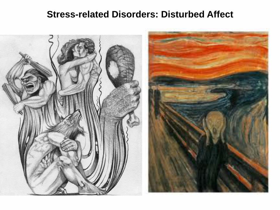
Stress-related Disorders: Disturbed Affect

Basic Neurobiology & Physiology
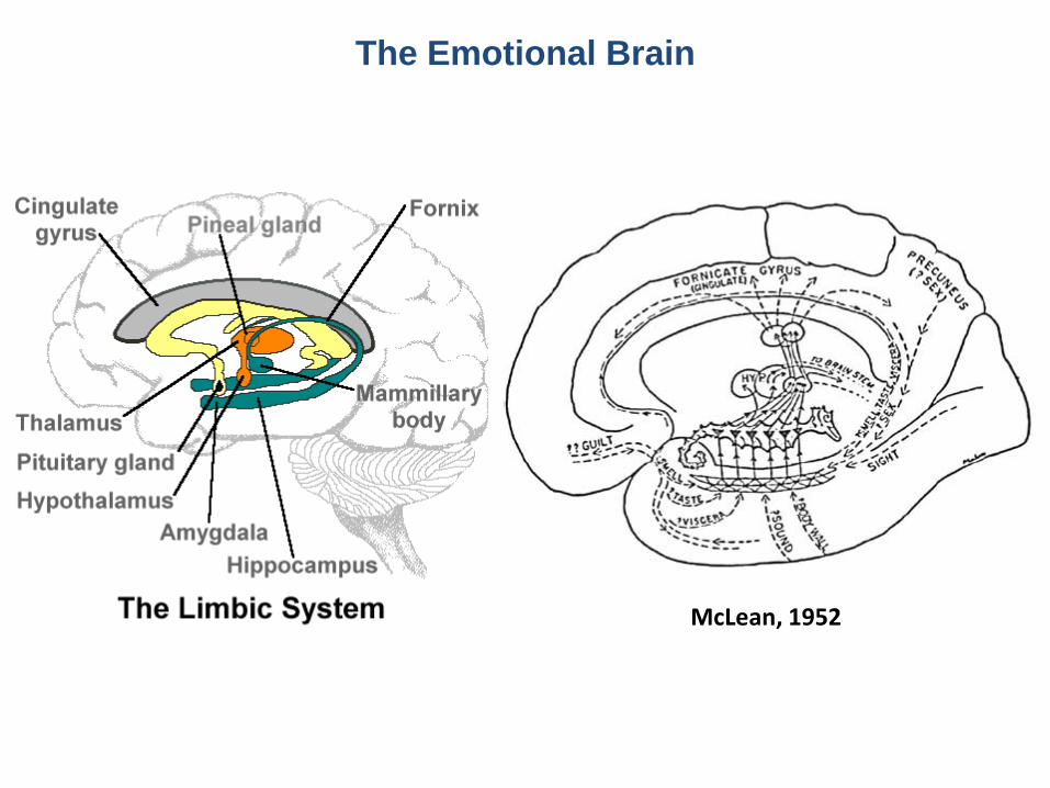
The Emotional Brain
McLean, 1952
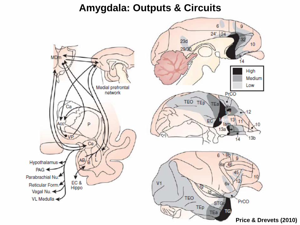
Amygdala: Outputs & Circuits
Price & Drevets (2010)
Biological Sciences
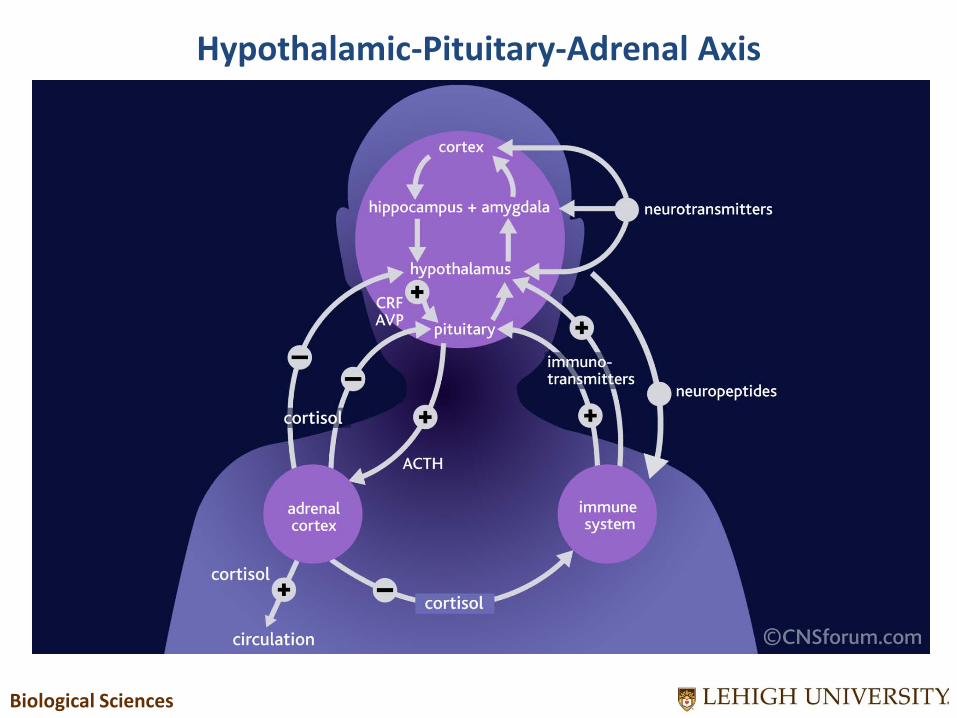
Hypothalamic-Pituitary-Adrenal Axis
Biological Sciences
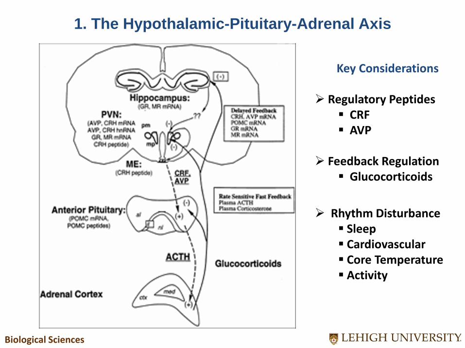
1. The Hypothalamic-Pituitary-Adrenal Axis
Key Considerations Regulatory Peptides
CRF AVP
Feedback Regulation
Glucocorticoids
Rhythm Disturbance Sleep Cardiovascular Core Temperature Activity
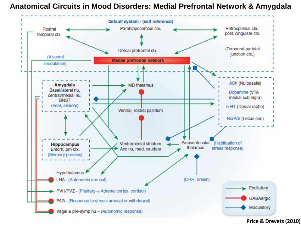
Anatomical Circuits in Mood Disorders: Medial Prefrontal Network & Amygdala
Price & Drevets (2010)
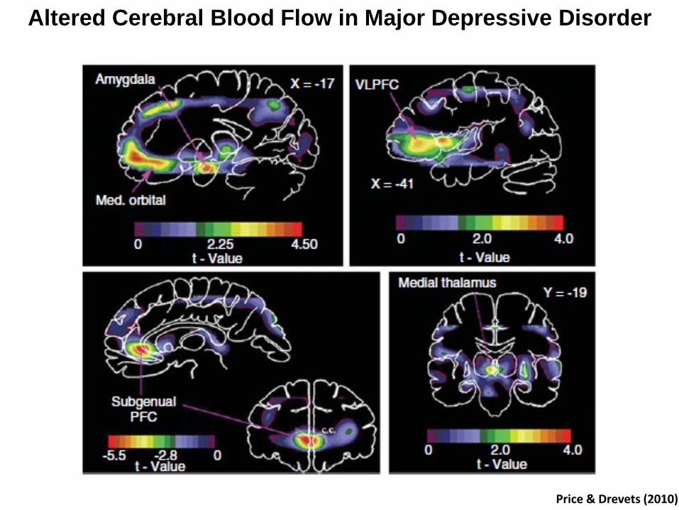
Altered Cerebral Blood Flow in Major Depressive Disorder
Price & Drevets (2010)
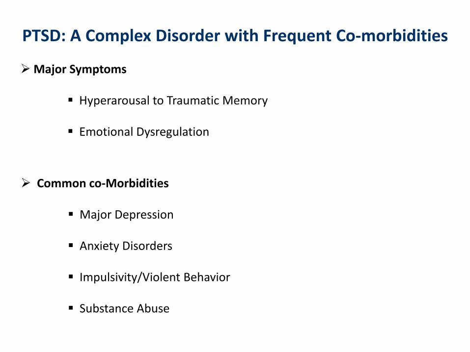
Common co-Morbidities
Major Depression
Anxiety Disorders
Impulsivity/Violent Behavior
Substance Abuse
PTSD: A Complex Disorder with Frequent Co-morbidities
Major Symptoms
Hyperarousal to Traumatic Memory
Emotional Dysregulation
Biological Sciences
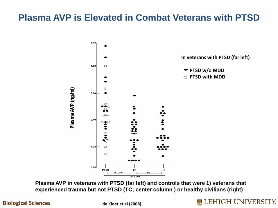
Plasma AVP is Elevated in Combat Veterans with PTSD
In veterans with PTSD (leftmost column):
de Kloet et al (2008)
Plas
ma A
VP (n
g/m
l)
In veterans with PTSD (leftmost column):
In veterans with PTSD (far left) PTSD w/o MDD PTSD with MDD
Plasma AVP in veterans with PTSD (far left) and controls that were 1) veterans that experienced trauma but not PTSD (TC; center column ) or healthy civilians (right)

Predatory Conditioned Fear – A Model of PTSD
sable ferret rat

Vasopressin Receptor Blockade is Effective in a Conditioned Fear PTSD Model
V1a Blockade
V1a receptor block significantly reduced hyperarousal in brain regions mediating fear & memory two weeks after traumatic fear conditioning Normal fear responses & arousal patterns were unaffected
Unconditioned Fear Response
Conditioned Fear Response
V1a Blockade Effect
15
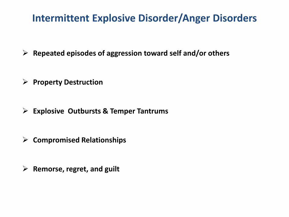
Intermittent Explosive Disorder/Anger Disorders
Repeated episodes of aggression toward self and/or others
Property Destruction
Explosive Outbursts & Temper Tantrums
Compromised Relationships
Remorse, regret, and guilt

Mat
e &
Intr
uder
Str
ess
Para
digm
Pr
etre
atm
ent w
ith
AVP
Bl
ocke
r bef
ore
Stre
ss
Amygdala Hippocampus Thalamus
Vasopressin Blockade: Neuroimaging in Major Brain Regions Linked to Stress-related Disorders
From Ferris et al. (2008)
AVP Blockade attenuates arousal, stress, fear, and aggressive motivation
Sexual motivation and performance remain intact
17
Cortex

Correlation between Aggression Against Persons (the fighting and assault items) scores on the Life History of Aggression (LHA) assessment and cerebrospinal fluid (CSF) arginine vasopression (AVP) concentrations in 26 individuals who met the DSM-IV criteria for personality disorder.
CSF AVP is Correlated with Fighting & Assault Scores
Coccaro et al (1997)
Summary: Peptides & Stress-related Disorders
CNS AVP receptors are implicated in stress-related disorders through preclinical models & human results
Human studies suggest the involvement of the vasopressin system
Disease-specific circuitry remains to be characterized
Social Neurobiology can potentially identify new pathways for intervention
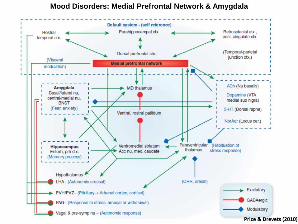
Mood Disorders: Medial Prefrontal Network & Amygdala
Price & Drevets (2010)
Alzheimer’s Disease

Clinical & Biomarker Changes in Dominantly Inherited Alzheimer’s Disease
Bateman et al (2012)
Autosomal Dominant Alzheimer's Disease
Amyloid Hypothesis: 3 genes can cause altered processing (APP, PSEN1, PSEN2)
Compared Carriers & Non-Carriers (n =128)
Clinical, Cognitive, Imaging, & Biochemical Assessments
Normalized against parental age of onset
Follows ADNI protocols and standards
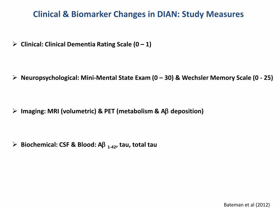
Clinical & Biomarker Changes in DIAN: Study Measures
Bateman et al (2012)
Clinical: Clinical Dementia Rating Scale (0 – 1)
Neuropsychological: Mini-Mental State Exam (0 – 30) & Wechsler Memory Scale (0 - 25)
Imaging: MRI (volumetric) & PET (metabolism & Aβ deposition)
Biochemical: CSF & Blood: Aβ 1-42, tau, total tau
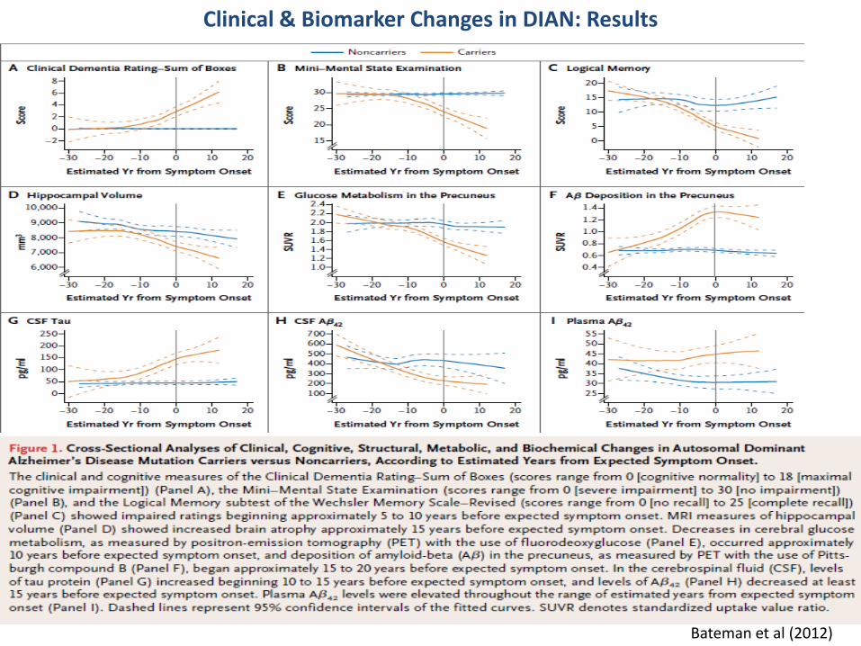
Clinical & Biomarker Changes in DIAN: Results
Bateman et al (2012)

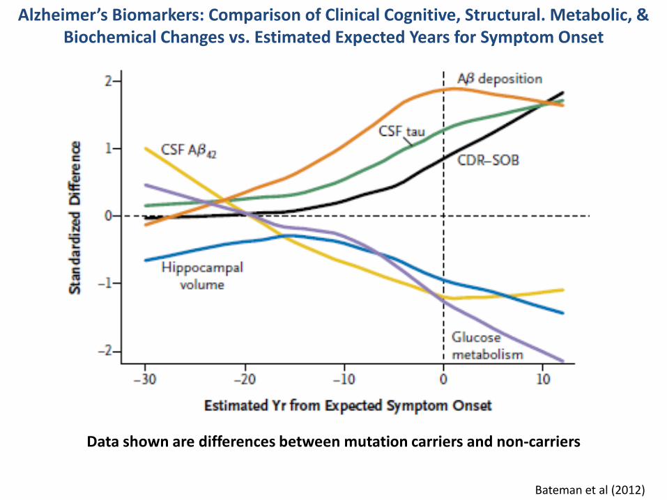
Alzheimer’s Biomarkers: Comparison of Clinical Cognitive, Structural, Metabolic, & Biochemical Changes vs. Estimated Expected Years for Symptom Onset
Bateman et al (2012)
Clinical Dementia Rating: + 5 years
Imaging MRI: Hippocampal Brain Atrophy + 15 years
Cerebral Metabolism: +10 years
Amyloid-β Deposition: + 15 years
Biochemistry
CSF Aβ 1-42: +20 years
CSF Tau: + 15 years
Precuneus
Alzheimer’s Biomarkers: Comparison of Clinical Cognitive, Structural. Metabolic, & Biochemical Changes vs. Estimated Expected Years for Symptom Onset
Bateman et al (2012)
Data shown are differences between mutation carriers and non-carriers
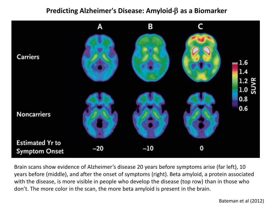
Brain scans show evidence of Alzheimer’s disease 20 years before symptoms arise (far left), 10 years before (middle), and after the onset of symptoms (right). Beta amyloid, a protein associated with the disease, is more visible in people who develop the disease (top row) than in those who don’t. The more color in the scan, the more beta amyloid is present in the brain.
Predicting Alzheimer's Disease: Amyloid-β as a Biomarker
Bateman et al (2012)

What Would You Do?

Thank you for your time and attention


