DNA Methylation Profiling Reveals Correlation of Differential Methylation Patterns with Gene Expression in Human Epilepsy Liang Wang 1 & Xinwei Fu 1 & Xi Peng 2 & Zheng Xiao 1 & Zhonggui Li 1 & Guojun Chen 1 & Xuefeng Wang 1 Received: 4 December 2015 /Accepted: 4 March 2016 /Published online: 12 April 2016 # Springer Science+Business Media New York 2016 Abstract DNA methylation plays important roles in regulat- ing gene expression and has been reported to be related with epilepsy. This study aimed to define differential DNA meth- ylation patterns in drug-refractory epilepsy patients and to investigate the role of DNA methylation in human epilepsy. We performed DNA methylation profiling in brain tissues from epileptic and control patients via methylated-cytosine DNA immunoprecipitation microarray chip. Differentially methylated loci were validated by bisulfite sequencing PCR, and the messenger RNA (mRNA) levels of candidate genes were evaluated by reverse transcriptase PCR. We found 224 genes that showed differential DNA methylation between ep- ileptic patients and controls. Among the seven candidate genes, three genes (TUBB2B, ATPGD1, and HTR6) showed relative transcriptional regulation by DNA methylation. TUBB2B and ATPGD1 exhibited hyperme- thylation and decreased mRNA levels, whereas HTR6 displayed hypomethylation and increased mRNA levels in the epileptic samples. Our findings suggest that cer- tain genes become differentially regulated by DNA methylation in human epilepsy. Keywords Human . Epilepsy . DNA methylation . Microarray . Gene expression Introduction Epilepsy is a common and refractory neurological disorder, which affects approximately 0.5–1 % of the world population and often requires lifelong medication (Ngugi et al., 2011). In a previous microarray-based study, we identified 243 genes that were differentially expressed in the anterior temporal neocortex of drug-refractory epilepsy patients relative to control patients (Xi et al., 2009). It is not possible to conclude that the differential expression of these genes either caused or resulted from epileptic seizures. Furthermore, the factors regulating the differentially expressed genes were unclear. Epigenetic mechanisms are one means by which gene expression is regulated, and in the central nervous system (CNS), epigenetic mechanisms are critical for promoting brain development, neural stem cell maintenance, pro- liferation, neurogenesis, gliogenesis, cellular migration, and syn- aptic and neural network connectivity (Delcuve et al., 2009). DNA methylation is a major epigenetic modification and plays important roles in regulating gene expression via reversible and dynamic chromatin remodeling processes. Kobow and Blümcke (2011) and Kobow et al. (2013b) first presented a methylation hypothesis for epileptogenesis and pharmacoresistance in epilep- sy, and recent experimental studies provide some evidence for a relationship between abnormal DNA methylation and epileptogenesis. DNA methylation is catalyzed by DNA meth- yltransferases (DNMTs). In our previous study, we showed that DNMT1 and DNMT3a were upregulated in the brain tissue of human epilepsy cases (Zhu et al., 2012). Kobow et al. (2009) examined reelin (RELN) promoter methylation in hippocampal subregions of temporal lobe specimens obtained from epilepsy patients, and deep sequencing revealed increased DNA Electronic supplementary material The online version of this article (doi:10.1007/s12031-016-0735-6) contains supplementary material, which is available to authorized users. * Xuefeng Wang [email protected]1 Department of Neurology, The First Affiliated Hospital of Chongqing Medical University, Chongqing Key Laboratory of Neurology, Chongqing 400016, China 2 Department of Neurology, The Second Affiliated Hospital of Chongqing Medical University, Chongqing 400010, China J Mol Neurosci (2016) 59:68–77 DOI 10.1007/s12031-016-0735-6
Transcript
DNA Methylation Profiling Reveals Correlation of DifferentialMethylation Patterns with Gene Expression in Human Epilepsy
Received: 4 December 2015 /Accepted: 4 March 2016 /Published online: 12 April 2016# Springer Science+Business Media New York 2016
Abstract DNA methylation plays important roles in regulat-ing gene expression and has been reported to be related withepilepsy. This study aimed to define differential DNA meth-ylation patterns in drug-refractory epilepsy patients and toinvestigate the role of DNA methylation in human epilepsy.We performed DNA methylation profiling in brain tissuesfrom epileptic and control patients via methylated-cytosineDNA immunoprecipitation microarray chip. Differentiallymethylated loci were validated by bisulfite sequencing PCR,and the messenger RNA (mRNA) levels of candidate geneswere evaluated by reverse transcriptase PCR. We found 224genes that showed differential DNA methylation between ep-ileptic patients and controls. Among the seven candidategenes, three genes (TUBB2B, ATPGD1, and HTR6)showed relative transcriptional regulation by DNAmethylation. TUBB2B and ATPGD1 exhibited hyperme-thylation and decreased mRNA levels, whereas HTR6displayed hypomethylation and increased mRNA levelsin the epileptic samples. Our findings suggest that cer-tain genes become differentially regulated by DNAmethylation in human epilepsy.
Keywords Human . Epilepsy . DNAmethylation .
Microarray . Gene expression
Introduction
Epilepsy is a common and refractory neurological disorder,which affects approximately 0.5–1 % of the world populationand often requires lifelong medication (Ngugi et al., 2011). In aprevious microarray-based study, we identified 243 genes thatwere differentially expressed in the anterior temporal neocortexof drug-refractory epilepsy patients relative to control patients (Xiet al., 2009). It is not possible to conclude that the differentialexpression of these genes either caused or resulted from epilepticseizures. Furthermore, the factors regulating the differentiallyexpressed genes were unclear. Epigenetic mechanisms are onemeans by which gene expression is regulated, and in the centralnervous system (CNS), epigenetic mechanisms are critical forpromoting brain development, neural stem cellmaintenance, pro-liferation, neurogenesis, gliogenesis, cellular migration, and syn-aptic and neural network connectivity (Delcuve et al., 2009).DNA methylation is a major epigenetic modification and playsimportant roles in regulating gene expression via reversible anddynamic chromatin remodeling processes. Kobow and Blümcke(2011) and Kobow et al. (2013b) first presented a methylationhypothesis for epileptogenesis and pharmacoresistance in epilep-sy, and recent experimental studies provide some evidence for arelationship between abnormal DNA methylation andepileptogenesis. DNA methylation is catalyzed by DNA meth-yltransferases (DNMTs). In our previous study, we showed thatDNMT1 and DNMT3a were upregulated in the brain tissue ofhuman epilepsy cases (Zhu et al., 2012). Kobow et al. (2009)examined reelin (RELN) promoter methylation in hippocampalsubregions of temporal lobe specimens obtained from epilepsypatients, and deep sequencing revealed increased DNA
Electronic supplementary material The online version of this article(doi:10.1007/s12031-016-0735-6) contains supplementary material,which is available to authorized users.
methylation in chronic rat epilepsy (Kobow et al., 2013a).Miller-Delaney et al. (2012) found genome-wide DNA methylationchanges after status epilepticus and in epileptic tolerance, whichmay contribute to regulating gene expression in the seizure-damaged hippocampus. Machnes et al. (2013) reported thatDNA methylation mediates persistent epileptiform activity invitro and in vivo and that pharmacological agents that blockDNA methylation inhibit epileptiform activity raising theprospect of DNA methylation inhibitors in epilepsytherapeutics. Belhedi et al. (2014) described increased CPA6promoter methylation in focal epilepsy and in febrile seizures.
Differential DNA methylation patterns may contribute to thediversity of phenotypes, pathogenesis, and progression of com-plex diseases. Strong association has been identified betweendifferential DNA methylation and cancer (Esteller, 2008), andthere are now ongoing efforts to investigate links betweenDNA methylation and other complex diseases. In the cur-rent study, we investigated the DNA methylation-basedregulation of gene expression in human epilepsy by com-paring global DNA methylation profiles in human braintissue between epileptic and control patients using amethylated-cytosine DNA immunoprecipitation microar-ray chip.
Materials and Methods
Patients
Human brain tissue was obtained from patients withdrug-refractory epilepsy who had undergone surgicalremoval of the epileptogenic zone in their temporalneocortex. The epileptogenic zones were determinedusing a combination of ictal semiology, brain magneticresonance imaging, video-EEG, sphenoidal electrodemonitoring, and intracranial EEG before surgeryand by intraoperative electrocorticography (EcoG).According to these analyses, the anterior temporalneocortical tissues capable of epileptiform dischargeswere resected. After resection, the EcoG electrodes wereplaced on the edges of the remaining tissues to ensurethat the dischargeable tissues had been resectedcompletely. All patients were refractory to maximaldoses of at least three anti-epilepsy drugs (AEDs).Brain magnetic resonance imaging with routine scansequences found no progressive lesions in the CNS.For comparison, histologically normal anterior temporalneocortex samples were obtained from patients whowere treated for post-trauma intracranial hypertension.These subjects had no history of epilepsy or of expo-sure to AEDs. Individual patient details are listed inSupplemental Table 1.
Human Brain Tissue Processing
The excised brain tissue was immediately placed in a cryovialcontaining buffered diethylpyrocarbonate and stored in liquidnitrogen until use.
Methylated-Cytosine DNA Immunoprecipitation Chip
According to previously described protocols (Rakyan et al.,2008, Schumacher et al., 2008), genomic DNAwas extractedfrom brain tissues using the DNeasy Blood & Tissue Kit ac-cording to the manufacturer’s instructions (Qiagen, Crawley,UK). Next, 11 μg global DNA (gDNA) of each sample wasdiluted in 300 μl TE buffer and sonicated into random ~500-bp fragments using a Bioruptor (Diagenode, Belgium).Immunoprecipitation of methylated DNA was performedusing a mouse monoclonal anti-5-methyl cytosine antibody(Diagenode) and Biomag magnetic beads (BangsLaboratories, USA). Five micrograms of anti-5-methyl cyto-sine antibody was added to the MeDIP sample, 5 μg mouseIgG (Jackson) to the mock IP sample, and nothing to the inputsample. The MeDIP quality was assessed by qPCR of H19(positive control) and GAPDH (negative control). Sampleswere rotated overnight at 4 °C, and then 50 μl of mag-netic beads coupled to anti-mouse IgG was added andmixed. Samples were rotated for an additional 2 h andthen washed three times with wash buffers before elut-ing with 200 μl elution buffer (1 mM Tris-HCl, pH 8.0,0.5 mM EDTA, 10 % SDS). The immunoprecipitatedDNA was purified by phenol chloroform extractionand ethanol precipitation.
For each sample, 1 μg of input and IP DNA was labeledwith Cy3- and Cy5-labeled random 9-mers, respectively, andco-hybridized to the Nimblegen HG 18 CpG promoter micro-array (Nimblegen, WI, USA). This Nimblegen microarray is asingle array design with ~385,000 probes that span all knownhuman CpG islands annotated on the UCSC genome browserand all well-characterized Human Refseq gene promoters (24,659) ranging from −2800 to +200 bp relative to the transcrip-tion start site (TSS). Scanning was performed with an AxonGenePix 4000B microarray scanner. Raw data were extractedas pair files by NimbleScan software (Nimblegene,USA). Several procedures were then sequentially per-formed to obtain peak data with specified parameters(sliding window width 750 bp; mini probes per peak 2; pvalue minimum cutoff 2; maximum spacing between nearbyprobes per peak 500 bp).
Bisulfite Sequencing PCR
The methylation status of the CpG islands in candidategene promoters was determined by bisulfite sequencingas described previously (Carr et al., 2007, Movassagh
J Mol Neurosci (2016) 59:68–77 69
et al., 2010). The primers were designed using Methprimersoftware (http://www.urogene.org/methprimer). Primer in-formation is shown in Supplemental Table 2. Bisulfite con-version of gDNA was performed using the EZ DNAMethylation-Gold kit (Zymo Research, Orange, CA,USA) according to the manufacturer’s protocol. Afterpurification, 5 μl of bisulfite-treated DNA was amplifiedby PCR. PCR conditions were as follows: 95 °C for 5 min;42 cycles of 95 °C for 30 s, 57 °C for 30 s, and 72 °C for40 s; and a final extension at 72 °C for 10 min. ThePCR products were purified and cloned using a Pmd19-T vector kit (Takara). To determine the methylationstatus of candidate gene promoters, an average offive positive clones were sequenced using the M13reverse primer and an automated ABI prism 3730xlGenetic Analyzer (Applied Biosystems, USA). The
sequencing results were analyzed using QUMA software(http://quma.cdb.riken.jp).
RT-PCR
Total RNA was isolated from brain tissue using Trizolreagent according to the manufacturer’s protocol(Invitrogen, Paisley, UK). Next, 20 μl complementaryDNA (cDNA) was synthesized from 1 μg total RNAusing the PrimeScript RT reagent Kit (Takara). Primerswere designed with Primer 5.0 and synthesized bySangon Biotech (Shanghai, China). Primer informationis shown in Supplemental Table 3. For PCR reactions,2 ng cDNA were amplified in a 25-μl reaction volumecontaining 0.5 μM of each forward and reverse primerand 12.5 μl 2 × PCR TaqMix (TianGen Biotech,
Fig. 1 DNA methylation profiles of epileptic patients and controls. Allprobes were mapped onto the human genome (NCBIv35) by BLASTandare depicted as vertical lines. The methylation profile of epileptic patientsis shown above the line and that of controls is displayed below the line.
Unmethylated regions are marked blue, yellow represents intermediatelymethylated (IM) regions, red represents densely methylated regions, andgreen represents centromeres
Fig. 2 Unsupervised clusteringfor six samples (UMunmethylation; M methylation)
70 J Mol Neurosci (2016) 59:68–77
Beijing, China) under the following conditions: 5 minof denaturation at 95 °C; followed by 35 cycles of 30 sat 95 °C, 30 s at 60 °C, and 60 s at 72 °C; and a finalextension at 72 °C for 10 min. The amplified productswere separated on 2 % agarose gels. Densitometryanalysis was performed with Quantity One software(Bio-Rad Laboratories, USA).
Data Analysis
Analysis of the Nimblegen methylated-cytosine DNAimmunoprecipitation chip (MeDIP-chip) data wasperformed using the ACME algorithm as previouslydescribed (Scacheri et al., 2006). The ACME algorithmcan be employed in cases where a fixed-length windowis slid along the length of each chromosome, using aone-sided Kolmogorov–Smirnov (KS) test at each probelocation to determine whether the surrounding windowis enriched for high-intensity probes relative to the restof the array. Each probe has a corresponding p valuescore (−log 10), and a threshold is set to select regionsenriched in the test sample. The data generated by thesoftware were exported in a spreadsheet format andprocessed using Microsoft Excel. The differentiallymethylated genes were analyzed using gene ontology(GO) functional categories based on the DAVIDdatabase (Huang da et al., 2009).
Data are reported as means ± standard deviation(SD). Statistical analysis was performed using SPSS
11.5. The Mann–Whitney U test was used to analyzethe small sample cohorts, and Student’s t test was usedto analyze real-time PCR (RT-PCR) results. A two-tailedp value of less than 0.05 was considered statisticallysignificant.
Results
Demographic and Clinical Characteristics of Subjects
The epileptic patients consisted of 12 males and 13females and had a mean age of 26.04 ± 8.69 years. Ofthese patients, 8 % had a history of seizure recurrence ofless than 5 years, 56 % had a history of between 5 and15 years, and 36 % had a clinical history lasting morethan 15 years. In addition, 16 % of the patients had lessthan four seizures per month, 64 % had four to tenseizures per month, and 20 % of patients had more thanten seizures per month. The control group had an averageage of 34.50 ± 14.05 years and consisted of six males andfour females. Supplemental Table 1 summarizes the
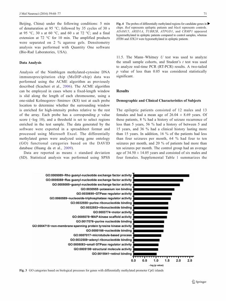
Fig. 3 GO categories based on biological processes for genes with differentially methylated promoter CpG islands
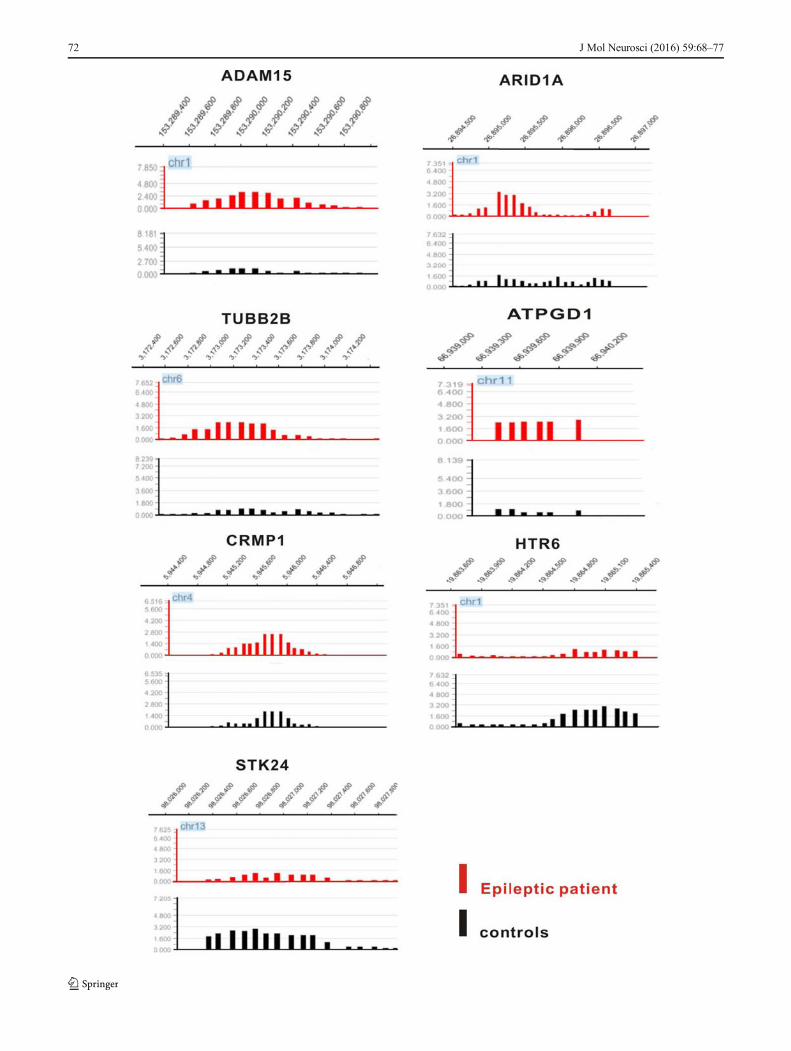
�Fig. 4 The probes of differentially methylated regions for candidate genes inchips. Red represents epileptic patients and black represents controls.ADAM15, ARID1A, TUBB2B, ATPGD1, and CRMP1 appearedhypermethylated in epileptic patients compared to control samples, whereasHTR6 and STK24 were hypomethylated in epileptic patients
J Mol Neurosci (2016) 59:68–77 71
72 J Mol Neurosci (2016) 59:68–77
demographic and clinical characteristics of all subjectswho participated in this study.
DNA Methylation Profiling of Brain Tissuesfrom Epileptic Patients
To obtain a comprehensive profile of aberrant DNAmethylation patterns in the epileptic patients, we usedthe Nimblegen HG 18 CpG promoter microarray chip toanalyze samples from three epileptic patients and threecontrols. Based on the ACEM algorithm, peaks weredefined as regions containing DNA methylation. Eachprobe had a p value, and –log (p value) represented thepeak score; when the peak score was more than 2, theprobe was selected. If several adjacent probes hadp values <0.01, then the group was classified as one peak.We used this cutoff to identity the peaks. By these criteria,we found 1676, 2138, 2371, 2148, 2299, and 2391 peaksfor patient 1 (P1), P2, P3, control 1 (C1), C2, and C3,with 578, 645, 740, 656, 613, and 736 peaks inpromoters, respectively. In terms of mean number ofpeaks in promoters, there was no significant differencebetween epileptic patients and controls (p > 0.05).Figure 1 shows the DNA methylation profile of braintissues from epileptic patients. Figure 2 displays themethylation profile clustering of the six samples.
Candidate genes were identified by peaks overlapping thepromoter region of the relevant transcript (approximately −800to +200 bp). Differentially methylated genes were defined asgenes with two or three peaks in the promoter region that weredifferential between epileptic patients and the controlgroup. Under these criteria, 224 genes were selected (seeSupplemental Table 5). Differentially methylated genes wereclassified by gene ontology (Fig. 3 and SupplementalTable 4). Rho guanyl-nucleotide exchange factor activitywas the most significant GO term.
DNA Methylation Analysis of Candidate Genes
We used bisulfite sequencing to verify the microarray resultsfor seven selected genes (ADAM15, ARID1A, TUBB2B,ATPGD1, CRMP1, and STK24), which are implicated in epi-lepsy or seizure according to the literature (Ortiz et al., 2005,Freitas et al., 2009, Jaglin and Chelly, 2009, Luo et al., 2012,Miyaji et al., 2012). The CpG islands were located in theestablished promoters of these genes. For bisulfite sequenc-ing, regions within CpG islands of approximately 400 bp wereselected. ARID1A, TUBB2B, ATPGD1, and CRMP1 werehypermethylated in epileptic patients compared to controlsamples, whereas HTR6 and STK24 were hypomethylated inepileptic patients (Fig. 4). Figure 5a–g shows the sequencing
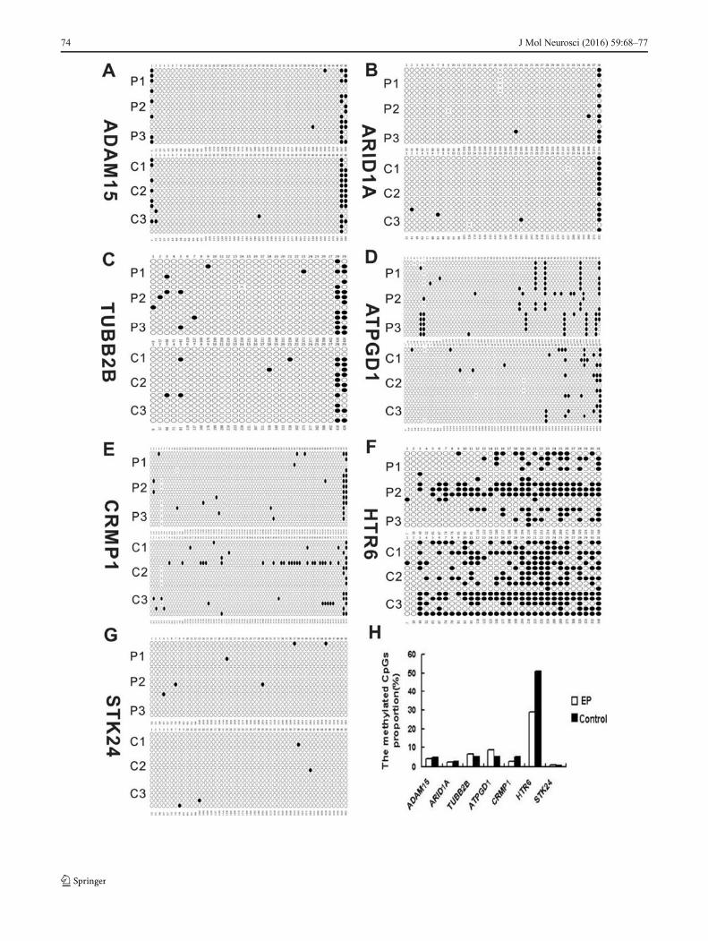
results of five positive clones from among the six samples forthe seven candidate genes. The proportion of methylatedCpGs in TUBB2B, ATPGD1, and HTR6 was 6.7, 8.7, and29.2 % in epileptic patients, respectively, and 5.1, 5, and50.8 % in controls, respectively (Fig. 5h). For TUBB2B,ATPGD1, and HTR6, the bisulfite sequencing results wereconsistent with the MeDIP-chip data. Methylation ofADAM15, ARID1A, CRMP1, and STK24 promoters was notsignificantly different between epileptic and control samples.
Transcriptional Levels of Candidate Genes
Hypomethylation of DNA in the promoter region of a gene isconsidered to be associated with increased transcription, whereashypermethylation in the body of a gene is associated with activetranscription. We therefore set out to determine the expressionlevels of the candidate genes in 25 epileptic samples and 10control samples by RT-PCR. The messenger RNA (mRNA)levels of candidate genes are shown in Fig. 6. mRNA levels ofCRMP1 and HTR6 were upregulated, while those of ADAM15,TUBB2B, and ATPGD1 were downregulated in epileptic sam-ples relative to controls. Expression of ARID1A and STK24 wasnot significantly different between epileptic and control samples.Combining the microarray and bisulfite sequencing results, weobserved that in the epileptic samples, TUBB2B and ATPGD1exhibited relatively hypermethylated promoters and decreasedmRNA levels, while HTR6 displayed a relativelyhypomethylated promoter and higher mRNA levels.
Discussion
In this study, we performed a DNA methylation profile anal-ysis using MeDIP-chip of brain tissues from epileptic patientsand controls. This screen detected several genes that weredifferentially methylated between epileptic patients andcontrols.
The methylated DNA immunoprecipitation microarray(MeDIP-chip) is a genome-wide, high-resolution approachto detect DNA methylation in the whole genome or specifi-cally in CpG islands. We used this method to profile DNAmethylation patterns in the brain tissue of epileptic patients.Surprisingly, the total number of methylated regions was notsignificantly different between epileptic patients and controls.
�Fig. 5 Detailed DNA methylation patterns of the candidate genes weredetermined by bisulfite sequencing PCR. a–g The sequencing results offive positive clones of seven candidate genes among the six samples.Each circle represents a CG dinucleotide, with white indicatingunmethylated residues and black indicating methylated residues. h Theproportion of methylated CpG islands of candidate genes in epilepticpatients and controls
J Mol Neurosci (2016) 59:68–77 73
74 J Mol Neurosci (2016) 59:68–77
This result suggests that recurrent epileptic seizures in patientsdo not induce genome-wide changes in DNA methylation inbrain tissues, unlike the general hypomethylation found incancer (Esteller, 2007). However, hundreds of differentiallymethylated regions were detected between epileptic patientsand controls, with some hypermethylated and somehypomethylated. Some studies reported that the methylationstatus of the BDNF and RELN promoters in brain tissue cor-related with changes in transcriptional activity (Chen et al.,2003, Grayson et al., 2005). Notably, increased RELN promot-er methylation was associated with granule cell dispersion inhuman temporal lobe epilepsy (Kobow et al., 2009). However,we did not detect differential methylation at these promoters inour samples.
GO analysis allows the investigation of functionally linkedbiological pathways in microarray datasets. In our microarraydata, several interesting GO categories were identified amongthe differentially methylated genes, including regulation ofRho, microtubule-based processes, and mitogen-activated pro-tein kinase (MAPK) scaffold activity. These biological processesplay important roles in axonal growth, neurotransmitter release,and synapse reorganization. It was previously reported thatRhoA was activated in the cortex and hippocampus after trau-matic brain injury and kainic acid-induced seizures (Dubreuil
et al., 2006). Likewise, we found increased expression ofRhoA in human temporal lobe epilepsy (Yuan et al., 2010).The neuronal cytoskeleton consists of microtubules, actin fila-ments, intermediate filaments, and associated proteins. A numberof mutations and altered levels of microtubule-associated pro-teins are likely to contribute to the pathogenesis of epilepsythrough mechanisms such as increased neurotrophic support toneurons and increased sprouting of mossy fibers (Gardiner andMarc, 2010). Finally, activation of MAPK signaling causedspontaneous epileptic seizures in an animal model (Nateri et al.,2007), and we determined that the extracellular signal-regulatedkinases (ERK) 1, ERK2, and phosphorylated ERK were upreg-ulated in human intractable epilepsy (Xi et al., 2007).
Bisulfite sequencing is a method for positioning and quanti-fying DNA methylation in promoter regions. We performed thistechnique to verify the results of the MeDIP-chip. The TUBB2B,ATPGD1, and HTR6 promoters exhibited differential methyla-tion between epileptic patients and controls, consistent with themicroarray data. However, the methylation status of the othercandidate genes was not different between epileptic patientsand controls. The microarray is a high-throughput screeningtechnology that is expected to yield a certain amount false-positive results; however, it can still reveal global DNA methyl-ation patterns. Abnormal hypermethylation of promoter CpG
Fig. 6 Differential mRNA expression levels of candidate genes inepileptic patients (n = 25) and controls (n = 10). Lane 1 is marker, lanes2–6 represent controls, and lanes 7–11 represent epileptic patients.mRNA levels of CRMP1 and HTR6 were upregulated and those of
ADAM15, TUBB2B, and ATPGD1 were downregulated in epilepticsamples relative to controls (*p < 0.05; **p < 0.01). Expression ofARID1A and STK24 was not significantly different between epilepticpatients and controls
J Mol Neurosci (2016) 59:68–77 75
islands results in transcriptional silencing,while hypomethylationcorrelates with increased transcription (El-Osta and Wolffe,2000). Interestingly, we found that in epileptic patients, hyper-methylation of the TUBB2B and ATPGD1 promoters correlatedwith reduced gene expression and hypomethylation of theHTR6promoter correlated with increased expression. This result sug-gests that the expression of TUBB2B, ATPGD1, and HTR6 isregulated by DNA methylation. With regard to the other candi-date genes, transcription of CRMP1 was increased, ADAM15was decreased, and ARID1A and STK24 were not altered inepileptic patients. DNA methylation is not the only epigeneticregulator of gene transcription, as histone acetylation andmicroRNAs are also important epigenetic mechanisms for mod-ulating gene transcription (Gibney and Nolan, 2010). For thesegenes, the degree of DNA methylation may, therefore, not nec-essarily correlate with transcriptional levels because they couldbe regulated by other mechanisms.
Functionally, the TUBB2B protein belongs to the β-tubulinfamily, which is the major constituent of microtubules. TUBB2Bhas been implicated in neuronal migration (Jaglin et al., 2009).TUBB2B mutations and aberrant tubulin heterodimer assemblylead to a large spectrum of neuronal migration disorders, includ-ing refractory epilepsy (Uribe, 2010). ATPGD1 is another namefor carnosine synthase 1, which is mainly found in skeletal mus-cle and the CNS and catalyzes the synthesis of carnosine (withthe conversion of ATP to AMP and inorganic pyrophosphate).Carnosine inhibits pentylenetetrazol-induced seizures by hista-minergic mechanisms in histidine decarboxylase knock-out mice(Zhu et al., 2007). HTR6 is one of several receptors for 5-hydroxytryptamine (serotonin). Functions of HTR6 includemodulation of cholinergic and dopaminergic neurotransmission.There is increasing evidence that serotonergic neurotransmissionmodulates a wide variety of experimentally induced seizures(Bagdy et al., 2007). The roles of HTR1A, HTR2C, HTR3,and HTR7 subtypes in epileptogenesis and/or propagation havebeen described (Bagdy et al., 2007). Our current findings revealthat DNA methylation regulates TUBB2B, ATPGD1, and HTR6gene expression and that these genes might play important rolesin epileptogenesis by affecting neuronal migration, carnosinemetabolism, and serotonergic neurotransmission, respectively.
In the present study, we profiled DNAmethylation patterns inbrain tissues of epileptic patients. Because of the limitations as-sociated with studying human brain tissue, some experimentaldetails require explanation. As control subjects, we used histo-logically normal anterior temporal neocortex samples of patientswith traumatic brain injury. However, we should consider thatepilepsy can be a potential complication of such patients and thatinjury causes broad molecular changes, including epigeneticmodifications (Diaz-Arrastia et al., 2009, Lundberg et al.,2009). Furthermore, DNA methylation is dynamically regulatedand influenced by a variety of factors, including gender, age, andsocial environment (Christensen et al., 2009). Methylation pat-terns also differ between brain regions and neuronal cell types
(Ladd-Acosta et al., 2007, Siegmund et al., 2007). We selectedthe right anterior temporal neocortex in six males of similar agefor DNA methylation analysis to reduce interference factors.However, we used DNA extracted from whole brain tissue ho-mogenates. Brain tissue comprises multiple types of neurons,glia, and other cells, and specific cell populations are thought tobe differentially affected in the pathoetiology of epilepsy. We,therefore, only obtained the composite DNAmethylation patternfor the epileptic brain tissues. In addition, all patients were refrac-tory to anti-epileptic drugs. Effects of the drugs, especiallyvalproic acid, on DNA methylation cannot be excluded.
Conclusions
We found differential DNA methylation profiles in epilepticpatients. The transcription of certain genes was regulated byDNA methylation. Further study of the involvement of DNAmethylation and other epigenetic mechanisms in epilepsy rep-resents an important and promisingmeans for identifying nov-el disease biomarkers for this disorder and for designing ratio-nal treatment strategies that overcome the serious limitationsof current therapeutic approaches.
Acknowledgments This work was supported by the National NaturalScience Foundation of China (grant numbers 81201003, 81271445). Wethank the patients and their families for their participation in this study.We also thank the KangChen Bio-tech for the technical assistance withthe MeDIP-chip.
Compliance with Ethical Standards Informed consent was obtainedfrom the participants for the use of their brain tissues for research, andapproval was obtained from the ethics committee of the First AffiliatedHospital of Chongqing Medical University.
Conflict of Interest The authors declare that they have no conflict ofinterest.
References
Bagdy G, Kecskemeti V, Riba P, Jakus R (2007) Serotonin and epilepsy. JNeurochem 100:857–873
Belhedi N, Perroud N, Karege F, Vessaz M, Malafosse A, Salzmann A(2014) Increased CPA6 promoter methylation in focal epilepsy andin febrile seizures. Epilepsy Res 108:144–148
Carr IM, Valleley EM, Cordery SF, Markham AF, Bonthron DT (2007)Sequence analysis and editing for bisulphite genomic sequencingprojects. Nucleic Acids Res 35:e79
Chen WG, Chang Q, Lin Y, Meissner A, West AE, Griffith EC, JaenischR, Greenberg ME (2003) Derepression of BDNF transcription in-volves calcium-dependent phosphorylation ofMeCP2. Science 302:885–889
Christensen BC, Houseman EA, Marsit CJ, Zheng S, Wrensch MR,Wiemels JL, Nelson HH, Karagas MR, Padbury JF, Bueno R,Sugarbaker DJ, Yeh RF, Wiencke JK, Kelsey KT (2009) Agingand environmental exposures alter tissue-specific DNA methylationdependent upon CpG island context. PLoS Genet 5:e1000602
76 J Mol Neurosci (2016) 59:68–77
Delcuve GP, Rastegar M, Davie JR (2009) Epigenetic control. J CellPhysiol 219:243–250
Diaz-Arrastia R, Agostini MA, Madden CJ, Van Ness PC (2009)Posttraumatic epilepsy: the endophenotypes of a human model ofepileptogenesis. Epilepsia 50(Suppl 2):14–20
Dubreuil CI, Marklund N, Deschamps K, McIntosh TK, McKerracher L(2006) Activation of Rho after traumatic brain injury and seizure inrats. Exp Neurol 198:361–369
El-Osta A,Wolffe AP (2000) DNAmethylation and histone deacetylationin the control of gene expression: basic biochemistry to human de-velopment and disease. Gene Expr 9:63–75
Esteller M (2007) Cancer epigenomics: DNA methylomes and histone-modification maps. Nat Rev Genet. 8:286–298
Esteller M (2008) Epigenetics in cancer. N Engl J Med 358:1148–1159Freitas RL, Ferreira CM, Urbina MA, Marino AU, Carvalho AD, Butera
G, deOliveira AM, Coimbra NC (2009) 5-HT1A/1B, 5-HT6, and 5-HT7 serotonergic receptors recruitment in tonic-clonic seizure-in-duced antinociception: role of dorsal raphe nucleus. Exp Neurol217:16–24
Gardiner J, Marc J (2010) Disruption of normal cytoskeletal dynamicsmay play a key role in the pathogenesis of epilepsy. Neuroscientist16:28–39
Gibney ER, Nolan CM (2010) Epigenetics and gene expression. Heredity105:4–13
GraysonDR, Jia X, ChenY, Sharma RP,Mitchell CP, Guidotti A, Costa E(2005) Reelin promoter hypermethylation in schizophrenia. ProcNatl Acad Sci U S A 102:9341–9346
Huang da W, Sherman BT, Lempicki RA (2009) Systematic and integra-tive analysis of large gene lists using DAVID bioinformatics re-sources. Nat Protoc 4:44–57
Jaglin XH, Poirier K, Saillour Y, Buhler E, Tian G, Bahi-Buisson N,Fallet-Bianco C, Phan-Dinh-Tuy F, Kong XP, Bomont P,Castelnau-Ptakhine L, Odent S, Loget P, Kossorotoff M, Snoeck I,Plessis G, Parent P, Beldjord C, Cardoso C, Represa A, Flint J,Keays DA, Cowan NJ, Chelly J (2009) Mutations in the beta-tubulin gene TUBB2B result in asymmetrical polymicrogyria. NatGenet 41:746–752
Kobow K, Jeske I, Hildebrandt M, Hauke J, Hahnen E, Buslei R,Buchfelder M, Weigel D, Stefan H, Kasper B, Pauli E, Blumcke I(2009) Increased reelin promoter methylation is associated withgranule cell dispersion in human temporal lobe epilepsy. JNeuropathol Exp Neurol 68:356–364
Kobow K, Blümcke I (2011) The methylation hypothesis: do epigeneticchromatin modifications play a role in epileptogenesis? Epilepsia52(Suppl 4):15–19
Kobow K, El-Osta A, Blümcke I (2013b) The methylation hypothesis ofpharmacoresistance in epilepsy. Epilepsia 54(Suppl 2):41–47
Kobow K, Kaspi A, Harikrishnan KN, Kiese K, Ziemann M, Khurana I,Fritzsche I, Hauke J, Hahnen E, Coras R, Muhlebner A, El-Osta A,Blumcke I (2013a) Deep sequencing reveals increased DNA meth-ylation in chronic rat epilepsy. Acta Neuropathol 126:741–756
Ladd-Acosta C, Pevsner J, Sabunciyan S, Yolken RH, Webster MJ,Dinkins T, Callinan PA, Fan JB, Potash JB, Feinberg AP (2007)DNA methylation signatures within the human brain. Am J HumGenet 81:1304–1315
Lundberg J, Karimi M, von Gertten C, Holmin S, Ekstrom TJ, Sandberg-Nordqvist AC (2009) Traumatic brain injury induces relocalizationof DNA-methyltransferase 1. Neurosci Lett 457:8–11
Luo J, Zeng K, Zhang C, Fang M, Zhang X, Zhu Q, Wang L, Wang W,Wang X, Chen G (2012) Down-regulation of CRMP-1 in patientswith epilepsy and a rat model. Neurochem Res 37:1381–1391
Machnes ZM, Huang TC, Chang PK, Gill R, Reist N, Dezsi G, Ozturk E,Charron F, O'Brien TJ, Jones NC, McKinney RA, Szyf M (2013)DNA methylation mediates persistent epileptiform activity in vitroand in vivo. PLoS ONE 8:e76299
Miller-Delaney SF, Das S, Sano T, Jimenez-Mateos EM, Bryan K,Buckley PG, Stallings RL, Henshall DC (2012) Differential DNAmethylation patterns define status epilepticus and epileptic toler-ance. J Neurosci. 32:1577–1588
Miyaji T, Sato M, Maemura H, Takahata Y, Morimatsu F (2012)Expression profiles of carnosine synthesis-related genes in miceafter ingestion of carnosine or ss-alanine. J Int Soc Sports Nutr 9:15
Movassagh M, Choy MK, Goddard M, Bennett MR, Down TA, Foo RS(2010) Differential DNA methylation correlates with differentialexpression of angiogenic factors in human heart failure. PLoSONE 5:e8564
Nateri AS, Raivich G, Gebhardt C, Da Costa C, Naumann H,Vreugdenhil M, Makwana M, Brandner S, Adams RH, JefferysJG, Kann O, Behrens A (2007) ERK activation causes epilepsy bystimulating NMDA receptor activity. EMBO J 26:4891–4901
Ngugi AK, Kariuki SM, Bottomley C, Kleinschmidt I, Sander JW,Newton CR (2011) Incidence of epilepsy: a systematic review andmeta-analysis. Neurology 77:1005–1012
Ortiz RM, Karkkainen I, Huovila AP, Honkaniemi J (2005) ADAM9,ADAM10, and ADAM15 mRNA levels in the rat brain after kainicacid-induced status epilepticus. Brain Res Mol Brain Res 137:272–275
Rakyan VK, Down TA, Thorne NP, Flicek P, Kulesha E, Graf S,Tomazou EM, Backdahl L, Johnson N, Herberth M, Howe KL,Jackson DK, Miretti MM, Fiegler H, Marioni JC, Birney E,Hubbard TJ, Carter NP, Tavare S, Beck S (2008) An integratedresource for genome-wide identification and analysis of humantissue-specific differentially methylated regions (tDMRs). GenomeRes 18:1518–1529
Scacheri PC, Crawford GE, Davis S (2006) Statistics for ChIP-chip andDNase hypersensitivity experiments onNimbleGen arrays. MethodsEnzymol 411:270–282
Schumacher A, Weinhausl A, Petronis A (2008) Application of microar-rays for DNA methylation profiling. Methods Mol Biol 439:109–129
Siegmund KD, Connor CM, Campan M, Long TI, Weisenberger DJ,Biniszkiewicz D, Jaenisch R, Laird PW, Akbarian S (2007) DNAmethylation in the human cerebral cortex is dynamically regulatedthroughout the life span and involves differentiated neurons. PLoSONE 2:e895
Uribe V (2010) The beta-tubulin gene TUBB2B is involved in a largespectrum of neuronal migration disorders. Clin Genet 77:34–35
Xi ZQ,WangXF, He RQ, LiMW, Liu XZ,Wang LY, Zhu X, Xiao F, SunJJ, Li JM, Gong Y, Guan LF (2007) Extracellular signal-regulatedprotein kinase in human intractable epilepsy. Eur J Neurol 14:865–872
Xi ZQ, Xiao F, Yuan J, Wang XF, Wang L, Quan FY, Liu GW (2009)Gene expression analysis on anterior temporal neocortex of patientswith intractable epilepsy. Synapse 63:1017–1028
Yuan J, Wang LY, Li JM, Cao NJ, Wang L, Feng GB, Xue T, Lu Y,WangXF (2010) Altered expression of the small guanosine triphosphataseRhoA in human temporal lobe epilepsy. J Mol Neurosci 42:53–58
Zhu Q, Wang L, Zhang Y, Zhao FH, Luo J, Xiao Z, Chen GJ, Wang XF(2012) Increased expression of DNA methyltransferase 1 and 3a inhuman temporal lobe epilepsy. J Mol Neurosci 46:420–426
Zhu YY, Zhu-Ge ZB, Wu DC, Wang S, Liu LY, Ohtsu H, Chen Z (2007)Carnosine inhibits pentylenetetrazol-induced seizures by histamin-ergic mechanisms in histidine decarboxylase knock-out mice.Neurosci Lett 416:211–216