40
1 DNA Replication CHE.167 Genetics Taken from: J.E. Krebs, E.S. Goldstein, S.T. Kilpatrick; „Lewin´s Genes XI“; Jones&Bartlett Learning

1 DNA Replication
CHE.167 Genetics
Taken from: J.E. Krebs, E.S. Goldstein, S.T. Kilpatrick; „Lewin´s Genes XI“; Jones&Bartlett Learning

22
Taken from: J.E. Krebs, E.S. Goldstein, S.T. Kilpatrick; „Lewin´s Genes XI“; Jones&Bartlett Learning

3
Taken from: J.E. Krebs, E.S. Goldstein, S.T. Kilpatrick; „Lewin´s Genes XI“; Jones&Bartlett LearningTaken from: B. Lewin, Essential Genes, Pearson Ed. International

4
DNA Polymerases of E.coli
Type Structure Biochemical
Function
Function in cell
DNA Polymerase I
Pol I
1 Subunit
928 aa
103 kDa
DNA Polymerase
3‘-5‘ Exonuclease
Gap filling
(Okazaki fragments)
DNA Repair
DNA Polymerase II
Pol II
88 kDa DNA Polymerase DNA Repair??
DNA Polymerase III
Pol III
10 different subunits
DNA Polymerase
3‘-5‘ Exonuclease
The replicationpolymerase
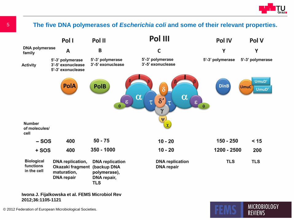
5 The five DNA polymerases of Escherichia coli and some of their relevant properties.
Iwona J. Fijalkowska et al. FEMS Microbiol Rev
2012;36:1105-1121
© 2012 Federation of European Microbiological Societies.

6
http://crowngene.tistory.com/category/
DNA polymerase I (E.coli)
Klenow fragment
The three functional domains of DNA polymerase I: DNA-polymerase and 3´-5´-exonuclease at the 3´-OH end and 5´-3´-exonuclease at the 5´-NH2-end.

7
DNA replication fork- simplified model
Taken from: D.P. Snustad, M.J. Simmons; Principles of Genetics, 5th edition; Wiley
8
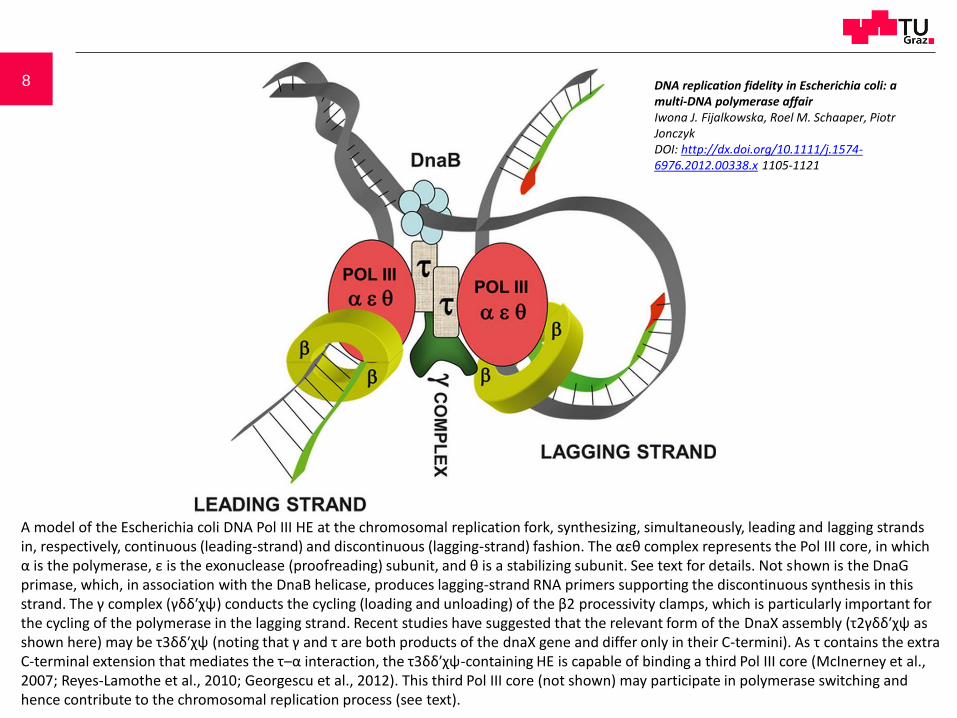
A model of the Escherichia coli DNA Pol III HE at the chromosomal replication fork, synthesizing, simultaneously, leading and lagging strands in, respectively, continuous (leading-strand) and discontinuous (lagging-strand) fashion. The αεθ complex represents the Pol III core, in which α is the polymerase, ε is the exonuclease (proofreading) subunit, and θ is a stabilizing subunit. See text for details. Not shown is the DnaGprimase, which, in association with the DnaB helicase, produces lagging-strand RNA primers supporting the discontinuous synthesis in this strand. The γ complex (γδδ′χψ) conducts the cycling (loading and unloading) of the β2 processivity clamps, which is particularly important for the cycling of the polymerase in the lagging strand. Recent studies have suggested that the relevant form of the DnaX assembly (τ2γδδ′χψ as shown here) may be τ3δδ′χψ (noting that γ and τ are both products of the dnaX gene and differ only in their C-termini). As τ contains the extra C-terminal extension that mediates the τ–α interaction, the τ3δδ′χψ-containing HE is capable of binding a third Pol III core (McInerney et al., 2007; Reyes-Lamothe et al., 2010; Georgescu et al., 2012). This third Pol III core (not shown) may participate in polymerase switching and hence contribute to the chromosomal replication process (see text).
DNA replication fidelity in Escherichia coli: a multi-DNA polymerase affairIwona J. Fijalkowska, Roel M. Schaaper, Piotr JonczykDOI: http://dx.doi.org/10.1111/j.1574-6976.2012.00338.x 1105-1121

9
DNA is pulled through primosome
Taken from: J.E. Krebs, E.S. Goldstein, S.T. Kilpatrick; „Lewin´s Genes XI“; Jones&Bartlett Learning

10 DNA Methylation Status Controls Replication Initiation
Taken from: J.E. Krebs, E.S. Goldstein, S.T. Kilpatrick; „Lewin´s Genes XI“; Jones&Bartlett Learning
Taken from: B. Lewin, Essential Genes, Pearson Ed. International

11
Taken from: J.E. Krebs, E.S. Goldstein, S.T. Kilpatrick; „Lewin´s Genes XI“; Jones&Bartlett Learning
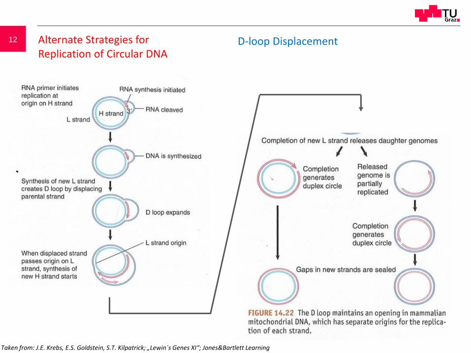
12 Alternate Strategies for Replication of Circular DNA
D-loop Displacement
Taken from: J.E. Krebs, E.S. Goldstein, S.T. Kilpatrick; „Lewin´s Genes XI“; Jones&Bartlett Learning

13 Alternate Strategiesfor Replication ofCircular DNA
Rolling Circle Mechanism
no RNA Primer 3‘ OH generated y nicking different types of DNA
generated- ds circular DNA- ss circular DNA- concatemeric linear DNA
Taken from: J.E. Krebs, E.S. Goldstein, S.T. Kilpatrick; „Lewin´s Genes XI“; Jones&Bartlett Learning

14
Taken from: J.E. Krebs, E.S. Goldstein, S.T. Kilpatrick; „Lewin´s Genes XI“; Jones&Bartlett Learning
Taken from: B. Lewin, Essential Genes, Pearson Ed. International

1515 10.11.15

16
Taken from: B. Lewin, Essential Genes, Pearson Ed. International

17
Taken from: B. Lewin, Essential Genes, Pearson Ed. International

18
Taken from: B. Lewin, Essential Genes, Pearson Ed. International
Sequence repeats at the telomeres
Organism Sequence
Sequence repeats at the telomeres
Organism Sequence
Homo sapiens

19
T
Telomeres have specific
structures:
Looping by Hoogsten
base pairing
No 3‘/5‘ free ends
Backfolding allows
priming for synthesis of
reverse strand
20
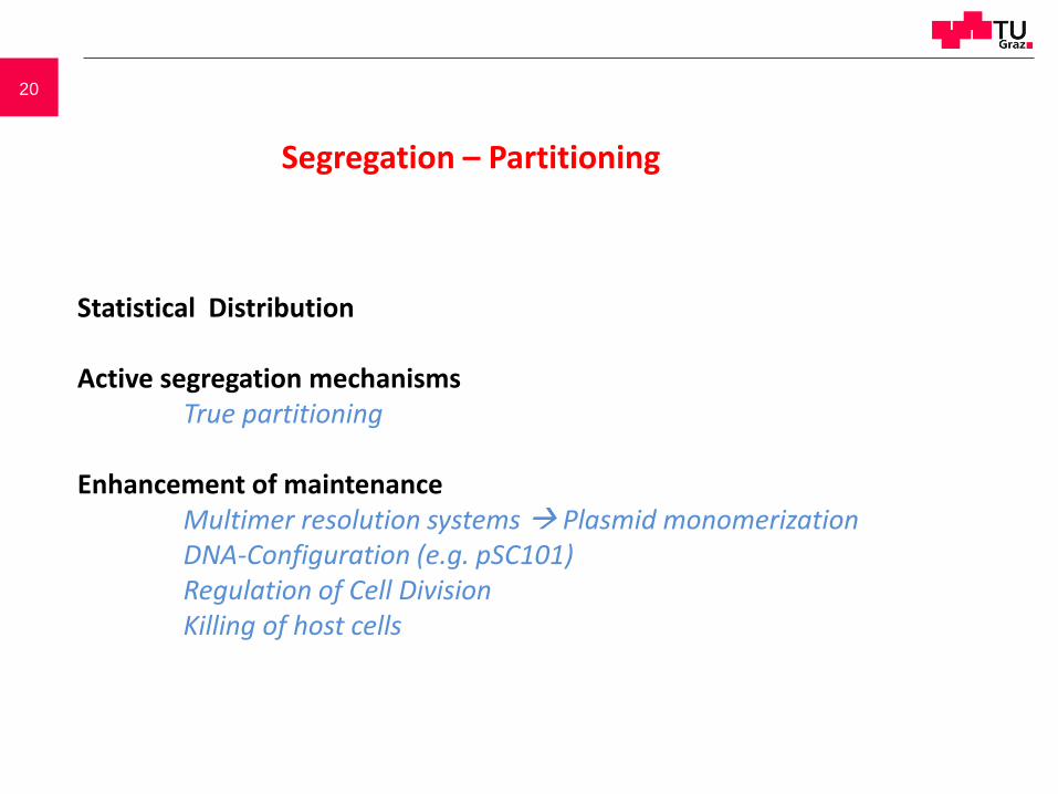
Statistical Distribution
Active segregation mechanismsTrue partitioning
Enhancement of maintenanceMultimer resolution systems Plasmid monomerizationDNA-Configuration (e.g. pSC101)Regulation of Cell DivisionKilling of host cells
Segregation – Partitioning
21
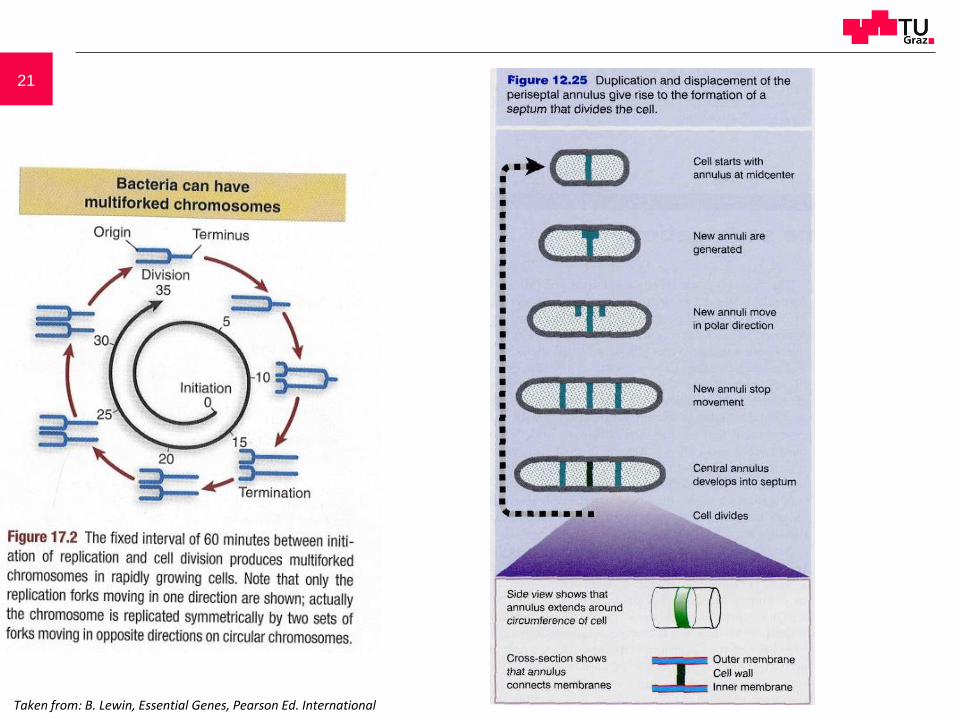
Taken from: B. Lewin, Essential Genes, Pearson Ed. International

22
23
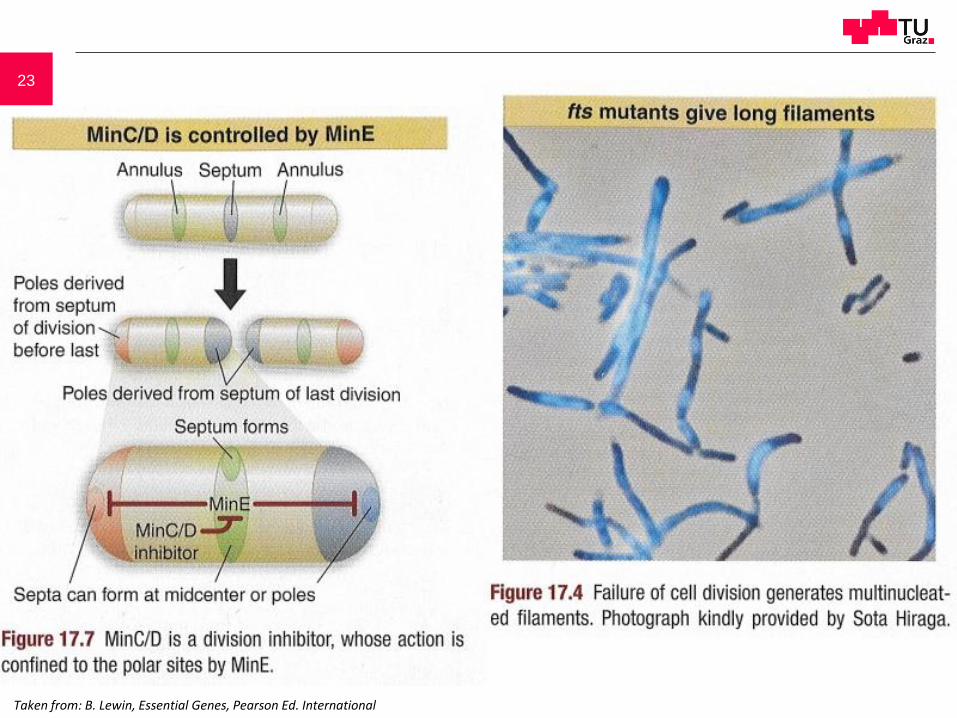
Taken from: B. Lewin, Essential Genes, Pearson Ed. International
24
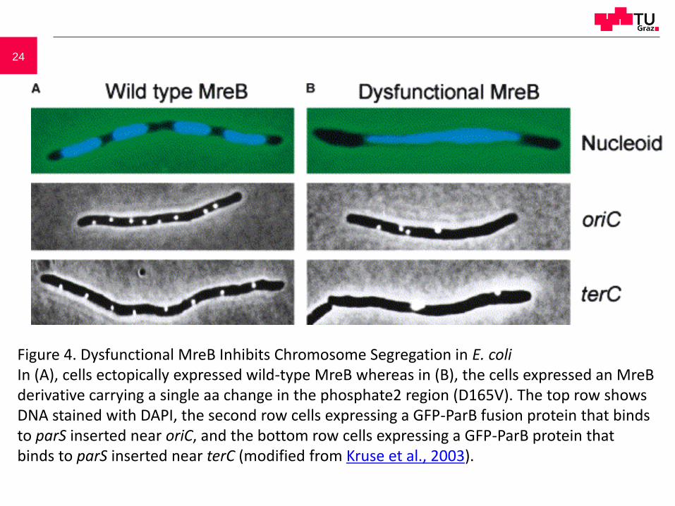
Figure 4. Dysfunctional MreB Inhibits Chromosome Segregation in E. coliIn (A), cells ectopically expressed wild-type MreB whereas in (B), the cells expressed an MreB derivative carrying a single aa change in the phosphate2 region (D165V). The top row shows DNA stained with DAPI, the second row cells expressing a GFP-ParB fusion protein that binds to parS inserted near oriC, and the bottom row cells expressing a GFP-ParB protein that binds to parS inserted near terC (modified from Kruse et al., 2003).

25
parA, parB: protein coding genesparC (parS) : protein binding site on the DNA
Taken from: B. Lewin, Essential Genes, Pearson Ed. International

26Bacterial Mitotic Machineries.Cell, Volume 116, Issue 3, Pages 359-366 K.Gerdes, J. Møller-Jensen, G. Ebersbach, T.Kruse, K. Nordström
Figure 1. Genetic Structure and Componentsof Type I (P1, F, and pB171) and Type IIPartitioning Loci (R1)In par of R1, ParR bindsto two times five direct repeats flanking thepromoter region in the parC region andthereby autoregulates transcription of theparMR operon. The parC region acts as acentromere-like site and has partitioningactivity when ParM and ParR are donated intrans (Dam and Gerdes, 1994). In par/sop ofP1 and F, the A proteins bind to the par/soppromoter region and autoregulatetranscription. The B proteins, when boundto the parS/sopC sites, enhanceautoregulation by the A proteins (Hao andYarmolinsky 2002 and Yates et al. 1999). Thepar region of pB171 has two cis-actingcentromere-like sites to which ParBpresumably binds (Ebersbach and Gerdes,2001). Binding of ParB of pB171 to parC1autoregulates transcription of the parABoperon.

27
Figure 2. Actin-Like ParM Filaments In Vivo and In Vitro In Vivo: (A) and (B) show cells with polar plasmids (red) located at the tip of ParM filaments (green) visualized by IFM. (C) shows decay of the filaments from mid-cell toward the cell poles. In (D), a single plasmid focus is located at mid-cell without a ParM filament (Møller-Jensen et al., 2003).In Vitro: (E) shows a 3D reconstruction of a straightened ParM filament obtained by electron microscopy (modified from van den Ent et al., 2002).
ParM: actin family ATPase

28
Figure 3. Model Explaining R1 par-Mediated Plasmid Partitioning during the Cell CyclePlasmids (red) are replicated by the host cell replication machinery, which is located at mid-cell. Replicated plasmids are paired by ParR bound to parC (yellow) thereby forming a partitioning complex (I). The partitioning complex forms a nucleation point for ParM filamentation. Continuous addition of ATP-ParM (green) to the filament poles provides the force for active movement of plasmids to opposite cell poles (II). Within the filaments, ATP is hydrolyzed, leading to destabilization of the ParM polymer (III). Nucleotide exchange is required to recharge the ADP-ParM (blue) molecules for a subsequent round of partitioning (IV). Modified from Møller-Jensen et al., 2003.
29
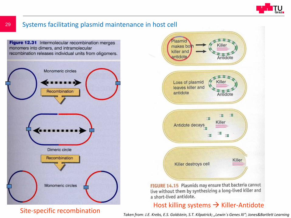
Site-specific recombinationHost killing systems Killer-Antidote
Taken from: J.E. Krebs, E.S. Goldstein, S.T. Kilpatrick; „Lewin´s Genes XI“; Jones&Bartlett Learning
Systems facilitating plasmid maintenance in host cell

30
Hok protein: toxic, kills cellsExpression of Hok protein is triggered at the translational level
by antisense RNA soksok RNA is less stable than hok RNA

31
Site specific resolution: parA, (parB), res
Killer – Antidote: parE, parD

32
Yeasts have two opposite mating types (a and α for S. cerevisiae), and can exist in both haploid and diploid states. Depending on the environment, yeast undergo sexual or asexual reproductive life cycles to maintain or switch their ploidy. When nutrients are abundant, yeasts propagate using asexual reproduction. For S. cerevisiae, this is done via budding, where the daughter cell originates as a small bud on the mother cell and continues to grow until the daughter separates from the mother. This is why S. cerevisiae is commonly known as budding yeast. When nutrients are limiting or during other high-stress conditions, yeasts undergo meiosis to generate haploid spores, which are contained in an ascus (in the case of S. cerevisiae).
http://www.singerinstruments.com/resource/what-is-yeast/
Eukaryotic Life Cycle

33

34 Cell cycle

35 Eukaryotic Life Cycle

36
Cell cycle mutants. a Saccharomyces cerevisiae (baker´s yeast). The initiation of DNA replication, the duplication of the mitotic spindle and the formation of the bud occur approximately at the same time. Thus, S-phase, G2-phase and mitosis cannot be differentiated clearly from one another. The „daughter“ cell, arisen from the bud, is initially smaller than the „mother“-cell. b Saccharomyces pombe (fission yeast). Note that „START“ is overstepped as soon as the genes CDC28 (S. cerevisiae) or cdc2 (S. pombe) are active.
Taken from: R. Knippers, Molekulare Genetik, 9th Ed., Thieme

37
http://csls-text.c.u-tokyo.ac.jp/active/12_02.html

38 Mitosis: maintaining the diploid status
Mitosis. During the early prophase thecentrioles move to opposite positions at thenuclear membrane and the chromatin startsto condense so that initially elongatedchromosomes become visible. During theprophase, chromosomes contract more andmore, the two chromatides become apparentand the nucleolus disintegrates. In the laterprophase the nuclear envelope dissolves, themitotic spindle forms and the chromosomesmigrate to the equatorial plane of the formernucleus. In the metaphase, all chromosomesare located in the equatorial plane.Homologous chromosomes are in generaldistributed accidentally and unpaired. In theanaphase, the chromatides separate andmigrate to opposite spindle poles. Thisensures that each daughter cell gets a full setof chromosomes. In the late anaphase thechromatides are located close to the spindlepoles and the constriction of the cell begins.In the telophase the new nuclear membraneis re-formed, centrioles duplicate andchromosome decondensation takes place.During the interphase, chromosomesdecondensate and build a new chromatinmatrix in the nucleus. The nucleolus was re-built. This scheme shows an animal cell.
late prophase
early prophase
interphase
telophase
late anaphase
early anaphaseprometaphase
metaphase
spindle
nucleolusnuclear membranecentriole

39 Meiosis: reduction to haploid status
During the first meiotic division homologous chromosomes areseparated (pre-reduction), during the second meiotic divisionchromatides of the single chromosomes are separated. Each diploidprimary meiocyte results in four haploid meiosis products. In males,these four haploid post-meiotic cells develop into spermatozoa. Infemales, three of the haploid meiosis products degenerate whereas thefourth cell differentiates into an oocyte. In some organisms, the haploidpost-meiotic cells undergo additional mitotic divisions. The prophase ofthe first meiotic division is morphologically divided into several stageswhich occur in most of the higher organisms as characteristic meioticchromosome states. Recombination during the first meiotic prophaseleads to a post-reduction of certain chromosome regions meaning to adistribution of paternal and maternal alleles not until the second meioticdivision. For some genetic analyses, this post-reduction mechanism hasexperimental consequences. This scheme shows the meiosis of animals.
Leptotene Zygotene
Pachytene Diplotene Diakinesis
40
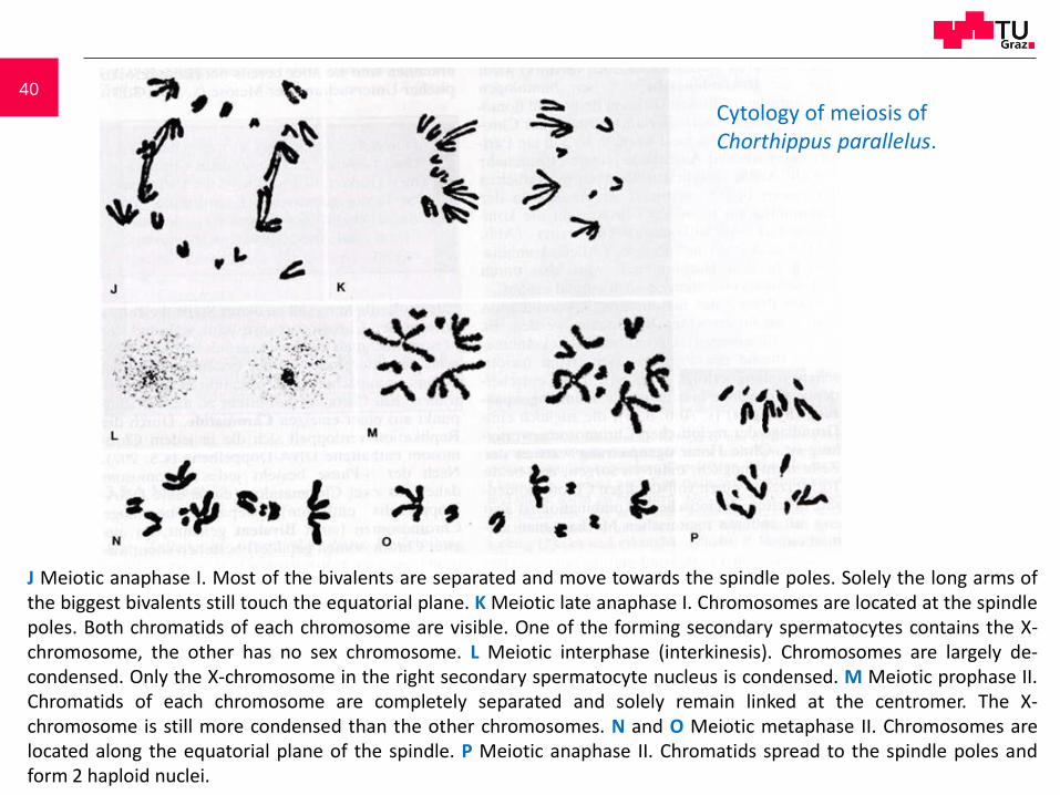
J Meiotic anaphase I. Most of the bivalents are separated and move towards the spindle poles. Solely the long arms ofthe biggest bivalents still touch the equatorial plane. K Meiotic late anaphase I. Chromosomes are located at the spindlepoles. Both chromatids of each chromosome are visible. One of the forming secondary spermatocytes contains the X-chromosome, the other has no sex chromosome. L Meiotic interphase (interkinesis). Chromosomes are largely de-condensed. Only the X-chromosome in the right secondary spermatocyte nucleus is condensed. M Meiotic prophase II.Chromatids of each chromosome are completely separated and solely remain linked at the centromer. The X-chromosome is still more condensed than the other chromosomes. N and O Meiotic metaphase II. Chromosomes arelocated along the equatorial plane of the spindle. P Meiotic anaphase II. Chromatids spread to the spindle poles andform 2 haploid nuclei.
Cytology of meiosis ofChorthippus parallelus.


