Submitted 4 March 2018, Accepted 28 May 2018, Published 29 June 2018
Corresponding Author: K.D. Hyde – e-mail – [email protected] 152
Taxonomic circumscription and phylogenetics of novel
didymellaceous taxa with brown muriform spores
Wanasinghe DN1,2,3, Jeewon R4, Peršoh D5, Jones EBG6, Camporesi E7,8,9,
Bulgakov TS10, Gafforov YS11 and Hyde KD1,2,3*
1Key Laboratory for Plant Biodiversity and Biogeography of East Asia (KLPB), Kunming Institute of Botany, Chinese
Academy of Science, Kunming 650201, Yunnan China 2Center of Excellence in Fungal Research, Mae Fah Luang University, Chiang Rai, 57100, Thailand 3World Agro Forestry Centre, East and Central Asia, 132 Lanhei Road, Kunming 650201, Yunnan China 4Department of Health Sciences, Faculty of Science, University of Mauritius, Reduit, Mauritius 5AG Geobotany, Faculty of Biology and Biotechnology, Ruhr-Universitat Bochum, Universitatsstraße 150, 44801
Bochum, Germany; 6Nantgaredig, 33B St. Edwards Road, Southsea, Hants., PO5 3DH, UK; 7Società per gli Studi Naturalistici della Romagna, C.P. 144, Bagnacavallo (RA), Italy; 8A.M.B. Gruppo Micologico Forlivese “Antonio Cicognani”, Via Roma 18, Forlì, Italy; 9A.M.B. Circolo Micologico “Giovanni Carini”, C.P. 314, Brescia, Italy; 10Russian Research Institute of Floriculture and Subtropical Crops, Sochi, 354002, Yana Fabritsiusa street, 2/28,
Krasnodar region, Russia; 11Laboratory of Mycology, Institute of Botany, Academy of Sciences of the Republic of Uzbekistan, 32 Durmon yuli
Street, Tashkent 100125, Uzbekistan
Wanasinghe DN, Jeewon R, Peršoh D, Jones EBG, Camporesi E, Bulgakov TS, Gafforov YS,
Hyde KD 2018 – Taxonomic circumscription and phylogenetics of novel didymellaceous taxa with
brown muriform spores. Studies in Fungi 3(1), 152–175, Doi 10.5943/sif/3/1/17
Abstract Sexual morph of didymellaceous taxa are characterized by their ascomata with relatively thin
peridium, cylindric-clavate to clavate, short-pedicellate or apedicellate asci, hyaline to brown, 1-
septate to muriform ascospores. Its asexual morphs are coelomycetous and comprising pycnidial or
acervulus conidiomata, phialidic, hyaline conidiogenous cells and hyaline or pale brown, septate or
aseptate conidia. The majority of these cosmopolitan species are plant associated fungi which can
be pathogens on a wide range of hosts and some species are of particular relevance for quarantine
measures. Recent studies have significantly improved the taxonomy and systematics of
didymellaceous taxa based on molecular phylogenetics. In contrast to the accurate and detailed
studies on the asexual morphs which are common obligate pathogens, information on their usually
saprobic sexual morphs is still limited. Among these phenotypically diverse species, spore
characteristics are quite unique as most have hyaline spores with 0–1 septum, while only
Neomicrosphaeropsis and Didymellocamarosporium are reported as producing pigmented,
muriform spores. These dematiaceous muriform spores are characteristic of a considerable number
of species that may be quite divergent in other characters. During taxonomic investigations on the
diversity of didymellaceous taxa, we have isolated species from Alhagi pseudalhagi, Coronilla
emerus, Cytisus sp., Elaeagnus angustifolia and Spartium junceum in Italy, Russia and Uzbekistan.
A comprehensive phylogeny, based on four loci (ITS, LSU, rpb2 and tub2) is used to infer species
relationships. Comprehensive morphological descriptions and in-depth phylogenetic investigations
of five new species viz. Ascochyta coronillae-emeri, Microsphaeropsis spartii-juncei,
Neomicrosphaeropsis alhagi-pseudalhagi, N. cytisicola and N. elaeagni are presented.
Studies in Fungi 3(1): 152–175 (2018) www.studiesinfungi.org ISSN 2465-4973
Article
Doi 10.5943/sif/3/1/17
Copyright © Mushroom Research Foundation
153
Keywords – five new species – coelomycetes – Italy – multi-gene – phylogeny – Pleosporales –
Russia – saprobic – taxonomic-ambiguity – Uzbekistan
Introduction
The family Didymellaceae was proposed by de Gruyter et al. (2009) to accommodate phoma-
like taxa, viz. Ascochyta, Didymella and Phoma, which probably diverged in the Jurassic or earlier
from an ancestor whose origin can be estimated about 63 mya (crown age) or 115 mya (stem age)
ago (Liu et al. 2017). Didymellaceae is one of the most species-rich families in the fungal kingdom
and includes 4956 and 4713 taxon epithets listed in MycoBank and Index Fungorum, respectively
(2017). More than 50% from the total epithets are listed as Phoma and over 30% are recorded as
Ascochyta. In a recent study, Chen et al. (2017) revised Didymellaceae and improved our
understanding of their distribution and biodiversity. They have proposed 19 genera in the family
and currently the family comprises 31 genera, including Cumuliphoma, Didymellocamarosporium,
Didysimulans, Ectophoma, Endocoryneum, Juxtiphoma, Neodidymella, Pseudoascochyta,
Pseudohendersonia, Remotididymella, Similiphoma and Vacuiphoma (Ariyawansa et al. 2015,
Crous et al. 2016, Wijayawardene et al. 2016, 2018, Tibpromma et al. 2017, Valenzuela-Lopez et
al. 2018). The majority of members in Didymellaceae are plant associated fungi which can be
pathogens on a wide range of hosts, largely causing leaf and stem lesions, with some of particular
relevance for quarantine measures (Aveskamp et al. 2008, 2010, Chen et al. 2015, 2017).
Didymellaceae are cosmopolitan and able to adapt to extreme environmental conditions i.e.
temperature, nutrients, moisture, absolute darkness and they can grow in exposed habitats such as
air, soil, water, limestone from caves (Chen et al. 2017) and inorganic materials including asbestos,
cement and paint (Aveskamp et al. 2008). Given their ubiquitous nature, additional taxonomic and
ecological knowledge are prerequisites to understand their biology and their significance in the
environment, especially in agriculture.
In contrast to the accurate and detailed studies on their asexual morphs, information is still
limited on their sexual morphs, which usually grow as saprobes, in contrast to their pathogenic
asexual counterparts (Chen et al. 2017). Determining the phylogenetic placement of sexual morphs
is crucial to properly define the taxonomic boundaries within the polyphyletic and morphologically
homogeneous genera (i.e. Ascochyta, Didymella and Phoma). Knowledge of the sexual-asexual
relationships will considerably improve our understanding of many of the specific biological
features. Of the 28 genera in this family, sexual morphs are known for 12 genera (Jayasiri et al.
2017) and their ascospores are mostly hyaline and 1-septate. There is only one sexual morph
recorded in this family with pigmented muriform spores, Neomicrosphaeropsis tamaricicola (=
Phoma tamaricicola), introduced by Crous et al. (2014). Pigmented muriform spores are
characteristic for a considerable number of species being divergent in other characters. For asexual
morphs, Didymellocamarosporium tamaricis (Wijayawardene et al. 2016) is the only asexual
member recorded with pigmented muriform conidia in this family.
We are investigating the diversity of microfungi that produce brown, muriform spores with
the aim of clarifying their taxonomy based on morphology coupled with multigene phylogeny
(Wanasinghe et al. 2014a, b, 2015, 2016, 2017a, b, 2018). As part of this study, we have isolated
taxa from Alhagi pseudalhagi, Coronilla emerus, Cytisus sp., Elaeagnus angustifolia and Spartium
junceum species in Italy, Russia and Uzbekistan which belong to the family Didymellaceae. Here
we present comprehensive morphological descriptions and in-depth phylogenetic investigation of
those taxa.
Materials and Methods
Sampling, examination and isolation
The novel strains were isolated from Alhagi pseudalhagi, Coronilla emerus, Cytisus sp.,
Elaeagnus angustifolia and Spartium junceum in Italy and Russia. Uzbekistan specimens were
loaned from Tashkent Mycological Herbarium (TASM) of the Institute of Botany, Academy of
154
Sciences of Uzbekistan, Tashkent. These collections were examined and isolated following the
methods used by Wanasinghe et al. (2017a). Type and isotype specimens of new species in this
study are deposited in the Mae Fah Luang University (MFLU) Herbarium. Living cultures are
deposited at the Culture Collection of Mae Fah Luang University (MFLUCC) and duplicated in
International Collection of Microorganisms from Plants (ICMP), Landcare Research, Auckland,
New Zealand.
DNA isolation, amplification and phylogenetic analyses
Total genomic DNA was extracted from fresh mycelia using the protocol described by
Wanasinghe et al. (2017a). When fungi failed to grow in culture, DNA was extracted directly from
ascomycete fruiting bodies by following the protocol described by Wanasinghe et al. (2018). DNA
to be used as template for PCR were stored at 4 °C for use in regular work and duplicated at -20 °C
for long term storage. The primers ITS5 and ITS4 (White et al. 1990) were used to amplify part of
rDNA 18S (3' end), the first internal transcribed spacer (ITS1), the 5.8S rRNA gene, the second ITS
region (ITS2), and part of the 28S rRNA (5' end); the primers LR0R (Rehner & Samuels 1994),
LR5 (Vilgalys & Hester 1990) were used for LSU amplification; Btub2Fd and Btub4Rd
(Woudenberg et al. 2009) for the partial β-tubulin (tub2) gene region, and RPB2-5F (Sung et al.
2007) and fRPB2-7cR (Liu et al. 1999) for the RNA polymerase II second largest subunit (rpb2).
Amplicons for ITS and LSU locus were generated following the protocols listed in Wanasinghe et
al. (2017a) and the protocols of Chen et al. (2015) were used to amplify tub2 and rpb2.
Sequencing was conducted in both directions with the same primer pair used for
amplification at BGI, Ltd., Shenzhen, P.R. China. Consensus sequences were assembled in BioEdit
v. 7.0.5.2 (Hall 1999) and additional reference sequences were obtained from GenBank (Table 1).
Subsequent alignments for each locus were generated with MAFFT v. 7
(http://mafft.cbrc.jp/alignment/server/index.html; Kuraku et al. 2013, Katoh et al. 2017), and
manually corrected when necessary in BioEdit v7.0.9 (Hall 1999). Each locus and the concatenated
aligned dataset were analysed separately using Maximum Likelihood (ML), Maximum Parsimony
(MP) and Bayesian Inference (BI). The best-fit models of evolution for the four loci tested
(GTR+I+G for all gene regions) were estimated by MrModeltest v. 2.3 (Nylander 2004).
Parsimony analysis was carried out with the heuristic search option in PAUP (Phylogenetic
Analysis Using Parsimony) v. 4.0b10 with the following parameter settings: characters unordered
with equal weight, random taxon addition, branch swapping with tree bisection-reconnection (TBR)
algorithm, branches collapsing if the maximum branch length was zero. Alignment gaps were
treated as missing characters in the analysis of the combined data set, where they occurred in
relatively conserved regions. Trees were inferred using the heuristic search option with 1000
random sequence additions, with maxtrees set at 5000. Descriptive tree statistics for parsimony;
tree length (TL), consistency index (CI), retention index (RI), relative consistency index (RC) and
homoplasy index (HI) were calculated for trees generated under different optimality criteria. The
Kishino-Hasegawa tests (Kishino & Hasegawa 1989) were performed in order to determine
whether trees were significantly different. Other details pertaining to analyses (e.g. consideration of
TT ratios, comparison of tree topologies and selection of outgroups) are outlined in Jeewon et al.
(2003a, b, 2004, 2013).
Bayesian (BI) analyses were performed on MrBayes v. 3.2.1 (Ronquist et al. 2012) based on
the models selected by the MrModeltest. The Markov Chain Monte Carlo (MCMC) algorithm of
six chains was initiated for 5 M generations in parallel from a random tree topology. The trees were
sampled every 200th generation. The distribution of log-likelihood scores was examined to
determine the stationary phase for each search and to decide if extra runs were required to achieve
convergence, using the program Tracer v. 1.5 (Rambaut & Drummond 2007). All sampled
topologies beneath the asymptote (10 %) were discarded as part of a burn-in procedure; the
remaining trees were used for calculating PP in the majority rule consensus tree. Posterior
probabilities values of the BI analyses (BYPP) over 0.95 were considered significant.
155
The ML analyses were conducted with RAxML-HPC BlackBox (v. 8.2.8) (Stamatakis et al.
2008, Stamatakis 2014) in the CIPRES Science Gateway platform (Miller et al. 2010) using a
GTR+I+G substitution model with 1 000 bootstrap replicates. The robustness of the analyses was
evaluated by bootstrap support (MLBS).
Phylograms were visualized with FigTree v1.4.0 program (Rambaut 2012) and reorganized in
Microsoft power point (2007) and Adobe Illustrator® CS5 (Version 15.0.0, Adobe®, San Jose, CA).
One hundred and twenty-six taxa are used (including our newly generated sequences) as
ingroup taxa, Leptosphaeria conoidea (CBS 616.75) and L. doliolum (CBS 505.75) were selected
as outgroup taxa. Sequences generated in this study were deposited in GenBank (Table 1), the final
matrices and trees in TreeBASE (accession number: 22328), (Study Accession URL:
http://purl.org/phylo/treebase/phylows/study/TB2:S22328) and novel taxonomic descriptions and
nomenclature in Faces of Fungi and Index Fungorum as outlined in Jayasiri et al. (2015), Index
Fungorum (2018). New species were established based on recommendations outlined by Jeewon &
Hyde (2016).
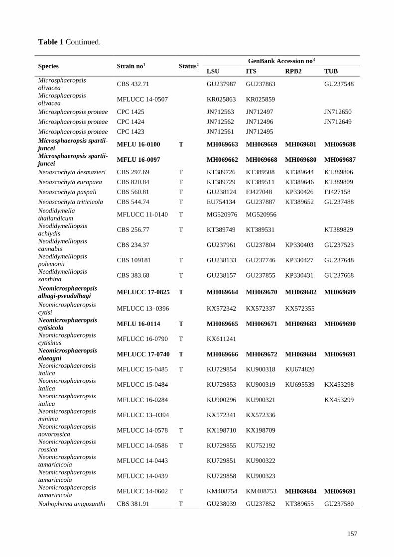
Table 1 Taxa used in the phylogenetic analysis and their corresponding GenBank numbers. The
newly generated sequences are indicated in bold.
Species Strain no1 Status2 GenBank Accession no3
LSU ITS RPB2 TUB
Allophoma minor CBS 325.82 T GU238107 GU237831 KT389553 GU237632
Allophoma nicaraguensis CBS 506.91 T GU238058 GU237876 KT389551 GU237596
Allophoma piperis CBS 268.93 T GU238129 GU237816 KT389554 GU237644
Allophoma tropica CBS 436.75 T GU238149 GU237864 KT389556 GU237663
Ascochyta boeremae CBS 372.84 T KT389697 KT389480
KT389774
Ascochyta boeremae CBS 373.84
KT389698 KT389481 KT389560 KT389775
Ascochyta coronillae-
emeri MFLUCC 13-0820 T MH069661 MH069667 MH069679 MH069686
Ascochyta herbicola CBS 629.97 R GU238083 GU237898 KP330421 GU237614
Ascochyta medicaginicola
var. macrospora CBS 112.53 T GU238101 GU237749
GU237628
Ascochyta medicaginicola
var. macrospora BRIP 45051
KY742198 KY742044 KY742132 KY742286
Ascochyta medicaginicola
var. medicaginicola MFLUCC 16-0599
KX698025 KX698036 KX698033 KX698029
Ascochyta phacae CBS 184.55 T KT389692 KT389475
KT389769
Ascochyta pisi CBS 122751
KP330444 KP330432 EU874867 KP330388
Ascochyta rabiei CBS 206.30
KT389695 KT389478 KT389559 KT389772
Ascochyta rabiei CBS 237.37 T KT389696 KT389479
KT389773
Ascochyta rabiei CBS 534.65
GU237970 GU237886 KP330405 GU237533
Boeremia exigua var.
heteromorpha CBS 443.94 T GU237935 GU237866 KT389573 GU237497
Boeremia exigua var.
opuli CGMCC 3.18354 T KY742199 KY742045 KY742133 KY742287
Boeremia hedericola CBS 367.91 R GU237949 GU237842 KT389579 GU237511
Briansuttonomyces
eucalypti CBS 114879 T KU728519 KU728479
KU728595
Briansuttonomyces
eucalypti CBS 114887
KU728520 KU728480
KU728596
Calophoma aquilegiicola CBS 107.96 R GU238041 GU237735 KT389586 GU237581
Calophoma clematidina CBS 102.66
FJ515630 FJ426988 KT389587 FJ427099
Calophoma clematidina CBS 108.79 T FJ515632 FJ426989 KT389588 FJ427100
156
Table 1 Continued.
Species Strain no1 Status2 GenBank Accession no3
LSU ITS RPB2 TUB
Calophoma rosae CGMCC 3.18347 T KY742203 KY742049 KY742135 KY742291
Cumuliphoma indica CBS 654.77 T GU238122 FJ427043 LT623261 FJ427153
Cumuliphoma omnivirens CBS 341.86 T LT623214 FJ427042 LT623260 FJ427152
Cumuliphoma pneumoniae CBS 142454 T LN907392 LT592925 LT593063 LT592994
Didymella aquatica CGMCC 3.18349 T KY742209 KY742055 KY742140 KY742297
Didymella arachidicola CBS 333.75 T GU237996 GU237833 KT389598 GU237554
Didymella exigua CBS 183.55 T EU754155 GU237794 EU874850 GU237525
Didymella heteroderae CBS 109.92 T GU238002 FJ426983 KT389601 FJ427098
Didymella macrophylla CGMCC 3.18357 T KY742224 KY742070 KY742154 KY742312
Didymellocamarosporium
tamaricis MFLUCC 14-0241 T KU848183
Didysimulans italica MFLUCC 15-0059 T KY496730 KY496750 KY514408
Didysimulans mezzanensis MFLUCC 15-0067 T KY496733 KY496753 KY514411
Ectophoma multirostrata CBS 274.60 T GU238111 FJ427031 LT623265 FJ427141
Ectophoma multirostrata CBS 368.65
GU238112 FJ427033 LT623266 FJ427143
Ectophoma pomi CBS 267.92 T GU238128 GU237814 LT623263 GU237643
Endocoryneum festucae MFLUCC 14-0461 T KU848203
Epicoccum brasiliense CBS 120105 T GU238049 GU237760 KT389627 GU237588
Epicoccum camelliae CGMCC 3.18343 T KY742245 KY742091 KY742170 KY742333
Epicoccum huancayense CBS 105.80 T GU238084 GU237732 KT389630 GU237615
Epicoccum latusicollum CGMCC 3.18346 T KY742255 KY742101 KY742174 KY742343
Epicoccum nigrum CBS 173.73 T GU237975 FJ426996 KT389632 FJ427107
Heterophoma
verbascicola CGMCC 3.18364 T KY742273 KY742119 KY742187 KY742361
Heterophoma
verbascicola LC 8164
KY742274 KY742120 KY742188 KY742362
Heterophoma adonidis CBS 114309
KT389724 KT389506 KT389637 KT389803
Heterophoma
dictamnicola CBS 507.91
GU238065 GU237877 KT389638 GU237603
Juxtiphoma eupyrena CBS 374.91
GU238072 FJ426999 LT623268 FJ427110
Juxtiphoma eupyrena CBS 527.66
GU238073 FJ427000 LT623269 FJ427111
Leptosphaeria conoidea CBS 616.75
JF740279 JF740201 KT389639 KT389804
Leptosphaeria doliolum CBS 505.75 T GQ387576 JF740205 KT389640 JF740144
Leptosphaerulina
americana CBS 213.55
GU237981 GU237799 KT389641 GU237539
Leptosphaerulina
arachidicola CBS 275.59
GU237983 GU237820
GU237543
Leptosphaerulina australis CBS 317.83
EU754166 GU237829 GU371790 GU237540
Leptosphaerulina trifolii CBS 235.58
GU237982 GU237806
GU237542
Macroventuria
anomochaeta CBS 502.72
GU237985 GU237873
GU237545
Macroventuria
anomochaeta CBS 525.71 T GU237984 GU237881 GU456346 GU237544
Macroventuria wentii CBS 526.71 T GU237986 GU237884 KT389642 GU237546
Microsphaeropsis
olivacea CBS 442.83
EU754171 GU237865
GU237547
Microsphaeropsis
olivacea CBS 233.77 GU237988 GU237803 KT389643 GU237549
157
Table 1 Continued.
Species Strain no1 Status2 GenBank Accession no3
LSU ITS RPB2 TUB
Microsphaeropsis
olivacea CBS 432.71
GU237987 GU237863
GU237548
Microsphaeropsis
olivacea MFLUCC 14-0507
KR025863 KR025859
Microsphaeropsis proteae CPC 1425
JN712563 JN712497
JN712650
Microsphaeropsis proteae CPC 1424
JN712562 JN712496
JN712649
Microsphaeropsis proteae CPC 1423
JN712561 JN712495
Microsphaeropsis spartii-
juncei MFLU 16-0100 T MH069663 MH069669 MH069681 MH069688
Microsphaeropsis spartii-
juncei MFLU 16-0097
MH069662 MH069668 MH069680 MH069687
Neoascochyta desmazieri CBS 297.69 T KT389726 KT389508 KT389644 KT389806
Neoascochyta europaea CBS 820.84 T KT389729 KT389511 KT389646 KT389809
Neoascochyta paspali CBS 560.81 T GU238124 FJ427048 KP330426 FJ427158
Neoascochyta triticicola CBS 544.74 T EU754134 GU237887 KT389652 GU237488
Neodidymella
thailandicum MFLUCC 11-0140 T MG520976 MG520956
Neodidymelliopsis
achlydis CBS 256.77 T KT389749 KT389531
KT389829
Neodidymelliopsis
cannabis CBS 234.37
GU237961 GU237804 KP330403 GU237523
Neodidymelliopsis
polemonii CBS 109181 T GU238133 GU237746 KP330427 GU237648
Neodidymelliopsis
xanthina CBS 383.68 T GU238157 GU237855 KP330431 GU237668
Neomicrosphaeropsis
alhagi-pseudalhagi MFLUCC 17-0825 T MH069664 MH069670 MH069682 MH069689
Neomicrosphaeropsis
cytisi MFLUCC 13–0396
KX572342 KX572337 KX572355
Neomicrosphaeropsis
cytisicola MFLU 16-0114 T MH069665 MH069671 MH069683 MH069690
Neomicrosphaeropsis
cytisinus MFLUCC 16-0790 T KX611241
Neomicrosphaeropsis
elaeagni MFLUCC 17-0740 T MH069666 MH069672 MH069684 MH069691
Neomicrosphaeropsis
italica MFLUCC 15-0485 T KU729854 KU900318 KU674820
Neomicrosphaeropsis
italica MFLUCC 15-0484
KU729853 KU900319 KU695539 KX453298
Neomicrosphaeropsis
italica MFLUCC 16-0284
KU900296 KU900321
KX453299
Neomicrosphaeropsis
minima MFLUCC 13–0394
KX572341 KX572336
Neomicrosphaeropsis
novorossica MFLUCC 14-0578 T KX198710 KX198709
Neomicrosphaeropsis
rossica MFLUCC 14-0586 T KU729855 KU752192
Neomicrosphaeropsis
tamaricicola MFLUCC 14-0443
KU729851 KU900322
Neomicrosphaeropsis
tamaricicola MFLUCC 14-0439
KU729858 KU900323
Neomicrosphaeropsis
tamaricicola MFLUCC 14-0602 T KM408754 KM408753 MH069684 MH069691
Nothophoma anigozanthi CBS 381.91 T GU238039 GU237852 KT389655 GU237580
158
Table 1 Continued.
Species Strain no1 Status2 GenBank Accession no3
LSU ITS RPB2 TUB
Nothophoma arachidis-
hypogaeae CBS 125.93 R GU238043 GU237771 KT389656 GU237583
Nothophoma gossypiicola CBS 377.67
GU238079 GU237845 KT389658 GU237611
Nothophoma infossa CBS 123395 T GU238089 FJ427025 KT389659 FJ427135
Nothophoma quercina CBS 633.92
EU754127 GU237900 KT389657 GU237609
Paraboeremia adianticola CBS 187.83
GU238035 GU237796 KP330401 GU237576
Paraboeremia camellae CGMCC 3.18106 T KX829042 KX829034 KX829050 KX829058
Paraboeremia litseae CGMCC 3.18109 T KX829037 KX829029 KX829045 KX829053
Paraboeremia
oligotrophica CGMCC 3.18111 T KX829039 KX829031 KX829047 KX829055
Paraboeremia
selaginellae CBS 122.93 T GU238142 GU237762
GU237656
Phoma herbarum CBS 134.96
KT389753 KT389535 KT389661 KT389834
Phoma herbarum CBS 274.37
KT389754 KT389537 KT389662 KT389835
Phoma herbarum CBS 377.92
KT389756 KT389536 KT389663 KT389837
Phoma herbarum CBS 502.91
GU238082 GU237874 KP330419 GU237613
Phoma herbarum CBS 615.75 R EU754186 FJ427022 KP330420 FJ427133
Phomatodes aubrietiae CBS 383.67 R GU238044 GU237854
GU237584
Phomatodes aubrietiae CBS 627.97 T GU238045 GU237895 KT389665 GU237585
Phomatodes nebulosa CBS 117.93
GU238114 GU237757 KP330425 GU237633
Phomatodes nebulosa CBS 740.96
KT389758 KT389540 KT389667 KT389839
Phomatodes nebulosa CBS 100191
KP330446 KP330434 KT389666 KP330390
Pseudoascochyta novae-
zelandiae CBS 141689
LT592893 LT592892 LT592895 LT592894
Pseudohendersonia
galiorum
MFLUCC 14 –
0452 T KU848207
Remotididymella
anthropophila CBS 142462 T LN907421 LT592936 LT593075 LT593005
Remotididymella
destructiva CBS 133.93
GU238064 GU237779 LT623257 GU237602
Remotididymella
destructiva CBS 378.73 T GU238063 GU237849 LT623258 GU237601
Similiphoma crystallifera CBS 193.82 T GU238060 GU237797 LT623267 GU237598
Stagonosporopsis actaeae CBS 106.96 T GU238166 GU237734 KT389672 GU237671
Stagonosporopsis
crystalliniformis CBS 713.85 T GU238178 GU237903 KT389675 GU237683
Stagonosporopsis dennisii CBS 631.68 T GU238182 GU237899 KT389677 GU237687
Stagonosporopsis
helianthi CBS 200.87 T KT389761 KT389545 KT389683 KT389848
Vacuiphoma bulgarica CBS 357.84 T GU238050 GU237837 LT623256 GU237589
Vacuiphoma oculihominis UTHSC DI16-308 T LN907451 LT592954 LT593093 LT593023
Xenodidymella applanata CBS 195.36 T KT389764 KT389548
KT389852
Xenodidymella applanata CBS 115577
KT389762 KT389546 KT389688 KT389850
Xenodidymella catariae CBS 102635 GU237962 GU237727 KP330404 GU237524 1 BRIP: Plant Pathology Herbarium, Department of Employment, Economic, Development and Innovation,
Queensland, Australia; CBS: Westerdijk Fungal Biodiversity Institute (formerly CBSKNAW), Utrecht, The
Netherlands; CGMCC: China General Microbiological Culture Collection, Beijing, China; CPC: Culture collection of
Pedro Crous, housed at CBS; LC: Corresponding author's personal collection deposited in laboratory, housed at CAS,
China; MFLUCC: Mae Fah Luang University Culture Collection, Chiang Rai, Thailand; UTHSC, Fungus Testing
Laboratory at the University of Texas Health Science Center, San Antonio, Texas, USA.
159
2 T: ex-type strain; R: representative strain. 3 ITS: internal transcibed spacer regions 1 & 2 including 5.8S nrDNA gene; LSU: 28S large subunit of the nrRNA
gene; rpb2: RNA polymerase II second subunit; tub2: ß-tubulin.
Results and Discussion
Phylogenetic analyses
Topologies of trees (under ML, MP and BI criteria) recovered for each gene dataset were
visually compared and the overall tree topology was congruent to those obtained from the
combined dataset.
The RAxML analysis of the combined dataset yielded a best scoring tree (Fig. 1) with a final
ML optimization likelihood value of -23881.01104. The matrix had 734 distinct alignment patterns,
with 8.95 % proportion of gaps and completely undetermined characters in this alignment.
Parameters for the GTR + I + G model of the combined LSU, ITS, rpb2 and tub2 were as follows:
Estimated base frequencies were as follows: A = 0.238058, C = 0.241410, G = 0.27525, T =
0.245283; substitution rates AC = 1.943648, AG = 6.96474, AT = 2.220889, CG = 0.925886, CT =
14.019529, GT = 1.000; proportion of invariable sites I = 0.63074; gamma distribution shape
parameter α = 0.584276. The maximum parsimonious dataset for the combined gene sequences
consisted of 2231 characters, of which 1560 were constant, 615 (27.6 %) parsimony-informative
and 56 parsimony-uninformative. The parsimony analysis of the data matrix resulted in the
maximum of 2325 equally most parsimonious trees with a length of 4662 steps (CI = 0.238, RI =
0.636, RC = 0.151, HI = 0.762) in the first tree. The Bayesian analysis resulted in 25001 trees after
5 M generations with 0.009735 as the average standard deviation of split frequency. Therefore, the
first 2500 trees, representing the burn-in phase of the analyses, were discarded, while the remaining
22501 trees were used or calculating posterior probabilities in the majority rule consensus tree.
Newly generated sequences from two Microsphaeropsis isolates (MFLU 16-0100 and MFLU
16-0097) grouped with isolates currently circumscribed as Microsphaeropsis olivacea and M.
proteae (de Gruyter et al. 2009, Aveskamp et al. 2010, Crous et al. 2011, Verkley et al. 2014, Chen
et al. 2015). These taxa formed an isolated clade (Clade A, Fig 1) within Didymellaceae, but poorly
supported in multi-gene analyses (69% in ML, <60 % in MP and <0.95 in BI). Within Clade A (Fig
1), our novel isolates are closely related and monophyletic with Microsphaeropsis olivacea (CBS
442.83, CBS 432.71, CBS 233.77) and retrieved 67% (ML), 86% (MP), 1.00 (BI) bootstrap support
for this lineage (Subclade A1).
Ascochyta coronillae-emeri (MFLUCC 13-0820), showed a close phylogenetic affinity to A.
rabiei (CBS 206.30, CBS 237.37, CBS 534.65), A. phacae (CBS 184.55) and A. herbicola (CBS
629.97) in the combined phylogeny (Subclade B1) and this relationship retrieved 96% ML, 92%
MP and 1.00 BI support.
Three newly generated sequences, Neomicrosphaeropsis alhagi-pseudalhagi (MFLUCC 17-
0825), N. cytisicola (MFLU 16-0114) and N. elaeagni (MFLUCC 17-0740), grouped with
Didymellocamarosporium tamaricis and eleven Neomicrosphaeropsis isolates. These taxa form a
monophyletic clade (Clade C) in Didymellaceae with poor statistical support (65% in ML, <60 %
in MP and <0.95 in BI). Didymellocamarosporium tamaricis, Neomicrosphaeropsis elaeagni sp.
nov., N. italica, N. novorossica, N. rossica and N. tamaricicola forms a subclade (Subclade C1) in
the combined phylogeny with 86% ML 77% MP and 1.00 BI support. Neomicrosphaeropsis cytisi,
N. cytisicola sp. nov., N. cytisinus and N. minima forms a separate cluster (Subclade C3) within
Clade C with high statistical support (91% ML, 84% MP and 1.00 BI). Neomicrosphaeropsis
alhagi-pseudalhagi sp. nov. nested in between subclades C1 and C3.
162
Fig. 1 – RAxML tree based on analysis of a combined dataset of LSU, ITS, rpb2 and tub2 partial
sequence data. Bootstrap support values for ML and MP equal to or greater than 60 %, Bayesian
posterior probabilities (PP) equal to or greater than 0.95 are defined as ML/MP/PP above the nodes.
Genera, where known, and selected regions are indicated with coloured blocks. The new isolates
are in blue. The ex-type strains are noted with superscripted T and representative strains are noted
with superscripted R. The scale bar represents the expected number of nucleotide substitutions per
site.
Taxonomy
Based on the results of the combined multi-gene phylogenies (Fig. 1), morphological
observations, five novel species are described.
Ascochyta coronillae-emeri Wanas., Camporesi, E.B.G. Jones & K.D. Hyde, sp. nov. Fig. 3
Index Fungorum number: IF554394; Facesoffungi number: FoF 04466
Etymology – Name reflects the host species Coronilla emerus, from which the species was
isolated.
Holotype – MFLU 16-0163.
Saprobic on Coronilla emerus L. Sexual morph: Ascomata 120–150 µm high, 150–220 µm
diam. (x̅ = 133.1 × 186.1 µm, n = 5), immersed to semi-erumpent, globose or subglobose, dark
brown to black, coriaceous. Peridium 10–15 µm wide at the base, 15–20 µm wide at the sides,
comprising reddish to dark brown cells of textura angularis. Hamathecium comprising numerous,
2–3 µm wide, filamentous, branched, septate, pseudoparaphyses. Asci 90–110 × 25–35 µm (x̅ =
100.3 × 29.1 µm, n = 20), 8-spored, bitunicate, fissitunicate, clavate, pedicellate, thick-walled at the
apex, with minute ocular chamber. Ascospores 36–35 × 13–15 µm (x̅ = 32.7 × 13.8 µm, n = 30),
overlapping biseriate, mostly ellipsoidal, muriform, 4−6-transversely septate, with 1 vertical
septum, slightly constricted at the septa, initially hyaline to pale yellow, becoming brown to dark
brown at maturity, upper part wider than the lower part, rounded at both end, surrounded by a thick
mucilaginous sheath (20–30 µm wide). Asexual morph: coelomycetous. Conidiomata superficial or
immersed in the agar, pale brown to dark brown, 0.5–1 mm diam, simple, or complex with several
merging cavities. Conidiomatal wall composed of textura angularis cells. Conidiogenous cells
discrete, assembled into protruding masses of cells, or integrated in very compact conidiophores.
Conidia 6–7 × 1.9–2.4 µm (x̅ = 6.3 × 2.1 µm, n = 30), ellipsoidal or short-cylindrical, hyaline,
straight or slightly curved, rounded at both ends, 1-celled, with 1–2 small, guttules.
Known distribution – On Coronilla emerus, Italy.
163
Fig. 2 – Habitats. a. Italy (Bagno di Cetica). b-d Russia (c, d Elaeagnus angustifolia L.).
e Uzbekistan. Photos by Erio Camporesi, Timur Bulgakov and Yusufjon Gafforov.
164
Fig. 3 – Ascochyta coronillae-emeri (MFLU 16-0163, holotype). a Appearance of ascomata on host
substrate. b Section of ascoma. c Peridium. d-f Asci. g Pseudoparaphyses. i-n Ascospores (Note the
ascospore stained with Indian Ink in l). o, p Culture on PDA (note p reverse). q Conidia. Scale bars:
b–f, h–n = 20 µm, g, q = 10 µm.
Material examined – ITALY, Forlì-Cesena Province, Bagno di Romagna, Valbonella, on
dead aerial branch of Coronilla emerus (Fabaceae) 23 August 2013, E. Camporesi IT 1422 (MFLU
16-0163, holotype); ex-type living culture, MFLUCC 13-0820.
Notes – Muriform ascospores are reported here for the first time in this genus. The new
fungus was collected from Coronilla emerus in Italy and it morphologically resembles most of the
Pleosporaceae taxa (e.g. Alternaria, Comoclathris, Pleospora) by its clavate, pedicellate asci with
thick-walled at the apex and mostly ellipsoidal, muriform, brown ascospores. However,
165
phylogenetically it has a close affinity to Ascochyta herbicola, A. phacae and A. rabiei in
Didymellaceae (subclade B1, Fig. 1). Among them, the sexual morph is known only for Ascochyta
phacae, which differs from our new isolate in having cylindrical to subclavate asci and hyaline,
uniseptate ascospores. Though the ascospore characters are different of our new isolate from all
other Ascochyta species, its thin peridium and asexual morph characteristics (ellipsoidal or short-
cylindrical, hyaline conidia) are in agreement with its phylogenetic placement within Ascochyta.
Microsphaeropsis spartii-juncei Wanas., Camporesi, E.B.G. Jones & K.D. Hyde, sp. nov. Fig. 4
Index Fungorum number: IF554395; Facesoffungi number: FoF 04467
Etymology – Name reflects the host species Spartium junceum, from which the species was
isolated.
Holotype – MFLU 16-0100.
Saprobic on Spartium junceum L. Sexual morph: Ascomata 180–250 µm high, 180–220 µm
diam. (x̅ = 219.7 × 206.9 µm, n = 5), immersed to semi-erumpent, globose or subglobose, dark
brown to black, coriaceous. Peridium 10–15 µm wide at the base, 15–30 µm wide at the sides,
comprising reddish to dark brown cells of textura angularis. Hamathecium comprising numerous,
2–3 µm wide, filamentous, branched, septate, pseudoparaphyses. Asci 120–140 × 28–35 µm (x̅ =
133.4 × 31.3 µm, n = 20), 8-spored, bitunicate, fissitunicate, clavate, pedicellate, thick-walled at the
apex, with minute ocular chamber. Ascospores 32–36 × 13–15 µm (x̅ = 34.7 × 13.7 µm, n = 30),
overlapping biseriate, mostly ellipsoidal, muriform, 6−7-transversely septate, with 1−2 vertical
septa, slightly constricted at the septa, initially hyaline to pale yellow, becoming brown to dark
brown at maturity, rounded at both end, surrounded by a thick mucilaginous sheath (15–20 µm
wide). Asexual morph: coelomycetous. Conidiomata superficial or immersed in the agar, pale
brown to dark brown, 0.5–1 mm diam, simple, or complex with several merging cavities.
Conidiomatal wall composed of textura angularis cells. Conidiogenous cells discrete, assembled
into protruding masses of cells, or integrated in very compact conidiophores. Conidia 4.5–5.5 ×
2.5–3.5 µm (x̅ = 4.8 × 3.2 µm, n = 30), ellipsoidal or globose, straight or slightly curved, rounded at
both ends, 1-celled, with 1–2 small, guttules, and with thin and smooth walls that are hyaline at
secession, becoming light brown and rough-walled.
Known distribution – On Spartium junceum, Italy.
Material examined – ITALY, Arezzo Province, Pieve Santo Stefano, Valsavignone, on dead
aerial twigs of Spartium junceum (Fabaceae), 27 May 2012, E. Camporesi IT 384 (MFLU 16-0100,
holotype); ITALY, Forlì-Cesena Province, Premilcuore, Fiumicello, on dead aerial branch of
Spartium junceum (Fabaceae), 1 April 2012, E. Camporesi IT 208 (MFLU 16-0097).
Notes – Microsphaeropsis is one of the oldest genera in Didymellaceae which was introduced
by von Höhnel (1917). The exact familial placement of this genus was uncertain and it has been
considered as an asexual morph of Phaeosphaeriaceae (Barr 1987) and Didymosphaeriaceae (Zhang
et al. 2012, Thambugala et al. 2017). However, with further morpho-phylo debates,
Microsphaeropsis has been referred as a member of Didymellaceae (De Gruyter et al. 2013, Hyde
et al. 2013). In a recent study, Chen et al. (2015) reported Microsphaeropsis as a distinct lineage
basal to Didymellaceae and the family Microsphaeropsidaceae was introduced. Taxa in
Microsphaeropsis produce ‘pale greenish brown, finely roughened conidia’ (Chen et al. 2015),
which differ from most other taxa in Didymellaceae which have mainly hyaline, smooth conidia
(phoma-like). Nevertheless, many species of Microsphaeropsis are still unknown from culture or
DNA sequence data and Chen et al. (2015), while introducing Microsphaeropsidaceae,
recommended that further studies are needed to clarify its precise taxonomic identity and species
boundaries.
During our investigation on the diversity of microfungi in Italy, two isolates (MFLU 16-0100,
MFLU 16-0097) were recovered from Spartium junceum in Arezzo and Forli-Cesena Provinces.
These new isolates share similarities to other Pleosporaceae taxa in their asci and ascospore
characteristics, but they share a close phylogenetic affinity to Microsphaeropsis species in our
sequence data analyses (Clade A, Fig. 1). However, in this study, Microsphaeropsis species could
166
not be segregated from Didymellaceae, in contrast to the results of Chen et al. (2015). Larger
datasets of each gene region (ITS, rpb2, tub2) basically yielded the same major clades as those
derived from the concatenated dataset (Fig. 1). Among them, LSU did not provide a better
resolution at the generic level and the taxa of Calophoma, Didysimulans, Macroventuria,
Microsphaeropsis, Neomicrosphaeropsis, Paraboeremia, Phomatodes and Pseudoascochyta
grouped together in an unsupported clade. Although we analysed larger datasets incorporating other
family members, we could not find support for segregating Microsphaeropsis from Didymellaceae
neither from individual ITS, rpb2 and tub2 data, nor from concatenated multi-gene analyses.
Among the various genes analysed, we noted that rpb2 and tub2 DNA sequence data yielded rather
well-resolved topologies to support intergeneric relationships within Didymellaceae and especially
in connection with Microsphaeropsis (data not shown).
Even though the asci and ascospore characters of our new isolates are different from all other
Microsphaeropsis species, its asexual morph characteristics are in agreement with the phylogenetic
placement, as it has conidia similar to Microsphaeropsis. In concatenated data analyses, our new
strains resemble Microsphaeropsis olivacea strains (CBS 233.77, CBS 432.71, CBS 442.83). These
strains are however unrelated to any type material and therefore we introduce our new isolates as
Microsphaeropsis spartii-juncei sp. nov. Unfortunately, we could not manage to maintain a living
culture as subsequent attempts to subculture failed, and hence a living culture is unavailable.
We admit that our phylogeny generated herein does not exactly translate into an appropriate
scenario to really demarcate our species but we still recognize it as a different single species
occupying a totally different ecological niche. As stated in our paper, there are some degrees of
morphological differences in the ascospore characters (despite similarities in conidial characters),
which support our new species. However, neither Microsphaeropsis olivacea nor M. proteae have
sexual characteristics to compare with M. spartii-juncei. Under circumstances where compelling
evidence are not available, we follow Jeewon & Hyde et al. (2016) herein to justify our new
species. We note 100% and 99% similarity for LSU and ITS in Microsphaeropsis species. There
was a 17/334 (5.1 %) difference in the TUB region. There are no RPB2 sequences for
Microsphaeropsis olivacea and M. proteae. We suspect herein that the genes analysed and the
taxon sampling used generating phylogenies could have had an impact and fail to resolve that
clade. It is beyond the scope of the study to resolve these. It might also not be a surprise if future
discoveries of more species within Microsphaeropsis split the clade and there is a need to segregate
one species into several. We have recently witnessed such a phenomenon with Dematiopleospora
(Huang et al. 2017). Unless we do some extensive taxonomic reassessment, we would not be
tempted to synonymise any extant taxa here.
Neomicrosphaeropsis alhagi-pseudalhagi Wanas., Gafforov & K.D. Hyde, sp. nov. Fig. 5
Index Fungorum number: IF554396; Facesoffungi number: FoF 04468
Etymology – Name reflects the host species Alhagi pseudoalhagi, from which the species was
isolated.
Holotype – TASM 6134.
Saprobic on Alhagi pseudalhagi (M. Bieb.) Fisch. Sexual morph: Undetermined. Asexual
morph: coelomycetous. Conidiomata 150–220 µm high × 40–70 µm diam. (x̅ = 187 × 52 μm, n =
6), acervuli, hemispherical to spherical, composed of brown to reddish-brown,
pseudoparenchymatous cells. Conidiophores reduced to conidiogenous cells. Conidiogenous cells
7–12 × 8–10 μm (x̅ = 10.8 × 9.1 μm, n = 20), holoblastic, phialidic, ampulliform to cylindrical,
unbranched, pale brwon, smooth. Conidia 30–45 × 18–22 μm (x̅ = 37.2 × 20.7 μm, n = 30),
variable and irregular, mostly ellipsoidal, terminal, solitary, muriform, 3−5-transversely septate,
with 1−3 vertical septa, deeply constricted at the middle septum, slightly constricted at remaining
septa, initially pale brown, becoming dark brown at maturity, upper part wider than lower part,
rounded at upper end, with flat lower end.
Known distribution – On Alhagi pseudalhagi, Uzbekistan.
167
Fig. 4 – Microsphaeropsis spartii-juncei (MFLU 16-0100, holotype). a Appearance of ascomata on
host substrate. b Section of ascoma. c Peridium. d Pseudoparaphyses. e-h Asci. i, j Ascospores
(Note the ascospore stained with Indian Ink in j). k, l Culture on PDA (note l reverse).
m Conidiama on PDA. n Conidia. Scale bars: b = 100 µm, c, e–h = 20 µm, d, i, j = 10 µm, n = 5
µm.
168
Fig. 5 – Neomicrosphaeropsis alhagi-pseudalhagi (TASM 6134, holotype). a, b Appearance of
conidiomata on host substrate. c, d Conidia and conidiogenous cells. e-i Conidia. Scale bars: c–i =
10 µm.
Material examined – UZBEKISTAN, Surxondaryo Province, Boysun District, Omonxona
Village, South-Western Hissar Mountains, on branches of Alhagi pseudalhagi (Fabaceae), 13 May
2016, Yusufjon Gafforov YG-S24-2 (TASM 6134, holotype; MFLU 17-0190, isotype).
Notes – Neomicrosphaeropsis alhagi-pseudalhagi, collected from Alhagi pseudalhagi in
Uzbekistan, is in an independent lineage with good support and phylogenetically distinct from other
extant species of Neomicrosphaeropsis (subclade C1, Fig. 1). This new species differs from other
taxa in Neomicrosphaeropsis in having acervulus type conidiomata and conidia with 1−3 vertical
septa and a deep constriction at the middle septum, whereas other species have pycnidial
conidiomata, conidia with 1−2 vertical septa and slight constrictions at their septa.
169
Neomicrosphaeropsis cytisicola Wanas., Camporesi, E.B.G. Jones & K.D. Hyde, sp. nov. Fig. 6
Index Fungorum number: IF554397; Facesoffungi number: FoF 04469
Etymology – Name reflects the host genus Cytisus, from which the species was isolated.
Holotype – MFLU 16-16-0114.
Saprobic on Cytisus sp. Sexual morph: Ascomata 180–250 µm high, 180–220 µm diam. (x̅ =
319.6 × 265.7 µm, n = 5), immersed to semi-erumpent, globose or subglobose, dark brown to black,
coriaceous, ostiolate. Ostioles 60–80 long, 100–120 µm wide, apapillate, central, filled with hyaline
to brown cells. Peridium 10–15 µm wide at the base, 15–20 µm wide at the sides, comprising
reddish to dark brown cells of textura angularis. Hamathecium comprising numerous, 2–2.5 µm
wide, filamentous, branched, septate, pseudoparaphyses. Asci 140–160 × 30–40 µm (x̅ = 146.6 ×
35.6 µm, n = 20), 8-spored, bitunicate, fissitunicate, clavate, pedicellate, thick-walled at the apex,
with minute ocular chamber. Ascospores 32–38 × 13–18 µm (x̅ = 35.8 × 15.4 µm, n = 30),
overlapping biseriate, mostly ellipsoidal, muriform, 6−7-transversely septate, with 2−3 vertical
septa, slightly constricted at the septa, initially hyaline to pale yellow, becoming brown to dark
brown at maturity, narrowly rounded at upper end and rounded at lower end, guttulate, surrounded
by a thick mucilaginous sheath (20–30 µm wide). Asexual morph: coelomycetous. Conidiomata
superficial or immersed in the agar, pale brown to dark brown, 0.5–1 mm diam, simple, or complex
with several merging cavities. Conidiomatal wall composed of textura angularis cells.
Conidiophores occasionally present, hyaline, doliiform to ampulliform, arising from inner layers of
the pycnidial wall. Conidiogenous cells enteroblastic, phialidic, doliiform or cylindrical to
ampulliform, with a periclinal wall thickening at the tip, hyaline, smooth. Conidia 4–7 × 2.5–3.5
µm (x̅ = 5.1 × 3.1 µm, n = 30), ellipsoidal, straight or slightly curved, rounded at both ends, 1-
celled, with 1–2 small guttules, and with thin and smooth walls that are hyaline at secession,
becoming light brown.
Known distribution – On Cytisus sp., Italy.
Material examined – ITALY, Arezzo Province, Bagno di Cetica, on dead aerial branches of
Cytisus sp. (Fabaceae), 1 October 2012, E. Camporesi IT 762 (MFLU 16-0114, holotype); ex-type
living culture, MFLUCC 18-0355.
Notes – Neomicrosphaeropsis cytisicola also a novel taxon in this study, which has muriform
ascospores, but resembles Laburnicola species in Didymosphaeriaceae more closely than
Pleosporaceae taxa in its ascospore characteristics. This novel taxon has closer phylogenetic
affinities to Neomicrosphaeropsis cytisi, N. cytisinus and N. minima (subclade C3, Fig. 1). All these
mentioned species were isolated from Cytisus and Verbascum species in Italy. Our new species is
the first record of sexual morph of taxa in Subclade C3 (Fig. 1) and it differs from the other
remaining sexual morph (Neomicrosphaeropsis tamaricicola) in having comparatively larger
ascospores (32–38 × 13–18 µm) with more septa (6−7 transverse septa, with 2−3 vertical septa),
while N. tamaricicola has smaller ascospores (15–20 × 7–10 µm) with less septa (4–6 transverse
septa, with 1 vertical septum). All taxa in Neomicrosphaeropsis produce aseptate brown conidia
similar to taxa in Microsphaeropsis including the sexual morph we observed from N. cytisicola sp.
nov.
While our two new taxa, Neomicrosphaeropsis alhagi-pseudalhagi and N. cytisicola are well-
supported and resolved, we noted within clade C, where Neomicrosphaeropsis is interspersed,
interspecies relationships are obscure. Even our multigene phylogeny fails to properly delineate
species in this clade and all species cluster together despite bearing morphological differences.
There is a need to redefine species delimitation among these species, possibly using a different
approach. In addition, it is also noteworthy to point out taxa in subclade C1 and C3 can also be
considered as different genera, but until more samples are collected, analysed and typification
reevaluated, we refrain from revising the current taxonomic concept.
Neomicrosphaeropsis elaeagni Wanas., Bulgakov, E.B.G. Jones & K.D. Hyde, sp. nov. Figs 2, 7
Index Fungorum number: IF554398; Facesoffungi number: FoF 04470
Etymology – Name reflects the host genus Elaeagnus, from which the species was isolated.
170
Holotype – MFLU 16-2389.
Necrotrophic/saprobic on dying branches of Elaeagnus angustifolia L. Sexual morph:
Undetermined. Asexual morph: coelomycetous. Conidiomata pycnidial, 350−400 μm high,
450−550 μm diam (x̅ = 378.7 × 500.1 µm, n = 10), black, superficial to semi-immersed,
Fig. 6 – Neomicrosphaeropsis cytisicola (MFLU 16-0114, holotype). a Appearance of ascomata on
host substrate. b Section of ascomata. c Peridium. d Pseudoparaphyses. e-h Asci. i-m Ascospores
(Note the ascospore stained with Indian Ink in m). n, o Culture on PDA (note o reverse). p Conidia.
Scale bars: b = 100 µm, c, e–h = 20 µm, d, i–m = 10 µm, p = 5 µm.
171
Fig. 7 – Neomicrosphaeropsis elaeagni (MFLU 16-2389, holotype). a Conidiomata on host surface.
b Vertical section through conidioma. c Conidiomata wall. d, e. Conidiogenous cells producing
conidia. f–j Conidia. Scale bars: a = 1 mm; b = 200 μm; c = 100 μm; d–j = 10 μm.
confluent, gregarious, sometimes scattered beneath the host periderm or on decorticated wood, fully
or partly erumpent, globose, ostiolate. Ostiole central, 100−130 μm long, 50−80 μm diam (x̅ =
117.1 × 62.7 µm, n = 10), central, long, smooth, sometimes ostiolar canal filled with hyaline or pale
brown cells. Pycnidial wall multi-layered, 20–30 µm wide at the base, 30–40 µm wide in sides,
thick, comprising two layers, outer layer heavily pigmented, thick-walled, comprising blackish to
dark reddish-brown cells of textura angularis, cells towards the inside lighter, inner layer composed
172
of hyaline, thin-walled cells of textura angularis. Conidiophores reduced to conidiogenous cells.
Conidiogenous cells enteroblastic, annellidic, doliiform, integrated, solitary, hyaline, smooth-
walled, and formed from the inner layer of pycnidium wall. Conidia 16−20 × 7−9 μm (x̅ = 17.5 ×
7.7 μm; n = 50), oblong, straight, rounded at both ends, sometimes narrowly rounded ends, 3–5-
transversely septate, one longitudinal septum, smooth-walled, initially hyaline, becoming brown to
dark brown at maturity.
Known distribution – On Elaeagnus angustifolia, European Russia (Krasnodar region).
Material examined – RUSSIA, Krasnodar region, Novorossiysk, trees near Sudzhuk lagoon
(N 44.68114°, E 37.79712°), on twigs of Elaeagnus angustifolia L. (Elaeagnaceae), 14 June 2016,
Timur S. Bulgakov NK-081 (MFLU 16-2389, holotype).
Notes – Neomicrosphaeropsis elaeagni is a novel species which was recovered from
Elaeagnus angustifolia in Russia. It was identified as a camarosporium-like taxon by its
morphology and further sequence analyses indicate a strong affinity to taxa related to
Neomicrosphaeropsis (subclade C1, Fig. 1). Didymellocamarosporium tamaricis also clusters in
this clade as another camarosporium-like species. Wijayawardene et al. (2016) proposed
Didymellocamarosporium as a monotypic genus based on rDNA sequence data available from
GenBank for the type, D. tamaricis. Both Neomicrosphaeropsis elaeagni and
Didymellocamarosporium tamaricis are morphologically similar in their conidiomata,
conidiogenous cells and conidial characteristics. However, taxa in this subclade C1 are
heterogenous and we could not demarcate Didymellocamarosporium and Neomicrosphaeropsis into
two separate genera from our multi-gene phylogenetic analyses. It is therefore necessary to collect
more fungi similar to Didymellocamarosporium and Neomicrosphaeropsis in different geographic
regions, isolate them into culture, describe their morphology, analyse their DNA sequences and
investigate their phylogenetic relationships to better identify and classify them.
Acknowledgements
Dhanushka Wanasinghe would like to thank the Molecular Biology Experimental Center at
Kunming Institute of Botany for facilities for molecular work. We thank Pranami Abeywickrama
for her valuable assistance. Shaun Pennycook is thanked for nomenclatural advices. Rajesh Jeewon
thanks the University of Mauritius and Mae Fah Luang University for research support. Yusufjon
Gafforov acknowledges the Committee for Coordination Science and Technology Development
under the Cabinet of Ministers of Uzbekistan for research support (#P3-2014-0830174425).
References
Ariyawansa HA, Hyde KD, Jayasiri SC, Buyck B et al. 2015 – Fungal diversity notes 111–252
taxonomic and phylogenetic contributions to fungal taxa. Fungal Diversity 75, 27–274.
Aveskamp MM, De Gruyter J, Crous PW. 2008 – Biology and recent developments in the
systematics of Phoma, a complex genus of major quarantine significance. Fungal Diversity
31, 1–18.
Aveskamp MM, De Gruyter J, Woudenberg JHC, Verkley GJM et al. 2010 – Highlights of the
Didymellaceae: a polyphasic approach to characterise Phoma and related pleosporalean
genera. Studies in Mycology 65, 1–60.
Barr ME. 1987 – Prodromus to Class Loculoascomycetes. Published by the Author, Amherst,
Massachusetts; University of Massachusetts, U.S.A.
Chen Q, Hou LW, Crous PW, Cai L. 2017 – Didymellaceae revisited. Studies in Mycology 87,
105-159.
Chen Q, Jiang JR, Zhang GZ, Cai L et al. 2015 – Resolving the Phoma enigma. Studies in
Mycology 82, 137–217.
Crous PW, Summerell BA, Swart L, Denman S et al. 2011 – Fungal pathogens of Proteaceae.
Persoonia 27, 20–45.
173
Crous PW, Wingfield MJ, Burgess TI, Hardy GESt J. 2016 – Fungal Planet description sheets:
469–557. Persoonia 37, 218–403.
Crous PW, Wingfield MJ, Schumacher RK, Summerell BA et al. 2014 – Fungal planet description
sheets: 281–319. Persoonia 33, 212–289
De Gruyter J, Aveskamp MM, Woudenberg JHC, et al. 2009 – Molecular phylogeny of Phoma and
allied anamorph genera: towards a reclassification of the Phoma complex. Mycological
Research 113, 508–519.
De Gruyter J, Woudenberg JHC, Aveskamp AA, Verkley GJM, et al. 2013 – Redisposition of
Phoma-like anamorphs in Pleosporales. Studies in Mycology 75, 1–36.
Hall TA. 1999 – BioEdit: a user-friendly biological sequence alignment editor and analysis
program for Windows 95/98/NT. In: Nucleic Acids Symposium Series 41, 95–98.
Hyde KD, Jones EBG, Liu JK, Ariyawansa H et al. 2013 – Families of Dothideomycetes. Fungal
Diversity 63, 1–313.
Huang S, Jeewon R, Wanasinghe DN, Manawasinghe IS et al. 2017 – Phylogenetic taxonomy of
Dematiopleospora fusiformis sp. nov. (Phaeosphaeriaceae) from Russia. Phytotaxa, 316, 239–
249.
Index Fungorum. 2018 – http://www.indexfungorum.org/Names/Names.asp.
Jayasiri SC, Hyde KD, Ariyawansa HA, Bhat J et al. 2015 – The Faces of Fungi database: fungal
names linked with morphology, phylogeny and human impacts. Fungal Diversity 74, 3–18.
Jayasiri SC, Hyde KD, Jones EBG, Jeewon R et al. 2017 – Taxonomy and multigene phylogenetic
evaluation of novel species in Boeremia and Epicoccum with new records of Ascochyta and
Didymella (Didymellaceae). Mycosphere 8, 1080–1101.
Jeewon R, Hyde KD. 2016 – Establishing species boundaries and new taxa among fungi:
recommendations to resolve taxonomic ambiguities. Mycosphere 7, 1669–1677.
Jeewon R, Ittoo J, Mahadeb D, Jaufeerally-Fakim Y et al. 2013 – DNA based identification and
phylogenetic characterisation of endophytic and saprobic fungi from Antidesma
madagascariense, a medicinal plant in mauritius. Journal of Mycology 2013, 1–10.
Jeewon R, Liew E, Hyde K. 2004 – Phylogenetic evaluation of species nomenclature of
Pestalotiopsis in relation to host association. Fungal Diversity 17, 39–55.
Jeewon R, Liew ECY, Hyde KD. 2003a – Molecular systematics of the Amphisphaeriaceae based
on cladistic analyses of partial LSU rDNA gene sequences. Mycological Research 107, 1392–
1402.
Jeewon R, Liew EC, Simpson JA, Hodgkiss IJ et al. 2003b – Phylogenetic significance of
morphological characters in the taxonomy of Pestalotiopsis species. Molecular Phylogenetics
and Evolution 27, 372–383.
Katoh k, Rozewicki J, Yamada KD. 2017 – MAFFT online service: multiple sequence alignment,
interactive sequence choice and visualization, Briefings in Bioinformatics, bbx108,
https://doi.org/10.1093/bib/bbx108
Kishino H, Hasegawa M. 1989 – Evaluation of the maximum likelihood estimate of the
evolutionary tree topologies from DNA sequence data, and the branching order in
hominoidea. Journal of Molecular Evolution 29, 170–179.
Kuraku S, Zmasek CM, Nishimura O, Katoh K. 2013 – aLeaves facilitates on-demand exploration
of metazoan gene family trees on MAFFT sequence alignment server with enhanced
interactivity. Nucleic Acids Res. 41(Web Server issue), W22–W28.
Liu JK, Hyde KD, Jeewon R, Phillips AJ et al. 2017 – Ranking higher taxa using divergence times:
a case study in Dothideomycetes. Fungal Diversity 84, 75–99.
Liu YJ, Whelen S, Hall BD. 1999 – Phylogenetic relationships among ascomycetes evidence from
an RNA polymerase II subunit. Molecular Biology and Evolution 16, 1799–1808.
Miller MA, Pfeiffer W, Schwartz T. 2010 – Creating the CIPRES Science Gateway for inference of
large phylogenetic trees. In: Proceedings of the gateway computing environments workshop
(GCE). Institute of Electrical and Electronics Engineers, New Orleans, LA, 14 Nov, pp 1–8.
MycoBank. 2017 – http://www.mycobank.org/quicksearch.aspx.
174
Nylander JAA. 2004 – MrModeltest 2.0. Program distributed by the author. Evolutionary Biology
Centre, Uppsala University.
Rambaut A, Drummond AJ. 2007 – Tracer v1, 4. Available from: http://beast.bio.ed.ac.uk/Tracer.
Rambaut A. 2012 – FigTree version 1.4.0. Available at http://tree.bio.ed.ac.uk/software/figtree/
Rehner SA, Samuels GJ. 1994 – Taxonomy and phylogeny of Gliocladium analysed from nuclear
large subunit ribosomal DNA sequences. Mycological Research 98, 625–634.
Ronquist F, Teslenko M, van der Mark P, Ayres DL et al. 2012 – MrBayes 3.2: Efficient Bayesian
phylogenetic inference and model choice across a large model space. Systematic Biology 61,
539–542.
Stamatakis A 2014 – RAxML version 8: a tool for phylogenetic analysis and post-analysis of large
phylogenies. Bioinformatics 30, 1312–1313.
Stamatakis A, Hoover P, Rougemont J. 2008 – A rapid bootstrap algorithm for the RAxML web
servers. Systematic Biology 57, 758–771.
Sung GH, Sung JM, Hywel-Jones NL, Spatafora JW. 2007 – A multi-gene phylogeny of
Clavicipitaceae (Ascomycota, Fungi): identification of localized incongruence using a
combinational bootstrap approach. Molecular Phylogenetics and Evolution 44, 1204–1223.
Thambugala KM, Daranagama DA, Phillips AJL, Balgakov TS et al. 2017– Microfungi on
Tamarix. Fungal Diversity 82, 239–306.
Tibpromma S, Hyde KD, Jeewon R, Maharachchikumbura SSN et al. 2017 – Fungal diversity notes
491–602: taxonomic and phylogenetic contributions to fungal taxa. Fungal Diversity 83, 1–
261.
Valenzuela-Lopez N, Cano-Lira JF, Guarro J, Sutton DA et al. 2018 – Coelomycetous
Dothideomycetes with emphasis on the families Cucurbitariaceae and Didymellaceae. Studies
in Mycology 90, 1–69.
Verkley GJM, Dukik K, Renfurm R, Göker M et al. 2014 – Novel genera and species of
coniothyrium-like fungi in Montagnulaceae (Ascomycota). Persoonia 32, 25–51.
Vilgalys R, Hester M. 1990 – Rapid genetic identification and mapping of enzymatically amplified
ribosomal DNA from several Cryptococcus species. Journal of Bacteriology 172, 4238–4246.
Von Höhnel F. 1917 – Fungi imperfecti. Beiträge zur Kenntnis derselben Hedwigia 59, 236–284.
Wanasinghe DN, Hyde KD, Jeewon R, Crous et al. 2017a – Phylogenetic revision of
Camarosporium (Pleosporineae, Dothideomycetes) and allied genera. Studies in Mycology
87, 207–256.
Wanasinghe DN, Jones EBG, Camporesi E, Boonmee S et al. 2014a – An exciting novel member
of Lentitheciaceae in Italy from Clematis vitalba. Cryptogamie Mycologie 35, 323–337.
Wanasinghe DN, Jones EBG, Camporesi E, Boonmee S et al. 2014b – Dematiopleospora mariae
gen sp nov from Ononis spinosa in Italy. Cryptogamie Mycologie 35, 105–117.
Wanasinghe DN, Jones EBG, Camporesi E, Dissanayake AJ et al. 2016 – Taxonomy and
phylogeny of Laburnicola gen. nov. and Paramassariosphaeria gen. nov.
(Didymosphaeriaceae, Massarineae, Pleosporales). Fungal Biology 120, 1354–1373.
Wanasinghe DN, Jones EG, Camporesi E, Mortimer PE et al. 2015 – The genus Murispora.
Cryptogamie Mycologie 36, 419–448.
Wanasinghe DN, Phookamsak R, Jeewon R, Li WJ et al. 2017b – A family level rDNA based
phylogeny of Cucurbitariaceae and Fenestellaceae with descriptions of new Fenestella
species and Neocucurbitaria gen. nov. Mycosphere 8, 397–414.
Wanasinghe DN, Phukhamsakda C, Hyde KD, Jeewon R et al. 2018 – Fungal diversity notes 709–
839: taxonomic and phylogenetic contributions to fungal taxa with an emphasis on fungi on
Rosaceae. Fungal Diversity 89, 1–238.
White TJ, Bruns T, Lee J, Taylor SB. 1990 – Amplification and direct sequencing of fungal
ribosomal RNA genes for phylogenetics. In: Innis MA, Gelfand DH, Sninsky JJ, White TJ
(eds), PCR protocols: a guide to methods and applications: 315–322. Academic Press, San
Diego, California, USA.
175
Wijayawardene NN, Hyde KD, Lumbsch HT, Liu JK et al. 2018 – Outline of Ascomycota: 2017
Fungal Diversity 88, 167–263.
Wijayawardene NN, Hyde KD, Wanasinghe DN, Papizadeh M et al. 2016 – Taxonomy and
phylogeny of dematiaceous coelomycetes. Fungal Diversity 77, 1–316.
Woudenberg JHC, Aveskamp MM, De Gruyter J, Spiers AG et al. 2009 – Multiple Didymella
teleomorphs are linked to the Phoma clematidina morphotype. Persoonia 22, 56–62.
Zhang Y, Crous PW, Schoch CL, Hyde KD. 2012 – Pleosporales. Fungal Diversity 53, 1–221.