UNIVERSITA’ DEGLI STUDI DI NAPOLI FEDERICO II DOTTORATO DI RICERCA IN BIOLOGIA AVANZATA XXIII ciclo Ying Zhang “Molecular genetic approaches to the study of early sex determination in the Mediterranean fruit fly Ceratitis capitata”. Relatore Prof. Giuseppe Saccone Correlatore Prof. Catello Polito Correlatore Prof. Marco Salvemini
Transcript
UNIVERSITA’ DEGLI STUDI DI NAPOLI
FEDERICO II
DOTTORATO DI RICERCA
IN BIOLOGIA AVANZATA
XXIII ciclo
Ying Zhang
“Molecular genetic approaches to the study of early sex determination
in the Mediterranean fruit fly Ceratitis capitata”.
Relatore Prof. Giuseppe Saccone
Correlatore Prof. Catello Polito
Correlatore Prof. Marco Salvemini
Index
Introduction…………………………………………………………………….....pag. 1
Results and Discussion……………………………………………………………pag. 22
Conclusions…………………………………………………………………..……pag. 50
Acknowledgements…………………………………………………………….….pag. 52
Materials and Methods…………………………………………………..………..pag. 53
References……………………………………………………………………..….pag. 60
Introduction
1.1 Insect Pests Control
Insects represent the most abundant group of organisms on earth, comprising
about 800,000 described species. A small number of these species cause devastating
crop losses or transmit disease to crops, animals and humans. So the insect pests
continue to pose a major threat to agriculture.
Of more than 2000 serious insect pest species, 90% remain for which effective
natural enemies have not been found. Pest control is at least as old as agricultural
production, as there has always been a need to keep crops free from pests. In order to
maximize food production, it is advantageous to protect crops from competing species
of plants, as well as from carnivores competing with humans. Pest control interventions
today are increasingly being implemented within the concept of Integrated Pest
Management, known as “IPM”. IPM relies on a combination of practices to reduce
damage by insects and related pests. As usually practiced, IPM can also include
judicious use of chemical pesticides applied only after scouting reveals pests at
economically damaging threshold levels. It also includes evaluation of the temporal
distribution of the pest to determine the periods when the pest is most susceptible to
preventive, rather than remedial interventions. For now, the Sterile Insect Technique
(SIT) is the new strategy which improves specificity in the insect pest control and
reduces any detrimental effects on the environment.
1.2 Fruit Flies
Fruit flies (Insecta; Neoptera; Diptera; Brachycera; Muscomorpha; Tephritidae)
are the most agriculturally important family of flies. About 70 species of fruit flies are
considered important agricultural pests, and many others are minor or potential pests
(White and Elson-Harris, 1992). These pest species cause heavy losses annually because
1
of the phytophagous behaviour of their larvae, whereas other species are beneficial
biological control agents of weeds. Fruits are the most important crops attacked,
including citrus, mango, apples, and many others. Actually the fly's life cycle damages
the crop. The female fly lays hundreds of eggs inside the fruit that is still healthy. Within
few days, they hatch into hungry larvae, that gobble the pulp and destroy the crop. The
larvae feed for 1-2 weeks in fruit and develop into pupae after larvae exit the fruit to
pupate in the soil.
After 1-2 weeks the transformation from larva to adult is complete. Around 2
weeks later, adult flies emerge to mate and resume the cycle. Economic effects of pest
species include not only direct loss by the larval activity and fruit damage, but also the
cost of constructing and maintaining fruit treatment such as low oxygen and anoxia
treatment to eradicate infestations in the fruit; and the loss in terms of exportations. In
fact, to prevent the spread of the fruit fly species, in many countries that are free of that
pest, the import of most commercial fruit from affected countries is severely restricted
by quarantine laws.
In fact, among these pest species of fruit flies, the Tephritidae family is the
dipteran group including most of the agricultural pest species, to which belong the
genera Ceratitis, Bactrocera, Rhagoletis and Anastrepha. Ceratitis species are mostly
restricted to Africa, except for the Mediterranean fruit fly (Ceratitis capitata), also
known as Medfly, which has spread to many tropical and subtropical parts of the world.
Ceratitis capitata is the most notorious pest species in the genus, and it is one of the
most polyphagous and widespread species of Tephritidae. The genus Bactrocera, about
40 species, is local to Africa, the Mediterranean region, Australia, and the Pacific. One
of theses species, the oriental fruit fly, Bactrocera dorsalis (Hendel), is a very
destructive pest of fruit in asiatic and other areas where it occurs. Rhagoletis includes 17
species of which were listed as pests. The most serious are the apple maggot (R.
pomonella), the European and eastern cherry fruit flies (R. cerasi and cingulata), the
blueberry maggot (R. mendax), the walnut husk fly (R. completa), R. striatella, a pest of
husk tomato, and R. tomatis, a pest of tomato (White & Elson-Harris, 1992).
2
1.3 Sterile Insect Technique
In recent years, molecular mechanisms regulating sex determination of species
such as Ceratitis capitata, have received special attention due their potential use in SIT
(Sterile Insect Technique) programs for the control and eradication of insect pests
(Robinson et al., 1999; Saccone et al., 2002). SIT involves mass production of the target
pest, sterilization by irradiation and sustained release over entire regions of large
numbers of sterilized insects, which reduce the native population through infertile
matings. Ideally, sterile insects competitively mate with the target population, and the
subsequent reduction in the number of feral population is proportional to the number of
sterile insects released.
The idea that populations of economically important insect species might be
controlled, managed or eradicated through genetic manipulation was conceived by an
American entomologist Dr. Edward F. Knipling in the late 1930s. A similar concept was
published independently by the Soviet geneticist Serebrovsky (1940). The best example
of a success of SIT is the New World screwworm (Cochliomyia hominivorax), which
over the last fifty years has been eradicated from the U.S., Mexico and recently also
from all of the central America and most of Panama (Wyss, 2000). The screwworm prey
on warm-blooded animals, including humans, but especially cattle herds. In the 1950s,
it was projected at about 200 million dollar annual losses to meat and dairy supplies in
America , because the larvae of screwworm could attack open wound and eat into
animal flesh, the flies could kill the cattle within 10 days of infection. Knipling and his
colleague Bushland tried to find a best and the most efficient way to eliminate the entire
screwworm population. Bushland researched chemical treatment of screwworm-infested
wound in cattle, Knipling developed the theory of autocidal control-breaking the life
cycle of the pest itself. In 1954, the technique was first successfully used in the field to
control the screwworm fly in Curacao (Netherlands Antilles). Since then, SIT was used
to control and eradication of others pest species in many countries, for example, it has
been used against the Mediterranean fruit fly in Mexico and California, melon fly
(Bactrocera cucurbitae), tsetse fly (Glossina species), and so many other insect of
different genera. SIT was advanced and promoted by the International Atomic Energy
3
Agency (IAEA) and the Food and Agricultural Organization of the United Nations
(FAO).
SIT is the first method involving insect genetics for population control, and it is
amongst the ecofriendly pest control methods. Unlike some other biologically-based
methods, it is species specific and does not release exotic agents into new environment
neither introduce new genetic variability into existing populations as the release
organisms are sterile (Hendrichs, 2002). In SIT program, the item 'sexually sterile ' dose
not indicate that the individuals do not produce any gametes but refers to the
transmission of dominant lethal mutations, caused by X or gamma rays treatmens at
pupal stage, that kill their progeny. It increases in effectiveness with decreasing density
of the target pest ( i.e. is inversely density-dependent ), making it more useful in
biosecurity applications in the early or final stages of eradication (Wimmer, 2005).
The application of SIT against medfly focused initially on the concept of
eradication, following the successful example of the screwworm. In 1977, the first large
SIT program against medfly was initiated in Southern Mexico, with the construction of
a 500 million sterile fly mass rearing facility in Tapachula. The aim of the so called
Moscamed program was to prevent the spread of medfly, which had become established
in Central America, into Mexico and the U.S.A.
A potential problem with SIT is that it relies on the release of large numbers of
sterile insects, but in some cases the adult females may themselves be unwanted or even
hazardous. Mass rearing facilities initially produce equal numbers of two sexes, but
generally try to separate and discard females before release. Possible reasons for such
separation are to avoid assortative mating; to avoid any increase in the size of feral
population during a genetic control procedure; to eliminate females which cause
damage by the ovipositor to the fruitcrops or which may be disease vectors, as in the
case of mosquitoes (Barlett and Staten, 1996). For this reason if the females can be
removed from the production and release procedures then considerable economic
advantages would accrue (Robinson, 1983). A variety of classical genetic approaches
have been used to try to achieve this with several genetic sexing strains being developed
and used in operational SIT programs (Bailey et al., 1980; Robinson et al., 1999). Such
4
methods are known as Genetic Sexing Mechanism (GSMs) or Genetic Sexing Strains
(GSSs). All the genetic sexing strains used in operational programs involved the use of a
chromosomal translocation to link the wild type allele of selectable/visible mutation to
the male determining Y chromosome. In these systems, females are homozygous for the
selectable mutation and males heterozygous. Current medfly genetic sexing strains
(GSSs) contain two components: the Y-autosome translocation and the temperature
sensitive lethal mutation (tsl, Franz et al., 1994). The tsl is used to eliminate the females
by raising the temperature during egg incubation. The tsl mutation was recovered in a
white pupae (wp, Rossler and Rosenthal, 1990) strain; both mutations are closely linked
on the right arm of chromosome 5. A corresponding autosomal segment bearing wild
type wp and tsl alleles (wp+ and tsl+) has been translocated on to the Y chromosome,
conferring both brown colour to pupae and heat shock resistance to XY. Hence XX
embryos are killed after a thermal shock to a 34°C temperature during late
embryogenesis, 24-48 hours after eggs ovoposition. On the contrary because of the
presence of wp+ pupae marker and of the tsl+ allele, males survive emerging from
brown pupae. This sexing system is made possible by the close linkage of these two
markers. However recombination does occur between wp+ and tsl+ resulting in a
breakdown of the sexing procedure (Robinson A.S., 2002). Even in C. capitata the
recombination in males is essentially absent, this is not the case when genetic sexing
strains are reared in very large number (Robinson et al., 1999). Transgenesis can offer
novel solutions to develop potentially more stable transgenic sexing strains (TSS). Sex
separation methods based on female-specific expression of a conditional dominant
lethal gene or the phenotypic transformation of females into males seem to be promising
alternatives to the classical GSSs (Saccone et al.,2002; Horn and Wimmer, 2003).
1.4 Ceratitis capitata as pest
Ceratitis capitata, the Mediterranean fruit fly, or medfly, has capable of causing
extensive damage to a wide range of fruit. Now the Medfly is seem to be the one of the
world's most destructive fruit pests because of its global distribution, its wide range of
hosts, its rapid dispersion through human transport, and its tolerance of colder climates
5
than most species of tropical fruit flies (Figure 1.1).
The Mediterranean fruit fly is native to tropical west Africa, but has spread to
other parts of the world including America, Southern Europe, Australia, and the New
World tropics. It has been recorded infesting over 300 cultivated and wild fruits. The
host list includes apple, apricot, avocado, bell pepper, carambola, coffee, dates, fig,
transcripts of Ceratitis capitata, including hopefully transcripts corresponding to the
Male Determining Factor, by applying a suppressive subtractive hybridization (SSH)
technique (Diatchenko et al., 1996; Gurskaya et al., 1996) on two different RNA
samples extracted respectively from mixed XY/XX and XX-only embryos populations.
This technique has been used with success to isolate differentially expressed genes for
example in different social insect castes (Donnell and Strand, 2006), in specific insect
tissues (Wolfner et al., 1997), as well as, in other eukaryotic systems (Beilinson et al.,
2005; Bree et al., 2005). The advantage of this method is that it allows also the isolation
of ESTs expressed a very low levels in a specific tissue.
The first step of the experimental procedure was to obtain two different
populations of embryos of Ceratitis capitata to be used in subtraction experiments: the
first one, composed of XY and XX embryos, and hence containing also Y-chromosome
derived transcripts, and the second one, made of XX embryos, without them.
In our laboratory a transgenic strain of Ceratitis capitata was developed to
produce XX only progeny (Saccone et al., 2007). In this strain the action of a transgene
(inserted by a PiggyBac vector, Pane et al., unpub. res.), able to produce dsRNA
molecule of Cctra gene during ovogenesis, prevents the early production of maternal
and zygotic CcTRA protein in female XX embryos (Saccone et al., 2007). The transient
lack of this early protein cause the lack of the establishment of the Cctra autoregulatory
positive loop in XX embryos and the development of XX pseudomales. The transgenic
individuals are identified through the presence of a fluorescent marker, the dsRed. The
progeny of a cross between a transgenic female (bearing one copy of the transgene) and
a non transgenic male is then composed of XY normal males (50% transgenic, 50 non
transgenic) and XX pseudomales (also 50% transgenic, 50 non transgenic), which are
all fertile. Hence by crossing individually these males (either transgenic or not; the
transgene acts through the mother, not the father) with non transgenic females, it is
possible to identify those cages having an XX male, being the progeny composed then
of all XX females. With this peculiar method we crossed non-transgenic XX pseudo-
males with XX non transgenic females and we obtained only XX embryos that
developed as female only progeny.
29
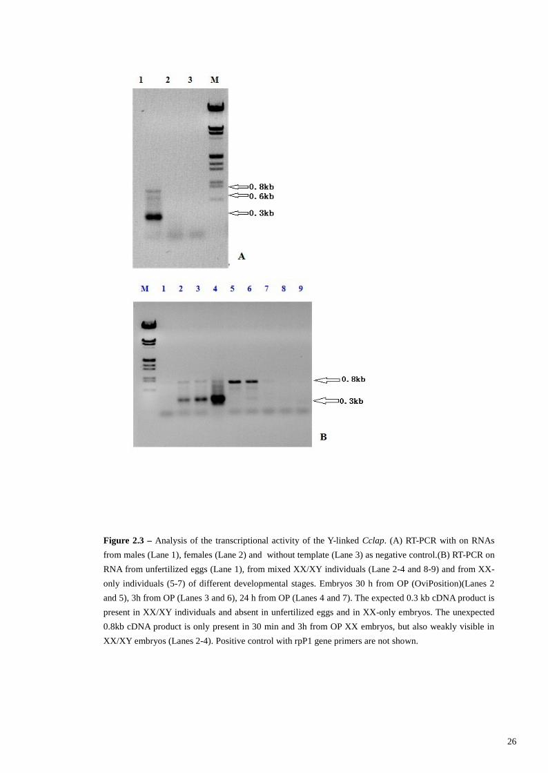
For the subtraction procedure are required RNA polyA+ amount of at least 2 g
for each of the two samples. To obtain this quantity of RNA polyA+ we have set up two
crosses for medium scale production of Ceratitis embryos with XX only karyotype
(from which to extract the XX-only RNA, named driver RNA) and embryos with mixed
karyotypes XY/XX (from which to extract the mixed XY/XX RNA, named tester
RNA). The two crosses were set up in cages of size 60x60x70 cm to contain about 2400
flies each: 800 XX pseudo-male and 1600 XX wild-type females with in the “driver”
cage and 800 XY wild- type males and 1600 XX wild-type females with in the “tester”
cage. We collected about 1 ml of embryos from each cross. Embryos collection was
carried out through two hours long intervals and the collected embryos were left to
develop until they reach the stage of 8-10 hours after ovideposition. Then we extracted
total RNA with cesium chloride density gradient protocol and we purified by affinity
chromatography molecules of RNA polyA+. Figure 2.5 shows an electrophoresis of
total RNA samples extracted by ultracentrifugation and respective RNA polyA+
obtained after chromatography.
Utilizing the PCR-Select Subtractive Hybridization Kit (Clontech, Palo Alto, CA)
we produced two subtracted libraries. The first one, named forward subtracted
library, is constituted of XY-XX 8-10h old Ceratitis capitata embryos cDNAs
population subtracted with XX-only 8-10h old Ceratitis capitata embryos cDNAs
population. This SSH library should contain early male-specific expressed transcripts
and hopefully MDG transcripts. The second library, named reverse subtracted library
and required as a control for the successive differential screening procedure, was
produced by subtracting the XX-only 8-10 old Ceratitis capitata embryos cDNAs
population with the XY-XX 8-10h old Ceratitis capitata embryos cDNAs population.
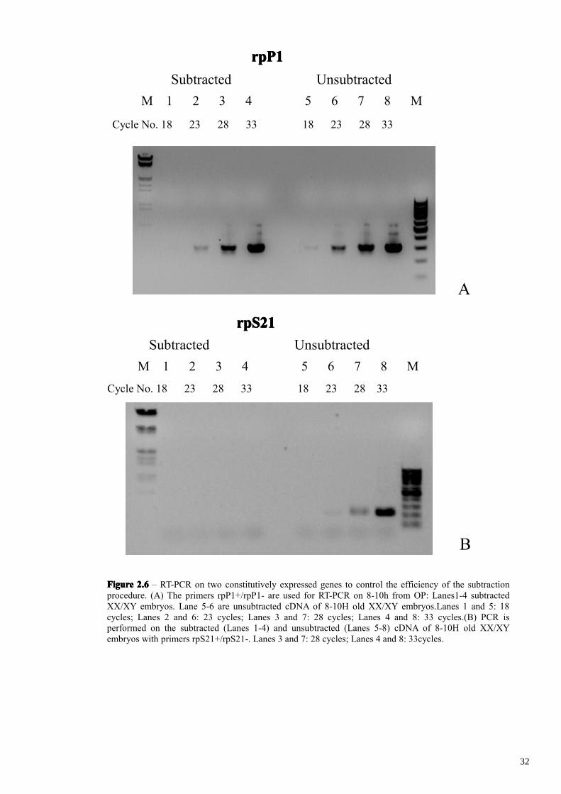
The validity of a SSH library can be confirmed by providing that subtraction had
indeed taken place. We evaluated our forward subtracted library by comparing the
abundance of two housekeeping genes, rpP1 (Gagou et al., 1999) and rpS21 (Verras et
al., 2004) in the subtracted respect to non-subtracted cDNA by PCR amplifications.
rpP1 transcripts were detected in the non-subtracted cDNA after 18 cycles of
amplification and rpS21 transcripts were detected after 28 cycles (Fig. 2.6). In
30
FigureFigureFigureFigure 2.52.52.52.5 – Gel electrophoresis of total RNA (Lanes 1 and 3) and poly (A+) RNA (Lanes 2 and 4), fromXX/XY (Lanes 1 and 2) and XX-only embryos (Lanes 3 and 4) 8-10h from OP .
1 2 3 4 M
31
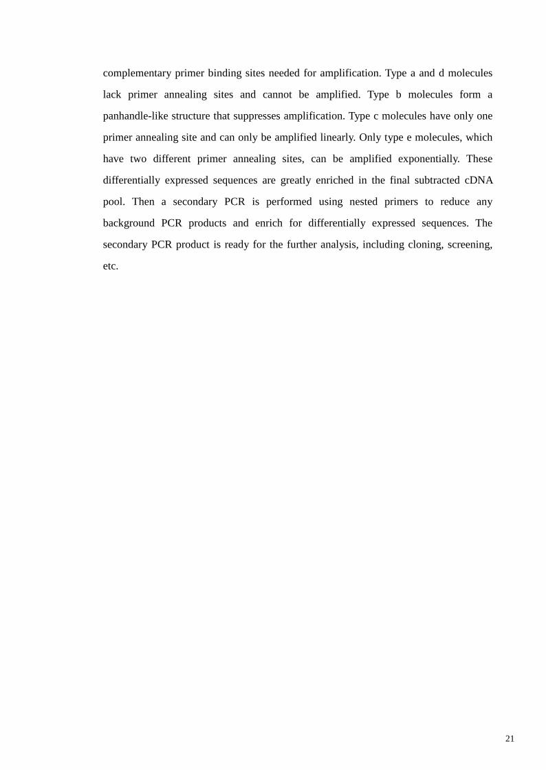
A
FigureFigureFigureFigure 2.62.62.62.6 – RT-PCR on two constitutively expressed genes to control the efficiency of the subtractionprocedure. (A) The primers rpP1+/rpP1- are used for RT-PCR on 8-10h from OP: Lanes1-4 subtractedXX/XY embryos. Lane 5-6 are unsubtracted cDNA of 8-10H old XX/XY embryos.Lanes 1 and 5: 18cycles; Lanes 2 and 6: 23 cycles; Lanes 3 and 7: 28 cycles; Lanes 4 and 8: 33 cycles.(B) PCR isperformed on the subtracted (Lanes 1-4) and unsubtracted (Lanes 5-8) cDNA of 8-10H old XX/XYembryos with primers rpS21+/rpS21-. Lanes 3 and 7: 28 cycles; Lanes 4 and 8: 33cycles.
Cycle No. 18 23 28 33 18 23 28 33
rpP1rpP1rpP1rpP1
M 1 2 3 4 5 6 7 8 MSubtracted Unsubtracted
Cycle No. 18 23 28 33 18 23 28 33
rpS21rpS21rpS21rpS21
M 1 2 3 4 5 6 7 8 MSubtracted Unsubtracted
B
32
subtracted cDNA rpP1 transcripts were detected after 23 cycles of amplification while
rpS21 transcripts were not detected at all. This marked reduction in the abundance of
both housekeeping genes in the SSH subtracted cDNA indicates that subtraction had
indeed correctly taken place.
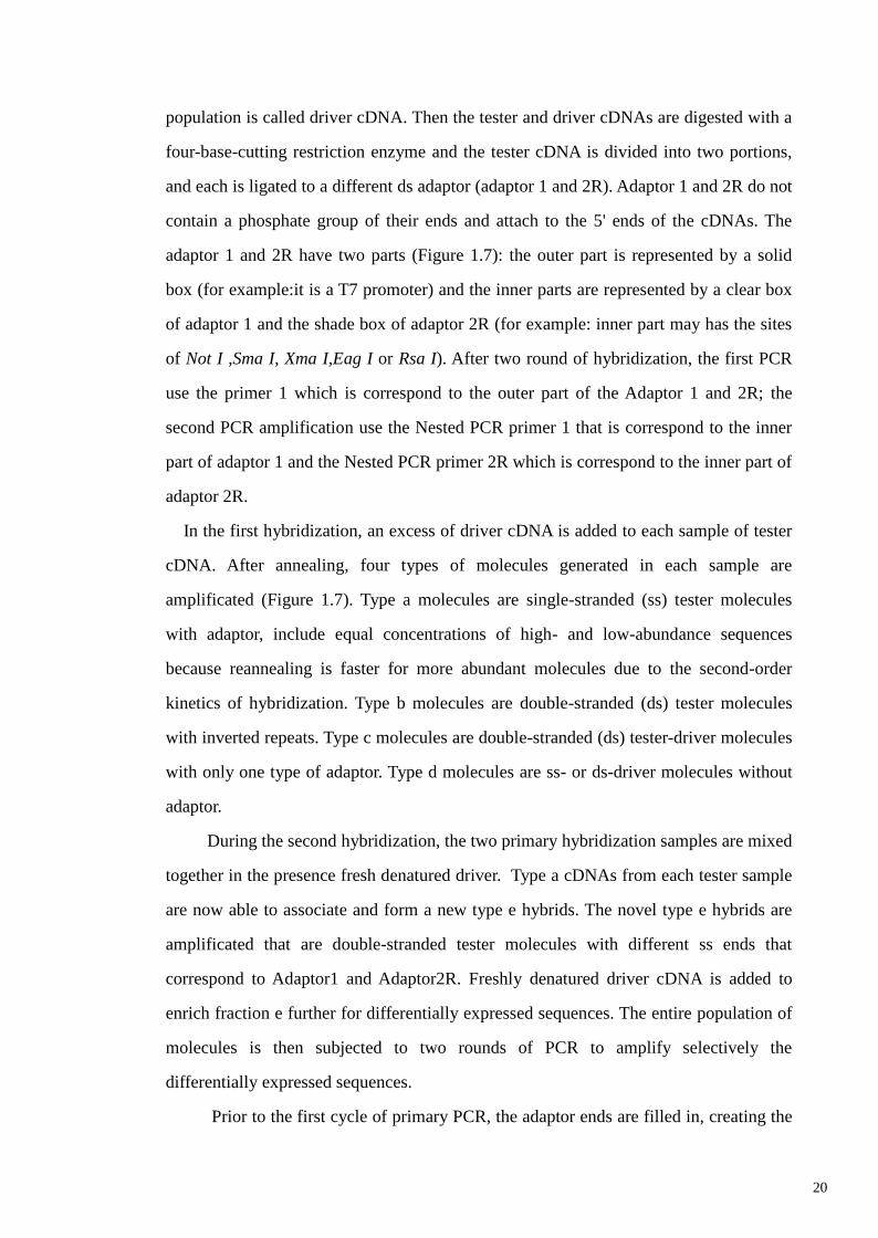
Mirror Orientated Selection
One of the major drawbacks of subtraction methods is the isolation of false
positive clones. These background clones are generated from non-specific annealing of
PCR primers or non-ligated adaptors (type-I background) and from redundant cDNA
molecules that evade elimination by hybridization (type-II background). In order to
reduce the number of background clones we applied also Mirror Oriented Selection
(MOS) procedure (Rebrikov et al., 2000). MOS utilizes the principle that background
molecules have only one orientation of the present adaptor sequence, whereas truly
differentially expressed molecules have many progenitors with adaptor sequences
present in both orientations. The result is achieved by removal of one adaptor by
restriction digestion, heat denaturation and re-annealing of the resulting molecules. In
this case only hybrid molecules with the remaining adaptor at the opposite ends are
amplified. MOS procedure hence eliminates background molecules reducing the
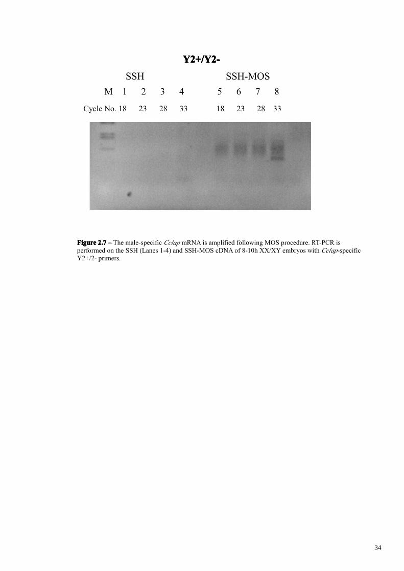
complexity of the remaining cDNA mixture. We confirmed the validity of MOS
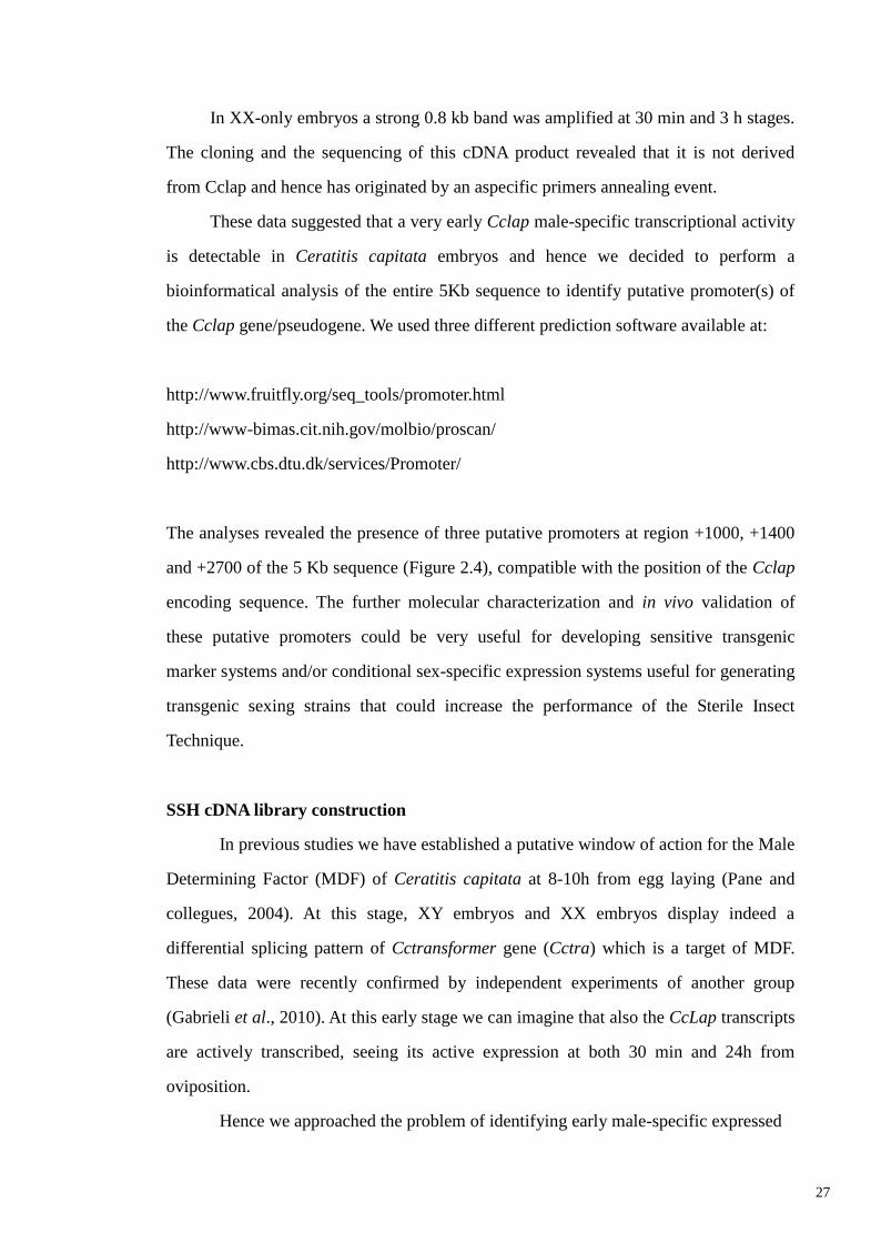
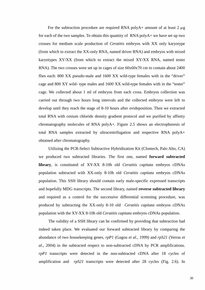
procedure using the male-specific CcLap transcript as positive control. As evidenced by
expression analysis at embryonic stages the CcLap 0.3 Kb amplification product is
amplifiable only in Y chromosome-containing samples, from early stage of
development (30 minutes). For this reason this transcript should be enriched during the
molecular subtraction procedure in the XY/XX minus XX sample. In SSH subtracted
cDNA this transcript is surprisingly not detectable at all by PCR amplification, most
probably due to the high complexity of the subtracted cDNA mixture due to abundant
background molecules. In addition SSH method has a better efficiency for genes
differentially expressed at low levels then for those having high expression such as
Cclap. After the MOS procedure instead CcLap trascripts are detected after 33 cycles of
amplification (Figure 2.7). These finding suggest us that MOS background reduction
33
FigureFigureFigureFigure 2.72.72.72.7 –––– The male-specific Cclap mRNA is amplified following MOS procedure. RT-PCR isperformed on the SSH (Lanes 1-4) and SSH-MOS cDNA of 8-10h XX/XY embryos with Cclap-specificY2+/2- primers.
Cycle No. 18 23 28 33 18 23 28 33
Y2+/Y2-Y2+/Y2-Y2+/Y2-Y2+/Y2-
M 1 2 3 4 5 6 7 8SSH SSH-MOS
34
had indeed correctly taken place and that male-specific transcripts are present in the
subtracted library.
The forward subtracted SSH-MOS cDNAs were directly cloned into a T/A
cloning vector (pDrive Vector – Qiagen) and electroporated into ultra competent E. coli
cells, resulting in the forward subtracted library. With homolog procedure we
produced also the second library, named reverse subtracted library.
We estimated the forward subtracted library size by plating an aliquot of the
transformed cells on 10 LB-agarose plates. We get a medium colony number of 643 for
each plate and a relative library size of 2,5 x 10⁴ clones.
Differential screening.
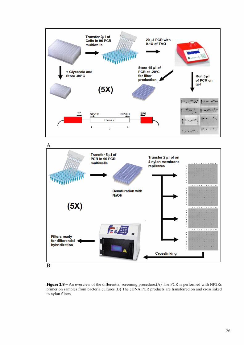
Differential screening of the subtracted cDNA library was performed by cDNA dot
blots of PCR products obtained with NP2Rs primer amplification of 480 forward
subtracted randomly chosen clones (NP2Rs primer is utilized during the MOS
procedure and is present at both sides of all the subtracted clones). In figure 2.8A-B is
presented a graphical overview of the differential screening procedure. 480 forward
subtracted plated colonies were picked out and incubated in five Eppendorf 1.2 ml 96-
well plates containing LB medium and ampicillin (named Plate A to E). The bacterial
cultures were analyzed individually by PCR amplification of plasmid inserts that were
analysed on agarose gel and then blotted on nylon filter replicates (4 for each plate)
(Figures 2.9-2.11). The electrophoresis analysis revealed that 38 clones out of 480 were
empty cloning vector and 32 clones out of the remaining 442 clones contain two or
more cloned products, which were excluded from the analysis. This led to a total
number of 410 single cloned subtracted cDNA fragments. It’s important to analyze
further by differential hybridization only those clones having a single fragment derived
from a specific mRNA. Indeed the presence of two independent cDNA fragments,
derived from two different mRNAs in the same plasmid, will hide a possible differential
hybridization of one of them. Considering that 10-30% of the clones are expected to be
truly positives, most probably only one of the two fragments would be differentially
expressed in vivo. The other cDNA fragment (false positive) would be expressed
35
A
B
FigureFigureFigureFigure 2.82.82.82.8 –––– An overview of the differential screening procedure.(A) The PCR is performed with NP2Rsprimer on samples from bacteria cultures.(B) The cDNA PCR products are transferred on and crosslinkedto nylon filters.
36
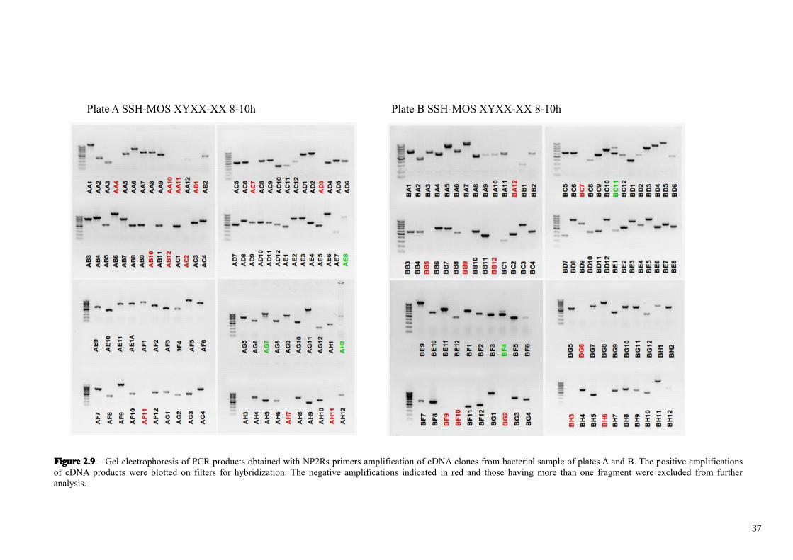
FigureFigureFigureFigure 2.92.92.92.9 – Gel electrophoresis of PCR products obtained with NP2Rs primers amplification of cDNA clones from bacterial sample of plates A and B. The positive amplificationsof cDNA products were blotted on filters for hybridization. The negative amplifications indicated in red and those having more than one fragment were excluded from furtheranalysis.
Plate A SSH-MOS XYXX-XX 8-10h Plate B SSH-MOS XYXX-XX 8-10h
37
FigureFigureFigureFigure 2.102.102.102.10 – Gel electrophoresis of PCR products obtained with NP2Rs primers amplification of cDNA clones from bacterial sample of plates C and D. The positive amplificationsof cDNA products were blotted on filters for hybridization. The negative amplifications indicated in red and those having more than one fragment were excluded from furtheranalysis.
Plate C SSH-MOS XYXX-XX 8-10h Plate D SSH-MOS XYXX-XX 8-10h
38
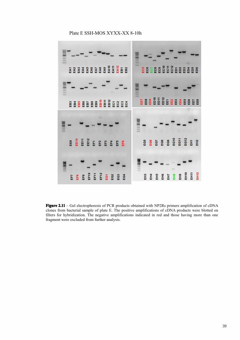
FigureFigureFigureFigure 2.112.112.112.11 – Gel electrophoresis of PCR products obtained with NP2Rs primers amplification of cDNAclones from bacterial sample of plate E. The positive amplifications of cDNA products were blotted onfilters for hybridization. The negative amplifications indicated in red and those having more than onefragment were excluded from further analysis.
Plate E SSH-MOS XYXX-XX 8-10h
39
similarly in both tester and driver and hence would hybridize with both probes on dot
spotted filters, hiding the presence of a second positive cDNA fragment.
4 identical filters were produced for each 96-well plate by arraying 2l of each
PCR product on nylon filter. Each filter replicate was hybridized with one of the four
following probes: 1) forward-subtracted tester probe (XY/XX 8-10h cDNA minus XX
8-10h cDNA), which identifies differentially expressed clones plus false positives. 2)
reverse-subtracted tester probe (XX 8-10h cDNA minus XY/XX 8-10h cDNA, which
identifies only false positives which hybridize also with the first probe, 3) unsubtracted-
tester probe (XY/XX 8-10h cDNA) which identifies differentially expressed clones plus
which identifies only false positives highly expressed,
Hence the clones that hybridize only with the forward-subtracted tester probe
(XX/XY) can correspond to differentially expressed cDNA clones. The clones that
hybridize with the forward-subtracted (XX/XY) and unsubtracted tester probes
(XX/XY), but not with the reverse-subtracted (XX minus XX/XY) or unsubtracted
driver probes (XX), usually correspond to differentially expressed genes, namely real
positive clones. Those clones having no detectable hybridization signals with any probe
could represent differentially expressed transcripts or false positives having a very low
abundance. Finally, those hybridizing equally with both subtracted probes and
unsubtracted probes, were the most highly expressed clones, including either real
positive clones or false positive. The results of the differential screening are shown in
Figures. 2.12-2.14. In our experiment, from 410 clones, 25 of them were identified as
hybridizing with the forward-subtracted tester probe (XX/XY minus XX). A further
clone was identified as hybridizing also with unsubtracted tester probe (XX/XY). All 26
clones either failed to hybridize or weakly hybridized with the reverse-subtracted (XX
minus XX/XY) and driver (XX) probes.
The 26 clones were sequenced with T7 and SP6 plasmid primers and aligned via
Macaw software. The sequence and length of AE5 and BE6 clones are respectively
identical to those of DB4 and BE8 clones. Hence we have isolated 24 different clones
and we analyzed them by computational analysis. A BLASTx analysis revealed that 18
40
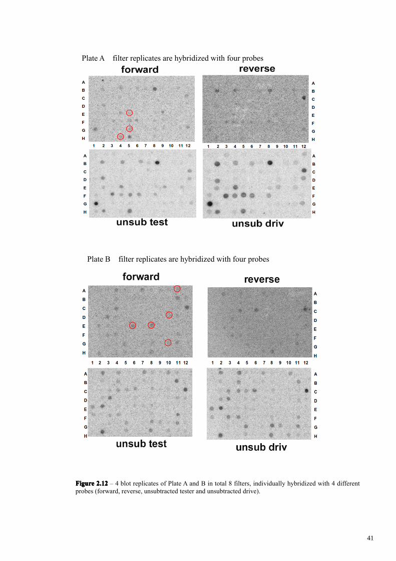
FigureFigureFigureFigure 2.122.122.122.12 – 4 blot replicates of Plate A and B in total 8 filters, individually hybridized with 4 differentprobes (forward, reverse, unsubtracted tester and unsubtracted drive).
Plate A filter replicates are hybridized with four probes
Plate B filter replicates are hybridized with four probes
41
FigureFigureFigureFigure 2.132.132.132.13 – 4 blot replicates of Plate C and D in total 8 filters, individually hybridized with 4 differentprobes (forward, reverse, unsubtracted tester and unsubtracted drive).
Plate D filter replicates are hybridized with four probes
Plate C filter replicates are hybridized with four probes.
42
FigureFigureFigureFigure 2.142.142.142.14 – 4 blot replicates of Plate E in total 4 filters, individually hybridized with 4 different probes(forward, reverse, unsubtracted tester and unsubtracted drive).
Plate E filter replicates are hybridized with four probes
43
transcripts encode for putative protein domains with significant homology with known
proteins of varying functions. The remaining 6 cDNA clones seem to encode either
unknown or low conserved proteins or to correspond to untranslated regions of the
transcripts. The results of these analyses are reported in Table 1.
Real Time PCR Validation
To validate the differential screening results and confirm the differential
expression of the 24 clones, we use quantitative real time PCR analysis. The Ambion
RetroScript Kit was used to prepare cDNA from the same XY/XX RNA polyA+ and
XX-only polyA+ samples used for the SSH-MOS procedure. cDNA were diluted 1:5
and 1 l was used in each reaction for Real-Time PCR using SYBR Green PCR
Mastermix (Applied Biosystem). rpP1 specific primers were used for normalization
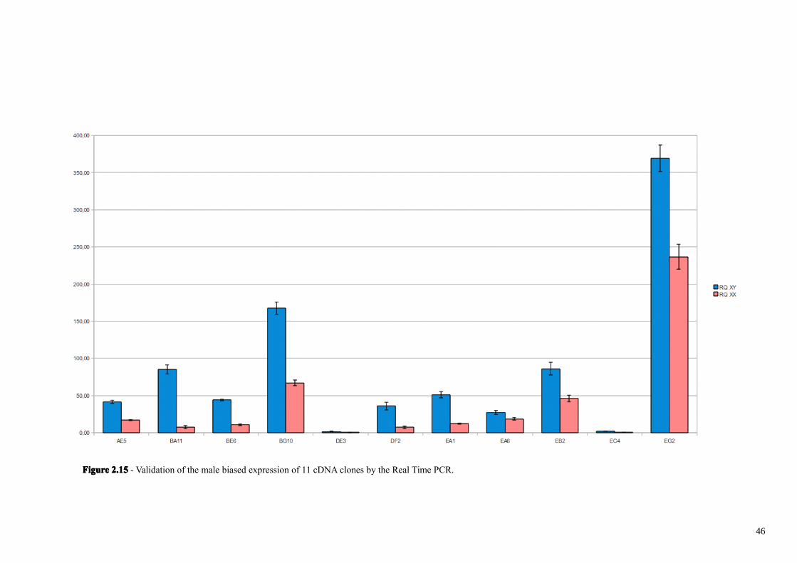
step. As reported in figure 2.15, 11 out of 26 clones are male-biased, with expression
levels ranging from 1,47 to 11,58 fold (Tab. 2).
We performed on these 11 differentially expressed clones, a Blast2Go analysis
(Götz et al., 2008), which automatically finds similarity between sequences (either
nucleotidic or aminoacidic), extracts the Gene Ontology (GO) terms associated to each
of the obtained hits and returns an evaluated GO annotation on putative biological
function (F), molecular process (P) and cellular component (C) for the query
sequence(s). The results are reported in Tab. 3. We have also performed a Blastx
analysis in Flybase, a Drosophila genome database (data not shown). Unfortunately, the
clone BA11, showing the most –male biased expression (11 times more expressed in
males), as also DE3 and EC4 clones have apparently no significant homology with
DNA or proteins. These 3 clones however could correspond to untranslated regions of
the corresponding transcripts.
EG2 clone encodes an aminoacidic sequence which has significant homology with
a blastoderm-specific protein of Drosophila, whose molecular function is unknown
(data not shown). Clone AE5 (2 times more expressed in males) and clone EA1 (4
times more expressed in males) encode aminoacidic sequences having possibly related
to functions such as spermatogenesis and in Drosophila are both related to Twin of m4
44
TableTableTableTable 1111 - Similarity sequence analyses of 24 clones by BlastN and BlastX algorithms on NCBI diptera databases.
45
FigureFigureFigureFigure 2.152.152.152.15 - Validation of the male biased expression of 11 cDNA clones by the Real Time PCR.
46
TableTableTableTable 2222 – Relative quantization (RQ) in XY embryos versus XX embryos of the 24 SSH-MOS subtractedclones. Clones shaded in light gray are more expressed in male embryos respect to female embryos.
47
TableTableTableTable 3333 - Analysis of 11 differentially expressed clones by Blast2Go tool.
48
and Ocho proteins, whose molecular functions are unknown but either for genetic
mutation or for similarity they are known to be involved in biological processes such as
sensory organ precursor cell fate determination, sensory organ development,
establishment of planar polarity, Notch signaling pathway and cell fate specification.
Clone BG10 encodes an aminoacidic sequence having similarity to transcription
factors such as the Drosophila DAN protein. The dan gene, distal antenna, encodes a
protein which has a transcription factor activity and protein binding and seems to be
involved in the biological processes such as segment specification and compound eye
development. EB2 encodes an aminoacidic sequence showing homology to Profilin, an
actin binding protein and in Drosophila to the Chickadee, a Profilin-related protein,
encoded by a gene showing various mutant alleles with mutant phenotypes interestingly
in the female and male germ lines and in the nervous system.
49
Conclusions
We have identified and analysed the expression of the first Y-linked gene in
Ceratitis capitata, Cclap, which appears to be transcribed very early during
embryogenesis (starting from the very first stages). Putative promoters of Cclap have
been identified which will be investigated for their use as early drivers of transgene
expression during embryogenesis. Further functional RNAi analysis of Cclap will
clarify its possible function during very early embryogenesis.
We used a Ceratitis transgenic line which can produce male only progeny by an in
vivo maternal RNAi specific for the Cctra master gene to obtain XX non transgenic
males. These males have been crossed to non transgenic XX females to produce XX-
only embryos. These method to produce female only progeny was very useful to
approach the problem of identifying male-specific or male-biased genes in Ceratitis
capitata through a PCR-based molecular subtractive technique.
We have produced a subtracted cDNA library from XX/XY embryos of 8-10h
from oviposition, in which we have found by differential hybridization out of 410 dot
spotted cDNA clones, 26 putative differentially expressed cDNAs out of which 11
cDNAs were real positive clones, showing a male-biased expression confirmed by real
time PCR. The overall efficiency of our SSH-MOS in leading to isolate cDNA clones
having putative differential expression is 6,3% (26/410), which is 1/3 of the one
obtained by Rebrikov et al., (2000). The observed efficiency of the MOS in reducing the
false positive with the respect of the real ones, is about 42% (11/26), a value which is
half of the one observed by Rebrikov. We observed indeed that 11 out of 26 clones
showed by real time PCR a significant differential expression, Considering the different
complexity and the different expected number of differentially expressed genes in the
Rebrikov and our studies, we think that the method worked very good in our case and
that interesting genes having differential expression in males have been identified.
We apparently failed to identify by the SSH strategy novel Y-linked genes,
considering that none of the clones showed an expression exclusively in the XX/XY
sample versus XX one, such as for example the Cclap gene. If the Y-linked M factor
50
corresponds to a non polyadenilated mRNA such as microRNAs, then a novel SSH
dedicated to this type of RNAs will be necessary to approach this new challenge.
However it still possible that one of the 11 isolated clones correspond to a Y-linked
gene, such as M, which however could have for example also a copy on an autosomal
localization. In this case mRNAs would be present in both sexes but biased in the male
one, because of the Y-linked extra copy, which could have evolved a male determining
or a male-specific function. These 11 Ceratitis capitata genes showing a male-biased
expression will be in future investigated also in their in vivo functions during
embryogenesis by applying transient RNAi on XX-only and on XX/XY mixed
embryos. Their functional study will contribute to a better understanding of the genetic
and molecular differences underlying the first stages of embryogenesis when male sex
determination takes place in Ceratitis capitata. This knowledge will be possibly useful
not only to understand evolution of sex determination in different dipteran species, but
also to develop novel strategies of biological control for this so relevant agricultural pest
insect.
51
Acknowledgment
I wish to sincerely thank all the people that have helped me during this three years of
Ph.D. experimental thesis.
I thank Prof. Catello Polito, Dr. Giuseppe Saccone and Dr. Marco Salvemini for their
high value scientific support, helpful discussions and for critically reading of the
manuscript.
I thank all my lab friends and colleagues Andreina Milano, Simona Capuozzo, Rocco
D’Amato, Domenica Ippolito.
52
Materials and Methods
Fly Strains
The medfly were reared in standard laboratory conditions at 25°C, 70% relative
humidity and 12:12 h light-dark regimen. 800 males mated 1600 females were
maintained in every sex cages (60cm×60cm×70cm).After 3-4 days, eggs were collected
in water dishes for 8-10H and 23-25H.
RNA isolation
The total RNA was extracted from embryos, using the standard guanidinium
isothiocyanate procedure (T. Maniatis, E. F. Fritsch and J. Sambrook, 1982). The 1ml
embryos are mixed with guanidinium isothiocyanate solution at the ratio of 1:7 (1 ml
embryos : 7 ml solution) and centrifuge at 10,000×g for 10 min at 4°C.The remove the
supernatant in a new tube with a sterile pipette. The supernatant fraction is highly
enriched for the denatured RNases and must be removed carefully to avoid bringing the
floating film into contact with the RNA pellet. The supernatant is mixed with 4 mL 5 M
CsCl and centrifuge at 31,000×g for 16 h at 18°C, then discard the supernatant fraction,
the RNA pellet is at the bottom of the tube. Dry the pellet at the room temperature and
dissolve the pellet in 500-1000 μl DEPC-treated 1 mM EDTA (pH 7.5) solution (5 to 50
μl is recommended). The concentration and purity of the RNA concentration were
determined spectrophotometrically by measuring the absorbance at 260 and 280 nm,
and the integrity of the RNA was assessed using denaturing agarose gel electrophoresis.
Reverse Transcription PCR
RT-PCR was performed using RNA from embryos with Advantage® RT-for-PCR Kit
(Clontech). 1 μl of RNA from each sample was treated with 1 μl of Dnase I (2 U/μl,
53
Ambion) to remove contaminating DNA following the manufacturer's instruction, and
then reverse transcribed using 1 μl of oligo (dT), incubate the sample at 70°C for 2 min.
Then the sample is mixed with 5× reaction buffer, dNTP mix, RNase inhibitor, MMLV
reverse transcriptase in the total volume of 20 μl. The mixture was incubated in a
thermal cycle at 42°C for 1 h and 94°C for 5 min. The primer RpP1(ribosomal protein
P1) was used as the positive control (RpP1+:5'-TTGCGTTTACGTTGCTCTCG-
3';RpP1-:5'-AATCGAAGAGACCGAAACCC-3'). The following PCR cycles were
performed: 5 min at 94°C, 35 cycles with 1 min at 94°C, 1 min at 60°C, 1 min at 72°C,
10 min at 72°C. RT-PCR expression analysis was performed with the following primers: