35
THE MUSCULAR SYSTEM Dr Idara
| Date post: | 17-Dec-2015 |
| Category: |
Documents |
| Upload: | joel-campbell |
| View: | 217 times |
| Download: | 0 times |

THE MUSCULAR SYSTEM
Dr Idara
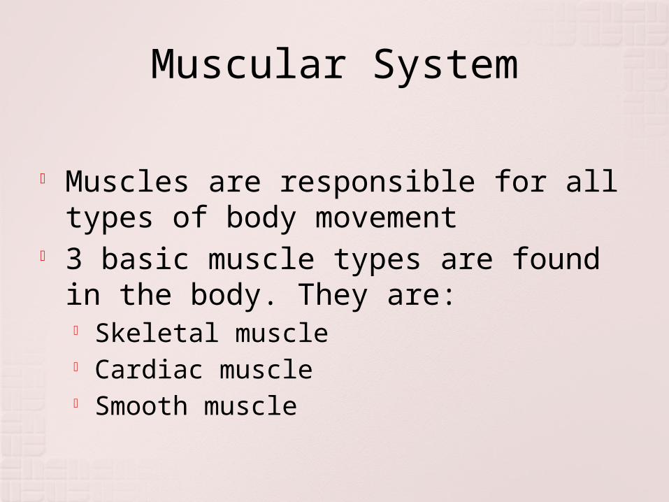
Muscular System
Muscles are responsible for all types of body movement
3 basic muscle types are found in the body. They are:
Skeletal muscle Cardiac muscle Smooth muscle

Characteristics of Muscles
Muscle cells are elongated (muscle cell = muscle fiber)
Contraction of muscles is due to the movement of microfilaments
All muscles share some terminology Prefix myo refers to muscle Prefix mys refers to muscle Prefix sarco refers to flesh

Muscle histology (cont.)
Scaterred throughout the sarcoplasm is the sarcoplasmic reticulum
It stores Calcium ions The sarcoplasm contains myoglobin
Red pigmented protein related to Hemoglobin that carries oxygen
Along entire length are myofibrils Myofibrils made of protein filaments
Come in thick and thin filaments
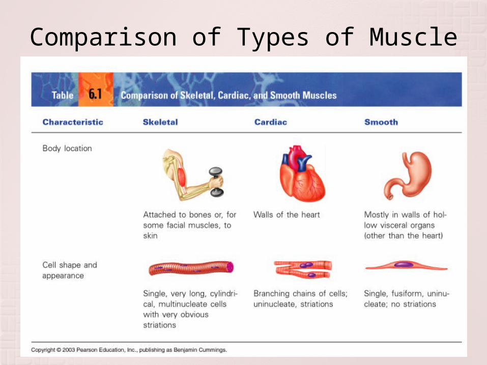
Comparison of Types of Muscle

Types of Muscle, cont

Skeletal Muscle Characteristics
Most attach to bones by tendon
Cells are multinucleate
Striated—have visible binding
Voluntary Cells surrounded &
bundled by connective tissue
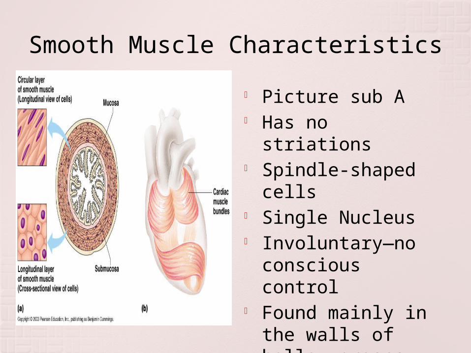
Smooth Muscle Characteristics
Picture sub A Has no striations Spindle-shaped
cells Single Nucleus Involuntary—no
conscious control Found mainly in
the walls of hollow organs
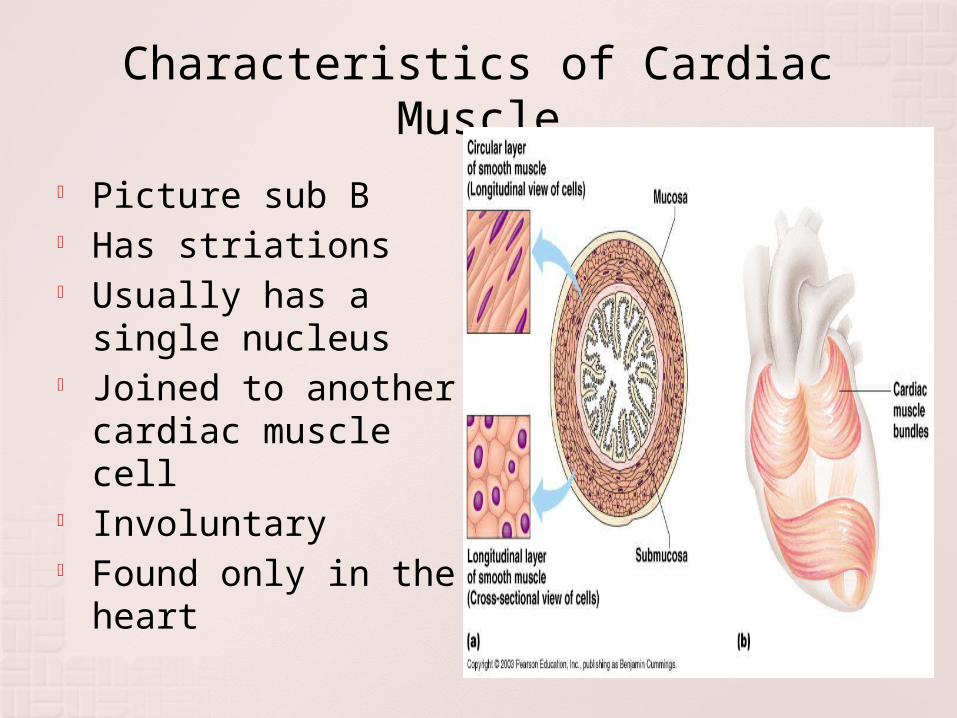
Characteristics of Cardiac Muscle
Picture sub B Has striations Usually has a
single nucleus Joined to another
cardiac muscle cell Involuntary Found only in the
heart

Functions of Skeletal Muscle
Produce Movement Maintain posture Stabilize joints Generate Heat
Sites of Muscle Attachment
Bones Cartilage Connective tissue
coverings
Muscle Fibers blend into a connective tissue attachment
Tendon—cordlike structure Aponeurosis—sheet-like
structure Properties of Muscle
Irritability – ability to receive and respond to a stimulus
Contractibility – ability to shorten when an adequate stimulus is received
Extensibility – ability to lengthen when an adequate stimulus is received
Elasticity – ability to return to normal shape

To Note:
A muscle cell is also called a muscle Fiber
A discrete bundle of muscle cells is called a Fascicle
The thin connective tissue that surrounds each muscle cell is called Endomysium
The connective tissue that surrounds each Fascicle is called Perimysium
The connective tissue surrounding the entire muscle is called Epimysium
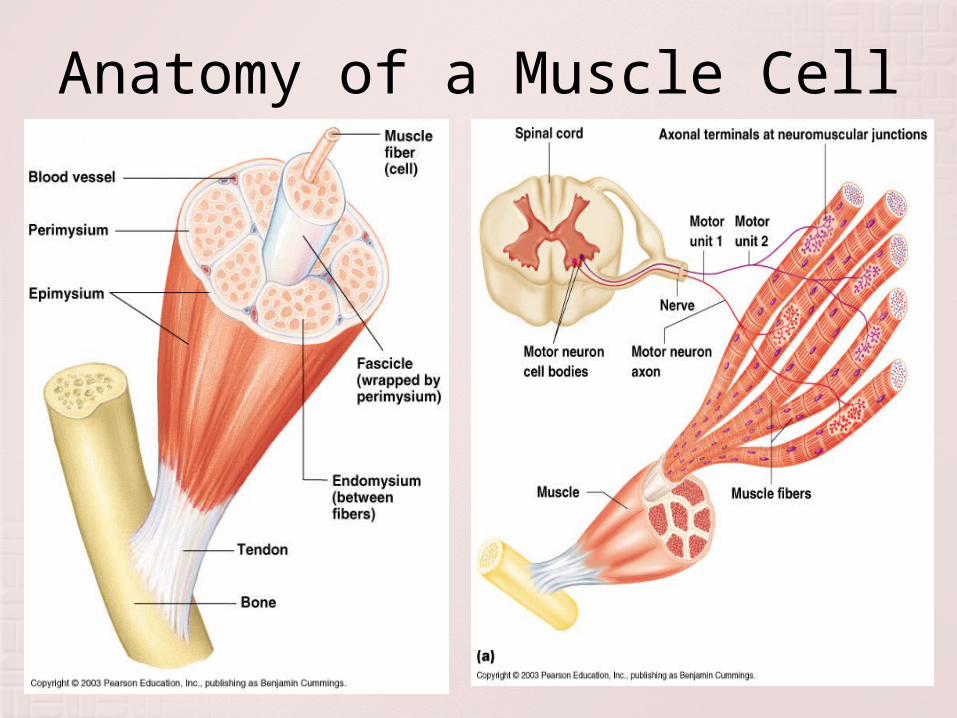
Anatomy of a Muscle Cell

Naming Skeletal Muscles
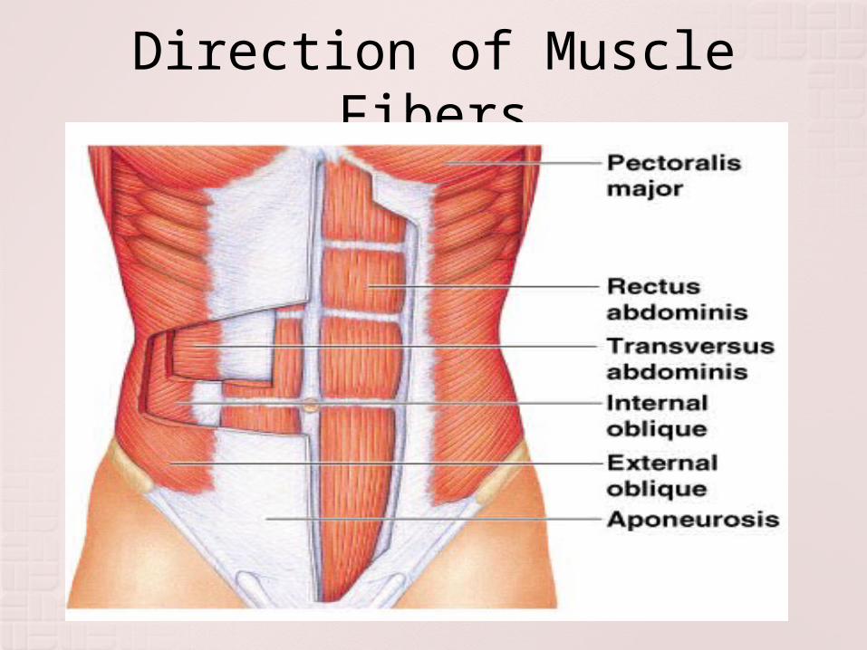
Direction of Muscle Fibers
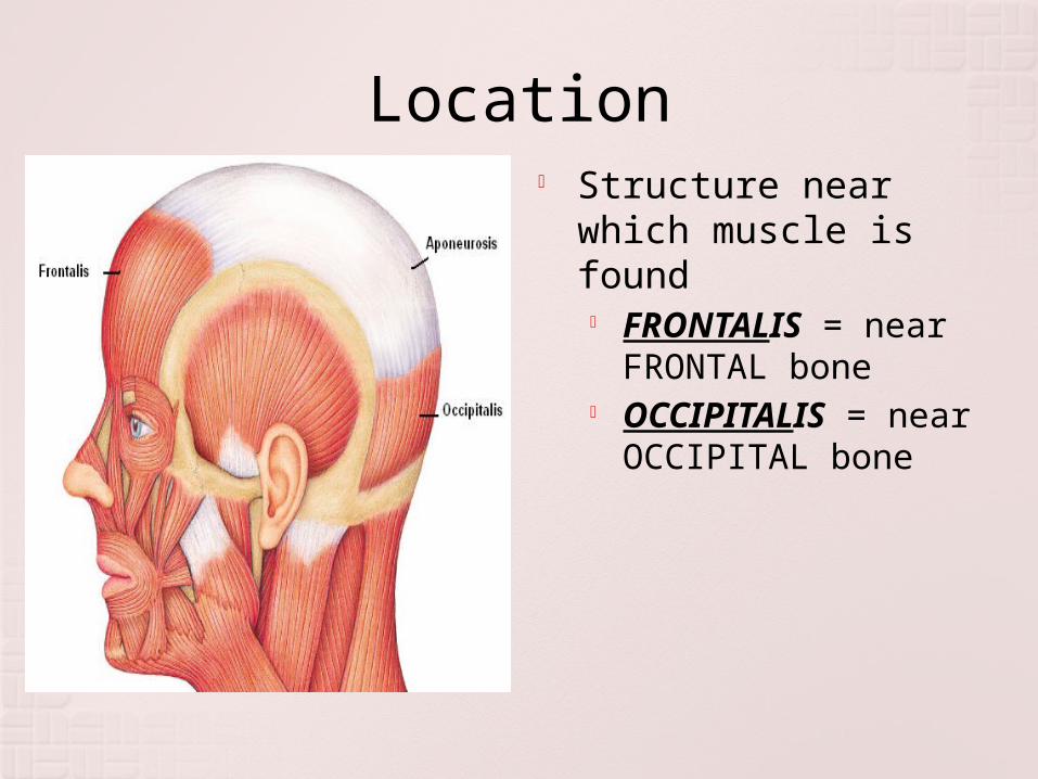
Location Structure near which
muscle is found FRONTALIS = near
FRONTAL bone OCCIPITALIS =
near OCCIPITAL bone
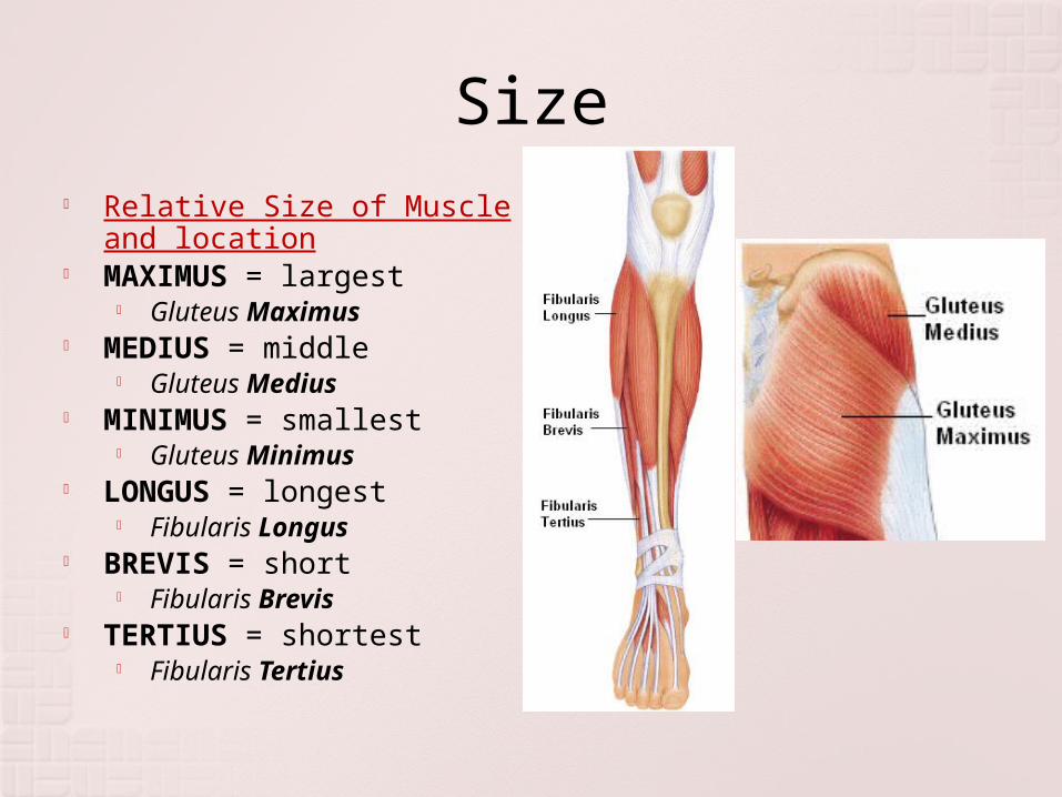
Size Relative Size of Muscle and
location MAXIMUS = largest
Gluteus Maximus MEDIUS = middle
Gluteus Medius MINIMUS = smallest
Gluteus Minimus LONGUS = longest
Fibularis Longus BREVIS = short
Fibularis Brevis TERTIUS = shortest
Fibularis Tertius
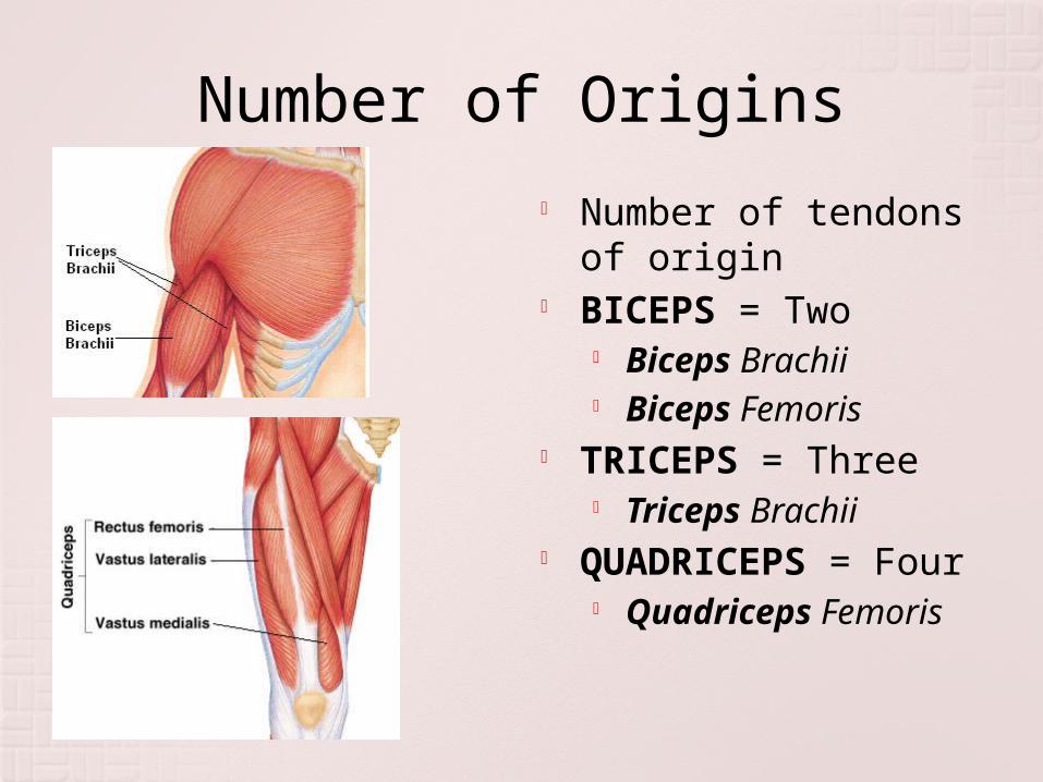
Number of Origins Number of tendons
of origin BICEPS = Two
Biceps Brachii Biceps Femoris
TRICEPS = Three Triceps Brachii
QUADRICEPS = Four
Quadriceps Femoris
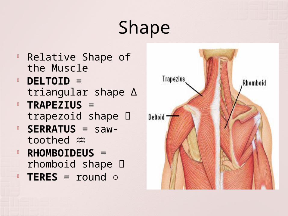
Shape Relative Shape of
the Muscle DELTOID =
triangular shape Δ TRAPEZIUS =
trapezoid shape SERRATUS = saw-
toothed ♒ RHOMBOIDEUS =
rhomboid shape TERES = round ○

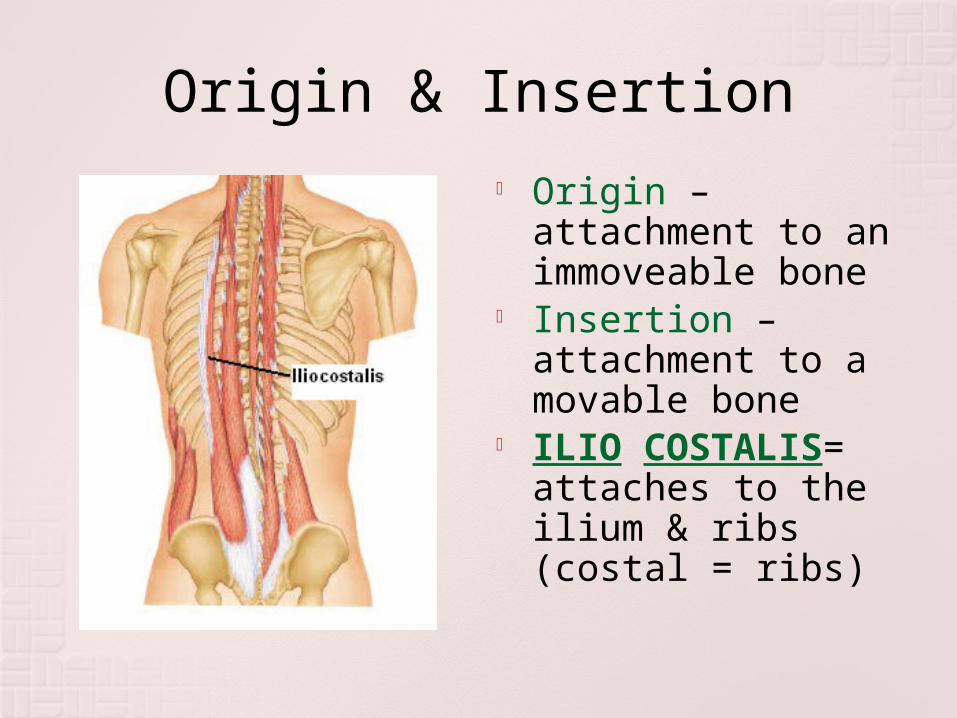
Origin & Insertion Origin –
attachment to an immoveable bone
Insertion – attachment to a movable bone
ILIO COSTALIS= attaches to the ilium & ribs (costal = ribs)
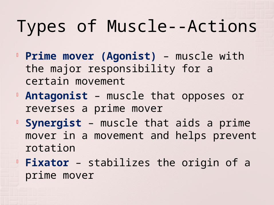
Types of Muscle--Actions
Prime mover (Agonist) – muscle with the major responsibility for a certain movement
Antagonist – muscle that opposes or reverses a prime mover
Synergist – muscle that aids a prime mover in a movement and helps prevent rotation
Fixator – stabilizes the origin of a prime mover
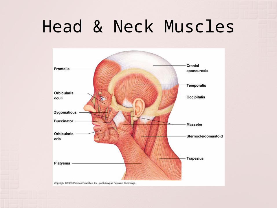
Head & Neck Muscles

Key Muscles of Facial Expression
Smiling Muscles Orbicularis Oculi Nasalis Levator Labii
Superioris Levator Anguli
Superioris Zygomaticus Risorius
Frowning Muscles Frontalis Orbicularis Oris Depressor Anguli
Oris Depressor Labii
Inferioris Mentalis Platysma


Frontalis - Used in frowning
Orbicularis Oculi – Surrounds the eye, eyelid and orbit. Used to close the eyes
Zygomaticus - Used in smiling
Buccinator- A flat muscle of the cheek, draws in
the cheek and puffs up the cheeks. Called the Trumpeter’s Muscle.
Orbicularis Oris- surrounds the mouth, closes the lips when contracted and used for whistling and kissing.
Masseter- Is the Prime mover for jaw closure. Also used for chewing.
Temporalis - is the synergist for jaw closure and assists with chewing.

Muscles of the Axial Skeleton
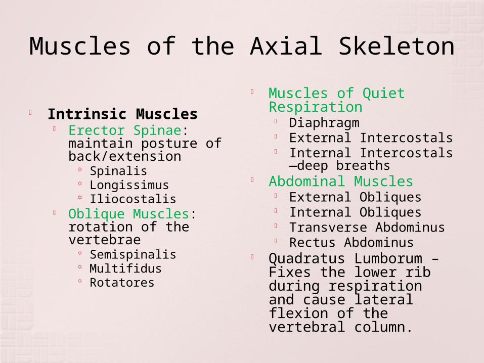
Muscles of the Axial Skeleton
Intrinsic Muscles Erector Spinae:
maintain posture of back/extension
Spinalis Longissimus Iliocostalis
Oblique Muscles: rotation of the vertebrae
Semispinalis Multifidus Rotatores
Muscles of Quiet Respiration
Diaphragm External Intercostals Internal Intercostals—
deep breaths Abdominal Muscles
External Obliques Internal Obliques Transverse Abdominus Rectus Abdominus
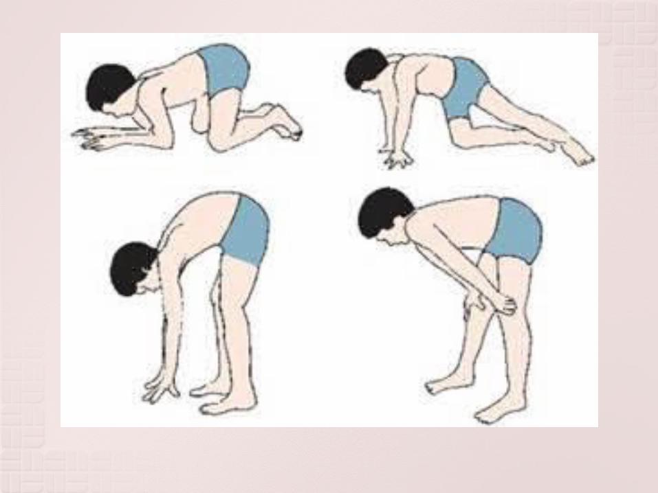
Quadratus Lumborum – Fixes the lower rib during respiration and cause lateral flexion of the vertebral column.


Diseases and Disorders of the Muscular System
Cerebral Palsy: This disorder is characterized by paralysis and or weakened muscles due to loss of muscle tone.
It can be caused due to lack of oxygen to the region of the motor region of the cerebrum of the brain which controls conscious control of muscles.
This is often attributed to complication during birth.

Others Myalgia: Muscle pain due to strain, tearing
of muscle fibers. It also is a symptom of an immune response along with a fever.
Myositis: Inflammation of muscle tissue due to injury or disease.
Charley Horse (fibromyositis): Inflamation of muscle tissue and the tendons associated with that muscle due to injury (tear or severe bruising- contusion).
Cramps: Painful, involuntary muscle contraction, typically caused by fatigue or strain.
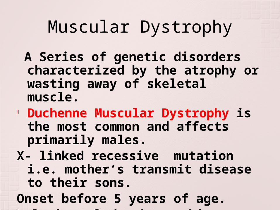
Muscular Dystrophy
A Series of genetic disorders characterized by the atrophy or wasting away of skeletal muscle.
Duchenne Muscular Dystrophy is the most common and affects primarily males.
X- linked recessive mutation i.e. mother’s transmit disease to their sons.
Onset before 5 years of age.Deletion of the dystrophin gene


Becker’s Muscular Dystrophy
Onset in adolesence or early adulthood.
Mutated dystrophin gene


