Department of Nuclear Physics and Biophysics, Faculty of Mathematics, Physics, and Informatics, Comenius University, Mlynska dolina F1, 842 48 Bratislava, Slovakia
Received: September 30, 2009; Accepted: November 27, 2009
Key words: Magnetic nanoparticles/Cancer therapy/Drug targeting/Gene Therapy/Controlled drug release
Background: Nanomaterials are at the leading edge of the rapidly developing field of nanotechnology. Magneticnanoparticles for cancer therapy and diagnosis have been developed on the basis of their unique physico-chemicalproperties not present in other materials. Their versatility is widely exploited in such diverse techniques as cell andmacromolecule separation and purification, immunoassays, targeted drug delivery, controlled material release, electro-
magnetic hyperthermia, gene therapy, or magnetic resonance imaging. In this review we concentrate on the physicalprinciples of magnetic drug targeting and biomedical applications of this technique.
Methods and results: We examined several databases, PubMed, ISI Web of Knowledge, and Scopus, for the period1985–2009, with specific attention to studies that used targeting of magnetic nanoparticles especially in the therapyand diagnostics of tumors. We have also presented several of our own results on theoretical simulations of magneticparticle motion in external magnetic field.
Conclusions: We found growing number of published papers in this field of nanomedicine, showing the almostunlimited potential of magnetic nanoparticles in the field of experimental and clinical oncology.
INTRODUCTION
The history of magnetism dates back to earlier than
600 B.C., but it is only in the twentieth century that sci-entists have begun to understand it, and develop tech-nologies based on this understanding. Magnetism wasmost probably first observed in a form of the mineralmagnetite called lodestone, which consists of iron oxidea chemical compound of iron and oxygen. The ancientGreeks were the first known to have used this mineral,which they called a magnet (from Greek μαγνήτης λίθος,"Magnesian stone") because of its ability to attract otherpieces of the same material and iron. Curiously, the firstsystematic study of magnetism using scientific methodswas made not by a physicist but by a physician William
Gilbert (1540–1603) the most distinguished man of sci-ence in England during the reign of Queen Elizabeth I,and the father of electric and magnetic science. Gilbert'sprincipal work is his treatise on magnetism, entitled De
magnete, magneticisque corporibus, et de magno magnete
tellure (London, 1600). This work, which embodied theresults of many years research, was distinguished by itsstrict adherence to the scientific method of investigationby experiment, and by the originality of its matter, con-taining, as it does, an account of the author's experimentson magnets and magnetic bodies and on electrical attrac-tions, and also his great conception that the earth is noth-ing but a large magnet, and that it is this which explains,not only the direction of the magnetic needle north andsouth, but also the variation and dipping or inclinationof the needle. Gilbert's is therefore not merely the first,
but the most important, systematic contribution to thesciences of electricity and magnetism.
One of the most promising fields of research lying on
the border of medicine, biology, physics, chemistry, andengineering – “nanomedicine”, according to the defini-tion given by USA National Institutes of Health is basedon the applications of nanotechnology for treatment, di-agnosis, monitoring, and control of biological systems.In the forefront of this field is research into the rationaldelivery and targeting of pharmaceutical, therapeutic,and diagnostic agents. The oldest and still most vivid sub-field is applications of magnetic nanoparticles (MNs) inbiology and medicine. Multifunctional MNs which arethe topic of this review have diverse potential applica-tions in many biological and medical applications such
as cell separation, drug targeting, electromagnetic hyper-thermia, magnetic resonance contrast enhancement. Webegan this introduction with the British physician WilliamGilbert, and we want to close it with an another physician,
Vilém Laufberger (1890–1986) from Charles Universityin Prague who in 1937 isolated for the first time proteinferritin containing 4 nm large magnetic nanoparticle1. Inour view the first nanotechnology pioneer should be con-sidered V. Lauberger, although generally is accepted as abeginning of nanotechnology Feynmann visionary lecture“There is a plenty of room at the bottom“ delivered in1959. Ferritin besides its key role in iron body metabo-lism2 has been used for multifold purposes. Let us mentionthe pathogenesis of Alzheimer3 and Parkinson diseases4,magnetic cell separation5, magnetic drug targeting6, MRIcontrast agents7, electromagnetic hyperthermia8, ultrasen-sitive diagnosis9, and macroscopic quantum tunelling10.
7/23/2019 Drug Delivery Magnetic Nanoparticle Review 2009
A magnet is a material or object that produces a mag-netic field. This magnetic field is invisible but is respon-sible for the most notable property of a magnet: a forcethat pulls on other ferromagnetic materials and attracts
or repels other magnets. Permanent magnet materialsare widely used in a range of devices from electric andelectronic appliances for domestic use to peripheral ter-minal devices for large-scaled computers. In view of recentneeds for miniaturization and high efficiency of electricand electronic equipment, there has been an increasingdemand for upgrading of permanent magnet materials. Apermanent magnet is one made from a material that staysmagnetized. Materials that can be magnetized, which arealso the ones that are strongly attracted to a magnet, arecalled ferromagnetic11. These include iron, nickel, cobalt,some rare earth metals and some of their alloys (e.g
Alnico), and some naturally occurring minerals such aslodestone. Permanent magnets are made from "hard" fer-romagnetic materials which are designed to stay magnet-ized, while "soft" ferromagnetic materials like soft iron areattracted to a magnet but do not tend to stay magnetized.
An electromagnet is made from a coil of wire which actsas a magnet when an electric current passes through it,but stops being a magnet when the current stops. Oftenan electromagnet is wrapped around a core of ferromag-netic material like steel, which enhances the magneticfield produced by the coil. Because human tissues havea very low level of susceptibility to static magnetic fields,there is no direct scientific evidence showing a health
hazard associated with exposure to these f ields. However,if a ferromagnetic foreign body is present in human tissue,the magnetic field will interact with it, which can pose aserious safety risk. Specifically, if a pacemaker has beenembedded in a patient's chest, care should be taken tokeep it away from magnetic fields. It is for this reason thata patient with the device installed cannot be tested withthe use of an MRI, which is a magnetic imaging device.The most commonly used ceramic, or ferrite, magnets aremade of a sintered composite of powdered iron oxide andbarium/strontium carbonate ceramic. Given the low costof the materials and manufacturing methods, inexpensive
magnets (or nonmagnetized ferromagnetic cores, for usein electronic components such as radio antennas, for ex-ample) of various shapes can be easily mass-produced.The resulting magnets are noncorroding, but brittle andmust be treated like other ceramics. To the another classbelongs alnico magnets which are made by casting orsintering a combination of aluminium, nickel and cobaltwith iron and small amounts of other elements addedto enhance the properties of the magnet. Sintering of-fers superior mechanical characteristics, whereas castingdelivers higher magnetic fields and allows for the designof intricate shapes. Alnico magnets resist corrosion andhave physical properties more pliable than ferrite, but not
quite as desirable as a metal.
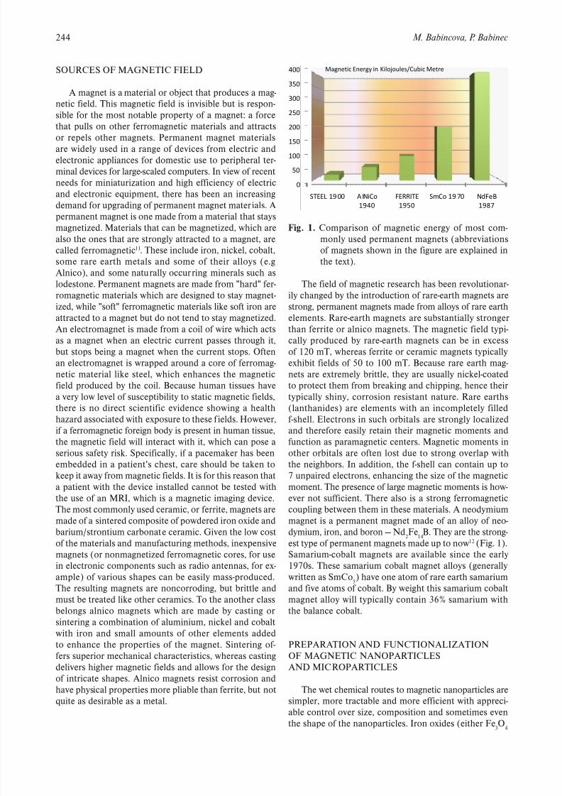
The field of magnetic research has been revolutionar-ily changed by the introduction of rare-earth magnets arestrong, permanent magnets made from alloys of rare earthelements. Rare-earth magnets are substantially strongerthan ferrite or alnico magnets. The magnetic field typi-cally produced by rare-earth magnets can be in excessof 120 mT, whereas ferrite or ceramic magnets typicallyexhibit fields of 50 to 100 mT. Because rare earth mag-nets are extremely brittle, they are usually nickel-coatedto protect them from breaking and chipping, hence theirtypically shiny, corrosion resistant nature. Rare earths(lanthanides) are elements with an incompletely filled
f-shell. Electrons in such orbitals are strongly localizedand therefore easily retain their magnetic moments andfunction as paramagnetic centers. Magnetic moments inother orbitals are often lost due to strong overlap withthe neighbors. In addition, the f-shell can contain up to7 unpaired electrons, enhancing the size of the magneticmoment. The presence of large magnetic moments is how-ever not sufficient. There also is a strong ferromagneticcoupling between them in these materials. A neodymiummagnet is a permanent magnet made of an alloy of neo-dymium, iron, and boron — Nd
2Fe
14B. They are the strong-
est type of permanent magnets made up to now 12 (Fig. 1).
Samarium-cobalt magnets are available since the early1970s. These samarium cobalt magnet alloys (generallywritten as SmCo
5) have one atom of rare earth samarium
and five atoms of cobalt. By weight this samarium cobaltmagnet alloy will typically contain 36% samarium withthe balance cobalt.
PREPARATION AND FUNCTIONALIZATIONOF MAGNETIC NANOPARTICLES
AND MICROPARTICLES
The wet chemical routes to magnetic nanoparticles are
simpler, more tractable and more efficient with appreci-able control over size, composition and sometimes eventhe shape of the nanoparticles. Iron oxides (either Fe
3O
4
0
50
100
150
200
250
300
350
400
STEEL 1900 A INiCo
1940
FERRITE
1950
SmCo 19 70 NdFeB
1987
Magnetic Energy in Kilojoules/Cubic Metre
Fig. 1. Comparison of magnetic energy of most com-monly used permanent magnets (abbreviationsof magnets shown in the figure are explained inthe text).
7/23/2019 Drug Delivery Magnetic Nanoparticle Review 2009
245 Magnetic drug delivery and targeting: principles and applications
or g-Fe2O
3) can be synthesized through the co-precipita-
tion of Fe2+ and Fe3+ aqueous salt solutions by additionof a base13. The control of size, shape and composition ofnanoparticles depends on the type of salts used (e.g. chlo-rides, sulphates, nitrates, perchlorates, etc.), Fe2+ and Fe3+ ratio, pH and ionic strength of the media. Conventionally,
magnetite is prepared by adding a base to an aqueousmixture of Fe2+ and Fe3+ chloride at a 1:2 molar ratio.The precipitated magnetite is black in colour. The overallreaction may be written as follows:
Fe2+ + Fe3+ + 8OH– Fe3O
4 + 4H
2O
According to the thermodynamics of this reaction,a complete precipitation of Fe
3O
4 should be expected
between pH 9 and 14, while maintaining a molar ratioof Fe2+ : Fe3+ is 2:1 under a non-oxidizing oxygen free en-
vironment. Otherwise, Fe3O
4 might also be oxidized as
4Fe3O
4 + O
2 + 18H
2O 12Fe(OH)
3
This would critically affect the physical and chemi-cal properties of the nanosized magnetic particles. Inorder to prevent them from possible oxidation in air aswell as from agglomeration, Fe
3O
4 nanoparticles produ-
ced by these reactions are usually coated with organicor inorganic molecules during the precipitation process.Several authors have reported the magnetic iron oxidenanoparticles coated with silica14. An advantage of havinga surface enriched in silica is the presence of surface sila-nol groups that can easily react with alcohols and silane
coupling agents15. Various biological molecules such asantibodies, proteins, targeting ligands, etc., may also bebound to the polymer surfaces onto the nanoparticles by
chemically coupling via amide or ester bonds to make theparticles target specific (Fig. 2). The possibilities of tar-geting protein coatings are numerous16–21. Another possi-bility is represented by liposomes 22–26 with the ability toencapsulate a large number of magnetic nanoparticles anddeliver them together, avoiding dilution, to a target site.
Combining a therapeutic agent in the payload further en-hances the multifunctionality of these delivery vehicles.
MAGNETIC DRUG TARGETING
The major problems of chemotherapeutics come es-sentially from the relative lack of specificity derived fromtheir systemic biodistribution and the subsequent sideeffects provoked by the drug attacking both healthy andtarget cells27.
There are several forces acting on magnetic particles in viscous environment and magnetic field, such as magneticforce due to all field sources, Stokes’ viscous drag force,inertia, buoyancy and gravity, thermal kinetics (Brownianmotion), and of course particle fluid interactions and in-terparticle effects like magnetic dipole interactions, elec-tric double layer interactions, and van der Vaal?Wallsforce. Apart from magnetic and viscous drag force thereare the others interactions negligible for magnetic micro-or nanoparticles28, 29. For this reason we assume in thispaper only these two effects. Given the considerationsmentioned before, we can predict the trajectory of motionof magnetic particle in magnetic field and viscous fluidambient using Newton’s law:
sm
p
pdt
dm FF
v+= , (1)
where m p and v
p are the mass and velocity of the particle,
and Fm and F
s are the magnetic and Stoke’s drag forces,
respectively.Magnetic force acting on a magnetic particle is deter-
mined by using the method of “effective” dipole moment,in which a magnetic particle is replaced by “equivalent”point dipole moment m
p,eff localized at the center of parti-
cle. According to this method, force acting on the dipoleis given by
aeff pm HmF , , (2)
where μ is permeability of fluid ambient, m p,eff
is “effec-tive” dipole moment of the particle, which depends onexternally applied magnetic field intensityH
a at the center
of particle, where the dipole moment is localized. A mag-netic force is therefore a function of external magneticfield gradient and the magnetization of the particle. Belowthe saturation the particles are linearly magnetized withtheir magnitude of magnetic moment increasing in thedirection of external field. Beyond the saturation point,magnetic moment magnitude tends to a constant value.
According to this magnetization model of particles basedon self demagnetization and magnetic saturation devel-oped by Furlani group30 the effective dipole moment canbe expressed as
Fig. 2. An example of multifunctionalized (F1 and F2
are examples two different functional groups)magnetic nanoparticle with ferrite core coveredby stabilization shell, e.g. SiO
2, and conjugated
functional groups at the surface.
7/23/2019 Drug Delivery Magnetic Nanoparticle Review 2009
we consider magnetic particle with radius R p and volume
V p, and a function
(|χ f |<<1),
3
3)( H
3
3)(
3)(
)(3
)(
α
+−≥
+−
<+−
−
=
sp
p
f p
a sp
sp p
f p
a f p
f p
a
M H M
M H H f
χ
χ χ
χ
χ χ
χ χ
χ χ
(4)
Where χ p and χ
f are the magnetic susceptibilities of the
particle and ambient fluid, respectively, M sp
is saturationmagnetization of the particle, and aa H H .
We assume nonmagnetic fluid ( 0 f ) and high sus-ceptibility of magnetic particles, i.e. 1 p , which is inthe case of water or air as fluid ambient, and magnetite
(Fe3O4) as particles accomplished; hence
≥
<=
3
3 3 )(
sp sp
sp
M B BM
M B B f
µ µ
µ , (5)
where B is magnetic flux density of external field and is valid: a H B .
As an illustration we could calculate trajectory of mag-netite particles in the magnetic field of permanent quadru-pole. Magnetic flux density was modeled as magnetostaticproblem by finite element method (FEM).
Magnetostatic problems are those in which the fieldsare time-independent. In this case, the field intensity (H)and flux density (B) must obey equations
JH , (6)
0 B , (7)
with a constitutive relationship between B and H for eachmaterial
HB . (8)
If a material is nonlinear (e.g. saturating iron or alnicomagnets), the permeability, µ is actually a function of B :
)( B H
B
. (9)
Finite Element Method Magnetics (FEMM) goesabout finding a field that satisfies all these 4 equations
via a magnetic vector potential approach. Flux density iswritten in terms of the vector potential, A , as:
AB . (10)Then the first equation can be rewritten as:
JA
B
)(
1
. (11)
For a linear isotropic material ( .const ) will pre- vious expression reduce in Coulomb gauge ( 0 A )to
JA
1, (12)
in that AAA )()( .We consider solved problems as planar ones, f lux den-
sity defined in x–y plane, i.e. we regard z -coordinate ofmagnetic flux density, B
z , equals to zero, which means
that magnetic vector potential is equal to ),0,0( AA .We also confine to current density parallel to z -directionof coordinate system. In this simplification last equationleads to scalar elliptic partial differential equation
J A
1 , (13)
Fig. 3. Permanent quadrupole realized as an octapolar magnet, and its flux density obtained from FEMM model.
7/23/2019 Drug Delivery Magnetic Nanoparticle Review 2009
247 Magnetic drug delivery and targeting: principles and applications
the solution to which using finite elements method is thebasis of the David Meeker FEMM31 program used in thisstudy.
We have created a planar model of permanent quad-rupole using this software, which is not 3D, but it repre-sents the distribution of flux density in transversal plane
of infinite long permanent magnets arranged to quadru-pole with zero value of perpendicular component of fluxdensity. Our quadrupole consists of eight sphenoid blocks,in the section, of uniformly magnetized neodymium rare-earth magnets with magnetic energy product 37 MG.Oe(megagauss-oersted; NdFeB N37) and with magnetizationorientation revolved in 135° between adjacent blocks. Thegeometry of quadrupole is determined by the radii of in-scribed and circumscribed circle, i.e. 1.4 and 4.5 cm inour case, respectively. The maximal size of mesh elementsin FEM model was 10–4 m.
We have already built such a quadrupolar magneticcircuit (Fig. 3) for the puposes of magnetic targeting intolung, as illustrated on Fig. 4.
In our model we consider in addition to magneticforce fluidic force acting on a moving particle in fluidmedium. Its magnitude is determined by Stokes’ law forthe drag on a sphere with radius R
p in uniform flow,
)(6 f p p s R vvF −−= πη , (14)
where η and v f are the viscosity and the velocity of the
fluid, respectively, and v p is the velocity of the particle. In
our case is the fluid ambient quiescent, i.e. –1m.s0= f v .In the next step computed magnetic flux density of
permanent quadrupole by FEM analysis were extractedfrom FEMM and used for calculation of particles tra- jectory. Movement of magnetic particles in the plane inmagnetic field with f lux density B in fluid ambient with
viscosity η, which is not moving, is described by systemof ordinary differential equations (ODE):
Fig. 4. Principle of magnetic gene targeting to lung tis-sue.
(14). Gradient of flux density components occurred inmentioned expression was calculated numerically in eachpoint of trajectory from the definition of derivation byreplacing limit of space element by the element of di-mension ten times smaller than maximal size of meshelements of the FEM model.
For setting the trajectory of magnetite (Fe3O
4) parti-
cles, with density a –3kg.m5000= p ρ and a saturationmagnetization -15 A.m1078.4 ×= sp
M , in the magneticfield created by quadrupole consisting of permanent
NdFeB N37 magnets, which is represented by magneticflux density lines and flux density B in Fig. 3., in non-moving and nonmagnetic fluid, with viscosity equal to
that of water or air, i.e.-23
N.s.m10003.1 −
×=η or-25 N.s.m1082.1
−×=η , respectively, when we haveconsidered the theoretical model described in previoussection, by solving ODE system (7), we have used ode23snumerical solver of software Matlab 7.0 (The MathWorks,2004). Computations were done for nanoparticles withradius 50 nm, which are often used in magnetic drug tar-geting, as well as microparticles with radius 10 μm, whichcan be used for magnetic separation.
There are shown snapshots of trajectories of hundredmagnetite particles of each size in the air as fluid ambient
( )
( )
−
∂
∂+
+
∂
∂+=
−
∂
∂+
+ ∂∂+=
=
=
y p p
y
y
y
x
p
p
y p
x p p x
y
x x
p
p
x p
y p
x p
v R y
y x B y x B
x
y x B y x B y x B f
V
mdt
dv
v R y
y x B y x B
x y x B y x B y x B f
V
mdt
dv
vdt
d y
vdt
d x
,
,
,
,
,
,
6),(
),(
),(),(),(
1
6),(
),(
),(),(),(1
πη
µ
πη
µ
, (15)where p p p V m and
334 p p RV are the massand the volume of particle, respectively. This system wasobtained by combination of expressions (1)–(3), (5) and
7/23/2019 Drug Delivery Magnetic Nanoparticle Review 2009
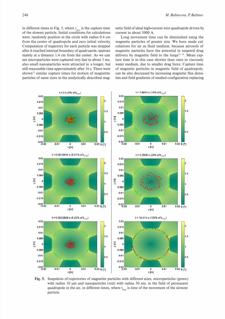
is the capture timeof the slowest particle. Initial conditions for calculationswere: randomly position in the circle with radius 0.4 cmfrom the center of quadrupole and zero initial velocity.Computation of trajectory for each particle was stoppedafter it reached internal boundary of quadrupole, approxi-
mately at a distance 1.4 cm from the center. As we cansee microparticles were captured very fast in about 3 ms,also small nanoparticles were attracted in a longer, butstill reasonable time approximately after 16 s. There wereshown32 similar capture times for motion of magnetiteparticles of same sizes in the analytically described mag-
Fig. 5. Snapshots of trajectories of magnetite particles with different sizes, microparticles (green)with radius 10 μm and nanoparticles (red) with radius 50 nm, in the field of permanentquadrupole in the air, in different times, where t
max is time of the movement of the slowest
particle.
netic field of ideal high-current wire quadrupole driven bycurrent in about 5000 A.
Long movement time can be diminished using themagnetic particles of greater size. We have made cal-culations for air as fluid medium, because aerosols ofmagnetic particles have the potential in targeted drug
delivery by magnetic field to the lungs33, 34
. Mean cap-ture time is in this case shorter than ones in viscouslywater medium, due to smaller drag force. Capture timeof magnetic particles in magnetic field of quadrupole,can be also decreased by increasing magnetic flux densi-ties and field gradients of studied configuration replacing
7/23/2019 Drug Delivery Magnetic Nanoparticle Review 2009
249 Magnetic drug delivery and targeting: principles and applications
permanent magnets by pulsed electromagnets, similar tothose pulsed quadrupole lenses using for focusing heavyion beams in accelerator physics. These results equallyapply also to magnetic separation of proteins, DNA, andwhole cells35,36.
The first study about the magnetic drug targeting was
performed 30 years ago37
, but the revival of the field, dueto apperance of a cheap and very powerful neodymiummagnets, started in the the new millenium38–44. These prin-ciples are used also in magnetofection (MF) which is amethod in which magnetic nanoparticles associated with
vector DNA are transfected into cells by the influence ofan external magnetic field. For this purpose, magneticparticles might be coated with the polycation polyethyle-nimine. These complexes readily associate with negativelycharged DNA since the magnetic particles are positivelycharged due to the polyethylenimine. Whether viral ornonviral vectors, MF has been shown to enhance the ef-ficiency of the vectors up to several thousand times45,46.
Also combination of magnetic targeting with controlledrelease seems to be a very promising field47–51. Moreovermagnetic nanoparticles provide magnetic resonance con-trast enhancement (i.e., changes in signal intensity) byshortening both the longitudinal and transverse relaxa-tion of surrounding protons, and widely available MRIequipment can be used for their noninvasive tracking inthe organism52–54.
CONCLUSIONS
Functional magnetic nanoparticles offer improvedspatio-temporal control over drug kinetics and distribu-tion, thus opening the prospect of safer and more specifictherapies. We found an explosive growth of published pa-pers from this field of nanomedicine, showing the almostunlimited potential of magnetic nanoparticles in the fieldof experimental and clinical oncology.
ACKNOWLEDGEMENTS
This work was supported by VEGA No.: grant 1/0082/08
and Magseletofection project of 6. FP of EU under the con- tract No.: LSHB-CT-2006–019038.
REFERENCES
1. Laufberger V. Sur la cristallisation de la ferritine. Bull Soc ChimBiol 1937;19:1575.
2. Kohgo Y, Ikuta K, Ohtake T, Torimoto Y, Kato J. Body iron me-tabolism and pathophysiology of iron overload. Int J Hematol 2008;88(1):7–15.
3. Pankhurst Q, Hautot D, Khan N, Dobson J. Increased levels ofmagnetic iron compounds in Alzheimer's disease. J Alzheimer'sDis 2008 ;13(1):49–52.
4. Babincová M, Babinec P. Dopamine mediated iron release fromferritin is enhanced at higher temperatures: Possible implica-tions for fever-induced Parkinson's disease. J Magn Magn Mater2005;293(1):341–4.
5. Kronick P, Gilpin RW. Use of superparamagnetic particles for isola-tion of cells. J Biochem Biophys Methods 1986;12(1–2):73–80.
6. Simsek E, Akif Kilic M. Magic ferritin: A novel chemotherapeuticencapsulation bullet. J Magn Magn Mater 2005;293(1):509–13.
7. Gilad AA, Winnard Jr. PT, van Zijl PCM, Bulte JWM. DevelopingMR reporter genes: Promises and pitfalls. NMR Biomed2007;20(3):275–90.
8. Babincová M, Leszczynska D, Sourivong P, Babinec P. Selectivetreatment of neoplastic cells using ferritin-mediated electromag-netic hyperthermia. Med Hypotheses 2000;54(2):177–9.
9. Lee S, Lee H, Park J, Choi H, Han K, Seo H, et al. A novel ap-proach to ultrasensitive diagnosis using supramolecular proteinnanoparticles. FASEB Journal 2007;21(7):1324–34.
10. Gider S, Awschalom DD, Douglas T, Mann S, Chaparala M.Classical and quantum magnetic phenomena in natural and artifi-cial ferritin proteins. Science 1995; 268(5207):77–80.
11. Morrish AH. Physical Principles of Magnetism. New York, IEEEPress;2001.
12. Jiles D. Introduction to Magnetism and Magnetic Materials,London, Chapman and Hall;1991.
13. Schwertmann U, Cornell RM. Iron oxides in the laboratory: pre-paration and characterization. Weinheim, VCH;1991.
14. Gupta AK, Gupta M. Synthesis and surface engineering of ironoxide nanoparticles for biomedical applications. Biomaterials2005;26(18):3995–4021.
15. Berry CC, Curtis ASG. Functionalisation of magnetic nanoparti-cles for applications in biomedicine. J Phys D 2003;36(13):R198–206.
16. Sajja HK, East MP, Mao H, Wang YA, Nie S, Yang L. Developmentof multifunctional nanoparticles for targeted drug delivery and non-invasive imaging of therapeutic effect. Current Drug DiscoveryTechnologies 2009;6(1):43–51.
17. Kikumori T, Kobayashi T, Sawaki M, Imai T. Anti-cancer effectof hyperthermia on breast cancer by magnetite nanoparticle-loaded anti-HER2 immunoliposomes. Breast Cancer Res Treat2009;113(3):435–41.
18. Veiseh O, Kievit FM, Gunn JW, Ratner BD, Zhang M. A ligand-
mediated nanovector for targeted gene delivery and transfection incancer cells. Biomaterials 2009;30(4):649–57.19. Kim D, Kim K, Kim K, Lee Y. Targeting to carcinoma cells with
chitosan- and starch-coated magnetic nanoparticles for magnetichyperthermia. Journal of Biomedical Materials Research – Part A2009;88(1):1–11.
20. Cheng C, Chu P, Chuang K, Roffler SR, Kao C, Tseng W.Hapten-derivatized nanoparticle targeting and imaging of geneexpression by multimodality imaging systems. Cancer Gene Ther2009;16(1):83–90.
21. Mohapatra S, Mallick SK, Maiti TK, Ghosh SK, Pramanik P.Synthesis of highly stable folic acid conjugated magnetite nano-particles for targeting cancer cells. Nanotechnology 2007;18(38).
22. Babincová M. Microwave induced leakage of magnetolipo-somes. possible clinical implications. Bioelectrochem Bioenerget
1993;32(2):187–9.23. Babincová M. Simple preparation and separation of magnetolipo-
somes. Chem Listy 1998;92(4):323.24. Babincová M, Machová E. Magnetoliposomes may be useful
for elimination of HIV from infected individuals. Zeitschrift fur Naturforschung – Section C Journal of Biosciences 1998;53(9–10):935–6.
25. Babincová M, Babinec P. Controlled drug delivery using magnetoli-posomes. Cellular and Molecular Biology Letters 1997;2(1):3–7.
26. Babincova M, Babinec P. Possibility of magnetic targeting of drugsusing magnetoliposomes. Pharmazie 1995;50(12):828–9.
27. Ferrari M. Cancer nanotechnology: Opportunities and challenges. Nature Reviews Cancer 2005;5(3):161–71.
28. Babincová M, Babinec P, Bergemann C. High-gradient magneticcapture of ferrofluids: Implications for drug targeting and tumor
embolization. Zeitschrift fur Naturforschung – Section C Journalof Biosciences 2001;56(9–10):909–11.
29. Pankhurst QA, Connolly J, Jones SK, Dobson J. Applicationsof magnetic nanoparticles in biomedicine. J Phys D2003;36(13):R167–81.
7/23/2019 Drug Delivery Magnetic Nanoparticle Review 2009
30. Furlani EP, Sahoo Y. Analytical model for the magneticfield and force in a magnetophoretic microsystem. J Phys D2006;39(9):1724–32.
31. Meeker D. software: Finite Elements Methods Magnetics, V 4.2. Available from: http://femm.foster-miller.net
32. Krafčík A, Babincová M, Babinec P. Theoretical analysis of mag-netic particle trajectory in high-current pulsed quadrupole: implica-tions for magnetic cell separation, drug targeting, and gene therapy.Optoel. Adv. Mater. – Rapid Commun 2009;3(11): 226–231.
33. Babincová M, Babinec P. Aerosolized VEGF in combination withintravenous magnetically targeted delivery of DNA-nanoparticlecomplex may increase efficiency of cystic fibrosis gene therapy.Med Hypotheses 2006;67(4):1002.
34. Dames P, Gleich B, Flemmer A, Hajek K, Seidl N, Wiekhorst F,et al. Targeted delivery of magnetic aerosol droplets to the lung. Nature Nanotechnology 2007;2(8):495–9.
35. Šafařík I, Šafaříková M. Use of magnetic techniques for the isola-tion of cells. Journal of Chromatography B: Biomedical Sciencesand Applications 1999;722(1–2):33–53
36. Šafařík I, Šafaříková M. Magnetic nanoparticles and biosciences.Monatshefte fur Chemie 2002;133(6):737–59.
37. Widder KJ, Senyei AE, Ranney DF. In vitro release of biologically
active adriamycin by magnetically responsive albumin microsphe-res. Cancer Res 1980;40(10):3512–7. 38. Gupta PK, Hung CT. Magnetically controlled targeted microcarrier
Possinger K, et al. Clinical experiences with magnetic drug target-ing: A phase I study with 4'-epidoxorubicin in 14 patients withadvanced solid tumors. Cancer Res 1996;56(20):4686–93.
40. Lübbe AS, Bergemann C, Brock J, McClure DG. Physiologicalaspects in magnetic drug-targeting. J Magn Magn Mater1999;194(1):149–55.
41. Babincová M, Altanerová V, Lamper t M, Altaner C, MachováE, Šrá mka M, et al. Site-specif ic in vivo targeting of magnetoli-posomes using externally applied magnetic field. Zeitschrift fur Naturforschung – Section C Journal of Biosciences 2000;55(3–
4):278–81.42. Babincová M, Leszczynska D, Sourivong P, Babinec P, LeszczynskiJ. Principles of magnetodynamic chemotherapy. Med Hypotheses2004;62(3):375–80.
43. Babincova M, Altanerova V, Altaner C, Bergemann C, Babinec P.In vitro analysis of cisplatin functionalized magnetic nanoparticlesin combined cancer chemotherapy and electromagnetic hyperther-mia. IEEE Transactions on Nanobioscience 2008;7(1):15–9.
44. Misra RDK. Magnetic nanoparticle carrier for targeted drug de-livery: Perspective, outlook and design. Materials Science andTechnology 2008;24(9):1011–9.
45. Scherer F, Anton M, Schillinger U, Henke J, Bergemann C, Kruger A, Gansbacher B, Plank C. Magnetofection: enhancing and targe-ting gene delivery by magnetic force in vitro and in vivo. Gene Ther2002;9:102–9.
46. Mykhaylyk O, Zelphati O, Hammerschmid E, Anton M, RoseneckerJ, Plank C. Recent advances in magnetofection and its potential todeliver siRNAs in vitro. Methods Mol Biol 2009;487:111–46.
47. Babincová M, Leszczynska D, Sour ivong P, Babinec P. Picosecondlaser pulses mediated drug release from magnetoliposomes.Cellular and Molecular Biology Letters 1999;4(4):625–30.
48. Babincová M, Leszczynska D, Sourivong P, Čičmanec P, Babinec P.Superparamagnetic gel as a novel material for electromagneticallyinduced hyperthermia. J Magn Magn Mater 2001;225(1–2):109–12.
P. AC-magnetic field controlled drug release from magnetoli-posomes: Design of a method for site-specific chemotherapy.Bioelectrochemistry 2002;55(1–2):17–9.
50. Babincová M, Altanerová V, Altaner C, Čičmanec P, Babinec P.In vivo heating of magnetic nanoparticles in alternating magneticfield. Med Phys 2004;31(8):2219–21.
51. Babinec P, Babincová M, Sourivong P, Leszczynska D. Eff icienttreatment of pigmented B16 melanoma using photosensitizedlong-circulating magnetofullerenosomes. J Magn Magn Mater2005;293(1):394–7.
52. Chouly C, Pouliquen D, Lucet I, Jeune JJ, Jallet P. Developmentof superparamagnetic nanoparticles for MRI: Effect of particlesize, charge and surface nature on biodistribution. J Microencapsul1996;13(3):245–55.
53. Babinec P, Babincová M. Towards multimodal nanoparticle labels
for molecular imaging of biological processes. Med Hypotheses2007;69(3):703–4.54. Syková E, Jendelová P, Herynek V. MR tracking of stem cells in
living recipients. Methods Mol Biol 2009;549:197–215.