ISJ 13: 140-152, 2016 ISSN 1824-307X RESEARCH REPORT Effects of exposure to zinc oxide nanoparticles in freshwater mussels in the presence of municipal effluents C Gagnon, M Pilote, P Turcotte, C André, F Gagné Aquatic Contaminants Research Division, Water Science and Technology, Environment Canada, 105 McGill, Montreal, Quebec, Canada Accepted May 12, 2016 Abstract Zinc oxide (nano-ZnO) nanoparticles are used in the production of transparent sunscreens and cosmetics, which are released into surface waters and municipal wastewater effluent. The purpose of this study was to examine the toxicity of nano-ZnO in the presence of municipal effluents to freshwater mussels Elliptio complanata. Mussels were exposed for 21 days at 15 o C to nano-ZnO and ZnCl 2 in the presence of 10 % dilution of primary-treated municipal effluent. After the exposure period and 24-h depuration step, mussels were analyzed for total Zn in gills and digestive gland, free Zn, metallothioneins (MT), oxidative stress (glutathione S-transferase and LPO), endoplasmic reticulum stress (heat shock proteins and protein ubiquitination) and genotoxicity. The data revealed that although total Zn loadings did not change with these treatments, Zn levels in digestive gland were elevated in mussels exposed to nano-ZnO but not with ZnCl 2 in the presence of municipal effluent. Free Zn levels in the gills were elevated in mussels exposed to the municipal effluent, but decreased in mussels exposed to nano-ZnO. MT in digestive gland showed a similar pattern and was negatively associated with free and total Zn. GST activity was significantly reduced by both nano-ZnO and municipal effluent and was negatively correlated with MT levels, suggesting the involvement of MT in the sequestration of reactive oxygen species. Discriminant function analysis showed that the municipal effluent related effects differed from the unexposed mussels and nano-ZnO exposed mussels in terms of the following responses: free Zn in gills and digestive gland and GST activity. Nano-ZnO related effects also involved GST activity, MT and protein ubiquitination, which suggests a combination of oxidative stress and reticulum endoplasmic stress. In respect with oxidative stress, the oxidative properties of nano-ZnO and ZnCl 2 are dampened in the presence of the municipal effluent. Key Words: zinc oxide nanoparticles; municipal effluent; freshwater mussels; oxidative stress; reticulum endoplasmic stress Introduction Nanotechnology is an exponentially growing industry with applications in many fields, such as electronics, optical instruments, drug delivery vectors and diagnostics (Lee et al., 2008). Nanomaterials are also used in consumer products, such as cosmetics, sunscreens and textiles (Contado, 2015). The presence of nanomaterials from personal care products in complex matrices, ___________________________________________________________________________ Corresponding authors: Christian Gagnon Francois Gagne Aquatic Contaminants Research Division Water Science and Technology Environment Canada, 105 McGill Montreal, Quebec, Canada H2Y 2E7 E-mails: [email protected]; [email protected]such as urban runoff and municipal effluents, complicates the hazard assessment process. Indeed, at the exposure characterization step, the identification and quantitation of low concentrations of nanoparticles in complex environmental samples (sediments, effluents) poses a major challenge at the analytical level. Hence, the concerns raised by the public and regulatory community about their safety, fate and ecotoxicity are legitimate. Zinc oxide nanoparticles (nano-ZnO) are used worldwide in sunscreens given their strong UV-light absorbing properties compared to other sunscreen formulations (Mitchnick et al., 1999). Sunscreen creams typically contain bulk particles of ZnO and titanium dioxide, which are in the form of white pastes. Creams composed of nano-ZnO diminish light scattering on the skin and appear more transparent (Mitchnick et al., 1999). Moreover, 140
Transcript
ISJ 13: 140-152, 2016 ISSN 1824-307X
RESEARCH REPORT Effects of exposure to zinc oxide nanoparticles in freshwater mussels in the presence of municipal effluents C Gagnon, M Pilote, P Turcotte, C André, F Gagné Aquatic Contaminants Research Division, Water Science and Technology, Environment Canada, 105 McGill, Montreal, Quebec, Canada
Accepted May 12, 2016
Abstract Zinc oxide (nano-ZnO) nanoparticles are used in the production of transparent sunscreens and
cosmetics, which are released into surface waters and municipal wastewater effluent. The purpose of this study was to examine the toxicity of nano-ZnO in the presence of municipal effluents to freshwater mussels Elliptio complanata. Mussels were exposed for 21 days at 15 oC to nano-ZnO and ZnCl2 in the presence of 10 % dilution of primary-treated municipal effluent. After the exposure period and 24-h depuration step, mussels were analyzed for total Zn in gills and digestive gland, free Zn, metallothioneins (MT), oxidative stress (glutathione S-transferase and LPO), endoplasmic reticulum stress (heat shock proteins and protein ubiquitination) and genotoxicity. The data revealed that although total Zn loadings did not change with these treatments, Zn levels in digestive gland were elevated in mussels exposed to nano-ZnO but not with ZnCl2 in the presence of municipal effluent. Free Zn levels in the gills were elevated in mussels exposed to the municipal effluent, but decreased in mussels exposed to nano-ZnO. MT in digestive gland showed a similar pattern and was negatively associated with free and total Zn. GST activity was significantly reduced by both nano-ZnO and municipal effluent and was negatively correlated with MT levels, suggesting the involvement of MT in the sequestration of reactive oxygen species. Discriminant function analysis showed that the municipal effluent related effects differed from the unexposed mussels and nano-ZnO exposed mussels in terms of the following responses: free Zn in gills and digestive gland and GST activity. Nano-ZnO related effects also involved GST activity, MT and protein ubiquitination, which suggests a combination of oxidative stress and reticulum endoplasmic stress. In respect with oxidative stress, the oxidative properties of nano-ZnO and ZnCl2 are dampened in the presence of the municipal effluent.
Nanotechnology is an exponentially growing industry with applications in many fields, such as electronics, optical instruments, drug delivery vectors and diagnostics (Lee et al., 2008). Nanomaterials are also used in consumer products, such as cosmetics, sunscreens and textiles (Contado, 2015). The presence of nanomaterials from personal care products in complex matrices, ___________________________________________________________________________
Corresponding authors: Christian Gagnon Francois Gagne Aquatic Contaminants Research Division Water Science and Technology Environment Canada, 105 McGill Montreal, Quebec, Canada H2Y 2E7 E-mails: [email protected]; [email protected]
such as urban runoff and municipal effluents, complicates the hazard assessment process. Indeed, at the exposure characterization step, the identification and quantitation of low concentrations of nanoparticles in complex environmental samples (sediments, effluents) poses a major challenge at the analytical level. Hence, the concerns raised by the public and regulatory community about their safety, fate and ecotoxicity are legitimate. Zinc oxide nanoparticles (nano-ZnO) are used worldwide in sunscreens given their strong UV-light absorbing properties compared to other sunscreen formulations (Mitchnick et al., 1999). Sunscreen creams typically contain bulk particles of ZnO and titanium dioxide, which are in the form of white pastes. Creams composed of nano-ZnO diminish light scattering on the skin and appear more transparent (Mitchnick et al., 1999). Moreover,
nano-ZnO possesses anti-microbial properties, which is perceived as an additional benefit for the consumer. As explained above, the release, fate and toxicity of these sunscreens are difficult to assess at present time and the resulting effects of nano-ZnO in complex environmental media, such as municipal effluent, are not well understood at the present time.
With respect to nanomaterials, mussels and clams (bivalves) are considered to be at risk from contaminants associated with fine particles and colloids (Canesi et al., 2012). These invertebrates are more susceptible to nanoparticles since they are sessile, live for long periods (years to decades) and especially feed on suspended particles, therefore accumulating large quantities of particles including nanoparticles. Copper oxide nanoparticles were shown to accumulate in the digestive gland and initiate toxicity (lipid peroxidation or LPO) in the mussel Mytilus galloprovincialis (Gomes et al., 2012). In another study, mussels exposed to CeO2 nanoparticles and nano-ZnO accumulated as much as 21 and 63 mg/g of total Ce and Zn respectively, indicating that these nanoparticles are readily available to mussels (Montes et al., 2012). The toxicity of metal-based nanoparticles cannot always be explained by the released component which shows different toxic properties. These toxic properties are associated with other properties of nanoparticles, such as size, form and surface properties (Oberdörster et al., 2007). Recent studies have shown that many nanoparticles of different sizes, forms and surface coatings are cytotoxic and genotoxic, leading to reticulum endoplasmic stress, oxidative stress and DNA damage. Reticulum endoplasmic stress involves protein chaperones (heat shock proteins 70 or HSP70) to restore the conformational integrity of proteins and tagging of denatured proteins by ubiquitin via the ubiquitin-proteasome pathways involved in the elimination of irreversibly damaged proteins (McDonagh and Sheehan, 2006). Ubiquitination also plays a role in the calcium carbonate biomineralization process of shells (Fang et al., 2012). Oxidative stress involves the mobilization of metals, such as iron (Fe3+), copper (Cu+) and Zn (Zn2+), leading to the production of reactive oxygen species. These processes are controlled by antioxidants and metallothioneins (MT) in the sequestration of divalent heavy metals and reactive oxygen species. The uncontrolled release of reactive oxygen species could lead to lipid peroxidation (LPO) and DNA damage via the formation of 8-oxo-guanine (Rocha et al., 2015; Valko et al., 2006; Formigari et al., 2007). Recent evidence suggests that nano-ZnO and municipal effluents could induce oxidative stress in mussels, but less is known about the combined effects in freshwater mussels located near urban waste discharges.
The purpose of this study was to examine the cumulative effects of nano-ZnO and municipal effluent exposure in freshwater mussels. Mussels were exposed to dilutions of municipal effluent in freshwater and to instilled nano-ZnO given the poor solubility of this nanoparticle in freshwater (Majedi et al., 2014). For comparison purposes, mussels were
also exposed to ZnCl2. Zn bioavailability was assessed in mussel tissues and biomarkers of stress were determined. Biomarkers of stress were labile Zn in tissues, MT, glutathione S-transferase (GST, a marker of oxidative stress and conjugation), reticulum endoplasmic stress (HSP70 and protein ubiquitylation), LPO and DNA damage. An attempt was made to highlight cumulative effects or interactions of a typical chemically-treated municipal effluent and nano-ZnO for 21 days based on effluent concentration and forms of dietary Zn in freshwater mussels.
Materials and Methods
Mussel handling and exposure to municipal effluents
Mussels (Elliptio complanata) were collected by hand during the first week of June 2012 in a pristine lake in the Laurentians under a provincial permit. The mussels were transported dry in coolers at 4 oC and transferred to 300-L tanks filled with City of Montreal tap water, which was charcoal-filtered and UV-treated. The mussels were held in the tanks at 15 °C under constant aeration for 4 to 10 weeks before initiating exposure. The mussels were fed three times a week with commercial coral reef feed enriched with 100 million/mL of Pseudokirchneriella subcapitata algal suspensions. For the exposure experiments, mussels (n = 10 individuals) were placed in 60-L tanks receiving a continuous flow (0.1 - 0.2 L/h) of physico-chemically treated municipal effluent from a city of 1.5 million people. Three aquarium per treatment was also included. The physical/chemical treatment consisted in reducing suspended matter down to the mg/L range using grid traps, flocculation (surfactants) and sieving. The municipal effluent exposure concentrations were 0, 3 and 10 % prepared in dechlorinated UV-treated tap water from the city of Montreal (Quebec, Canada). Mussels were exposed to 1 and 10 µg/L Zn as nano-ZnO or ZnCl2 using an “instillation” technique. During exposure, 1 and 10 µg/L zinc solutions were added to 60 L of aquarium water. To ensure contact of the zinc with mussel tissues, 60 and 600 µg of either nano-ZnO or ZnCl2 were dissolved in 20 mL of aquarium water and placed directly over the mussel siphons (each mussel received 1 mL). The mussels were held under static conditions for one hour prior to continuous exposure to the municipal effluent. This process was repeated every 3 days for 21 days. At the end of the exposure period, mussels were allowed to depurate in clean aquarium water overnight (24 h). Morphological characteristics were measured for mussel weight and shell length. Soft tissue, gills, gonad and digestive glands were dissected on ice and weighed. Sex was determined by examination of gonad smears on glass slides under a binocular microscope at 200x magnification. A portion of the digestive, gill and gonad tissues were processed for heavy metal analysis (including Zn) using ICP-mass spectrometry and nitric acid digestions as previously described (Gagnon et al., 2006). The digestive gland and gills were homogenized on ice using a Teflon pestle tissue
142
grinder in 100 mM NaCl containing 20 mM Hepes-NaOH, pH 7.4, 10 µg/mL aprotinin (protease inhibitor) and 1 mM dithiothreitol. A part of the homogenate was centrifuged at 15000xg for 30 min at 4 oC and the supernatant (S15 fraction) was removed and stored at -85 oC until biomarker analysis. Total proteins were determined using the protein-dye binding principle with standard solutions of bovine serum albumin for calibration (Bradford, 1976). Basic physico-chemical characteristics were determined using standard methods for total coliform counts, conductivity, pH, biochemical oxygen demand, dissolved organic carbon and turbidity.
Xenobiotic metabolism
Xenobiotic metabolism was characterized by monitoring changes in glutathione S-transferase (GST) and metallothioneins (MT) in the digestive gland. GST activity was determined using the absorbance microplate methodology as described elsewhere (Boryslawskyj et al., 1988). The 15000xg supernatant or S15 fraction (50 µL) was mixed with 200 µL of 1 mM glutathione (GSH) and 1 mM of 1-chloro-2,4-dinitrobenzene in 125 mM NaCl containing 20 mM Hepes-NaOH, pH 6.5. The increase in absorbance at 340 nm was measured at 0, 10, 20 and 30 min at 30 °C (Synergy 4, Dynatek Instrument, USA). Enzyme activity was expressed as the change in absorbance/min/mg protein. Metallothionein (MT) levels in the digestive gland were determined using a modified spectrophotometric assay (Viarengo et al., 1997; Gagné et al., 2010). Briefly, total MT levels were determined by the addition of a strong reducing agent, phosphine, in the S15 fraction for 15 min prior to the addition of ethanol-chloroform solvent. The data were expressed as µmole of GSH/mg protein. The levels of labile Zn were also determined in the digestive gland extracts using the fluoresecent probe methodology (Gagné and Blaise, 1996). Briefly, 20 μl of the homogenate was mixed with 180 μl N-(6-methoxy-8 quinolyl)-p-toluenesulfonamide (TSQ) probe in phosphate-buffered saline (140 mM NaCl, 5 mM KH2PO4, pH 7.4) containing 20 % dimethyl sulfoxide (DMSO). Fluorescence values were determined at 370 nm excitation and 490 nm emission. Blanks consisting of 20 % DMSO-phosphate-buffered saline and standard solutions of ZnSO4 were used for calibration. Data were expressed as nanograms Zn per milligram protein.
Endoplasmic reticulum stress
The levels of heat shock proteins of the 72-kDa family (Hsp72) and total ubiquitin levels were assessed using the enzyme immunoassay described previously (Louis et al., 2010). Briefly, the S15 fraction was diluted to 1 µg total proteins in 50 mM carbonate buffer (100 µL) pH 9.6, then added to high binding microplate wells (Immulon-4) and allowed to stand 10 - 12 h at 4 oC in darkness. The wells were then washed twice with 200 µL PBS and blocked with PBS containing 1 % albumin for 30 min at room temperature. The wells were washed once with PBS, and 100 µL of either
Hsp72 or polyubiquitin polyclonal antibody (recombinant human Hsp72 IgG SPA-812 and rabbit ubiquitin antibody; Stressgen, USA) at 1/1000 and 1/5000 dilution in PBS containing 0.5 % albumin were added in each well. The wells were incubated at 37 °C for 60 min. The wells were washed three times in PBS, and 100 µL of secondary antibody (rabbit IgC antibody linked to peroxidase) were added at 1/5000 dilution in PBS containing 0.5 % albumin. After 30 min at 37 °C, the wells were washed four times in PBS, and peroxidase activity was determined using 1 µM luminol and 10 µM H2O2. Readings were taken after 1 min using a luminescence microplate reader, at 2 and 5 min (Synergy-4, Dynatek). The data were expressed as the rate of increase in luminescence/min. Oxidative stress and DNA damage
Lipid peroxidation (LPO) was determined in the liver homogenates using the thiobarbituric acid method (Wills, 1987). A volume of 50 µL of the homogenate was mixed with 250 µL of 10 % trichloroacetic acid containing 1 mM FeSO4 and 75 µL of 0.7 % thiobarbituric acid (Sigma Chemical Company) and heated at 70 oC for 10 min. The mixture was cooled at room temperature and centrifuged at 10 000xg for 5 min. A 100 - 200 µl aliquot of the supernatant was transferred to a 96-well dark microplate and fluorescence readings were taken at 520 nm excitation and 600 nm emission. Standard solutions of tetrametoxypropane (stabilized form of malonaldehyde) were prepared for calibration in the blank (homogenization buffer). Results were expressed as µmole thiobarbituric acid reactants (TBARS)/mg total protein in the homogenate. DNA damage in liver was determined using the alkaline precipitation assay (Olive, 1988), which is based on potassium detergent precipitation of DNA-proteins. Protein-free DNA strand breaks that remain in the supernatant were determined using fluorescence spectroscopy (at 360 nm excitation and 450 nm emission) in the presence of diluent to prevent interference from the detergent (Bester et al., 1994). The diluent consisted of 0.4 M NaCl, 0.1 M Tris-acetate, pH 8.2, 4 mM sodium cholate and 10 μM SYBR® Green dye. Standard solutions of salmon sperm DNA were prepared for calibration. The data were expressed as µg DNA strand/mg total protein in homogenate. Data analysis
The exposure experiment consisted of 10 mussels per treatment aquarium and three replicate aquarium per conditions were used. Tissue biomarkers were performed on 10 mussels using 2-way factorial analysis of variance (municipal effluent concentration and form of zinc) after verifying for homogeneity of variance and normality using the Levene’s and Shapiro-Wilks tests respectively. Correlation analysis was also performed using the Pearson product-moment method. The physiological changes induced by exposure to the municipal effluent, dissolved Zn and ZnO nanoparticles were determined using discriminant function and factor analysis methods. All statistical tests were
performed using Statistica software (version 8). Significance was set at α = 0.05.
Results Mussels were exposed to a physico-chemically
treated municipal effluent from a relatively large urban area (1.5 million residents). The undiluted effluent had a conductivity of 835 ± 50 µScm-1 and a pH of 7.2 (Table 1). The suspended matter content and dissolved organic carbon content were 25 ± 5 and 82 ± 10 mg/L respectively. The biochemical oxygen demand was 35 ± 10 mg/L and the concentration of thermotolerant coliform bacteria was 2.9x10⁶ ± 5x10⁵ counts per 100 mL.
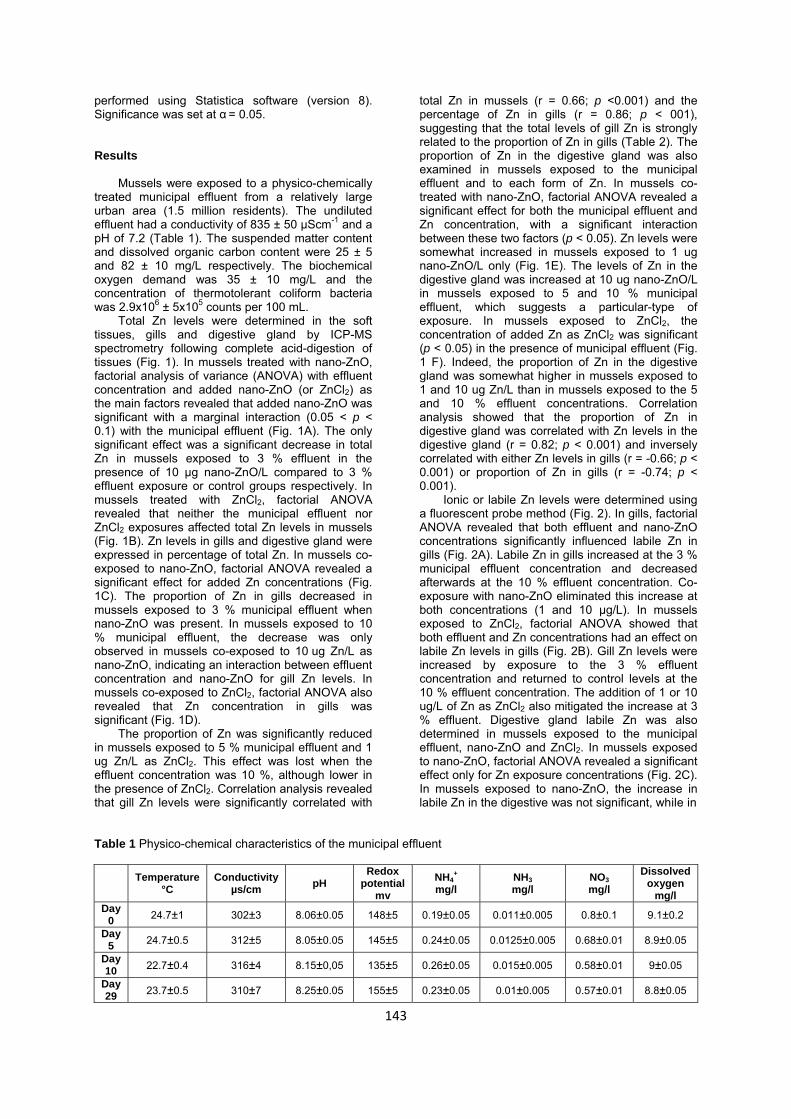
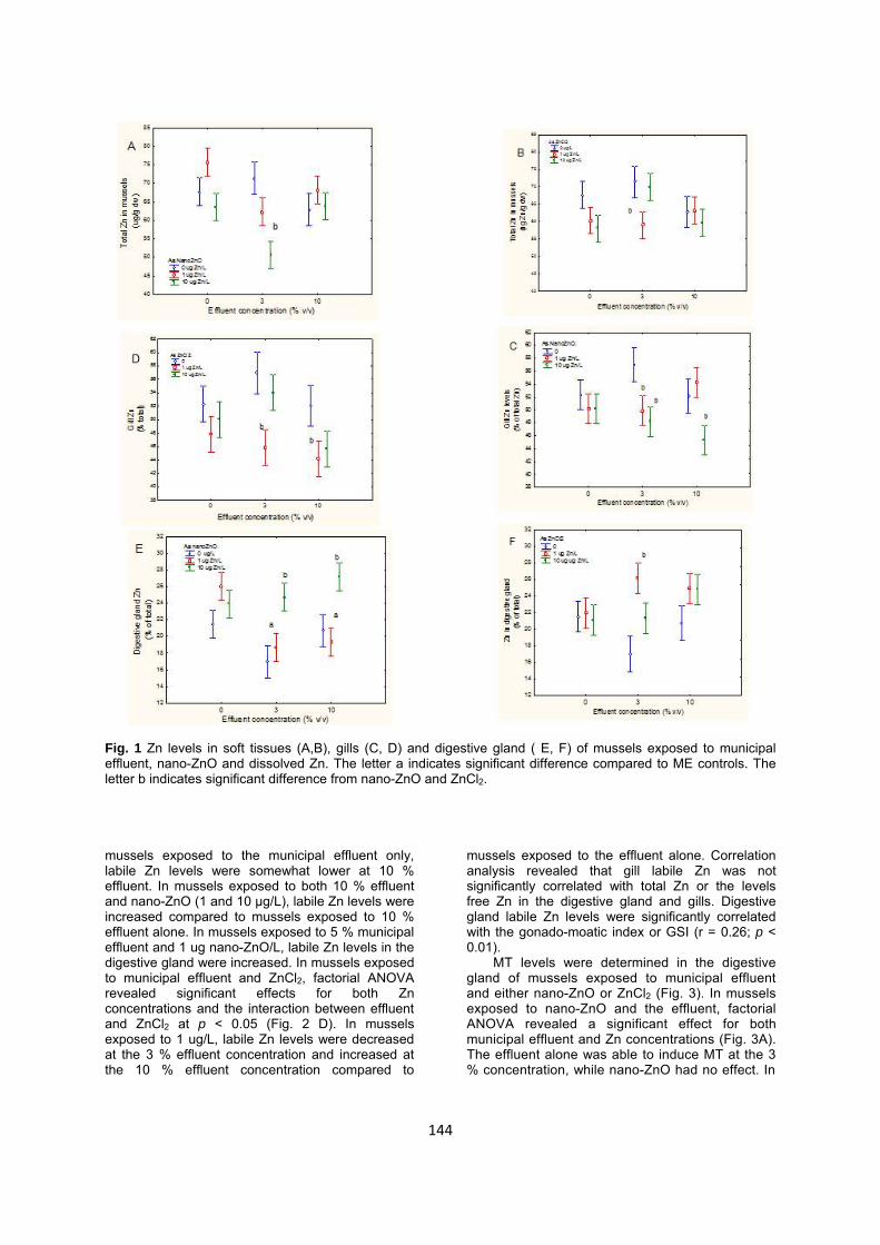
Total Zn levels were determined in the soft tissues, gills and digestive gland by ICP-MS spectrometry following complete acid-digestion of tissues (Fig. 1). In mussels treated with nano-ZnO, factorial analysis of variance (ANOVA) with effluent concentration and added nano-ZnO (or ZnCl2) as the main factors revealed that added nano-ZnO was significant with a marginal interaction (0.05 < p < 0.1) with the municipal effluent (Fig. 1A). The only significant effect was a significant decrease in total Zn in mussels exposed to 3 % effluent in the presence of 10 µg nano-ZnO/L compared to 3 % effluent exposure or control groups respectively. In mussels treated with ZnCl2, factorial ANOVA revealed that neither the municipal effluent nor ZnCl2 exposures affected total Zn levels in mussels (Fig. 1B). Zn levels in gills and digestive gland were expressed in percentage of total Zn. In mussels co-exposed to nano-ZnO, factorial ANOVA revealed a significant effect for added Zn concentrations (Fig. 1C). The proportion of Zn in gills decreased in mussels exposed to 3 % municipal effluent when nano-ZnO was present. In mussels exposed to 10 % municipal effluent, the decrease was only observed in mussels co-exposed to 10 ug Zn/L as nano-ZnO, indicating an interaction between effluent concentration and nano-ZnO for gill Zn levels. In mussels co-exposed to ZnCl2, factorial ANOVA also revealed that Zn concentration in gills was significant (Fig. 1D).
The proportion of Zn was significantly reduced in mussels exposed to 5 % municipal effluent and 1 ug Zn/L as ZnCl2. This effect was lost when the effluent concentration was 10 %, although lower in the presence of ZnCl2. Correlation analysis revealed that gill Zn levels were significantly correlated with
total Zn in mussels (r = 0.66; p <0.001) and the percentage of Zn in gills (r = 0.86; p < 001), suggesting that the total levels of gill Zn is strongly related to the proportion of Zn in gills (Table 2). The proportion of Zn in the digestive gland was also examined in mussels exposed to the municipal effluent and to each form of Zn. In mussels co-treated with nano-ZnO, factorial ANOVA revealed a significant effect for both the municipal effluent and Zn concentration, with a significant interaction between these two factors (p < 0.05). Zn levels were somewhat increased in mussels exposed to 1 ug nano-ZnO/L only (Fig. 1E). The levels of Zn in the digestive gland was increased at 10 ug nano-ZnO/L in mussels exposed to 5 and 10 % municipal effluent, which suggests a particular-type of exposure. In mussels exposed to ZnCl2, the concentration of added Zn as ZnCl2 was significant (p < 0.05) in the presence of municipal effluent (Fig. 1 F). Indeed, the proportion of Zn in the digestive gland was somewhat higher in mussels exposed to 1 and 10 ug Zn/L than in mussels exposed to the 5 and 10 % effluent concentrations. Correlation analysis showed that the proportion of Zn in digestive gland was correlated with Zn levels in the digestive gland (r = 0.82; p < 0.001) and inversely correlated with either Zn levels in gills (r = -0.66; p < 0.001) or proportion of Zn in gills (r = -0.74; p < 0.001).
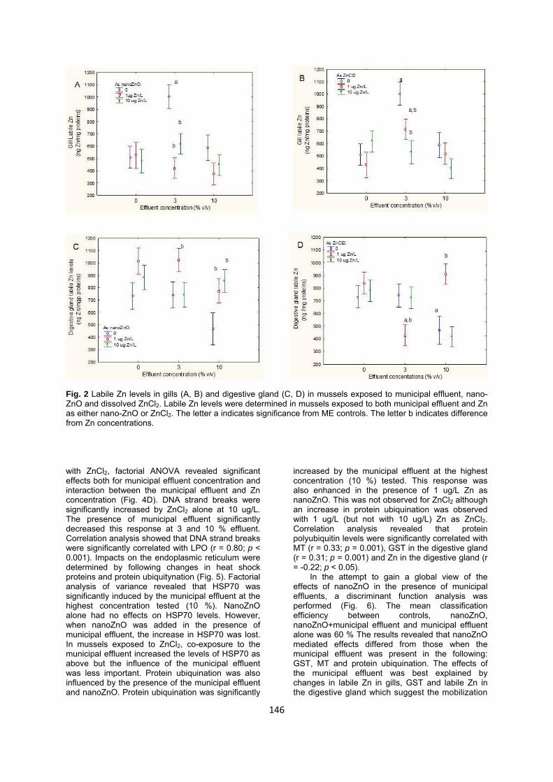
Ionic or labile Zn levels were determined using a fluorescent probe method (Fig. 2). In gills, factorial ANOVA revealed that both effluent and nano-ZnO concentrations significantly influenced labile Zn in gills (Fig. 2A). Labile Zn in gills increased at the 3 % municipal effluent concentration and decreased afterwards at the 10 % effluent concentration. Co-exposure with nano-ZnO eliminated this increase at both concentrations (1 and 10 µg/L). In mussels exposed to ZnCl2, factorial ANOVA showed that both effluent and Zn concentrations had an effect on labile Zn levels in gills (Fig. 2B). Gill Zn levels were increased by exposure to the 3 % effluent concentration and returned to control levels at the 10 % effluent concentration. The addition of 1 or 10 ug/L of Zn as ZnCl2 also mitigated the increase at 3 % effluent. Digestive gland labile Zn was also determined in mussels exposed to the municipal effluent, nano-ZnO and ZnCl2. In mussels exposed to nano-ZnO, factorial ANOVA revealed a significant effect only for Zn exposure concentrations (Fig. 2C). In mussels exposed to nano-ZnO, the increase in labile Zn in the digestive was not significant, while in
Table 1 Physico-chemical characteristics of the municipal effluent
Day 5 24.7±0.5 312±5 8.05±0.05 145±5 0.24±0.05 0.0125±0.005 0.68±0.01 8.9±0.05
Day 10 22.7±0.4 316±4 8.15±0,05 135±5 0.26±0.05 0.015±0.005 0.58±0.01 9±0.05
Day 29 23.7±0.5 310±7 8.25±0.05 155±5 0.23±0.05 0.01±0.005 0.57±0.01 8.8±0.05
143
Fig. 1 Zn levels in soft tissues (A,B), gills (C, D) and digestive gland ( E, F) of mussels exposed to municipal effluent, nano-ZnO and dissolved Zn. The letter a indicates significant difference compared to ME controls. The letter b indicates significant difference from nano-ZnO and ZnCl2. mussels exposed to the municipal effluent only, labile Zn levels were somewhat lower at 10 % effluent. In mussels exposed to both 10 % effluent and nano-ZnO (1 and 10 µg/L), labile Zn levels were increased compared to mussels exposed to 10 % effluent alone. In mussels exposed to 5 % municipal effluent and 1 ug nano-ZnO/L, labile Zn levels in the digestive gland were increased. In mussels exposed to municipal effluent and ZnCl2, factorial ANOVA revealed significant effects for both Zn concentrations and the interaction between effluent and ZnCl2 at p < 0.05 (Fig. 2 D). In mussels exposed to 1 ug/L, labile Zn levels were decreased at the 3 % effluent concentration and increased at the 10 % effluent concentration compared to
mussels exposed to the effluent alone. Correlation analysis revealed that gill labile Zn was not significantly correlated with total Zn or the levels free Zn in the digestive gland and gills. Digestive gland labile Zn levels were significantly correlated with the gonado-moatic index or GSI (r = 0.26; p < 0.01).
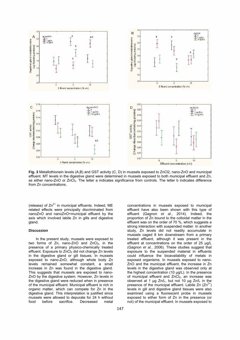
MT levels were determined in the digestive gland of mussels exposed to municipal effluent and either nano-ZnO or ZnCl2 (Fig. 3). In mussels exposed to nano-ZnO and the effluent, factorial ANOVA revealed a significant effect for both municipal effluent and Zn concentrations (Fig. 3A). The effluent alone was able to induce MT at the 3 % concentration, while nano-ZnO had no effect. In
144
145
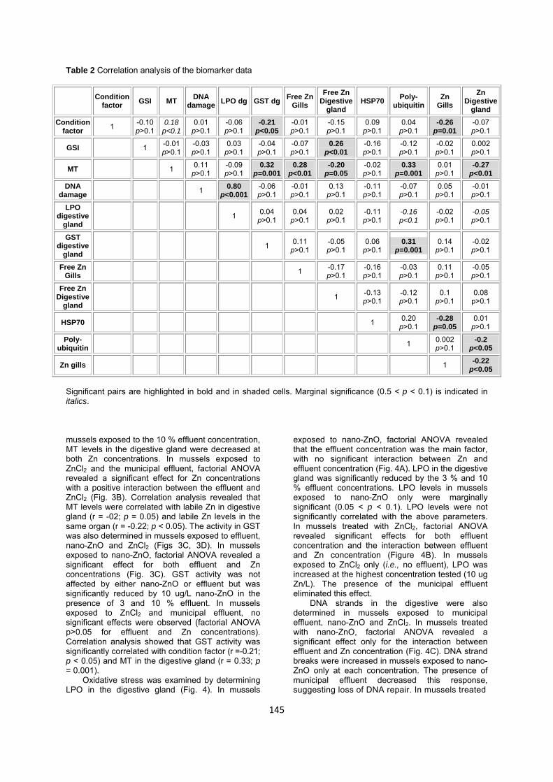
Table 2 Correlation analysis of the biomarker data
Condition factor GSI MT DNA
damage LPO dg GST dg Free Zn Gills
Free Zn Digestive
gland HSP70 Poly-
ubiquitin Zn
Gills Zn
Digestive gland
Condition factor 1 -0.10
p>0.1 0.18
p<0.1 0.01
p>0.1 -0.06 p>0.1
-0.21 p<0.05
-0.01 p>0.1
-0.15 p>0.1
0.09 p>0.1
0.04 p>0.1
-0.26 p=0.01
-0.07 p>0.1
GSI 1 -0.01 p>0.1
-0.03 p>0.1
0.03 p>0.1
-0.04 p>0.1
-0.07 p>0.1
0.26 p<0.01
-0.16 p>0.1
-0.12 p>0.1
-0.02 p>0.1
0.002 p>0.1
MT 1 0.11 p>0.1
-0.09 p>0.1
0.32 p=0.001
0.28 p<0.01
-0.20 p=0.05
-0.02 p>0.1
0.33 p=0.001
0.01 p>0.1
-0.27 p<0.01
DNA damage 1 0.80
p<0.001-0.06 p>0.1
-0.01 p>0.1
0.13 p>0.1
-0.11 p>0.1
-0.07 p>0.1
0.05 p>0.1
-0.01 p>0.1
LPO digestive
gland 1 0.04
p>0.1 0.04
p>0.1 0.02
p>0.1 -0.11 p>0.1
-0.16 p<0.1
-0.02 p>0.1
-0.05 p>0.1
GST digestive
gland 1 0.11
p>0.1 -0.05 p>0.1
0.06 p>0.1
0.31 p=0.001
0.14 p>0.1
-0.02 p>0.1
Free Zn Gills 1 -0.17
p>0.1 -0.16 p>0.1
-0.03 p>0.1
0.11 p>0.1
-0.05 p>0.1
Free Zn Digestive
gland 1 -0.13
p>0.1 -0.12 p>0.1
0.1 p>0.1
0.08 p>0.1
HSP70 1 0.20 p>0.1
-0.28 p=0.05
0.01 p>0.1
Poly- ubiquitin 1 0.002
p>0.1 -0.2
p<0.05
Zn gills 1 -0.22 p<0.05
Significant pairs are highlighted in bold and in shaded cells. Marginal significance (0.5 < p < 0.1) is indicated in italics. mussels exposed to the 10 % effluent concentration, MT levels in the digestive gland were decreased at both Zn concentrations. In mussels exposed to ZnCl2 and the municipal effluent, factorial ANOVA revealed a significant effect for Zn concentrations with a positive interaction between the effluent and ZnCl2 (Fig. 3B). Correlation analysis revealed that MT levels were correlated with labile Zn in digestive gland (r = -02; p = 0.05) and labile Zn levels in the same organ (r = -0.22; p < 0.05). The activity in GST was also determined in mussels exposed to effluent, nano-ZnO and ZnCl2 (Figs 3C, 3D). In mussels exposed to nano-ZnO, factorial ANOVA revealed a significant effect for both effluent and Zn concentrations (Fig. 3C). GST activity was not affected by either nano-ZnO or effluent but was significantly reduced by 10 ug/L nano-ZnO in the presence of 3 and 10 % effluent. In mussels exposed to ZnCl2 and municipal effluent, no significant effects were observed (factorial ANOVA p>0.05 for effluent and Zn concentrations). Correlation analysis showed that GST activity was significantly correlated with condition factor (r = -0.21; p < 0.05) and MT in the digestive gland (r = 0.33; p = 0.001).
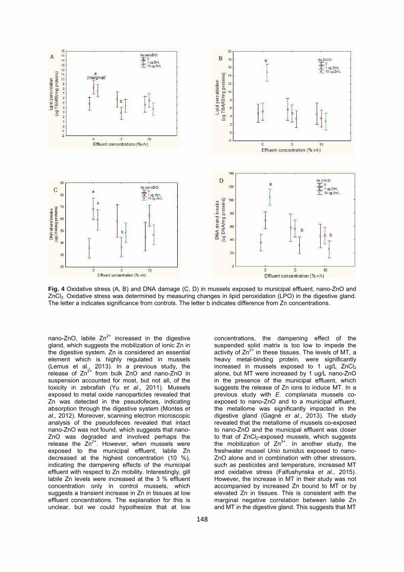
Oxidative stress was examined by determining LPO in the digestive gland (Fig. 4). In mussels
exposed to nano-ZnO, factorial ANOVA revealed that the effluent concentration was the main factor, with no significant interaction between Zn and effluent concentration (Fig. 4A). LPO in the digestive gland was significantly reduced by the 3 % and 10 % effluent concentrations. LPO levels in mussels exposed to nano-ZnO only were marginally significant (0.05 < p < 0.1). LPO levels were not significantly correlated with the above parameters. In mussels treated with ZnCl2, factorial ANOVA revealed significant effects for both effluent concentration and the interaction between effluent and Zn concentration (Figure 4B). In mussels exposed to ZnCl2 only (i.e., no effluent), LPO was increased at the highest concentration tested (10 ug Zn/L). The presence of the municipal effluent eliminated this effect.
DNA strands in the digestive were also determined in mussels exposed to municipal effluent, nano-ZnO and ZnCl2. In mussels treated with nano-ZnO, factorial ANOVA revealed a significant effect only for the interaction between effluent and Zn concentration (Fig. 4C). DNA strand breaks were increased in mussels exposed to nano-ZnO only at each concentration. The presence of municipal effluent decreased this response, suggesting loss of DNA repair. In mussels treated
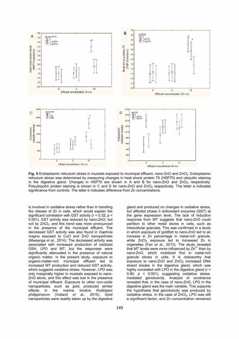
Fig. 2 Labile Zn levels in gills (A, B) and digestive gland (C, D) in mussels exposed to municipal effluent, nano-ZnO and dissolved ZnCl2. Labile Zn levels were determined in mussels exposed to both municipal effluent and Zn as either nano-ZnO or ZnCl2. The letter a indicates significance from ME controls. The letter b indicates difference from Zn concentrations. with ZnCl2, factorial ANOVA revealed significant effects both for municipal effluent concentration and interaction between the municipal effluent and Zn concentration (Fig. 4D). DNA strand breaks were significantly increased by ZnCl2 alone at 10 ug/L. The presence of municipal effluent significantly decreased this response at 3 and 10 % effluent. Correlation analysis showed that DNA strand breaks were significantly correlated with LPO (r = 0.80; p < 0.001). Impacts on the endoplasmic reticulum were determined by following changes in heat shock proteins and protein ubiquitynation (Fig. 5). Factorial analysis of variance revealed that HSP70 was significantly induced by the municipal effluent at the highest concentration tested (10 %). NanoZnO alone had no effects on HSP70 levels. However, when nanoZnO was added in the presence of municipal effluent, the increase in HSP70 was lost. In mussels exposed to ZnCl2, co-exposure to the municipal effluent increased the levels of HSP70 as above but the influence of the municipal effluent was less important. Protein ubiquination was also influenced by the presence of the municipal effluent and nanoZnO. Protein ubiquination was significantly
increased by the municipal effluent at the highest concentration (10 %) tested. This response was also enhanced in the presence of 1 ug/L Zn as nanoZnO. This was not observed for ZnCl2 although an increase in protein ubiquination was observed with 1 ug/L (but not with 10 ug/L) Zn as ZnCl2. Correlation analysis revealed that protein polyubiquitin levels were significantly correlated with MT (r = 0.33; p = 0.001), GST in the digestive gland (r = 0.31; p = 0.001) and Zn in the digestive gland (r = -0.22; p < 0.05).
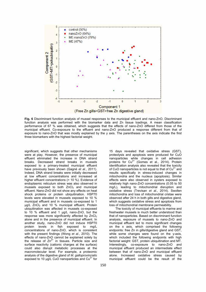
In the attempt to gain a global view of the effects of nanoZnO in the presence of municipal effluents, a discriminant function analysis was performed (Fig. 6). The mean classification efficiency between controls, nanoZnO, nanoZnO+municipal effluent and municipal effluent alone was 60 % The results revealed that nanoZnO mediated effects differed from those when the municipal effluent was present in the following: GST, MT and protein ubiquination. The effects of the municipal effluent was best explained by changes in labile Zn in gills, GST and labile Zn in the digestive gland which suggest the mobilization
146
Fig. 3 Metallothionein levels (A,B) and GST activity (C, D) in mussels exposed to ZnCl2, nano-ZnO and municipal effluent. MT levels in the digestive gland were determined in mussels exposed to both municipal effluent and Zn, as either nano-ZnO or ZnCl2. The letter a indicates significance from controls. The letter b indicates difference from Zn concentrations. (release) of Zn2+ in municipal effluents. Indeed, ME related effects were principally discriminated from nanoZnO and nanoZnO+municipal effluent by the axis which involved labile Zn in gills and digestive gland.
Discussion
In the present study, mussels were exposed to
two forms of Zn, nano-ZnO and ZnCl2, in the presence of a primary physico-chemically treated effluent. Exposure to ZnCl2 did not change Zn levels in the digestive gland or gill tissues. In mussels exposed to nano-ZnO, although whole body Zn levels remained somewhat constant, a small increase in Zn was found in the digestive gland. This suggests that mussels are exposed to nano-ZnO by the digestive system. However, Zn levels in the digestive gland were reduced when in presence of the municipal effluent. Municipal effluent is rich in organic matter, which can compete for Zn in the digestive gland. This interpretation is justified since mussels were allowed to depurate for 24 h without food before sacrifice. Decreased metal
concentrations in mussels exposed to municipal effluent have also been shown with this type of effluent (Gagnon et al., 2014). Indeed, the proportion of Zn bound to the colloidal matter in the effluent was on the order of 70 %, which suggests a strong interaction with suspended matter. In another study, Zn levels did not readily accumulate in mussels caged 8 km downstream from a primary treated effluent, although it was present in the effluent at concentrations on the order of 25 µg/L (Gagnon et al., 2006). These studies suggest that exposure to the suspended material in effluents could influence the bioavailability of metals in exposed organisms. In mussels exposed to nano-ZnO and the municipal effluent, the increase in Zn levels in the digestive gland was observed only at the highest concentration (10 µg/L). In the presence of municipal effluent and ZnCl2, an increase was observed at 1 µg Zn/L, but not 10 µg Zn/L in the presence of the municipal effluent. Labile Zn (Zn2+) levels in gill and digestive gland tissues were also examined using a fluorescent probe in mussels exposed to either form of Zn in the presence (or not) of the municipal effluent. In mussels exposed to
147
Fig. 4 Oxidative stress (A, B) and DNA damage (C, D) in mussels exposed to municipal effluent, nano-ZnO and ZnCl2. Oxidative stress was determined by measuring changes in lipid peroxidation (LPO) in the digestive gland. The letter a indicates significance from controls. The letter b indicates difference from Zn concentrations. nano-ZnO, labile Zn2+ increased in the digestive gland, which suggests the mobilization of ionic Zn in the digestive system. Zn is considered an essential element which is highly regulated in mussels (Lemus et al., 2013). In a previous study, the release of Zn2+ from bulk ZnO and nano-ZnO in suspension accounted for most, but not all, of the toxicity in zebrafish (Yu et al., 2011). Mussels exposed to metal oxide nanoparticles revealed that Zn was detected in the pseudofeces, indicating absorption through the digestive system (Montes et al., 2012). Moreover, scanning electron microscopic analysis of the pseudofeces revealed that intact nano-ZnO was not found, which suggests that nano-ZnO was degraded and involved perhaps the release the Zn2+. However, when mussels were exposed to the municipal effluent, labile Zn decreased at the highest concentration (10 %), indicating the dampening effects of the municipal effluent with respect to Zn mobility. Interestingly, gill labile Zn levels were increased at the 3 % effluent concentration only in control mussels, which suggests a transient increase in Zn in tissues at low effluent concentrations. The explanation for this is unclear, but we could hypothesize that at low
concentrations, the dampening effect of the suspended solid matrix is too low to impede the activity of Zn2+ in these tissues. The levels of MT, a heavy metal-binding protein, were significantly increased in mussels exposed to 1 ug/L ZnCl2 alone, but MT were increased by 1 ug/L nano-ZnO in the presence of the municipal effluent, which suggests the release of Zn ions to induce MT. In a previous study with E. complanata mussels co-exposed to nano-ZnO and to a municipal effluent, the metallome was significantly impacted in the digestive gland (Gagné et al., 2013). The study revealed that the metallome of mussels co-exposed to nano-ZnO and the municipal effluent was closer to that of ZnCl2-exposed mussels, which suggests the mobilization of Zn2+. In another study, the freshwater mussel Unio tumidus exposed to nano-ZnO alone and in combination with other stressors, such as pesticides and temperature, increased MT and oxidative stress (Falfushynska et al., 2015). However, the increase in MT in their study was not accompanied by increased Zn bound to MT or by elevated Zn in tissues. This is consistent with the marginal negative correlation between labile Zn and MT in the digestive gland. This suggests that MT
148
Fig. 5 Endoplasmic reticulum stress in mussels exposed to municipal effluent, nano-ZnO and ZnCl2. Endoplasmic reticulum stress was determined by measuring changes in heat shock protein 70 (HSP70) and ubiquitin staining in the digestive gland. Changes in HSP70 are shown in A and B for nano-ZnO and ZnCl2 respectively. Polyubiquitin protein staining is shown in C and D for nano-ZnO and ZnCl2 respectively. The letter a indicates significance from controls. The letter b indicates difference from Zn concentrations. is involved in oxidative stress rather than in handling the release of Zn in cells, which would explain the significant correlation with GST activity (r = 0.32; p < 0.001). GST activity was reduced by nano-ZnO, but not by ZnCl2, and this trend was more pronounced in the presence of the municipal effluent. The decreased GST activity was also found in Daphnia magna exposed to CuO and ZnO nanoparticles (Mwaanga et al., 2014). The decreased activity was associated with increased production of oxidized GSH, LPO and MT, but the responses were significantly attenuated in the presence of natural organic matter. In the present study, exposure to organic-matter-rich municipal effluent led to increased MT production and reduced GST activity, which suggests oxidative stress. However, LPO was only marginally higher in mussels exposed to nano-ZnO alone, and this effect was lost in the presence of municipal effluent. Exposure to other non-oxide nanoparticles, such as gold, produced similar effects in the marine bivalve Ruditapes philippinarum (Volland et al., 2015). Gold nanoparticles were readily taken up by the digestive
gland and produced no changes in oxidative stress, but affected phase II antioxidant enzymes (GST) at the gene expression level. The lack of induction response from MT suggests that nano-ZnO could partition to other metal stores in cells, such as intracellular granules. This was confirmed in a study in which exposure of goldfish to nano-ZnO led to an increase in Zn percentage in metal-rich granule, while ZnCl2 exposure led to increased Zn in organelles (Fan et al., 2013). The study revealed that MT levels were more influenced by Zn2+ than by nano-ZnO, which mobilized first in metal-rich granule stores in cells. It is noteworthy that exposure to nano-ZnO and ZnCl2 increased DNA strand breaks in the digestive gland, which was highly correlated with LPO in the digestive gland (r = 0.80; p < 0.001), suggesting oxidative stress-mediated genotoxicity. Analysis of covariance revealed that, in the case of nano-ZnO, LPO in the digestive gland was the main variable. This supports the hypothesis that genotoxicity was produced by oxidative stress. In the case of ZnCl2, LPO was still a significant factor, and Zn concentration remained
149
Fig. 6 Discriminant function analysis of mussel responses to the municipal effluent and nano-ZnO. Discriminant function analysis was performed with the biomarker data and Zn tissue loadings. A mean classification performance of 67 % was obtained, which suggests that the effects of nano-ZnO differed from those of the municipal effluent. Co-exposure to the effluent and nano-ZnO produced a response different from that of exposure to nano-ZnO that was mostly explained by the y axis. The parentheses on the axis indicate the first three biomarkers with the highest factorial weight. significant, which suggests that other mechanisms were at play. However, the presence of municipal effluent eliminated the increase in DNA strand breaks. Decreased strand breaks in mussels exposed to a primary-treated municipal effluent have previously been shown (Gagné et al., 2011). Indeed, DNA strand breaks were initially decreased at low effluent concentrations and increased at higher effluent concentrations (> 10 %). Evidence of endoplasmic reticulum stress was also observed in mussels exposed to both ZnCl2 and municipal effluent. Nano-ZnO did not show any effects on heat shock proteins or protein ubiquitination. HSP70 levels were elevated in mussels exposed to 10 % municipal effluent and in mussels co-exposed to 1 µg/L ZnCl2 and 10 % municipal effluent. Protein ubiquitination was affected in mussels co-exposed to 10 % effluent and 1 µg/L nano-ZnO, but the response was more significantly affected by ZnCl2 alone and in the presence of municipal effluent. In another study, nano-ZnO did not induce HSP70 protein levels in fish exposed to mg/L concentrations of nano-ZnO, which is consistent with the present findings (Wong et al., 2010). The effects of nano-ZnO cannot be explained solely by the release of Zn2+ in tissues. Particle size and surface reactivity (cationic charges at the surface) could also disrupt metabolic processes at the macromolecular level. For example, a proteomic analysis of the digestive gland of M. galloprovincialis exposed to 10 µg/L CuO nanoparticles and Cu2+ for
15 days revealed that oxidative stress (GST), proteolysis and apoptosis were produced for CuO nanoparticles while changes in cell adhesion proteins for Cu2+ (Gomes et al., 2014). Protein identification analysis also revealed that the toxicity of CuO nanoparticles is not equal to that of Cu2+ and results specifically in stress-induced changes in mitochondria and the nucleus (apoptosis). Similar effects were also observed in oysters exposed to relatively high nano-ZnO concentrations (0.05 to 50 mg/L), leading to mitochondrial disruption and oxidative stress (Trevisan et al., 2014). Swollen mitochondria and loss of mitochondrial cristae were observed after 24 h in both gills and digestive gland, which suggests oxidative stress and apoptosis from loss of mitochondrial membrane permeability.
The toxicity of municipal effluents to marine and freshwater mussels is much better understood than that of nanoparticles. Based on discriminant function analysis, exposure of mussels to nano-ZnO and municipal effluent led to more significant changes on the x axis, which comprised the following endpoints: free Zn in gills/digestive gland and GST, while some changes were found on the y-axis, which included the following endpoints with high factorial weight: GST, protein ubiquitination and MT. Interestingly, co-exposure to nano-ZnO and municipal effluent produced an intermediate effect between that of nano-ZnO and municipal effluent alone. Increased oxidative stress caused by municipal effluent could be the result of the
150
151
increased proportion of polyunsaturated lipids (Rocchetta et al., 2014). Freshwater clams (Diplodon chilensis) collected at sites downstream of a sewage discharge point had increased proportions of omega-3 fatty acids (EPA and DHA), which are targets for lipid oxidation. Levels of LPO and lipofuscin (age-related pigments) were also higher in clams collected downstream of the sewage discharge point. Caged mussels placed downstream of a municipal discharge showed increased AMP-activated protein kinase, indicating increased cellular energy consumption which, in turn, could increase oxidative stress (Goodchild et al. 2015). Oxidative stress was confirmed by increased superoxide dismutase and GST activity. The negative correlation between MT levels and both total Zn and free Zn in the digestive gland suggests that MT was involved in the sequestration of other metals or reactive oxygen species occurring in municipal wastewater. MT is also involved in the sequestration of reactive oxygen species, which confers antioxidant properties, and typically in the sequestration of heavy metals in cells (Gagné et al., 2008; Formigari et al., 2007). In conclusion, exposure of freshwater mussels to nano-ZnO released in municipal effluent could have an effect on the toxicity of municipal effluents. The oxidative properties of both nano-ZnO and ZnCl2 are reduced in the presence of municipal effluent. Although no clear signs of oxidative stress were observed at the LPO level, LPO levels were strongly correlated with DNA damage and GST activity was positively correlated with MT levels which were not associated with Zn binding in the digestive gland. The municipal effluent will likely change the oxidative stress response to nanoZnO and ZnCl2 in freshwater mussels.
Acknowledgements
The work was funded under the Chemical Management Plan and the St. Lawrence Action Plan of Environment Canada. The authors thank Sophie Trépanier for her technical assistance in mussel handling and exposure at the City of Montreal ecotoxicology laboratory.
References Boryslawskyj M, Garrood AC, Pearson JT. Elevation
of glutathione-S transferase activity as a stress response to organochlorine compounds in the freshwater mussel Sphaerium corneum. Mar. Environ. Res. 24: 101-104, 1998.
Balshaw DM, Philbert M, Suk WA. Research strategies for safety evaluation of nanomaterials. Part III: Nanoscale technologies for assessing risk and improving public health. Toxicol. Sci. 88: 298-306, 2005.
Bradford MM. A rapid and sensitive method for the quantitation of microgram quantities of protein utilizing the principle of protein-dye binding. Anal. Biochem. 72: 248-254, 1976.
Canesi L, Ciacci C, Fabbri R, Marcomini A, Pojana G, Gallo G. Bivalve molluscs as a unique target group for nanoparticle toxicity. Mar. Environ. Res. 76: 16-21, 2012.
Contado C. Nanomaterials in consumer products: a challenging analytical problem. Front. Chem. 3:48, 2015.
Falfushynska H, Gnatyshyna L, Yurchak I, Sokolova I, Stoliar O. The effects of zinc nanooxide on cellular stress responses of the freshwater mussels Unio tumidus are modulated by elevated temperature and organic pollutants. Aquat. Toxicol. 162: 82-93, 2015.
Fan W, Li Q, Yang X, Zhang L. Zn subcellular distribution in liver of goldfish (Carassius auratus) with exposure to zinc oxide nanoparticles and mechanism of hepatic detoxification. PLoS One. 8: e78123, 2013.
Fang D, Pan C, Lin H, Lin Y, Xu G, Zhang G, et al. Ubiquitylation functions in the calcium carbonate biomineralization in the extracellular matrix. PLoS One. 7: e35715, 2012.
Formigari A, Irato P, Santon A. Zinc, antioxidant systems and metallothionein in metal mediated-apoptosis: Biochemical and cytochemical aspects. Comp. Biochem. Physiol. 146C: 443-459, 2007.
Gagné F, André C, Blaise C. The dual nature of metallothioneins in the metabolism of heavy metals and reactive oxygen species in aquatic organisms: implications of use as a biomarker of heavy-metal effects in field investigations. Biochem. Insights 1: 31-41, 2008.
Gagné F, Gélinas M, Gagnon C, André C, Blaise C. Change in metallothioneins phosphorylation state in Mya arenaria clams: implication in metal metabolism and oxidative stress. Inv. Surv. J. 7: 22-31, 2010.
Gagné F, André C, Cejka P, Hausler R, Fournier M. 2011. Alterations in DNA metabolism in Elliptio complanata mussels after exposure to municipal effluents. Comp. Biochem. Physiol. 154C: 100-107, 2011.
Gagné F, Turcotte P, Auclair J, Gagnon C. The effects of zinc oxide nanoparticles on the metallome in freshwater mussels. Comp. Biochem. Physiol. 158C: 22-28, 2013.
Gagné F, Blaise C. Available intracellular Zn as a potential indicator of heavy metal exposure in rainbow trout hepatocytes. Environ. Toxicol. Wat. Qual. 11: 319-325, 1996.
Gagnon C, Turcotte P, Trépanier S, Gagné F, Cejka P-J. Impacts of municipal wastewater oxidative treatments: Changes in metal physical speciation and bioavailability. Chemosphere 97: 86-91, 2014.
Gagnon C, Gagné F, Turcotte P, Saulnier I, Blaise C, Salazar MH, Salazar SM. Exposure of caged mussels to metals in a primary-treated municipal wastewater plume. Chemosphere 62: 998-1010, 2006.
Gomes T, Pereira CG, Cardoso C, Pinheiro JP, Cancio I, Bebianno MJ. Accumulation and toxicity of copper oxide nanoparticles in the digestive gland of Mytilus galloprovincialis. Aquat. Toxicol. 118-119: 72-79, 2012.
Goodchild CG, Frederich M, Zeeman SI. AMP-activated protein kinase is a biomarker of energetic status in freshwater mussels exposed
to municipal effluents. Sci. Total Environ. 512-513: 201-209, 2015.
Gomes T, Chora S, Pereira CG, Cardoso C, Bebianno MJ. Proteomic response of mussels Mytilus galloprovincialis exposed to CuO NPs and Cu²+: an exploratory biomarker discovery. Aquat Toxicol. 155: 327-336, 2014.
Lee J, Yang J, Seo SB, Ko HJ, Suh JS, Huh YM, et al. Smart nanoprobes for ultrasensitive detection of breast cancer via magnetic resonance imaging. Nanotechnology 19: 485101, 2008.
Lemus M, Rojas N, Rojas-Astudillo L, Chung K. Metallothioneins in Perna viridis (Bivalvia: Mytilidae): seasonal variation and its relation to reproductive biology. Rev. Biol. Trop. 61: 701-709, 2013.
Louis S, Gagné F, Auclair J, Turcotte P, Gagnon C, Émond C. The characterization of the behaviour and gill toxicity of CdS/CdTe quantum dots in rainbow trout (Oncorhynchus mykiss). Int. J. Biomed. Nanosc. and Nanotechnol. 1, 52-69, 2010.
Majedi SM, Kelly BC, Lee HK. Combined effects of water temperature and chemistry on the environmental fate and behavior of nanosized zinc oxide. Sci. Total Environ. 496: 585-593, 2014.
McDonagh B, Sheehan D. Redox proteomics in the blue mussel Mytilus edulis: carbonylation is not a pre-requisite for ubiquitination in acute free radical-mediated oxidative stress. Aquat. Toxicol. 79: 325-333, 2006.
Mitchnick MA, Fairhurst D, Pinnell SR. Microfine zinc oxide (z-cote) as a photostable UVA/UVB sunblock agent. J. Am. Acad. Dermatol. 40, 85-89, 1999.
Montes MO, Hanna SK, Lenihan HS, Keller AA. Uptake, accumulation, and biotransformation of metal oxide nanoparticles by a marine suspension-feeder. J Hazard. Mater. 225-226: 139-145, 2012.
Mwaanga P, Carraway ER, van den Hurk P. The induction of biochemical changes in Daphnia magna by CuO and ZnO nanoparticles. Aquat. Toxicol. 150: 201-209, 2014.
Oberdörster G, Stone V, Donaldson K. Toxicology of nanoparticles: a historical perspective. Nanotoxicology 1: 2-25, 2007.
Olive PL. DNA precipitation assay: a rapid and
simple method for detecting DNA damage in mammalian cells. Environ. Mol. Mutagen. 11: 487-495, 1988.
Rocha TL, Gomes T, Sousa VS, Mestre NC, Bebianno MJ. Ecotoxicological impact of engineered nanomaterials in bivalve molluscs: An overview. Mar. Environ. Res. S0141-1136: 30002-30007, 2015.
Rocchetta I, Pasquevich MY, Heras H, Ríos de Molina Mdel C, Luquet CM. Effects of sewage discharges on lipid and fatty acid composition of the Patagonian bivalve Diplodon chilensis. Mar. Pollut. Bull. 79: 211-219, 2014.
Trevisan R, Delapedra G, Mello DF, Arl M, Schmidt ÉC, Meder F, et al. Gills are an initial target of zinc oxide nanoparticles in oysters Crassostrea gigas, leading to mitochondrial disruption and oxidative stress. Aquat. Toxicol. 153: 27-38, 2014.
Valko M, Rhodes CJ, Moncola J, Izakovic M, Mazura M. Free radicals, metals and antioxidants in oxidative stress-induced cancer. Chem. Biol. Interact. 160: 1-40, 2006.
Viarengo A, Ponzanon E, Dondero F, Fabbri R. A simple spectrophotometric method for metallothionein evaluation in marine organisms: an application to Mediterranean and Antarctic molluscs. Mar. Environ. Res. 44: 69-84, 1997.
Volland M, Hampel M, Martos-Sitcha JA, Trombini C, Martínez-Rodríguez G, Blasco J. Citrate gold nanoparticle exposure in the marine bivalve Ruditapes philippinarum: uptake, elimination and oxidative stress response. Environ. Sci. Pollut. Res. Int. 22: 17414-17424, 2015
Wills ED. Evaluation of lipid peroxidation in lipids and biological membranes. In: Snell K, Mullock B (eds), Biochemical toxicology: a practical approach. Washington, USA: IRL Press; pp 127-150, 1987.
Wong SW, Leung PT, Djurisić AB, Leung KM. Toxicities of nano zinc oxide to five marine organisms: influences of aggregate size and ion solubility. Anal. Bioanal. Chem..396: 609-618, 2010.
Yu LP, Fang T, Xiong DW, Zhu WT, Sima XF. Comparative toxicity of nano-ZnO and bulk ZnO suspensions to zebrafish and the effects of sedimentation, ˙OH production and particle dissolution in distilled water. J. Environ. Monit. 13: 1975-1982, 2011.