Efficient repair of bulky anti-BPDE DNA adducts fromnon-transcribed DNA strand requires functional p53
but not p21waf1/cip1 and pRb
Manzoor A. Wania,1, Mohammed A. El-Mahdya,d,1, Farid M. Hamadad,Gulzar Wania, Qianzheng Zhua, Qi-en Wanga, Altaf A. Wania,b,c,∗
a Department of Radiology, The Ohio State University, Columbus, OH 43210, USAb Biochemistry Program, 103 Wiseman Hall, 400 W 12th Avenue, The Ohio State University, Columbus, OH 43210, USA
c James Cancer Hospital and Research Institute, The Ohio State University, Columbus, OH 43210, USAd College of Pharmacy, Al-Azhar University, Cairo, Egypt
Received 22 February 2002; received in revised form 1 April 2002; accepted 25 April 2002
1These two authors contributed equally to the work.
1. Introduction
The integrity of genetic material in mammaliancells is continuously compromised by the expo-sure and action of exogenous and endogenousfactors, of both chemical and physical origin, re-sulting in a large variety of DNA structural alter-ations. Incessant exposure of cells to DNA-damagingagents is implicated in the pathogenesis of various
14 M.A. Wani et al. / Mutation Research 505 (2002) 13–25
cancers. Xenobiotic polycyclic aromatic hydrocar-bons, such as benzo[a]pyrene (B[a]P), the prod-ucts of incomplete combustion of organic matterare widespread and major environmental pollutants.Carcinogenic and mutagenic effects of B[a]P arewell documented in human and animal model sys-tems[1]. The diol-epoxide, (±)-anti-BPDE (racemic7,8-dihydroxy-anti-9,10-epoxy-7,8,9,10-tetrahydro-benzo[a]pyrene), is the highly reactive electrophilicmetabolite of B[a]P and is reported to cause muta-tions, cytotoxicity, and inhibition of DNA synthesisin both prokaryotic and eukaryotic cells[2]. Uponmetabolic activation in cells, anti-BPDE reacts atseveral nucleophilic sites in DNA, and the covalentN2-dG-anti-BPDE adduct is most abundant DNA baselesion [3]. In order to protect cells from deleteriouseffects of these physico–chemical DNA-damagingagents, a host of complex cellular DNA repair mech-anisms have evolved to counteract the harmful effectsof DNA damage in mammalian cells[4]. In additionto the existence of several detoxification systems,delays at specific stages of the cell cycle occur afterDNA damage, most likely for the efficient and promptremoval of DNA lesions from the cellular genome be-fore commencing of cell division[5,6]. DNA lesionsinitially lead to a transient inhibition of DNA repli-cation and transcription, while in the end unrepairedlesions may give rise to mutations. Such mutationalevents can affect the expression or functioning ofimportant cellular genes involved in the control ofproliferation and differentiation of cells, thus playinga critical role in the initiation of carcinogenesis.
It has been known that the p53 tumor suppressorprotein, although not an absolute requirement for nor-mal growth and development, is critical for the pre-vention of tumor development. p53 function is lost inmajority of the human cancers[7,8]. A lack of func-tional p53 causes genetic instability and accumulationof mutations in cells[9–11]. Inactivation of p53 by vi-ral oncoproteins has also been known to be related tohuman cancers. For example, a subset of human papil-lomaviruses (HPVs) referred to as high risk HPVs, areassociated with cervical carcinomas in humans. Twoviral proteins encoded byE6 and E7 genes, are ex-pressed in cells derived from HPV-associated cancers[12]. Nevertheless, p53 mutations are rarely found inthese tumors[13]. E6 andE7 gene products cooper-ate with each other in causing the immortalization or
transformation of cells. The transforming activity ofE6 and E7, at least in part, correlates with their inac-tivation of p53 and Rb, which regulate the importantbiological processes of cell division and proliferation[14,15]. E6 binds to p53 and mediates its degradation[13] while as E7 binds and inactivates pRb as well aspromotes its degradation[16,17].
In response to DNA damage, activation of p53 leadsto the suppression of cell growth, cell cycle arrest orapoptosis by transcriptionally activating a range of tar-get genes required for these cellular processes[18–21].Cell cycle arrest, mediated by activated p53, is be-lieved to allow DNA repair and prevent mutagenesisresulting from DNA damage. Functional p53, itself,has also been shown to be required for the removal ofDNA lesions from the non-transcribed strand (NTS)and specifically influence the global genomic repair(GGR), a repair sub-pathway of nucleotide excisionrepair (NER,[22,24]).
As pRb and p21waf1/cip1 are major participantsin the p53-dependent cellular response to DNAdamage, here we investigated the possible role ofpRb and p21waf1/cip1 in NER. Data shows that theHPV-16E6 expressing cells lacking functional p53and p21waf1/cip1 were inefficient for the removal ofanti-BPDE induced DNA lesions from the NTS andgenome overall as compared to normal and pRb de-ficient HPV-16E7 expressing cells. Moreover, wecompared the repair kinetics in HCT116 cells consti-tutively expressing wild-type p53 but having differentp21waf 1/cip1 genotypes. The results show no dis-cernible difference in the removal of anti-BPDE DNAadducts from the genome overall and the transcribedstrand (TS) and NTS irrespective of the presenceor absence of p21waf1/cip1 expression. These resultssupport the role of functional p53 for the efficient re-moval of bulky DNA lesions and further indicate thatp53 regulated NER is independent of p21waf1/cip1,pRb and cell cycle arrest.
2. Materials and methods
2.1. Culture and pre-labeling of cells
Human colon adenocarcinoma HCT116 cells(p21+/+, p21+/− and p21−/−) were kindly providedby Dr. Bert Vogelstein (The John Hopkins OncologyCenter, Baltimore, MD). The p21+/− and p21−/−
M.A. Wani et al. / Mutation Research 505 (2002) 13–25 15
cells were generated by homologous recombinationto create homozygous deletion of p21waf1/cip1 [25].These cells and normal human mammary epithelialcells (HMECs) were established and grown in cultureas described earlier[26]. The cognate HMEC trans-fectants, herein termed as 76E6 and 76E7, expressingeither HPV-16E6 or -16E7 oncoproteins, respectively,were kindly provided by Dr. Vimla Band (Tufts Uni-versity School of Medicine, Boston, MA). All thethree HMEC cell lines were grown in a humidifiedatmosphere of 5% CO2 in DCFI medium supple-mented with suitable nutrient and growth factors[27]. For some experiments, in order to separate thenewly synthesized daughter DNA from parental DNA,cells were pre-labeled with 25 nCi/ml [3H]-thymidine(86.0 Ci/mmol) for 3–7 days. The medium waschanged every 2–3 days.
2.2. Western blot analysis of proteins
Exponentially growing HMEC and HCT116cells were washed with PBS and exposed to 1�M(±)-anti-BPDE, a dose previously determined tomaximally induce p53 without killing the cells. Af-ter carcinogen treatment cells were maintained infresh medium for 2–24 h. At the indicated time pe-riods, the cells were recovered by gentle trypsiniza-tion and immediately lysed by boiling for 10 min insample buffer (2% sodium dodecyl sulfate (SDS);10% glycerol; 10 mM DTT in 62 mM Tris–HCl (pH6.8), 10�g/ml pepstatin, and 10�g/ml leupeptin).Proteins were quantitated using a Bradford assay(Biorad) and results were further corroborated byvisualization of sample bands using comassie bluestaining after SDS-polyacrylamide gel electrophoresis(PAGE). Western blot analysis of proteins separatedby SDS-PAGE was performed as described earlier[28]. For p53 protein detection, a mixture of anti-p53antibodies (Ab-2 and Ab-6 from hybridoma clones1801 and DO-1, Neomarkers, CA) was used at 1:200dilution. Antibodies for the detection of p21waf1/cip1
and pRb (clone DC60.2) and (clone 1F8) were alsoused at a 1:200 dilution. Following incubation withthe corresponding enzyme-conjugated secondary an-tibody (Boehringer Mannheim, Indianapolis, IN),the protein bands were detected using the enhancedchemiluminescence substrate reaction (Pierce, Rock-ford, IL) essentially according to the manufacturer’s
instructions using Kodak X-OMAT AR film. Rain-bow protein size markers were run in parallel in allthe experiments to localize the gel transfer regions forspecific proteins and determine the transfer efficiency.
2.3. Anti-BPDE treatment and incubation of cellsafter carcinogen exposure
For DNA damage and repair analysis the abovementioned cells were exposed to (±)-anti-BPDE asdescribed earlier[22]. Briefly, the confluent culturesof monolayer cells were washed with PBS and ex-posed to different doses of (±)-anti-BPDE (preparedfrom a fresh 100× stock in 95% ethanol) in 10 mmof Hank’s balanced salt solution (pH 7.0) for 30 minat 37◦C. The exposed cells were washed with PBSto remove the DNA-damaging agent and further incu-bated in fresh medium for varying post-treatment pe-riods. For some repair analysis experiments cells wereincubated in the medium containing 10�M bromod-eoxyuridine and 1�M fluorodeoxyuridine to densitylabel the DNA. After incubation at 37◦C for variousperiods of time, the treated cells were lysed for DNAisolation.
2.4. Isolation, purification and separation ofparental DNA
Cells were washed and harvested after desired in-cubation periods and immediately lysed for DNAisolation as described earlier[28,29]. The DNA waspurified by phenol:chloroform extractions followedby chloroform:isoamyl alcohol extraction, ethanolprecipitated and resuspended in TE buffer (1 mMEDTA/10 mM Tris–HCl, pH 8.0). For Southern blot-ting, the DNA was extensively digested to completionwith EcoRI (5 U/�g of DNA) at 37◦C for 6–8 h.Complete digestion was verified by electrophoresisof sample aliquots on agarose minigels. The digestedDNA was again purified by extraction with phe-nol:chloroform (3.5:6), precipitated with ethanol andquantitated. Replicated and non-replicated DNA wasthen separated and isolated using cesium chlorideisopycnic density gradient centrifugation. Gradientswere fractionated and radioactivity in samples wasmeasured in a scintillation counter to determine theamounts and position of parental and hybrid densityDNA. Fractions of parental DNA were pooled and
16 M.A. Wani et al. / Mutation Research 505 (2002) 13–25
dialyzed against TE. The dialyzed parental DNA wasethanol precipitated and quantitated by absorptionmeasurement at 260 nm. Only the non-replicated DNAwas used for strand-specific repair kinetics analysis.
2.5. Quantitation of anti-BPDE DNA adductsby ISB assay
Following carcinogen treatment of cells, GGR ofanti-BPDE induced DNA adducts was measured atdifferent post-exposure times using a non-competitiveimmuno-slot blot assay[22,30]. Briefly, cells were ex-posed to different doses of anti-BPDE for 30 min at37◦C. Cells were either immediately lysed for DNAisolation or incubated further in fresh medium fordifferent repair times. Genomic DNA was isolatedby phenol/chloroform extraction, followed by ethanolprecipitation and the DNA concentration was deter-mined spectrophotometrically at 260 nm and by themicrodiphenylamine assay[31,32]. For determiningthe extent of anti-BPDE–DNA adduct formation andrepair, equal amounts (100 ng) of denatured genomicDNA, from each individual sample, were immobi-lized onto nitrocellulose filters in duplicate using aslot blot manifold (BRL). The filters were sequen-tially exposed to primary polyclonal antibody BP1, de-veloped against anti-BPDE modified single-strandedDNA [33,34]and enzyme-labeled secondary antibody.Band color intensities were evaluated using LKB laserdensitometry and the data were transferred to an in-terfaced computer and analyzed using LKB GelscanXL software. Genomic DNA from untreated cells andanti-BPDE-modified DNA standard samples were runin parallel with all of the blots. The experiments wererepeated at least three times.
2.6. UvrABC nuclease treatment and Southernblot analysis
The UvrA, UvrB and UvrC proteins were purified asdescribed previously[22]. Equal amounts (20�g) ofDNA of each sample were either treated with UvrABCexcinuclease or enzyme buffer. Linearized plasmidDNA (5 pg) containing the p53 cDNA sequencewas added into each sample as an internal standard.The UvrABC reaction components and conditionswere the same as described earlier[22]. After incuba-tion of the reaction mixture at 37◦C for 1 h, the DNA
was purified by extraction with phenol/chloroformand precipitated with ethanol. The samples were thendissolved in denaturation buffer and incubated at37◦C for 30–45 min to denature the DNA followedby electrophoresis at 1.5 V/cm for 18–20 h in a 0.7%neutral agarose gel. The DNA in the gel was aciddepurinated, neutralized and transferred to a nylonmembrane[35]. Membranes were hybridized in 10 mlof solution containing 50% (v/v) formamide, 6× SSC,0.5% SDS, 5% dextran sulfate, denatured salmonsperm DNA (100�g/ml), and 1× 10⁸ to 2× 10⁸ cpm32P-labeled single stranded exon specific probes gen-erated by PCR[36]. After 20–22 h hybridization timeat 42◦C, the membranes were washed to final strin-gency at 62◦C in 1× SSC/1% SDS and exposed toa phosphorimager screen. The ratio of full-lengthrestriction fragment in the UvrABC treated and un-treated sample was determined by phosphorimageranalysis of integrated signals upon imaging and pro-cessing by Imagequant software (molecular dynam-ics). The average number of UvrABC sensitive sitesper fragment were calculated by Poisson distribu-tion as described earlier[37]. Each experiment wasindependently repeated at least two times.
3. Results
3.1. Anti-BPDE induced response of cell cycleregulatory proteins in HMEC
To investigate, the level of p53 and other cell cycleregulatory proteins in normal as well as HPV-16E6and -16E7 expressing HMEC, Western blot analysiswas performed with protein extracts from untreatedcells or cells treated with 1�M anti-BPDE and incu-bated for different time periods. The three cell linesshowed distinct induction responses with respect totheir p53, p21waf1/cip1 and pRb protein levels (Fig. 1).Within few hours of carcinogen treatment the p53protein levels were increased several fold in normaland HPV-16E7 transformed cells. The elevated pro-tein levels, peaking at 8–24 h, were sustained wellbeyond the 24 h time period. However, lack of de-tectable levels of pRb could be seen only in HPV-16E7cells, consistent with the expression of E7 oncopro-tein targeting pRb for degradation. Cells expressingHPV-16E6 showed a complete absence of p53 protein
M.A. Wani et al. / Mutation Research 505 (2002) 13–25 17
Fig. 1. Time dependent anti-BPDE induced response of p53, p21waf1/cip1 and pRb proteins in normal, HPV-16E6 and -16E7 expressinghuman mammary epithelial cells. Exponentially growing cells were treated with 1�M anti-BPDE for 30 min at 37◦C and maintained furtherin fresh medium. Protein extracts, at the indicated times were separated by SDS-PAGE, transferred onto PVDF membranes, processedwith monoclonal antibody specific for p53, p21waf1/cip1 and pRb proteins and visualized by enhanced chemiluminescence as described inSection 2. The figure is representative of results from experiments independently repeated thrice.
in untreated as well as treated cells and longer film ex-posures also failed to reveal any detectable p53 proteinin carcinogen treated cells. Higher basal pRb levelswere observed in HPV-16E6 expressing cells as com-pared to normal parental cells. Moreover, these cellsalso showed a clear increase in both phosphorylatedand hyperphosphorylated forms of pRb within 2 h af-ter carcinogen treatment. Consistent with the presenceof functional p53, normal and HPV-16E7 cells alsoshowed a p53-dependent increase in their p21waf1/cip1
protein levels that were fully sustained up to 24 h.Complete absence of p21waf1/cip1 protein, observed inHPV-16E6 expressing cells, was fully consistent withthe lack of functional p53 in these cells.
3.2. Global genomic repair of anti-BPDEadducts in HMEC
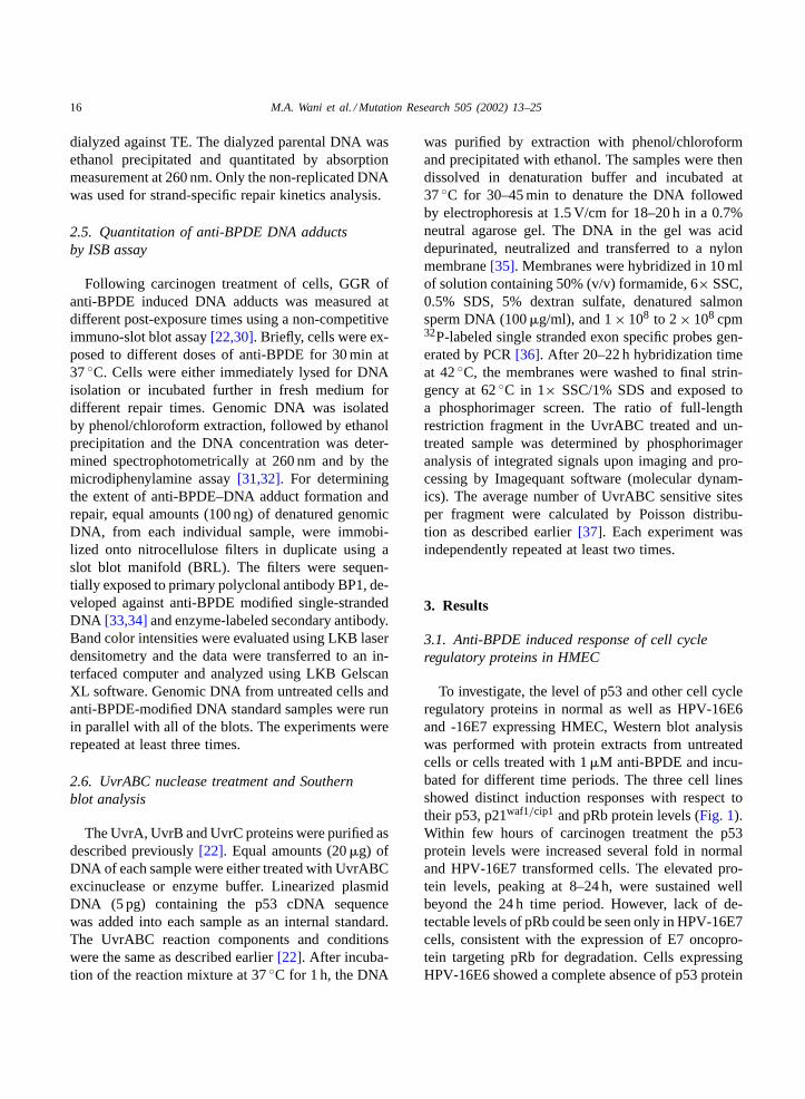
The GGR of anti-BPDE DNA adducts in normal,HPV-16E6 and -16E7 expressing cells was deter-mined by non-competitive immuno-slot blot assaysusing adduct specific polyclonal antibodies[33]. Adose dependent formation of anti-BPDE DNA adductswas observed following in vivo exposure of cells to in-creasing doses of the carcinogen (Fig. 2A). Although,the three cell lines did not seem to differ in the extentof formation of initial DNA adducts upon increas-ing doses of the carcinogen within their genome, therate at which the DNA lesions were repaired variedsignificantly between the p53 proficient and deficient
cells. Normal and HPV-16E7 expressing HMEC, bothcontaining wild-type p53, showed efficient removalof anti-BPDE DNA adducts from the genome overallas compared to HPV-16E6 transformed cells. Thesecells exhibited a significant reduction in their GGR ofanti-BPDE induced DNA lesions (Fig 2B). Quantita-tive comparison of anti-BPDE DNA adduct removalshowed that normal HMEC, HPV-16E7 and -16E6expressing cells repaired 61, 57 and 32% of the initialdamage within 24 h following anti-BPDE treatment.A similar response in repair profiles was observed atearlier time periods in all the three cell lines.
3.3. Transcription-coupled repair of anti-BPDEadducts in HMEC
We have previously reported that p53 has no sig-nificant role in transcription-coupled repair (TCR) butis required for the efficient GGR of CPD and chemi-cally induced DNA lesions. To determine whether theDNA adducts formed by anti-BPDE are also repairedin a p53-dependent manner in normal, HPV-16E6 and-16E7 expressing cells, strand-specific repair analysiswas performed within the individual strands of targethumanp53gene. All the three cell lines upon exposureto 2�M anti-BPDE, resulted in an initial frequencyof adducts in the range of 1–1.3/16 kb DNA fragment.The removal of anti-BPDE induced DNA lesions wasdetermined upon adduct specific cleavage by UvrABCexcinuclease assay. Representative autoradiograms of
18 M.A. Wani et al. / Mutation Research 505 (2002) 13–25
Fig. 2. Formation and repair of anti-BPDE DNA adducts in nor-mal, E6 and E7 expressing HMEC. (A) Confluent cultures ofmonolayer cells were exposed to increasing doses of anti-BPDEfor 30 min at 37◦C and lysed immediately for DNA isolation. Theextent of initial anti-BPDE adduct formation was determined fromspecific antibody binding sites. (B) Global NER of anti-BPDEadducts in HMEC. Following 1�M anti-BPDE treatment, cellswere incubated for indicated times and repair determined as theloss of antibody binding sites from the DNA. The amount of dam-age was calculated upon comparison with a standard reference andthe repair expressed as the percentage of initial damage remain-ing at the indicated times. Each experiment was repeated at leastthree times and the data expressed are mean± S.D.
the repair experiments with the three cell lines arepresented inFig. 3A. As reported earlier for CPDremoval, E6 mediated p53 degradation had no sig-nificant effect on the removal of anti-BPDE DNAadducts from the transcribed strand (TS) in HPV-16E6expressing cells. The rate and extent of efficient re-moval of adducts within the TS was comparativelysimilar to normal and HPV-16E7 expressing cells.Although, the repair being slower at 4 h in E6 express-ing cells, the amount of damage repaired in normal,HPV-16E6, and -16E7 within the TS was calculatedto be 65, 60 and 66% at 8 h followed by approxi-mately 90% of repair at 24 h in all the three cell lines(Fig. 3B).
In contrast to that of the TS, the repair withinthe NTS was severely compromised in HPV-16E6expressing cells. These cells repaired only 10 and30% adducts within 8 and 24 h in their NTS ascompared to 58 and 60% repair at 8 h in normaland HPV-16E7 expressing cells. Both the nor-mal and HPV-16E7 cells showed efficient repairof NTS that was comparable to the repair of theirTS at 24 h, indicating that pRb does not play asignificant role in the repair efficiency of eitherstrand.
3.4. Anti-BPDE induced response of p53 andp21waf 1/cip1 proteins in HCT116 cells
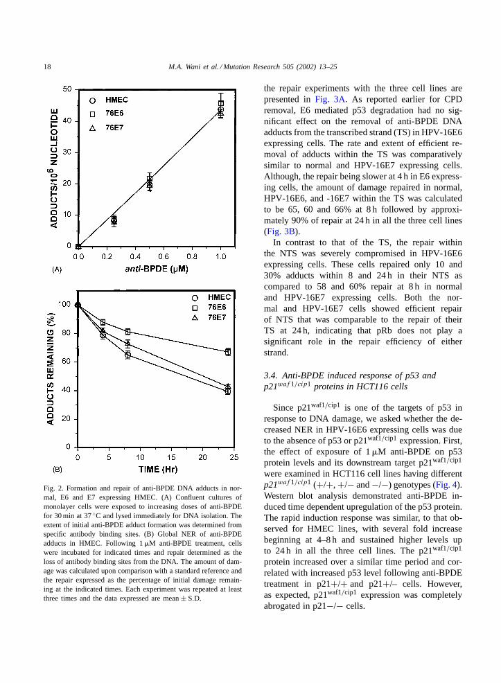
Since p21waf1/cip1 is one of the targets of p53 inresponse to DNA damage, we asked whether the de-creased NER in HPV-16E6 expressing cells was dueto the absence of p53 or p21waf1/cip1 expression. First,the effect of exposure of 1�M anti-BPDE on p53protein levels and its downstream target p21waf1/cip1
were examined in HCT116 cell lines having differentp21waf 1/cip1 (+/+, +/− and−/−) genotypes (Fig. 4).Western blot analysis demonstrated anti-BPDE in-duced time dependent upregulation of the p53 protein.The rapid induction response was similar, to that ob-served for HMEC lines, with several fold increasebeginning at 4–8 h and sustained higher levels upto 24 h in all the three cell lines. The p21waf1/cip1
protein increased over a similar time period and cor-related with increased p53 level following anti-BPDEtreatment in p21+/+ and p21+/– cells. However,as expected, p21waf1/cip1 expression was completelyabrogated in p21−/− cells.
M.A
.W
an
ie
ta
l./Mu
tatio
nR
ese
arch
50
5(2
00
2)
13
–2
519
20 M.A. Wani et al. / Mutation Research 505 (2002) 13–25
Fig. 4. Western blot analysis of p53 and p21waf1/cip1 protein levels from whole cell extracts in HCT116 cells treated with 1�M ofanti-BPDE as described inSection 2. Fast green staining of the bottom portion of protein blots indicates the loading control (LC). Therepresentative results were independently performed two times.
3.5. Global genomic repair in HCT116 cells
We next measured the rate and extent of DNArepair in the above mentioned HCT116 cell linesto ascertain that the decreased GGR in HPV-16E6expressing cells might be due to the absence ofp21waf1/cip1 protein or p21waf1/cip1 might be involvedin the observed p53 modulation of GGR. Cells ex-posed to 2�M anti-BPDE were allowed to repair fordifferent time periods, and the loss of antibody bind-ing sites was determined using polyclonal antibodiesspecific against anti-BPDE DNA adducts. The threecell lines did not differ significantly in the removalof anti-BPDE DNA adducts from the genome overall.Upon quantifying the repair rates, p21+/+, p21+/−and p21−/− showed about 56, 55 and 50% repairwithin 24 h following anti-BPDE treatment (Fig. 5A).To further address the question of whether the ob-served adduct elimination in all the three HCT116 celllines was actually due to repair and not due to DNAreplication, we also measured the GGR in parentalDNA after separation of daughter DNA by densitygradient sedimentation. Again the three cell lines ex-hibited an identical pattern of removal of anti-BPDEDNA adducts from their parental DNA (Fig. 5B).Clearly, the p21−/− cells, unlike p53-compromisedcells, did not show any significant loss of GGR ac-tivity for the removal of anti-BPDE induced DNAlesions.
3.6. Repair of anti-BPDE adducts in specific DNAstrands in HCT116 cells
In order to determine the ability of HCT116 cells,differing in their p21waf 1/cip1 gene status, to per-form TCR and to assess the repair in the NTS asanother measure of GGR, we examined these cellsfor the removal of anti-BPDE DNA adducts from thep53gene sequence. Using Southern hybridization andstrand-specific DNA probes, we found that the repairfrom both the TS and NTS in all the three HCT116cell lines (+/+, +/− and−/−) was mostly similar toeach other in the rate as well as extent of adduct re-moval (Fig. 6A). Presence or absence ofp21waf 1/cip1
gene status didnot seem to influence the repair of eitherstrand. Quantitation of the individuals bands showedthat the p21+/+, p21+/− and the p21−/− cells re-paired 70, 61 and 62% of DNA lesions in TS within8 h followed by about 95% repair within 24 h in all thethree cell lines (Fig. 6B). A similar extent of equallyproficient repair was observed for the NTS in all thethree cell lines further indicating that the p21waf1/cip1
does not play any role in modulating either the TCRor, more importantly, the GGR activity.
4. Discussion
NER is an important mechanism through whichcells remove a variety of DNA lesions following DNA
M.A. Wani et al. / Mutation Research 505 (2002) 13–25 21
Fig. 5. Global genomic repair of anti-BPDE adducts in HCT116cells. (A) Confluent cultures of HCT116 cells were treated with2�M anti-BPDE for 30 min at 37◦C and further incubated infresh medium. At the indicated time periods, DNA repair wasdetermined from constant amounts of isolated whole DNA byimmunoassay. The extent of DNA repair is expressed as the percentof initial damage remaining at the indicated times. (B) The relativeextent of DNA repair determined by immuno-slot blot assay in theunreplicated (parental) DNA after separation by density gradientsedimentation as described inSection 2.
damage. The human tumor suppressor p53 has beenshown to participate in NER of adducts induced byphysical and chemical agents[22–24,28,38,39]. In aneffort to determine the possible connection betweenp53 and its downstream target p21waf1/cip1 as well aspRb in DNA damage and repair response, we exam-
ined the repair kinetics of bulky DNA adducts usingHPV-16E6 or -16E7 expressing cells. We first deter-mined the DNA response of p53, pRb and p21waf1/cip1
in E6 or E7 expressing and normal parental cells andshowed that p53 accumulated in xenobiotic treatednormal parental cell lines. The cells expressing E6failed to induce either p53 or p21waf1/cip1 in responseto carcinogen treatment, although a time dependent in-crease in pRb levels was observed in these cells. Withrespect to the induction of p53 and p21waf1/cip1, E7expressing cells were indistinguishable from the con-trol cells, indicating that these cells also maintaineda normal p53 response pathway. However, these cellshad elevated levels of p53 relative to the parental cellline. On the other hand, these E7 expressing cellsfailed to show any detectable levels of pRb in treatedand untreated cells. These results were consistent withthe well-documented observations that E6 oncopro-tein mediates p53 whereas E7 oncoprotein mediatespRb degradation. Previous studies have clearly shownthat E6 expression does not regulate p53 at the tran-scriptional level. For example, treatment with actino-mycin D or ionizing radiation had no effect on p53or E6 mRNA levels[40]. Moreover, it has also beenshown that E6-facilitated degradation of p53 is spe-cific and not a generalized degradation of all the cel-lular proteins[41]. Measurement of GGR showed that61% of anti-BPDE adducts were repaired at 24 h fol-lowing carcinogen treatment in normal HMEC and E7expressing cells. Thus, cells lacking only in the pRbexpression displayed a reproducible and quantitativelysimilar extent of overall repair as exhibited by the nor-mal HMEC cells. On the contrary, E6 expressing cellsshowed a clear reduction in the initial rate as well assignificant decrease in the overall extent of DNA re-pair at 24 h, achieving only 50% of the repair seen innormal or E7 expressing cells. These results suggestthat p53 plays significant role in modulating GGR.Further analysis of the repair of DNA adducts from theTS and NTS of the humanp53gene also revealed thatdegradation of p53 mediated by E6 did not affect theTCR of DNA lesions. The rate and extent of efficientlesion removal from the TS was identical in all the celllines. In contrast the repair of NTS was significantlycompromised in E6 expressing cells, while as repairof NTS in E7 expressing cells was identical to profi-cient repair of normal control cells. Lack of efficientDNA repair in E6 expressing cells also correlated with
22 M.A. Wani et al. / Mutation Research 505 (2002) 13–25
Fig. 6. Repair of anti-BPDE adducts within the TS and NTS in human cells of varyingp21 genotype. HCT116 cells with differentp21waf 1/cip1 gene status were exposed to the carcinogen as described inSection 2. (A) Autoradiograms representing the extent of removalof anti-BPDE adducts in the TS and NTS of the humanp53 gene. Genomic DNA, isolated from untreated confluent cells (first two lanes ofeach panel) or from cells incubated for indicated time periods after 2�M anti-BPDE treatment was digested with EcoRI and the unreplicated(parental) DNA separated from hybrid (daughter) DNA by density gradient sedimentation. Samples (20�g) of EcoRI digested DNA werethen treated (+) or mock-treated (−) with UvrABC excinuclease and analyzed as inFig. 3A. Bands marked as TS and NTS correspond tothe 16 kb fragment of the humanp53 gene, and the bands marked as IM correspond to the DNA fragments serving as internal markers. (B)Repair profile for the rate of removal of anti-BPDE within the TS and NTS of the humanp53 gene. The rate of anti-BPDE adduct removalwas determined as described forFig. 3B. The points represent the average repair at each time point from two individual experiments.
enhanced cellular sensitivity and higher apoptosis fol-lowing genotoxin damage within these cells (data notshown). As repair of NTS in expressed genes is gener-ally reflected as the capacity of cells to repair overallgenome[42], these findings confirm the role of p53 inregulating repair of DNA lesions within the genome
and further indicate that pRb cannot have a significantinfluence on NER.
Contribution of p53 to GGR of CPDs and bulkyDNA adducts has been shown by several laboratoriesusing diverse model systems[22,23,38,43]. Throughits acidic transactivation domain p53 protein can
M.A. Wani et al. / Mutation Research 505 (2002) 13–25 23
interact with the components of the repair/replicationmachinery including RPA and the helicase com-ponents of TFIIH that are essential for NER andtranscription initiation [44–46]. These interactionswould seem to implicate p53 in direct regulation ofNER machinery and specifically modifying GGR re-sponse presumably through its effect on chromatinconfiguration and damage accessibility. It has beenalso suggested that following DNA damage p53might act indirectly to regulate DNA repair throughits downstream target genep21waf 1/cip1 [47]. How-ever, evidence related to the role of p21waf1/cip1
in NER has been controversial perhaps because ofthe different experimental approaches for assessingNER both in vitro and in vivo. Some studies havereported inhibition of PCNA mediated DNA replica-tion by p21waf1/cip1 without affecting NER[48,49],whereas, others using a similar in vitro approachreported inhibitory effect of p21waf1/cip1 on NER[50]. Investigations on the effects of over-expressingexogenous p21waf1/cip1 in p53 deficient DLD1 col-orectal carcinoma cells found increased clonogenicsurvival after UV-irradiation and suggested a rolefor p21waf1/cip1 in the repair of UV-induced DNAlesions[51]. However, it has been recently reportedthat the presence or absence of p21waf1/cip1 expres-sion does not affect the long-term survival responsesand GGR after UV-irradiation in human cells[52].To address the question of p21waf1/cip1 involvementin NER and to determine whether the lack of effi-cient DNA repair in E6 expressing cells is due toabsence of p53 or p21waf1/cip1, we further analyzedthe repair kinetics of chemically induced DNA le-sions in HCT116 human colon adenocarcinoma cellsdiffering in p21waf 1/cip1 gene status (+/+, +/−and −/−). These cell lines have wild-type p53 andshow normal p53 induction following genotoxin dam-age. p21waf1/cip1 levels also increase in p21+/+ andp21+/− cells in a p53-dependent manner and nop21waf1/cip1 protein was detectable in p21−/− cellsdue to the homozygous deletion ofp21waf 1/cip1
gene [25]. Our results, using UvrABC nucleasesensitive site assay, show that the p21waf1/cip1 defi-ciency in p21−/− cells did not affect the removalof anti-BPDE induced DNA lesions from the TSand NTS of thep53 gene. All the three cell linesremoved the chemically induced DNA lesions withequal efficiency in both the strands of thep53 gene.
Similar results were obtained with immunoassay ex-periments for the measurement of GGR. The threecell lines were similar in efficiency of removal ofDNA adducts from the genome overall. These re-sults, indicating the lack of p21waf1/cip1 modulationon NER, are consistent with recent findings where,upon UV-irradiation, p21waf1/cip1-null mouse embryofibroblasts exhibited normal levels of repair in eitherDNA strand[53]. These observations and our presentresults, provide strong evidence that p21waf1/cip1
is not required for p53-mediated efficient removalof DNA lesions from genome overall in humancells.
Both pRb and p21waf1/cip1 are cell cycle regulatoryproteins. Lack of any discernable influence of theseproteins on DNA repair raises an interesting possi-bility that cell cycle may be regulated independentlyfrom that of DNA repair processes. Upon DNA dam-age induction, neither E6 nor E7 expressing cell lines,used in this study, arrest in the G1 phase of the cellcycle [40,54]. So, the mechanism of bypassing DNAdamage checkpoint in these cells has remained un-clear. Bypass of DNA damage via E6 expression mayresult from E6 mediated degradation of p53, whichsubsequently affects the induction of p21waf1/cip1 inresponse to DNA damage. On the other hand, lack ofRb, due to E7 mediated degradation could release E2Ftranscription factors leading to the bypass of DNAdamage checkpoint for E7 expressing cells. Appar-ently, regulation of NER in E6 or E7 expressing cells isindependent of cell cycle regulation. This also seemsto be the case for p21−/− cells, since it is knownthat DNA damage induced and p53-dependent cell ar-rest is abolished in such p21waf1/cip1 knockout cells[25]. Therefore, it will be interesting to know howNER and cell cycle regulation are harmonized whencells experience DNA damage. Though in this context,p21waf1/cip1 may be a coordinator of DNA repair andcell cycle in switching DNA replication to DNA repairprocesses.
In summary, the present study shows that E6-mediated p53 degradation results in a significant de-crease in NER of the overall genome and the NTSwithout any effect on the TS. Furthermore, cell cycleregulatory proteins p21waf1/cip1 and pRb donot seemto play a major role in modulating TCR and GGR ac-tivities suggesting that the p53-associated NER couldbe independent of its cell cycle regulation.
24 M.A. Wani et al. / Mutation Research 505 (2002) 13–25
Acknowledgements
We are highly thankful to Dr. Bert Vogelstein(The John Hopkins Oncology Center) for providingHCT116 cells. We would also like to thank Dr. AzizSancar (University of North Carolina) for providingUvrA plasmid construct, Drs. Yue Zou and Ben VanHouten (University of Texas) for UvrB and UvrCconstructs and protein purification protocols, and Dr.Vimla Band (Tufts University School of Medicine,Boston, MA) for providing the 76E6 and 76E7 trans-fected HMEC lines. This work was supported by NIHgrants, ES6074 and CA93413, to AAW.
References
[1] H. Tokiwa, N. Sera, K. Horikawa, Y. Nakanishi, N.Shigematu, The presence of mutagens/carcinogens in theexcised lung and analysis of lung cancer induction,Carcinogenesis 14 (1993) 1933–1938.
[2] M.F. Denissenko, J. Cahill, T.B. Koudriakova, N. Gerber,G.P. Pfeifer, Quantitation and mapping of aflatoxinB1-induced DNA damage in genomic DNA using aflatoxinB1-8,9-epoxide and microsomal activation systems, Mutat.Res. 425 (1999) 205–211.
[3] M.R. Osborne, S. Jacobs, R.G. Harvey, P. Brookes, Minorproducts from the reaction of (+) and (−) benzo[a]pyrene-anti-diolepoxide with DNA, Carcinogenesis 2 (1981) 553–558.
[4] A. Sancar, DNA repair in humans, Ann. Rev. Genet. 29(1995) 69–105.
[5] X. Grana, E.P. Reddy, Cell cycle control in mammalian cells:role of cyclins, cyclin dependent kinases (CDKs), growthsuppressor genes and cyclin-dependent kinase inhibitors(CKIs), Oncogene 11 (1995) 211–219.
[6] M.B. Kastan, O. Onyekwere, D. Sidransky, B. Vogelstein,R.W. Craig, Participation of p53 protein in the cellularresponse to DNA damage, Cancer Res. 51 (1991) 6304–6311.
[7] M. Hollstein, D. Sidransky, B. Vogelstein, C.C. Harris, p53mutations in human cancers, Science 253 (1991) 49–53.
[8] M. Hollstein, K. Rice, M.S. Greenblatt, T. Soussi, R. Fuchs,T. Sorlie, E. Hovig, B. Smith-Sorensen, R. Montesano, C.C.Harris, Database ofp53 gene somatic mutations in humantumors and cell lines, Nucleic Acids Res. 22 (1994) 3551–3555.
[9] M.L. Agarwal, A. Agarwal, W.R. Taylor, G.R. Stark, p53controls both the G2/M, Proc. Natl. Acad. Sci. U.S.A. 92(1995) 8493–8497.
[10] L.R. Livingstone, A. White, J. Sprouse, E. Livanos, T. Jacks,T.D. Tlsty, Altered cell cycle arrest and gene amplificationpotential accompany loss of wild-type p53, Cell 70 (1992)923–935.
[11] N. Stewart, G.G. Hicks, F. Paraskevas, M. Mowat, Evidencefor a second cell cycle block at G2/M by p53, Oncogene 10(1995) 109–115.
[12] M. Durst, A. Kleinheinz, M. Hotz, L. Gissman, The physicalstate of human papillomavirus type 16 DNA in benign andmalignant genital tumors, J. Gen. Virol. 66 (1985) 1515–1522.
[13] M. Scheffner, K. Munger, J.C. Byrne, P.M. Howley, Thestate of the p53 and retinoblastoma genes in human cervicalcarcinoma cell lines, Proc. Natl. Acad. Sci. U.S.A. 88 (1991)5523–5527.
[14] D.V. Heck, C.L. Yee, P.M. Howley, K. Munger, Efficiencyof binding the retinoblastoma protein correlates with thetransforming capacity of the E7 oncoproteins of the humanpapillomaviruses, Proc. Natl. Acad. Sci. U.S.A. 89 (1992)4442–4446.
[16] S. Chellappan, V.B. Kraus, B. Kroger, K. Munger, P.M.Howley, W.C. Phelps, J.R. Nevins, Adenovirus E1A, simianvirus 40 tumor antigen, and human papillomavirus E7protein share the capacity to disrupt the interaction betweentranscription factor E2F and the retinoblastoma gene product,Proc. Natl. Acad. Sci. U.S.A. 89 (1992) 4549–4553.
[17] S.N. Boyer, D.E. Wazer, V. Band, E7 protein of humanpapilloma virus-16 induces degradation of retinoblastomaprotein through the ubiquitin-proteasome pathway, CancerRes. 56 (1996) 4620–4624.
[18] S. Bates, K.H. Vousden, p53 in signaling checkpoint arrestor apoptosis, Curr. Opin. Genet. Dev. 6 (1996) 12–18.
[19] Y. Sanchez, S.J. Elledge, Stopped for repairs, BioEssays 17(1995) 545–548.
[20] M.L. Smith, A.J. Fornace Jr., p53-mediated protectiveresponses to UV irradiation, Proc. Natl. Acad. Sci. U.S.A.94 (1997) 12255–12257.
[21] W.S. El-Deiry, T. Tokino, V.E. Velculescu, D.B. Levy, R.Parsons, J.M. Trent, D. Lin, W.E. Mercer, K.W. Kinzler,B. Vogelstein, WAF1, a potential mediator of p53 tumorsuppression, Cell 75 (1993) 817–825.
[22] M.A. Wani, Q.Z. Zhu, M. El-mahdy, S. Venkatachalam,A.A. Wani, Enhanced sensitivity to anti-benzo[a]pyrene-diol-epoxide DNA damage correlates with decreased genomicrepair attributable to abrogated p53 function in human cells,Cancer Res. 60 (2000) 2275–2280.
[23] J.M. Ford, P.C. Hanawalt, Expression of wild-type p53 isrequired for efficient global genomic nucleotide excisionrepair in UV-irradiated human fibroblasts, J. Biol. Chem. 272(1997) 28073–28080.
[24] Q.Z. Zhu, M.A. Wani, M. El-mahdy, A.A. Wani, DecreasedDNA repair efficiency by loss or disruption of p53 functionpreferentially affects removal of cyclobutane pyrimidinedimers from non-transcribed strand and slow repair sites intranscribed strand, J. Biol. Chem. 275 (2000) 11492–11497.
[25] T. Waldman, K.W. Kinzler, B. Vogelstein, p21 is necessaryfor the p53-mediated G1 arrest in human cancer cells, CancerRes. 55 (1995) 5187–5190.
[26] M.A. El-Mahdy, F.M. Hamada, M.A. Wani, Q.Z. Zhu,A.A. Wani, p53-degradation by HPV-16 E6 preferentially
M.A. Wani et al. / Mutation Research 505 (2002) 13–25 25
affects the removal of cyclobutane pyrimidine dimers fromnon-transcribed strand and sensitizes mammary epithelial cellsto UV-irradiation, Mutat. Res. DNA Repair 459 (2000) 135–145.
[27] V. Band, R. Sager, Distinctive traits of normal andtumor-derived human mammary epithelial cells expressed ina medium that supports long-term growth of both cell types,Proc. Natl. Acad. Sci. U.S.A. 86 (1989) 1249–1253.
[28] M.A. Wani, Q.Z. Zhu, M. El-mahdy, A.A. Wani, Influenceof p53 tumor suppressor protein on bias of DNA repair,Carcinogenesis 20 (1999) 765–772.
[29] S.A. Miller, D.D. Dykes, H.F. Polesky, A simple salting outprocedure for extracting DNA from human nucleated cells,Nucleic Acids Res. 16 (1988) 12–15.
[30] A.A. Wani, S.M. D’Ambrosio, N.K. Alvi, Quantitation ofpyrimidine dimers by immuno-slot blot following sublethalUV-irradiation of human cells, Photochem. Photobiol. 46(1987) 477–482.
[31] S. Venkatachalam, M.F. Denissenko, N. Alvi, A.A. Wani,Rapid activation of apoptosis in human promyelocyticleukemia cells by (±)-anti-benzo[a]pyrene-diol epoxideinduced DNA damage, Biochem. Biophys. Res. Commun.197 (1993) 722–729.
[32] S. Venkatachalam, M. Denissenko, A.A. Wani, Modulation of(+/−)-anti-BPDE mediated p53 accumulation by inhibitorsof protein kinase C and poly(ADP-ribose) polymerase,Oncogene 14 (1997) 801–809.
[33] S. Venkatachalam, A.A. Wani, Differential recognitionof stereochemically defined base adducts by antibodiesagainst anti-benzo[a]pyrene diol epoxide modified DNA,Carcinogenesis 15 (1994) 565–572.
[34] S. Venkatachalam, M.F. Denissenko, A.A. Wani, DNArepair in human cells: quantitative assessment of bulkyanti-BPDE DNA adducts by non-competitive immunoassays,Carcinogenesis 16 (1995) 2029–2036.
[35] J. Sambrook, E.F. Fritsh, T. Maniatis, Molecular Cloning:A Laboratory Manual, Cold Spring Harbor LaboratoryPublications, New York, 1989.
[36] H.J.T. Ruven, C.M.J. Seelen, P.H.M. Lohman, L.H.F.Mullenders, A.A. Van Zeeland, Efficient synthesis of32P-labeled single-stranded DNA probes using linear PCR:application of the method for analysis of strand-specific DNArepair, Mutat. Res. 315 (1994) 189–195.
[37] M.F. Denissenko, S. Venkatachalam, E.F. Yamasaki, A.A.Wani, Assessment of DNA damage and repair in specificgenomic regions by quantitative immuno-coupled PCR,Nucleic Acids Res. 22 (1994) 2351–2359.
[38] J.M. Ford, P.C. Hanawalt, Li-Fraumeni syndrome fibroblastshomozygous for p53 mutations are deficient in global DNArepair but exhibit normal transcription-coupled repair andenhanced UV resistance, Proc. Natl. Acad. Sci. U.S.A. 92(1995) 8876–8880.
[39] D.R. Lloyd, P.C. Hanawalt, p53-dependent global genomicrepair of benzo[a]pyrene-7,8-diol-9,10-epoxide adducts inhuman cells, Cancer Res. 60 (2000) 517–521.
[40] R.J.C. Slebos, M.H. Lee, B.S. Plunkett, T.D. Kessis, B.O.Williams, T. Jacks, L. Hedrick, M.B. Kastan, K.R. Cho,
p53-dependent G1 arrest involves pRB-related proteins andis disrupted by the human papillomavirus 16 E7 oncoprotein,Proc. Natl. Acad. Sci. U.S.A. 91 (1994) 5320–5324.
[41] M. Scheffner, J.M. Werness, J.M. Huibregtse, A.J. Levine,P.M. Howley, The E6 oncoprotein encoded by humanpapillomavirus type 16 or 18 promotes the degradation ofp53, Cell 63 (1990) 1129–1136.
[42] J.M. Ford, P.C. Hanawalt, Role of DNA excision repairgene defects in the etiology of cancer, Curr. Top. Microbiol.Immunol. 221 (1997) 47–70.
[43] M.L. Smith, I.-T. Chen, Q. Zhan, P.M. O’Connor, A.J. FornaceJr., Involvement of the p53 tumor suppressor in repair ofUV-type DNA damage, Oncogene 10 (1995) 1053–1059.
[44] L.A. Donehower, The p53-deficient mouse: a model for basicand applied cancer studies, Semin. Cancer Biol. 7 (1996)269–278.
[45] C. Cullinane, C.A. Weber, G. Dianov, V.A. Bohr, Restorationof preferential and strand specific gene repair in group 2Chinese hamster ovary mutants (UV5) by the XPD (ERCC2)gene, Carcinogenesis 18 (1997) 681–686.
[46] X.W. Wang, K. Forrester, H. Yeh, M.A. Feitelson, J.-R.Gu, C.C. Harris, Hepatitis B virus X protein inhibits p53sequence-specific DNA binding, transcriptional activity, andassociation with transcription factor ERCC3, Proc. Natl. Acad.Sci. U.S.A. 91 (1994) 2230–2234.
[47] D.P. Lane, Cancer p53, guardian of the genome, Nature 358(1992) 15–16.
[48] R. Li, S. Waga, G.J. Hannon, D. Beach, B. Stillman,Differential effects by the p21 CDK inhibitor onPCNA-dependent DNA replication, Nature 371 (1996) 534–537.
[49] M.K.K. Shivji, S.J. Grey, U.P. Strausfeld, R.D. Wood, J.J.Blow, Cip1 inhibits DNA replication but not PCNA-dependentnucleotide excision-repair, Curr. Biol. 4 (1994) 1062–1068.
[50] Z.Q. Pan, J.T. Reardon, L. Li, H. Flores-Rozas, R. Legerski,A. Sancar, J. Hurwitz, Inhibition of nucleotide excision repairby the cyclin-dependent kinase inhibitor p21, J. Biol. Chem.270 (1995) 22008–22016.
[51] M.S. Sheikh, Y.Q. Chen, M.L. Smith, A.J. Fornace Jr., Roleof p21waf1/cip1/sd1 in cell death and DNA repair as studiedusing a tetracycline-inducible system in p53-deficient cells,Oncogene 14 (1997) 1875–1882.
[52] S. Adimoolan, C.X. Lin, J.M. Ford, The p53-regulated cyclin-dependent proteins kinases inhibitor,p21(waf1,cip1,sid1), is not required for global genomicand transcription-coupled nucleotide excision repair ofUV-induced DNA photoproducts, J. Biol. Chem. 276 (2001)25813–25822.
[53] M.L. Smith, J.M. Ford, M.C. Hollander, R.A. Bortnick, S.A.Amundson, Y.R. Seo, C.X. Deng, P.C. Hanawalt, A.J. FornaceJr., p53-mediated DNA repair responses to UV radiation:studies of mouse cells lacking p53, p21, and/orGADD45genes, Mol. Cell Biol. 20 (2000) 3705–3714.
[54] D.A. Galloway, G.W. Demers, S.A. Foster, C.L. Halbert, K.Russell, Cell cycle checkpoint control is bypassed by humanpapillomavirus oncogenes, in: Proceedings of the Cold SpringHarbor Symposium, Quant. Biol. 59 (1994) 297–306.