43
Jesse Felts PGY2, not a cardiologist. EKG 101

Jesse Felts
PGY2, not a cardiologist.
EKG 101

Objectives
Approach to reading an EKG
Myocardial Ischemia
Blocks
Tachyarrhythmia and Bradyarrhythmia
Other Miscellaneous EKGs
Before you interpret an EKG
One of the most important parts of EKG interpretation is comparing
the current EKG with any previous EKGs available.
Minor changes in between EKGs can have huge implications (in
the right clinical context).
Reading an EKG can be intimidating but the key is forming a
system that works for you.
Take a DEEP Breath! (It’s an “EasyG,” Dr. Ortiz)
Approach to Reading an EKG
Step 1: Rate
Step 2: Rhythm
Step 3: Axis
Step 4: Intervals
Step 5: P wave
Step 6: QRS Complex
Step 7: ST segment-T wave
Step 8: Overall interpretation
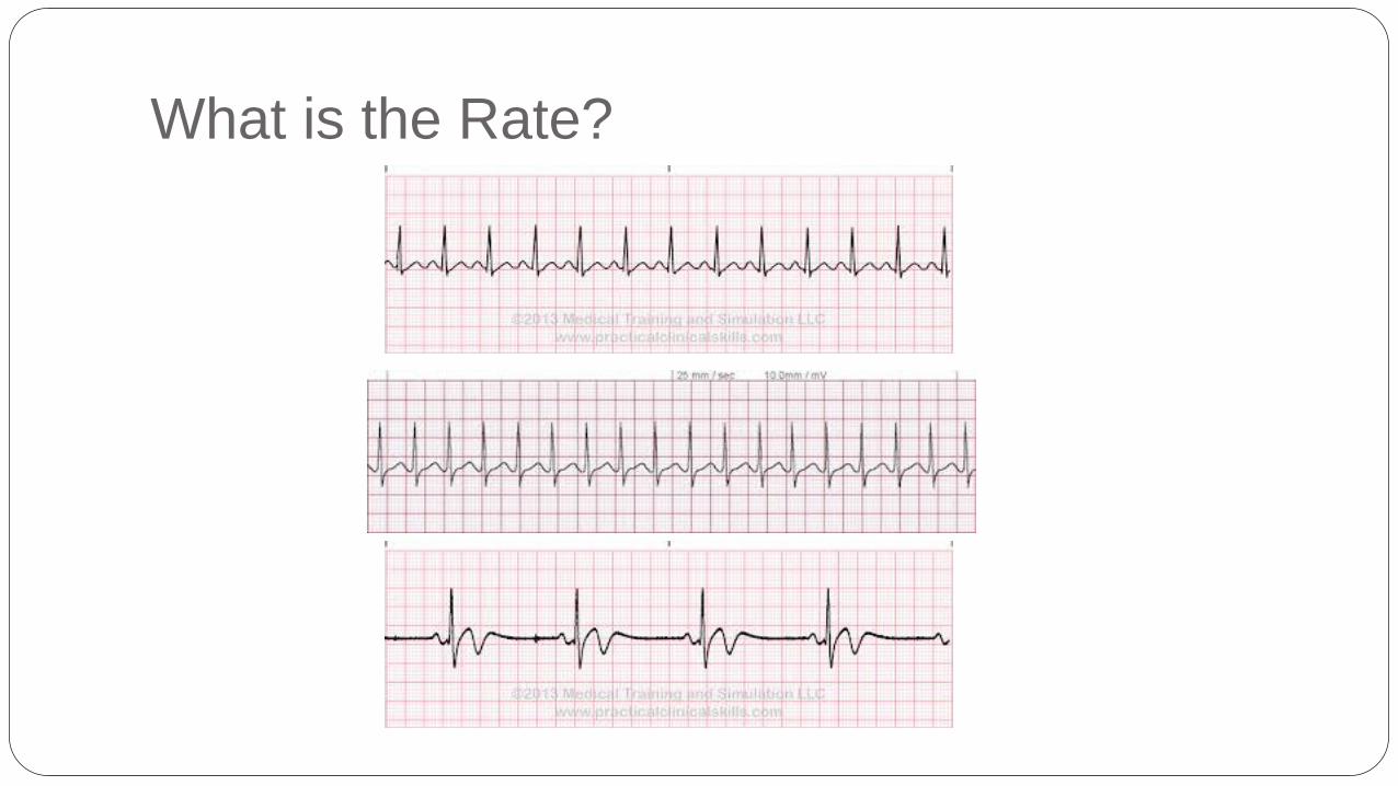
What is the Rate?
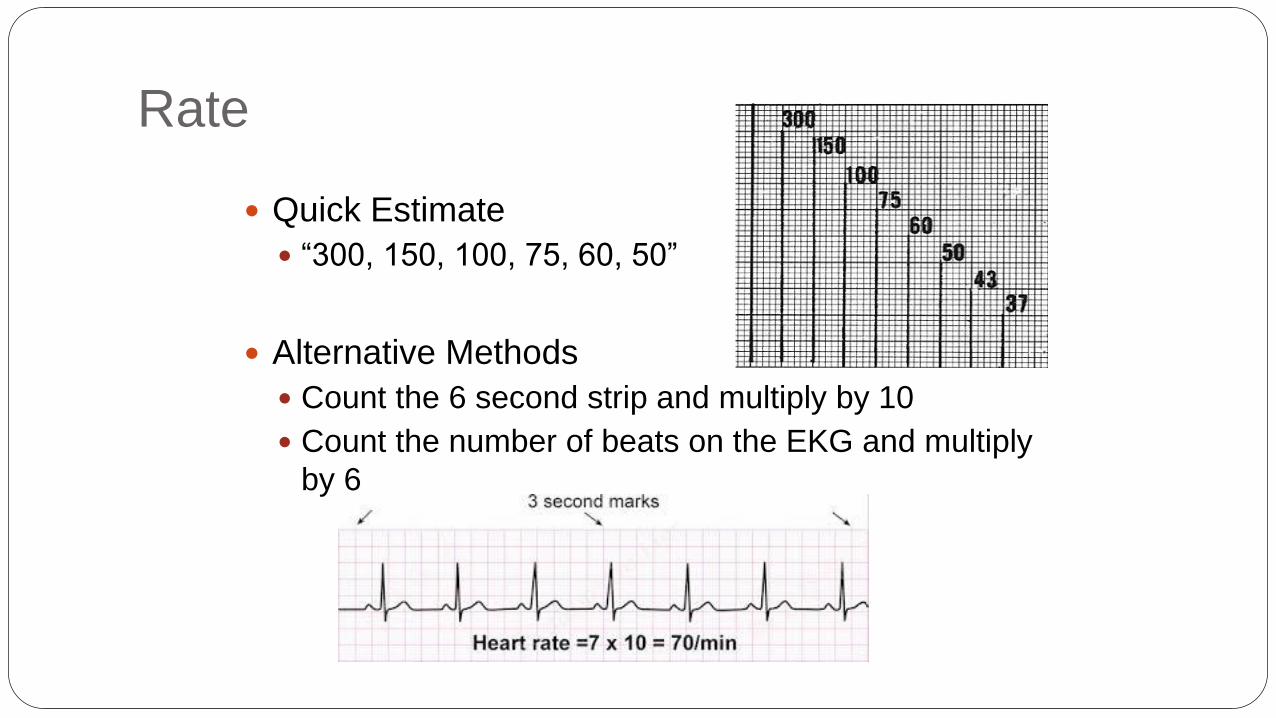
Rate
Quick Estimate
“300, 150, 100, 75, 60, 50”
Alternative Methods
Count the 6 second strip and multiply by 10
Count the number of beats on the EKG and multiply
by 6
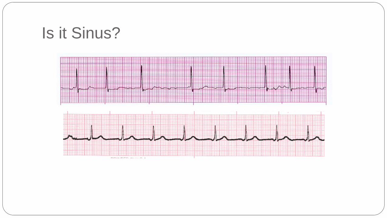
Is it Sinus?

Rhythm
P wave before every QRS?
Every P waves followed by QRS?
Regular Vs Irregular?
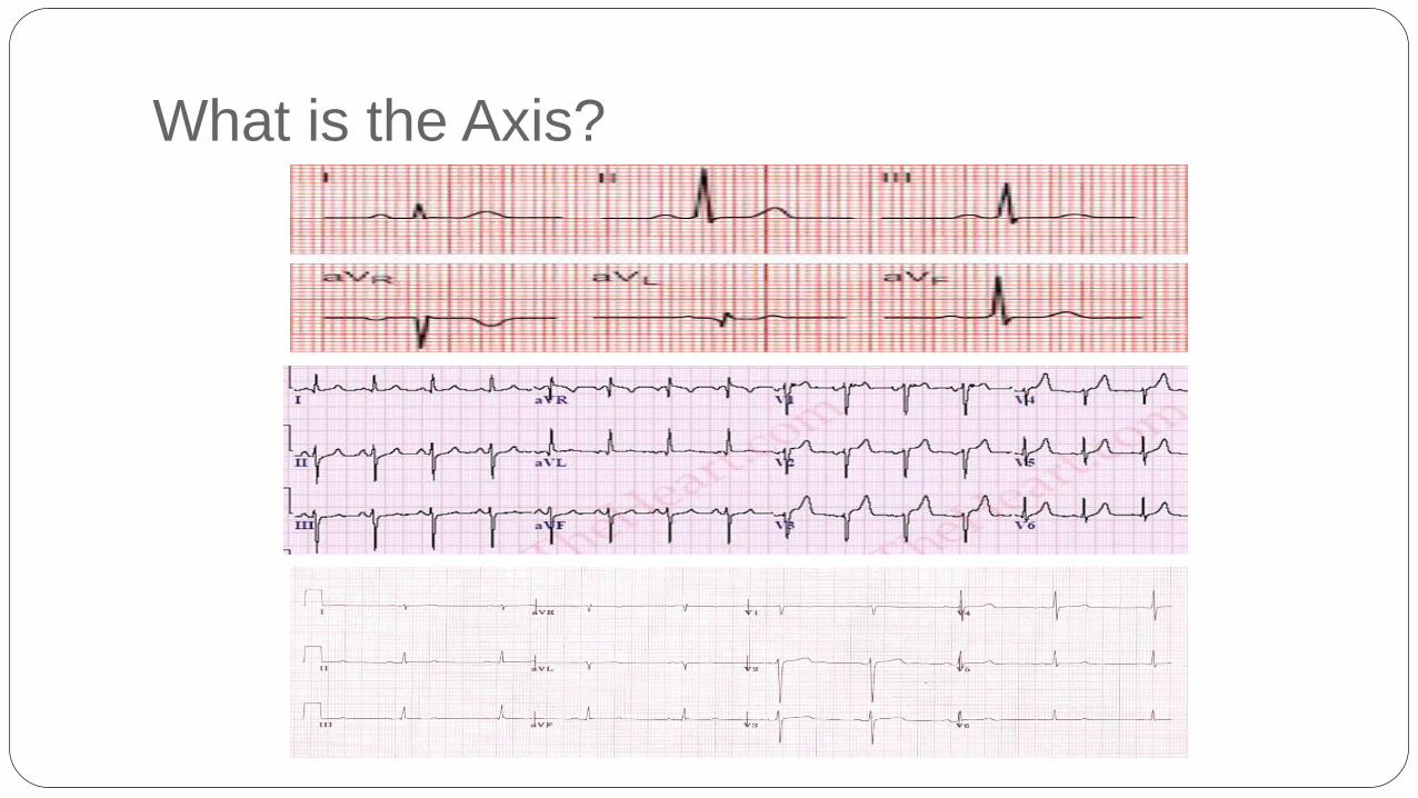
What is the Axis?
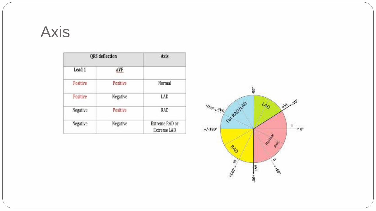
Axis


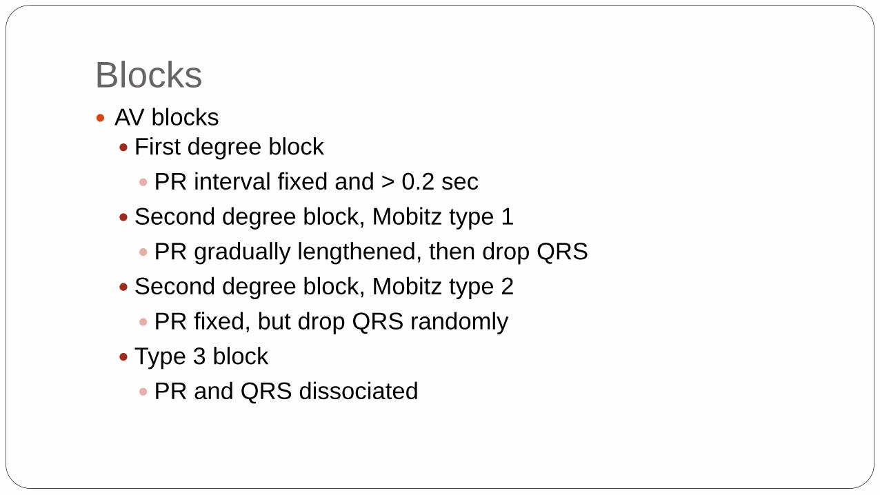
Blocks AV blocks
First degree block
PR interval fixed and > 0.2 sec
Second degree block, Mobitz type 1
PR gradually lengthened, then drop QRS
Second degree block, Mobitz type 2
PR fixed, but drop QRS randomly
Type 3 block
PR and QRS dissociated
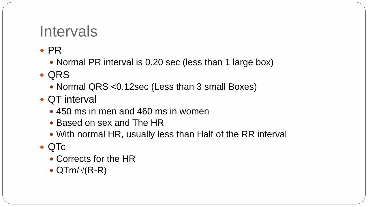
Intervals PR
Normal PR interval is 0.20 sec (less than 1 large box)
QRS Normal QRS <0.12sec (Less than 3 small Boxes)
QT interval 450 ms in men and 460 ms in women
Based on sex and The HR
With normal HR, usually less than Half of the RR interval
QTc Corrects for the HR
QTm/√(R-R)

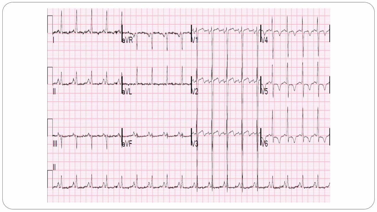
17 year old female found by her room mate
unconscious
Wissam Alajaji, Electrocardiogram Interpretation: A Brief Overview, July-21, 2015.

Causes of QT prolongation
Drugs (Na channel blockers), Antipsychotics
Hypocalcemia
Hypomagnesemia
Hypokalemia
Hypothermia
AMI
Congenital
Increased ICP

P Wave
Upright In Lead II Sinus rhythm
The P wave can also help with atrial enlargement
L Atrial Enlargement
Lead II: Bifid P wave with total P wave duration of >110ms
Lead V1: Biphasic P wave with terminal negative portion > 1mm deep
R Atrial Enlargement
Lead II: Peaked P waves >2.5mm
Lead I: Peaked P wave >1.5mm

QRS Complex
Dr. Mohan’s 4 things to look for in a QRS complex
Amplitude (Helps with LVH)
Duration (Bundle Branch)
Q waves (Old MIs)
R wave progression

Amplitude Add the larger S wave of V1 or V2 in mm, to the larger R wave of
V5 or V6.
Sum is > 35mm = LVH

Duration
Normal Duration <0.12 sec
If prolonged, have to think about RBBB or LBBB
LBBB
Dominant S wave in V1 and Broad monophasic R wave in lateral leads (I, aVL, V5-
V6)
RBBB
RSR’ pattern in V1-3 (‘M-shaped’ QRS complex) and Wide, slurred S wave in the
lateral leads (I, aVL, V5-6)

R wave Progression Usual Transition between V3-V4
Early Progression 3 major causes: RBBB, RVH and Posterior MI

R Wave progression Late R wave Progression
3 Major causes: LVH, LBBB and Anterior MI

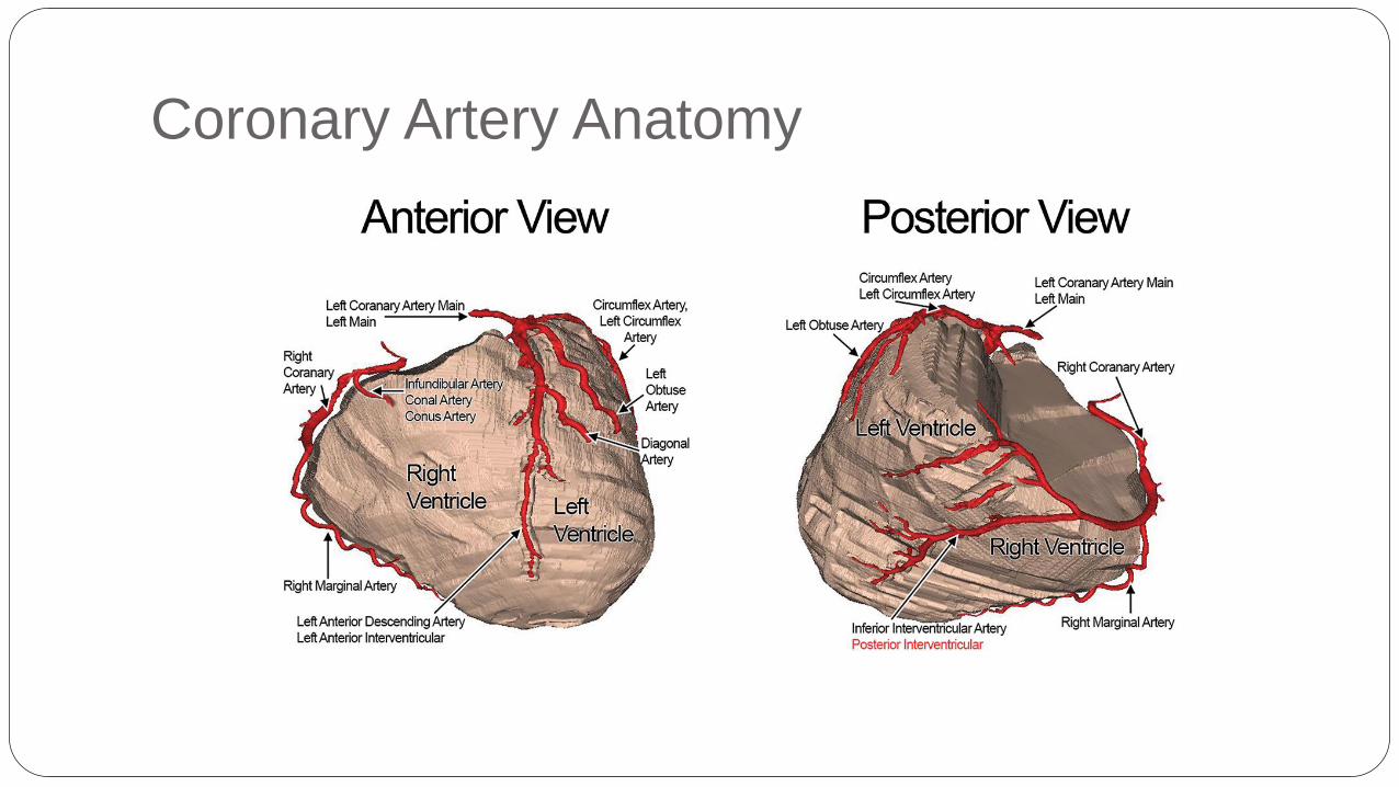
Coronary Artery Anatomy
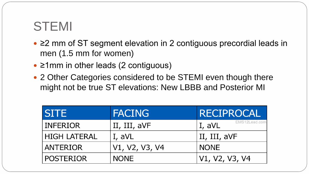
STEMI
≥2 mm of ST segment elevation in 2 contiguous precordial leads in
men (1.5 mm for women)
≥1mm in other leads (2 contiguous)
2 Other Categories considered to be STEMI even though there
might not be true ST elevations: New LBBB and Posterior MI

ST Depression ST depression can be either upsloping, downsloping, or horizontal (see diagram below).
Horizontal or downsloping ST depression ≥ 0.5 mm at the J-point in ≥ 2 contiguous
leads indicates myocardial ischemia.
ST depression ≥ 1 mm is more specific and conveys a worse prognosis.
ST depression ≥ 2 mm in ≥ 3 leads is associated with a high probability of NSTEMI and
predicts significant mortality (35% mortality at 30 days).
Upsloping ST depression is non-specific for myocardial ischemia.
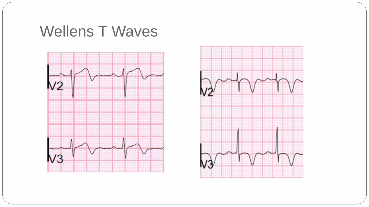
Wellens T Waves

T wave Inversion
At least 1 mm deep
Present in ≥ 2 continuous leads that have dominant R waves (R/S
ratio > 1)
Dynamic — not present on old ECG or changing over time
Wellens’ syndrome is a pattern of inverted or biphasic T waves in
V2-4 (in patients presenting with ischemic chest pain) that is highly
specific for critical Stenosis of the left anterior descending artery.

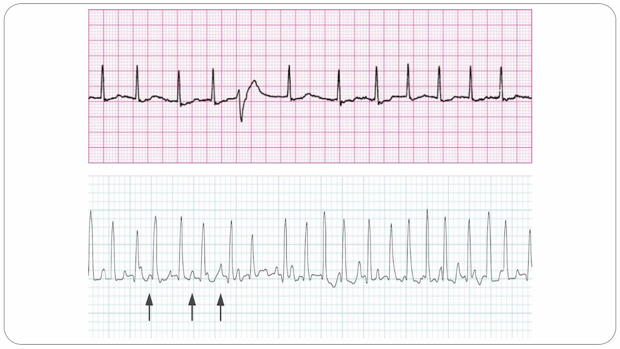
Tachycardia
Narrow Complex Vs. Wide Complex
Regular Vs. Irregular
Regular Narrow Complex Tachycardia: Sinus Tachycardia, Atrial
Tachycardia, A flutter, SVT, AVNRT
Irregular Narrow Complex Tachycardia: A Fib, A flutter with Variable
Block, MAT etc
Regular Wide complex Tachycardia: VT, V-Flutter, Tachycardia with
aberrancy, Hyperkalemia
Irregular Wide Complex Tachycardia: Torsades, V-Fib Etc.


Bradycardia
Narrow Vs Wide Complex
Regular Vs. Irregular
Regular narrow complex bradycardia: Sinus, Junctional, Complete
AV block (junctional escape), A-flutter with high degree block.
Irregular narrow complex bradycardia: Sinus, A-fib with slow
ventricular response, A-flutter with variable block, Type I and Type II
second degree block.
Regular wide complex bradycardia: Idioventricular rhythm,
Complete AV block (ventricular escape), Regular bradycardias with
aberrancy or bundle branch block
Irregular wide complex bradycardia: Type 1 and type 2 second
degree blocks, Irregular bradycardias with bundle branch block.

Hyperkalemia
> 5.5 mEq/L is associated with repolarization abnormalities
Peaked T waves
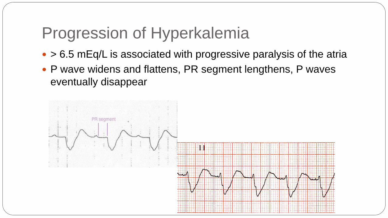
Progression of Hyperkalemia
> 6.5 mEq/L is associated with progressive paralysis of the atria
P wave widens and flattens, PR segment lengthens, P waves
eventually disappear

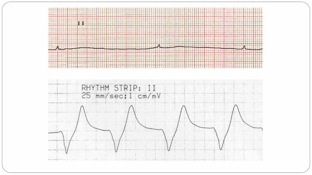
Hyperkalemia Continued
> 7.0 mEq/L is associated with conduction abnormalities and
bradycardia.
Prolonged QRS interval with bizarre QRS morphology, High-grade
AV block with slow junctional and ventricular escape rhythm, Any
kind of conduction block (bundle branch blocks, fascicular blocks),
Sinus bradycardia or slow AF, Development of a sine wave
appearance (a pre-terminal rhythm)

Hyperkalemia Continued
> 9.0 mEq/L causes cardiac arrest.
Asystole
Ventricular fibrillation
PEA with bizarre, wide complex rhythm

Wissam Alajaji, Electrocardiogram Interpretation: A Brief Overview, July-21, 2015.
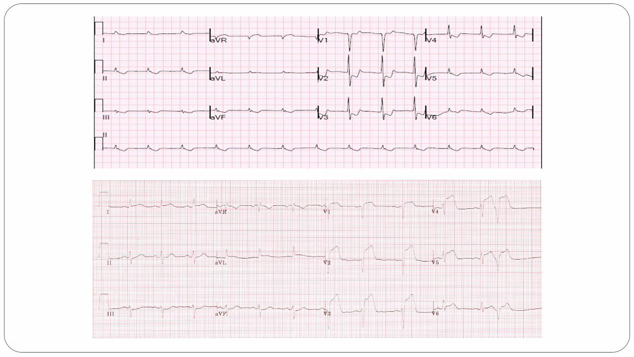
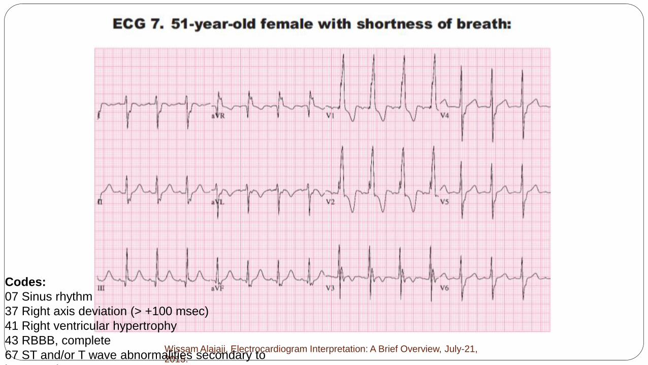
Codes:
07 Sinus rhythm
37 Right axis deviation (> +100 msec)
41 Right ventricular hypertrophy
43 RBBB, complete
67 ST and/or T wave abnormalities secondary to
hypertrophy

Objectives
Approach to reading an EKG
Myocardial Ischemia
Blocks
Tachyarrhythmia and Bradyarrhythmia
Other Miscellaneous EKGs


