Hindawi Publishing CorporationJournal of NanomaterialsVolume 2011, Article ID 348268, 15 pagesdoi:10.1155/2011/348268
Research Article
Electrospun Collagen: A Tissue Engineering Scaffold withUnique Functional Properties in a Wide Variety of Applications
Balendu Shekhar Jha,1 Chantal E. Ayres,2 James R. Bowman,3 Todd A. Telemeco,4
Scott A. Sell,5 Gary L. Bowlin,2 and David G. Simpson1
1 Department of Anatomy and Neurobiology, Virginia Commonwealth University, Richmond, VA 23298, USA2 Department of Biomedical Engineering, Virginia Commonwealth University, Richmond, VA 23298, USA3 School of Medicine, Virginia Commonwealth University, Richmond, VA 23298, USA4 Division of Physical Therapy, Shenandoah University, Winchester, VA 22601, USA5 Physical Medicine and Rehabilitation Service, Hunter Holmes McGuire VA Medical Center, Richmond, VA 23249, USA
Correspondence should be addressed to David G. Simpson, [email protected]
Type I collagen and gelatin, a derivative of Type I collagen that has been denatured, can each be electrospun into tissue engineeringscaffolds composed of nano- to micron-scale diameter fibers. We characterize the biological activity of these materials in avariety of tissue engineering applications, including endothelial cell-scaffold interactions, the onset of bone mineralization,dermal reconstruction, and the fabrication of skeletal muscle prosthetics. Electrospun collgen (esC) consistently exhibited uniquebiological properties in these functional assays. Even though gelatin can be spun into fibrillar scaffolds that resemble scaffolds ofesC, our assays reveal that electrospun gelatin (esG) lacks intact α chains and is composed of proinflammatory peptide fragments.In contrast, esC retains intact α chains and is enriched in the α 2(I) subunit. The distinct fundamental properties of the constituentsubunits that make up esC and esG appear to define their biological and functional properties.
1. Introduction
Electrospinning has been used to fabricate a variety of poly-mers, including natural proteins [1–3], sugars [4], syntheticpolymers [5, 6], and blends of native and synthetic polymers[7–9] into tissue engineering scaffolds composed of nano- tomicron-scale diameter fibers, a size-scale that approaches thefiber diameters observed in the native extracellular matrix(ECM). The physical, biochemical, and biological propertiesof these unique biomaterials can be regulated at severalsites in the electrospinning process. As this technology hasmatured, it has become apparent that many electrospunnanomaterials exhibit unusual, and often surprising, prop-erties.
For many polymers, physical properties, including fiberdiameter, pore dimension, and degree of scaffold anisotropy,can be regulated by controlling the composition of the elec-trospinning solvent, the air gap distance, accelerating voltage,
mandrel properties, and/or the identity, concentration, anddegree of chain entanglements (viscosity) present in thestarting solutions [10–12]. The ability to directly manipulatethese fundamental variables can have a dramatic impacton the structural and functional properties of electrospunmaterials. This is especially true when considering nativeproteins and blends of synthetic polymers and nativeproteins.
Collagen represents the most abundant protein of themammalian ECM. As such, this natural polymer has longbeen used as a biomaterial in a variety of tissue engineeringapplications. This crucial ECM protein, as well as a varietyof other native proteins, can be electrospun into fibers thatresemble the native state [1]. Not surprisingly, the fibers ofelectrospun collagen do not appear to fully reconstitute thestructural or mechanical properties of the parent material[12]. Simultaneously, it is unclear to what extent theelectrospun analog “must” recapitulate the native material
2 Journal of Nanomaterials
to be a functional tissue engineering scaffold. The nature ofthe electrospun collagen fiber is the subject of debate andthere are conflicting reports in the literature concerningits structural and functional properties [7, 12–15]. In thisstudy, we compare and contrast the functional characteristicsof electrospun collagen and electrospun gelatin (denaturedcollagen) in a variety of tissue engineering applications. Wethen explore how the procedures used to isolate and preparecollagen for the electrospinning process might ultimatelyimpact its functional profile once it has been processedinto a tissue engineering scaffold. We believe that it isessential to develop a more complete functional map of thesenovel materials to fully exploit them in the development ofclinically relevant products.
2. Materials and Methods
2.1. Collagen Preparation. Collagen was isolated at 4◦C. Calf-skin corium (Lampire Biologics, Pipersville, PA) was cut into1 mm2 blocks and stirred for 24 hr in acetic acid (0.5 M),processed in a blender into a slurry, and stirred for anadditional 24 hr. Solutions were filtered through cheesecloth,centrifuged at 10,000×G for 12 hr; supernatant was recov-ered and dialyzed against ice cold, ultra pure 18 MΩ-cmwater. Collagen isolates were frozen and lyophilized. Bovinegelatin Type B isolated from skin was purchased from Sigma(75 or 225 bloom).
2.2. Electrospinning: Collagen and Gelatin. Materials werepurchased through Sigma unless noted. Lyophilized col-lagen (at 55 mg/mL) and gelatin (Sigma, 225 bloomat 110 mg/mL) were solubilized for 12 hr in 1,1,1,3,3,3-hex-afluoro-2-propanol (HFP) and electrospun [1, 7, 10]. Con-ditions were adjusted to produce scaffolds composed offiber diameters that were nominally 1 μm in cross-sectionaldiameter. Solutions were charged to 22 kV and delivered(3–7 mL/hr) across a 25 cm air gap. Electrospun samples,designated “recovered” electrospun collagen (rEC) or “recov-ered” electrospun gelatin (rEG) were produced by dissolv-ing uncross-linked electrospun scaffolds immediately afterspinning in ice cold, 18 MΩ-cm water; the final protein con-centration was adjusted to 1.5 mg/mL. Collagen and gelatinstarting electrospinning concentrations were manipulatedto produce fibers of varying diameters. Where indicated,scaffolds were vapor cross-linked (1–12 hrs) in glutaralde-hyde, blocked in 0.1 M glycine, rinsed in PBS, and disin-fected in 70% alcohol prior to culture experimentation orimplantation.
2.3. Cell Culture: Endothelial Cells. Electrospun scaffoldswere cut into 12 mm diameter circular disks and cross-linked. A sterile 6 mm diameter glass cloning ring was placedon top of each disk and supplemented with 3,000 adulthuman microvascular endothelial cells (Invitrogen, C-011-5C) in a total volume of 100 μL. After 20 min the culturedishes were flooded with media to ensure that the cellswere immersed. Cloning rings were removed after 24 hr ofculture.
2.4. Cell Culture: Osteoblasts. Type I collagen and gelatinwere electrospun across a 25 cm gap and directed at agrounded 6 in diameter circular steel plate. Tissue culturedishes were placed between the source electrospinningsolutions and the grounded target to directly collect fiberson the culture surfaces. After cross-linking, equal numbersof osteoblasts (Clonetics, CC-2538) were plated onto eachsurface and cultured for 10 days in OBM basal media(CC-3208). As controls, cells were plated onto native tissueculture plastic or random gels composed of Type I collagen(Vitrogen: Cohesion Technologies), Simpson et al. [16].For SEM imaging, osteoblasts were cultured directly on6 mm diameter× 500 μm thick circular disks of electrospuncollagen or gelatin (conditions optimized for 1 μm diameterfibers).
2.5. Dermal Reconstruction. Adult guinea pigs were broughtto a surgical plane, fur was shaved and skin swabbed in beta-dine. Four 1 cm2 full-thickness dermal injuries (completeremoval of the dermis and hypodermis and bordered by thesuperficial fascia of the panniculus adiposus) were preparedon the dorsum of each animal. Injuries were treated withscaffolds composed of electrospun Type I collagen or gelatin(electrospinning conditions adjusted to produce scaffoldscomposed of fibers ranging from 250 nm to >2000 nmin average cross-sectional diameter). Scaffolds were vaporcross-linked to varying degrees. Each wound was treatedwith a candidate scaffold and covered with a piece of silvergauze that was sutured in place. Silver gauze remainedin place for 5–7 days. Animals recovered on a warmingpad and were provided with pain mitigation. Injuries werephotographed at intervals. Data on wound closure wasexpressed as the percent injury surface area observed at thetime of implantation. Representative samples were recoveredfor histological evaluation.
2.6. Muscle Fabrication. Three-day-old neonatal rats weredecapitated, skin was removed. Skeletal muscle was removed,minced into 1 mm2 pieces in sterile PBS and rinsed until clearof blood. Tissue was incubated in a sterile flask supplementedwith 0.25% trypsin (Invitrogen) in a shaking (100 RPM)37◦C water bath. At 10 min intervals, tissue was cannulatedand allowed to settle, and supernatant was removed andcentrifuged at 800×G for 6 min. Cell pellets were pooledin DMEM plus 10% FBS and 1.2% Antibiotic/Antimycotic(Invitrogen 15240). A 60 minute interval of differentialadhesion to tissue culture plastic was used to reducefibroblast contamination. Myoblasts were cultured for 3–5days under conditions that minimized cell to cell contacts. Incell labeling assays, myoblast cultures were incubated in DiO(Invitrogen, L-7781) overnight according to manufacturer’srecommendations.
Electrospun scaffolds were prepared on a 4 mm diameterround mandrel. With conditions optimized to produce 1 μmdiameter fibers, cylindrical constructs were fabricated witha wall thickness of 200–400μm [7]. Scaffolds were cross-linked. Myoblasts were recovered from culture and rinsed2x in PBS by centrifugation (800×G, 6 min). Electrospun
Journal of Nanomaterials 3
cylinders were sutured shut and suspended myoblasts wereinjected into the lumen of the constructs. Adult 150–180 gmSprague Dawley rats were brought to a surgical plane. Fur onthe hindlimb was shaved and skin was swabbed in betadine.In short-term studies (3 wks), a 4 mm× 15 mm long cylindersupplemented with cells was inserted directly into a channel(“intramuscular” position) prepared in the vastus lateralismuscle after the methods of Telemeco et al. [7]. In long termstudies, a hemostat was passed deep to the quadriceps musclegroup; engineered tissue (4 mm × 40 mm) was passed underthe existing muscle mass and sutured (in an extramuscularposition) to the proximal and distal tendons of origin andinsertion for the quadriceps. Incisions were repaired, skinwas stapled, and animals recovered on a warming pad.
2.7. Electrospinning: Nylon. To separate the fiber-formingproperties of the different protein fractions from theirfundamental biological properties, we applied collagen andgelatin fractions to electrospun scaffolds composed of nylon66 (Ambion). Electrospun nylon has a high surface areaand exhibits high protein binding capacity. Nylon was spunafter the methods of Manis et al. [17]. Conditions wereoptimized to produce charged nylon fibers ranging 1.0–1.5 μm in diameter.
2.8. Cell Culture: Adult Human Dermal Fibroblasts. Dermalfibroblasts (HDF), (Cascade Biologics: C-013-5C) werepassaged 3–5 times in basal dermal fibroblast medium106 supplemented with a low serum growth kit (CascadeBiologics, S-003-K) prior to experimentation.
2.9. Cell Adhesion Assays. Electrospun nylon scaffolds wereimmersed in 20% methanol/phosphate buffered saline (PBS)[17], rinsed 3x in PBS and installed in a dot blotter manifold(Topac Model DHM-48). Wells were supplemented with50 μL of collagen (control samples or fractions thermallydenatured at 50, 60, 70, 80 or 90◦C for 1 hr) or gelatinfractions containing equal amounts of protein. After 5 min,solutions were sucked through the membrane using avacuum pump. Scaffolds coated with 1% bovine serumalbumin (BSA) were used as controls. All wells were blockedwith 100 μL of 1% BSA solution for 5 min and rinsed in PBSprior to use. In each assay, 3000 HDFs were suspended in100 μL of media and applied to each surface for 1 hr at 37◦C(dot blotter was used as a culture vessel; no vacuum wasapplied to the cell suspensions).
After the plating interval, the dot blotter was invertedto remove nonadherent cells, scaffolds were removed, rinsedin PBS, and fixed in ice cold methanol (20 min). Foranalysis, scaffolds were rinsed 5x in PBS plus 0.5% Triton,and incubated overnight at 4◦C in primary goat antirabbitGAPDH antibody (Sigma # G9545, 1 : 5000). All antibodieswere diluted in LiCor Odyssey Blocking Buffer (L-OiBB),and LOiBB plus 0.1% Tween-20 was used in all rinses.Cultures were rinsed 5x in L-OiBB, counter-stained with goatantirabbit IRDye 800 secondary antibody (LiCor 1 : 1000)for 1 hr and rinsed 5x. Data sets were captured at a lineresolution of 169 μm using a Li-Cor Odyssey Infrared Imager.
Adhesion was expressed as “Integrated Intensity” (signal-mm2). Integrated intensity values were extrapolated to cellnumber using a standard curve of cells plated in parallel withthe unknowns. Data sets were screened by one-way ANOVA(P < .01), Dunn’s Method (P < .05), and a Mann-WhitneyRank Sum test (P < .001) was used in post hoc analysis.In cyclic RGD competition experiments, HDFs (10,000 cellsper treatment) were incubated for 15 min at 37◦C with 0.01,0.1, or 1 μg/mL cyclic RGD peptide (Bachem, H-2574) or1 μg/mL control RGD peptide (Bachem, H-4088). The cellswere then plated for 1 hr on the different surfaces and wereprocessed to image GAPDH as described.
2.10. Alpha Chain Analysis. Collagen samples were diluted to0.15 mg protein/mL in Laemmli sample buffer and separatedby SDS interrupted gel electrophoresis using 10% poly-acrylamide gels. Gels were run until the dye front reachedthe base of the stacking gel, 1 mL of Laemmli buffer sup-plemented with 20% β-mercaptoethanol was added to thegel stacker and incubated for 30 min at room temperature.The separation run was then completed. Gels were stainedwith Coomassie brilliant blue overnight, de-stained andphotographed. Densitometric analysis was conducted withNIH ImageJ software.
2.11. Cross-Linking Assays. See Newton et al. [12] for detailsof this assay. Percent cross-linking was calculated from theformula
% cross-linked = 1− Absc/massc
Absnc/massnc, (1)
where Absc = absorbance of the controls at 345 nm; the unitof mass is given in mg. Absnc = absorbance of the unknownsat 345 nm; again the unit of mass is given in mg. All data isexpressed as percent of cross-linking observed in electrospunscaffolds (controls) that have not been exposed to cross-linking reagents.
2.12. Scanning Electron Microscopy. Samples were sputter-coated and imaged with a Zeiss EVO 50 XVP scanningelectron microscope (SEM) equipped with digital imageacquisition. Average fiber diameter and pore area data wasdetermined from representative samples using NIH Image-Tool (UTHSCSA version 3). All fiber diameter measurementswere taken perpendicular to the long axis of electrospunfibers [10, 11].
2.13. Transmission Electron Microscopy. Samples were im-mersed in 2% glutaraldehyde for 12 hr at 4◦C and postfixedin 1.0% osmium plus or minus 2.5% potassium ferricyanide[6, 7]. All samples were subjected to a graded series ofdehydration and embedded in Poly/Bed (Polysciences).
3. Results
3.1. Functional Performance of Electrospun Collagen. To com-pare and contrast the biological properties of electrospun
4 Journal of Nanomaterials
collagen and electrospun gelatin, we conducted a series of invitro and in vivo functional assays.
3.1.1. Endothelial Cell Growth. Critical to the bioengineeringparadigm is the development of tissue engineering scaffoldsthat can support the proliferation and penetration of vascu-lar elements. To evaluate this characteristic in vitro, we platedmicrovascular endothelial cells onto electrospun scaffolds ofType I collagen and electrospun gelatin composed of varyingfiber diameters. During the initial plating phase, and overtime in culture, cell shape, on both surfaces (collagen andgelatin), was modulated by the fiber size (and likely the porecharacteristics that “travel” with the fiber size that is presentin an electrospun scaffolds [10]) (Figure 1). Electrospunscaffolds of collagen and gelatin composed of small diameterfibers induced the expression of a highly flattened andstellate cell shape. This cell shape was retained through-out the culture interval on both surfaces (e.g., Figure 1compare (a) = day 1 with (b) = day 7 as well as (i) and(j)). With increasing fiber diameter, the cells assumed amore rounded and elongated phenotype. After 10 days,microvascular endothelial cells cultured on collagen- orgelatin-based scaffolds with average cross-sectional fiberdiameter less than about 1.0–1.50μm remained on the dorsalsurfaces of the constructs (Figure 1 (q), (r), (u) and (v)).As fiber size exceeded this threshold value and pore sizeincreased to about 10,000 nm2, the cells began to penetrateinto the scaffolds (Figure 1 (s), (t), (w) and (x)). These resultssuggest that the physical arrangement of fibers (i.e., the porecharacteristics) plays a role in regulating the infiltration ofendothelial cells into an electrospun scaffold.
3.1.2. Osteoblast Differentiation. The 67 nm banding patterntypical of native Type I collagen is associated with theformation of nucleation and binding sites critical to themineralization process in bone [18]. Superficially, electro-spun fibers of collagen exhibit a similar structural motif. Toexamine the potential functional consequences of this motif,we plated osteoblasts onto surfaces coated with electrospuncollagen and electrospun gelatin. Cultures plated onto sur-faces coated with electrospun collagen exhibited low rates ofproliferation and failed to form a confluent cell layer, evenafter 8 days. Phase bright crystals were present throughoutthese cultures (Figure 2). Cells plated onto surfaces coatedwith electrospun gelatin, Type I collagen gels, or nativetissue culture plastic proliferated and formed a confluent celllayer over this same culture interval. Phase bright crystalswere infrequently observed in any of these cultures. Thesedata suggest electrospun collagen contains structural motifsnecessary and sufficient to induce osteoblast differentiationand subsequent formation of hydroxyapatite crystals.
3.1.3. Dermal Reconstruction. From an architectural stand-point, tissue engineering electrospun scaffolds are theoreti-cally very well suited for applications in dermal reconstruc-tion. These constructs are deposited as nonwoven, fibrillarstructures that exhibit an extensive void volume and poresthat are completely interconnected with one another. To
evaluate the efficacy of using electrospun collagen as a dermaltemplate, we treated full thickness dermal injuries withvarious permutations of this material. To track the healingprocess, we measured total wound surface area as a functionof treatment and time. We have used this metric becauseinterventions that reduce wound contraction (as measuredby an increased retention of wound surface area once theinjury is healed) are associated with less scarring and morecomplete tissue regeneration [19].
In the first series of experiments, we treated wounds(1 cm2) with dermal templates fabricated from electrospuncollagen under conditions that produced fibers ranging 750–1,000 nm. These scaffolds were postprocessed to cross-linkapproximately 50% of the available sites. Wound closuretook place in 16 days with these constructs (Figure 3(a)).When the extent of cross-linking was increased to 70% inthese constructs, wound healing was delayed modestly andwound surface area was dramatically increased, a featureindicative of increased regeneration (Figures 3(a) and 3(c)).Histological examination of the tissue reconstituted withtemplates composed of electrospun collagen consistentlyrevealed a smooth continuum of infiltrating cells. There wereno overt signs of inflammation or fibrosis along the interfaceof the implanted templates and the adjacent uninjuredtissue, regardless of the fiber diameter composition presentin electrospun constructs of Type I collagen (Figures 4(a)–4(c)). Our tissue culture experiments demonstrated thatthe intrinsic architectural features present in an electrospunscaffold can modulate the extent to which endothelial cellscan penetrate into these constructs. We observed a similartrend in our dermal reconstruction experiments. Injuriestreated with templates composed of fibers less than 500 nmin diameter were less densely populated than templatescomposed of fibers greater than 750–1000 nm in diameter(Figure 4).
In parallel experiments conducted with a wide varietyof electrospun scaffolds composed of gelatin (average fiberdiameters ranging from 250 nm to approximately 3000 nm),we were unable to replicate these results. Wounds treatedwith electrospun gelatin consistently healed in the classic Xshaped configuration (Figure 3(d)) that develops in responseto wound contraction, this feature developed regardless ofthe fiber diameter or the degree of cross-linking presentin the gelatin-based scaffolds. These scaffolds were consis-tently infiltrated by foreign body giant cells, indicating aninflammatory response to the implanted electrospun gelatin(Figures 4(d)–4(f)). These data indicate that architecturalfeatures and the biochemical identity of an electrospunscaffold composed of collagen interact to define its uniquefunctional characteristics.
3.1.4. Muscle Engineering. To evaluate the efficacy of usingelectrospun collagen in a cell-based tissue engineering appli-cation, we used an in situ strategy to fabricate skeletal muscleprosthetics. In these experiments we electrospun Type I col-lagen onto a rotating 4 mm diameter mandrel; this resultedin the formation of a hollow cylinder with walls composed of1 μm diameter fibers of electrospun collagen. These cylinders
Journal of Nanomaterials 5
(s) (t) (w) (x)
(q) (r) (u) (v)
(g) (h) (o) (p)
(e) (f) (m) (n)
(c) (d) (k) (l)
(a) (b) (i) (j)
Figure 1: Endothelial interactions with electrospun collagen ((a)–(h)) and gelatin ((i)–(p)). Endothelial cell shape varied as a function ofincreasing fiber diameter on both electrospun collagen (Day 1: (a), (c), (e), (g) & Day 7: (b), (d), (f), (h)) and electrospun gelatin (Day 1:(i), (k), (m), (o) & Day 7: (j), (l), (n), (p)). Cell shape established during the early stages of plating persisted throughout the entire cultureinterval (e.g., for each scaffold cells at day 1 appeared to exhibit a similar cell shape after 7 days of culture). Cells expressed and retained ahighly flattened and stellate shape when plated onto scaffolds composed of fibers less than 1,500 nm ((a)–(l)). At larger fiber sizes the cellsexhibited a more elongated phenotype, this was especially evident on the collagen-based scaffolds ((e), (f), (g) and (h)). Penetration into thescaffolds was primarily regulated by average fiber diameter and pore size. TEMs of cross-sectional images of cells plated onto electrospuncollagen ((q)–(t)) and electrospun gelatin ((u)–(x)) for 10 days. Average fiber diameters for collagen (a) & (q) = 449 ± 122 nm, (c) & (r) =1,187± 297 nm, (e) & (s) = 1,886± 513 nm and (g) & (t) = 2,756± 855 nm. In gelatin (i) & (u) = 198± 50 nm, (k) & (v) = 491± 114 nm, (m)& (w) = 1,252 ± 302 nm, and (o) & (x) = 1,619 ± 414 nm (all fiber measurements from dry scaffolds prior to processing for cross-linking).Note that penetration was not evident until a nominal average fiber diameter of about 1,800 nm was achieved in the scaffolds (arrows in (r),(t), (x) indicate fibers in cross section). Scale bar in (a) = 20 μm.
6 Journal of Nanomaterials
(a) (b) (c) (d)
(e) (f) (g) (h)
1370 15 kV x2,500 10 μm WD13
(i)
1362 15 kV x1,000 10 μm WD13
(j)
1363 15 kV x3,000 10 μm WD13
(k)
Figure 2: Osteoblast interactions with electrospun collagen and electrospun gelatin. Human osteoblasts plated for 1 (a)–(d) or 8 days(e)–(h). Cells plated on electrospun collagen ((a), (e)), electrospun gelatin ((b), (f)), collagen gel ((c), (g)), or tissue culture plastic ((d),(h)). Cells plated onto electrospun collagen remain subconfluent after 8 days of culture (e) and accumulated phase bright crystals. At theultrastructural level, osteoblasts plated onto electrospun gelatin expressed a rounded cell shape (i); when plated onto electrospun collagen,the cells were covered with elaborate arrays of plate-like structures typical of hydroxyapatite crystals ((j), (k)).
were then supplemented with myoblasts and implanteddirectly into the vastus lateralis muscle. After 3 weeks, tissuefabricated with electrospun collagen was densely populatedwith cells. Nascent myotubes and functional blood vesselswere evident throughout these implants (Figure 5). As withthe dermal-templates, we observed a smooth continuumbetween the surrounding tissue and the engineered musclewith no evidence of fibrosis. In contrast to these results,tissue fabricated with gelatin based materials was necrotic,exhibited extensive fibrosis at the tissue interface and amassive infiltration of lymphocytes.
Given these results we next prepared engineered muscletissue fabricated with electrospun collagen and directlysutured the constructs to the tendons of origin and insertionfor the quadriceps muscle. These constructs were placed inan extramuscular position; in effect we are converting thequadriceps muscle into a “quintriceps” muscle. After 8 weeks,the engineered muscle was densely packed with fully dif-ferentiated myotubes that were distributed into stacked andlinear parallel arrays that mimicked native tissue (Figure 6).This developing muscle tissue displayed well-formed myofib-rillar elements. However, a range of cytoskeletal structural
patterns was observed. For example, some areas of the tis-sue displayed loosely packed arrays of myofibrils (Figures6(d) and 6(e)); other domains differentially took up thestains used to enhance contrast for light (Figure 6(g)) andtransmission electron microscopy (Figure 6(h)). We suspectthis differential staining is a reflection of protein density orprotein identity with respect to the myofibrillar subunits.Collagen bundles were evident along the borders of theimplanted tissue.
3.2. Analysis of Collagen Alpha Chain Structure and Function
3.2.1. Protein Analysis. We next conducted experiments tocharacterize how various processing conditions impact acid-soluble collagen and how these manipulations might regulatethe evolution of the functional properties of an electrospunfiber. We first examined the effects of thermal denaturationon Type I collagen. These experiments were conducted toprovide us with a benchmark for the evaluation of collagenstructure and its α chain content in response to varioussteps in the electrospinning process. Collagen was isolatedfrom calfskin corium using classical acid extraction methods,
Figure 3: Dermal Reconstruction. Rates of wound closure in lesions treated with electrospun collagen (a) or electrospun gelatin (b). Notethat increasing the extent of cross-linking has a modest effect on slowing wound closure and greatly increases total wound surface area inanimals treated with electrospun collagen (a) and (c). Rates of wound closure were similar when injuries were treated with a wide varietyof gelatin-based constructs (b) and (d). Panel (c) depicts the typical wound healing course for injuries treated with electrospun collagenin which approximately 70% of the available sites are cross-linked. Note the retention of wound surface area in this example. Panel (d)depicts the typical wound healing course for injuries treated with electrospun gelatin in which approximately 70% of the available sites arecross-linked. Note the classically X-shaped wound typical of a lesion that has undergone contraction.
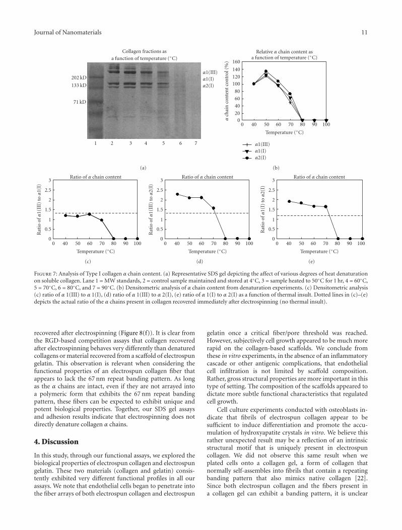
a procedure routinely used to prepare collagen as a biomate-rial and for use in the electrospinning process [1, 7]. Figure 7illustrates an SDS gel depicting the α chain content of theacid soluble parent extract with respect to fractions that havebeen subjected to varying degrees of thermal denaturation.
Protein fractions were held at 4◦C or heated to 50, 60,70, 80, or 90◦C for 1 hr. Detectable changes in the proteinbanding patterns present on the SDS gels were visible inall samples subjected to heating. Densitometric analysisof the separated protein fractions revealed that 50% ofthe collagen α chains were lost within 1 hr when solublecollagen was exposed to 70◦C (Figure 7). Exposure to higher
temperatures accelerated the loss of the collagen α chainsfrom the soluble fractions and resulted in progressively moreα chain fragmentation and smearing in the gel lanes typicalof a sample that has been broken down into a heterogeneousmixture of peptides. At temperatures of 80◦C and greater,the protein bands corresponding to the individual α chainswere completely lost after the 1 hr incubation interval.Commercially procured gelatin (collagen that has beenheated and denatured during isolation) samples exhibitedlittle or no protein banding associated with intact α chainson the SDS gels; these samples ran nearly exclusively as acontinuous smear of proteins (not shown, see [7]).
8 Journal of Nanomaterials
(a) (b)
(c) (d)
(e) (f)
Figure 4: Dermal Reconstruction. Healing response to electrospun collagen (a)–(c) and electrospun gelatin (d)–(f) as a function of fiberdiameter and pore dimension. Collagen-based implants exhibited a smooth continuum of cells at the interface of the lesion and thesurrounding tissue. In each of the images depicted in this figure, native, uninjured tissue appears to the left of each data image (delineated bythe large blue staining collagen bundles in the histological preparations, arrows (b) and (c)). Gelatin based-scaffolds were consistently lessheavily infiltrated and exhibited evidence of an inflammatory response and accumulated foreign body giant cells (arrow head (d)). (a) and(d) average fiber diameter = 240–280 nm, average pore dimension =1500–2000 nm2, (b) and (e) average fiber diameter 500–600 nm, averagepore dimension 3000–5000 nm2, (c) and (f) average fiber diameter 800–1000 nm, average pore dimension 4000–5000 nm.
To examine how the electrospinning process (solvents,electric field, and the flash-lyophilization of proteins thatoccurs during fiber formation) might alter collagen αchain content, we next prepared scaffolds composed ofelectrospun Type I collagen or commercially sourced gelatin(conditions optimized to produce average fiber diameters= 1 μm). As judged by scanning electron microscopy, thesescaffolds were superficially identical. However, transmissionelectrospun microscopy reveals that scaffolds of electrospun
collagen exhibit the 67 nm banding pattern typical of thenative fibril (Figures 8(a)–8(d)). In contrast, fibers of theelectrospun gelatin lack this distinctive structure and arenearly homogenous in appearance.
For protein characterization of the electrospun scaffolds,we redissolved the collagen and gelatin-based scaffolds andseparated the resulting extracts by SDS gel electrophoresis.The protein banding patterns on the gels and the specificcompliment of α chains in the electrospun collagen and
Journal of Nanomaterials 9
M
TZ
(a)
∗
∗
(b)
∗∗
(c)
∗∗
(d)
CC
(e)
CC
FC
(f)
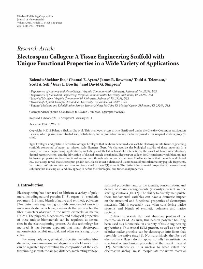
Figure 5: Muscle Fabrication: 3 Weeks. (a) Cross-section through the wall of cylindrical construct placed within the tissue of the rat vastuslateralis. M = endogenous host muscle tissue, TZ = transitional zone occupied by the wall of the electrospun construct. Note the lack of afibrotic capsule. ((b), (c)) Sections taken from within the lumen of the tissue, dense cell populations are evident as are nascent myotubes(∗). Scattered vascular elements are apparent and intermingle with the surrounding cell population. (d) TEM cross-section of developingmyotube with forming myofibrillar elements (arrows). (e) Cell labeling experiments indicate that few cells migrate out of the implantedtissues (arrowhead). The bulk of the labeled cells are retained within the lumen of the electrospun constructs. (f) Tissue fabricated fromgelatin undergoes necrosis and develops a fibrotic capsule (FC). CC = central core of implant.
the control starting material (Acid soluble Type I Collagen:Figure 7) were similar, but not identical. Samples of elec-trospun collagen exhibited subtle derangements in α chaincontent and, as judged by densitometric analysis, it wasenriched in the α2 (I) content (see Figure 7) with respectto the other α chains. Protein separation analysis of theelectrospun gelatin revealed, as expected, nearly completeα chain fragmentation (these samples displayed completefragmentation even prior to the electrospinning process) and
was comparable to the calfskin collagen samples heated to90◦C for 1 hr (Figure 7).
3.2.2. Alpha Chain Functional Properties. Next we examinedhow thermal manipulation impacted the adhesion propertiesof acid-soluble collagen and its electrospun variants. Inthese experiments, we coated electrospun fibers of chargednylon with equal amounts of the various protein fractionsdescribed in Figure 7. This strategy made it possible to
10 Journal of Nanomaterials
(a) (b) (c)
(d) (e) (f)
(g) (h) (i)
C
B
∗ ∗
∗
∗
∗ ∗∗
Figure 6: Muscle Fabrication: 8 Weeks. (a) Low magnification survey image depicting the terminal portions of muscle tissue engineered insitu. Images depicted in panels (b) and (c) were taken from the cross-sectional area denoted as “B” and “C”, respectively in panel (a). Thetissue distal to the attachment sites exhibited stacked arrays of myotubes ((b), asterisks). Tissue alignment is disrupted to some degree atthe distal attachment sites as a consequence of the sutures used to place the tissue ((c), arrows indicate functional blood vessels). Myofibrildensity is observed to vary within cells that are in close proximity to one another ((d), arrowheads and asterisk; also the myofibrillar densityin (d) and (e) to (g) and (h)) and some subdomains of the tissue stain differentially at both the light (g) and EM levels (h) with stainsdesigned to enhanced contrast. The tissue is highly biosynthetic as evidence by the accumulation of proteins in the vicinity of the nucleiand golgi (g, asterisk). A subset of engineered tissue exhibits sarcomeres (f) with unusual cytoarchitectural patterns at the Z bands (arrows)and H zones (arrowheads) characterized by accessory electron dense structures. At the ultrastructural level, the implants are surrounded bycollagen fibrils ((i) asterisks).
separate the fiber-forming capacity of each fraction fromthe biological activity that it displayed while presenting eachfraction in a “matrix” that exhibited identical architecturalpatterns (fiber size, pore properties, and material properties).Equal numbers of dermal fibroblasts were then plated ontoprotein-coated electrospun nylon fibers. Rates of adhesion tothe parent collagen fraction were approximately 50% greaterthan rates of adhesion to BSA-coated surfaces (Figure 8(e)).With one exception, thermal manipulation of the acid-soluble collagen fractions did not dramatically impact func-tional performance in these assays. Samples heated to 70◦Csupported substantially higher rates of adhesion than theBSA-coated surfaces and all of the other collagen fractions
(P < .002), the gelatin samples (P < .001), and the samplesrecovered from electrospun scaffolds (P < .001). Rates ofadhesion to Type I collagen recovered after electrospinningwere not statistically different from the parent controlsand were qualitatively much higher than gelatin and theelectrospun gelatin samples.
The denaturation of collagen α chains uncovers RGDbinding sites [20, 21]. To test for the presence of these sitesin the different samples, we challenged cells in the adhesionassays with increasing amounts of cyclic RGD peptide. TheRGD peptide reduced adhesion in the control samples byabout 20% and nearly 60% in collagen samples that beenheated to 70◦C. No inhibition was detected in the collagen
Journal of Nanomaterials 11
7654321
Collagen fractions asa function of temperature (◦C)
202 kD
133 kD
71 kD
α1(III)α1(I)α2(I)
(a)
1009080706050400
Temperature (◦C)
Relative α chain content asa function of temperature (◦C)
020
40
60
80
100120
140
160
αch
ain
con
ten
tco
ntr
ol(%
)
α1(III)α1(I)α2(I)
(b)
1009080706050400
Temperature (◦C)
Ratio of α chain content
0
0.5
1
1.5
2
2.5
3
Rat
ioofα
1(II
I)toα
1(I)
(c)
1009080706050400
Temperature (◦C)
Ratio of α chain content
0
0.5
1
1.5
2
2.5
3
Rat
ioofα
1(II
I)toα
2(I)
(d)
1009080706050400
Temperature (◦C)
Ratio of α chain content
0
0.5
1
1.5
2
2.5
3
Rat
ioofα
1(I)
toα
2(I)
(e)
Figure 7: Analysis of Type I collagen α chain content. (a) Representative SDS gel depicting the affect of various degrees of heat denaturationon soluble collagen. Lane 1 = MW standards, 2 = control sample maintained and stored at 4◦C, 3 = sample heated to 50◦C for 1 hr, 4 = 60◦C,5 = 70◦C, 6 = 80◦C, and 7 = 90◦C. (b) Densitometric analysis of α chain content from denaturation experiments. (c) Densitometric analysis(c) ratio of α 1(III) to α 1(I), (d) ratio of α 1(III) to α 2(I), (e) ratio of α 1(I) to α 2(I) as a function of thermal insult. Dotted lines in (c)–(e)depicts the actual ratio of the α chains present in collagen recovered immediately after electrospinning (no thermal insult).
recovered after electrospinning (Figure 8(f)). It is clear fromthe RGD-based competition assays that collagen recoveredafter electrospinning behaves very differently than denaturedcollagens or material recovered from a scaffold of electrospungelatin. This observation is relevant when considering thefunctional properties of an electrospun collagen fiber thatappears to lack the 67 nm repeat banding pattern. As longas the α chains are intact, even if they are not arrayed intoa polymeric form that exhibits the 67 nm repeat bandingpattern, these fibers can be expected to exhibit unique andpotent biological properties. Together, our SDS gel assaysand adhesion results indicate that electrospinning does notdirectly denature collagen α chains.
4. Discussion
In this study, through our functional assays, we explored thebiological properties of electrospun collagen and electrospungelatin. These two materials (collagen and gelatin) consis-tently exhibited very different functional profiles in all ourassays. We note that endothelial cells began to penetrate intothe fiber arrays of both electrospun collagen and electrospun
gelatin once a critical fiber/pore threshold was reached.However, subjectively cell growth appeared to be much morerapid on the collagen-based scaffolds. We conclude fromthese in vitro experiments, in the absence of an inflammatorycascade or other antigenic complications, that endothelialcell infiltration is not limited by scaffold composition.Rather, gross structural properties are more important in thistype of setting. The composition of the scaffolds appeared todictate more subtle functional characteristics that regulatedcell growth.
Cell culture experiments conducted with osteoblasts in-dicate that fibrils of electrospun collagen appear to besufficient to induce differentiation and promote the accu-mulation of hydroxyapatite crystals in vitro. We believe thisrather unexpected result may be a reflection of an intrinsicstructural motif that is uniquely present in electrospuncollagen. We did not observe this same result when weplated cells onto a collagen gel, a form of collagen thatnormally self-assembles into fibrils that contain a repeatingbanding pattern that also mimics native collagen [22].Since both electrospun collagen and the fibers present ina collagen gel can exhibit a banding pattern, it is unclear
12 Journal of Nanomaterials
(a) (b)
(c) (d)
∗∗∗∗
EG
EC
G-2
25
G-7
5
CO
L-80
CO
L-70
CO
L-60
CO
L-50
CO
L
BSA
Protein sample
0
50
100
150
200
250
300
350
Adh
esio
nB
SA(%
)
Adhesion of dermal fibroblasts to acid soluble collagen
(e)
10.10.010
Peptide (μg/mL)
0
20
40
60
80
100
120
140
160
Adh
esio
nas
afu
nct
ion
ofn
op
epti
de(%
)
RGD competition assay
Acid soluble collagenSoluble collagen denatured at 70◦CCollagen solubilized after electrospinningGelatin solubilized after electrospinning
(f)
Figure 8: Ultrastructural and functional characteristics of collagen. Representative scanning electron and transmission electron micrographsof electrospun collagen ((a), (b)) and gelatin ((c), (d)). (e) Cell adhesion to various forms of collagen; maximal adhesion was observedin samples heated to 70◦C. (F) RGD competition binding assay. Cyclic RGD peptides inhibited adhesion to denatured collagens in aconcentration-dependent manner and had minor effects on control acid-soluble starting material and no effect on collagen recovered fromelectrospun scaffolds. Scale bars in (a) and (c) = 10 μm. Scale bar in (d) for (b) and (d) = 100 nm.
what drives these results. It is clear that these two types offibers have subtle structural differences; fibers of electrospuncollagen are not stable in an aqueous environment, unlessthey are cross-linked. In contrast, the fibers of a collagengel are fully stable under these conditions and do not re-quire such treatment. Fiber stability in a collagen gel is
linked to the exothermic reaction that occurs during fiberpolymerization; this leads to the partial denaturation ofthe constituent α chains and results in a more stable fiberstructure. In the electrospinning process, collagen alphachains essentially undergo flash lyophilization. These inher-ent differences in the fiber formation process likely result in
Journal of Nanomaterials 13
the production of fibers exhibiting very different fine struc-tures.
In dermal applications, wounds treated with electrospuncollagen and electrospun gelatin underwent resolution overa similar time course. The nature of the tissue that resulted atthe conclusion of wound closure was very different and var-ied as a function of fiber composition and the extent of cross-linking that was introduced in the scaffolds. Injuries treatedwith lightly cross-linked electrospun collagen and all samplesof electrospun gelatin underwent varying degrees of woundcontraction. Introducing additional cross-linking into thecollagen-based materials likely stiffens the constructs andmakes them less susceptible to wound contraction. All of thecollagen-based scaffolds were infiltrated by fibroblasts andexhibited numerous functional blood vessels. In contrast,injuries treated with electrospun gelatin were consistentlyless densely populated and showed an accumulation offoreign body giant cells at the borders of the wound. Thisadverse response in the gelatin-based materials is undoubt-edly related to the proinflammatory peptides present inhighly denatured collagens [23].
The essential capacity of electrospun collagen to supportrapid cell infiltration was readily apparent from the exper-iments in which we fabricated skeletal muscle in situ. Theinterconnected nature of the pores present in a scaffold ofelectrospun collagen appears to provide more than enoughpassive nutrient exchange to support the donor cell popula-tion. A nascent vascular supply developed in tandem withmuscle differentiation to support the increased metabolicdemand associated with this process. Consistent with thisconclusion, we observed functional blood vessels traversingthe external walls of the implanted tissue and penetratinginto the internal aspects of the constructs. In contrast, onceagain, tissue fabricated with electrospun gelatin induced amarked inflammatory response, and by 3 weeks the tissuewas largely necrotic.
To develop an understanding of the basis of biologicalproperties of electrospun collagen, we compared and con-trasted the structure and function of collagen α chains insamples subjected to varying degrees of thermal denatura-tion and electrospinning. Not surprisingly, α chain contentis dramatically altered in response to heating. At 70◦Cfor 1 hour, 50% of the α chains are lost; at 80◦C thereis essentially complete α chain fragmentation and specificbands corresponding to α1(III), α1(II), and α2(I) are nolonger detectable. The α chain content of collagen subjectedto electrospinning is subtlety altered from the patternobserved in the starting materials (acid-soluble collagen).Based on our analysis,we have concluded that electrospunsamples become enriched in α2(I) content. This conclusionis based on the observation that the ratio of α1(III): α1(I) isnormal in the electrospun samples, α1(III): α2(I) is reducedand α1(I): α2(I) is reduced. Since the ratio of α1(III): α1(I) isunchanged we must assume that no change in α1(III) contenthas occurred as a consequence of the electrospinning process.Given this assumption and the observation that both α1(III):α2(I) and α1(I): α2(I) are depressed; the common factor is achange in α2(I) content; an increase in α2(I) is the simplestexplanation for the observed results. As the denominator in
both ratios, any increase in α2(I) content results in a decreasein these ratios. It is unclear how this enrichment may occur;it is possible that differences in alpha chain solubility (orstability) in the electrospinning solutions may exist, leadingto a preferential enrichment of the α2(I) chain. This resultawaits further investigation.
Our baseline adhesion experiments failed to definitivelyidentify a specific binding profile that might be used toevaluate the functional profile of the collagen α chains.A number of integrins (α1β1, α2β1, α3β1) bind to TypeI collagen in an RGD-independent manner and the αvβ3integrin binds with high affinity to denatured collagen inan RGD-dependent manner [21]. In competition bindingassays, RGD challenge had little or no effect in controlsamples of collagen or collagen that had been recovered afterelectrospinning, suggesting that electrospinning in and ofitself does not induce damage in a fashion that specificallyuncovers cryptic RGD binding sites. We saw a dramaticdecrease in adhesion when these same experiments wereconducted with collagen fractions that had been heated to70◦C, these samples exhibited clear evidence of denaturationin our SDS gel studies.
5. Conclusions
Biophysical and structural evaluations demonstrate that elec-trospinning does not truly reconstitute the native structureof the collagen fibril [13]. Additionally, our data suggeststhat fibers of electrospun collagen become enriched in α2(I)content. However, our functional assays demonstrate thatelectrospun collagen has unique biological activity in a widevariety of tissue engineering applications. These data arguethat it is not necessary to fully recapitulate the structureof the native fibril to generate a biologically relevant tissueengineering scaffold.
We believe that there are likely three critical variablesthat ultimately interact to determine the structural andfunctional properties of the electrospun collagen fibril. Theseinclude the quality of the starting material, the specificelectrospinning conditions and, finally, the postprocessingmanipulations that are used to prepare the material foruse in a tissue engineering application. The first consid-eration is paramount; the starting material must not bedenatured during any of the processes used to isolate andprepare the collagen for electrospinning (acid extraction,centrifugation steps, lyophilization and storage). The use ofpartially denatured collagen will obviously compromise thefunctional profile of the final product. Gelatin, a material thatis composed of highly fragmented peptides readily spins intofibers but, it is highly proinflammatory in many settings.
Second, the role of fiber size in the formation of theultrastructural organization of electrospun collagen has notbeen explored to any extent. The 67 nm banding patternobserved in electrospun samples appears to be most promi-nent in constructs composed of small diameter fibers. Somelaboratories report that this banding pattern is confinedto relatively small domains in an electrospun scaffold orthat it is absent altogether [13]. We would argue that
14 Journal of Nanomaterials
the 67 nm repeat does not have to be present to impartpotent functional properties onto the electrospun collagenfiber. Our cell adhesion experiments demonstrated thatnative and denatured collagen α chains have very distinctivebiological properties, even when they are not assembled intofibrils. Given the dramatic differences in performance thatdistinguish electrospun collagen from electrospun gelatin ina broad spectrum of tissue engineering applications, it seemspremature to discount the functional significance of thismaterial [13].
Finally, during electrospinning collagen α chains are sub-jected to a very high strength electric field. This electricfield must place these peptides in a high energy stateas they traverse the charged electrospinning field; theseprotein subunits are then frozen and trapped in this highenergy state by the flash lyophilization process that makesfiber formation possible. This residual energy may havedirect role in determining the fine structure (banding)of the resulting fibers. Electrospun fibers of collagen thathave been modestly cross-linked will undergo coiling whenplaced into an aqueous solution; this coiling is dramaticallyreduced as a function of increasing degrees of cross-linking[12]. At very high levels of cross-linking, fibers of elec-trospun collagen retain a nearly linear conformation whenhydrated.
Modest cross-linking conditions appear to stabilize grossfiber structure (sufficient to keep the fibers from dissolvingin an aqueous buffer); however, a modest degree of cross-linking does not appear to be sufficient to completelysuppress α chain reorganization as these subunits returnto a basal energy state. Upon hydration, the shedding ofthis excess energy appears to drive coiling. At high levels ofcross-linking, fibril coiling is suppressed, suggesting the αchains are trapped in a very different tertiary configurationas compared to the fibers of more modestly cross-linkedstructures that can undergo molecular reorganization. Thepotential contribution of these variables in the formation ofstructure, and functional considerations in the electrospuncollagen fibril, awaits further investigation. Given the potentbiological activity of electrospun collagen, in a broad spec-trum of applications, we can anticipate the development ofunique tissue engineering scaffolds and the introduction of anew generation of tissue engineering products in the clinicalmarket place.
Acknowledgments
This study is supported in part by NIH EB003087 (Simpson)and USAMRMC 9918006 (Simpson). Electron microscopywas conducted at the VCU Department of Neurobiology andAnatomy Microscopy Facility, supported in part by NIH-NCRR Shared Instrument Grant (1S10RR022495) and NIH-NINDS Center Core Grant (5P30NS04763).
References
[1] J. A. Matthews, G. E. Wnek, D. G. Simpson, and G. L. Bowlin,“Electrospinning of collagen nanofibers,” Biomacromolecules,vol. 3, no. 2, pp. 232–238, 2002.
[2] C. P. Barnes, M. J. Smith, G. L. Bowlin et al., “Feasibilityof electrospinning the globular proteins hemogloblin andmyoglobin,” Journal of Engineered Fibers and Fabrics, vol. 1,no. 2, pp. 16–28, 2006.
[3] M. McManus, E. Boland, S. Sell et al., “Electrospun nanofibrefibrinogen for urinary tract tissue reconstruction,” BiomedicalMaterials, vol. 2, no. 4, pp. 257–262, 2007.
[4] S. W. Rothwell, E. Sawyer, J. Dorsey et al., “Wound healingand the immune response in swine treated with a hemostaticbandage composed of salmon thrombin and fibrinogen,”Journal of Materials Science, vol. 20, no. 10, pp. 2155–2166,2009.
[5] E. D. Boland, B. D. Coleman, C. P. Barnes, D. G. Simpson, G.E. Wnek, and G. L. Bowlin, “Electrospinning polydioxanonefor biomedical applications,” Acta Biomaterialia, vol. 1, no. 1,pp. 115–123, 2005.
[6] B. S. Jha, R. J. Colello, J. R. Bowman et al., “Two poleair gap electrospinning: fabrication of highly aligned, three-dimensional scaffolds for nerve reconstruction,” Acta Bioma-terialia, vol. 7, no. 1, pp. 203–215, 2011.
[7] T. A. Telemeco, C. Ayres, G. L. Bowlin et al., “Regulation ofcellular infiltration into tissue engineering scaffolds composedof submicron diameter fibrils produced by electrospinning,”Acta Biomaterialia, vol. 1, no. 4, pp. 377–385, 2005.
[8] S. A. Sell, M. J. McClure, C. P. Barnes et al., “Electrospunpolydioxanone-elastin blends: potential for bioresorbable vas-cular grafts,” Biomedical Materials, vol. 1, no. 2, pp. 72–80,2006.
[9] M. J. McClure, S. A. Sell, C. E. Ayres, D. G. Simpson,and G. L. Bowlin, “Electrospinning-aligned and randompolydioxanone-polycaprolactone-silk fibroin-blended scaf-folds: geometry for a vascular matrix,” Biomedical Materials,vol. 4, no. 5, 2009.
[10] C. Ayres, G. L. Bowlin, S. C. Henderson et al., “Modulationof anisotropy in electrospun tissue-engineering scaffolds:analysis of fiber alignment by the fast Fourier transform,”Biomaterials, vol. 27, no. 32, pp. 5524–5534, 2006.
[11] C. E. Ayres, G. L. Bowlin, R. Pizinger, L. T. Taylor, C. A.Keen, and D. G. Simpson, “Incremental changes in anisotropyinduce incremental changes in the material properties ofelectrospun scaffolds,” Acta Biomaterialia, vol. 3, no. 5, pp.651–661, 2007.
[12] D. Newton, R. Mahajan, C. Ayres, J. R. Bowman, G. L. Bowlin,and D. G. Simpson, “Regulation of material properties inelectrospun scaffolds: role of cross-linking and fiber tertiarystructure,” Acta Biomaterialia, vol. 5, no. 1, pp. 518–529, 2009.
[13] D. I. Zeugolis, S. T. Khew, E. S. Y. Yew et al., “Electro-spinningof pure collagen nano-fibres—just an expensive way to makegelatin?” Biomaterials, vol. 29, no. 15, pp. 2293–2305, 2008.
[14] S. Heydarkhan-Hagvall, K. Schenke-Layland, A. P. Dhana-sopon et al., “Three-dimensional electrospun ECM-basedhybrid scaffolds for cardiovascular tissue engineering,” Bioma-terials, vol. 29, no. 19, pp. 2907–2914, 2008.
[15] L. Yang, C. F. C. Fitie, K. O. van der Werf, M. L. Bennink,P. J. Dijkstra, and J. Feijen, “Mechanical properties of singleelectrospun collagen type I fibers,” Biomaterials, vol. 29, no. 8,pp. 955–962, 2008.
[16] D. G. Simpson, L. Terracio, M. Terracio, R. L. Price, D. C.Turner, and T. K. Borg, “Modulation of cardiac myocytephenotype in vitro by the composition and orientation of theextracellular matrix,” Journal of Cellular Physiology, vol. 161,no. 1, pp. 89–105, 1994.
[17] A. E. Manis, J. R. Bowman, G. L. Bowlin, and D. G. Simpson,“Electrospun nitrocellulose and nylon: design and fabrication
Journal of Nanomaterials 15
of novel high performance platforms for protein blottingapplications,” Journal of Biological Engineering, vol. 1, article2, 2007.
[18] S. Weiner and W. Traub, “Organization of hydroxyapatitecrystals within collagen fibrils,” FEBS Letters, vol. 206, no. 2,pp. 262–266, 1986.
[19] I. V. Yannas, “Regeneration templates,” in The BiomedicalEngineering Handbook, J. D. Bronzino, Ed., CRC press LLC,Fla, USA, 2000.
[20] M. V. Agrez, R. C. Bates, A. W. Boyd, and G. F. Burns, “Arg-Gly-Asp-containing peptides expose novel collagen receptorson fibroblasts: implications for wound healing,” Cell Regula-tion, vol. 2, no. 12, pp. 1035–1044, 1991.
[21] G. E. Davis, “Affinity of integrins for damaged extracellularmatrix: Alpha v beta 3 binds to denatured collagen typeI through RGD sites,” Biochemical and Biophysical ResearchCommunications, vol. 182, no. 3, pp. 1025–1031, 1992.
[22] D. A. Cisneros, C. Hung, C. M. Franz, and D. J. Muller,“Observing growth steps of collagen self-assembly by time-lapse high-resolution atomic force microscopy,” Journal ofStructural Biology, vol. 154, no. 3, pp. 232–245, 2006.
[23] K. R. Stevens, N. J. Einerson, J. A. Burmania, and W. J. Kao,“In vivo biocompatibility of gelatin-based hydrogels andinterpenetrating networks,” Journal of Biomaterials Science,vol. 13, no. 12, pp. 1353–1366, 2002.


![fiber context - fibers without scheduler · 11 fiber_context f1{[&f2]{12 f2.resume(); 13 }}; 14 pf1=&f1; 15 f1.resume(); In the pseudo-code example above, a chain of fibers is](https://static.documents.pub/doc/80x56/5fc71e5b51035f3c5f7450d0/iber-context-ibers-without-11-fibercontext-f1f212-f2resume-13.jpg)


