Grant Government Medical College & Sir J.J. Hospitals, Mumbai, IndiaK.E.M. Hospital, Department of ENT, Parel, Mumbai, IndiaRajiv Gandhi Medical College & Chhatrapati Shivaji Maharaj Hospital, Kalwa, Thane, India
eceived 10 June 2018; accepted 11 December 2018vailable online 20 February 2019
AbstractIntroduction: Cartilage is the grafting material of choice for certain disorders of the middleear. The indications for its routine use remain controversial due to the possible detrimentaleffect on post-operative hearing.Objective: The present study was carried out to report a personal experience with ‘‘tragalcartilage shield’’ tympanoplasty to compare the results, in terms of graft uptake and hearingimprovement, of endoscopic cartilage shield technique using either partial thickness or fullthickness tragal cartilage for type 1 tympanoplasty and to highlight the tips for single-handedendoscopic ear surgery.Methods: Fifty patients with safe chronic suppurative otitis media, assisted at out-patientdepartment from February 2014 to September 2015 were selected. They were randomly allo-cated into two groups, 25 patients were included in group A where a full thickness tragalcartilage was used and 25 patients included in group B where a partial thickness tragal car-
tilage was used. Audiometry was performed 2 months after the surgery in all cases and thepatients were followed for one year.Results: Out of the total of 50 patients 39 (78%) had a successful graft take up, amongst these22 belonged to group A and 17 belonged to the group B. The hearing was similar in both groups.
� Please cite this article as: Parelkar K, Thorawade V, Marfatia H, Shere D. Endoscopic cartilage tympanoplasty: full thickness and partial
hickness tragal graft. Braz J Otorhinolaryngol. 2020;86:308---14.�� Level of evidence for the study: Level II.∗ Corresponding author.
E-mail: [email protected] (K. Parelkar).Peer Review under the responsibility of Associacão Brasileira de Otorrinolaringologia e Cirurgia Cérvico-Facial.
The tragal cartilage is often preferred for revision surgeries,attic reconstructions, atelectasis and cases of suspectedeustachian tube dysfunction. The use of tragal cartilage inmiddle ear surgery is not a new concept however, during thelast decade it has gained greater approval across the globe.The main reason behind this is not only its reliability andstability but also the growing demand for minimally inva-sive procedures. The tragal cartilage is an excellent graftespecially in endoscopic tympanoplasties. The transcanalapproach is scarless and the field is bloodless. The cartilageshield being relatively rigid is possible to place single hand-
edly with ease and precision. The single main controversythat the use of this graft faces is related to its thickness.The literature on the acoustic benefits of slicing the tra-gal cartilage is scant. Technique of SHEES, advantages and
MThd
isadvantages of slicing the cartilage have been discussed inhis article.
ethods
atient population and evaluationFrom February 2014 to September 2015, endoscopic tra-
al cartilage tympanoplasty was performed in 50 patients21 females and 29 males, age range 18---70 years) whoresented to the ENT out-patient department of ourertiary-care hospital.
Patients with safe type of Chronic Suppurative Otitis
edia (CSOM) and conductive hearing loss were chosen.hose with unsafe disease, active infection, sensorineuralearing loss, recurrent perforations and co-morbidities likeiabetes or compromised immune system were excluded.
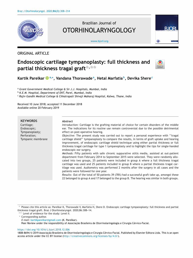
igure 1 A full thickness tragal cartilage with perichondriumn both sides being harvested.
These cases were randomly allocated into Group A androup B of 25 patients each. Patients in Group A underwentndoscopic Type 1 tragal cartilage tympanoplasty with aull thickness graft (∼0.9 mm) while those in the Group
underwent the same procedure with a partial thicknessraft (∼0.4 mm). All the cases had a dry ear for atleast 2onths pre-operatively and an intact ossicular chain.The following parameters were assessed after 2 months
f surgery: graft take-up rate and the hearing improve-ent in both the groups. After this assessment the patients
ollowed up for routine ear examination up to 1 year. Auccessful result was defined as having closure of perfo-ation, no medialization or lateralization and having goodascularity over the cartilage graft. Failure included resid-al/recurrent perforation and no hearing improvement oreterioration in hearing. The Pure Tone Audiogram (PTA)
esting calculated the average hearing at 500, 1000 and000 Hz pre-operatively and 2 months post-operatively inll cases. Institutional ethical review board permission (UIN:256) and patients consent were obtained for this study.
flfte
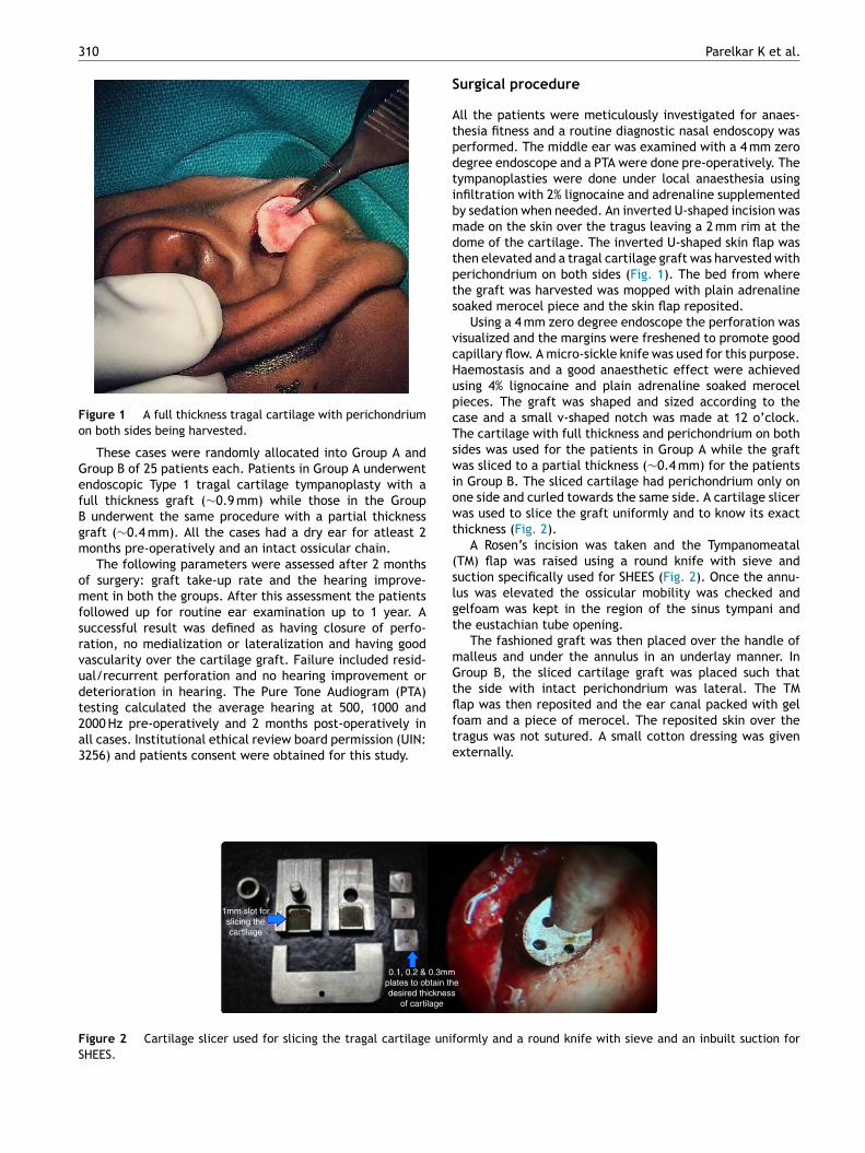
1mm slot forslicing thecartilage
0.1, 0.2 & 0.3mmplates to obtain thdesired thickness
of cartilage
igure 2 Cartilage slicer used for slicing the tragal cartilage unifHEES.
Parelkar K et al.
urgical procedure
ll the patients were meticulously investigated for anaes-hesia fitness and a routine diagnostic nasal endoscopy waserformed. The middle ear was examined with a 4 mm zeroegree endoscope and a PTA were done pre-operatively. Theympanoplasties were done under local anaesthesia usingnfiltration with 2% lignocaine and adrenaline supplementedy sedation when needed. An inverted U-shaped incision wasade on the skin over the tragus leaving a 2 mm rim at theome of the cartilage. The inverted U-shaped skin flap washen elevated and a tragal cartilage graft was harvested witherichondrium on both sides (Fig. 1). The bed from wherehe graft was harvested was mopped with plain adrenalineoaked merocel piece and the skin flap reposited.
Using a 4 mm zero degree endoscope the perforation wasisualized and the margins were freshened to promote goodapillary flow. A micro-sickle knife was used for this purpose.aemostasis and a good anaesthetic effect were achievedsing 4% lignocaine and plain adrenaline soaked merocelieces. The graft was shaped and sized according to thease and a small v-shaped notch was made at 12 o’clock.he cartilage with full thickness and perichondrium on bothides was used for the patients in Group A while the graftas sliced to a partial thickness (∼0.4 mm) for the patients
n Group B. The sliced cartilage had perichondrium only onne side and curled towards the same side. A cartilage sliceras used to slice the graft uniformly and to know its exact
hickness (Fig. 2).A Rosen’s incision was taken and the Tympanomeatal
TM) flap was raised using a round knife with sieve anduction specifically used for SHEES (Fig. 2). Once the annu-us was elevated the ossicular mobility was checked andelfoam was kept in the region of the sinus tympani andhe eustachian tube opening.
The fashioned graft was then placed over the handle ofalleus and under the annulus in an underlay manner. Inroup B, the sliced cartilage graft was placed such that
he side with intact perichondrium was lateral. The TMap was then reposited and the ear canal packed with geloam and a piece of merocel. The reposited skin over theragus was not sutured. A small cotton dressing was given
xternally.
e
ormly and a round knife with sieve and an inbuilt suction for
Endoscopic cartilage tympanoplasty 311
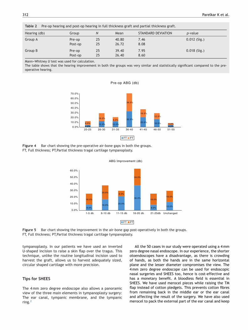
RESIDUAL PIN-HOLEPERFORATION
in-hole residual perforations at the edge of a partial thickness tragal
Table 1 Comparison of graft uptake between full thicknessgraft (A) and partial thickness graft (B).
Take up Group Total
A B
Fail 3 8 1112.0% 32.0% 22.0%
Good 22 17 3988.0% 68.0% 78.0%
Total 25 25 50100.0% 100.0% 100.0%
p-value = −0.017 (Sig.).Fisher exact test was used for calculation.The table shows the comparison of graft take rate in both thegroups. The uptake rate of full thickness was 88% and that of
oTahGBrat
D
A
Tst
Figure 3 A successful full thickness tragal graft take-up and pgraft.
Post-operative care
Patients were advised to take water precautions and avoidvigorous nose blowing. The external cotton and merocel inthe ear canal was removed after 1 week and antibiotic-steroid ear drops were started then. Patients were kept onoral antibiotics for 1 week. The gelfoam was allowed toresolve gradually on its own. The operated patients wereexamined at 1 week, 3 weeks and then 2 months post-operatively. A PTA was repeated at 2 months once a viablegraft was visible and patient was asymptomatic. Thesepatients followed up for routine ear examination up to 1year after the surgery.
Patients who had upper respiratory tract infection andotitis media with effusion post-operatively were managedwith nasal steroidal sprays and an effort was made to pre-vent these conditions from hampering the take of the graft.
Results
Graft take-up
Out of the total of 50 patients 39 (78%) had a successful grafttake up (Fig. 3), amongst these 22 (88%) belonged to the fullthickness i.e. Group A and 17 (68%) belonged to the partialthickness i.e. Group B. Among the 11 cases which had prob-lems with respect to the take of the graft, 3 (12%) belongedto Group A and 8 (32%) belonged to Group B. Of these, all 3of the Group A and 3 of the Group B had small pin-hole resid-ual perforations at the edge of the cartilage graft (Fig. 3)while remaining 5 of the Group B had either medializationof the graft or a large residual. The graft take up rate of fullthickness cartilage was significantly better than that of thepartial thickness graft; p-value = 0.017 (Table 1). This takeup rate essentially remained the same at 1 year follow-uptoo.
Acoustic benefit
The average pre-operative hearing was 40.80 ± 7.46 dB forGroup A and 39.40 ± 7.95 dB for Group B. The post-operativePTA done 2 months after surgery showed an average hearing
ciaa
partial thickness was 68% which is statistically significant; thetake-up with full thickness graft is better than that with partialthickness graft.
f 26.72 ± 8.08 for Group A and 26.40 ± 8.60 for Group B.he hearing improvement in both the groups was very similarnd statistically significant compared to the pre-operativeearing (p-value = 0.012 for Group A and p-value = 0.018 forroup B) (Table 2). The details hearing status in terms of Air-one Gap (ABG) are shown in (Figs. 4 and 5). The PTA was notepeated at 1 year follow up, as these patients did not noteny significant change in their hearing levels, compared tohe hearing assessment done 2 months after the surgery.
iscussion
bout the tragal cartilage
he use of cartilage is experiencing a renaissance in earurgery because of its reliability as a graft material and alsohe increasing use of endoscope in ear surgeries. The tragal
artilage is nourished by diffusion and eventually becomesncorporated in the tympanic membrane.1 The graft is easilyccessible, adequate and its rigidity makes it easy to fashionnd manipulate, reducing the learning curve in endoscopic
312 Parelkar K et al.
Table 2 Pre-op hearing and post-op hearing in full thickness graft and partial thickness graft.
Hearing (db) Group N Mean STANDARD DEVIATION p-value
Group A Pre-op 25 40.80 7.46 0.012 (Sig.)Post-op 25 26.72 8.08
Group B Pre-op 25 39.40 7.95 0.018 (Sig.)Post-op 25 26.40 8.60
Mann---Whitney U test was used for calculation.The table shows that the hearing improvement in both the groups was very similar and statistically significant compared to the pre-operative hearing.
12.0%
8.0%16.0%
0.0%
10.0%
20.0%
30.0%
40.0%
50.0%
60.0%
70.0%
20-25 26-30 31-35 36-40 41-45 46-50 51-554.0%
12.0%
32.0%20.0% 16.0%
4.0%8.0%
36.0%
16.0%12.0%
4.0%
Pre-op ABG (db)
FT PT
Figure 4 Bar chart showing the pre-operative air-bone gaps in both the groups.FT, Full thickness; PT,Partial thickness tragal cartilage tympanoplasty.
0.0%
10.0%
20.0%
30.0%
40.0%
50.0%
60.0%
1-5 db 6-10 db 11-15 db 16-20 db 21-25db Unchanged
8.0%16.0%
20.0%
36.0%
8.0%12.0%
16.0%
20.0%8.0%
24.0%
16.0%16.0%
ABG Improvement (db)
FT PT
F gap
F plast
tUthc
T
TvTr
zoop4nhS
igure 5 Bar chart showing the improvement in the air-boneT, Full thickness; PT,Partial thickness tragal cartilage tympano
ympanoplasty. In our patients we have used an inverted-shaped incision to raise a skin flap over the tragus. Thisechnique, unlike the routine longitudinal incision used toarvest the graft, allows us to harvest adequately sized,ircular shaped cartilage with more precision.
ips for SHEES
he 4 mm zero degree endoscope also allows a panoramiciew of the three main elements in tympanoplasty surgery:he ear canal, tympanic membrane, and the tympanicing.2
flfam
post-operatively in both the groups.y.
All the 50 cases in our study were operated using a 4 mmero degree nasal endoscope. In our experience, the shortertoendoscopes have a disadvantage, as there is crowdingf hands, as both the hands are in the same horizontallane and the lesser diameter compromises the view. The
mm zero degree endoscope can be used for endoscopicasal surgeries and SHEES too, hence is cost-effective andas a monetary benefit. A bloodless field is essential inHEES. We have used merocel pieces while raising the TM
ap instead of cotton pledgets. This prevents cotton fibresrom remaining back in the middle ear or the ear canalnd affecting the result of the surgery. We have also usederocel to pack the external part of the ear canal and keep
Endoscopic cartilage tympanoplasty 313
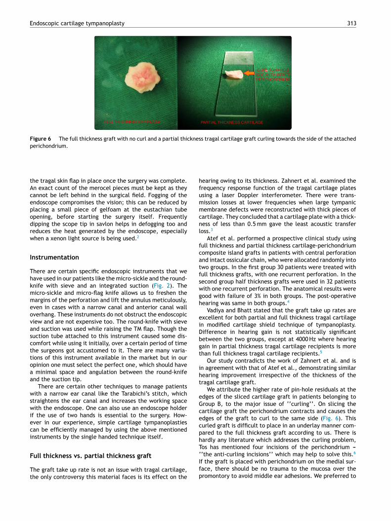
FULL THICKNESS CARTILAGE PARTIAL THICKNESS CARTILAGE
CURL TOWARDSSIDE WITH INTACTPERICHONDRIUM
Figure 6 The full thickness graft with no curl and a partial thickness tragal cartilage graft curling towards the side of the attachedperichondrium.
hfummcnl
fcatfswgh
eiDbgt
iht
eGcecphT
the tragal skin flap in place once the surgery was complete.An exact count of the merocel pieces must be kept as theycannot be left behind in the surgical field. Fogging of theendoscope compromises the vision; this can be reduced byplacing a small piece of gelfoam at the eustachian tubeopening, before starting the surgery itself. Frequentlydipping the scope tip in savlon helps in defogging too andreduces the heat generated by the endoscope, especiallywhen a xenon light source is being used.2
Instrumentation
There are certain specific endoscopic instruments that wehave used in our patients like the micro-sickle and the round-knife with sieve and an integrated suction (Fig. 2). Themicro-sickle and micro-flag knife allows us to freshen themargins of the perforation and lift the annulus meticulously,even in cases with a narrow canal and anterior canal walloverhang. These instruments do not obstruct the endoscopicview and are not expensive too. The round-knife with sieveand suction was used while raising the TM flap. Though thesuction tube attached to this instrument caused some dis-comfort while using it initially, over a certain period of timethe surgeons got accustomed to it. There are many varia-tions of this instrument available in the market but in ouropinion one must select the perfect one, which should havea minimal space and angulation between the round-knifeand the suction tip.
There are certain other techniques to manage patientswith a narrow ear canal like the Tarabichi’s stitch, whichstraightens the ear canal and increases the working spacewith the endoscope. One can also use an endoscope holderif the use of two hands is essential to the surgery. How-ever in our experience, simple cartilage tympanoplastiescan be efficiently managed by using the above mentionedinstruments by the single handed technique itself.
Full thickness vs. partial thickness graft
The graft take up rate is not an issue with tragal cartilage,the only controversy this material faces is its effect on the
‘Ifp
earing owing to its thickness. Zahnert et al. examined therequency response function of the tragal cartilage platessing a laser Doppler interferometer. There were trans-ission losses at lower frequencies when large tympanicembrane defects were reconstructed with thick pieces of
artilage. They concluded that a cartilage plate with a thick-ess of less than 0.5 mm gave the least acoustic transfeross.3
Atef et al. performed a prospective clinical study usingull thickness and partial thickness cartilage-perichondriumomposite island grafts in patients with central perforationnd intact ossicular chain, who were allocated randomly intowo groups. In the first group 30 patients were treated withull thickness grafts, with one recurrent perforation. In theecond group half thickness grafts were used in 32 patientsith one recurrent perforation. The anatomical results wereood with failure of 3% in both groups. The post-operativeearing was same in both groups.4
Vadiya and Bhatt stated that the graft take up rates arexcellent for both partial and full thickness tragal cartilagen modified cartilage shield technique of tympanoplasty.ifference in hearing gain is not statistically significantetween the two groups, except at 4000 Hz where hearingain in partial thickness tragal cartilage recipients is morehan full thickness tragal cartilage recipients.5
Our study contradicts the work of Zahnert et al. and isn agreement with that of Atef et al., demonstrating similarearing improvement irrespective of the thickness of theragal cartilage graft.
We attribute the higher rate of pin-hole residuals at thedges of the sliced cartilage graft in patients belonging toroup B, to the major issue of ‘‘curling’’. On slicing theartilage graft the perichondrium contracts and causes thedges of the graft to curl to the same side (Fig. 6). Thisurled graft is difficult to place in an underlay manner com-ared to the full thickness graft according to us. There isardly any literature which addresses the curling problem,os has mentioned four incisions of the perichondrium ---
‘the anti-curling incisions’’ which may help to solve this.6
f the graft is placed with perichondrium on the medial sur-ace, there should be no trauma to the mucosa over theromontory to avoid middle ear adhesions. We preferred to
3
po
L
Thcttotsctrfdfaw
moohftp
C
Scwip
h
cnrt
E
Eieam
a
C
T
R
1
2
3
4
5
14
lace the sliced cartilage grafts with intact perichondriumn the lateral side in our cases belonging to Group B.
imitation of our study
here have been many studies reporting 100 percent or veryigh take up rates in primary tympanoplasties with tragalartilage as the graft of choice. In our study the overallake up rate is 78%, we attribute this lower rate not onlyo patient factors such as low socio-economic class, thatur hospital caters but also to the learning curve of thisechniques. Being a teaching tertiary-care hospital, not allurgeries were done by a single senior surgeon; this alsoould have been one of the factors in our opinion, thoughhe technique and protocol of the procedure essentiallyemained the same. In order to reduce the learning curveor endoscopic ear surgery, we encourage our junior residentoctors to routinely use the 4 mm zero degree endoscopeor diagnostic examinations of the ear. Once they develop
good hand-eye co-ordination they are trained to go-aheadith minor procedures.
A potential drawback of the tragal graft is its opacity, as itay be more difficult to detect an iatrogenic cholesteatoma
r post-operative secretory otitis media. We have had two ofur failures due to secretory otitis media too. These patientsad evidence of glue coming out from the residual per-oration at the edge of the cartilage graft. However, theemporalis fascia too is often not completely transparentost-operatively.
onclusion
ingle Handed Endoscopic Ear Surgery (SHEES) with tragalartilage graft is one of the best modality to manage patientsho require a simple Type 1 tympanoplasty. It is minimally
nvasive, scarless, time-saving, cost-effective and has a highatient compliance in our opinion.
The thickness of the tragal cartilage does not affect theearing results significantly according to our results. The
6
Parelkar K et al.
ondition of the middle ear, patient selection and the tech-ique of the surgery along with post-operative care areesponsible for a successful result rather than the grafthickness.
thical standards
thical approval: All procedures performed in this studynvolving human participants were in accordance with thethical standards of the institutional research committeend with the 1964 Helsinki declaration and its later amend-ents or comparable ethical standards.Informed consent: Informed consent was obtained from
ll participants included in the study.
onflicts of interest
he authors declare no conflicts of interest.
eferences
. Levinson RM. Cartilage-perichondrial composite graft tym-panoplasty in the treatment of posterior marginal and atticretraction pockets. Laryngoscope. 1987;97:1069---74.
. Tarabichi M. Endoscopic transcanal middle ear surgery. Indian JOtolaryngol Head Neck Surg. 2010;62:6---24.
. Zahnert T, Hüttenbrink KB, Mürbe D, Bornitz M. Experimentalinvestigations of the use of cartilage in tympanic membranereconstruction. Otol Neurotol. 2000;21:322---8.
. Atef A, Talaat N, Fathi A, Mosleh M, Safwat S. Effect of thethickness of the cartilage disk on the hearing results afterperichondrium/cartilage island flap tympanoplasty. ORL J Otorhi-nolaryngol Relat Spec. 2007;69:207---11.
. Vadiya S, Bhatt S. Comparison of partial thickness and fullthickness tragal cartilage graft during modified cartilage shieldtympanoplasty for type I procedures. Indian J Otolaryngol Head
Neck Surg. 2016;68:30---3.
. Tos M. Cartilage tympanoplasty: classification of methods-techniques-results. 1st ed. New York, NY: Thieme; 2009. p.353---64.


![Cartilage - facultymembers.sbu.ac.irfacultymembers.sbu.ac.ir/rajabi/ppt toPDF/Cartilage [Compatibility Mode].pdfFibrocartilage • Fibrous Cartilage • is a form of connective tissue](https://static.documents.pub/doc/80x56/6012989a4318862a0e5813ae/cartilage-topdfcartilage-compatibility-modepdf-fibrocartilage-a-fibrous.jpg)

![Triple-c Cartilage Tympanoplasty: Case Series · [1] Glasscock, M.E. and Shambaugh, G.E. Surgery of the Ear 5th Edition Pathology and Clinical Course of Inflammatory Disease of the](https://static.documents.pub/doc/80x56/5f1437e8dc43ae01c032f5f5/triple-c-cartilage-tympanoplasty-case-series-1-glasscock-me-and-shambaugh.jpg)


