Enhanced exfoliation efficiency of graphite into few-layer graphenevia reduction of graphite edge
Liangwei Yang a, Fuzhen Zhao a, b, Yan Zhao a, Yangyong Sun a, Guangwu Yang b,Lianming Tong a, *, Jin Zhang a, **
a Center for Nanochemistry, Beijing Science and Engineering Center for Nanocarbons, Beijing National Laboratory for Molecular Sciences, College ofChemistry and Molecular Engineering, Peking University, Beijing, 100871, Chinab College of Science, China University of Petroleum, Qingdao, 266580, China
a r t i c l e i n f o
Article history:Received 6 June 2018Received in revised form23 July 2018Accepted 25 July 2018Available online 25 July 2018
Graphene has received abundant attentions in many applications because of its extraordinary properties.Liquid-phase exfoliation is one of the most important methods to obtain graphene from graphite.However, due to strong van der Waals interaction force, it is still extremely challenging to furtherimprove the exfoliation efficiency. Here we report that by carrying out reductions with the oxygenicfunctional groups on the pristine graphite edges using hydrazine hydrate and hydrogen at 800 �C as apretreatment approach, the exfoliation efficiency can be improved by ~37% using a lithium-assistedliquid-phase exfoliation approach. The change of the functional groups is characterized by X-rayphotoelectron spectroscopy and Fourier transform infrared spectroscopy. The graphene sheets show highdegree of structural integrity and few defects. A thin film is prepared from the exfoliated graphene byvacuum assisted filtration and treated by annealing and mechanical compression. The graphene thin filmshows a high thermal conductivity of 1707Wm�1 K�1, which is superior to that of common metalmaterials, such as Cu and Al.
Graphene is a single atomic-layer of hexagonal sp2-bondedcarbon atoms. Since its discovery, graphene has been attractingappreciable attention owing to its unique physical properties,including ultrahigh charge-carrier mobility (~106 cm2 V�1 s�1) [1],superior thermal conductivity (~5300Wm�1 K�1) [2], opticaltransmittance (~97.7%) [3] and extremely intrinsic strength(~130 GPa) [4]. The mechanical exfoliation (“scotch-tape” method)of graphite into single and few-layer graphene nanosheets wasdiscovered in 2004 [1e5]. Since then, many methods have alsobeen developed to prepare large-scale or massive graphene.Chemical vapor deposition (CVD) was introduced to synthesizelarge-scale single-crystalline graphene films [6e8]. However, suchCVD-grown graphene is usually costly and used in some particularapplications such as optoelectronics and flexible electronics.Liquid-phase exfoliation of bulk graphite is acknowledged as the
most widely used method for massive production of graphene[9e11]. Even though the quality of exfoliated graphene is relativelylower, the low-cost and mass production are extraordinarily ad-vantageous for widespread applications in many fields such aspolymer composites and clean energy devices.
Graphite is composited of graphene layers bonded togethervertically through van der Waals force. In 2006, a solution-basedmethod was reported to produce graphene oxide (GO) via chemi-cal modification (strong oxidizing agents) of graphite, which hasreceived great attention due to the capability of scalable production[12e14]. Reduced graphene oxide (rGO) can also be produced viathe reduction of GO. GO can be well dispersed in the aqueous so-lution due to the existence of oxygenic functional groups, which, onthe other hand, also destroy the structural integrity and bring manydefects that cannot be completely repaired although after thereduction processes [15]. However, residual oxygenic functionalgroups of rGO are helpful in the application fields on the electro-chemical catalysis [16] and composite with polymer [17]. Never-theless, the oxide and reduce processes utilize abundant oxidizingand reducing agents, the post-treatment of which may bring upissues of high energy consumption and high pollution. Therefore, it
is necessary to develop non-oxidizing method to producegraphene.
The Coleman group and others developed a simple non-oxideliquid-phase exfoliation of pristine graphite to produce high-quality graphene flakes using surfactants or organic solvents[9,18,19]. Although it is also considered as a promising method formass production, the exfoliation efficiency is still too low to beindustrialized. Alternatively, graphite can be intercalated to pro-duce graphite intercalation compounds (GICs) by using variousintercalates, such as alkali metals, organic molecules and transitionmetal halides [20]. GICs have been exfoliated to produce graphitenanoplatelets with thicknesses down to 2e10 nm [21e23]. Ac-cording to the research of Jeon group, graphene flakes could begenerated by using a eutectic system of ternary KCl�NaCl�ZnCl2salts through the intercalation process [24]. Li-GICs, formed by theinsertion of lithium atoms in graphite, is a widely used anodematerial for Li-ion batteries (LIB). Because of the high activity oflithium, the exfoliation methods were inspired by the electro-chemical reactions of negative electrodes in the rechargeable LIB[25,26]. Song et al. developed the lithium-assisted approach for theexfoliation of pristine graphite [27]. In this approach, Li/liquidammonia solution was utilized to synthesize a lithium intercalatedgraphite precursor. Among all the methods that have been devel-oped, the key challenge is still to increase the exfoliation efficiencyof pristine graphite.
Graphite ore, the raw material of graphite powder, is mined byboth open pit and underground processes (Figure S1). In thesecomplicated processes, the regrinding and ball milling can decreasethe size of raw materials at relatively high temperature, but mayintroduce numerous oxygenic functional groups on the graphiteedges [28]. The regrinding and ball milling damage C-C bonds ofgraphite to form highly active carbon radical, which can be oxidizedinto oxygenic functional groups (carbonyl, epoxy and carboxyl) inthe atmosphere. The heat produced by friction in the regrindingand ball milling provides enough energy for the functionalizationreaction. As a result, the edges of the graphite used for LPE aredefective and functionalized by oxygenic functional groups. To acertain extent, the stereo-hindrance effect of oxygenic functionalgroups will hinder the insertion of atoms and molecules. Hence, formore atoms to intercalate into the graphite interlayers for exfolia-tion, it is extremely needed to remove these oxygenic functionalgroups on the graphite edges.
Herein, we report a pre-reduction based approach to enhancethe exfoliation efficiency of graphite into few-layer graphene viareduction of graphite edge. Before the exfoliation process, theoxygenic functional groups on the graphite edges were reduced bysolution (hydrazine hydrate) and thermal (hydrogen, high tem-perature) approaches. After reduction, the stereo-hindrance effectof graphite edges was decreased and more Li atoms could inter-calate into graphite interlayers in the lithium-assisted physicalinsertion. The change of the functional groups caused by thereduction reaction can be accurately characterized by X-rayphotoelectron spectroscopy (XPS) and Fourier transform infraredspectroscopy (FT-IR). Due to the removal of oxygenic functionalgroups on the graphite edges, the exfoliation efficiency of lithium-assisted liquid-phase exfoliation was enhanced by ~37%. Theaverage size of exfoliated graphene sheets was about 1 mm, andover 94% had no more than 5 layers. The electron diffraction pat-terns in Transmission Electron Microscope (TEM) shows that gra-phene sheets maintain high degree of structural integrity. A thinfilm of graphene was fabricated from the exfoliated graphenesheets using an annealing and mechanical compression process.The thermal conductivity of the film was found to be1707Wm�1 K�1, which is superior to common metal films (Cu andAl). This work demonstrates a feasible route to enhance the
efficiency of LPE, and at the same time preserve the high quality ofgraphene.
2. Experimental
2.1. Materials and methods
Graphite powder (Figure S2) was bought from BTR new energymaterials Inc. The following chemicals were used without furtherpurification: lithium foil (AR, China Energy Lithium Co., LTD), hy-drazine hydrate (N2H4$H2O, 85%, Sinopharm Chemical Reagent Co.,LTD), ethanol (C2H5OH, Bejing Tong Guang Fine Chemicals Com-pany), liquid ammonia (NH3, Beijing Nanfei Industry and Trade Co.LTD.), N-methyl pyrrolidone (NMP, Xilong Scientific Co., LTD), hy-drochloric acid (HCl, ~37%, Xilong Scientific Co., LTD), AAO mem-brane (60mm in diameter, 0.2 mm in pore size, Whatman).
Themorphology and structure of graphene sheets and graphenefilms were characterized by Scanning Electron Microscopy (SEM,Hitachi S-4800) at an acceleration voltage of 1 kV and TEM (FEITecnai F30) at 300 kV. Raman spectroscopy (Horiba Jobin YvonLabRAM HR 800, 514.5 nm) was used to characterize the defect andquality of exfoliated graphene sheets. XPS (Kratos Analytical Axis-Ultra spectrometer with Al Ka X-ray source) and FT-IR (Nicoletis50, ThermoFisher) were used to characterize the change ofoxygenic functional groups on the pristine graphite and reducedgraphite. Sheet resistance was measured by means of an Agilent4155C semiconductor characterization system. Ultravioletevisspectroscopy (UVevis, Perkin Elmer Lambda 950) was used tomeasure the transmittance of the graphene dispersion. Atomicforce microscopy (AFM, Bruker Dimension Icon) was carried out tomeasure the thickness of graphene sheets and statistics the thick-ness distribution. At room temperature, the thermal conductivitywas carried out by a self-heating method. The thermal image wasinvestigated by thermal infrared imager (Optris PI 160).
2.2. Reduction of graphite powder with hydrazine hydrate andhydrogen
The powder of pristine graphite (1 g) with an average diameterof 200 mm was dispersed into deionized water (500ml). Smallamount of ethanol was also introduced to homogeneously dispersethe graphite powder. The hydrazine hydrate (1ml) was added andthe resultant mixture was further stirred (~25 rpm) for 3 h at 90 �C.Then the graphite powder was filtrated and dried. The resultantgraphite powder was put on a quartz boat and placed within aquartz tube inside the furnace. The tubewas purged with argon (Ar,300 sccm). Then the furnace was heated to 800 �C under Ar (300sccm) and H2 (100 sccm) flow at ambient pressure. The graphitepowder were held at 800 �C for 1 h. The hydrazine hydrate andthermal annealing can ensure removing oxygenic functional groupsas many as possible and residual materials (ethanol and hydrazinehydrate). Finally we can get the reduced graphite.
2.3. Preparation of graphene sheets and measurement of electricalconductivity
Reduced graphite was added to lithium in liquid ammonia,which was prepared by the cooling of ammonia at an acetone/dryice bath. Themixturewas stirred for 2 h, and then the ammoniawasslowly evaporated bymeans of thermostatic bath after the acetone/dry ice bath was removed. Then hydrochloric acid (HCl, ~37%) wasadded into the residule with mild sonication. The black productswas cleaned by deionized water and ethanol. Finally, the graphenesheets was dispersed into NMP. The graphene dispersion wascentrifuged at 1000 rpm for 1 h and the supernatant dispersionwas
L. Yang et al. / Carbon 138 (2018) 390e396392
carefully collected.The graphene dispersions in NMP were dip-coated onto a fresh
siliconwafer and then annealed in argon at 800 �C for 2 h. AFMwasused to measure the height of graphene sheet, and a single-layergraphene sheet of 0.8 nm was chosen to characterize the elec-trical properity. Micro-gap Au (600 nm)/Cr (80 nm) electrodes witha gap of ~300 nm were fabricated onto the single-layer graphenesheet via electron beam lithography. Based on the resultant I-Vprofiles, the sheet resistance value Rs can be calculated by theequation Rs¼ RW/L, where R, W and L represent the resistancevalue, width and electrode gap length of the single-layer graphenesheet, respectively.
2.4. Preparation of graphene film
The graphene filmwas prepared by vacuum assisted filtration ofwell-dispersed graphene solution through AAO membrane. Thenthe graphene film was dried under vacuum at 80 �C for overnightbefore peeling off from the AAO membrane. The graphene filmwasannealed at 3000 �C for 3 h under Ar in a graphite furnace (ZhuzhouHongya Electric-heating Equipment Co., LTD) and then compressedat 30MPa for 1 h.
3. Results and discussion
3.1. Reduction of graphite powder and characterization
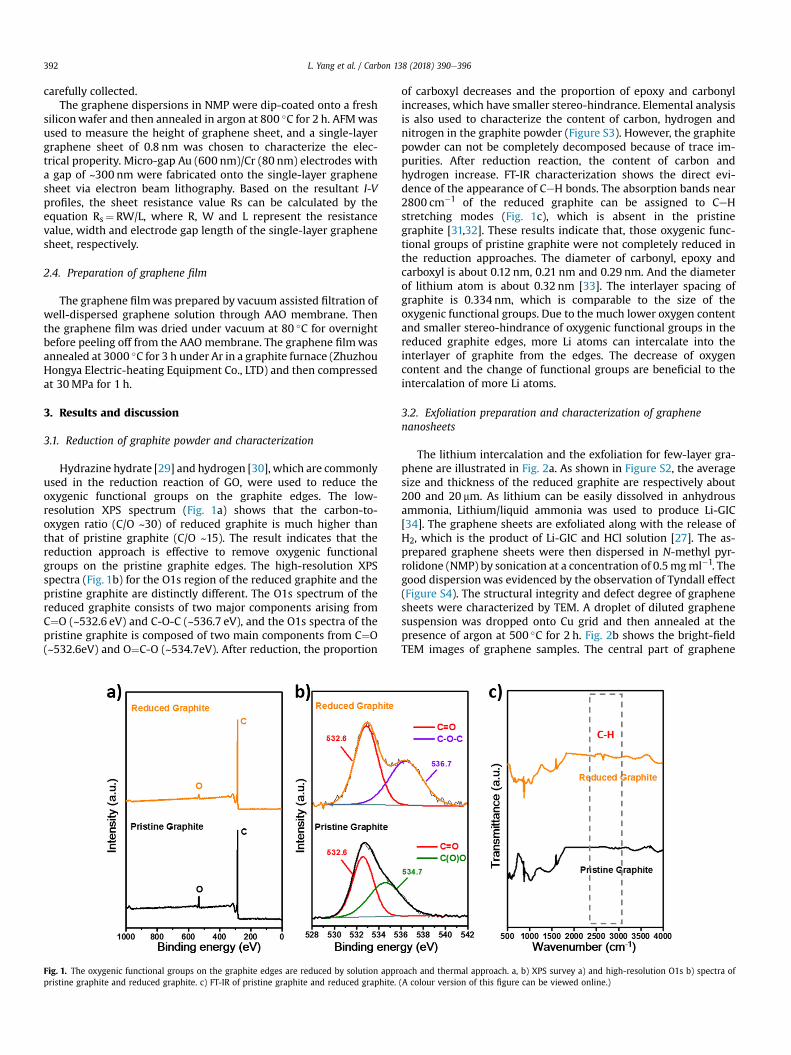
Hydrazine hydrate [29] and hydrogen [30], which are commonlyused in the reduction reaction of GO, were used to reduce theoxygenic functional groups on the graphite edges. The low-resolution XPS spectrum (Fig. 1a) shows that the carbon-to-oxygen ratio (C/O ~30) of reduced graphite is much higher thanthat of pristine graphite (C/O ~15). The result indicates that thereduction approach is effective to remove oxygenic functionalgroups on the pristine graphite edges. The high-resolution XPSspectra (Fig. 1b) for the O1s region of the reduced graphite and thepristine graphite are distinctly different. The O1s spectrum of thereduced graphite consists of two major components arising fromC¼O (~532.6 eV) and C-O-C (~536.7 eV), and the O1s spectra of thepristine graphite is composed of two main components from C¼O(~532.6eV) and O¼C-O (~534.7eV). After reduction, the proportion
Fig. 1. The oxygenic functional groups on the graphite edges are reduced by solution apprpristine graphite and reduced graphite. c) FT-IR of pristine graphite and reduced graphite.
of carboxyl decreases and the proportion of epoxy and carbonylincreases, which have smaller stereo-hindrance. Elemental analysisis also used to characterize the content of carbon, hydrogen andnitrogen in the graphite powder (Figure S3). However, the graphitepowder can not be completely decomposed because of trace im-purities. After reduction reaction, the content of carbon andhydrogen increase. FT-IR characterization shows the direct evi-dence of the appearance of CeH bonds. The absorption bands near2800 cm�1 of the reduced graphite can be assigned to CeHstretching modes (Fig. 1c), which is absent in the pristinegraphite [31,32]. These results indicate that, those oxygenic func-tional groups of pristine graphite were not completely reduced inthe reduction approaches. The diameter of carbonyl, epoxy andcarboxyl is about 0.12 nm, 0.21 nm and 0.29 nm. And the diameterof lithium atom is about 0.32 nm [33]. The interlayer spacing ofgraphite is 0.334 nm, which is comparable to the size of theoxygenic functional groups. Due to the much lower oxygen contentand smaller stereo-hindrance of oxygenic functional groups in thereduced graphite edges, more Li atoms can intercalate into theinterlayer of graphite from the edges. The decrease of oxygencontent and the change of functional groups are beneficial to theintercalation of more Li atoms.
3.2. Exfoliation preparation and characterization of graphenenanosheets
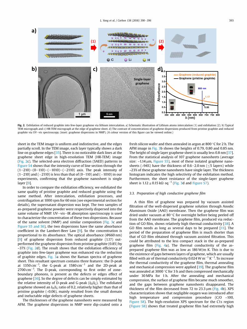
The lithium intercalation and the exfoliation for few-layer gra-phene are illustrated in Fig. 2a. As shown in Figure S2, the averagesize and thickness of the reduced graphite are respectively about200 and 20 mm. As lithium can be easily dissolved in anhydrousammonia, Lithium/liquid ammonia was used to produce Li-GIC[34]. The graphene sheets are exfoliated along with the release ofH2, which is the product of Li-GIC and HCl solution [27]. The as-prepared graphene sheets were then dispersed in N-methyl pyr-rolidone (NMP) by sonication at a concentration of 0.5mgml�1. Thegood dispersion was evidenced by the observation of Tyndall effect(Figure S4). The structural integrity and defect degree of graphenesheets were characterized by TEM. A droplet of diluted graphenesuspension was dropped onto Cu grid and then annealed at thepresence of argon at 500 �C for 2 h. Fig. 2b shows the bright-fieldTEM images of graphene samples. The central part of graphene
oach and thermal approach. a, b) XPS survey a) and high-resolution O1s b) spectra of(A colour version of this figure can be viewed online.)
Fig. 2. Exfoliation of reduced graphite into few-layer graphene via lithium intercalation. a) Schematic illustration of Lithium atoms intercalation (1) and exfoliation (2). b) TypicalTEM micrograph and c) HR-TEM micrograph at the edge of graphene sheet. d) The contrast of concentrations of graphene dispersions produced from pristine graphite and reducedgraphite via UVevis spectroscopy. (inset: graphene dispersions in NMP). (A colour version of this figure can be viewed online.)
L. Yang et al. / Carbon 138 (2018) 390e396 393
sheet in the TEM image is uniform and indistinctive, and the edgespartially scroll. In the TEM image, each layer typically shows a darkline on graphene edges [35]. There is no noticeable dark lines at thegraphene sheet edge in high-resolution TEM (HR-TEM) image(Fig. 2c). The selected-area electron diffraction (SAED) patterns inFigure S4 shows that the intensity curve of line section through the(1e210)e(0e110)e(e1010)e(e2110) axis. The peak intensity of(1e210) and (�2110) is less than that of (0e110) and (�1010) in ourexperiments, confirming that the graphene nanosheet is singlelayer [9].
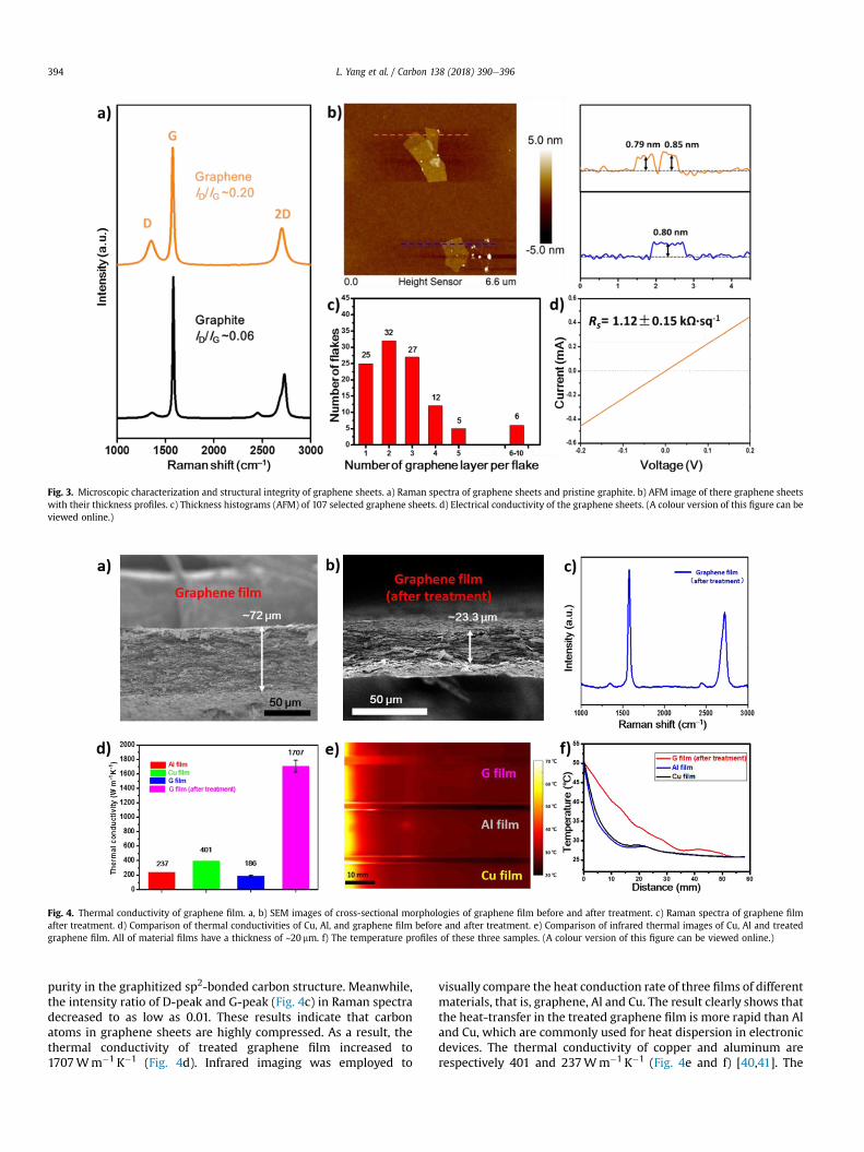
In order to compare the exfoliation efficiency, we exfoliated thesame quality of pristine graphite and reduced graphite using thesame method. After intercalation, exfoliation processes, andcentrifugation at 1000 rpm for 60min (see experimental section fordetails), the supernatant dispersion was kept. The two samples ofas-prepared graphene powder were respectively dispersed into thesame volume of NMP. UVeviseIR absorption spectroscopy is usedto characterize the concentration of these two dispersions. Becauseof the same solvent (NMP) and similar composition (Fig. 3c andFigure S5 and S6), the two dispersions have the same absorbancecoefficient in the Lambert-Beer Law [9]. So the concentration isproportional to its absorbance. The optical absorbance (@660 nm)[9] of graphene dispersion from reduced graphite (1.17) out-performed the graphene dispersion from pristine graphite (0.85) by~37% (Fig. 2d). The result shows that the exfoliation efficiency ofgraphite into few-layer graphene was enhanced via the reductionof graphite edges. Fig. 3a shows the Raman spectra of graphenesheet. This resultant spectrum contains three features: the D-peakat 1350 cm�1, the G-peak at 1580 cm�1 and the 2D-peak at2700 cm�1. The D-peak, corresponding to first order of zone-boundary phonons, is present as the defects or edges effect ofgraphene [36]. So the degree of defects can be simply estimated bythe relative intensity of D-peak and G-peak (ID/IG). The exfoliatedgraphene showed an ID/IG ratio of 0.2, relatively higher than that ofpristine graphite (~0.06), mainly resulted from the small domainand ineluctable edge defects of graphene sheets.
The thicknesses of the graphene nanosheets were measured byAFM. The graphene dispersions in NMP were dip-coated onto a
fresh siliconwafer and then annealed in argon at 800 �C for 2 h. TheAFM image in Fig. 3b shows the heights of 0.79, 0.80 and 0.85 nm.The height of single layer graphene sheet is usually less 0.8 nm [37].From the statistical analysis of 107 graphene nanosheets (averagesize: ~1.14 mm, Figure S5), most of these isolated graphene nano-sheets (~94%) have the thickness of 0.6e2.0 nm (�5 layers) while~23% of these graphene nanosheets have single layer. The thicknesshistogram indicates the high selectivity of the exfoliation method.Furthermore, the sheet resistance of the single-layer graphenesheet is 1.12± 0.15 kU$sq�1 (Fig. 3d and Figure S7).
3.3. Preparation of high conductive graphene film
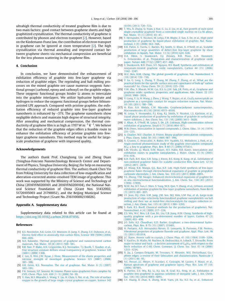
A thin film of graphene was prepared by vacuum assistedfiltration of the well-dispersed graphene solution through AnodicAluminum Oxide (AAO) membrane. Then the graphene film wasdried under vacuum at 80 �C for overnight before being peeled offfrom the AAO membrane. The graphene film, produced via reduc-tion of GO film, shows relatively high thermal conductivity [38]. AGO film needs as long as several days to be prepared [11]. Theperiod of the preparation of graphene film is much shorter thanthat of GO film obtained through vacuum assisted filtration. Thiscould be attributed to the less compact stack in the as-preparedgraphene film (Fig. 4a). The thermal conductivity of the as-prepared graphene film was as low as 186W/m K. This is due tothe existence of gaps between layers of graphene, which are usuallyfilled with air of thermal conductivity 0.024Wm�1 K�1. To increasethe thermal conductivity of the graphene film, thermal annealingand mechanical compression were applied [39]. The graphene filmwas annealed at 3000 �C for 3 h and then compressed mechanicallyunder 30MPa for 1 h. After the annealing and mechanicalcompression, the surface of graphene film became much smoother,and the gaps between graphene nanosheets disappeared. Thethickness of the film decreased from 72 to 23.3 mm (Fig. 4b). XPScharacterization shows that negligible oxygenwas introduced afterhigh temperature and compression procedure (C/O ~100,Figure S8). The high-resolution XPS spectrum for the C1s region(Figure S8) shows that treated graphene film had extremely high
Fig. 3. Microscopic characterization and structural integrity of graphene sheets. a) Raman spectra of graphene sheets and pristine graphite. b) AFM image of there graphene sheetswith their thickness profiles. c) Thickness histograms (AFM) of 107 selected graphene sheets. d) Electrical conductivity of the graphene sheets. (A colour version of this figure can beviewed online.)
Fig. 4. Thermal conductivity of graphene film. a, b) SEM images of cross-sectional morphologies of graphene film before and after treatment. c) Raman spectra of graphene filmafter treatment. d) Comparison of thermal conductivities of Cu, Al, and graphene film before and after treatment. e) Comparison of infrared thermal images of Cu, Al and treatedgraphene film. All of material films have a thickness of ~20 mm. f) The temperature profiles of these three samples. (A colour version of this figure can be viewed online.)
L. Yang et al. / Carbon 138 (2018) 390e396394
purity in the graphitized sp2-bonded carbon structure. Meanwhile,the intensity ratio of D-peak and G-peak (Fig. 4c) in Raman spectradecreased to as low as 0.01. These results indicate that carbonatoms in graphene sheets are highly compressed. As a result, thethermal conductivity of treated graphene film increased to1707Wm�1 K�1 (Fig. 4d). Infrared imaging was employed to
visually compare the heat conduction rate of three films of differentmaterials, that is, graphene, Al and Cu. The result clearly shows thatthe heat-transfer in the treated graphene film is more rapid than Aland Cu, which are commonly used for heat dispersion in electronicdevices. The thermal conductivity of copper and aluminum arerespectively 401 and 237Wm�1 K�1 (Fig. 4e and f) [40,41]. The
L. Yang et al. / Carbon 138 (2018) 390e396 395
ultrahigh thermal conductivity of treated graphene film is due totwo main factors: good contact between graphene sheets and highgraphitized crystallization. The thermal conductivity of graphene iscontributed by phonon and electron transport [2]. However, basedon the Kiedemann-Franz law, the contribution of electron transportin graphene can be ignored at room temperature [2]. The highcrystallization via thermal annealing and improved contact be-tween graphene sheets via mechanical compression are beneficialfor the less phonon scattering in the graphene film.
4. Conclusion
In conclusion, we have demonstrated the enhancement ofexfoliation efficiency of graphite into few-layer graphene viareduction of graphite edges. The regrinding and ball milling pro-cesses on the mined graphite ore cause numerus oxygenic func-tional groups (carbonyl, epoxy and carboxyl) on the graphite edges.These oxygenic functional groups hinder Li atoms to intercalateinto the graphite interlayers. We utilize hydrazine hydrate andhydrogen to reduce the oxygenic functional groups before lithium-assisted LPE approach. Compared with pristine graphite, the exfo-liation efficiency of reduced graphite into few-layer graphenenanosheets is enhanced by ~37%. The graphene nanosheets shownegligible defects and maintain high degree of structural integrity.After annealing and mechanical compression, the thermal con-ductivity of graphene film is as high as 1707Wm�1 K�1. We believethat the reduction of the graphite edges offers a feasible route toenhance the exfoliation efficiency of pristine graphite into few-layer graphene nanosheets. This method may be useful for large-scale production of graphene with improved quality.
Acknowledgements
The authors thank Prof. Changhong Liu and Zheng Duan(Tsinghua-Foxconn Nanotechnology Research Center and Depart-ment of Physics, Tsinghua University, Beijing) for help onmeasuringthermal conductivity of graphene film. The authors thank Chen Yinfrom Peking University for data collection of low-magnification andaberration-corrected atomic-resolved TEM image of graphene. Thiswork was supported by the Ministry of Science and Technology ofChina (2016YFA0200101 and 2016YFA0200104), the National Nat-ural Science Foundation of China (Grant Nos. 51432002,51720105003 and 21790052), and the Beijing Municipal Scienceand Technology Project (Grant No. Z161100002116026).
Appendix A. Supplementary data
Supplementary data related to this article can be found athttps://doi.org/10.1016/j.carbon.2018.07.056.
References
[1] K.S. Novoselov, A.K. Geim, S.V. Morozov, D. Jiang, Y. Zhang, S.V. Dubonos, et al.,Electric field effect in atomically thin carbon films, Science 306 (5696) (2004)666e669.
[2] A.A. Balandin, Thermal properties of graphene and nanostructured carbonmaterials, Nat. Mater. 10 (8) (2011) 569e581.
[3] R.R. Nair, P. Blake, A.N. Grigorenko, K.S. Novoselov, T.J. Booth, T. Stauber, et al.,Fine structure constant defines visual transparency of graphene, Science 320(5881) (2008) 1308.
[4] C. Lee, X. Wei, J.W. Kysar, J. Hone, Measurement of the elastic properties andintrinsic strength of monolayer graphene, Science 321 (5887) (2008)385e388.
[5] A.K. Geim, K.S. Novoselov, The rise of graphene, Nat. Mater. 6 (3) (2007)183e191.
[6] P.R. Somani, S.P. Somani, M. Umeno, Planer nano-graphenes from camphor byCVD, Chem. Phys. Lett. 430 (1e3) (2006) 56e59.
[7] Y. Hao, M.S. Bharathi, L. Wang, Y. Liu, H. Chen, S. Nie, et al., The role of surfaceoxygen in the growth of large single-crystal graphene on copper, Science 342
(6159) (2013) 720e723.[8] T. Wu, X. Zhang, Q. Yuan, J. Xue, G. Lu, Z. Liu, et al., Fast growth of inch-sized
single-crystalline graphene from a controlled single nucleus on Cu-Ni alloys,Nat. Mater. 15 (1) (2016) 43e47.
[9] Y. Hernandez, V. Nicolosi, M. Lotya, F.M. Blighe, Z. Sun, S. De, et al., High-yieldproduction of graphene by liquid-phase exfoliation of graphite, Nat. Nano-technol. 3 (9) (2008) 563e568.
[10] K.R. Paton, E. Varrla, C. Backes, R.J. Smith, U. Khan, A. O'Neill, et al., Scalableproduction of large quantities of defect-free few-layer graphene by shearexfoliation in liquids, Nat. Mater. 13 (6) (2014) 624e630.
[11] D.A. Dikin, S. Stankovich, E.J. Zimney, R.D. Piner, G.H. Dommett,G. Evmenenko, et al., Preparation and characterization of graphene oxidepaper, Nature 448 (7152) (2007) 457e460.
[12] S. Stankovich, R.D. Piner, S.T. Nguyen, R.S. Ruoff, Synthesis and exfoliation ofisocyanate-treated graphene oxide nanoplatelets, Carbon 44 (15) (2006)3342e3347.
[13] W.C. Ren, H.M. Cheng, The global growth of graphene, Nat. Nanotechnol. 9(10) (2014) 726e730.
[14] Y. Lu, G. Long, L. Zhang, T. Zhang, M. Zhang, F. Zhang, et al., What are thepractical limits for the specific surface area and capacitance of bulk sp2 carbonmaterials? Sci. China Chem. 59 (2) (2016) 225e230.
[15] Y.W. Zhu, S. Murali, W.W. Cai, X.S. Li, J.W. Suk, J.R. Potts, et al., Graphene andgraphene oxide: synthesis, properties, and applications, Adv. Mater. 22 (35)(2010) 3906e3924.
[16] Y. Liang, Y. Li, H. Wang, J. Zhou, J. Wang, T. Regier, et al., Co3O4 nanocrystals ongraphene as a synergistic catalyst for oxygen reduction reaction, Nat. Mater.10 (10) (2011) 780e786.
[17] H. Kim, A.A. Abdala, C.W. Macosko, Graphene/polymer nanocomposites,Macromolecules 43 (16) (2010) 6515e6530.
[18] M. Lotya, Y. Hernandez, P.J. King, R.J. Smith, V. Nicolosi, L.S. Karlsson, et al.,Liquid phase production of graphene by exfoliation of graphite in surfactant/water solutions, J. Am. Chem. Soc. 131 (10) (2009) 3611e3620.
[19] U. Khan, A. O'Neill, M. Lotya, S. De, J.N. Coleman, High-concentration solventexfoliation of graphene, Small 6 (7) (2010) 864e871.
[20] M.B. Dines, Intercalation in layered compounds, J. Chem. Educ. 51 (4) (1974)221e223.
[21] A. Charlier, M.F. Charlier, D. Fristot, Binary graphite-intercalation compounds,J. Phys. Chem. Solid. 50 (10) (1989) 987e996.
[22] A. Gruneis, C. Attaccalite, A. Rubio, D.V. Vyalikh, S.L. Molodtsov, J. Fink, et al.,Angle-resolved photoemission study of the graphite intercalation compoundKC8: a key to graphene, Phys. Rev. B 80 (7) (2009) 075431.
[24] K.H. Park, B.H. Kim, S.H. Song, J. Kwon, B.S. Kong, K. Kang, et al., Exfoliation ofnon-oxidized graphene flakes for scalable conductive film, Nano Lett. 12 (6)(2012) 2871e2876.
[25] J.Z. Wang, K.K. Manga, Q.L. Bao, K.P. Loh, High-Yield synthesis of few-layergraphene flakes through electrochemical expansion of graphite in propylenecarbonate electrolyte, J. Am. Chem. Soc. 133 (23) (2011) 8888e8891.
[27] M.W. Xu, H.T. Sun, C. Shen, S. Yang, W.X. Que, Y. Zhang, et al., Lithium-assistedexfoliation of pristine graphite for few-layer graphene nanosheets, Nano Res 8(3) (2015) 801e807.
[28] I.Y. Jeon, H.J. Choi, S.M. Jung, J.M. Seo, M.J. Kim, L.M. Dai, et al., Large-scaleproduction of edge-selectively functionalized graphene nanoplatelets via ballmilling and their use as metal-free electrocatalysts for oxygen reduction re-action, J. Am. Chem. Soc. 135 (4) (2013) 1386e1393.
[29] S. Park, R.S. Ruoff, Chemical methods for the production of graphenes, Nat.Nanotechnol. 4 (4) (2009) 217e224.
[30] Z.S. Wu, W.C. Ren, L.B. Gao, B.L. Liu, C.B. Jiang, H.M. Cheng, Synthesis of high-quality graphene with a pre-determined number of layers, Carbon 47 (2)(2009) 493e499.
[31] J.O. Sofo, A.S. Chaudhari, G.D. Barber, Graphane: a two-dimensional hydro-carbon, Phys. Rev. B 75 (15) (2007) 153401.
[32] H. Peelaers, A.D. Hernandez-Nieves, O. Leenaerts, B. Partoens, F.M. Peeters,Vibrational properties of graphene fluoride and graphane, Appl. Phys. Lett. 98(5) (2011) 051914.
[33] J.C. Slater, Atomic radii in crystals, J. Chem. Phys. 41 (10) (1964) 3199e3204.[34] Y. Vasil'ev, D. Wallis, M. Nuchter, B. Ondruschka, A. Lobach, T. Drewello, From
major to minor and back - a decisive assessment of C60H36 with respect to theBirch reduction of C-60, Chem. Commun. (J. Chem. Soc. Sect. D) 14 (2000)1233e1234.
[35] X. Jia, J. Campos-Delgado, M. Terrones, V. Meunier, M.S. Dresselhaus, Gra-phene edges: a review of their fabrication and characterization, Nanoscale 3(1) (2011) 86e95.
[36] A.C. Ferrari, J.C. Meyer, V. Scardaci, C. Casiraghi, M. Lazzeri, F. Mauri, et al.,Raman spectrum of graphene and graphene layers, Phys. Rev. Lett. 97 (18)(2006) 187401.
[37] K. Parvez, Z.S. Wu, R.J. Li, X.J. Liu, R. Graf, X.L. Feng, et al., Exfoliation ofgraphite into graphene in aqueous solutions of inorganic salts, J. Am. Chem.Soc. 136 (16) (2014) 6083e6091.
[38] S.Y. Huang, B. Zhao, K. Zhang, M.M. Yuen, J.B. Xu, X.Z. Fu, et al., Enhanced
reduction of graphene oxide on recyclable Cu foils to fabricate graphene filmswith superior thermal conductivity, Sci. Rep. 5 (2015) 14260.
[39] C. Teng, D. Xie, J. Wang, Z. Yang, G. Ren, Y. Zhu, Ultrahigh conductive graphenepaper based on ball-milling exfoliated graphene, Adv. Funct. Mater. 27 (20)
(2017) 1700240.[40] D. Lide, CRC Handbook of Chemistry and Physics, 84th ed., Crc Press, Boca
Raton, 2003.[41] C. Kittel, Introduction to Solid State Physics, sixth ed., Wiley, New York, 1986.