Contents lists available at SciVerse ScienceDirect
Biochimica et Biophysica Acta
j ourna l homepage: www.e lsev ie r .com/ locate /bbad is
Enhanced parkin levels favor ER-mitochondria crosstalk and guaranteeCa2+ transfer to sustain cell bioenergetics
Tito Calì a,1, Denis Ottolini b,1, Alessandro Negro b, Marisa Brini a,⁎a Department of Comparative Biomedicine and Food Science, University of Padova, Padova, Italyb Department of Biomedical Sciences, University of Padova, Padova Italy
⁎ Corresponding author at: Department of ComparativeUniversity of Padova, Viale G. Colombo, 3, 35131 Padovafax: +39 049 8276125.
Loss-of-function mutations in PINK1 or parkin genes are associated with juvenile-onset autosomal recessiveforms of Parkinson disease. Numerous studies have established that PINK1 and parkin participate in a com-mon mitochondrial-quality control pathway, promoting the selective degradation of dysfunctional mito-chondria by mitophagy. Upregulation of parkin mRNA and protein levels has been proposed as protectivemechanism against mitochondrial and endoplasmic reticulum (ER) stress. To better understand how parkincould exert protective function we considered the possibility that it could modulate the ER–mitochondriainter-organelles cross talk. To verify this hypothesis we investigated the effects of parkin overexpressionon ER–mitochondria crosstalk with respect to the regulation of two key cellular parameters: Ca2+ homeosta-sis and ATP production. Our results indicate that parkin overexpression in model cells physically and func-tionally enhanced ER–mitochondria coupling, favored Ca2+ transfer from the ER to the mitochondriafollowing cells stimulation with an 1,4,5 inositol trisphosphate (InsP3) generating agonist and increasedthe agonist-induced ATP production. The overexpression of a parkin mutant lacking the first 79 residues(ΔUbl) failed to enhance the mitochondrial Ca2+ transients, thus highlighting the importance of theN-terminal ubiquitin like domain for the observed phenotype. siRNA-mediated parkin silencing caused mito-chondrial fragmentation, impaired mitochondrial Ca2+ handling and reduced the ER–mitochondria tether-ing. These data support a novel role for parkin in the regulation of mitochondrial homeostasis, Ca2+
Mitochondrial dysfunction and endoplasmic reticulum (ER) stresscontribute to the pathogenesis of Parkinson disease (PD) [1,2]. Al-though PD is mostly idiopathic, our understanding of the molecularmechanisms that lead to this pathology has dramatically improvedafter the discovery of rare familial forms of PD, mainly associated tomutations in leucine-rich repeat kinase 2 (LRRK2), α-synuclein,DJ-1, PINK1 and parkin genes, being mutations in parkin gene respon-sible for the 50% of autosomal recessive PD cases.
It is now well established that PINK1 and parkin participate in mi-tochondrial quality control: PINK1 accumulation at the outer mito-chondrial membrane of dysfunctional mitochondria recruits parkin,which in turn promotes their selective degradation by mitophagy[3–6].
Biomedicine and Food Science,, Italy. Tel.: +39 049 8276150;
rights reserved.
Parkin is a cytosolic E3 ubiquitin ligase [7], that mediatesubiquitylation of several targeted proteins through both classicalK48-linked polyubiquitin chains associated with the ubiquitin-proteasome system (UPS) [8] andnon-classical K63-linked polyubiquitinchains associated with the activation of the autophagic machinery [9],thus underlining a dual role for parkin-mediated ubiquitination and afunctional link between UPS and autophagy [10,11]. Several outermitochondrial membrane proteins are ubiquitinated by parkin,among them the voltage dependent anion channel VDAC1 [10,12],mitofusins [10,13–15], Drp1 [16], Bcl-2 [17] and, more recently,Bax ubiquitination by parkin has been demonstrated to preventBax translocation to mitochondria and possibly apoptosis induction[18].
Parkinhas been found cytoprotective indifferent conditions [19–26],butwhether parkin could have a role onmitochondria under physiolog-ical conditions, i.e., in the absence of their damage, is instead less clear.Interestingly, parkin becomes transcriptionally upregulated during con-ditions that inducemitochondrial and ER stress, and its downregulationincreased the vulnerability of cells to ER stress-induced mitochondrialdysfunctions [27]. Accordingly, exogenous parkin overexpression wasprotective against mitochondrial fragmentation and cell death inducedby thapsigargin and tunicamycin treatment. Notably, it has been shown
496 T. Calì et al. / Biochimica et Biophysica Acta 1832 (2013) 495–508
that this action was independent from the activation of the proteasomeand that it specifically prevented mitochondrial damage and cell deathwithout decreasing the level of ER stress, thus suggesting a functionallink between parkin, ER and mitochondria [27]. Mitochondria play acentral role in cell biology both as ATP producers and as regulators ofCa2+ signal. ER physically and functionally interacts withmitochondriato influence cellular physiology and viability, and the ER–mitochondriaclose relationship is essential to guarantee ER–mitochondrial Ca2+
transfer and bioenergetics [28,29].Disrupted ER–mitochondria communication with consequent
deregulation of Ca2+ homeostasis has been linkedwith the pathogene-sis of several neurodegenerative diseases [30,31], including PD [32–34].
Thus, since ER-mitochondria interaction represents a privilegedcommunication in the modulation of Ca2+ fluxes, we decided to in-vestigate whether parkin could play a role in this relationship andin the control of mitochondrial-related activities such as ATP synthe-sis. Parkin was overexpressed in HeLa or neuroblastoma SH-SY5Ycells together with the selectively targeted recombinant probeaequorin or luciferase to monitor Ca2+ fluxes or ATP production, re-spectively [35,36]. We have found that parkin overexpression en-hanced mitochondrial Ca2+ transients generated by cell stimulationwith a Ca2+ mobilizing agonist. Its overexpression also enhancedATP production following cell stimulation, thus supporting a role forparkin in energy maintenance. We explored different possibilities toexplain these effects and we have found that parkin overexpressionfavored ER–mitochondria tethering, thus indicating an important func-tional role for parkin in ER–mitochondria communication. Our dataalso suggest that the N-terminal ubiquitin-like domain is requiredto mediated the observed phenotypes since the overexpression of aΔUbl parkin mutant failed to enhance mitochondrial Ca2+ transients.Furthermore, our data revealed that proper ER–mitochondria commu-nication is essential to sustainmitochondrial integrity and propermito-chondrial Ca2+ handling under physiological conditions, since siRNAmediated parkin down regulation severely compromised these param-eters in SH-SY5Y cells.
2. Materials and methods
2.1. DNA constructs, antibodies, cell cultures and transfection
Human parkin full-length cDNA was cloned from a human braincDNA library (Clontech, Palo Alto-CA, USA) by PCR. The followingprimers were used to amplify the cDNA: forward, 5′ CTG GCTAGCATGATAGTGTTTGTCAGGTTCAAC- 3′ and reverse, 5′ CCGAATTCCCTGGCTAC ACGTCGAACCAGTG-3′. The PCR product was cleaved byNheI and EcoRI and cloned in pEGFP-C1 plasmid removing EGFP cDNA(Clontech) to obtain parkin expression plasmid. ΔUbl parkin mutantwas obtained by amplifying wt parkin by PCR using the followingprimers: forward 5′-GAAGTTCGAATTCATGAATGCAACTGGAGGCGACGACCCC-3′ and reverse 5′-ACTTCTCATCTAGACTACACGTCGAACCAGTGGTCCCCC-3′. All the constructs were controlled by sequencing. Plasmidsencoding recombinant targeted aequorin probes and luciferase werepreviously described [35,36].
Mouse monoclonal anti-parkin antibody (sc-32282, Santa CruzBiotechnology, Inc.) was used at 1:50 dilution in immunocytochemis-try analysis and at 1:750 dilution in Western blotting analysis. Rabbitpolyclonal anti-HA1 (Cat. H6908 Sigma) was used at 1:100 dilution inimmunocytochemistry analysis. Mouse monoclonal anti-β-actin (AC-15,Sigma)was used at 1:90,000 dilution; rabbit polyclonal anti-VDAC1 an-tibody (PAB1231, Abnova) was used at 1:2000; mouse monoclonalanti-α-tubulin (Cat. T6074 clone B-5-1-2, Sigma) was used at 1:1000and rabbit polyclonal anti-mitofusin2 (AB50832, Abcam) was used at1:1000 in Western blotting analysis.
HeLa cells and SH-SY5Y neuroblastoma cells were grown inDulbecco's modified Eagle's mediumHigh Glucose (DMEM, Euroclone),supplemented with 10% fetal bovine serum (FBS, Euroclone), 100 U/ml
penicillin and 100 μg/ml streptomycin; 12 hours before transfection,cells were seeded onto 13 mm (for Ca2+ and ATP measurements) or24 mm (for TMRM and ER–mitochondria contact sites analysis) glasscoverslips and allowed to grow to 60–80% confluence. For Ca2+, ATPand TMRMmeasurements HeLa cells were co-transfectedwith aequorin,luciferase and GFP constructs respectively and empty pcDNA3 vector(mock) or parkin expressing plasmids in a 1:2 ratio with the calcium-phosphate procedure as previously described [37]. SH-SY5Yneuroblasto-ma cells were transfected by using the Attractene reagent (Qiagen)according to the manufacturer's instructions. Ca2+ and ATP measure-ments were performed 36 h later. Cells plated for Western blottingwere collected 24–36 h after transfection.
2.2. Immunocytochemistry analysis
Transfected HeLa or SH-SY5Y cells plated on coverslips were fixedwith 3.7% formaldehyde in phosphate-buffered saline (PBS; 140 mMNaCl, 2 mM KCl, 1.5 mM KH2PO4, 8 mM Na2HPO4, pH 7.4) for 20 minor 3 min, respectively, and washed three times with PBS. Where indi-cated, before fixing, the cells were incubated with 10 μM CCCP inDMEM for two hours in CO2 cell incubator, an equal volume of DMSOwas added to control cells. Cell permeabilization was performed by20 min incubation in 0.1% Triton X-100/PBS, followed by 30 min washin 1% gelatin (type IV, from bovine skin, Sigma) in PBS at room temper-ature. The coverslips were then incubated for 90 min at 37 °C in a wetchamber with the specific antibody diluted in PBS. Staining was re-vealed by the incubation with specific AlexaFluor 488 or AlexaFluor594 secondary antibodies for 45 min at room temperature (1:100 di-lution in PBS; Invitrogen). Fluorescence was analyzed with a ZeissAxiovert microscope equipped with a 12-bit digital cooled camera(Micromax-1300Y; Princeton Instruments Inc., Trenton, NJ) or LeicaConfocal SP5 microscope. Images were acquired by using Axiovision3.1 or Leica AS software.
2.3. Western blotting analysis
Where indicated, before proceeding with the preparation of cellextracts, the cells were incubated with 10 μM CCCP in DMEM for2 hours in CO2 cell incubator and an equal volume of DMSO wasadded to untreated control cells.
HeLa and SH-SY5Y cells were flooded, on ice, with 20 mM ice-coldN-ethylmaleimide in PBS to prevent post-lysis oxidation of free cysteines.Cell extracts were prepared by solubilizing cells in ice-cold lysis buffer(150 mM NaCl, 50 mM Tris/HCl, 1 mM EGTA/Tris, 1% Triton, pH 7.4)containing N-ethylmaleimide, 1 mM PMSF and cocktail protease inhibi-tors (Sigma). Postnuclear supernatants were collected after 10 mincentrifugation at 10,000g at 4 °C. The total protein content wasdetermined by the Bradford assay (Biorad). Samples were loadedon a 10% SDS-PAGE Tris/HCl gel, transferred onto PVDF membrane(Biorad) and incubated overnight with the specific primary antibodydiluted in TBS-T (Tris Buffered Saline-Tween, 20 mM Tris, 0.137 MNaCl, pH 7.6, 0.1 % Tween 20) at 4 °C after 1 h of blocking in 5 %milk in TBS-T. Detection was carried out by incubation with second-ary horseradish peroxidase-conjugated anti-mouse IgG antibody(Santa Cruz Biotechnology) for 2 h at room temperature followedby incubation with the chemiluminescent reagent Immobilon Western(Millipore). Densitometric analyses were performed by using Kodak1Dimage analysis software (Kodak Scientific Imaging Systems, New Haven,CT). Means of densitometric measurements of at least three independentexperiments, normalized by the endogenous β-actin values, were com-pared by Student's t test.
2.4. Subcellular fractionation
Preparation of mitochondrial fraction from HeLa cells: all sampleswere maintained in ice and all centrifugations were performed at
497T. Calì et al. / Biochimica et Biophysica Acta 1832 (2013) 495–508
4 °C. Cells were scraped in PBS and centrifuged at 1000g for 4 min.Then, the cells were re-suspended in HEPES buffer (10 mM HEPES,0.25 M sucrose, pH 7.4, protease inhibitor cocktail was added attime of use) and mechanically lysed using potter. Samples werecentrifuged at 1000g for 4 min to eliminate nuclei. Post nuclear su-pernatant (PNS) was centrifuged at 1000g for 6 min to separate mito-chondria from cytosolic fraction. The supernatant was centrifugedthree times and carefully collected without perturbing the bottomof the tube. Mitochondria were washed three times with 500 μlHEPES buffer and re-suspended in TES buffer (10 mM TES, 0.5%NP40, pH 7.4, protease inhibitor cocktail was added at time of use).
2.5. Aequorin measurements
Mitochondrial low-affinity aequorin (mtAEQ) and cytosolic wtaequorin (cytAEQ) were reconstituted by incubating cells for 3 h(cytAEQ) or 1.5 h (mtAEQ) with 5 μM wt coelenterazine (Invitrogen)in DMEM supplemented with 1% fetal bovine serum at 37 °C in a 5%CO2 atmosphere. To functional reconstitute low affinity ER targetedaequorin (erAEQ), the ER Ca2+ content had to be drastically reduced.To this end, cells were incubated for 1.5 h at 4 °C in Krebs Ringermodified buffer (KRB, 125 mM NaCl, 5 mM KCl, 1 mM Na3PO4, 1 mMMgSO4, 5.5 mM glucose, 20 mM HEPES, pH 7.4, 37 °C), supplementedwith the Ca2+ ionophore ionomycin (5 μM), 600 μM EGTA and 5 μMcoelenterazine n (Invitrogen). Cells were then extensively washedwith KRB supplemented with 2% bovine serum albumin and 1 mMEGTA [35]. After reconstitution cells were transferred to the chamberof a purpose-built luminometer and Ca2+ measurements were startedin KRB medium added with 1 mM CaCl2 or 100 μM EGTA accordingthe different protocols and aequorin probes. 100 μM histamine inHeLa cells or 100 nM bradykinin (BK) were added as specified in theFigure legends. For mitochondrial Ca2+measurements in permeabilizedcells, after reconstitution, cells were transferred in an intracellular buffer(IB) (130 mM KCl, 10 mM NaCl, 0.5 mM KH2PO4, 1 mM MgSO4, 5 mMsodium succinate, 3 mM MgCl2, 20 mM HEPES, 5.5 mM glucose, 1 mMpyruvic acid supplemented with 2 mM EGTA and 2 mM HEDTA,pH 7.0) and then permeabilized in the same buffer supplementedwith 25 μM digitonin for 1 min. Cells were then perfused withIB/EGTA containing 1 mM ATP (IB/EGTA-ATP) and transferred tothe luminometer chamber. Ca2+ uptake into mitochondria wasinitiated by replacing IB/EGTA-ATP buffer with IB containing a 2 mMEGTA-HEDTA-buffered Ca2+ of 1 μM, prepared as elsewhere described[38,39].
All the experiments were terminated by cell lysis with 100 μMdigitonin in a hypotonic Ca2+-rich solution (10 mM CaCl2 in H2O)to discharge the remaining reconstituted active aequorin pool. Thelight signal was collected and calibrated off-line into Ca2+ concentra-tion values, using a computer algorithm based on the Ca2+ responsecurve of wt and mutant aequorin as previously described [40,41].
2.6. TMRM analysis
The TMRM “non-quenching”methodwas used,which is adequate forthe comparison of the membrane potential between two populations ofcells [42], and thus a decrease in TMRM signal reflected mitochondrialdepolarization. HeLa cells seeded on coverslip were co-transfectedwith parkin expressing plasmid and cytosolic-GFP as a marker ofco-transfection, 30 h after transfection, cells were loaded with20 nM TMRM for 30 min at 37 °C in KRB containing 1 mM CaCl2and 5.5 mM glucose. TMRM fluorescence was registered at thewavelength of 510 nm with a Leica SP2 confocal microscope at40× magnification. The normalized TMRM fluorescence intensity wasobtained by acquiring images before and after application of proton ion-ophore FCCP (10 μM) to redistribute TMRM away from mitochondria.Measurements were corrected for residual TMRM fluorescence after fullΔψmit collapse with FCCP. Basal average TMRM signal was normalized
to the average remaining signal obtained upon FCCP treatment. Regionsof interest (ROIs) were off-line positioned across the peripheral cellarea; TMRM fluorescence was analyzed using ImageJ software.
2.7. ER–mitochondria contact sites and mitochondrial network integrityanalysis
HeLa and SH-SY5Y cells plated on 24 mm diameter coverslipswere transfected with erGFP and mtRFP together with empty orparkin expressing vectors or with parkin siRNA 10 or Scr siRNA. Fluo-rescence was analyzed in living cells by Leica SP2 confocal micro-scope. Cells were excited separately at 488 nm or 543 nm, and thesingle images were recorded. Confocal stacks were acquired every0.2 μm along the z-axis (for a total of 30–40 images) with a 63× ob-jective. Cells were maintained in KRB containing 1 mM CaCl2 and5.5 mM glucose during acquisition of images. For mitochondria–ERinteraction analysis, stacks were automatically thresholded usingImageJ, deconvoluted, 3D reconstructed and surface rendered byusing VolumeJ (ImageJ). Interactions were quantified by Manders'colocalization coefficient as already described [43,44]. Mitochondrialcircularity, perimeter and area, are calculated as described elsewhere[45].
2.8. Luciferase assays
Luciferase luminescence was measured as previously published [36].HeLa cells co-transfectedwith a cytosolic or mitochondrial luciferase chi-mera (cytLuc or mtLuc) were perfused at 37 °C with KRB containing20 μM luciferin and 1 mM CaCl2 and supplemented with either 5.5 mMglucose. Where indicated, the agonist histamine (100 μM) was added tothe perfusion medium to evoke cellular response. The light output of acoverslip of transiently transfected cells was 500–5000 cps versus a back-ground output of less than 10 cps.
2.9. siRNA mediated knockdown
SH-SY5Y cells were transfected with two different validatedpre-designed siRNA oligonucleotides (Qiagen) directed against humanparkin (Hs_PARK2_9 siRNA, SI04240397 and Hs_PARK2_10 siRNA,SI04246550) using the Attractene reagent (Qiagen), which is also high-ly suitable for co-transfection of plasmid DNA with siRNA. The siRNAtransfection protocol was adjusted according to the manufacturer's in-structions (12–18 pmol of each siRNA oligo were added for each 2 μlof Attractene). RNAi negative control duplex, i.e., scramble siRNA,(which sequencematched no knownmRNA sequence in the vertebrategenome) was purchased from Qiagen (AllStars Neg. siRNA AF 488,1027284). Briefly, the day before the experiment the cells were seededat 70–80% confluence in a 24-well plate in 500 μl of culture mediumcontaining serum and antibiotics. The day after, transfection complexeswere added drop-wise, and incubated under normal growth conditions.Gene down regulation and mitochondrial Ca2+ transients were moni-tored 36–48 h after transfection.
2.10. Statistical analysis
Data are reported as means±S.E.M. Statistical differences wereevaluated by Student's two-tailed t test for impaired samples, withp value 0.05 being considered statistically significant.
To understandwhether increased parkin expression couldmodulatethe ER–mitochondria interplay, we first analyzed Ca2+ homeostasis in
498 T. Calì et al. / Biochimica et Biophysica Acta 1832 (2013) 495–508
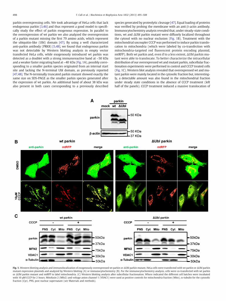
parkin overexpressing cells. We took advantage of HeLa cells that lackendogenous parkin [3,46] and thus represent a good model to specifi-cally study the effect of parkin exogenous expression. In parallel tothe overexpression of wt parkin we also analyzed the overexpressionof a parkin mutant missing the first 79 amino acids, which representthe ubiquitin-like (Ubl) domain [47]. By using a well characterizedanti-parkin antibody (PRK8) [5,48], we found that endogenous parkinwas not detectable by Western blotting analysis in empty vectortransfected HeLa cells, while exogenously introduced wt parkin wasdetected as a doublet with a strong immunoreactive band at ~50 kDaand aweaker fastermigrating band at ~40 kDa (Fig. 1A), possibly corre-sponding to a smaller parkin species originated from an internal startsite and lacking the N-terminal Ubl domain, as previously reported[47,48]. The N-terminally truncated parkin mutant showed exactly thesame size on SDS-PAGE as the smaller parkin species generated afterthe expression of wt parkin. An additional band of about 38 kDa wasalso present in both cases corresponding to a previously described
Fig. 1.Western blotting analysis and immunolocalization of exogenously overexpressed wt pmutant expression plasmids and analyzed by Western blotting (A) or immunocytochemistror ΔUbl parkin mutant and mtRFP to label mitochondria. (C) Western blotting analysis aftwith 10 μM CCCP for 2 hours. Mitofusin 2 (Mfn2) and voltage anion channel 1 (VDAC1) werfraction (Cyt). PNS, post nuclear supernatant (see Materials and methods).
species generated by proteolytic cleavage [47]. Equal loading of proteinswas verified by probing the membrane with an anti β-actin antibody.Immunocytochemistry analysis revealed that, under steady-state condi-tions, wt and ΔUbl parkin mutant were diffusely localized throughoutthe cytosol with no nuclear exclusion (Fig. 1B). Treatment with themitochondrial uncoupler CCCPwas performed to induce parkin translo-cation to mitochondria (which were labeled by co-transfection withmitochondria-targeted red fluorescent protein encoding plasmid,mtRFP). Both wt parkin and, even if to a less extent, ΔUbl parkin mu-tant were able to translocate. To better characterize the intracellulardistribution of our overexpressedwt andmutant parkin, subcellular frac-tionation experiments were performed in control and CCCP treated cells(Fig. 1C). Western blot analysis revealed that overexpressed wt andmu-tant parkinweremainly located in the cytosolic fraction but, interesting-ly, a detectable amount was also found in the mitochondrial fractionunder steady state conditions in the absence of CCCP treatment (lefthalf of the panels). CCCP treatment induced a massive translocation of
arkin or ΔUbl parkin mutant. HeLa cells were transfected with wt parkin or ΔUbl parkiny (B). For the immunocytochemistry analysis, cells were co-transfected with wt parkiner subcellular fractionation. Where indicated the different cell batches were incubatede used as positive controls for mitochondria fraction (Mito), α-tubulin for the cytosolic
499T. Calì et al. / Biochimica et Biophysica Acta 1832 (2013) 495–508
exogenous wt parkin to the mitochondrial fraction, accordingly to thedata shown by immunocytochemistry analysis. Mitochondrial transloca-tion was also appreciated for the parkin mutant, even if less pronouncedin respect to wt parkin, suggesting that the deletion of the Ubl domainimpaired (but did not abolish) the translocation of parkin to mitochon-dria under depolarization conditions. Mitofusin 2 (Mfn2), the outermitochondrial membrane voltage gated anion channel VDAC1 andα-tubulin were used as markers for the mitochondrial and the cytosolicfractions, respectively.
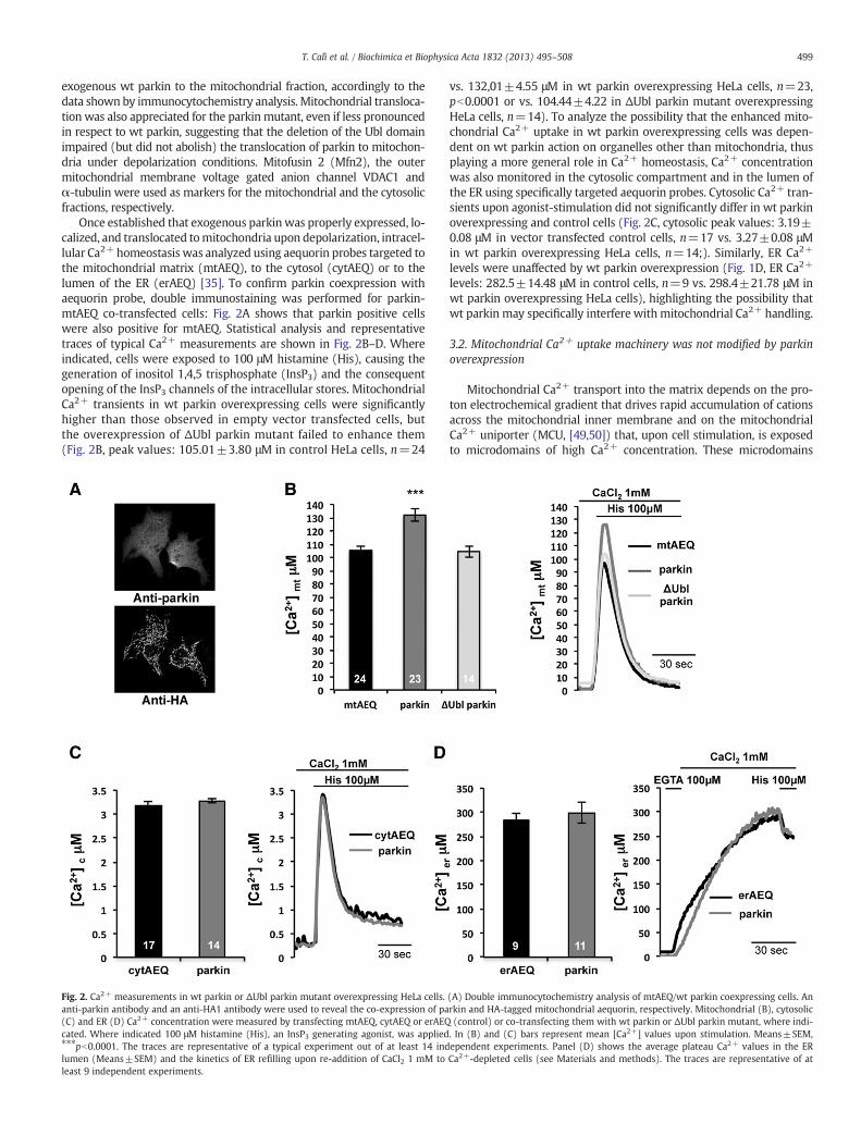
Once established that exogenous parkin was properly expressed, lo-calized, and translocated tomitochondria upon depolarization, intracel-lular Ca2+ homeostasis was analyzed using aequorin probes targeted tothe mitochondrial matrix (mtAEQ), to the cytosol (cytAEQ) or to thelumen of the ER (erAEQ) [35]. To confirm parkin coexpression withaequorin probe, double immunostaining was performed for parkin-mtAEQ co-transfected cells: Fig. 2A shows that parkin positive cellswere also positive for mtAEQ. Statistical analysis and representativetraces of typical Ca2+ measurements are shown in Fig. 2B–D. Whereindicated, cells were exposed to 100 μM histamine (His), causing thegeneration of inositol 1,4,5 trisphosphate (InsP3) and the consequentopening of the InsP3 channels of the intracellular stores. MitochondrialCa2+ transients in wt parkin overexpressing cells were significantlyhigher than those observed in empty vector transfected cells, butthe overexpression of ΔUbl parkin mutant failed to enhance them(Fig. 2B, peak values: 105.01±3.80 μM in control HeLa cells, n=24
Fig. 2. Ca2+ measurements in wt parkin or ΔUbl parkin mutant overexpressing HeLa cells.anti-parkin antibody and an anti-HA1 antibody were used to reveal the co-expression of pa(C) and ER (D) Ca2+ concentration were measured by transfecting mtAEQ, cytAEQ or erAEQcated. Where indicated 100 μM histamine (His), an InsP3 generating agonist, was applied⁎⁎⁎pb0.0001. The traces are representative of a typical experiment out of at least 14 indlumen (Means±SEM) and the kinetics of ER refilling upon re-addition of CaCl2 1 mM toleast 9 independent experiments.
vs. 132,01±4.55 μM in wt parkin overexpressing HeLa cells, n=23,pb0.0001 or vs. 104.44±4.22 in ΔUbl parkin mutant overexpressingHeLa cells, n=14). To analyze the possibility that the enhanced mito-chondrial Ca2+ uptake in wt parkin overexpressing cells was depen-dent on wt parkin action on organelles other than mitochondria, thusplaying a more general role in Ca2+ homeostasis, Ca2+ concentrationwas also monitored in the cytosolic compartment and in the lumen ofthe ER using specifically targeted aequorin probes. Cytosolic Ca2+ tran-sients upon agonist-stimulation did not significantly differ in wt parkinoverexpressing and control cells (Fig. 2C, cytosolic peak values: 3.19±0.08 μM in vector transfected control cells, n=17 vs. 3.27±0.08 μMin wt parkin overexpressing HeLa cells, n=14;). Similarly, ER Ca2+
levels were unaffected by wt parkin overexpression (Fig. 1D, ER Ca2+
levels: 282.5±14.48 μM in control cells, n=9 vs. 298.4±21.78 μM inwt parkin overexpressing HeLa cells), highlighting the possibility thatwt parkin may specifically interfere withmitochondrial Ca2+ handling.
3.2. Mitochondrial Ca2+ uptake machinery was not modified by parkinoverexpression
Mitochondrial Ca2+ transport into the matrix depends on the pro-ton electrochemical gradient that drives rapid accumulation of cationsacross the mitochondrial inner membrane and on the mitochondrialCa2+ uniporter (MCU, [49,50]) that, upon cell stimulation, is exposedto microdomains of high Ca2+ concentration. These microdomains
(A) Double immunocytochemistry analysis of mtAEQ/wt parkin coexpressing cells. Anrkin and HA-tagged mitochondrial aequorin, respectively. Mitochondrial (B), cytosolic(control) or co-transfecting them with wt parkin or ΔUbl parkin mutant, where indi-. In (B) and (C) bars represent mean [Ca2+] values upon stimulation. Means±SEM,ependent experiments. Panel (D) shows the average plateau Ca2+ values in the ERCa2+-depleted cells (see Materials and methods). The traces are representative of at
Fig. 3. Mitochondrial Ca2+ uptake in permeabilized cells and mitochondrial membranepotential in parkin overexpressing HeLa cells. (A) Representative traces (left) and averageplateau [Ca2+]m values (right, Means±SEM) reached in permeabilized cells exposed to1 μM Ca2+-buffered solution. Where indicated, the medium was switched from IB/EGTA-ATP to IB/1 μMCa2+. The traces are representative of at least 10 independent exper-iments. (B) Immunocytochemistry analysis on permeabilized HeLa cells overexpressingparkin. The stainingwithmonoclonal antibody against parkinwas revealed by AlexaFluor488 conjugated antibody. (C) HeLa cells (control and overexpressing parkin) were loadedwith TMRM probe to determine themitochondrial membrane potential (ΔΨ). Bars repre-sent the average TMRM fluorescence signals subtracted of signals remaining after FCCPtreatment to collapseΔΨ, and expressed as % Δ fluorescence (Means±SEM). The analysiswas performed on n=124mock cells and on n=264 parkin overexpressing cells, 3 inde-pendent experiments, ⁎pb0.05.
500 T. Calì et al. / Biochimica et Biophysica Acta 1832 (2013) 495–508
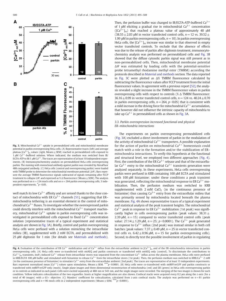
well match its low Ca2+ affinity and are sensed thanks to the close con-tact of mitochondria with ER Ca2+ channels [51], suggesting that ER–mitochondria tethering is an essential element in the control of mito-chondrial Ca2+ fluxes. To investigatewhether the overexpressed parkincould directly interfere with the mitochondrial Ca2+ transport machin-ery, mitochondrial Ca2+ uptake in parkin overexpressing cells was in-vestigated in permeabilized cells exposed to fixed Ca2+ concentrationsolution (representative traces of a typical experiment and statisticalanalysis are shown in Fig. 3A). Briefly, control and parkin overexpressingHeLa cells were perfused with a solution mimicking the intracellularmilieu (IB), supplemented with 2 mM EGTA, and permeabilized with25 μM digitonin for 1 min (for details see Materials and methods).
Fig. 4. Evaluation of the contribution of ER Ca2+ mobilization and of Ca2+ influx from theoverexpressing cells. (A) HeLa cells were co-transfected with mtAEQ and parkin constru[Ca2+]m transients, InsP3-induced Ca2+ release from intracellular stores was separated fromin KRB/EGTA 100 μM buffer and stimulated with histamine to release Ca2+ from the intrace(in the continuous presence of histamine) to stimulate Ca2+ entry from the extracellular amBars represent normalized mean [Ca2+] values upon stimulation, Means±SEM. ⁎⁎⁎pb0.0transfected with mtRFP/erGFP only (mock). Single plane images and three-dimensional recor in controls as indicated in each panel. Cells were excited separately at 488 nm or 543 nm,condition. Yellow indicates colocalization of the two organelles. Insets at higher magnificattotal of 40 images) with a 63× objective. (C) Manders’ coefficient for colocalization, caoverexpressing cells and n=96 mock cells in 2 independent experiments (Means±SEM, ⁎
Then, the perfusion buffer was changed to IB/EGTA-ATP-buffered Ca2+
of 1 μM eliciting a gradual rise in mitochondrial Ca2+ concentration([Ca2+]m) that reached a plateau value of approximately 40 μM(38.55±2.03 μM in vector transfected control cells, n=12 vs. 39.52±1.99 μMinparkin overexpressing cells, n=10). In parkin overexpressingHeLa cells, the [Ca2+]m increase was similar to that observed in emptyvector transfected controls. To exclude that the absence of effectswas due to the release of parkin after digitonin treatment, immunocyto-chemistry analysis was performed on permeabilized cells and Fig. 3Bshowed that the diffuse cytosolic parkin signal was still present as innon-permeabilized cells. Then, mitochondrial membrane potentialΔΨ was estimated by loading cells with the potential-sensitiveprobe tetramethyl rhodamine methyl ester (TMRM) according theprotocols described inMaterial andmethods section. The data reportedin Fig. 3C were plotted as Δ% TMRM fluorescence calculated bysubtracting thefluorescence values after FCCP treatment from the initialfluorescence values. In agreement with a previous report [52], the analy-sis revealed a slight increase in the TMRM fluorescence values in parkinoverexpressing cells with respect to controls (% Δ TMRM fluorescence:36.16±0.99 in vector transfected control cells, n=124 vs. 40.24±0.70in parkin overexpressing cells, n=264, pb0.05) that is consistent withamild increase in the driving force formitochondrial Ca2+ accumulation,that however did not influence the intrinsic capacity of mitochondria totake up Ca2+ in permeabilized cells as shown in Fig. 3A.
3.3. Parkin overexpression increased functional and physicalER–mitochondria interactions
The experiments on parkin overexpressing permeabilized cells(Fig. 3A) excluded a direct involvement of parkin in the modulation ofthe activity of mitochondrial Ca2+ transporters. A possible explanationfor the action of parkin on mitochondrial Ca2+ homeostasis couldmatch with a role in the formation and/or the stabilization of ER–mitochondria interactions. To verify this hypothesis at the functionaland structural level, we employed two different approaches (Fig. 4).First, the contribution of the ER Ca2+ release and that of the extracellu-lar Ca2+ entry to the mitochondrial Ca2+ transients generation wereanalyzed separately. In these experiments, HeLa cells overexpressingparkin were perfused in KRB containing 100 μM EGTA and stimulatedwith 100 μM histamine: under these conditions a peak transientwas generated, reflecting themitochondrial response to ER Ca2+mo-bilization. Then, the perfusion medium was switched to KRBsupplemented with 2 mM CaCl2 (in the continuous presence ofhistamine) thus causing Ca2+ entry from the extracellular milieu thatwas primarily sensed by mitochondria located beneath the plasmamembrane. Fig. 4A shows representative traces of a typical experimentand statistical analysis of the peak transient heights. The mitochondrialCa2+ peak in response to ER Ca2+ mobilization (1st peak) was signifi-cantly higher in cells overexpressing parkin (peak values: 38.31±2.39 μM, n=15) compared to vector transfected control cells (peakvalue: 27.14±1.29 μM, n=25; pb0.0001). The Ca2+ peak generatedin response to Ca2+ influx (2nd peak) was instead similar in both cellbatches (peak values: 7.37±0.49 μM, n=25 in vector transfected con-trol cells vs. 6.42±0.96 μM, n=15 for parkin overexpressing cells).Second, to directly test the possible involvement of parkin in regulating
extracellular ambient to [Ca2+]m and of the ER–mitochondria interactions in parkincts or transfected with mtAEQ only (control). To discriminate the contributions tothe concomitant Ca2+ influx across the plasma membrane. HeLa cells were perfused
llular stores (1st peak). Then, the perfusion medium was switched to KRB/Ca2+ 2 mMbient (2nd peak). The traces are representative of at least 15 independent experiments.001. (B) HeLa cells were co-transfected with mtRFP/erGFP and parkin constructs oronstruction of mitochondria (red) and ER (green) in HeLa cells overexpressing parkinand the single images were recorded. The merging of the two images is shown for eachion are also shown. Confocal stacks were acquired every 0.2 μm along the z-axis (for alculated from z-axis confocal stacks. The analysis was performed on n=81 parkin⁎⁎pb0.0001).
501T. Calì et al. / Biochimica et Biophysica Acta 1832 (2013) 495–508
ER–mitochondria contact sites, we performed live-cell confocal micros-copy three-dimensional reconstructions of ER and mitochondria. HeLacells were co-transfected with mtRFP and the ER-targeted GFP (erGFP)together with either parkin or empty vector and ER–mitochondriaco-localization was assessed. A detailed confocal microscopy analysis
carried out by acquiring 25 to 30 confocal z-axis stacks and applying vol-ume rendering on the 3D reconstructions showed anenhancement in theER/mitochondria co-localization in parkin overexpressing cells comparedto vector transfected control cells. Fig. 4B shows representative images atthe single plane level and after rendering of the z-stacks. The area of
502 T. Calì et al. / Biochimica et Biophysica Acta 1832 (2013) 495–508
co-localization of the two organelles is clearly appreciated in the mergeand inset panels. A quantification of the area of co-localization wasobtained by calculating Manders' coefficient [44] and revealed an in-crease of about 10% in parkin overexpressing cells compared to vector ex-pressing control cells (Fig. 4C, mock cells, 100±1.34%, n=96 and parkin109.38±1.10%, n=81 cells, pb0.0001). Altogether, the data presentedso far indicated that parkin is able to enhance mitochondrial Ca2+ tran-sients by potentiating the ER–mitochondria connections.
3.4. Parkin overexpression enhanced agonist-stimulated ATP production
To assess whether the favored ER–mitochondrial coupling, and theconsequent enhancement in mitochondrial Ca2+ transients couldhave a role in energetic cellular balance, we monitored cytosolicand mitochondrial ATP levels with specifically targeted luciferasescytLuc and mtLuc [36] both in control and parkin overexpressingcells. Briefly, cells were perfused with KRB, supplemented with glu-cose (5.5 mM) and 1 mM CaCl2 and then shifted to the same buffersupplemented with 20 μM luciferin until a plateau was reached,where indicated cells were challenged with histamine (100 μM) anda second plateau is generated corresponding to the amount ofnewly synthetized ATP. The data are represented as % of counts persecond (cps) normalized with respect to the first plateau. Fig. 5Ashowed that cytosolic ATP levels were significantly enhanced inparkin overexpressing cells as compared to control cells in response
Fig. 5. Functional analysis of energetic metabolism in parkin overexpressing HeLa cells. Cytosand mtLuc recombinant luciferase and after histamine stimulation. Data are expressed as pesecond. Data are typical of at least 8 independent experiments which gave the same result
to agonist stimulation, (128.48±1.22 %, n=8 in vector transfected cellsvs. 135.16±0.81 %, n=12 in parkin overexpressing cells, pb0.0001).However, mtLuc probe failed to revealed differences in mitochondrialATP production between the two cell batches (Fig. 5B, 146.45±1.51 %,n=20 in vector transfected cells and 145.10±1.70 %, n=13 in parkinoverexpressing cells).
These data indicate that parkin not only may have role inguaranteeing homeostatic Ca2+ fluxes from ER to mitochondria butalso in supporting ATP production.
3.5. siRNA-mediated parkin down-regulation caused mitochondrialfragmentation, compromised mitochondrial Ca2+ transients and reducedER–mitochondria contact sites in human dopaminergic neuroblastomacells
To clarify whether the observed enhancement of ER–mitochondriacommunication was a condition that had functional consequencesonly after parkin overexpression, or may also have a constitutiverole inmaintaining ER–mitochondria functionswe decided to performsiRNA-mediated parkin silencing in human dopaminergic SH-SY5Yneuroblastoma cells, a cellular model harboring endogenous parkinprotein, albeit at low levels [12,21,53]. Before performing experimentsin parkin-silenced cells, the effects of parkin overexpression inSH-SY5Y cells were tested. Figs. 6A and B show Western blotting andimmunocytochemistry analysis performed in control cells and in
olic (A) and mitochondrial (B) ATP production was evaluated by co-transfecting cytLucrcent of cytLuc or mtLuc light output of cells before agonist stimulation. cps, counts pers. Bars indicate Means±SEM, ⁎⁎⁎pb0.0001.
Fig. 6.Western blotting analysis, immunolocalization and Ca2+ measurements in parkin overexpressing SH-SY5Y cells. SH-SY5Y cells were transfected with parkin expression plas-mid and analyzed by Western Blotting (A) or immunocytochemistry (B). mtRFP was co-transfected to visualize mitochondria and where indicated CCCP treatment was performedto induce parkin translocation to mitochondria. Cells were transfected with mtAEQ, cytAEQ or erAEQ (control) or co-transfected with mtAEQ, cytAEQ or erAEQ and parkin. Mito-chondrial (C), cytosolic (D) and ER (E) Ca2+ concentration in SH-SY5Y cells overexpressing parkin. Where indicated 100 nM bradykinin (BK), an InsP3 generating agonist, was ap-plied. In (C) and (D) bars represent mean [Ca2+] values upon stimulation. Means±SEM, ⁎⁎pb0.001. The traces are representative of a typical experiment out of at least 3independent experiments. Panel (E) shows the average plateau Ca2+ values in the ER lumen (Means±SEM) and the kinetics of ER refilling upon re-addition of CaCl2 1 mM toCa2+-depleted cells (see Materials and methods). The traces are representative of at least 5 independent experiments.
503T. Calì et al. / Biochimica et Biophysica Acta 1832 (2013) 495–508
parkin overexpressing cells. Parkin overexpressing cells clearlyrevealed the presence of a doublet at ~50 kDa and ~40 kDa, asobserved in transfected HeLa cells. Endogenous parkin was not visible,possibly due to its very low level of expression and to the strong immu-noreactivity of the overexpressed protein that could obscure its detec-tion (see below). Immunocytochemistry analysis revealed a diffusecytosolic signal and, similarly to what observed for HeLa cells,overexpressed parkin displayed the ability to translocate to mitochon-dria upon CCCP treatment (Fig. 6B). Mitochondria, cytosolic and ERCa2+ levels were thus monitored in SH-SY5Y cells with specificallytargeted aequorins and the results are reported in Fig. 6C, D and E,respectively.
Mitochondrial Ca2+ transients in parkin SH-SY5Y overexpressingcells were significantly higher than those measured in empty vector
transfected cells (Fig. 6C, peak values: 133.42±5.38 μM in parkinoverexpressing SH-SY5Y cells, n=6 vs 107.48±6.48 μM in controlcells, n=7, pb0.001).
Cytosolic Ca2+ transients upon agonist-stimulation and ER Ca2+
levels resulted not significantly different in parkin overexpressingand control cells (Fig. 6D, cytosolic peak values: 1.95±0.03 μM invector transfected control cells, n=3 vs. 1.78±0.04 μM in parkinoverexpressing SH-SY5Y cells, n=4; Fig. 6E, ER Ca2+ levels: 235±10.38 μM in control cells, n=7 vs. 266±16.66 μM in parkinoverexpressing SH-SY5Y cells, n=5). These data indicated that the ef-fects on mitochondrial Ca2+ homeostasis observed following parkinoverexpression were not cell type-specific and supported the evidencethat the modulation of mitochondrial Ca2+ homeostasis represents aspecific target of parkin action.
504 T. Calì et al. / Biochimica et Biophysica Acta 1832 (2013) 495–508
Then, we considered siRNA experiments. Fig. 7A showed theWesternblotting analysis of siRNA dose-dependent reduction of parkin expres-sion level following incubation with different amount of two indepen-dent validated parkin siRNA (siRNA 9 and siRNA 10). The quantificationwas carried out by densitometric analysis and the amounts parkin levelsare indicated by the numbers below the image as values normalizedwithrespect to the β-actin levels. In this case, endogenous parkin levels werevisible, even if it was necessary to overexpose the nitrocellulose mem-brane to reveal it.
Once confident on the parkin siRNA efficiency, we performed 18pmol siRNA treatments in SH-SY5Y control cells and analyzed their
Fig. 7. Silencing of parkin in SH-SY5Y cells impairs mitochondrial morphology and Ca2+ transiRNA (scr siRNA) and parkin siRNA transfected SH-SY5Y cells. Two doses (12 pmol and 18 pevaluate the siRNA efficiency. The numbers below the blotting refer to normalized parkin/β−siRNA 10 or scr siRNA and mtRFP were co-transfected in SH-SY5Y cells. After 36–48 h cells,The Panel displays representative mitochondrial phenotypes observed by monitoring mtRFmitochondrial circularity (C), perimeter (D) and area (E), see [45] for details. The valuesexpressed as %. Means±SEM, *pb0.01, at least n=47 cells/conditions, three independentsiRNA 9 or parkin siRNA 10 or scramble siRNA SH-SY5Y treated cells; where indicated 10The traces are representative of at least 7 independent experiments. Means±SEM, ⁎⁎pb0.0
mitochondrial morphology by monitoring the fluorescence signal ofthe co-transfected mtRFP. The analysis of the mitochondrial networkintegrity revealed that, when the scramble siRNA was applied, themajority of the cells showed an intact network of tubular mitochon-dria, but when the cells were incubated with parkin siRNA 9 orsiRNA 10 mitochondria lose their tubular morphology and appearedtruncated and fragmented, as shown in Fig. 7B and in agreement towhat previously reported in several different cell types [54,55]. Aquantitative analysis of the mitochondrial network integrity wasperformed using the “Mito-Morphology” macro of the Image J soft-ware [45]. The measure of mitochondrial morphological parameters
sients. (A) Western blotting and quantification of siRNA-mediated silencing in scramblemol, respectively) of two different parkin siRNA (siRNA 9 and siRNA 10) were applied toactin ratio±SEM obtained in 3 independent experiments. (B) Parkin siRNA 9 or parkinwere observed under fluorescence microscope to evaluate mitochondrial morphology.P fluorescence. (C, D and E) Quantification of mitochondrial morphology by calculatingwere normalized with respect to the values calculated in scr siRNA treated cells andexperiments. (F) Mitochondrial Ca2+ transients induced by cell stimulation in parkin0 nM bradykinin (BK) was applied. Bars indicate the average heights of peak values.01, ⁎⁎⁎pb0.0001.
505T. Calì et al. / Biochimica et Biophysica Acta 1832 (2013) 495–508
showed a significant increase in mitochondrial average circularity(Fig. 7C) in both parkin siRNA treated cell batches and a concomitantsignificant decrease in the average perimeter and area of the singleobjects analyzed (Figs. 7D and E, respectively). These results stronglyindicated that endogenous parkin was required to maintain the prop-er mitochondrial tubular architecture. Mitochondrial Ca2+ measure-ments were then performed. As shown in Fig. 7 F, parkin siRNA 9and 10 but not scramble siRNA treatment affected the ability of mito-chondria to take up Ca2+, being the mitochondrial peak height of98.60±2.48 μM in scrambled siRNA control cells, n=21 vs. 78.71±3.26 μM in parkin siRNA 9 treated cells, n=22, pb0.001 and77.39±2.54 in parkin siRNA 10 treated cells, n=7, pb0.0001.
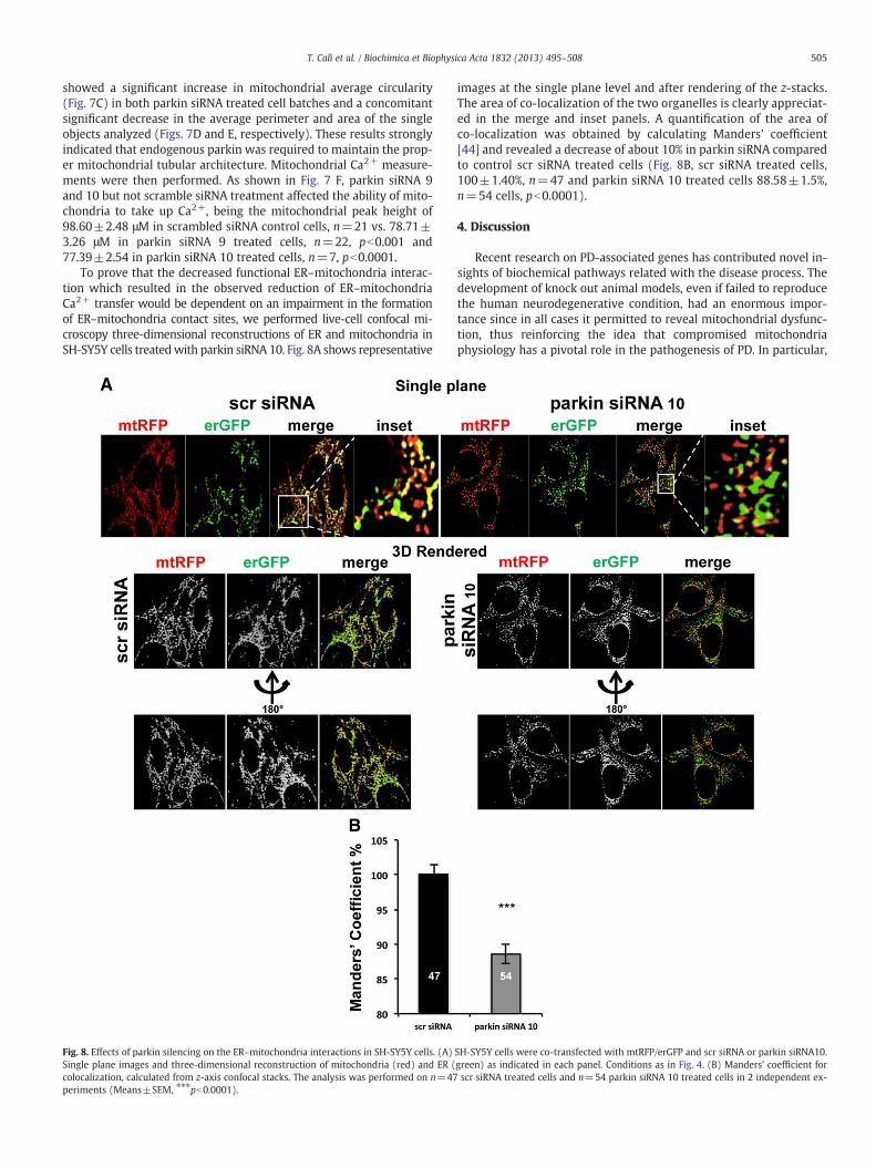
To prove that the decreased functional ER–mitochondria interac-tion which resulted in the observed reduction of ER–mitochondriaCa2+ transfer would be dependent on an impairment in the formationof ER–mitochondria contact sites, we performed live-cell confocal mi-croscopy three-dimensional reconstructions of ER and mitochondria inSH-SY5Y cells treatedwith parkin siRNA 10. Fig. 8A shows representative
Fig. 8. Effects of parkin silencing on the ER–mitochondria interactions in SH-SY5Y cells. (A)Single plane images and three-dimensional reconstruction of mitochondria (red) and ER (colocalization, calculated from z-axis confocal stacks. The analysis was performed on n=47periments (Means±SEM, ⁎⁎⁎pb0.0001).
images at the single plane level and after rendering of the z-stacks.The area of co-localization of the two organelles is clearly appreciat-ed in the merge and inset panels. A quantification of the area ofco-localization was obtained by calculating Manders' coefficient[44] and revealed a decrease of about 10% in parkin siRNA comparedto control scr siRNA treated cells (Fig. 8B, scr siRNA treated cells,100±1.40%, n=47 and parkin siRNA 10 treated cells 88.58±1.5%,n=54 cells, pb0.0001).
4. Discussion
Recent research on PD-associated genes has contributed novel in-sights of biochemical pathways related with the disease process. Thedevelopment of knock out animal models, even if failed to reproducethe human neurodegenerative condition, had an enormous impor-tance since in all cases it permitted to reveal mitochondrial dysfunc-tion, thus reinforcing the idea that compromised mitochondriaphysiology has a pivotal role in the pathogenesis of PD. In particular,
SH-SY5Y cells were co-transfected with mtRFP/erGFP and scr siRNA or parkin siRNA10.green) as indicated in each panel. Conditions as in Fig. 4. (B) Manders' coefficient forscr siRNA treated cells and n=54 parkin siRNA 10 treated cells in 2 independent ex-
506 T. Calì et al. / Biochimica et Biophysica Acta 1832 (2013) 495–508
reduced levels of mitochondrial proteins involved in mitochondrialoxidative phosphorylation were reported in parkin-knockout mice,which exhibited normal brain morphology, but increased striatalextracellular dopamine levels [56]. Increased parkin levels wereassociated with protection from cellular stress and cell cycle regulation[57–59] and overexpression of parkin in cultured cells not only rescuedmitochondrial dysfunction caused by PINK1 loss of function, but exerteda cytoprotective role against different toxic stressor [19–23,26]. Severalstudies have elucidated the PINK1/parkin pathway, demonstrating thatPINK1 acted as sensor ofmitochondrial damage [60] necessary to specif-ically recruit parkin on dysfunctionalmitochondria and to promote theirautophagic degradation [3–5,12]. If the action of parkin on damagedmi-tochondria is well recognized, its role in physiological condition is stillobscure. The finding that, after ER stress induction, parkin gene was in-duced and mitochondrial degeneration, which normally occurred inthese conditions, was prevented [27], suggested that parkin may alsohave a cytoprotective role independently to its mitochondria transloca-tion and consequent mitophagy activation. We searched for this role byinvestigating the ER–mitochondria relationship in terms of Ca2+ signal-ing, since Ca2+ dyshomeostasis and ER–mitochondria coupling play aprominent role in neuronal cell death.
To date, only one study reported possible involvement of parkin inCa2+ homeostasis. In the study, parkin silenced cells and cells ex-pressing parkin mutants displayed increased basal cytosolic Ca2+
levels and longer lasting responses following agonist stimulation.These alterations in Ca2+ homeostasis were attributed to increasedlevels of phospholipase Cγ1 in parkin knock out cells, and the possi-bility that impaired ability of mitochondria to take up Ca2+ could par-ticipate in compromising buffering capacity was excluded [21].
Our study demonstrated that parkin overexpression specifically in-duced an increase in the mitochondrial Ca2+ transients evoked by cellstimulation and that such increase was not observed by overexpressinga parkin N-terminal truncation mutant lacking the Ubl domain,suggesting that this domain is necessary to this parkin function. In-terestingly, the entity of the transients was not only compatiblewith physiological levels of mitochondrial Ca2+ concentration, butalso positively modulated ATP production thus sustaining cell bioen-ergetics. Themechanism underlining enhanced cytosolic ATP levels arepresently unclear: it could bedependent on a secondary effect related toincreased ATP translocation from mitochondria to cytosol through theATP/ADP exchanger, possibly enhanced by the slight mitochondrial hy-perpolarization [61] (accordingly to the increased TMRM fluorescence)in parkin overexpressing cells. The effect on mitochondrial membranepotential was previously observed and it was attributed to enhancedcomplex I levels and activity [52,62].
The rate of mitochondrial ATP synthesis in parkin overexpressingcells could keep pace with that of ATP translocation from mitochon-dria to cytoplasm, thus making changes in mitochondrial ATP similarto those measured in control cells.
Our results suggested that parkin is essential to maintain mito-chondrial network integrity possibly by sustaining ER–mitochondriaconnections and guaranteeing the proper ER–mitochondria Ca2+
transfer. Interestingly, we have obtained similar evidence also forα-synuclein [63] and DJ-1 (unpublished data), suggesting that thisaction may represent a common mechanism shared by differentPD-related proteins.
We have also found that, when this action was missing (i.e., inparkin downregulated cells), the ER–mitochondria tethering was re-duced, the agonist-stimulated Ca2+ entry in mitochondrial matrixwas compromised, and cells showed alterations of the mitochondrialnetwork, underlining that molecular mechanisms that coordinate theinterplay between ER and mitochondria are relevant to the control ofmitochondria integrity. Even if our study was limited to the analysisof the ER–mitochondria Ca2+ transfer, which is only one of multipleparameters that could be compromised by the impairment of ER–mitochondrial tethering, our data on ATP are consistent with the
finding that mitochondria from parkin KO mice displayed reducedmitochondrial respiratory activity and that signs of oxidative dam-age have been detected in brains of KO mice [56] and in fibroblast ofPD patients with parkin mutations [54,64].
The data presented here not only indicated that parkin is requiredfor guaranteeing ER–mitochondria contact sites, maintaining mito-chondrial Ca2+ homeostasis and network integrity, but also that itsoverexpression favored the ER–mitochondria connections and conse-quently the ER–mitochondria Ca2+ transfer, thus enhancing ATP pro-duction following cell stimulation. Parkin overexpression has beenshown to represent a strategic mechanism adopted by the cells to over-come ER and mitochondrial stress [27], however it was not clear theprecise role of parkin in the communication between these two organ-elles. The enhanced cytosolic ATP levels could in turn provide fuel toaliment cell protective responses, and prolong cell survival.
The next step will be to figure out the precise mechanism of parkinin mediating ER–mitochondria contact sites: suggestions come fromthe finding that parkin, similarly to α-synuclein and DJ-1 [65], hasbeen shown to interact with grp75 (mortalin) and to rescue mitochon-drial dysfunctions induced by its down regulation [66]. Since grp75 hasbeen demonstrated to couple InsP3 receptors with VDAC1, and in thisway regulate the mitochondria Ca2+ uptake machinery [67], the possi-bility that parkin could act downstream grp75 must be considered. Thepossible involvement of mitofusins is also intriguing, since mitofusin 2has been demonstrated to tether ER tomitochondria [43]. Parkin medi-ated mitofusin ubiquitination may provide a mechanism to label mito-chondria destined to degradation by mitophagy but also act as firsttentative of rescue by sustaining cellular bioenergetics through the en-gagement of ER–mitochondria connections. Subcellular fractionationexperiments of parkin overexpressing cells indicated that, in ouroverexpression conditions (and under basal conditions) overexpressedparkin ismostly cytosolic. However, the detection of parkin signal in themitochondrial fraction, where Mfn2 and VDAC1 (two proteins of theouter mitochondrial membrane that also participate to the formationof the ER–mitochondria contact sites) were specifically detected,may suggest a possible preferentially parkin localization near theER–mitochondria contact sites in respect to other cytosolic proteins,as documented by the absence of α-tubulin in that fraction.
On the basis of these results, it could be proposed that parkin mayhave a versatile role according to its localization that, in turn, dependson the cell/mitochondria status. When mitochondria were severelycompromised (depolarized), parkin is selectively recruited to damagedmitochondria and promotes their removal by mitophagy; when theyare still functional, parkin stays in the cytoplasm (and possibly nearthe ER–mitochondria contact sites) and promotes mitochondria activi-ties by regulating Ca2+ fluxes enhancing ER–mitochondria coupling.
5. Conclusions
In summary, we have shown that parkin, by favoring ER–mitochondria tethering, also favored Ca2+ transfer from the ER to mi-tochondria, sustained organellemorphology andATP production.Whenthe protein is defective, ER–mitochondria contact sites are reduced,Ca2+ uptake is impaired and mitochondria undergo massive fragmen-tation. These data account for a role for parkin independent from itsmi-tochondrial translocation, suggesting that it could exert its protectiverole at two different levels. The first level may be activated by mecha-nisms that increase parkin expression, as documented by Bouman andcoworkers [27]. Here we propose that parkin overexpression couldcompensate for possible mitochondria impairment by enhancing ATPproduction thank to the ability to modulate ER–mitochondria Ca2+
transfer. This aspect is emerging as a key element in cell bioenergeticsas recently documented by studies showing that mitochondria elonga-tion occurring during autophagy is determinant to increase ATPproduc-tion and sustain cell viability [68]. The second level of defense isactivatedwhen this compensationwas not sufficient, and cellular stress
507T. Calì et al. / Biochimica et Biophysica Acta 1832 (2013) 495–508
is too extended to be counteracted: at this point the removal of dam-aged mitochondria become necessary and inevitable to preserve thehealthy population, thus parkin is recruited to mitochondria and pro-mote mitophagy according to a well-recognized pathway.
Acknowledgements
The authors wish to thank Dr. Laura Fedrizzi (University of Padova)for performing Ca2+ measurements on SH-SY5Y cells overexpressingparkin. The work was supported by the Italian Ministry of Universityand Research (PRIN 2008) and the local founding of the University ofPadova (Progetto di Ateneo 2008 CPDA082825) to M.B.
References
[1] M.T. Lin, M.F. Beal, Mitochondrial dysfunction and oxidative stress in neurode-generative diseases, Nature 443 (2006) 787–795.
[2] A.H. Schapira, Mitochondria in the aetiology and pathogenesis of Parkinson's dis-ease, Lancet Neurol. 7 (2008) 97–109.
[3] D. Narendra, A. Tanaka, D.F. Suen, R.J. Youle, Parkin is recruited selectively to im-pairedmitochondria and promotes their autophagy, J. Cell Biol. 183 (2008) 795–803.
[4] C. Vives-Bauza, C. Zhou, Y. Huang, M. Cui, R.L. de Vries, J. Kim, J. May, M.A.Tocilescu, W. Liu, H.S. Ko, J. Magrane, D.J. Moore, V.L. Dawson, R. Grailhe, T.M.Dawson, C. Li, K. Tieu, S. Przedborski, PINK1-dependent recruitment of Parkin tomitochondria in mitophagy, Proc. Natl. Acad. Sci. U. S. A. 107 (2010) 378–383.
[5] N. Matsuda, S. Sato, K. Shiba, K. Okatsu, K. Saisho, C.A. Gautier, Y.S. Sou, S. Saiki, S.Kawajiri, F. Sato, M. Kimura, M. Komatsu, N. Hattori, K. Tanaka, PINK1 stabilizedby mitochondrial depolarization recruits Parkin to damaged mitochondria andactivates latent Parkin for mitophagy, J. Cell Biol. 189 (2010) 211–221.
[6] S.R. Yoshii, C. Kishi, N. Ishihara, N. Mizushima, Parkin mediates proteasome-dependent protein degradation and rupture of the outer mitochondrial mem-brane, J. Biol. Chem. 286 (2011) 19630–19640.
[7] H. Shimura, N. Hattori, S. Kubo, Y. Mizuno, S. Asakawa, S. Minoshima, N. Shimizu,K. Iwai, T. Chiba, K. Tanaka, T. Suzuki, Familial Parkinson disease gene product,parkin, is a ubiquitin-protein ligase, Nat. Genet. 25 (2000) 302–305.
[8] H. Xiong, D. Wang, L. Chen, Y.S. Choo, H. Ma, C. Tang, K. Xia, W. Jiang, Z. Ronai, X.Zhuang, Z. Zhang, Parkin, PINK1, and DJ-1 form a ubiquitin E3 ligase complex pro-moting unfolded protein degradation, J. Clin. Invest. 119 (2009) 650–660.
[9] K.L. Lim, K.C. Chew, J.M. Tan, C. Wang, K.K. Chung, Y. Zhang, Y. Tanaka, W. Smith,S. Engelender, C.A. Ross, V.L. Dawson, T.M. Dawson, Parkin mediates nonclassical,proteasomal-independent ubiquitination of synphilin-1: implications for Lewybody formation, J. Neurosci. 25 (2005) 2002–2009.
[10] N.C. Chan, A.M. Salazar, A.H. Pham, M.J. Sweredoski, N.J. Kolawa, R.L. Graham, S.Hess, D.C. Chan, Broad activation of the ubiquitin–proteasome system by Parkinis critical for mitophagy, Hum. Mol. Genet. 20 (2011) 1726–1737.
[11] V.I. Korolchuk, A. Mansilla, F.M. Menzies, D.C. Rubinsztein, Autophagy inhibitioncompromises degradation of ubiquitin–proteasome pathway substrates, Mol.Cell 33 (2009) 517–527.
[12] S. Geisler, K.M.Holmstrom, D. Skujat, F.C. Fiesel, O.C. Rothfuss, P.J. Kahle,W. Springer,PINK1/Parkin-mediated mitophagy is dependent on VDAC1 and p62/SQSTM1, Nat.Cell Biol. 12 (2010) 119–131.
[13] A. Tanaka, M.M. Cleland, S. Xu, D.P. Narendra, D.F. Suen, M. Karbowski, R.J. Youle,Proteasome and p97 mediate mitophagy and degradation of mitofusins inducedby Parkin, J. Cell Biol. 191 (2010) 1367–1380.
[14] M.E. Gegg, J.M. Cooper, K.Y. Chau, M. Rojo, A.H. Schapira, J.W. Taanman, Mitofusin1 and mitofusin 2 are ubiquitinated in a PINK1/parkin-dependent manner uponinduction of mitophagy, Hum. Mol. Genet. 19 (2010) 4861–4870.
[15] E. Ziviani, R.N. Tao, A.J. Whitworth, Drosophila parkin requires PINK1 for mito-chondrial translocation and ubiquitinates mitofusin, Proc. Natl. Acad. Sci. U. S. A.107 (2010) 5018–5023.
[16] H. Wang, P. Song, L. Du, W. Tian, W. Yue, M. Liu, D. Li, B. Wang, Y. Zhu, C. Cao, J.Zhou, Q. Chen, Parkin ubiquitinates Drp1 for proteasome-dependent degradation:implication of dysregulated mitochondrial dynamics in Parkinson disease, J. Biol.Chem. 286 (2011) 11649–11658.
[17] D. Chen, F. Gao, B. Li, H. Wang, Y. Xu, C. Zhu, G. Wang, Parkin mono-ubiquitinatesBcl-2 and regulates autophagy, J. Biol. Chem. 285 (2010) 38214–38223.
[18] B.N. Johnson, A.K. Berger, G.P. Cortese, M.J. Lavoie, The ubiquitin E3 ligase parkinregulates the proapoptotic function of Bax, Proc. Natl. Acad. Sci. U. S. A. 109(2012) 6283–6288.
[19] H. Jiang, Y. Ren, J. Zhao, J. Feng, Parkin protects human dopaminergic neuroblastomacells against dopamine-induced apoptosis, Hum. Mol. Genet. 13 (2004) 1745–1754.
[20] T. Hasegawa, A. Treis, N. Patenge, F.C. Fiesel, W. Springer, P.J. Kahle, Parkinprotects against tyrosinase-mediated dopamine neurotoxicity by suppressingstress-activated protein kinase pathways, J. Neurochem. 105 (2008) 1700–1715.
[21] A. Sandebring, N. Dehvari, M. Perez-Manso, K.J. Thomas, E. Karpilovski, M.R.Cookson, R.F. Cowburn, A. Cedazo-Minguez, Parkin deficiency disrupts calcium ho-meostasis bymodulating phospholipase C signalling, FEBS J. 276 (2009) 5041–5052.
[22] F. Darios, O. Corti, C.B. Lucking, C. Hampe, M.P. Muriel, N. Abbas, W.J. Gu, E.C. Hirsch, T.Rooney,M. Ruberg, A. Brice, Parkin preventsmitochondrial swelling and cytochrome crelease in mitochondria-dependent cell death, Hum. Mol. Genet. 12 (2003) 517–526.
[23] L. Petrucelli, C. O'Farrell, P.J. Lockhart, M. Baptista, K. Kehoe, L. Vink, P. Choi, B.Wolozin, M. Farrer, J. Hardy, M.R. Cookson, Parkin protects against the toxicity as-sociated with mutant alpha-synuclein: proteasome dysfunction selectively af-fects catecholaminergic neurons, Neuron 36 (2002) 1007–1019.
[24] Y. Imai, M. Soda, H. Inoue, N. Hattori, Y. Mizuno, R. Takahashi, An unfolded puta-tive transmembrane polypeptide, which can lead to endoplasmic reticulumstress, is a substrate of Parkin, Cell 105 (2001) 891–902.
[25] Y. Imai, M. Soda, R. Takahashi, Parkin suppresses unfolded protein stress-inducedcell death through its E3 ubiquitin–protein ligase activity, J. Biol. Chem. 275(2000) 35661–35664.
[26] M. Bian, J. Liu, X. Hong, M. Yu, Y. Huang, Z. Sheng, J. Fei, F. Huang, Overexpression ofparkin ameliorates dopaminergic neurodegeneration induced by 1- methyl-4-phenyl-1,2,3,6-tetrahydropyridine in mice, PLoS One 7 (2012) e39953.
[27] L. Bouman, A. Schlierf, A.K. Lutz, J. Shan, A. Deinlein, J. Kast, Z. Galehdar, V.Palmisano, N. Patenge, D. Berg, T. Gasser, R. Augustin, D. Trumbach, I. Irrcher,D.S. Park, W. Wurst, M.S. Kilberg, J. Tatzelt, K.F. Winklhofer, Parkin is transcrip-tionally regulated by ATF4: evidence for an interconnection between mitochon-drial stress and ER stress, Cell Death Differ. 18 (2011) 769–782.
[28] C. Cardenas, R.A. Miller, I. Smith, T. Bui, J. Molgo, M. Muller, H. Vais, K.H. Cheung, J.Yang, I. Parker, C.B. Thompson, M.J. Birnbaum, K.R. Hallows, J.K. Foskett, Essentialregulation of cell bioenergetics by constitutive InsP3 receptor Ca2+ transfer tomitochondria, Cell 142 (2010) 270–283.
[29] G. Csordas, G. Hajnoczky, SR/ER–mitochondrial local communication: calciumand ROS, Biochim. Biophys. Acta 1787 (2009) 1352–1362.
[30] D.J. Surmeier, Calcium, ageing, and neuronal vulnerability in Parkinson's disease,Lancet Neurol. 6 (2007) 933–938.
[31] L. Bojarski, J. Herms, J. Kuznicki, Calcium dysregulation in Alzheimer's disease,Neurochem. Int. 52 (2008) 621–633.
[33] T. Cali, D. Ottolini, M. Brini, Mitochondria, calcium, and endoplasmic reticulumstress in Parkinson's disease, Biofactors 37 (2011) 228–240.
[34] T. Cali, D. Ottolini, M. Brini, Mitochondrial Ca(2+) and neurodegeneration, CellCalcium 52 (2012) 73–85.
[35] M. Brini, Calcium-sensitive photoproteins, Methods 46 (2008) 160–166.[36] L.S. Jouaville, P. Pinton, C. Bastianutto, G.A. Rutter, R. Rizzuto, Regulation of mito-
chondrial ATP synthesis by calcium: evidence for a long-term metabolic priming,Proc. Natl. Acad. Sci. U. S. A. 96 (1999) 13807–13812.
[37] R. Rizzuto, M. Brini, C. Bastianutto, R. Marsault, T. Pozzan, Photoprotein-mediatedmeasurement of calcium ion concentration in mitochondria of living cells,Methods Enzymol. 260 (1995) 417–428.
[38] R. Rizzuto, M. Brini, M. Murgia, T. Pozzan, Microdomains with high Ca2+ close toIP3-sensitive channels that are sensed by neighboring mitochondria, Science 262(1993) 744–747.
[39] E. Rapizzi, P. Pinton, G. Szabadkai, M.R. Wieckowski, G. Vandecasteele, G. Baird,R.A. Tuft, K.E. Fogarty, R. Rizzuto, Recombinant expression of the voltage-dependent anion channel enhances the transfer of Ca2+ microdomains to mito-chondria, J. Cell Biol. 159 (2002) 613–624.
[40] M. Brini, R. Marsault, C. Bastianutto, J. Alvarez, T. Pozzan, R. Rizzuto, Transfectedaequorin in the measurement of cytosolic Ca2+ concentration ([Ca2+]c). A criticalevaluation, J. Biol. Chem. 270 (1995) 9896–9903.
[41] M.J. Barrero, M. Montero, J. Alvarez, Dynamics of [Ca2+] in the endoplasmic retic-ulum and cytoplasm of intact HeLa cells. A comparative study, J. Biol. Chem. 272(1997) 27694–27699.
[42] M.R. Duchen, A. Surin, J. Jacobson, Imaging mitochondrial function in intact cells,Methods Enzymol. 361 (2003) 353–389.
[43] O.M. de Brito, L. Scorrano, Mitofusin 2 tethers endoplasmic reticulum to mito-chondria, Nature 456 (2008) 605–610.
[44] E.M. Manders, F.J. Verbeek, J.A. Aten, Measurement of co-localization of objects indual-colour confocal images, J. Microsc. 169 (1993) 375–382.
[45] R.K. Dagda, S.J. Cherra III, S.M. Kulich, A. Tandon, D. Park, C.T. Chu, Loss of PINK1function promotes mitophagy through effects on oxidative stress and mitochon-drial fission, J. Biol. Chem. 284 (2009) 13843–13855.
[46] S.R. Denison, F. Wang, N.A. Becker, B. Schule, N. Kock, L.A. Phillips, C. Klein, D.I.Smith, Alterations in the common fragile site gene Parkin in ovarian and othercancers, Oncogene 22 (2003) 8370–8378.
[47] I.H. Henn, J.M. Gostner, P. Lackner, J. Tatzelt, K.F. Winklhofer, Pathogenic mutationsinactivate parkin by distinct mechanisms, J. Neurochem. 92 (2005) 114–122.
[48] A.C. Pawlyk, B.I. Giasson, D.M. Sampathu, F.A. Perez, K.L. Lim, V.L. Dawson, T.M.Dawson, R.D. Palmiter, J.Q. Trojanowski, V.M. Lee, Novel monoclonal antibodiesdemonstrate biochemical variation of brain parkin with age, J. Biol. Chem. 278(2003) 48120–48128.
[49] J.M. Baughman, F. Perocchi, H.S. Girgis, M. Plovanich, C.A. Belcher-Timme, Y.Sancak, X.R. Bao, L. Strittmatter, O. Goldberger, R.L. Bogorad, V. Koteliansky, V.K.Mootha, Integrative genomics identifies MCU as an essential component of themitochondrial calcium uniporter, Nature 476 (2011) 341–345.
[50] D. De Stefani, A. Raffaello, E. Teardo, I. Szabo, R. Rizzuto, A forty-kilodalton proteinof the inner membrane is the mitochondrial calcium uniporter, Nature 476(2011) 336–340.
[51] R. Rizzuto, P. Pinton, W. Carrington, F.S. Fay, K.E. Fogarty, L.M. Lifshitz, R.A. Tuft, T.Pozzan, Close contacts with the endoplasmic reticulum as determinants of mito-chondrial Ca2+ responses, Science 280 (1998) 1763–1766.
[52] Y. Kuroda, T. Mitsui, M. Kunishige, T. Matsumoto, Parkin affects mitochondrialfunction and apoptosis in neuronal and myogenic cells, Biochem. Biophys. Res.Commun. 348 (2006) 787–793.
508 T. Calì et al. / Biochimica et Biophysica Acta 1832 (2013) 495–508
[53] Y. Machida, T. Chiba, A. Takayanagi, Y. Tanaka, M. Asanuma, N. Ogawa, A. Koyama,T. Iwatsubo, S. Ito, P.H. Jansen, N. Shimizu, K. Tanaka, Y. Mizuno, N. Hattori, Com-mon anti-apoptotic roles of parkin and alpha-synuclein in human dopaminergiccells, Biochem. Biophys. Res. Commun. 332 (2005) 233–240.
[54] H. Mortiboys, K.J. Thomas, W.J. Koopman, S. Klaffke, P. Abou-Sleiman, S. Olpin,N.W. Wood, P.H. Willems, J.A. Smeitink, M.R. Cookson, O. Bandmann, Mitochon-drial function and morphology are impaired in parkin-mutant fibroblasts, Ann.Neurol. 64 (2008) 555–565.
[55] A.K. Lutz, N. Exner, M.E. Fett, J.S. Schlehe, K. Kloos, K. Lammermann, B. Brunner, A.Kurz-Drexler, F. Vogel, A.S. Reichert, L. Bouman, D. Vogt-Weisenhorn, W. Wurst, J.Tatzelt, C. Haass, K.F. Winklhofer, Loss of parkin or PINK1 function increasesDrp1-dependent mitochondrial fragmentation, J. Biol. Chem. 284 (2009)22938–22951.
[56] J.J. Palacino, D. Sagi, M.S. Goldberg, S. Krauss, C. Motz, M. Wacker, J. Klose, J. Shen,Mitochondrial dysfunction and oxidative damage in parkin-deficient mice, J. Biol.Chem. 279 (2004) 18614–18622.
[57] T.M. Dawson, V.L. Dawson, The role of parkin in familial and sporadic Parkinson'sdisease, Mov. Disord. 25 (Suppl. 1) (2010) S32–S39.
[59] A. Ulusoy, D. Kirik, Can overexpression of parkin provide a novel strategy forneuroprotection in Parkinson's disease? Exp. Neurol. 212 (2008) 258–260.
[60] D.P. Narendra, S.M. Jin, A. Tanaka, D.F. Suen, C.A. Gautier, J. Shen, M.R. Cookson,R.J. Youle, PINK1 is selectively stabilized on impaired mitochondria to activateParkin, PLoS Biol. 8 (2010) e1000298.
[61] M. Klingenberg, The ADP and ATP transport in mitochondria and its carrier,Biochim. Biophys. Acta 1778 (2008) 1978–2021.
[62] Y. Kuroda, W. Sako, S. Goto, T. Sawada, D. Uchida, Y. Izumi, T. Takahashi, N.Kagawa, M. Matsumoto, R. Takahashi, R. Kaji, T. Mitsui, Parkin interacts withKlokin1 for mitochondrial import and maintenance of membrane potential,Hum. Mol. Genet. 21 (2012) 991–1003.
[63] T. Cali, D. Ottolini, A. Negro, M. Brini, alpha-Synuclein controls mitochondrial cal-cium homeostasis by enhancing endoplasmic reticulum–mitochondria interac-tions, J. Biol. Chem. 287 (2012) 17914–17929.
[64] A. Grunewald, L. Voges, A. Rakovic, M. Kasten, H. Vandebona, C. Hemmelmann, K.Lohmann, S. Orolicki, A. Ramirez, A.H. Schapira, P.P. Pramstaller, C.M. Sue, C. Klein,Mutant Parkin impairs mitochondrial function and morphology in human fibro-blasts, PLoS One 5 (2010) e12962.
[65] J. Jin, G.J. Li, J. Davis, D. Zhu, Y. Wang, C. Pan, J. Zhang, Identification of novel pro-teins associated with both alpha-synuclein and DJ-1, Mol. Cell Proteomics 6(2007) 845–859.
[66] H. Yang, X. Zhou, X. Liu, L. Yang, Q. Chen, D. Zhao, J. Zuo, W. Liu, Mitochondrialdysfunction induced by knockdown of mortalin is rescued by Parkin, Biochem.Biophys. Res. Commun. 410 (2011) 114–120.
[67] G. Szabadkai, K. Bianchi, P. Varnai, D. De Stefani, M.R. Wieckowski, D. Cavagna, A.I.Nagy, T. Balla, R. Rizzuto, Chaperone-mediated coupling of endoplasmic reticu-lum and mitochondrial Ca2+ channels, J. Cell Biol. 175 (2006) 901–911.
[68] L.C. Gomes, G. Di Benedetto, L. Scorrano, During autophagy mitochondria elon-gate, are spared from degradation and sustain cell viability, Nat. Cell Biol. 13(2011) 589–598.