34
Enterobacteriaceae Chapter 31
| Date post: | 14-Dec-2015 |
| Category: |
Documents |
| Upload: | ellen-grubb |
| View: | 221 times |
| Download: | 0 times |
Enterobacteriaceae
Chapter 31
Introduction “Enteric Bacteria” Gram-negative rods Ubiquitous Cause 30%-35% of all septicemias, more
than 70% of UTIs, and many intestinal infections
Pathogens: Normal flora – opportunistic infections Animal reservoirs Human carriers
Box 31-1
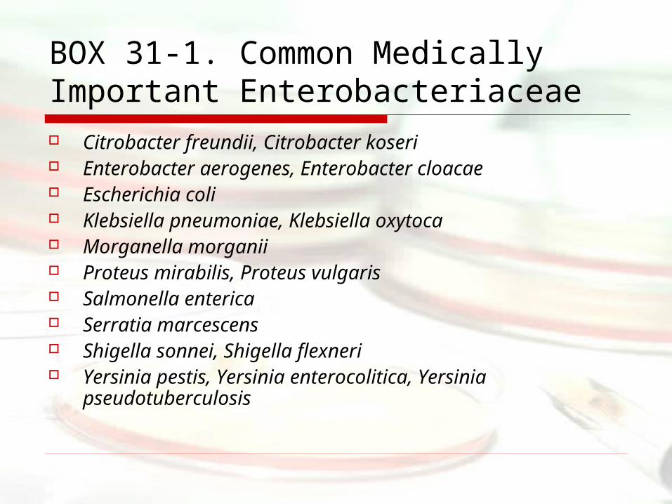
BOX 31-1. Common Medically Important Enterobacteriaceae Citrobacter freundii, Citrobacter koseri Enterobacter aerogenes, Enterobacter cloacae Escherichia coli Klebsiella pneumoniae, Klebsiella oxytoca Morganella morganii Proteus mirabilis, Proteus vulgaris Salmonella enterica Serratia marcescens Shigella sonnei, Shigella flexneri Yersinia pestis, Yersinia enterocolitica, Yersinia
pseudotuberculosis
Physiology and Structure
Facultative anaerobes Ferment glucose, are catalase
positive, and oxidase negative Lactose fermenting strains (e.g.
Escherichia, Klebsiella, Enterobacter) Non-lactose fermenting (e.g.
Salmonella, Shigella, and Yersinia)
Differentiating Similar Strains
Antigen detection O polysaccharides
Part of LPS Capsular K Flagellar H proteins E. coli O157:H7
Pathogenesis and ImmunityCommon virulence factors Endotoxin
Lipid A portion of LPS causes many of the systemic manifestations of infection
Capsule Interferes with antibody binding Capsular antigens are hydrophilic (phagocytic cell
surface is hydrophobic) Poor antigenicity
Antigenic phase variation Capsular K and flagellar H antigens are under genetic
control Can be expressed or not expressed
Pathogenesis and Immunity
Sequestration of growth factors Iron: Bacteria produce competitive
siderophores or iron-chelating compounds, hemolysins
Escherichia coli
Pathogenesis (Box 31-3) Adhesins: Essential for colonization
Prevents the organism from being flushed out of the urinary or gastrointestinal tract
Exotoxins Specific target tissue Result in altered cell function or cell
death
Epidemiology and Clinical Diseases Many infections are endogenous (septicemia and
UTI’s) Septicemia-originate from UT or GI infections leading
to intraabdominal infection Neonatal meningitis, Intraabdominal infections UTIs-originate in the colon -> contaminate urethra ->
ascend into the bladder Production of adhesins ~80% of all community-acquired UTIs
Gastroenteritis-caused by five major groups May include: watery diarrhea, abdominal cramps,
fever, and vomiting (Table 31-1)
Gastroenteritis (ETEC) Estimated 80,000 cases in US travelers annually (650
million worldwide) In small intestine; watery diarrhea, cramps, vomiting, fever Occurs in developing countries usually in children or
travelers (traveler’s diarrhea) 1-2 day incubation, 3-4 duration Infectious dose is high so person to person spread does not
occur Two classes of enterotoxins: heat-labile (LT-I, LT-II) and
heat-stable (STa, STb) LT-I increases secretion of chloride and inhibits absorption of
sodium and chloride (the same as cholera toxin) STa causes a hypersecretion of fluids
Both contributing to watery diarrhea Disease similar to cholera, but milder
Gastroenteritis (ETEC)
Imodium mode of action:http://en.wikipedia.org/wiki/Loperamide
Gastroenteritis (EHEC) 73,000 cases with 60 deaths annually In large intestine; vomiting, abdominal cramps, fever Severity ranges from diarrhea to hemorrhagic
colitis (bacterial dysentery) 3-4 day incubation, 4-10 day duration Infectious dose is less than 100 bacteria, O157:H7
serotype is the most common Read text page 329 Shiga toxins (Stx-1, Stx-2)
Bind to 28S rRNA and disrupt protein synthesis Tissue destruction leads to the symptoms (bloody
diarrhea)
Gastroenteritis (EHEC) Spinach Outbreak Information http://www.cdc.gov/mmwr/preview/m
mwrhtml/mm55d926a1.htm
http://www.cfsan.fda.gov/~dms/spinacqa.html#howmany
Salmonella Characteristics
Similar to E. coli except no lactose fermentation
Historically there have been many different species (~2000) All are really one species: Salmonella enterica
Salmonella Virulence factors
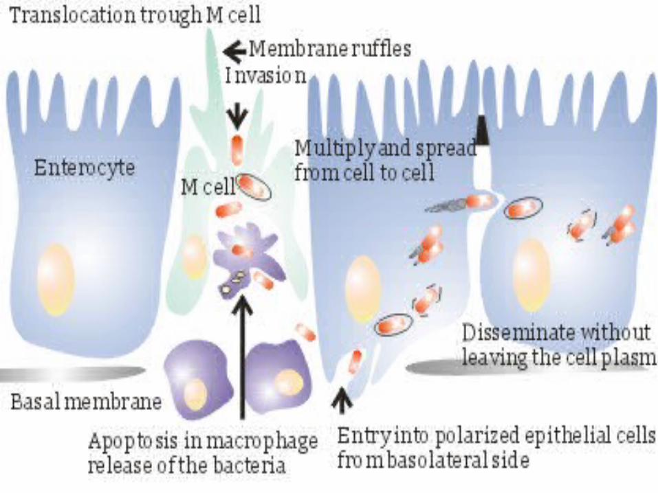
Box 31-2 and 31-5 Some bacteria can survive stomach
acid Able to enter M cells (peyer’s
patches) Cause cell death and spread to
surrounding cells. Figure
SalmonellaEpidemiology
Colonize virtually all animal species

Salmonella Diseases(Gastroenteritis) Most common form of disease
40,000 cases in the US in 2004 Mostly spread by eating contaminated food (Poultry, eggs, dairy products) Can be fecal-oral in children Infectious dose 106 to 108
Symptoms 6-48hrs after consumptionnausea, vomiting, non-bloody
diarrhea, fever, abdominal cramps Usually ends without intervention in a week
or less
Salmonella Diseases(Typhoid Fever)
Typhoid Fever Human reservoir (person-to-person spread) Pass through intestinal lining and engulfed
by phagocytes Replicate in liver, spleen, bone marrow
Cause fever, myalgia, gastroenteritis Asymptomatic colonization (1-5% patients)
Story Time - “Typhoid Mary”
Salmonella Treatment
Preventative - safe food preparation Antibiotics not recommended for
enteritis Typhoid Fever - antibiotics
Shigella
Characteristics Gram - facultative anaerobe, rod DNA hybridization reveals they’re actually
biogroups of E. coli. Don’t ferment lactose Intracellular pathogen

Shigella Virulence Factors Adhere to, invade, and replicate in M cells
(Peyer’s Patches). Spread to macrophages and cause lysis of
phagocytic vacuole They then replicate in the cytoplasm
Cause apoptosis, and release of IL-1β which attract polymorphonulear leukocytes which destroy intestinal tissue.
Shiga toxin—disrupts protein synthesis Remember E. coli O157:H7
Shigella Epidemiology Estimated 450,000 cases in U.S.
(2003) 150 million world wide
Spread by fecal oral route (yummy). Primarily a pediatric disease
70% occur in children 15 and under. Highest risk in daycares, nurseries,
custodial institutions Low infectious dosage (~200 cells)
Shigellosis Symptoms appear 1-3 days after ingestion Begin with watery diarrhea. Progress to abdominal cramps and pus in
bloody stool. Usually clears up on its own
Antibiotics are given to reduce the chance of spread
Small percentage of asymptomatic colonization
Yersinia Species Y. pestis – causes the plague
Highly virulent pathogen causing a systemic disease
Y. enterocolitica - causes enterocolitis
Yersinia Virulence Factors Found on plasmids Capsule Antiphagocytic proteins Proteins which cause apoptosis in
macrophages Proteases which inactivate compliment
proteins Fibrinases which break down blood clots
Yersinia Epidemiology Humans are accidental hosts
Most infections in other animals are fatal (not normal flora)
Y. enterocolitica Reservoir rabbits, rodents, pigs, livestock Primarily in colder climates 90% infections associated with ingestion
of contaminated meat, milk, water Mostly in children
Yersinia entercolitica Symptoms include: diarrhea, abdominal
pain, fever Can mimic acute appendicitis
Usually lasts 1 to 2 weeks Because of growth at low temperature
(4°C) can spread in blood products
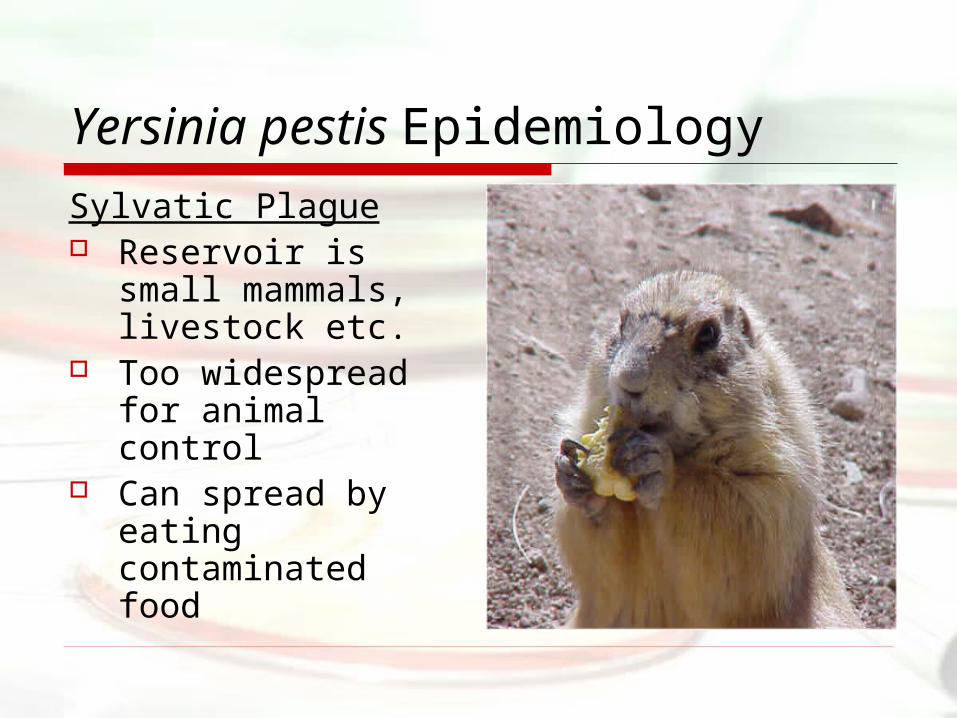
Yersinia pestis Epidemiology
Sylvatic Plague Reservoir is small
mammals, livestock etc.
Too widespread for animal control
Can spread by eating contaminated food
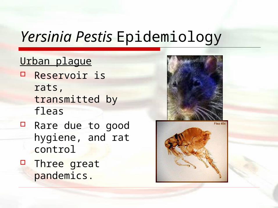
Yersinia Pestis Epidemiology
Urban plague Reservoir is rats,
transmitted by fleas
Rare due to good hygiene, and rat control
Three great pandemics.
Plague History Egypt 541 AD. lasted 200yrs
Spread to most of the “old world” Killed a majority of the population
1320s, over 5 year period 25 million died in Europe (30-40% of population)
China 1860s spread world wide About 10 cases in the U.S. per year
Sylvatic plague
Yersinia Diseases
Bubonic Plague incubation of no more than 7 dayscause bubos (swelling of lymph nodes) in groin and armpit75% mortality in untreated cases
Yersinia Diseases
Pneumonic Plagueshort (2-3 day) incubationfever, malaise, pulmonary signshighly infectious90% mortality for untreated patients
Yersinia Treatment
Y. pestis–streptomycin, tetracyclines, chloroamphenicol
Enteric infections usually clear on there own
Urban plague is controlled by reducing the rodent population


