Diagnostic and Interventional Imaging (2015) 96, 1261—1278
CONTINUING EDUCATION PROGRAM: FOCUS. . .
Entrapment and traumatic neuropathies ofthe elbow and hand: An imaging approach
A. Deniel a,∗, A. Causereta, T. Moserb, Y. Rollandc,T. Dréanod, R. Guillina
a Department of Medical Imaging, Rennes University Hospitals, Sud Hospital, 16, boulevard deBulgarie, 35203 Rennes cedex 2, Franceb Department of Radiology, Montreal University Hospital Centre, 1560, rue Sherbrooke-Est,Montreal, Quebec H2 4M1, Canadac Department of Medical Imaging, Eugène Marquis Centre, avenue de laBataille-Flandres-Dunkerque, 35000 Rennes, Franced Department of Orthopaedics and Traumatology, Rennes University Hospitals, 2, rueHenri-Le-Guilloux, 35000 Rennes, France
Abstract Ultrasound and magnetic resonance imaging currently offer a detailed analysis ofthe peripheral nerves. Compressive and traumatic nerve injuries are the two main indicationsfor imaging investigation of nerves with several publications describing the indications, tech-nique and diagnostic capabilities of imaging signs. Investigation of entrapment neuropathieshas three main goals, which are to confirm neuronal distress, search for the cause of nervecompression and exclude a differential diagnosis on the entire nerve. For traumatic nerveinjuries, imaging, predominantly ultrasound, occasionally provides essential information formanagement including the type of nerve lesion, its exact site and local extension.
Upper limb nerves can be subjected to various injuries; the great majority of them involvecompression and trauma. Apart from the median, ulnar and radial nerves, several lesswell-known motor and/or sensory nerves must not be disregarded. Whilst magnetic res-onance imaging (MRI) can achieve these aims because of a high contrast resolution [1],
ultrasound is the most commonly used tool in clinical practice because of its spatial res-olution, accessibility and limited costs. The goal of this review was to describe the mainpoints arising from the international literature about compressive and traumatic upperlimb neuropathies before or after surgery.
eripheral nerves are made of many fascicles which them-elves are made of numerous nerve fibers. Each nerve fibers contained in a supporting tissue layer known as thendoneurium and each fascicle is surrounded by the per-neurium, whereas the nerve is contained within a peripheralheath known as the epineurium. The epineurium receivesrterioles and venules, which provide neuronal vasculariza-ion [2].
ltrasound appearance of peripheral nerves
he internal architecture of the nerve on ultrasoundas a ‘‘pseudo-ovular’’ appearance. The fascicles areypoechogenic surrounded by hyperechogenic epineuralupporting tissue. This appearance differs from the moreyperechogenic, finely fibrillar and anisotropic appearancef tendons. The thickness of the nerve is influenced by gen-er [3—5] and possibly by weight [5] and patient age [3,4].
RI anatomy of peripheral nerves
he signal of normal peripheral nerves is identical to thatf adjacent muscles on T1- and T2-weighted MR images butay be slightly more intense on T2-weighted images [6,7].
ike the tendons, nerves are subjected to ‘‘magic angle’’rtifacts [8]. Because of the existence of the ‘‘blood-nerve’’arrier, the normal nerve does not substantially enhancefter intravenous administration of gadolinium chelate [6].
ntrapment neuropathies
athophysiology of nerve compression
erve compression results in a pathophysiological cascaden which microcirculatory disorders play a key role. Itas been shown in animal models that nerve compressionapidly reduces the circulation in venules [9]. Prolonged
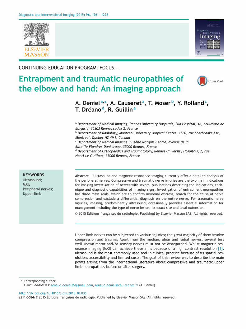
igure 1. Ultrasound signs of nerve distress in a patient with carpal
rea of the median nerve (dots) measuring 13 mm2 with hypoechogenicroximal to the entrance to the carpal tunnel (arrow); c: Doppler ultras
ttop
A. Deniel et al.
ompression results in ischemia, which itself causeslood vessel endothelial permeability abnormalities in thendoneurium, which are responsible for intrafasciculardema [10—12]. The resultant raised intrafascicular fluidressure persists and maintains neuronal compressionecause of the blood-nerve barrier and the small numberf lymph vessels within the endoneurium [2].
At an early stage of compression, neuronal ischemiaesults in axonal transport abnormalities, which cause onlyymptomatic reversible damage [2]. At a later stage, thepineural and endoneural interstitial edema is combinedith structural changes in the nerve, initially involving dam-ge to the myelin sheath (persistent or recurrent symptoms)nd then later, axonal damage causing actual signs of dener-ation (sensory disorders and muscle wastage). Recoveryf the nerve by axonal growth is then slow and occasion-lly incomplete at this stage. In the long term, peri- andndoneural edema is believed to activate fibroblast recruit-ent, leading to fibrosis of the supporting tissues [10].Because the nerve is fixed as a result of the edema, the
nhibition of gliding may induce stretching lesions which arelso harmful [2]. In addition, proximal compression of theerve renders it more liable to distal compression, and viceersa. This effect is known by the term of ‘‘double crushyndrome’’ [13], which should be considered if treatmentails at a given level.
ltrasound appearance of nerve compression
nvestigation of nerve canal symptoms has three purposes:o confirm nerve distress, investigate for an anatomical orxtrinsic cause for the neuronal compression and to look for
differential diagnosis throughout the length of the nerveparticularly a neurogenic tumor).
The signs of compressive nerve distress consist of localhickening of the nerve, hypoechogenicity with loss of itsasciculated appearance and intra-neural hypervasculariza-
tunnel syndrome confirmed by electromyogram. a: cross-sectionality of the nerve; b: longitudinal section shows increased diameteround in color mode shows intra-neural hyperemia.
ion in color or power Doppler (Fig. 1). Pathophysiologically,he hypoechogenicity and increased diameter of the nerveccur as a result of the edema developing in the endo- anderineural spaces (Fig. 2).
Entrapment and traumatic neuropathies of the elbow and hand: An imaging approach 1263
rve d
aiaMsbt
tissatdmimicfwapas
M
Ctuou
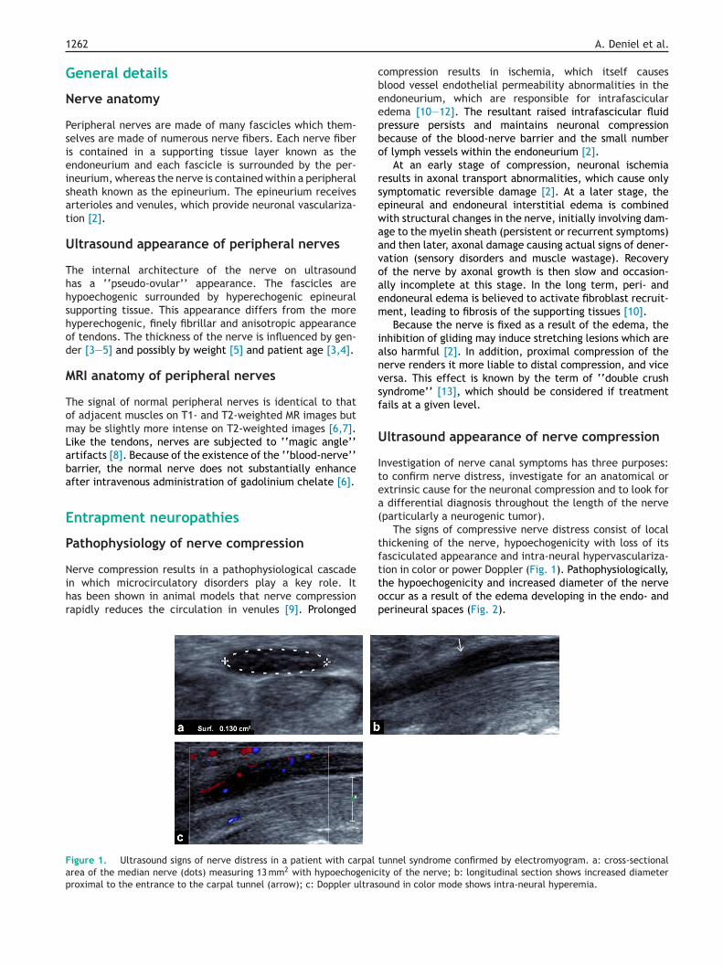
Figure 2. Pathophysiology and ultrasound signs of compressive ne
By consensus, nerve thickening is quantified by measur-ing the cross-sectional area (CSA). Measurement using acontinuous trace is preferable to an elliptical ROI, definingthe contour of the nerve from inside its peripheral hyper-echogenic rim. This technique has been shown to offergood reproducibility for both the median and ulnar nerves[14—17]. The increase in diameter, often extending alongthe nerve, results in a characteristic imprint longitudinallywhen it passes beneath the compressive arcade.
Intra-neural hypervascularization is clearly visible onhigh-resolution ultrasound. Any intra-neural Doppler signalshould be considered as an indicator of abnormal hypervas-cularization [17—19]. Power Doppler appears more effectivethan color Doppler to detect slow flow in the nerve [20].
Whilst direct visualization of compressive fibrous arcadesis often considered to be difficult, investigation for anextrinsic compressive mass is the second key objective ofultrasound. This includes, for example, supernumerary mus-cles, arthrosynovial cysts and joint or tendon sheath tumors.Although the precise level of the lesion is often strongly sug-gested by electromyography, routine scanning of the nerveover its entire length using the ‘‘lift’’ technique should beused systematically in order to not miss a remote lesion suchas a neurogenic tumour.
Finally, dynamic investigation of the nerve is a usefuladjunct to static examination, because it may show reducedneuronal mobility as a result of compression (for exampleat the wrist) or abnormally increased mobility because ofchronic instability (particularly at the elbow).
MRI appearance of nerve compression
Visualization of direct signs of nerved distress on MRI isinconstant [6,21—24]. An increased signal on T2-weighted
images indicates neuronal edema although this feature is notspecific because it can be observed in asymptomatic patients[6,7,25,26]. Conversely, a decrease in signal intensity on T2-weighted images occurs less commonly in neuronal fibrosis
di(s
istress.
t an advanced stage [25]. Nerve enhancement is absentn the normal state and may be seen after intravenousdministration of gadolinium chelate in nerve distress [6].orphological nerve changes may be visible although mea-
urement of these is less satisfactory than with ultrasoundecause of poor spatial resolution and difficulties visualizinghe nerve in its length.
In experimental nerve damage, signs of muscle denerva-ion appear early (from 24 h) as a moderate rise in signalntensity on STIR images in the affected area [27]. Thisign, which has limited sensitivity in chronic nerve compres-ion, may be helpful when direct signs of nerve distress arebsent [28], pending a knowledge of motor territories ofhe different upper limb nerves [29]. In advanced chronicamage, muscle atrophy and fatty changes are detectedore easily than on ultrasound. MRI is only a second-line
nvestigation in addition to ultrasound to look for signs ofuscle denervation, to identify a compressive mass and
n a postoperative context [30]. The investigation proto-ol is often limited to T1- and T2-weighted images withat suppression in the transverse plane, combined with T1-eighted fat-suppressed images obtained after intravenousdministration of gadolinium chelate in the transverse planereoperatively for lesion characterization although thesere used routinely postoperatively, particularly to search formall fibrous scars.
edian nerve at the wrist
ompression of the median nerve at the wrist known ashe ‘‘carpal tunnel syndrome’’ (CTS), is the most commonpper limb compressive neuropathy and occurs as a resultf compression of the nerve beneath the flexor retinac-lum. Although it is usually idiopathic, many causes are
escribed, particularly including tumors and pseudomassesnside the canal, including tumors of the median nerve itselfschwannoma, fibrolipoma), tenosynovial hypertrophy andupernumerary muscles. The 2013 guidelines of the French
1
Nnbosna
PUIrtw[tmvtmttrssTg
mtosKoCfa
hbwtnio(
saflwssCbiai≥Ts[i
ub[vdw
arrpsspdcaiCttcttt
Fop
264
ational Health Authority (HAS) indicate that carpal tun-el imaging is useless in case of typical clinical scenariout is indicated when unusual symptoms are present such asccurrence during an effort, a young age, a sudden onset, auspected responsible underlying disease and in case of diag-ostic uncertainty (negative EMG), or failure or recurrencefter surgery [31].
reoperative imagingltrasound
ncreased cross-sectional area (CSA) of the median nerve isecognized to be the best performing parameter, althoughhere is no consensus about the cut-off value to be used,hich ranges depending on the study between 9 and 15 mm2
14,15,17—19,32—44]. A cut-off of 11 mm2 however appearso be often used (Table 1). A uniform cut-off value howeveray be questioned because of interindividual physiological
ariations [3—5]. Some authors have proposed standardizinghe technique by incorporating the difference in nerve CSAeasured in the canal tunnel and more proximally, 12 cm
owards the elbow [45] or next to the pronator quadra-us muscle, which is easier to perform [41]. Klauser et al.eported that a difference greater than 2 mm2 results in aensitivity of 99% and a specificity of 100% for the diagno-is of carpal tunnel syndrome [42]. Hunderfund et al. andajika et al. confirmed that this diagnostic method offersood diagnostic performance [43,44].
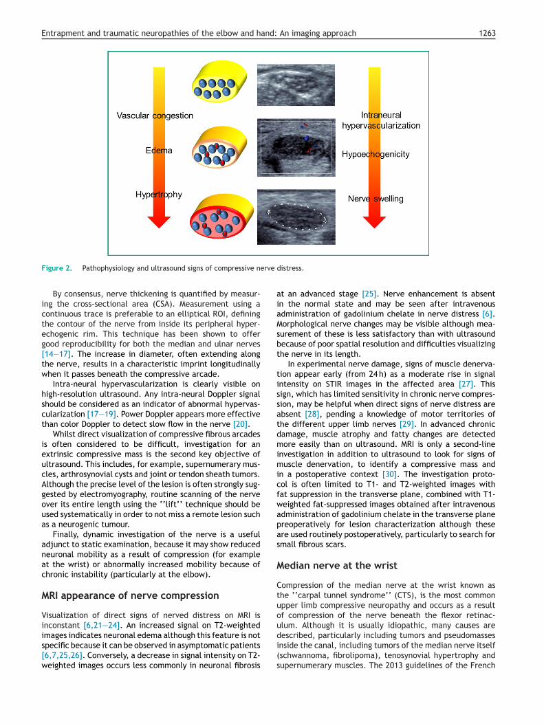
In the specific case of a bifid median nerve, the opti-al threshold values are the same or slightly greater than
hose found without a bifid nerve. Using a threshold valuef 11 mm2 Bayrak et al. found a sensitivity of 90% and apecificity of 99% [46]. Using a threshold value of 12 mm2
lauser et al. found a sensitivity of 84.9% and a specificityf 46.5% (Fig. 3) [47]. Klauser et al. proposed using a deltaSA of 4 mm2 in the forearm to improve the diagnostic per-ormance of the measurement, yielding a sensitivity of 92.5%nd a specificity of 94.6% [47].
Measurement of the CSA at the carpal tunnel outletas also been discussed. Some authors consider this toe of limited value and difficult to perform [15,43,48],hereas others believe that it increases the sensitivity of
he investigation [49,50]. In any event, ultrasound exami-
ation of the canal to its distal end is essential to detectsolated nerve distress at this level and to identify a deepccult compressive cause, such as a synovial palmar cystFig. 4).
tnTo
igure 3. Bifid median nerve in a patient with carpal tunnel syndromef the cross-sectional areas of the two components (dots) is measuredower mode confirms patency of median artery.
A. Deniel et al.
Intra-neural hyperemia is a useful sign for the diagno-is of nerve distress with sensitivity ranging from 41 to 95%nd specificity from 71 to 100% [18,19,51—53]. These dif-erences are probably due to variations in protocols, whichimit its validity [20]. Overall, hypoechogenicity of the nerveith loss of fasciculated structure, although subjective, is a
trong variable when combined with the above-mentionedigns. In addition to these three most classical signs ofTS, increased carpal tunnel pressure may result in palmarulging of the flexor retinaculum. This is assessed by measur-ng the distance between the end point of the retinaculumnd a virtual line traced between the apex of the trapez-um tubercle and the hamulus of the hamate. A value of
2—4 mm is significantly associated with CTS [15,17,19,35].he flattening index, which is the ratio of the greater tomaller diameter of the nerve, is pathological if it is over 317,19,33,37]. These criteria, however, are not widely usedn everyday practice.
Finally, dynamic flexion-extension of fingers can besed to assess transverse mobility of the median nerveeneath the flexor retinaculum and may be reduced in CTS17,54,55]. This is assessed subjectively in the absence of aalidated quantitative criterion and may provide additionaliagnostic information in some cases. A comparative analysisith the contralateral side is often very useful.
All studies agree that in the absence of a consensusbout the diagnostic ultrasound criteria and in view of theecognized performance of EMG, it cannot at present beeplaced by ultrasound [56]. Whilst EMG provides importantrognostic information before surgical release regarding theeverity of nerve damage, ultrasound is difficult to use in thisituation because of conflicting results [57—60]. One indis-utable advantage of ultrasound, however, is its ability toepict a compressive mass. Of these, supernumerary mus-les are a rare cause of CTS occurring on effort and resolvingt rest. The muscles must be seen within the carpal tunneln the resting position to be incriminated as the cause ofTS. Other masses arising both from joints (cysts or synovialumors), or in the tendon sheaths (tenosynovitis, sheathumors, etc.) may be seen on ultrasound (Fig. 5). Two tipsan be recommended for this on ultrasound examination ofhe carpal tunnel. In order to circumvent the anisotropism ofhe flexor tendons due to their oblique path running deeply,he first tip is to incline the probe upwards and downwards
o recognize them and ensure that there is no ectopic masson-subject to the anisotropic effect present in the canal.he second tip is to examine the canal to its distal end inrder to not miss a deep compressive lesion vel.
confirmed on electromyogram. a: on B mode ultrasound, the sum (in this patient the surface is 12 mm2); b: Doppler ultrasound in
Entrapment
and traum
atic neuropathies
of the
elbow and
hand: An
imaging
approach
1265
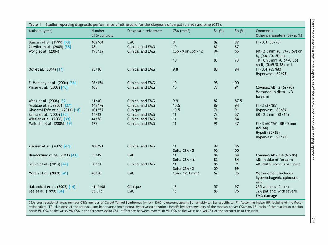
Table 1 Studies reporting diagnostic performance of ultrasound for the diagnosis of carpal tunnel syndrome (CTS).
Nakamichi et al. (2002) [14] 414/408 Clinique 13 57 97 235 women/40 menLee et al. (1999) [34] 65 CTS EMG 15 88 96 32% patients with severe
EMG damage
CSA: cross-sectional area; number CTS: number of Carpal Tunnel Syndromes (wrist); EMG: electromyogram; Se: sensitivity; Sp: specificity; FI: flattening index; BR: bulging of the flexorretinaculum; TR: thickness of the retinaculum; hypervasc.: intra-neural Hypervascularization; HypoE: hypoechogenicity of the median nerve; CSAmax/AB: ratio of the maximum mediannerve MN CSA at the wrist/MN CSA in the forearm; delta CSA: difference between maximum MN CSA at the wrist and MN CSA at the forearm or at the wrist.
1266 A. Deniel et al.
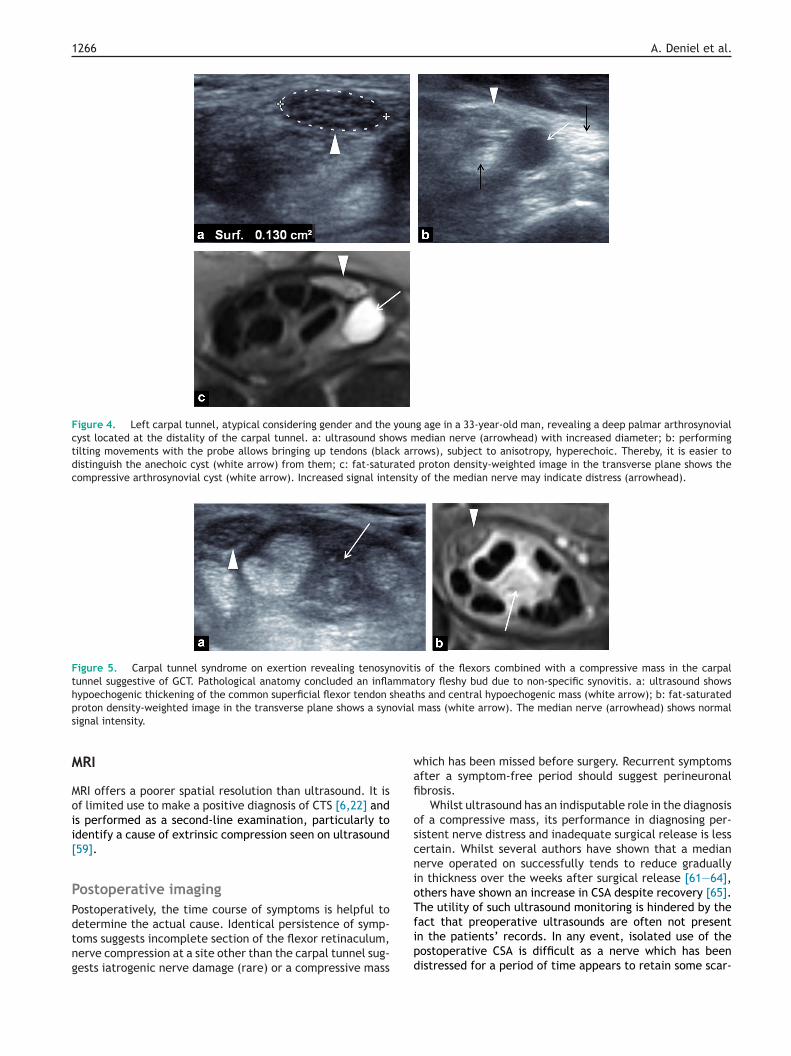
Figure 4. Left carpal tunnel, atypical considering gender and the young age in a 33-year-old man, revealing a deep palmar arthrosynovialcyst located at the distality of the carpal tunnel. a: ultrasound shows median nerve (arrowhead) with increased diameter; b: performingtilting movements with the probe allows bringing up tendons (black arrows), subject to anisotropy, hyperechoic. Thereby, it is easier todistinguish the anechoic cyst (white arrow) from them; c: fat-saturated proton density-weighted image in the transverse plane shows thecompressive arthrosynovial cyst (white arrow). Increased signal intensity of the median nerve may indicate distress (arrowhead).
Figure 5. Carpal tunnel syndrome on exertion revealing tenosynovitis of the flexors combined with a compressive mass in the carpaltunnel suggestive of GCT. Pathological anatomy concluded an inflammatory fleshy bud due to non-specific synovitis. a: ultrasound showshypoechogenic thickening of the common superficial flexor tendon sheaths and central hypoechogenic mass (white arrow); b: fat-saturatedp ovials
M
Moii[
PPdtng
wafi
oscnioT
roton density-weighted image in the transverse plane shows a synignal intensity.
RI
RI offers a poorer spatial resolution than ultrasound. It isf limited use to make a positive diagnosis of CTS [6,22] ands performed as a second-line examination, particularly todentify a cause of extrinsic compression seen on ultrasound59].
ostoperative imagingostoperatively, the time course of symptoms is helpful to
etermine the actual cause. Identical persistence of symp-oms suggests incomplete section of the flexor retinaculum,erve compression at a site other than the carpal tunnel sug-ests iatrogenic nerve damage (rare) or a compressive mass
fipd
mass (white arrow). The median nerve (arrowhead) shows normal
hich has been missed before surgery. Recurrent symptomsfter a symptom-free period should suggest perineuronalbrosis.
Whilst ultrasound has an indisputable role in the diagnosisf a compressive mass, its performance in diagnosing per-istent nerve distress and inadequate surgical release is lessertain. Whilst several authors have shown that a medianerve operated on successfully tends to reduce graduallyn thickness over the weeks after surgical release [61—64],thers have shown an increase in CSA despite recovery [65].he utility of such ultrasound monitoring is hindered by the
act that preoperative ultrasounds are often not presentn the patients’ records. In any event, isolated use of theostoperative CSA is difficult as a nerve which has beenistressed for a period of time appears to retain some scar-
and:
U
Tunarbrla[au
PDoctrmtrtac(gsaasadt(
IUItt
Entrapment and traumatic neuropathies of the elbow and h
ring hypertrophy despite symptoms disappearing [64—66].In a prospective series of 24 patients and 44 wrists treatedsurgically, Kim et al. reported a mean preoperative CSA of14.5 mm2, compared to 13 mm2 at 3 weeks and 11.5 mm2 at3 months after surgery [67]. The presence of increased signalintensity of the nerve on T2-weighed images is not specificafter surgery [30].
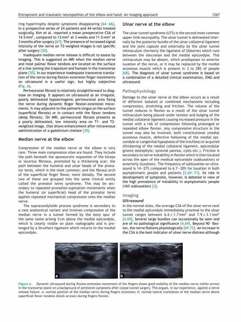
Inadequate median nerve release is difficult to assess byimaging. This is suggested on MRI when the median nerveand most palmar flexor tendons are located on the surfaceof a line joining the trapezium and hamate in the transverseplane [30]. In our experience inadequate transverse transla-tion of the nerve during flexion-extension finger movementson ultrasound is a useful sign, but highly subjective(Fig. 6).
Perineuronal fibrosis is relatively straightforward to diag-nose on imaging. It appears on ultrasound as an irregular,poorly delineated hypoechogenic perineuronal area fixingthe nerve during dynamic finger flexion-extension move-ments. It may adjacent to the palmaris longus on the surface(superficial fibrosis) or in contact with the flexor tendons(deep fibrosis). On MRI, perineuronal fibrosis presents asa poorly delineated, low intensity area on T1- and T2-weighted image, that shows enhancement after intravenousadministration of a gadolinium chelate [30].
Median nerve at the elbow
Compression of the median nerve at the elbow is veryrare. Three main compression sites are found. They includethe path beneath the aponeurotic expansion of the bicepsor lacertus fibrosus, promoted by a thickening scar; thepath between the humeral and ulnar heads of the prona-tor teres, which is the most common; and the fibrous archof the superficial finger flexor, more distally. The secondtwo of these are grouped into the same clinical entitycalled the pronator teres syndrome. This may be sec-ondary to repeated pronation-supination movements whenthe humeral (or superficial) head of the pronator teresapplies repeated mechanical compression onto the mediannerve.
The supracondyloide process syndrome is secondary toa rare anatomical variant and involves compression of themedian nerve in a tunnel formed by the bony spur of
the same name arising 5 cm above the medial epicondyle,which is clearly visible on plain radiographs and is pro-longed by a Struthers ligament which returns to the medialepicondyle.
[ait
Figure 6. Dynamic ultrasound during flexion-extension movement of tin the transverse plane on a background of persistent symptoms after carelease failure. a: normal position of the median nerve during fingers esuperficial flexor tendons (black arrows) during fingers flexion.
An imaging approach 1267
lnar nerve at the elbow
he ulnar tunnel syndrome (UTS) is the second most commonpper limb neuropathy. The ulnar tunnel is delineated inter-ally by the posterior bundle of the ulnar collateral ligamentnd the joint capsule and externally by the ulnar tunneletinaculum (formerly the ligament of Osborne) which runsetween the olecranon and the medial epicondyle. Thisetinaculum may be absent, which predisposes to anterioruxation of the nerve, or it may be replaced by the medialconeous muscle which is present in 3 to 28% of people68]. The diagnosis of ulnar tunnel syndrome is based on
combination of a detailed clinical examination, EMG andltrasound.
athophysiologyamage to the ulnar nerve at the elbow occurs as a resultf different isolated or combined mechanisms includingompression, stretching and friction. The volume of theunnel reduces in flexion as a result of the ulnar tunneletinaculum being placed under tension and bulging of theedial collateral ligament causing increased pressure in the
unnel with a risk of compression following prolonged orepeated elbow flexion. Any compressive structure in theunnel may also be involved, both constitutional (medialconeous muscle, defective hollowing of the medial epi-ondyle or congenital hypoplasia of the trochlea) or acquiredthickening of the medial collateral ligament, epicondylarroove osteophyte, synovial pannus, cysts etc.). Friction isecondary to nerve instability in flexion which is then locatedcross the apex of the medical epicondyle (subluxation) ornteriorly (luxation). The frequency of subluxation on ultra-ound is 14—27% compared to 6.7—20% for luxation in bothsymptomatic people and patients [3,69—71]. Its role inevelopment of symptoms, however, is debated in view ofhe high prevalence of instability in asymptomatic people16% subluxation) [3].
magingltrasound
n the normal state, the average CSA of the ulnar nerve nexto the medial epicondyle immediately proximal to the ulnarunnel ranges between 6.6 ± 1.7 mm2 and 7.9 ± 3.1 mm2
4,69]. Several large bundles can occasionally be seen andre of no pathological significance [4,69]. Beyond 90◦ flex-on, the nerve flattens physiologically [69,72]. An increase inhe CSA is the best indicator of ulnar nerve distress although
he fingers shows good mobility of the median nerve (white arrow)rpal tunnel surgery. This argues, in our experience, against a nervextension; b: normal lateral translation of the median nerve above
1
tHg
CghnHiapa[
uwt
MMuoiooA
dau
PTsaitRms
Us
TlGfafi
Fii
268
here is no consensus about the best cut-off value [72—81].owever, a value of 10 or even 11 mm2 appears to offer aood compromise (Fig. 7) (Table 2).
Similar to the carpal tunnel, a comparison of maximumSA of the nerve at the elbow against a reference value for aiven patient measured in the forearm or middle third of theumerus is proposed by some authors and offers good diag-ostic performance with a thickening ratio > 1.5 [74,77,79].owever, it does not appear to perform better than measur-
ng the CSA alone only at the site of maximum thickening,lthough may be particularly useful in unusually short or talleople. Ultimately, the simultaneous use of the CSA valuend a CSA ratio would offer the best diagnostic performances74,76,79].
A dynamic study should be performed routinely duringltrasound in order to search for instability of the nerve,hich although of uncertain pathological significance needs
o be known by the surgeon (Fig. 8).
RIRI must be performed with the elbow in extension. Thelnar nerve is identified on T1-weighted images as a roundr ovoid structure. The isolated presence of an increased
ntensity of the nerve on T2-weighted images at the levelf the medial epicondyle should not lead to the diagnosisf UTS as this is seen in 60% of asymptomatic people [7].
raised signal intensity of muscle on STIR images reflects
tfii
igure 7. Signs of nerve distress on ultrasound in a patient with ulnmage in the transverse plane shows a cross-sectional surface area of 15ncreased diameter of the nerve immediately proximal to its path benea
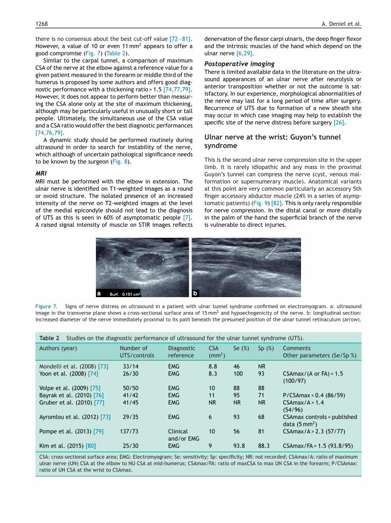
Table 2 Studies on the diagnostic performance of ultrasound
Authors (year) Number ofUTS/controls
Diagnosticreference
Mondelli et al. (2008) [73] 33/14 EMG
Yoon et al. (2008) [74] 26/30 EMG
Volpe et al. (2009) [75] 50/50 EMG
Bayrak et al. (2010) [76] 41/42 EMG
Gruber et al. (2010) [77] 41/45 EMG
Ayromlou et al. (2012) [73] 29/35 EMG
Pompe et al. (2013) [79] 137/73 Clinicaland/or EMG
Kim et al. (2015) [80] 25/30 EMG
CSA: cross-sectional surface area; EMG: Electromyogram; Se: sensitivityulnar nerve (UN) CSA at the elbow to NU CSA at mid-humerus; CSAmaxratio of UN CSA at the wrist to CSAmax.
A. Deniel et al.
enervation of the flexor carpi ulnaris, the deep finger flexornd the intrinsic muscles of the hand which depend on thelnar nerve [6,29].
ostoperative imaginghere is limited available data in the literature on the ultra-ound appearances of an ulnar nerve after neurolysis ornterior transposition whether or not the outcome is sat-sfactory. In our experience, morphological abnormalities ofhe nerve may last for a long period of time after surgery.ecurrence of UTS due to formation of a new sheath siteay occur in which case imaging may help to establish the
pecific site of the nerve distress before surgery [26].
lnar nerve at the wrist: Guyon’s tunnelyndrome
his is the second ulnar nerve compression site in the upperimb. It is rarely idiopathic and any mass in the proximaluyon’s tunnel can compress the nerve (cyst, venous mal-
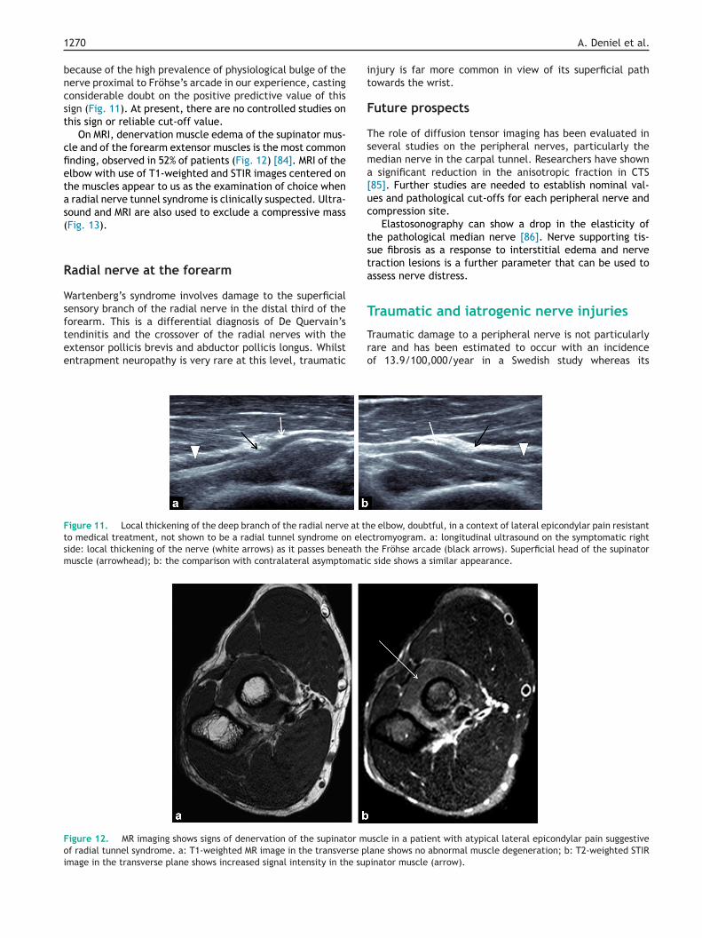
ormation or supernumerary muscle). Anatomical variantst this point are very common particularly an accessory 5thnger accessory abductor muscle (24% in a series of asymp-
omatic patients) (Fig. 9) [82]. This is only rarely responsibleor nerve compression. In the distal canal or more distallyn the palm of the hand the superficial branch of the nerves vulnerable to direct injuries.
ar tunnel syndrome confirmed on electromyogram. a: ultrasound mm2 and hypoechogenicity of the nerve. b: longitudinal section:th the presumed position of the ulnar tunnel retinaculum (arrow).
for the ulnar tunnel syndrome (UTS).
CSA(mm2)
Se (%) Sp (%) CommentsOther parameters (Se/Sp %)
8.8 46 NR8.3 100 93 CSAmax/(A or FA) > 1.5
(100/97)10 88 8811 95 71 P/CSAmax < 0.4 (86/59)NR NR NR CSAmax/A > 1.4
(54/96)6 93 68 CSAmax controls < published
data (5 mm2)10 56 81 CSAmax/A > 2.3 (57/77)
9 93.8 88.3 CSAmax/FA > 1.5 (93.8/95)
; Sp: specificity; NR: not recorded; CSAmax/A: ratio of maximum/FA: ratio of maxCSA to max UN CSA in the forearm; P/CSAmax:
Entrapment and traumatic neuropathies of the elbow and hand: An imaging approach 1269
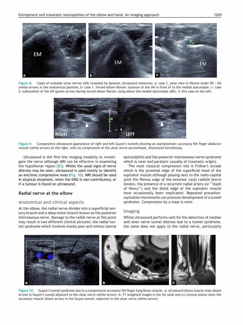
Figure 8. Cases of unstable ulnar nerves (UN) revealed by dynamic ultrasound maneuver. a: case 1, axial view in flexion under 90◦: UN(white arrow) in the anatomical position; b: case 1, forced elbow flexion: luxation of the UN in front of to the medial epicondyle. c: case2: subluxation of the UN (green arrow) during forced elbow flexion, lying above the medial epicondyle (ME), in this case on the left.
uyonr ner
ew
wsjtohss
Figure 9. Comparative ultrasound appearance of right and left Gmuscle (white arrow) on the right, with no compression of the ulna
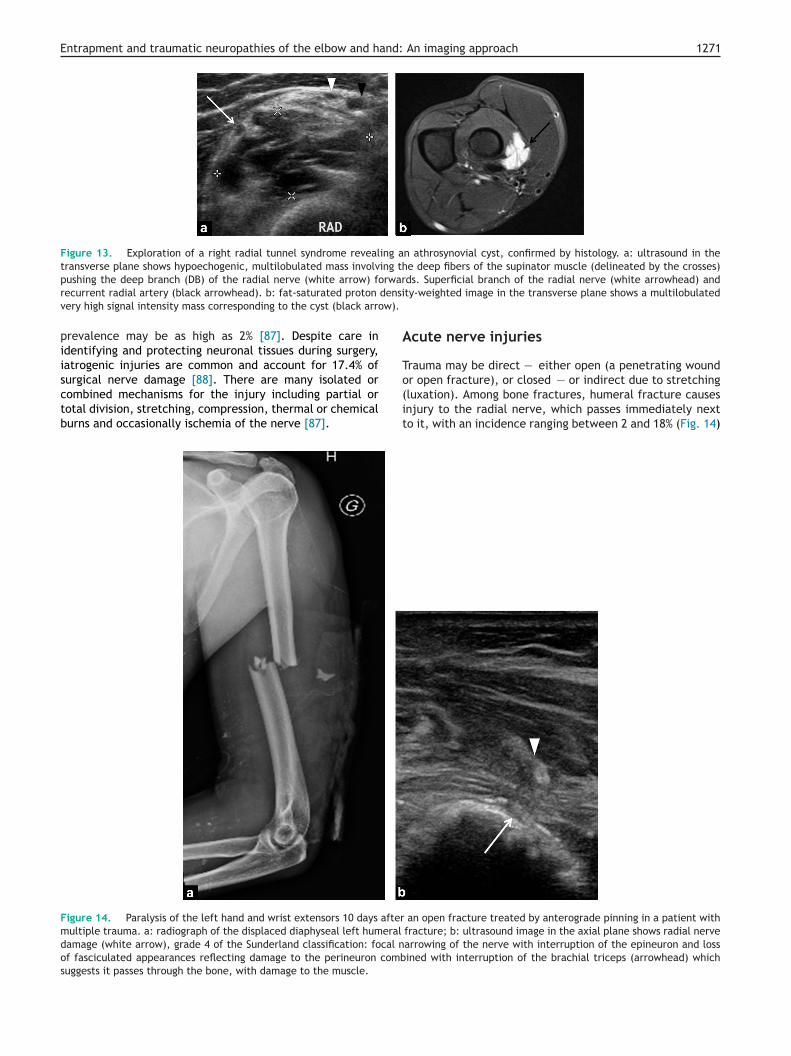
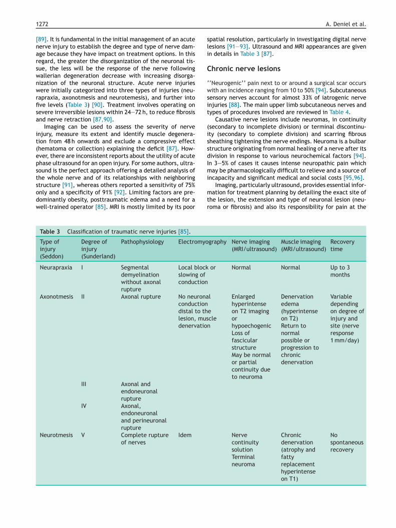
Ultrasound is the first-line imaging modality to investi-gate the nerve although MRI can be effective in examiningthe hypothenar region [83]. Whilst the usual signs of nervedistress may be seen, ultrasound is used mostly to identifyan extrinsic compressive mass (Fig. 10). MRI should be usedin atypical situations, when the EMG is non-contributory, orif a tumour is found on ultrasound.
Radial nerve at the elbow
Anatomical and clinical aspectsAt the elbow, the radial nerve divides into a superficial sen-
sory branch and a deep motor branch known as the posteriorinterosseous nerve. Damage to the radial nerve at this pointmay result in two different clinical pictures: the radial tun-nel syndrome which involves mostly pain and mimics lateral
IWat
Figure 10. Guyon’s tunnel syndrome due to a compressive accessory 5tarrow) in Guyon’s tunnel adjacent to the ulnar nerve (white arrow); b: Taccessory muscle (black arrow) in the Guyon tunnel, adjacent to the uln
picondylitis and the posterior interosseous nerve syndromehich is rarer and paralytic (usually of traumatic origin).
The most classical compression site is Fröhse’s arcadehich is the proximal edge of the superficial head of the
upinator muscle although passing next to the radio-capitaloint the fibrous edge of the extensor carpi radialis brevisendon, the presence of a recurrent radial artery (or ‘‘leashf Henry’’) and the distal edge of the supinator muscleave occasionally been implicated. Repeated pronation-upination movements can promote development of a tunnelyndrome. Compression by a mass is rarer.
maginghilst ultrasound performs well for the detection of median
nd ulnar nerve tunnel distress due to a tunnel syndrome,he same does not apply to the radial nerve, particularly
h finger long flexor muscle. a: ultrasound shows muscle mass (black1-weighted images in the (b) axial and (c) coronal planes show thear nerve (white arrow).
1
bncst
cfietas(
R
Wsftee
it
F
Tsma[uc
tsta
T
Ftsm
Foi
270
ecause of the high prevalence of physiological bulge of theerve proximal to Fröhse’s arcade in our experience, castingonsiderable doubt on the positive predictive value of thisign (Fig. 11). At present, there are no controlled studies onhis sign or reliable cut-off value.
On MRI, denervation muscle edema of the supinator mus-le and of the forearm extensor muscles is the most commonnding, observed in 52% of patients (Fig. 12) [84]. MRI of thelbow with use of T1-weighted and STIR images centered onhe muscles appear to us as the examination of choice when
radial nerve tunnel syndrome is clinically suspected. Ultra-ound and MRI are also used to exclude a compressive massFig. 13).
adial nerve at the forearm
artenberg’s syndrome involves damage to the superficialensory branch of the radial nerve in the distal third of the
orearm. This is a differential diagnosis of De Quervain’sendinitis and the crossover of the radial nerves with thextensor pollicis brevis and abductor pollicis longus. Whilstntrapment neuropathy is very rare at this level, traumatic
Tro
igure 11. Local thickening of the deep branch of the radial nerve at tho medical treatment, not shown to be a radial tunnel syndrome on eleide: local thickening of the nerve (white arrows) as it passes beneath tuscle (arrowhead); b: the comparison with contralateral asymptomatic
igure 12. MR imaging shows signs of denervation of the supinator muf radial tunnel syndrome. a: T1-weighted MR image in the transverse plmage in the transverse plane shows increased signal intensity in the sup
A. Deniel et al.
njury is far more common in view of its superficial pathowards the wrist.
uture prospects
he role of diffusion tensor imaging has been evaluated ineveral studies on the peripheral nerves, particularly theedian nerve in the carpal tunnel. Researchers have shown
significant reduction in the anisotropic fraction in CTS85]. Further studies are needed to establish nominal val-es and pathological cut-offs for each peripheral nerve andompression site.
Elastosonography can show a drop in the elasticity ofhe pathological median nerve [86]. Nerve supporting tis-ue fibrosis as a response to interstitial edema and nerveraction lesions is a further parameter that can be used tossess nerve distress.
raumatic and iatrogenic nerve injuries
raumatic damage to a peripheral nerve is not particularlyare and has been estimated to occur with an incidencef 13.9/100,000/year in a Swedish study whereas its
e elbow, doubtful, in a context of lateral epicondylar pain resistantctromyogram. a: longitudinal ultrasound on the symptomatic righthe Fröhse arcade (black arrows). Superficial head of the supinator
side shows a similar appearance.
scle in a patient with atypical lateral epicondylar pain suggestiveane shows no abnormal muscle degeneration; b: T2-weighted STIRinator muscle (arrow).
Entrapment and traumatic neuropathies of the elbow and hand: An imaging approach 1271
Figure 13. Exploration of a right radial tunnel syndrome revealing an athrosynovial cyst, confirmed by histology. a: ultrasound in thetransverse plane shows hypoechogenic, multilobulated mass involving the deep fibers of the supinator muscle (delineated by the crosses)
rwardensiw).
A
T
pushing the deep branch (DB) of the radial nerve (white arrow) forecurrent radial artery (black arrowhead). b: fat-saturated proton
very high signal intensity mass corresponding to the cyst (black arro
prevalence may be as high as 2% [87]. Despite care inidentifying and protecting neuronal tissues during surgery,iatrogenic injuries are common and account for 17.4% of
surgical nerve damage [88]. There are many isolated orcombined mechanisms for the injury including partial ortotal division, stretching, compression, thermal or chemicalburns and occasionally ischemia of the nerve [87].
o(it
Figure 14. Paralysis of the left hand and wrist extensors 10 days aftermultiple trauma. a: radiograph of the displaced diaphyseal left humeral
damage (white arrow), grade 4 of the Sunderland classification: focal nof fasciculated appearances reflecting damage to the perineuron combsuggests it passes through the bone, with damage to the muscle.
ds. Superficial branch of the radial nerve (white arrowhead) andty-weighted image in the transverse plane shows a multilobulated
cute nerve injuries
rauma may be direct — either open (a penetrating wound
r open fracture), or closed — or indirect due to stretchingluxation). Among bone fractures, humeral fracture causesnjury to the radial nerve, which passes immediately nexto it, with an incidence ranging between 2 and 18% (Fig. 14)
an open fracture treated by anterograde pinning in a patient withfracture; b: ultrasound image in the axial plane shows radial nervearrowing of the nerve with interruption of the epineuron and lossined with interruption of the brachial triceps (arrowhead) which
1
[narswnwrfisa
it(epstsodw
sli
C
‘wsit
(issdImi
272
89]. It is fundamental in the initial management of an acuteerve injury to establish the degree and type of nerve dam-ge because they have impact on treatment options. In thisegard, the greater the disorganization of the neuronal tis-ue, the less will be the response of the nerve followingallerian degeneration decrease with increasing disorga-ization of the neuronal structure. Acute nerve injuriesere initially categorized into three types of injuries (neu-
apraxia, axonotmesis and neurotemesis), and further intove levels (Table 3) [90]. Treatment involves operating onevere irreversible lesions within 24—72 h, to reduce fibrosisnd nerve retraction [87,90].
Imaging can be used to assess the severity of nervenjury, measure its extent and identify muscle degenera-ion from 48 h onwards and exclude a compressive effecthematoma or collection) explaining the deficit [87]. How-ver, there are inconsistent reports about the utility of acutehase ultrasound for an open injury. For some authors, ultra-ound is the perfect approach offering a detailed analysis ofhe whole nerve and of its relationships with neighboring
tructure [91], whereas others reported a sensitivity of 75%nly and a specificity of 91% [92]. Limiting factors are pre-ominantly obesity, posttraumatic edema and a need for aell-trained operator [85]. MRI is mostly limited by its poor
mtr
Table 3 Classification of traumatic nerve injuries [85].
Type ofinjury(Seddon)
Degree ofinjury(Sunderland)
Pathophysiology Electromyo
Neurapraxia I Segmentaldemyelinationwithout axonalrupture
Local blockslowing ofconduction
Axonotmesis II Axonal rupture No neuronaconductiondistal to thlesion, musdenervatio
III Axonal andendoneuronalrupture
IV Axonal,endoneuronaland perineuronalrupture
Neurotmesis V Complete ruptureof nerves
Idem
A. Deniel et al.
patial resolution, particularly in investigating digital nerveesions [91—93]. Ultrasound and MRI appearances are givenn details in Table 3 [87].
hronic nerve lesions
‘Neurogenic’’ pain next to or around a surgical scar occursith an incidence ranging from 10 to 50% [94]. Subcutaneous
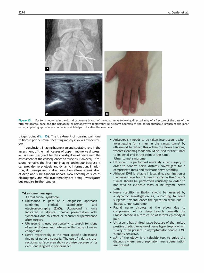
ensory nerves account for almost 33% of iatrogenic nervenjuries [88]. The main upper limb subcutaneous nerves andypes of procedures involved are reviewed in Table 4.
Causative nerve lesions include neuromas, in continuitysecondary to incomplete division) or terminal discontinu-ty (secondary to complete division) and scarring fibrousheathing tightening the nerve endings. Neuroma is a bulbartructure originating from normal healing of a nerve after itsivision in response to various neurochemical factors [94].n 3—5% of cases it causes intense neuropathic pain whichay be pharmacologically difficult to relieve and a source of
ncapacity and significant medical and social costs [95,96].
ation for treatment planning by detailing the exact site ofhe lesion, the extension and type of neuronal lesion (neu-oma or fibrosis) and also its responsibility for pain at the
graphy Nerve imaging(MRI/ultrasound)
Muscle imaging(MRI/ultrasound)
Recoverytime
or Normal Normal Up to 3months
l
eclen
Enlargedhyperintenseon T2 imagingorhypoechogenicLoss offascicularstructureMay be normalor partialcontinuity dueto neuroma
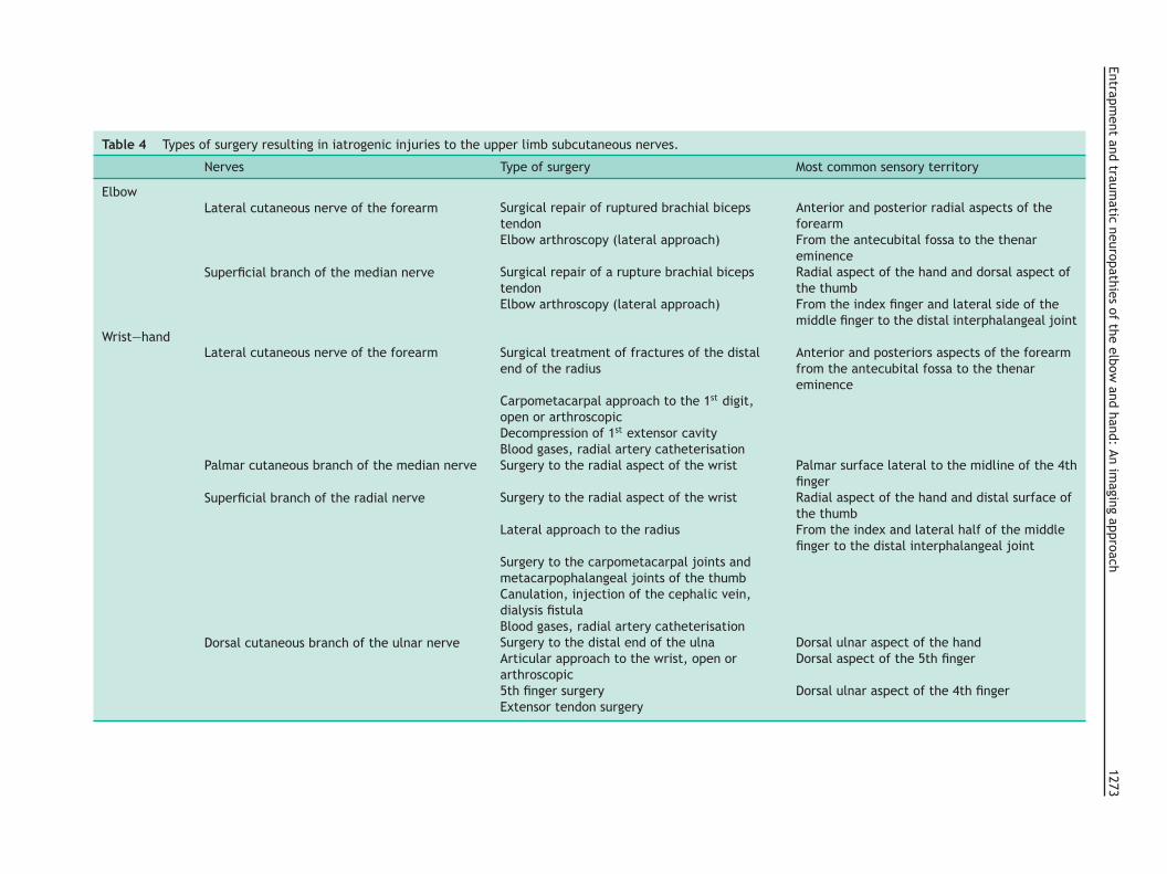
Table 4 Types of surgery resulting in iatrogenic injuries to the upper limb subcutaneous nerves.
Nerves Type of surgery Most common sensory territory
ElbowLateral cutaneous nerve of the forearm Surgical repair of ruptured brachial biceps
tendonAnterior and posterior radial aspects of theforearm
Elbow arthroscopy (lateral approach) From the antecubital fossa to the thenareminence
Superficial branch of the median nerve Surgical repair of a rupture brachial bicepstendon
Radial aspect of the hand and dorsal aspect ofthe thumb
Elbow arthroscopy (lateral approach) From the index finger and lateral side of themiddle finger to the distal interphalangeal joint
Wrist—handLateral cutaneous nerve of the forearm Surgical treatment of fractures of the distal
end of the radiusAnterior and posteriors aspects of the forearmfrom the antecubital fossa to the thenareminence
Carpometacarpal approach to the 1st digit,open or arthroscopicDecompression of 1st extensor cavityBlood gases, radial artery catheterisation
Palmar cutaneous branch of the median nerve Surgery to the radial aspect of the wrist Palmar surface lateral to the midline of the 4thfinger
Superficial branch of the radial nerve Surgery to the radial aspect of the wrist Radial aspect of the hand and distal surface ofthe thumb
Lateral approach to the radius From the index and lateral half of the middlefinger to the distal interphalangeal joint
Surgery to the carpometacarpal joints andmetacarpophalangeal joints of the thumbCanulation, injection of the cephalic vein,dialysis fistulaBlood gases, radial artery catheterisation
Dorsal cutaneous branch of the ulnar nerve Surgery to the distal end of the ulna Dorsal ulnar aspect of the handArticular approach to the wrist, open orarthroscopic
Dorsal aspect of the 5th finger
5th finger surgery Dorsal ulnar aspect of the 4th fingerExtensor tendon surgery
1274 A. Deniel et al.
Figure 15. Fusiform neuroma in the dorsal cutaneous branch of the ulnar nerve following direct pinning of a fracture of the base of thefi aph; b: fusiform neuroma of the dorsal cutaneous branch of the ulnarn neuroma.
tty
aMasctoeb
• Anisotropism needs to be taken into account wheninvestigating for a mass in the carpal tunnel byultrasound to detect this within the flexor tendons,whereas scanning mode should be used for the tunnelto its distal end in the palm of the hand.Ulnar tunnel syndrome
• Ultrasound is performed routinely after surgery inorder to confirm nerve distress, investigate for acompressive mass and estimate nerve stability.
• Although EMG is reliable in localizing, examination ofthe nerve throughout its length as far as the Guyon’stunnel should be performed routinely in order tonot miss an extrinsic mass or neurogenic nervetumor.
• Nerve stability in flexion should be assessed bya dynamic investigation as, according to somesurgeons, this influences the operation technique.Radial tunnel syndrome
• Radial nerve distress at the elbow due tocompression of its deep branch beneath theFröhse arcade is a rare cause of lateral epicondylarpain.
• Ultrasound has limited value because of the limitedpositive predictive value of nerve hypertrophy, whichis very often present in asymptomatic people. EMGis poorly sensitive.
•
fth metacarpal bone and the hamatum. a: postoperative radiogrerve; c: photograph of operation scar, which helps to localize the
rigger point (Fig. 15). The treatment of scarring pain dueo fibrous perineuronal sheathing mostly involves exoneurol-sis.
In conclusion, imaging has now an undisputable role in thessessment of the main causes of upper limb nerve distress.RI is a useful adjunct for the investigation of nerves and thessessment of the consequences on muscles. However, ultra-ound remains the first-line imaging technique because itan provide morphologic and dynamic information. In addi-ion, its unsurpassed spatial resolution allows examinationf deep and subcutaneous nerves. New techniques such aslastography and MRI tractography are being investigatedut require further studies.
Take-home messagesCarpal tunnel syndrome
• Ultrasound is part of a diagnostic approachcombining clinical examination andelectromyography (EMG). Ultrasound is onlyindicated in atypical clinical presentation withsymptoms due to effort or recurrence/persistenceafter surgery.
• Ultrasound is used particularly to search for signsof nerve distress and determine the cause of nervecompression.
• Nerve hypertrophy is the most specific ultrasound
finding of nerve distress is. The use of a delta cross-sectional surface area shows promise because of itsexcellent diagnostic performance.
MRI of the elbow is a valuable tool to guide thediagnosis when signs of supinator muscle denervationare present.
Entrapment and traumatic neuropathies of the elbow and hand:
Traumatic nerve injuries• Because of its good spatial resolution, ultrasound can
be used to provide a detailed assessment of acuteand chronic nerve injuries.
• In the acute phase it assesses the type (division,stretching or compression) and extent of the injury.
• In the chronic phase it is used to look for a neuromaand determine whether the nerve is partially ortotally discontinuous and assesses the extent of any
s
Md•••••
A
1ip
ahtn
concomitant peripheral fibrosis.
Clinical case
A 71 year-old-woman underwent surgery 6 months ago forclinically typical left carpal tunnel syndrome that was con-firmed by EMG. Despite surgery, she consulted 4 months laterfor persisting similar symptoms. Ultrasound examination wasrequested (Fig. 16).
Questions
1. Would the patient have benefitted from preoperativeimaging?
[p
t
Figure 16. Clinical case: postoperative ultrasound and MRI. a, b: ultrasoimages in the (c) sagittal and (d) transverse planes obtained after intrav
An imaging approach 1275
2. Does postoperative ultrasound allow drawing a conclu-ion regarding persistent nerve distress?
3. In view of ultrasound findings, the patient underwentRI examination (Fig. 16). Which of the following proposediagnoses would you make?palmar arthrosynovial cyst of the wrist;supernumerary muscle;synovitis of the wrist;algodystrophy;scarring perineural fibrosis.
nswers
. No. According to the March 2013 HAS guidelines imag-ng is useless in carpal tunnel syndrome when the clinicalresentation is typical [30].
2. No, persistent ultrasound signs of nerve distressppearing as a borderline cross-sectional surface area andypoechogenicity of the nerve do not allow concluding thathe damage is persistent, as moderate thickening of theerve may be seen during the initial months after surgery
64—67]. It is difficult to interpret this sign because theatient did not have a reference ultrasound before surgery.
3. Ultrasound and MRI show palmar synovial thickening ofhe wrist with peripheral enhancement on MRI after intra-
und images in the transverse plane; c, d: fat-saturated T1-weightedenous administration of a gadolinium chelate.
1
vspoc
D
T
R
[
[
[
[
[
[
[
[
[
[
[
[
[
[
[
[
[
[
[
[
[
[
[
[
[
[
[
[
[
276
enous administration of a gadolinium chelate suggestive ofynovitis of the wrist. This clinically unapparent synovitisrobably contributed to the development and persistencef the patient symptoms because of the mass effect on thearpal tunnel.
isclosure of interest
he authors declare that they have no competing interest.
eferences
[1] Kirchgesner T, Pesquer L, Larbi A, et al. Axial traction in mag-netic resonance arthrography of the wrist: how to do? DiagnInterv Imaging 2015;96:519—22.
[2] Lundborg G, Dahlin LB. Anatomy, function, and pathophysi-ology of peripheral nerves and nerve compression. Hand Clin1996;12:185—93.
[3] Okamoto M, Abe M, Shirai H, Ueda N. Morphology and dynam-ics of the ulnar nerve in the cubital tunnel. Observation byultrasonography. J Hand Surg [Br] 2000;25:85—9.
[4] Jacob D, Creteur V, Courthaliac C, Bargoin R, Sassus B, Bacq C,et al. Sonoanatomy of the ulnar nerve in the cubital tunnel: amulticentre study by the GEL. Eur Radiol 2004;14:1770—3.
[5] Thoirs K, Williams MA, Phillips M. Ultrasonographic measure-ments of the ulnar nerve at the elbow: role of confounders. JUltrasound Med 2008;27:737—43.
[6] Andreisek G, Crook DW, Burg D, et al. Peripheral neuropathiesof the median. Radial and ulnar nerves: MR imaging features.Radiographics 2006;26:1267—87.
[7] Husarik DB, Saupe N, Pfirrmann CW, Jost B, Hodler J, Zanetti M.Elbow nerves: MR findings in 60 asymptomatic subjects. Normalanatomy, variants and pitfalls. Radiology 2009;252:148—56.
[8] Chappell KE, Robson MD, Stonebrifge-Foster A, et al.Magic angle effects in MR neurography. Am J Neuroradiol2004;25:431—40.
[9] Rydevik B, Lundborg G, Bagge U. Effects of graded compressionon intraneural blood flow. An in vitro study on rabbit tibialnerve. J Hand Surg [Am] 1981;6:3—12.
10] Sunderland S. The nerve lesion in the carpal tunnel syndrome.J Neurol Neurosurg Psychiatry 1976;39:615—26.
11] Lundborg G, Myers R, Powell H. Nerve compression injuryand increase in endoneurial fluid pressure: a miniaturecompartment syndrome. J Neurol Neurosurg Psychiatry1983;46:1119—24.
12] Kobayashi S, Meir A, Baba H, Uchida K, Hayakawa K. Imag-ing of intraneural edema by using gadolinium-enhanced MRimaging: experimental compression injury. Am J Neuroradiol2005;26:973—80.
13] Upton AR, McComas AJ. The double crush in nerve entrapmentsyndromes. Lancet 1973;18:359—62.
14] Nakamichi K, Tachibana S. Ultrasonographic measure-ment of median nerve cross-sectional area in idiopathiccarpal tunnel syndrome: diagnostic accuracy. Muscle Nerve2002;26:798—803.
15] Wong SM, Griffith JF, Hui AC, Lo SK, Fu M, Wong KS. Carpal tun-nel syndrome: diagnostic usefulness of sonography. Radiology2004;232:93—9.
16] Aleman L, Berna JD, Reus M, et al. Reproducibility of sono-graphic measurements of the median nerve. J Ultrasound Med2008;27:193—7.
17] Ooi CC, Wong SK, Tan AB, et al. Diagnostic criteria ofcarpal tunnel syndrome using high-resolution ultrasonogra-phy: correlation with nerve conduction studies. Skeletal Radiol2014;43:1387—94.
[
A. Deniel et al.
18] Ghasemi-Esfe AR, Khalilzadeh O, Mazloumi M, et al. Combina-tion of high-resolution and color Doppler ultrasound in diagno-sis of carpal tunnel syndrome. Acta Radiol 2011;52:191—7.
19] Mallouhi A, Pülzl P, Trieb T, et al. Predictors of carpal tunnelsyndrome: accuracy of gray-scale and color Doppler sonogra-phy. AJR Am J Roentgenol 2006;186:1240—5.
20] Vanderschueren G, Meys V, Beekman R. Doppler sonographyfor the diagnosis of carpal tunnel syndrome: a critical review.Muscle Nerve 2014;50:159—63.
21] Monagle K, Dai G, Chu A, Burnham RS, Snyder RE, QuantitativeMR imaging of carpal tunnel syndrome. AJR Am J Roentgenol1999;172:1581—662.
22] Fleckenstein JL, Wolfe GI. MRI vs EMG: which has theupper hand in carpal tunnel syndrome? Neurology 2002;58:1583—4.
23] Jarvik JG, Yuen E, Haynor DR, et al. MR nerve imaging in aprospective cohort of patients with suspected carpal tunnelsyndrome. Neurology 2002;58:1597—602.
24] Zaidman CM, Seelig MJ, Baker JC, Mackinnon SE, Pestronk A.Detection of peripheral nerve pathology: comparison of ultra-sound and MRI. Neurology 2013;80:1634—40.
25] Kim S, Choi JY, Huh YM, et al. Role of magnetic resonance imag-ing in entrapment and compressive syndrome: what, where,and how to see the peripheral nerves on the muskuloskele-tal magnetic resonance image. II. Upper extremity. Eur Radiol2007;17:509—22.
26] Subhawong TK, Wang KC, Thawalt SK, et al. High resolutionimaging of tunnels by magnetic resonance neurography. Skele-tal Radiol 2012;41:15—31.
27] Bendszus M, Koltzenburg M, Wessig C, Solymosi L. SequentialMR imaging of denervated muscle: experimental study. Am JNeuroradiol 2002;23:1427—31.
28] Polak JF, Joresz FA, Adama DF. MRI of skeletal muscle: prolon-gation of TI and T2 subsequent to denervation. Invest Radiol1988;23:365—9.
29] Kim SJ, Hong SH, Jun WS, et al. MR imaging mapping ofskeletal muscle denervation in entrapment and compressiveneuropathies. Radiographics 2011;31:319—32.
30] Campagna R, Pessis E, Feydy A, et al. MRI assessment of recur-rent carpal tunnel syndrome after open surgical release of themedian nerve. AJR Am J Roentgenol 2009;193:644—50.
32] Wang LY, Leong CP, Huang YC, et al. Best diagnostic criterionin high-resolution ultrasonography for carpal tunnel syndrome.Chang Gung Med J 2008;31:469—76.
33] Duncan I, Sullivan P, Lomas F. Sonography in the diagnosis ofcarpal tunnel syndrome. AJR Am J Roentgenol 1999;173:681—4.
34] Lee D, van Holsbeeck MT, Janevski PK, et al. Diagnosis of carpaltunnel syndrome. Ultrasound versus electromyography. RadiolClin North Am 1999;37:859—72.
36] El Miedany YM, Aty SA, Ashour S. Ultrasonography versus nerveconduction study in patients with carpal tunnel syndrome:substantive or complementary tests? Rheumatology (Oxford)2004;43:887—95.
37] Yesildag A, Kutluhan S, Sengul N, et al. The role of ultrasono-graphic measurements of the median nerve in the diagnosis ofcarpal tunnel syndrome. Clin Radiol 2004;59:910—5.
38] Ziswiler HR, Reichenbach S, Vögelin E, et al. Diagnostic value ofsonography in patients with suspected carpal tunnel syndrome:a prospective study. Arthritis Rheum 2005;52:304—11.
39] Wiesler ER, Chloros GD, Cartwright MS, Smith BP, Rushing J,Walker FO. The use of diagnostic ultrasound in carpal tunnelsyndrome. J Hand Surg [Am] 2006;31:726—32.
Entrapment and traumatic neuropathies of the elbow and h
[40] Visser LH, Smidt MH, Lee ML. High-resolution sonography ver-sus EMG in the diagnosis of carpal tunnel syndrome. J NeurolNeurosurg Psychiatry 2008;79:63—7.
[41] Moran L, Perez M, Esteban A, Bellon J, Arranz B, del CerroM. Sonographic measurement of cross-sectional area of themedian nerve in the diagnosis of carpal tunnel syndrome:correlation with nerve conduction studies. J Clin Ultrasound2009;37:125—31.
[42] Klauser AS, Halpern EJ, De Zordo T, et al. Carpal tunnel syn-drome assessment with US: value of additional cross-sectionalarea measurements of the median nerve in patients versushealthy volunteers. Radiology 2009;250:171—7.
[43] Hunderfund AN, Boon AJ, Mandrekar JN, Sorenson EJ. Sonogra-phy in carpal tunnel syndrome. Muscle Nerve 2011;44:485—91.
[44] Tajika T, Kobayashi T, Yamamoto A, Kaneko T, Takagishi K.Diagnostic utility of sonography and correlation between sono-graphic and clinical findings in patients with carpal tunnelsyndrome. J Ultrasound Med 2013;32:1987—93.
[45] Hobson-Webb LD, Massey JM, Juel VC, et al. The ultrasono-graphic wrist-to- forearm median nerve area ratio in carpaltunnel syndrome. Clin Neurophysiol 2008;119:1353—7.
[46] Bayrack IK, Bayrak AO, Kale M, Turker H, Diren B. Bifid mediannerve in patients with carpal tunnel syndrome. J ultrasoundMed 2008;27:1129—36.
[47] Klauser AS, Halpern EJ, Faschingbauer R, et al. Bifid mediannerve in carpal tunnel syndrome: assessment with US cross-sectional area measurement. Radiology 2011;259:808—15.
[48] Mondelli M, Filippou G, Gallo A, Frediani B. Diagnostic utility ofultrasonography versus nerve conduction studies in mild carpaltunnel syndrome. Arthritis Rheum 2008;59:357—66.
[49] Therimadasamy A, Pin Peng Y, Wilder-Smith EP. Carpal tunnelsyndrome: median nerve enlargement restricted to the distalcarpal tunnel. Muscle Nerve 2012;46:455—7.
[50] Paliwal PR, Therimadasamy AK, Chan YC, et al. Does mea-suring the median nerve at the carpal tunnel outlet improveultrasound CTS diagnosis? J Neurol Sci 2014;339:47—51.
[51] Akcar N, Ozkan S, Mehmetoglu O, et al. Value of power Dopplerand gray-scale US in the diagnosis of carpal tunnel syndrome:contribution of cross-sectional area just before the tunnelinlet as compared with the cross-sectional area at the tunnel.Korean J Radiol 2010;11:632—9.
[52] Joy V, Therimadasamy AK, Chan YC, Wilder-Smith EP. CombinedDoppler and B-mode sonography in carpal tunnel syndrome. JNeurol Sci 2011;308:16—20.
[53] Dejaco C, Stradner M, Zauner D, et al. Ultrasound for diagnosisof carpal tunnel syndrome: comparison of different methods todetermine median nerve volume and value of power Dopplersonography. Ann Rheum Dis 2013;72:1934—9.
[54] Nakamichi K, Tachibana S. Restricted motion of the mediannerve in carpal tunnel syndrome. J Hand Surg [Br]1995;20:460—4.
[55] Erel E, Dilley A, Greening J, Morris V, Cohen B, Lynn B. Longitu-dinal sliding of the median nerve in patients with carpal tunnelsyndrome. J Hand Surg [Br] 2003;28:439—43.
[56] Fowler JR, Gaughan JP, Ilyas AM. The sensitivity and specificityof ultrasound for the diagnosis of carpal tunnel syndrome: ameta-analysis. Clin Orthop Relat Res 2011;469:1089—94.
[57] Ghasemi-Esfe AR, Khalilzadeh O, Vaziri-Bozorg SM, et al. Colorand power Doppler US for diagnosing carpal tunnel syndromeand determining its severity: a quantitative image processingmethod. Radiology 2011;26:499—506.
[58] Klauser AS, Abd Ellah MM, Halpern EJ, et al. Sonographiccross-sectional area measurement in carpal tunnel syn-drome patients: can delta and ratio calculations predict
severity compared to nerve conduction studies? Eur Radiol2015;25:2419—27.
[59] Touraine S, Wybier M, Sibileau E, Genah I, Petrover D, Parlier-Cuau C, et al. Non-traumatic calcifications/ossifications of the
[
An imaging approach 1277
bone surface and soft tissues of the wrist, hand and fingers: adiagnostic approach. Diagn Interv Imaging 2014;95:1035—44.
60] Zyluk A, Walaszek I, Szlosser Z. No correlation between sono-graphic and electrophysiological parameters in carpal tunnelsyndrome. J Hand Surg Eur Vol 2014;39:161—6.
61] El-Karabaty H, Hetzel A, Galla TJ, Horch RE, Lücking CH,Glocker FX. The effect of carpal tunnel release on mediannerve flattening and nerve conduction. Electromyogr Clin Neu-rophysiol 2005;45:223—7.
62] Abicalaf CA, De Barros N, Sernik RA, et al. Ultrasound eval-uation of patients with carpal tunnel syndrome before andafter endoscopic release of the transverse carpal ligament. ClinRadiol 2007;62:891—4.
63] Mondelli M, Filippou G, Aretini A, Frediani B, Reale F. Ultraso-nography before and after surgery in carpal tunnel syndromeand relationship with clinical and electrophysiological find-ings. A new outcome predictor? Scand J Rheumatol 2008;37:219—24.
64] Smidt MH, Visser LH. Carpal tunnel syndrome: clinical and sono-graphic follow-up after surgery. Muscle Nerve 2008;38:987—91.
65] Lee CH, Kim TK, Yoon ES, Dhong ES. Postoperative morpho-logic analysis of carpal tunnel syndrome using high-resolutionultrasonography. Ann Plast Surg 2005;54:143—6.
66] Naranjo A, Ojeda S, Rúa-Figueroa I, et al. Limited valueof ultrasound assessment in patients with poor outcomeafter carpal tunnel release surgery. Scand J Rheumatol2010;39:409—12.
67] Kim JY1, Yoon JS, Kim SJ, Won SJ, Jeong JS. Carpal tunnelsyndrome: clinical, electrophysiological, and ultrasonographicratio after surgery. Muscle Nerve 2012;45:183—8.
68] O’Driscoll SW, Horii E, Carmichael SW, Morrey BF. Thecubital tunnel and ulnar neuropathy. J Bone Joint Surg Br1991;73B:613—7.
69] Ozturk E, Sonmez G, Colak A, et al. Sonographic appearances ofthe normal ulnar nerve in the cubital tunnel. J Clin Ultrasound2008;36:325—9.
70] Filippou G, Mondelli M, Greco G, et al. Ulnar neuropathy atthe elbow: how frequent is the idiopathic form? An ultra-sonographic study in a cohort of patients. Clin Exp Rheumatol2010;28:63—7.
71] Van Den Berg PJ, Pompe SM, Beekman R, Visser LH. Sonographicincidence of ulnar nerve (sub)luxation and its associatedclinical and electrodiagnostic characteristics. Muscle Nerve2013;47:849—55.
72] Nakano K, Murata K, Omokawa S, et al. Dynamic analysis ofthe ulnar nerve in the cubital tunnel using ultrasonography. JShoulder Elbow Surg 2014;23:933—7.
73] Mondelli M, Filippou G, Frediani B, Aretini A. Ultrasono-graphy in ulnar neuropathy at the elbow: relationships toclinical and electrophysiological findings. Neurophysiol Clin2008;38:217—26.
74] Yoon JS, Walker FO, Cartwright MS. Ultrasonographic swellingratio in the diagnosis of ulnar neuropathy at the elbow. MuscleNerve 2008;38:1231—5.
75] Volpe A, Rossato G, Bottanelli M, et al. Ultrasound evaluationof ulnar neuropathy at the elbow: correlation with electrophys-iological studies. Rheumatology (Oxford) 2009;48:1098—101.
76] Bayrak AO, Bayrak IK, Turker H, Elmali M, Nural MS. Ultra-sonography in patients with ulnar neuropathy at the elbow:comparison of cross-sectional area and swelling ratio with elec-trophysiological severity. Muscle Nerve 2010;41:661—6.
77] Gruber H, Glodny B, Peer S. The validity of ultrasonographicassessment in cubital tunnel syndrome: the value of a cubital-to-humeral nerve area ratio (CHR) combined with morphologicfeatures. Ultrasound Med Biol 2010;36:376—82.
78] Ayromlou H, Tarzamni MK, Daghighi MH, et al. Diagnostic valueof ultrasonography and magnetic resonance imaging in ulnarneuropathy at the elbow. ISRN Neurol 2012;2012:491892.
79] Pompe SM, Roy Beekman R. Which ultrasonographic measurehas the upper hand in ulnar neuropathy at the elbow? ClinicalNeurophysiology 2013;124:190—6.
80] Kim JH, Won SJ, Rhee WI, Park HJ, Hong HM. Diagnostic cutoffvalue for ultrasonography in the ulnar neuropathy at the elbow.Ann Rehabil Med 2015;39:170—5.
81] Omejec G, Zgur T, Podnar S. Diagnostic accuracy of ultrasono-graphic and nerve conduction studies in ulnar neuropathy atthe elbow. Clin Neurophysiol 2015;126:1797—804.
82] Zeiss J, Guilliam-Haidet L. MR demonstration of anomalousmuscles about the volar aspect of the wrist and forearm. ClinImaging 1996;20:219—21.
83] Blum AG, Zabel JP, Kohlmann R, et al. Pathologic condi-tions of the hypothenar eminence: evaluation with mul-tidetector CT and MR imaging. Radiographics 2006;26:1021—44.
84] Ferdinand B, Rosenberg ZS, Schweitzer ME, et al. MRI fea-tures of radial tunnel syndrome: initial experience. Radiology2006;240:161—8.
85] Khalil C, Budzik JF, Kermarrec E, Balbi V, Le Thuc V, Cotten A.Tractography of peripheral nerves and skeletal muscles. Eur JRadiol 2010;76:391—7.
86] Miyamoto H, Halpern EJ, Kastlunger M, et al. Carpal tun-nel syndrome: diagnosis by means of median nerve elasticity:improved diagnostic accuracy of US with sonoelastography.
Radiology 2014;270:481—6.
87] Moser T. Imagerie des traumatismes nerveux. In: Lhoste-Trouilloud A, editor. Le nerf périphérique. Sauramps Médical;2015. p. 107—21.
[
A. Deniel et al.
88] Kretschmer T, Antoniadis G, Braun V, et al. Evaluation of iatro-genic lesions in 722 surgically treated cases of peripheral nervetrauma. J Neurosurg 2001;94:905—12.
89] Bodner G, Buchberger W, Schocke M, et al. Radial nerve palsyassociated with humeral shaft fracture: evaluation with US:initial experience. Radiology 2001;219:811—6.
90] Wavreille G, Clairemidi A, Sauvage A, et al. Lésions trauma-tiques des nerfs périphériques (plexus brachial exclu). EMC —Appar Locomoteur 2013;8:1—12.
91] Umans H, Kessler J, de la Lama M, et al. Sonographic assess-ment of volar digital nerve injury in the context of penetratingtrauma. AJR Am J Roentgenol 2010;194:1310—3.
92] Soubeyrand M, Biau D, Jomaah N, et al. Penetrating volarinjuries of the hand: diagnostic accuracy of US in depictingsoft-tissue lesions. Radiology 2008;249:228—35.
93] Tagliafico A, Pugliese F, Bianchi S, et al. High-resolution sonog-raphy of the palmar cutaneous branch of the median nerve.AJR Am J Roentgenol 2008;191:107—14.
94] Baptista C, Iniesta A, Nguyen P, Legré R, Gay AM. Greffe detissu adipeux autologue dans la prise en charge chirurgicaledes cicatrices douloureuses: résultats préliminaires. Chir Main2013;32:329—34.
95] Atherton DD, Taherzadeh O, Facer P, Elliot D, Anand P. Thepotential role of nerve growth factor (NGF) in painful neuromasand the mechanism of pain relief by their relocation to muscle.
J Hand Surg Br 2006;31:652—6.
96] Dworkin RH, Handlin DS, Richlin DM, et al. Unraveling theeffects of compensation, litigation, and employment on treat-ment response in chronic pain. Pain 1985;23:49—59.