Epilepsy James Bowman, Colorado State University, Fort Collins, Colorado, USA F Edward Dudek, Colorado State University, Fort Collins, Colorado, USA Mark Spitz, University of Colorado Health Science Center, Denver, Colorado, USA Epilepsy is a chronic neurological disorder characterized by recurrent epileptic seizures. Brain injury and genetic abnormalities underlie this disorder. Introduction Epilepsy is defined as the repeated occurrence of sudden, excessive and/or synchronous discharges in cerebral cortical neurons resulting in disruption of consciousness, disturbance of sensation, movements, impairment of mental function, or some combination of these signs. Because of their sudden nature, seizures are called ictal events, from the Latin ictus meaning ‘to strike’. The terms epilepsy, seizure and convulsion are not synonymous. A seizure always is a symptom of abnormal function in the central nervous system (CNS) rather than a disease in itself. A seizure discharge may be initiated in an entirely normal cerebral cortex by a variety of acute insults, such as withdrawal from alcohol, low blood sodium, or certain toxins. Seizures are to be distinguished from epilepsy, which is a chronic condition in which seizures occur repeatedly due to an underlying brain abnormality which persists between seizures. A convulsion is a forceful involuntary contraction of skeletal muscles. A convulsion is a physical manifestation of a seizure, but the term is inappropriate as a synonym for epilepsy when epilepsy may consist only of a temporary alteration of conscious- ness or sensation. Epilepsy occurs in approximately 0.7% of the popula- tion at any one time. More than two-thirds of seizure problems begin in childhood, with a second peak of onset in the elderly. Usually, epilepsy does not significantly alter life expectancy, but quality of life may be seriously compromised when seizures are not satisfactorily mana- ged. Epilepsy has many causes, but in most patients a cause cannot be identified. Among the pathologies most commonly considered to give rise to epilepsy are cerebro- vascular lesions, perinatal or postnatal trauma, infections of the CNS, and tumours or congenital malformations of the brain. This area is referred to as the epileptogenic lesion (Figure 1, A). The epileptogenic zone is where the seizures actually begin, and this area is usually in or near the epileptogenic lesion (Figure 1, C). The function of nerve cells and their circuits in the epileptogenic zone has been fundamentally altered, and some even destroyed, by the pathology. During an epileptic seizure, neurons in the epileptogenic zone begin to discharge hypersynchronous electrical signals at an excessively high rate and/or in an abnormal pattern. An epileptic seizure can originate only in certain structures of the brain (e.g. the cerebral cortex and amygdala) but the seizure may then spread to other structures of the CNS (e.g. the basal ganglia). Once a patient has developed epilepsy, individual seizures may be precipitated by a number of conditions and circumstances. Electroencephalography Electroencephalography (EEG) has been the most im- portant test in the diagnosis of epilepsy. It involves using a set of electrodes (channels) to record the electrical activity of just those neurons nearest each electrode. When electrical abnormalities are present in some channels of the electroencephalogram and not in others, this pattern localizes the site of the problem. EEG usually is able to detect signs of neuronal dysfunction, even between epileptic attacks (the interictal period), although many epileptic patients have normal interictal EEGs. Even ictal EEGs can be normal if the seizure is localized to a small area of cortex distant from the recording electrodes. The patient usually undergoes several activation proce- dures in an attempt to bring out EEG abnormalities. This routinely includes hyperventilation (deep, rapid breathing for 3–5 min) and photic stimulation (flashing lights). However, the most useful activation procedure is sleep or sleep deprivation. The activity of cortical neurons during certain stages of sleep becomes more synchronous than during waking, and this may lead to the appearance of specific abnormal electrical activity strongly suggestive of epilepsy. Classification of Seizures and Epilepsy Classification is critical to the optimal care of a patient. It provides information on aetiology, treatment and prog- nosis. Modern classification of the epilepsies is based on how the seizures begin. Two broad categories are recognized depending upon whether the entire brain Article Contents Introductory article . Introduction . Electroencephalography . Classification of Seizures and Epilepsy . The Brain Abnormality in Epilepsy . Precipitants of the Epileptic Attack . Treatment . Summary 1 ENCYCLOPEDIA OF LIFE SCIENCES / & 2001 Nature Publishing Group / www.els.net
Transcript
EpilepsyJames Bowman, Colorado State University, Fort Collins, Colorado, USA
F Edward Dudek, Colorado State University, Fort Collins, Colorado, USA
Mark Spitz, University of Colorado Health Science Center, Denver, Colorado, USA
Epilepsy is a chronic neurological disorder characterized by recurrent epileptic seizures.
Brain injury and genetic abnormalities underlie this disorder.
Introduction
Epilepsy is defined as the repeated occurrence of sudden,excessive and/or synchronous discharges in cerebralcortical neurons resulting in disruption of consciousness,disturbance of sensation, movements, impairment ofmental function, or some combination of these signs.Because of their sudden nature, seizures are called ictalevents, from the Latin ictus meaning ‘to strike’. The termsepilepsy, seizure and convulsion are not synonymous. Aseizure always is a symptom of abnormal function in thecentral nervous system (CNS) rather than a disease in itself.A seizure discharge may be initiated in an entirely normalcerebral cortex by a variety of acute insults, such aswithdrawal from alcohol, low blood sodium, or certaintoxins. Seizures are to be distinguished from epilepsy,which is a chronic condition in which seizures occurrepeatedly due to an underlying brain abnormality whichpersists between seizures. A convulsion is a forcefulinvoluntary contraction of skeletal muscles. A convulsionis a physical manifestation of a seizure, but the term isinappropriate as a synonym for epilepsy when epilepsymay consist only of a temporary alteration of conscious-ness or sensation.
Epilepsy occurs in approximately 0.7% of the popula-tion at any one time. More than two-thirds of seizureproblems begin in childhood, with a second peak of onsetin the elderly. Usually, epilepsy does not significantly alterlife expectancy, but quality of life may be seriouslycompromised when seizures are not satisfactorily mana-ged.
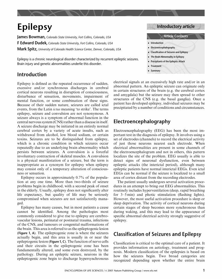
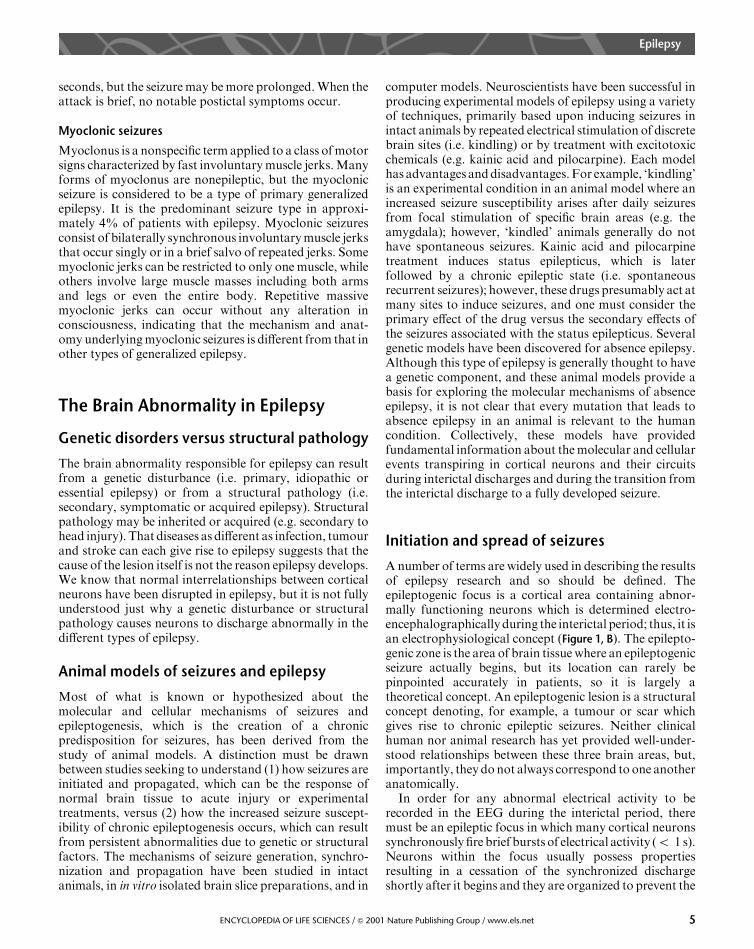
Epilepsy has many causes, but in most patients a causecannot be identified. Among the pathologies mostcommonly considered to give rise to epilepsy are cerebro-vascular lesions, perinatal or postnatal trauma, infectionsof the CNS, and tumours or congenital malformations ofthe brain. This area is referred to as the epileptogenic lesion(Figure 1, A). The epileptogenic zone is where the seizuresactually begin, and this area is usually in or near theepileptogenic lesion (Figure 1, C). The function of nerve cellsand their circuits in the epileptogenic zone has beenfundamentally altered, and some even destroyed, by thepathology. During an epileptic seizure, neurons in theepileptogenic zone begin to discharge hypersynchronous
electrical signals at an excessively high rate and/or in anabnormal pattern. An epileptic seizure can originate onlyin certain structures of the brain (e.g. the cerebral cortexand amygdala) but the seizure may then spread to otherstructures of the CNS (e.g. the basal ganglia). Once apatient has developed epilepsy, individual seizures may beprecipitated by a number of conditions and circumstances.
Electroencephalography
Electroencephalography (EEG) has been the most im-portant test in the diagnosis of epilepsy. It involves using aset of electrodes (channels) to record the electrical activityof just those neurons nearest each electrode. Whenelectrical abnormalities are present in some channels ofthe electroencephalogram and not in others, this patternlocalizes the site of the problem. EEG usually is able todetect signs of neuronal dysfunction, even betweenepileptic attacks (the interictal period), although manyepileptic patients have normal interictal EEGs. Even ictalEEGs can be normal if the seizure is localized to a smallarea of cortex distant from the recording electrodes.
The patient usually undergoes several activation proce-dures in an attempt to bring out EEG abnormalities. Thisroutinely includes hyperventilation (deep, rapid breathingfor 3–5 min) and photic stimulation (flashing lights).However, the most useful activation procedure is sleep orsleep deprivation. The activity of cortical neurons duringcertain stages of sleep becomes more synchronous thanduring waking, and this may lead to the appearance ofspecific abnormal electrical activity strongly suggestive ofepilepsy.
Classification of Seizures and Epilepsy
Classification is critical to the optimal care of a patient. Itprovides information on aetiology, treatment and prog-nosis. Modern classification of the epilepsies is based onhow the seizures begin. Two broad categories arerecognized depending upon whether the entire brain
Article Contents
Introductory article
. Introduction
. Electroencephalography
. Classification of Seizures and Epilepsy
. The Brain Abnormality in Epilepsy
. Precipitants of the Epileptic Attack
. Treatment
. Summary
1ENCYCLOPEDIA OF LIFE SCIENCES / & 2001 Nature Publishing Group / www.els.net
(generalized seizure) or only a restricted part of the brain(partial seizure) is involved in the discharge at its onset.Each category is further subdivided, depending on thesymptoms displayed by the patient during the seizure.Many patients have more than one seizure type, but it israre to have both partial onset and primary generalizedseizures.
Partial seizures
Partial seizures begin in a discrete cortical area. They arecategorized as simple when consciousness is preserved andcomplex when consciousness is altered. Simple partialseizures may evolve into complex partial seizures orsecondarily generalized tonic–clonic seizures as a resultof the spread of abnormal electrical activity.
Simple partial seizures
Simple partial seizures are the primary complaint in 10%of patients with epilepsy. They can occur frequently andmay result in little disability. Simple motor seizures resultfrom a discharging lesion in the precentral gyrus of thefrontal lobe of the cerebral hemisphere opposite the musclecontractions. Some are sustained (tonic) and othersintermittent (clonic), and they may involve any body partdepending on the location of the abnormal brain discharge.Simple sensory seizures indicate discharging nerve cells inthe appropriate primary sensory cortex of the hemisphere.Simple somatosensory seizures usually result from anepileptogenic zone in or near the postcentral gyrus of the
opposite cerebral hemisphere. Somatosensory seizures areusually described by the patient as the sensation of ‘pins-and-needles’. A simple seizure may remain restricted or‘march’ to other parts of the body in a sequence determinedby the somatotopic organization of the cortex, or it mayspread through other axonal pathways. Visual seizuresindicate an epileptogenic zone in or near the primary visualcortex of the occipital lobe. Spots or patterns areexperienced in the visual field opposite the side of theseizure focus. Seizures eliciting auditory, vestibular,olfactory or visceral sensations can also occur.
Complex partial seizures
Complex partial seizures are the predominant seizure typein about 20% of patients with epilepsy. The termspsychomotor, temporal lobe and limbic system seizureare older terms, somewhat synonymous with the termcomplex partial seizure, all designating a form of partialseizure in which consciousness is altered but not necessarilylost. During these seizures there is a period of alteredbehaviour for which the patient is later amnesic. Theamnesia for ictal events is a key factor for the diagnosis of acomplex partial seizure.
A typical complex partial seizure consists of severalphases. About 70% begin with an ‘aura’, a sometimescomplex psychic experience that may be manifest in one ormore of a wide variety of vivid forms: as an illusion,hallucination, dyscognitive state or emotional (affective)experience. This usually lasts a few seconds. Such psychicexperiences may comprise the entire seizure, which is then a
A
B C
Figure 1 The three cortical areas contributing to seizure onset. (A) The epileptogenic lesion, a structural abnormality that leads to the development ofepileptic foci (B). Each focus consists of a group of excessively discharging neurons (blue) surrounded by a zone of inhibited neurons (shaded area) whichkeeps each focus from spreading to neighbouring neurons (pink). (C) The epileptogenic zone, a large group of excessively discharging neurons (blue)created by the breakdown of the foci and responsible for seizure onset.
Epilepsy
2 ENCYCLOPEDIA OF LIFE SCIENCES / & 2001 Nature Publishing Group / www.els.net
simple partial seizure. However, when the seizure pro-gresses into a second phase with alteration of conscious-ness, it is defined as a complex partial seizure. Dystonic(twisted, stiff) posturing of the arm or leg on the sideopposite to where the seizure occurs is often observed.Primitive movements occur frequently. These are referredto as spontaneous automatisms, and may include aimlessfumbling with clothing or walking in a daze, and chewingor swallowing. However, reactive automatisms also mayoccur and these are not stereotyped because they aredetermined by environmental stimuli. Such automatismsare why the old term psychomotor seizures was coined.
Primary generalized seizures
Primary generalized seizures (also called generalizedseizures) involve widespread areas of the cerebral cortexfrom the onset. These terms must not be confused with theterm secondary generalized seizure, which refers to apartial onset seizure that spreads to wide areas of cortex.The abnormal electrical activity is the same in both the leftand right hemispheres (bilaterally symmetrical). General-ized seizures are further subdivided into convulsive andnonconvulsive types. Convulsive seizures are characterizedby sometimes violent and sustained contractions ofmuscles. Nonconvulsive seizures lack prominent motoractivity. Generalized tonic–clonic, clonic, and some tonicseizures are referred to as convulsive generalized seizures.The most common nonconvulsive generalized seizure is the
absence seizure, but the category also includes atonic, brieftonic, and myoclonic seizures.
Convulsive seizures
Tonic–clonic convulsions often begin with a piercing cry orchoking as the entire body musculature is seized in a strongcontraction and air is forced out through partially closedvocal cords. Patients fall to the ground in an unconsciousstate. Initial motor signs include brief flexion of the trunk,an opening of the mouth and eyelids, upward deviation ofthe eyes, and elevation of the arms. These are followed by arigid extension phase, involving the back, neck, arms andlegs, which lasts for 15–20 s. Involvement of the respiratorymuscles in the spasm results in a suspension of breathing,and in a few seconds the skin and mucous membranesbecome cyanotic. The patient often loses bowel andbladder control during this phase. This tonic phase of theconvulsive seizure is followed by the clonic phase, whichconsists of rhythmic muscle contractions lasting for 20–30 s. Autonomic signs are conspicuous: the pupils aredilated, blood pressure is raised, the pulse is rapid, andsalivation and sweating occur.
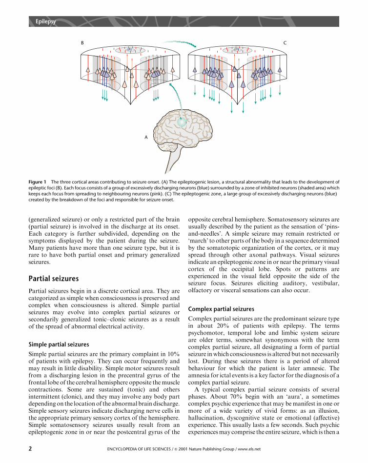
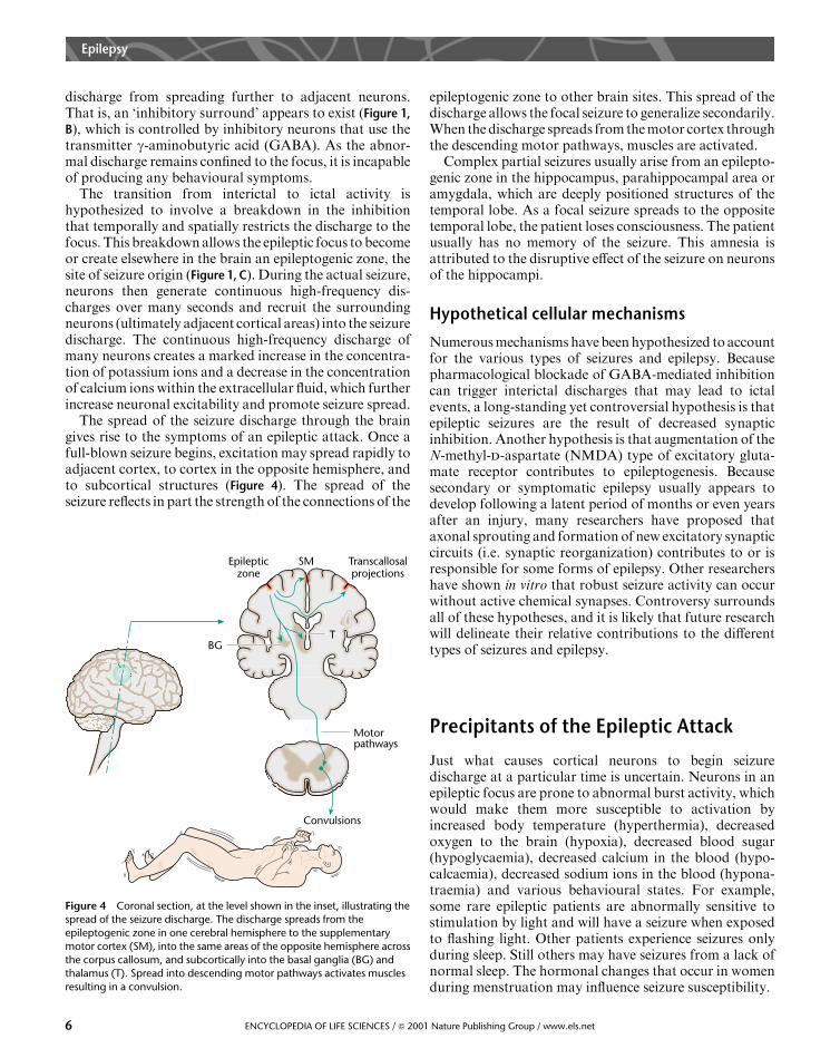
The onset of a grand mal seizure is characterizedelectroencephalographically by the simultaneous appear-ance of abnormal activity over large areas of the cerebralcortex in both hemispheres (Figure2). The EEG shows a fastrhythmic discharge (4 10 Hz), which decreases in fre-quency and increases in amplitude during the tonic phase.The cortical discharge then changes from continuous to
Figure 2 Simulated electroencephalogram (EEG) of a generalized seizure characterized by a tonic–clonic convulsion. Each line tracing (channel) isfrom electrodes positioned on the scalp as shown in the inset EEG montage. (Adapted from Gastaut H and Broughton R (1972) Epileptic Seizures.Springfield, Illinois: Charles C. Thomas.)
Epilepsy
3ENCYCLOPEDIA OF LIFE SCIENCES / & 2001 Nature Publishing Group / www.els.net
intermittent bursts of activity, which signals the beginningof the clonic phase of the seizure. These intermittent burstsof activity are referred to as grouped polyspikes and areseparated by quiet intervals. The bursts gradually decreasein frequency, which correlates behaviourally with thedecrease in the frequency of the repetitive clonic muscularjerks.
When the seizure is over, the patient does notimmediately return to normal. This postictal state, whichlasts for minutes to hours (rarely days), is first character-ized by the patient lying still and limp, and breathingquietly. The patient slowly regains consciousness, usuallyover the next several minutes, but is obviously confused.The patient may remain sleepy for several hours; if leftundisturbed, the patient may fall into a deep sleep for hoursand awaken with a postictal headache and fatigue. Oncerecovered, the patient may have no memory for any part ofthe seizure. However, the patient is aware that somethingtroublesome has happened because of a bitten tongue,injury from the fall, concern expressed by others, regainingconsciousness in different surroundings, and achingmuscles from the violent contractions. A small proportion(5–8%) of these patients in the future will experience aprolonged series of tonic–clonic seizures without fullyregaining consciousness between them. This condition istermed status epilepticus; it is life threatening and demandsurgent medical treatment.
Absence seizures
Absence (petit mal) seizures occur without warning andconsist of a sudden interruption of consciousness. Thehallmarks of absence seizures are their brevity, general lackof motor activity, frequency, and lack of a postictal period.
The seizures usually last from 2 to 10 s, occasionally longer.Patients are often unaware of them. An observer mayinterpret an absence as a moment of daydreaming. Theperson stops talking briefly in midsentence, stares, or stopsresponding. As many as several hundred such seizures mayoccur in 1 day. Absence seizures almost always begin inchildhood or adolescence. They often disappear beforeadulthood, presumably because of the biochemical orstructural changes associated with brain maturation.When they occur with high frequency in school, they oftenlead to poor performance.
During short absences the patient may be completelymotionless. With longer absences, subtle automatisms areoften observed such as blinking. When used correctly, theolder term petit mal is synonymous with absence seizures.However, the term is often applied incorrectly, as when apatient with complex partial seizures is said to have petitmal staring spells.
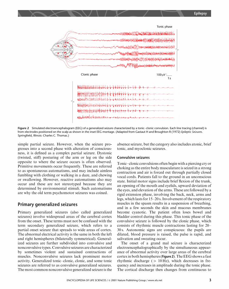
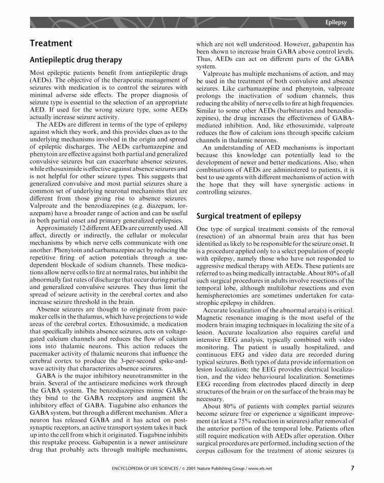
The EEG signature of a typical absence seizure is anabrupt onset and sudden cessation of a bilaterallysynchronous and regular discharge recorded over wide-spread areas of the scalp (Figure 3). The discharge consistsof spike- or multiple spike-and-slow wave complexes at afrequency of about 3 Hz.
Atonic seizures
Atonic seizures (drop attacks) manifest themselves as asudden loss of tone in postural muscles. In a mild variant,only the head drops. However, in the severe form, all of thepostural muscles lose tone, and the patient suddenlycollapses to the ground. Frequent falls may result ininjury, especially to the head, so protective headgear maybe needed. The duration of the attack is usually only a few
Figure 3 Simulated electroencephalogram of generalized 3-Hz spike-and-wave complexes during an absence seizure. Note the abrupt onset andsudden cessation of the bilaterally synchronous activity. (Adapted from Lothman EW and Collins RC (1990) In: Pearlman AL and Collins RC (eds)Neurobiology of Disease. New York: Oxford University Press.)
Epilepsy
4 ENCYCLOPEDIA OF LIFE SCIENCES / & 2001 Nature Publishing Group / www.els.net
seconds, but the seizure may be more prolonged. When theattack is brief, no notable postictal symptoms occur.
Myoclonic seizures
Myoclonus is a nonspecific term applied to a class of motorsigns characterized by fast involuntary muscle jerks. Manyforms of myoclonus are nonepileptic, but the myoclonicseizure is considered to be a type of primary generalizedepilepsy. It is the predominant seizure type in approxi-mately 4% of patients with epilepsy. Myoclonic seizuresconsist of bilaterally synchronous involuntary muscle jerksthat occur singly or in a brief salvo of repeated jerks. Somemyoclonic jerks can be restricted to only one muscle, whileothers involve large muscle masses including both armsand legs or even the entire body. Repetitive massivemyoclonic jerks can occur without any alteration inconsciousness, indicating that the mechanism and anat-omy underlying myoclonic seizures is different from that inother types of generalized epilepsy.
The Brain Abnormality in Epilepsy
Genetic disorders versus structural pathology
The brain abnormality responsible for epilepsy can resultfrom a genetic disturbance (i.e. primary, idiopathic oressential epilepsy) or from a structural pathology (i.e.secondary, symptomatic or acquired epilepsy). Structuralpathology may be inherited or acquired (e.g. secondary tohead injury). That diseases as different as infection, tumourand stroke can each give rise to epilepsy suggests that thecause of the lesion itself is not the reason epilepsy develops.We know that normal interrelationships between corticalneurons have been disrupted in epilepsy, but it is not fullyunderstood just why a genetic disturbance or structuralpathology causes neurons to discharge abnormally in thedifferent types of epilepsy.
Animal models of seizures and epilepsy
Most of what is known or hypothesized about themolecular and cellular mechanisms of seizures andepileptogenesis, which is the creation of a chronicpredisposition for seizures, has been derived from thestudy of animal models. A distinction must be drawnbetween studies seeking to understand (1) how seizures areinitiated and propagated, which can be the response ofnormal brain tissue to acute injury or experimentaltreatments, versus (2) how the increased seizure suscept-ibility of chronic epileptogenesis occurs, which can resultfrom persistent abnormalities due to genetic or structuralfactors. The mechanisms of seizure generation, synchro-nization and propagation have been studied in intactanimals, in in vitro isolated brain slice preparations, and in
computer models. Neuroscientists have been successful inproducing experimental models of epilepsy using a varietyof techniques, primarily based upon inducing seizures inintact animals by repeated electrical stimulation of discretebrain sites (i.e. kindling) or by treatment with excitotoxicchemicals (e.g. kainic acid and pilocarpine). Each modelhas advantages and disadvantages. For example, ‘kindling’is an experimental condition in an animal model where anincreased seizure susceptibility arises after daily seizuresfrom focal stimulation of specific brain areas (e.g. theamygdala); however, ‘kindled’ animals generally do nothave spontaneous seizures. Kainic acid and pilocarpinetreatment induces status epilepticus, which is laterfollowed by a chronic epileptic state (i.e. spontaneousrecurrent seizures); however, these drugs presumably act atmany sites to induce seizures, and one must consider theprimary effect of the drug versus the secondary effects ofthe seizures associated with the status epilepticus. Severalgenetic models have been discovered for absence epilepsy.Although this type of epilepsy is generally thought to havea genetic component, and these animal models provide abasis for exploring the molecular mechanisms of absenceepilepsy, it is not clear that every mutation that leads toabsence epilepsy in an animal is relevant to the humancondition. Collectively, these models have providedfundamental information about the molecular and cellularevents transpiring in cortical neurons and their circuitsduring interictal discharges and during the transition fromthe interictal discharge to a fully developed seizure.
Initiation and spread of seizures
A number of terms are widely used in describing the resultsof epilepsy research and so should be defined. Theepileptogenic focus is a cortical area containing abnor-mally functioning neurons which is determined electro-encephalographically during the interictal period; thus, it isan electrophysiological concept (Figure 1, B). The epilepto-genic zone is the area of brain tissue where an epileptogenicseizure actually begins, but its location can rarely bepinpointed accurately in patients, so it is largely atheoretical concept. An epileptogenic lesion is a structuralconcept denoting, for example, a tumour or scar whichgives rise to chronic epileptic seizures. Neither clinicalhuman nor animal research has yet provided well-under-stood relationships between these three brain areas, but,importantly, they do not always correspond to one anotheranatomically.
In order for any abnormal electrical activity to berecorded in the EEG during the interictal period, theremust be an epileptic focus in which many cortical neuronssynchronously fire brief bursts of electrical activity (5 1 s).Neurons within the focus usually possess propertiesresulting in a cessation of the synchronized dischargeshortly after it begins and they are organized to prevent the
Epilepsy
5ENCYCLOPEDIA OF LIFE SCIENCES / & 2001 Nature Publishing Group / www.els.net
discharge from spreading further to adjacent neurons.That is, an ‘inhibitory surround’ appears to exist (Figure 1,B), which is controlled by inhibitory neurons that use thetransmitter g-aminobutyric acid (GABA). As the abnor-mal discharge remains confined to the focus, it is incapableof producing any behavioural symptoms.
The transition from interictal to ictal activity ishypothesized to involve a breakdown in the inhibitionthat temporally and spatially restricts the discharge to thefocus. This breakdown allows the epileptic focus to becomeor create elsewhere in the brain an epileptogenic zone, thesite of seizure origin (Figure 1, C). During the actual seizure,neurons then generate continuous high-frequency dis-charges over many seconds and recruit the surroundingneurons (ultimately adjacent cortical areas) into the seizuredischarge. The continuous high-frequency discharge ofmany neurons creates a marked increase in the concentra-tion of potassium ions and a decrease in the concentrationof calcium ions within the extracellular fluid, which furtherincrease neuronal excitability and promote seizure spread.
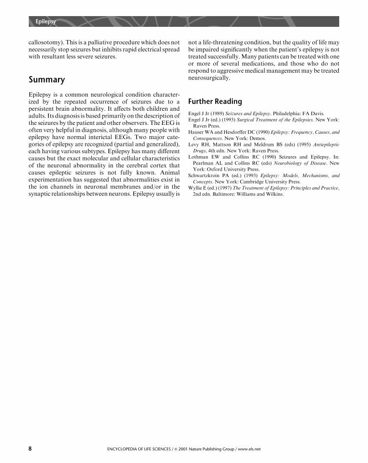
The spread of the seizure discharge through the braingives rise to the symptoms of an epileptic attack. Once afull-blown seizure begins, excitation may spread rapidly toadjacent cortex, to cortex in the opposite hemisphere, andto subcortical structures (Figure 4). The spread of theseizure reflects in part the strength of the connections of the
epileptogenic zone to other brain sites. This spread of thedischarge allows the focal seizure to generalize secondarily.When the discharge spreads from the motor cortex throughthe descending motor pathways, muscles are activated.
Complex partial seizures usually arise from an epilepto-genic zone in the hippocampus, parahippocampal area oramygdala, which are deeply positioned structures of thetemporal lobe. As a focal seizure spreads to the oppositetemporal lobe, the patient loses consciousness. The patientusually has no memory of the seizure. This amnesia isattributed to the disruptive effect of the seizure on neuronsof the hippocampi.
Hypothetical cellular mechanisms
Numerous mechanisms have been hypothesized to accountfor the various types of seizures and epilepsy. Becausepharmacological blockade of GABA-mediated inhibitioncan trigger interictal discharges that may lead to ictalevents, a long-standing yet controversial hypothesis is thatepileptic seizures are the result of decreased synapticinhibition. Another hypothesis is that augmentation of theN-methyl-d-aspartate (NMDA) type of excitatory gluta-mate receptor contributes to epileptogenesis. Becausesecondary or symptomatic epilepsy usually appears todevelop following a latent period of months or even yearsafter an injury, many researchers have proposed thataxonal sprouting and formation of new excitatory synapticcircuits (i.e. synaptic reorganization) contributes to or isresponsible for some forms of epilepsy. Other researchershave shown in vitro that robust seizure activity can occurwithout active chemical synapses. Controversy surroundsall of these hypotheses, and it is likely that future researchwill delineate their relative contributions to the differenttypes of seizures and epilepsy.
Precipitants of the Epileptic Attack
Just what causes cortical neurons to begin seizuredischarge at a particular time is uncertain. Neurons in anepileptic focus are prone to abnormal burst activity, whichwould make them more susceptible to activation byincreased body temperature (hyperthermia), decreasedoxygen to the brain (hypoxia), decreased blood sugar(hypoglycaemia), decreased calcium in the blood (hypo-calcaemia), decreased sodium ions in the blood (hypona-traemia) and various behavioural states. For example,some rare epileptic patients are abnormally sensitive tostimulation by light and will have a seizure when exposedto flashing light. Other patients experience seizures onlyduring sleep. Still others may have seizures from a lack ofnormal sleep. The hormonal changes that occur in womenduring menstruation may influence seizure susceptibility.
Epilepticzone
SM Transcallosalprojections
Convulsions
BG
Motorpathways
T
Figure 4 Coronal section, at the level shown in the inset, illustrating thespread of the seizure discharge. The discharge spreads from theepileptogenic zone in one cerebral hemisphere to the supplementarymotor cortex (SM), into the same areas of the opposite hemisphere acrossthe corpus callosum, and subcortically into the basal ganglia (BG) andthalamus (T). Spread into descending motor pathways activates musclesresulting in a convulsion.
Epilepsy
6 ENCYCLOPEDIA OF LIFE SCIENCES / & 2001 Nature Publishing Group / www.els.net
Treatment
Antiepileptic drug therapy
Most epileptic patients benefit from antiepileptic drugs(AEDs). The objective of the therapeutic management ofseizures with medication is to control the seizures withminimal adverse side effects. The proper diagnosis ofseizure type is essential to the selection of an appropriateAED. If used for the wrong seizure type, some AEDsactually increase seizure activity.
The AEDs are different in terms of the type of epilepsyagainst which they work, and this provides clues as to theunderlying mechanisms involved in the origin and spreadof epileptic discharges. The AEDs carbamazepine andphenytoin are effective against both partial and generalizedconvulsive seizures but can exacerbate absence seizures,while ethosuximide is effective against absence seizures andis not helpful for other seizure types. This suggests thatgeneralized convulsive and most partial seizures share acommon set of underlying neuronal mechanisms that aredifferent from those giving rise to absence seizures.Valproate and the benzodiazepines (e.g. diazepam, lor-azepam) have a broader range of action and can be usefulin both partial onset and primary generalized epilepsies.
Approximately 12 different AEDs are currently used. Allaffect, directly or indirectly, the cellular or molecularmechanisms by which nerve cells communicate with oneanother. Phenytoin and carbamazepine act by reducing therepetitive firing of action potentials through a use-dependent blockade of sodium channels. These medica-tions allow nerve cells to fire at normal rates, but inhibit theabnormally fast rates of discharge that occur during partialand generalized convulsive seizures. They thus limit thespread of seizure activity in the cerebral cortex and alsoincrease seizure threshold in the brain.
Absence seizures are thought to originate from pace-maker cells in the thalamus, which have projections to wideareas of the cerebral cortex. Ethosuximide, a medicationthat specifically inhibits absence seizures, acts on voltage-gated calcium channels and reduces the flow of calciumions into thalamic neurons. This action reduces thepacemaker activity of thalamic neurons that influence thecerebral cortex to produce the 3-per-second spike-and-wave activity that characterizes absence seizures.
GABA is the major inhibitory neurotransmitter in thebrain. Several of the antiseizure medicines work throughthe GABA system. The benzodiazepines mimic GABA;they bind to the GABA receptors and augment theinhibitory effect of GABA. Tiagabine also enhances theGABA system, but through a different mechanism. After aneuron has released GABA and it has acted on post-synaptic receptors, an active transport system takes it backup into the cell from which it originated. Tiagabine inhibitsthis reuptake process. Gabapentin is a newer antiseizuredrug that probably acts through multiple mechanisms,
which are not well understood. However, gabapentin hasbeen shown to increase brain GABA above control levels.Thus, AEDs can act on different parts of the GABAsystem.
Valproate has multiple mechanisms of action, and maybe used in the treatment of both convulsive and absenceseizures. Like carbamazepine and phenytoin, valproateprolongs the inactivation of sodium channels, thusreducing the ability of nerve cells to fire at high frequencies.Similar to some other AEDs (barbiturates and benzodia-zepines), the drug increases the effectiveness of GABA-mediated inhibition. And, like ethosuximide, valproatereduces the flow of calcium ions through specific calciumchannels in thalamic neurons.
An understanding of AED mechanisms is importantbecause this knowledge can potentially lead to thedevelopment of newer and better medications. Also, whencombinations of AEDs are administered to patients, it isbest to use agents with different mechanisms of action withthe hope that they will have synergistic actions incontrolling seizures.
Surgical treatment of epilepsy
One type of surgical treatment consists of the removal(resection) of an abnormal brain area that has beenidentified as likely to be responsible for the seizure onset. Itis a procedure applied only to a select population of peoplewith epilepsy, namely those who have not responded toaggressive medical therapy with AEDs. These patients arereferred to as being medically intractable. About 80% of allsuch surgical procedures in adults involve resections of thetemporal lobe, although multilobar resections and evenhemispherectomies are sometimes undertaken for cata-strophic epilepsy in children.
Accurate localization of the abnormal area(s) is critical.Magnetic resonance imaging is the most useful of themodern brain imaging techniques in localizing the site of alesion. Accurate localization also requires careful andintensive EEG analysis, typically combined with videomonitoring. The patient is usually hospitalized, andcontinuous EEG and video data are recorded duringtypical seizures. Both types of data provide information onlesion localization; the EEG provides electrical localiza-tion, and the video behavioural localization. SometimesEEG recording from electrodes placed directly in deepstructures of the brain or on the surface of the brain may benecessary.
About 80% of patients with complex partial seizuresbecome seizure free or experience a significant improve-ment (at least a 75% reduction in seizures) after removal ofthe anterior portion of the temporal lobe. Patients oftenstill require medication with AEDs after operation. Othersurgical procedures are performed, including section of thecorpus callosum for the treatment of atonic seizures (a
Epilepsy
7ENCYCLOPEDIA OF LIFE SCIENCES / & 2001 Nature Publishing Group / www.els.net
callosotomy). This is a palliative procedure which does notnecessarily stop seizures but inhibits rapid electrical spreadwith resultant less severe seizures.
Summary
Epilepsy is a common neurological condition character-ized by the repeated occurrence of seizures due to apersistent brain abnormality. It affects both children andadults. Its diagnosis is based primarily on the description ofthe seizures by the patient and other observers. The EEG isoften very helpful in diagnosis, although many people withepilepsy have normal interictal EEGs. Two major cate-gories of epilepsy are recognized (partial and generalized),each having various subtypes. Epilepsy has many differentcauses but the exact molecular and cellular characteristicsof the neuronal abnormality in the cerebral cortex thatcauses epileptic seizures is not fully known. Animalexperimentation has suggested that abnormalities exist inthe ion channels in neuronal membranes and/or in thesynaptic relationships between neurons. Epilepsy usually is
not a life-threatening condition, but the quality of life maybe impaired significantly when the patient’s epilepsy is nottreated successfully. Many patients can be treated with oneor more of several medications, and those who do notrespond to aggressive medical management may be treatedneurosurgically.
Further Reading
Engel J Jr (1989) Seizures and Epilepsy. Philadelphia: FA Davis.
Engel J Jr (ed.) (1993) Surgical Treatment of the Epilepsies. New York:
Raven Press.
Hauser WA and Hesdorffer DC (1990) Epilepsy: Frequency, Causes, and
Consequences. New York: Demos.
Levy RH, Mattson RH and Meldrum BS (eds) (1995) Antiepileptic
Drugs, 4th edn. New York: Raven Press.
Lothman EW and Collins RC (1990) Seizures and Epilepsy. In:
Pearlman AL and Collins RC (eds) Neurobiology of Disease. New
York: Oxford University Press.
Schwartzkroin PA (ed.) (1993) Epilepsy: Models, Mechanisms, and
Concepts. New York: Cambridge University Press.
Wyllie E (ed.) (1997) The Treatment of Epilepsy: Principles and Practice,
2nd edn. Baltimore: Williams and Wilkins.
Epilepsy
8 ENCYCLOPEDIA OF LIFE SCIENCES / & 2001 Nature Publishing Group / www.els.net