Episodic Occurrence of Antibodies against the Bovine LeukemiaVirus Rex Protein during the Course of Infection in Sheep
MAUREEN A. POWERS,"12 DEBORAH GROSSMAN,' LYNN C. KIDD,12 AND KATHRYN RADKE1*
Department ofAvian Sciences' and Biochemistry Graduate Group,2 University of California, Davis, California 95616-8532
Received 8 March 1991/Accepted 24 May 1991
Infection by bovine leukemia virus (BLV) is characterized by a long clinical latency after which some
individuals develop B-cell tumors. The contributions of the viral regulatory proteins Tax and Rex duringclinical latency and disease are incompletely understood. To learn about Rex expression in the host, we useda sensitive immunoprecipitation assay to detect Rex antibodies throughout the course ofBLV infection in sheep.Sixty percent of the infected animals produced Rex antibodies in intermittent episodes. This pattern differedmarkedly from that of antibodies to virion structural proteins, which were maintained in all animalsthroughout infection. Only one of two animals that developed tumors had detectable Rex antibodies at the time,although the other had previously demonstrated an especially strong Rex antibody response. We examined theRex response in the context of BLV infection by comparing it with the frequency of circulating mononuclearblood cells that could transcribe BLV RNA or produce infectious virus. Episodes of Rex antibody occurrence
followed some but not all increases in the number of BLV-transcribing cells. Since the appearance of circulatingantibodies requires that the intracellular Rex protein be available to serve as antigen, the episodic pattern ofoccurrence of Rex antibodies could result from intermittent killing by virus-specific cytotoxic cells. Fluctuationsin titer that were observed during some episodes of Rex response could be due to antibody retention by antigenpresent in lymphoid tissue.
Bovine leukemia virus (BLV), human T-cell leukemiavirus types I and II (HTLV-I and -II), and simian T-cellleukemia virus constitute a family of retroviruses that havesimilar genetic structures and modes of action. Infection bythese viruses is characterized by long clinical latency fol-lowed, in only a few infected individuals, by terminal disease(1, 2). Virus expression is latent within most infected cells;circulating lymphocytes carry integrated proviral DNA butdo not produce detectable virus (15). However, the continu-ing presence of antiviral antibodies throughout infectionindicates that virus production does occur, most probably inlymphoid tissues. The specific cellular events that activatevirus expression in the infected host are unknown, but inculture, polyclonal activators of lymphocytes stimulate virusproduction (11, 19, 29). This suggests that immune responsesstimulate viral replication in vivo and may play importantroles in the progression from clinical latency to disease.
Replication of these viruses is regulated by two virus-encoded proteins, Tax and Rex (10). Tax acts in concert withcellular factors to activate transcription from the viral longterminal repeat. Rex controls translational availability ofdifferent viral messages, although the mechanism is notunderstood. Since the Tax and Rex proteins regulate virusexpression, they presumably mediate the exit of the virusfrom cellular latency. These regulatory proteins are ex-
pressed at low levels in the host cell, making their detectiondifficult. However, the patterns of antibody response to Taxand Rex might serve as indicators of prior increases in theirproduction or availability in vivo.
Antibodies to virus-encoded regulatory proteins havebeen observed in both patients and asymptomatic carriersinfected with either HTLV-I or human immunodeficiencyvirus (HIV). Titers of antibodies to some HIV regulatoryproteins as well as to the virus capsid protein (CA; p24)
* Corresponding author.
change over the course of infection (17, 31, 32). In rare
cases, all HIV antibodies can synchronously disappear for a
time (8). Since studies most often have used only a fewserum samples from each of many patients, it is difficult tointerpret the results in the context of cellular activation anddisease progression. Longitudinal studies with frequent sam-
pling are required for such interpretation. Since little isknown about antibodies to Rex during either HTLV or BLVinfection, we assessed the presence of these antibodiesthroughout the course of BLV infection in sheep. Here we
present the results and place them in the context of severalcellular measures of infection.
MATERIALS AND METHODS
Plasmid construction. The 973-bp ClaI7113-to-RsaI8086 frag-ment (numbered according to reference 28), encompassingthe majority of the second exon of the rex open readingframe, was isolated from the Belgian tumor clone of BLV(4). After digestion of the plasmid pSP6-X3M (16) with BalIand HincII, blunt-end ligation with the BLV fragment re-
sulted in a construct (pSP6rex) which carries the SP6 bacte-riophage promoter followed by the P-globin 5' untranslatedregion and rex coding sequences. For in vitro transcription,the plasmid was linearized with SmaI and purified by usingGeneclean (BiolOl, La Jolla, Calif.).
In vitro transcription and translation. In vitro transcrip-tions were performed with SP6 polymerase in the Riboprobesystem (Promega Corp., Madison, Wis.). A trace amount of[35S]CTP was included in the 100-,ul reaction mixture toallow quantitation of yield. Typically, 10 to 20 ,ug of un-
capped RNA was transcribed from 2 to 3 ,ug of linearizedplasmid. The reaction mixture was then treated with RNase-free DNase (RQ DNase; Promega), phenol extracted, andethanol precipitated. To verify their sizes, 3"S-labeled tran-scription products were analyzed by electrophoresis through1% agarose in the presence of 2.2 M formaldehyde (22). The
4959
Vol. 65, No. 9
Dow
nloa
ded
from
http
s://j
ourn
als.
asm
.org
/jour
nal/j
vi o
n 16
Feb
ruar
y 20
22 b
y 1.
65.1
54.1
34.
4960 POWERS ET AL.
electrophoresed RNA was transferred to nitrocellulosewhich was then saturated with 20% 2,5-diphenyloxazole(PPO) in toluene, air dried, and exposed to film.
In vitro translations were performed with amino acid-depleted rabbit reticulocyte lysates (Amersham Corp., Ar-lington Heights, Ill.) supplemented with 100 mM potassiumacetate. RNA was denatured at 65°C for 10 min and quicklycooled on ice. Approximately 2 ,ug of RNA was used in 200,ul of translation reaction mixture. [35S]methionine (167 ,uCi;1,000 Ci/mmol; Amersham) was added along with aminoacids lacking methionine, and the reaction was incubated at30°C for 90 min. Synthesized protein was quantitated bytrichloroacetic acid precipitation after RNA was removed byincubation with 20 ,ug of RNase A at 30°C for 10 min.Translation products were analyzed by electrophoresisthrough 12.5% polyacrylamide gels containing sodium dode-cyl sulfate (SDS), as described below.
Immunoprecipitation. For use as antigen, "S-labeled Rexprotein was diluted with RIPA buffer (10 mM Tris hydro-chloride [pH 7.4], 0.15 M NaCl, 1.0% Nonidet P-40, 1.0%sodium deoxycholate, 0.1% SDS, 1% aprotinin). In eachassay, 3 ,ul of sheep serum was mixed with 50 ,ul of antigencontaining 80,000 cpm of Rex protein, an amount usuallyderived from 2 RI of the translation reaction mixture. Afterincubation at 4°C for 14 to 16 h, 50 ,ul of a 50% (vol/vol)suspension of protein G-Sepharose (Pharmacia, Piscataway,N.J.) in RIPA buffer was added, and the assay was furtherincubated for 2 h at 4°C on a rocking platform. The proteinG-Sepharose and bound antigen-antibody complexes werepelleted at 4°C and washed three times with ice-cold buffer(10 mM Tris hydrochloride [pH 7.4], 0.1 M NaCl, 0.1%Nonidet P-40). Gel sample buffer (25 RI) containing 50 mMdithiothreitol was added to the pellet. After boiling, 10 pI ofthe resulting supernatant was electrophoresed through anSDS-12.5% polyacrylamide gel (18). Gels were fixed, treatedwith En3Hance (New England Nuclear, Boston, Mass.), andthen exposed to film at -80°C for 24 h unless otherwiseindicated. Intensities of autoradiographic bands were quan-titated by using a Visage 70 densitometer (Biolmage, AnnArbor, Mich.). A positive control was included in all assays.
Collection of serum samples from infected sheep. Sera wereobtained from sheep that had been infected at 2 to 10 weeksof age with BLV derived from persistently infected bat lungcells, as previously described (20). Animals were bled byjugular venipuncture twice weekly for the first 3 months afterinfection and approximately once a month thereafter.
Detection of viral RNA and antiviral antibodies and deter-mination of virus production. Viral RNA was detected by insitu hybridization using "S-labeled antisense RNA probesspanning the BLV genome, as previously described (19).Mononuclear blood cells were cultured for 2 to 5 h at roomtemperature in Iscove's medium containing 10% fetal bovineserum before they were deposited onto slides and fixed for insitu hybridization. An infectious-center assay was used aspreviously described (20) to measure mononuclear cells thatproduced BLV within 24 to 48 h of being cultured in minimalEagle's medium supplemented with 5% fetal bovine serum.After day 642, infectious-center assays were also performedby suspending mononuclear cells in growth medium contain-ing 1% methylcellulose (25). This improved procedure re-vealed increased numbers of infectious centers.Immunoblots were performed as previously described (24)
by using an alkaline phosphatase detection system. Antiserawere tested at a 1/30 dilution. Signal intensities were as-signed values of 0 to 4 after two independent visual evalua-tions. Neutralizing antibodies were measured as described
A B92.5-68 -
43 -
mRNA
28S -
30 -
lBS-
14.3- uM Rex
FIG. 1. In vitro transcription and translation of Rex. (A) 35S-labeled rex RNA was transcribed, purified, and analyzed by 1%agarose-formaldehyde gel electrophoresis and fluorography. Thepositions of 18S and 28S rRNAs are indicated. (B) The products oftranslation of rex RNA were analyzed by 12.5% SDS-polyacryl-amide gel electrophoresis and fluorography. The positions of molec-ular size markers (sizes in kilodaltons) are indicated.
previously (24) by the ability of heat-treated serum to inhibitformation of syncytia induced by cell-free virus. The percentinhibition was determined by comparison with average num-bers of syncytia obtained from virus treated with culturemedium containing fetal bovine serum.
Production of rabbit antipeptide antibodies. A C-terminal12-amino-acid peptide (PSSGTAFFPGTT) of the Rex pro-tein was synthesized with an Applied Biosystems (FosterCity, Calif.) 430A peptide synthesizer. In the synthesis, acysteine was added to the N terminus for cross-linking thepeptide to keyhole limpet hemocyanin (KLH; Calbiochem,La Jolla, Calif.) through the bivalent agent m-maleimidoben-zoyl N-hydroxysulfosuccinimide ester (Pierce, Rockford,Ill.) (9). The extent of cross-linkage was estimated bycomparison of amino acid compositions of KLH and KLH-peptide conjugate.Three New Zealand White rabbits were injected intrader-
mally with 200 pug of KLH-peptide conjugate in Freund'scomplete adjuvant. Rabbits were boosted 7 months later andagain after 5 additional months by intramuscular injectionwith 100 ,ug of peptide conjugate in Freund's incompleteadjuvant. Rabbits were exsanguinated 12 days after the finalboost. All rabbits produced antibodies that recognized boththe C-terminal Rex peptide and the product of in vitrotranslation.
RESULTS
In vitro transcription and translation. The expressionvector pSP6rex was constructed by replacing the ,-globin-coding and 3' untranslated sequences of pSP6-X,M withthose of the rex gene of BLV. Transcription of the linearizedplasmid by SP6 polymerase produced primarily a singleRNA species of approximately the expected size of 1,064nucleotides when analyzed by denaturing gel electrophoresis(Fig. 1A). This RNA encodes 111 of the 154 amino acids ofthe native Rex protein, starting with Met-43. When the RNAwas translated in a reticulocyte lysate, the major radiola-beled product was a polypeptide migrating in SDS-polyacryl-amide gels at approximately 15,000 Da, somewhat largerthan the calculated molecular size of 11,740 (Fig. 1B).Smaller bands were sometimes present. They probablycorrespond to products of internal initiation from down-stream methionine codons, since these polypeptides could
J. VIROL.
Dow
nloa
ded
from
http
s://j
ourn
als.
asm
.org
/jour
nal/j
vi o
n 16
Feb
ruar
y 20
22 b
y 1.
65.1
54.1
34.
EPISODIC Rex ANTIBODIES 4961
92.5m-68
43
30
14.3-
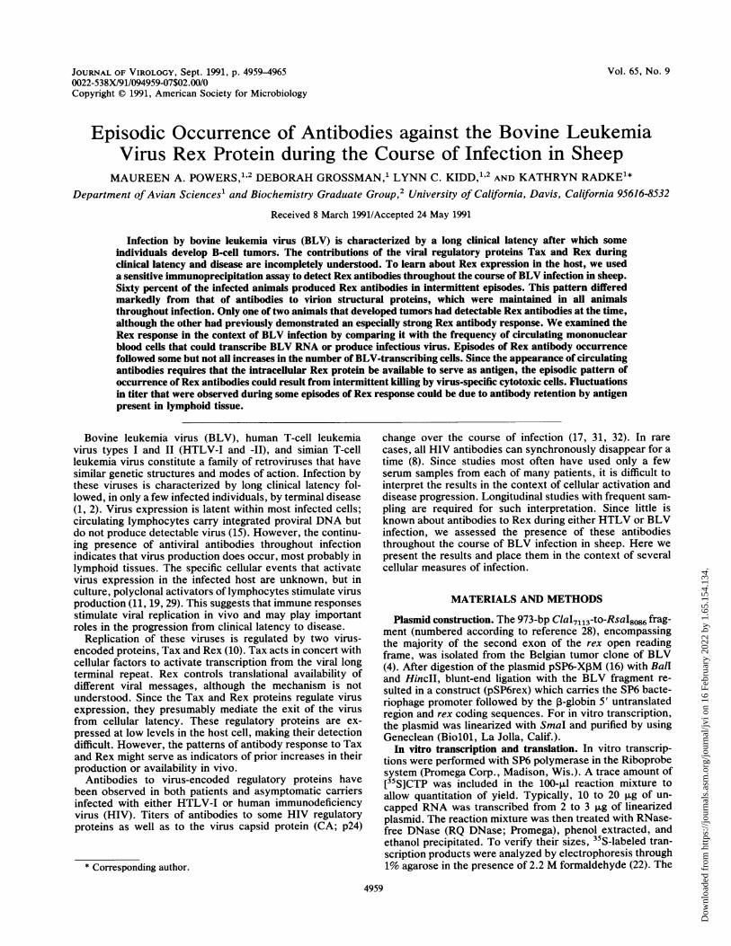
FIG. 2. Immunoprecipitation of Rex. Serial dilutions of a rabbitantipeptide serum were used to immunoprecipitate in vitro-trans-lated Rex protein. Precipitated protein was analyzed on a 12.5%SDS-polyacrylamide gel. Positions of molecular size markers (sizesin kilodaltons) are shown. The gel was exposed to film for 3 h.
be immunoprecipitated with Rex C-terminal antiserum (Fig.2).
Immunoprecipitation assay. To establish a sensitive immu-noprecipitation assay for Rex antibodies, we tested severaldilutions of a rabbit antipeptide serum which had been raisedagainst the 12 C-terminal amino acids of the Rex protein (PSSGTAFFPGTT). With the standard amount of antigen thatwe chose to use (80,000 cpm), a strong positive signal was
evident at a 1/5,000 dilution of the twice-boosted serum (Fig.2). This signal was seen after the gel containing radiolabeledprotein was exposed to film for only 3 h. The same antiserumexhibited a titer (50% of maximum signal) of 1/400 whentested in an enzyme-linked immunosorbent assay using thefree peptide as antigen (data not shown).Rex antibodies in BLV-infected sheep. We then used this
immunoprecipitation assay to determine when, during thecourse of infection, BLV-infected sheep produced antibod-ies specific for the Rex protein. In previous immunoblotanalyses, we had found that BLV structural protein antibod-ies have low titers early in infection (24). Since the regula-tory proteins of BLV are produced in small amounts and arenot incorporated into virions, we expected that titers ofantibodies to Rex would also be low. Immunoblot analysisusing in vitro-translated Rex as antigen yielded no convinc-ing signals from sheep serum. Consequently, immunoprecip-itations were performed with sheep serum at a dilution of1/17, and gels were exposed to film for 24 h. Controlexperiments showed that signal intensity decreased withantibody concentration. No significant background signalsarose from sera drawn prior to infection (Fig. 3A, day 0) orfrom uninfected animals. When sera drawn over periods ofup to 4 years were tested, an immune response to Rex wasdetected in 6 of 10 infected animals.
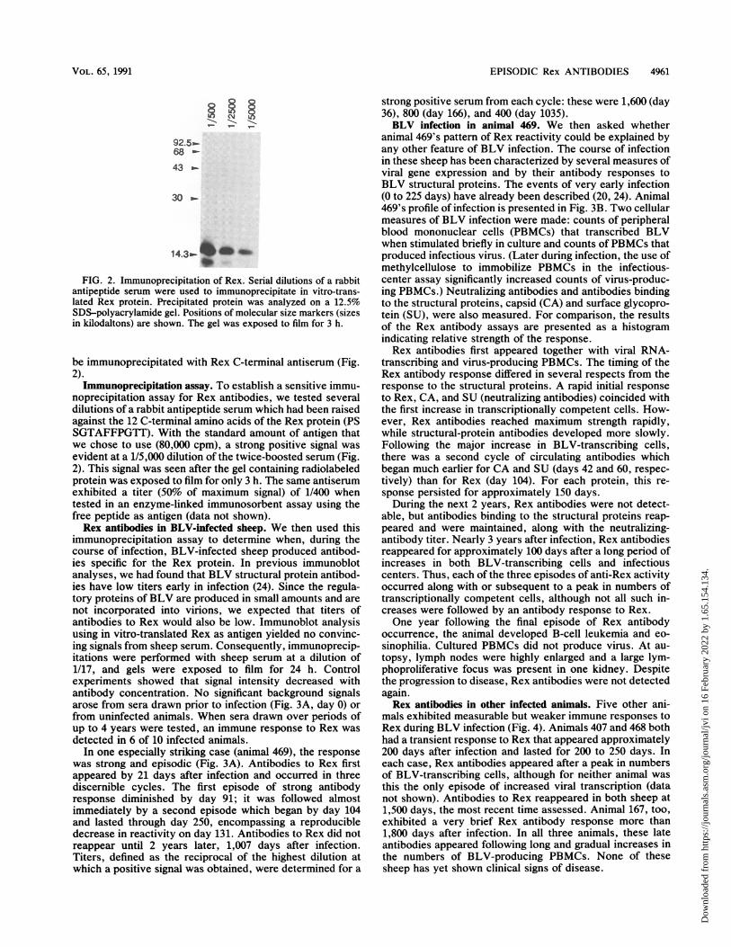
In one especially striking case (animal 469), the responsewas strong and episodic (Fig. 3A). Antibodies to Rex firstappeared by 21 days after infection and occurred in threediscernible cycles. The first episode of strong antibodyresponse diminished by day 91; it was followed almostimmediately by a second episode which began by day 104and lasted through day 250, encompassing a reproducibledecrease in reactivity on day 131. Antibodies to Rex did notreappear until 2 years later, 1,007 days after infection.Titers, defined as the reciprocal of the highest dilution atwhich a positive signal was obtained, were determined for a
strong positive serum from each cycle: these were 1,600 (day36), 800 (day 166), and 400 (day 1035).BLV infection in animal 469. We then asked whether
animal 469's pattern of Rex reactivity could be explained byany other feature of BLV infection. The course of infectionin these sheep has been characterized by several measures ofviral gene expression and by their antibody responses toBLV structural proteins. The events of very early infection(0 to 225 days) have already been described (20, 24). Animal469's profile of infection is presented in Fig. 3B. Two cellularmeasures of BLV infection were made: counts of peripheralblood mononuclear cells (PBMCs) that transcribed BLVwhen stimulated briefly in culture and counts ofPBMCs thatproduced infectious virus. (Later during infection, the use ofmethylcellulose to immobilize PBMCs in the infectious-center assay significantly increased counts of virus-produc-ing PBMCs.) Neutralizing antibodies and antibodies bindingto the structural proteins, capsid (CA) and surface glycopro-tein (SU), were also measured. For comparison, the resultsof the Rex antibody assays are presented as a histogramindicating relative strength of the response.Rex antibodies first appeared together with viral RNA-
transcribing and virus-producing PBMCs. The timing of theRex antibody response differed in several respects from theresponse to the structural proteins. A rapid initial responseto Rex, CA, and SU (neutralizing antibodies) coincided withthe first increase in transcriptionally competent cells. How-ever, Rex antibodies reached maximum strength rapidly,while structural-protein antibodies developed more slowly.Following the major increase in BLV-transcribing cells,there was a second cycle of circulating antibodies whichbegan much earlier for CA and SU (days 42 and 60, respec-tively) than for Rex (day 104). For each protein, this re-sponse persisted for approximately 150 days.During the next 2 years, Rex antibodies were not detect-
able, but antibodies binding to the structural proteins reap-peared and were maintained, along with the neutralizing-antibody titer. Nearly 3 years after infection, Rex antibodiesreappeared for approximately 100 days after a long period ofincreases in both BLV-transcribing cells and infectiouscenters. Thus, each of the three episodes of anti-Rex activityoccurred along with or subsequent to a peak in numbers oftranscriptionally competent cells, although not all such in-creases were followed by an antibody response to Rex.One year following the final episode of Rex antibody
occurrence, the animal developed B-cell leukemia and eo-sinophilia. Cultured PBMCs did not produce virus. At au-topsy, lymph nodes were highly enlarged and a large lym-phoproliferative focus was present in one kidney. Despitethe progression to disease, Rex antibodies were not detectedagain.Rex antibodies in other infected animals. Five other ani-
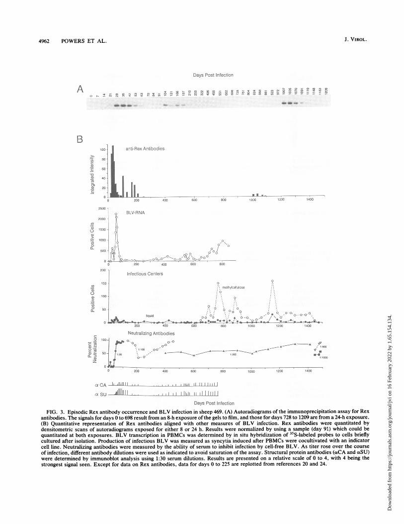
mals exhibited measurable but weaker immune responses toRex during BLV infection (Fig. 4). Animals 407 and 468 bothhad a transient response to Rex that appeared approximately200 days after infection and lasted for 200 to 250 days. Ineach case, Rex antibodies appeared after a peak in numbersof BLV-transcribing cells, although for neither animal wasthis the only episode of increased viral transcription (datanot shown). Antibodies to Rex reappeared in both sheep at1,500 days, the most recent time assessed. Animal 167, too,exhibited a very brief Rex antibody response more than1,800 days after infection. In all three animals, these lateantibodies appeared following long and gradual increases inthe numbers of BLV-producing PBMCs. None of thesesheep has yet shown clinical signs of disease.
VOL. 65, 1991
Dow
nloa
ded
from
http
s://j
ourn
als.
asm
.org
/jour
nal/j
vi o
n 16
Feb
ruar
y 20
22 b
y 1.
65.1
54.1
34.
J. VIROL.4962 POWERS ET AL.
Days Post Infectio;n
A
anti-Rex Anlibodies
L,2ll?00 4150060 a0 IJ:):Do I.220 1430
o BLV-RNA
V0~~~~~~~~~~~0200 40003
Infectious Centers
'?rn :oh r ioose
O
.liqu0 0-~~T4
200 430
Neutralzing Antibodies
.0
1 30) 0-0,
a3- 0
'0
I 0
Oo 0-0
See- 8001-I30
10.0. ,0 0 C)-- 0,
e 00
2. *-1400
120-0 1400
o -u3-0 ~--A T500
A -__--A *V-l A,
2W0 400 W00
e CA ILtAI1J-1I
ax SU i1 1-.ll
!11,11 I 11
Days Post Infection
FIG. 3. Episodic Rex antibody occurrence and BLV infection in sheep 469. (A) Autoradiograms of the immunoprecipitation assay for Rexantibodies. The signals for days 0 to 698 result from an 8-h exposure of the gels to film, and those for days 728 to 1209 are from a 24-h exposure.(B) Quantitative representation of Rex antibodies aligned with other measures of BLV infection. Rex antibodies were quantitated bydensitometric scans of autoradiograms exposed for either 8 or 24 h. Results were normalized by using a sample (day 91) which could bequantitated at both exposures. BLV transcription in PBMCs was determined by in situ hybridization of 3"S-labeled probes to cells brieflycultured after isolation. Production of infectious BLV was measured as syncytia induced after PBMCs were cocultivated with an indicatorcell line. Neutralizing antibodies were measured by the ability of serum to inhibit infection by cell-free BLV. As titer rose over the courseof infection, different antibody dilutions were used as indicated to avoid saturation of the assay. Structural protein antibodies (aCA and aSU)were determined by immunoblot analysis using 1:30 serum dilutions. Results are presented on a relative scale of 0 to 4, with 4 being thestrongest signal seen. Except for data on Rex antibodies, data for days 0 to 225 are replotted from references 20 and 24.
B>10
BOe-
C 60
-o40-
0 20
c I2000 I
-6C)
.6_0a-
1500
500
0 -
2DI
It5
1C
5S
40
50
00
50
4,
.LC)n
O
0-
C:
a) .N
0-f
z
10
5(
P. 0 0 - .Dm-e . § n cq 8 n p- M .'!
2 0't ;
0.; 8 - (. C) N 'n - c, = 10
P, , - C4) T n N U) 'n 8 0, ". .e '? (N Cj - .,- 9 S? -
p. '! Co to N M Q O a) cn- I -0 cnmC) ;; 04n , 0 .
- I Zn !. - - 04 " M -r
- -0
Dow
nloa
ded
from
http
s://j
ourn
als.
asm
.org
/jour
nal/j
vi o
n 16
Feb
ruar
y 20
22 b
y 1.
65.1
54.1
34.
EPISODIC Rex ANTIBODIES 4963
1.0
0.8
0.6
0.4
02a, _
169
II0 200 400
468
II Io - - Io 200 400 600 Soo 1000 1200 1400
1.0
).s 407
).6
).4
D2|.
0 200 400 600 o00 1000 1200 1400 1600
167
2X) 400 600 800 1000 1200 140 1800
Days Post lIfecton
FIG. 4. Rex antibodies in other infected sheep. Quantitative results of immunoprecipitation assays performed on sera drawn at least every100 days over the course of infection. The x axis extends to the final day tested, even when antibodies were not detected (animals 468, 407,and 167), or to the day of the animal's death (animals 470 and 169). Note that the scale for intensity is only 1/100 of that used in Fig. 3. Alldata were taken from 24-h exposures.
Animals 169 and 470 exhibited quite different Rex re-
sponses. Antibodies to Rex appeared in animal 169 onlyduring the terminal 2 weeks of a B-cell leukemia whichdeveloped relatively soon after infection. PBMCs producedsubstantial amounts of BLV in culture. Multicentric lym-phoproliferation and a solid tumor on the omasum were
revealed upon autopsy. In contrast, animal 470 developedantibodies to Rex by day 98 after infection, at the end of itsthird early peak of transcriptionally active cells (20). Whenthis animal was euthanized on day 246 because it had failedto thrive, lymphoproliferation was evident but no tumorswere found. Although Rex antibodies reached maximumstrength on days 117 and 138, detectable levels persisteduntil the animal's death.The remaining 4 of the 10 infected animals had no detect-
able Rex antibodies. This was not due to lack of virusproduction, as evidenced by the ability of their PBMCs toproduce BLV in culture and by their neutralizing-antibodytiters, which reflect both virus production in vivo andcapacity for immunological response (Table 1). Rex-respon-sive and nonresponsive animals showed similar ranges inthese measures of infection.
DISCUSSION
We delineated the pattern of antibodies against a retroviralregulatory protein to learn about the antigenic availability ofthe protein in vivo and potentially about its contribution todisease progression. Using a sensitive immunoprecipitationassay, we found that 6 of 10 BLV-infected sheep producedantibodies to Rex in an episodic fashion over the course of
infection and disease. In one animal, the response wasespecially strong. Both of the sheep that developed tumorsproduced Rex antibodies, although only one exhibited adetectable titer at the time of disease. This is consistent withthe results of Yoshinaka and Oroszlan (36), who found thatonly 2 of 11 cows with BLV-induced tumors had antibodiesto Rex.
Regulatory protein antibodies have been studied in otherretroviral infections. HIV encodes multiple regulatory pro-teins, including Tat and Rev, which are functionally analo-gous to Tax and Rex, as well as Nef, Vif, Vpu, and Vpr,whose functions are less well defined (34). Nef elicits animmune response more frequently than does Tat, Rev, orVif, but correlations between these antibodies and diseaseprogression have not been consistent (5, 17, 27, 32). In thesole longitudinal study of Rev antibodies (26), 30% ofasymptomatic HIV carriers seroconverted with antibodies toRev that persisted for the duration of the 42-month study,
TABLE 1. Similar ranges for measures of infection in Rex-responsive and nonresponsive animals
Rex Animal Day Infectious centers/ Neutralizingantibody postinfection 500,000 cellsa titer
a PBMCs were cultured in methylcellulose medium containing 50 pg oflipopolysaccharide per ml.
1.0 470
0.811
0.61
0.4
00 200
1.0
0.8
0.6
.4
12O0 0
'aD0 0O-1
0-
S
1.0
0X
0.6
0.4
02
0 I1800
VOL. 65, 1991
Dow
nloa
ded
from
http
s://j
ourn
als.
asm
.org
/jour
nal/j
vi o
n 16
Feb
ruar
y 20
22 b
y 1.
65.1
54.1
34.
4964 POWERS ET AL.
while 10% showed a transient or intermittent antibodyresponse.During infection by HTLV, which is closely related to
BLV, Tax antibodies have been found in 50 to 60% ofcarriers (13, 14, 35). In one study of intravenous drugabusers, 50% of those seropositive for HTLV recognizedonly Tax among the viral proteins (6). The presence of Taxantibodies may correlate with increased likelihood of virustransmission (3, 13). None of the HTLV studies have re-ported episodic Tax antibody responses, and Rex antibodieswere not investigated. Antibodies to the BLV Tax proteinhave been detected in a single serum from each of six carriersheep, from 25% of infected cows, and from 90% of tumor-bearing animals (33). We attempted to analyze Tax antibod-ies throughout BLV infection but found that Tax translatedin vitro bound nonspecifically to sheep immunoglobulins.Despite extensive modification of the assay, we could notprevent this binding.The Rex antibody response differed strikingly from the
response to the BLV structural proteins CA and SU. Allinfected animals produced antibodies to both structuralproteins, but only some responded to Rex. The Rex re-sponse was episodic, while CA and SU antibodies werecontinuously maintained. Antibodies to both types of pro-teins showed early fluctuations in animal 469. For thestructural-protein antibodies, this could result when immu-noglobulins are sequestered by binding to antigen, for exam-ple, in lymphoid tissues, where virus-producing cells mightbe concentrated. Such immune complexes have been foundcirculating in the blood during HIV infection (21) and arethought to reduce the titers of free antibodies to structuralproteins. Sequestering of immunoglobulins by antigen prob-ably contributed to the early fluctuations in animal 469's Rextiter at a time when virus production was extensive. How-ever, sequestering is less likely to explain the prolongedabsence of Rex antibodies, as this would require that Rexprotein be available for extended periods without eliciting adetectable antibody response.Although not yet prognostic, regulatory protein antibodies
may yield information about virus replication in the host.When comparing the results of Rex immunoprecipitationswith measures of viral transcription and infectivity, wefound that Rex antibodies appeared following some but notall peaks in number of virus-ttanscribing PBMCs. Increasesin circulating BLV transcription-competent PBMCs mostprobably reflect preceding bursts of viral replication withinlymphoid tissues along with recruitment of new host cells.Some periods of virus spread must make Rex available as anantigen.The fact that retroviral regulatory proteins elicit antibod-
ies raises the interesting question of how these intracellularproteins become available to the immune system as antigens.Virus-induced cytopathicity and cell lysis release HIV reg-ulatory proteins into the extracellular environment. How-ever, for the noncytopathic HTLVs and BLV, the route isless apparent. Episodes of antibody production may followbursts in killing of virus-producing cells by cytotoxic Tlymphocytes (CTLs) or natural killer cells. High levels ofthese lytic cells have been observed in seropositive HIVcarriers, where they represent an early antiviral responsewhich decreases prior to clinical deterioration (30). In con-trast, high levels of Tax-specific CTLs have been detected inHTLV-infected patients with tropical spastic paraparesis(12). No CTLs specific for HTLV proteins were detectedamong fresh PBMCs from three asymptomatic carriers (12),but this does not preclude their intermittent presence
throughout the many years of infection. Episodes of CTLactivity could release Rex from infected cells, producing thetransient surges in Rex antibodies seen in all six animals. Itis noteworthy that our sheep with the strongest response toRex showed the largest increases in numbers of virus-producing cells in the infectious-center assays performed inthe presence of methylcellulose. This viscous medium re-duces cell-to-cell contact and hence may block cell-mediatedkilling in the assay.Another way in which Rex could become extracellular is
by secretion from infected cells. Neither Tax nor Rexcontains a conventional secretory signal sequence; both ofthese proteins, in fact, contain sequences which direct themto the nucleus. However, recent work by Marriott andcoworkers (23) indicates that Tax is found in the medium ofHTLV-I-infected T-cells and stimulates proliferation of pri-mary lymphocytes in culture. A similar growth factor-likerole has been proposed for HIV Tat in Kaposi's sarcoma (7).Likewise, Rex could have a second, extracellular activity,but to our knowledge, the presence of Rex in supernatantsfrom either HTLV- or BLV-producing cultures has not beenexplored.
In summary, we find that Rex antibodies are producedduring brief episodes over the course of BLV infection. Thisis not representative of the humoral antiviral response, asanimals maintain almost continuous reactivity to other viralantigens. Intermittent CTL killing could explain the episodicnature of the Rex response. Characterization of the cell-mediated immune response in BLV infection should proveimportant in our understanding of latency and disease.
ACKNOWLEDGMENTS
We thank Lourdes Adamson for excellent assistance with bloodcollection and data analysis; Maria Lau for help with the immuno-blotting experiments; Donna Lagarias for in situ hybridizations;Frank Masiarz of Chiron Corp. for peptide synthesis and amino acidanalysis; Larry Hjelmeland for advice on antibody production;Harold Burger for valuable discussions; and Martin Privalsky andKathelyn Steimer for comments on the manuscript.
This investigation was supported by Public Health Service grantCA-46374 from the National Institutes of Health. M.A.P. was aRegents Graduate Fellow of the University of California. L.C.K.was a National Institutes of Health predoctoral trainee supported byPublic Health Service grant GM-07377.
REFERENCES1. Burny, A., Y. Cleuter, R. Kettman, M. Mammerickx, G. Mar-
baix, D. Portetelle, A. van den Broeke, L. Willems, and R.Thomas. 1988. Bovine leukaemia: facts and hypotheses derivedfrom the study of an infectious cancer. Vet. Microbiol. 17:197-218.
2. Cann, A. J., and I. S. Y. Chen. 1990. Human T-cell leukemiavirus types I and II, p. 1501-1527. In B. N. Fields and D. M.Knipe (ed.), Virology. Raven Press, New York.
3. Chen, Y.-M. A., A. Okayama, T.-H. Lee, N. Tachibana, N.Mueller, and M. Essex. 1991. Sexual transmission of humanT-cell leukemia virus type I associated with the presence ofanti-Tax antibody. Proc. Natl. Acad. Sci. USA 88:1182-1186.
4. Deschamps, J., R. Kettman, and A. Burny. 1981. Experimentswith cloned complete tumor-derived bovine leukemia virusinformation prove that the virus is totally exogenous to its targetanimal species. J. Virol. 40:605-609.
5. Devash, Y., K. Reagan, D. Wood, J. Turner, M. Parrington, andC. Y. Kang. 1990. Antibodies against AIDS proteins. Nature(London) 345:581.
6. Ehrlich, G., J. Glaser, M. Abbott, D. Slamon, D. Keith, M.Sliwkowski, J. Brandis, E. Keitelman, Y. Teramoto, L. Papsi-dero, H. Simpkins, J. Sninsky, and B. Poiesz. 1989. Detection ofanti-HTLV-I Tax antibodies in HTLV-I enzyme-linked im-
J. VIROL.
Dow
nloa
ded
from
http
s://j
ourn
als.
asm
.org
/jour
nal/j
vi o
n 16
Feb
ruar
y 20
22 b
y 1.
65.1
54.1
34.
EPISODIC Rex ANTIBODIES 4965
munosorbent assay-negative individuals. Blood 74:1066-1072.7. Ensoli, B., G. Barillari, S. Z. Salahuddin, R. C. Gallo, and F.
Wong-Staal. 1990. Tat protein of HIV-1 stimulates growth ofcells derived from Kaposi's sarcoma lesions of AIDS patients.Nature (London) 345:84-86.
8. Farzadegan, H., M. A. Polis, S. M. Wolinsky, C. R. Rinaldo,J. J. Sninsky, S. Kwok, R. L. Griffith, R. A. Kaslow, J. P. Phair,B. F. Polk, and A. J. Saah. 1988. Loss of human immunodefi-ciency virus type 1 (HIV-1) antibodies with evidence of viralinfection in asymptomatic homosexual men. Ann. Intern. Med.108:785-790.
9. Green, N., H. Alexander, A. Olson, S. Alexander, T. M. Shin-nick, J. G. Sutcliffe, and R. A. Lerner. 1982. Immunogenicstructure of the influenza virus hemagglutinin. Cell 28:477-487.
10. Green, P. L., and I. S. Y. Chen. 1990. Regulation of human Tcell leukemia virus expression. FASEB J. 4:169-175.
11. Hinuma, Y., Y. Gotoh, K. Sugamura, K. Nagata, T. Goto, M.Nakai, N. Kamada, T. Matsumoto, and K. Kinoshita. 1985. Aretrovirus associated with human adult T-cell leukemia: in vitroactivation. Jpn. J. Cancer Res. 73:341-344.
12. Jacobson, S., H. Shida, D. E. McFarlin, A. S. Fauci, and S.Koenig. 1990. Circulating CD8+ cytotoxic T lymphocytes spe-cific for HTLV-1 pX in patients with HTLV-1 associatedneurological disease. Nature (London) 348:245-248.
13. Kamihira, S., K. Toriya, T. Amagasaki, S. Momita, S. Ikeda, Y.Yamada, M. Tomonaga, M. Ichimaru, K. Kinoshita, and T.Sawada. 1989. Antibodies against p40' gene product of humanT-lymphotropic virus type-I (HTLV-I) under various conditionsof HTLV-I infection. Jpn. J. Cancer Res. 80:1066-1071.
14. Kashiwagi, S., W. Kajiyama, J. Hayashi, A. Noguchi, K. Na-kashima, H. Nomura, H. Ikematsu, T. Sawada, S. Kida, and A.Koide. 1990. Antibody to p40"'O protein of human T cell leuke-mia virus I and infectivity. J. Infect. Dis. 161:426-429.
15. Kettman, R., G. Marbaix, Y. Cleuter, D. Portetelle, M. Mam-merickx, and A. Burny. 1980. Genomic integration of bovineleukemia provirus and lack of viral RNA expression in the targetcells of cattle with different responses to BLV infection. Leuk.Res. 4:509-519.
16. Krieg, P. A., and D. A. Melton. 1984. Functional messengerRNAs are produced by SP6 in vitro transcription of clonedcDNAs. Nucleic Acids Res. 12:7057-7070.
17. Krone, W. J. A., C. Debouck, L. G. Epstein, P. Heutink, R.Meloen, and J. Goudsmit. 1988. Natural antibodies to HIV-1genes in vivo. J. Med. Virol. 26:261-270.
18. Laemmli, U. K. 1970. Cleavage of structural proteins during theassembly of the head of bacteriophage T4. Nature (London)227:680-685.
19. Lagarias, D. M., and K. Radke. 1989. Transcriptional activationof bovine leukemia virus in blood cells from experimentallyinfected, asymptomatic sheep with latent infections. J. Virol.63:2099-2107.
20. Lagarias, D. M., and K. Radke. 1990. Transient increases ofblood mononuclear cells that could express bovine leukemiavirus early after experimental infection of sheep. Microb.Pathog. 9:147-158.
21. Lange, J. M. A., D. A. Paul, F. de Wolf, R. A. Coutinho, and J.Goudsmit. 1987. Viral gene expression, antibody production andimmune complex formation in human immunodeficiency virus
infection. AIDS 1:15-20.22. Lehrach, H., D. Diamond, J. M. Wozney, and H. Boedtker. 1977.
RNA molecular weight determinations by gel electrophoresisunder denaturing conditions, a critical reexamination. Biochem-istry 16:4743-4751.
23. Marriott, S. J., P. F. Lindholme, and J. N. Brady. Personalcommunication.
24. Radke, K., D. Grossman, and L. C. Kidd. 1990. Humoralimmune response of experimentally infected sheep defines twoearly periods of bovine leukemia replication. Microb. Pathog.9:159-171.
25. Radke, K., T. J. Sigala, and D. Grossman. Submitted forpublication.
26. Reiss, P., A. de Ronde, J. M. A. Lange, F. de Wolf, J. Dekker,S. A. Danner, C. Debouck, and J. Goudsmit. 1989. Low antige-nicity of HIV-1 rev: rev-specific antibody response of limitedvalue as correlate of rev gene expression and disease progres-sion. AIDS Res. Hum. Retroviruses 5:621-628.
27. Reiss, P., A. de Ronde, J. M. Lange, F. de Wolf, J. Dekker, C.Debouck, and J. Goudsmit. 1989. Antibody response to the viralnegative factor (ne]) in HIV-1 infection: a correlate of levels ofHIV-1 expression. AIDS 3:227-233.
28. Rice, N. R., R. M. Stephens, and R. V. Gilden. 1987. Sequenceanalysis of the bovine leukemia virus genome, p. 115-144. In A.Burny and M. Mammerickx (ed.), Enzootic bovine leukosis andbovine leukemia virus. Martinus Nijhoff, Boston.
29. Stock, N. D., and J. F. Ferrer. 1972. Replicating C-type virus inphytohemagglutinin-treated buffy-coat cultures of bovine origin.J. Natl. Cancer Inst. 48:985-996.
30. Walker, B. D., and F. Plata. 1990. Cytotoxic T lymphocytesagainst HIV. AIDS 4:177-184.
31. Weber, J. N., R. A. Weiss, C. Roberts, I. Weller, R. S. Tedder,P. R. Clapham, D. Parker, J. Duncan, C. Carne, A. J. Pinching,and R. Cheingsong-Popov. 1987. Human immunodeficiency vi-rus infection in two cohorts of homosexual men: neutralisingsera and association of anti-gag antibody with prognosis. Lanceti:119-122.
32. Wieland, U., J. E. Kuhn, C. Jassoy, H. Rubsamen-Waigmann,V. Wolber, and R. Braun. 1990. Antibodies to recombinantHIV-1 vif, tat, and nef proteins in human sera. Med. Microbiol.Immunol. 179:1-11.
33. Willems, L., C. Bruck, D. Portetelle, A. Burny, and R. Kettman.1987. Expression of a cDNA clone corresponding to the longopen reading frame (XBL-1) of the bovine leukemia virus.Virology 16:55-59.
34. Wong-Staal, F. 1990. Human immunodeficiency viruses andtheir replication, p. 1529-1543. In B. N. Fields and D. M. Knipe(ed.), Virology. Raven Press, New York.
35. Yokota, T., M.-J. Cho, N. Tachibana, M. F. McLane, K.Takatsuki, T.-H. Lee, N. Mueller, and M. Essex. 1989. Theprevalence of antibody to p42 of HTLV-I among ATLL patientsin comparison with healthy carriers in Japan. Int. J. Cancer43:970-974.
36. Yoshinaka, Y., and S. Oroszlan. 1985. Bovine leukemia viruspost-envelope gene coded protein: evidence for expression innatural infection. Biochem. Biophys. Res. Commun. 131:347-354.