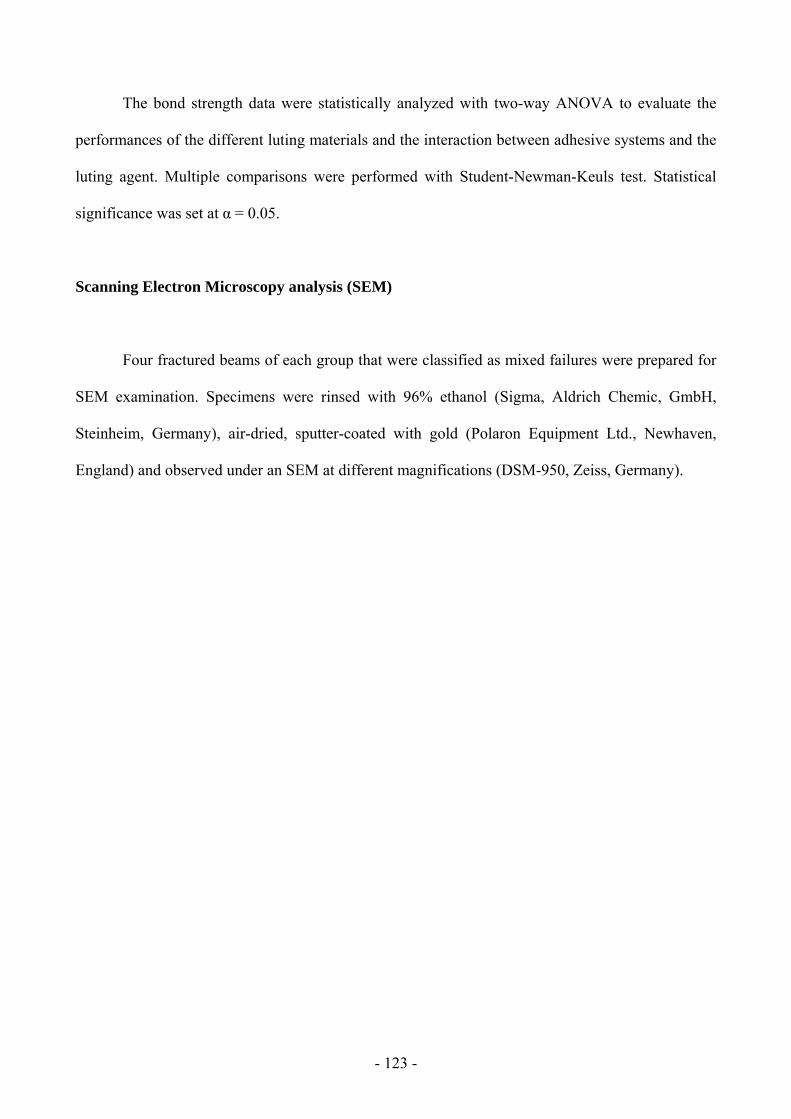
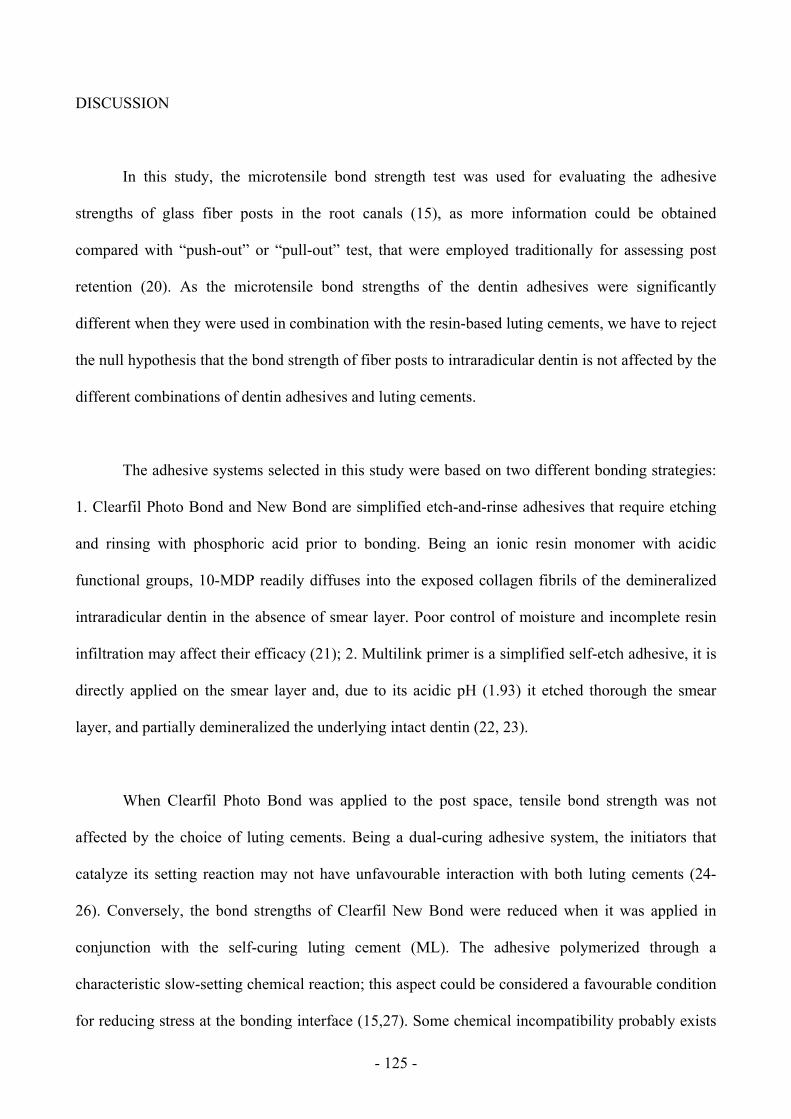
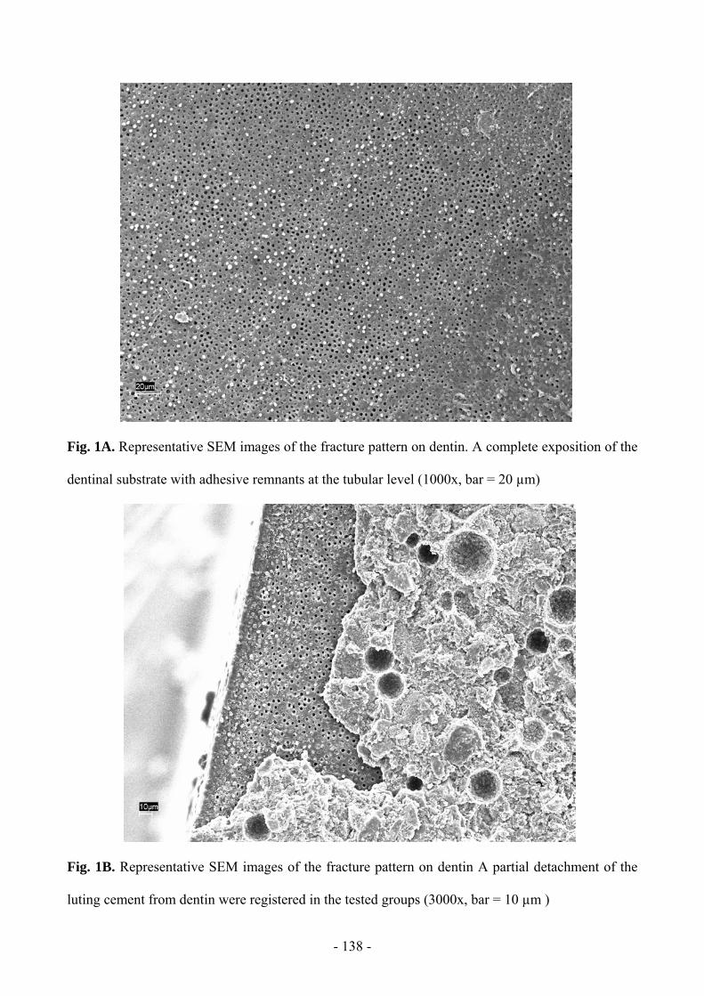
Page 1
- 1 -
Universidad de Granada Departamento de Estomatología
�������
Estudio in vitro de factores que afectan la durabilidad de la adhesión a dentina
Factors influencing resin-dentin bond
durability: An in vitro study
�������
Tesis Doctoral
Presentada por:
Alberto Albaladejo Martínez
Para optar al Título de Doctor en estomatología
Directores:
Manuel Toledano Pérez
Raquel Osorio Ruiz Marco Ferrari
Page 2
Editor: Editorial de la Universidad de GranadaAutor: Alberto Albaladejo MartínezD.L.: Gr. 659 - 2006ISBN: 84-338-3797-4
Page 3
- 2 -
Manuel TOLEDANO PÉREZ, Profesor Titular del Departamento de Estomatología
de la Universidad de Granada.
CERTIFICA
Que el trabajo de investigación titulado “Estudio in vitro de factores que afectan la
durabilidad de la adhesión a dentina” del que es autor D. Alberto Albaladejo Martínez, ha
sido realizado bajo mi dirección y supervisión, y reúne en su introducción, objetivos y
justificación, artículos, discusión, conclusión y resumen los requisitos para su defensa.
Y para que conste y surta efectos en el expediente correspondiente, expido la
presente en Granada a cinco de diciembre de dos mil cinco.
Fdo. Prof. Manuel Toledano Pérez
Page 4
- 3 -
Raquel OSORIO RUIZ, Profesor Titular del Departamento de Estomatología de la
Universidad de Granada.
CERTIFICA
Que el trabajo de investigación titulado “Estudio in vitro de factores que afectan la
durabilidad de la adhesión a dentina” del que es autor D. Alberto Albaladejo Martínez, ha
sido realizado bajo mi dirección y supervisión, y reúne en su introducción, objetivos y
justificación, artículos, discusión, conclusión y resumen los requisitos para su defensa.
Y para que conste y surta efectos en el expediente correspondiente, expido la
presente en Granada a cinco de diciembre de dos mil cinco.
Fdo. Profa. Raquel Osorio Ruiz.
Page 5
- 4 -
Marco FERRARI, Profesor Titular del Departamento de Estomatología de la
Universidad de Siena.
CERTIFICA
Que el trabajo de investigación titulado “Estudio in vitro de factores que afectan la
durabilidad de la adhesión a dentina” del que es autor D. Alberto Albaladejo Martínez, ha
sido realizado bajo mi dirección y supervisión, y reúne en su introducción, objetivos y
justificación, artículos, discusión, conclusión y resumen los requisitos para su defensa.
Y para que conste y surta efectos en el expediente correspondiente, expido la
presente en Granada a cinco de diciembre de dos mil cinco.
Fdo. Prof. Marco Ferrari.
Page 6
- 5 -
AGRADECIMIENTOS.
A la Profa. Raquel Osorio, por su dedicación, paciencia y enseñanza en la realización de este
trabajo.
Al Prof. Manuel Toledano, por las orientaciones y la confianza que siempre recibí de él.
Al Prof. Marco Ferrari por el rigor, precisión y hospitalidad que me dio.
A mi compañera y amiga la Profa. Estrella Osorio, por su continuo ánimo y tantas lecciones de la
vida que he aprendido junto a ella. Gracias.
A la Dra. Fátima Sánchez Aguilera, por introducirme y guiarme en esta universidad.
A mis compañeros y amigos Fernanda, Julio, Francesca, Tuchi y Carol por su continua ayuda e
incesable impulso.
A mis colegas de Siena, por su amabilidad y la buena acogida que me dieron.
A los proyectos de la comisión Interministerial de Ciencia y Tecnología CICYT/FEDER.MAT
2001-2843-CO2 y CICYT/FEDER.MAT 2004 06872-C03-02.RED CYTED VIII.J dentro de los
cuales se ha desarrollado este trabajo.
A Federica Papacchini por su ayuda en la traducción al italiano.
Page 7
- 6 -
A Gertrudis Gómez Villaescusa, por su ayuda en el laboratorio.
A las casas comerciales, por ceder sus productos.
A mis padres y hermano por el constante cariño y amor mostrado.
Page 8
- 7 -
ÍNDICE I. INTRODUCCIÓN. -10-
I.1. Concepto de adhesión y su proyección en Odontología. -10-
I.2. Adhesión a dentina. -12-
I.2.1. Histología de la dentina. -12-
I.2.1.1. Histología de la dentina coronal. -12-
I.2.1.2. Histología de la dentina radicular. -15-
I.2.1.3. Barrillo dentinario. -16-
I.2.2. Mecanismos de adhesión a dentina. -18-
I.2.3. Sistemas adhesivos. Clasificación y características. -21-
I.2.3.1. Adhesivos que eliminan el barrillo dentinario. -22-
I.2.3.2. Adhesivos que disuelven el barrillo dentinario. -24-
I.2.3.3. Adhesivos que modifican el barrillo dentinario. -26-
I.3. Métodos de medida de la eficacia adhesiva. -28-
I.3.1. Método de medida de la eficacia adhesiva a dentina coronal. -28-
I.3.2. Uso de pernos de fibra en la evaluación de la eficacia adhesiva
a dentina radicular. -29-
I.3.3. Degradación in vitro de la interfase adhesiva. -31-
I.3.3.1. Degradación mecánica de la interfase adhesiva. -32-
I.3.3.2. Degradación química de la interfase adhesiva. -32-
II. OBJETIVOS Y JUSTIFICACIÓN. -35-
OBJECTIVES AND JUSTIFICATION. -38-
Page 9
- 8 -
III. ADHESION TO CORONAL DENTIN: MECHANICAL AND CHEMICAL DEGRADATION
OF RESIN-DENTIN BONDS.
III. 1. Toledano M, Osorio R, Albaladejo A, Aguilera FS, Tay FR,
Ferrari M. Effect of cyclic loading on microtensile bond strengths of
total-etch and self-etch adhesives. Operative Dentistry 2005; (Aceptado
para supublicación).
-40-
III. 2. Toledano M, Osorio R, Albaladejo A, Aguilera FS, Osorio E.
Differential effect of in vitro degradation on resin-dentin bonds
produced by self-etch vs. total-etch adhesives. Journal of Biomedical
Materials Research, Applied Biomaterials (Part A) 2005; (Aceptado
para su publicación).
-65-
III.3. Albaladejo A, Osorio R, Toledano M, Papacchini, F, Ferrari M.
Micromorphology of total etching versus self-etching adhesive systems:
a SEM approach. 2005; International Dental Journal (Enviado para su
publicación).
-92-
IV. ADHESION TO ROOT CANAL DENTIN.
IV. 1. Monticelli f, Osorio R, Albaladejo A, Aguilera FS, Ferrari M, Tay F,
Toledano M. Effects of adhesive systems and luting agents on bonding of
fiber posts to root canal dentin. Journal of Biomedical Materials Research,
Applied Biomaterials (Part B) 2005; (Aceptado para su publicación).
-116-
Page 10
- 9 -
IV.2. Albaladejo A, Osorio R, Papacchini F, Goracci C, Toledano M,
Ferrari M. Post silanization improves bond strength of translucent posts to
the flowable composite resins. The International Journal of
Prosthodontics; 2005 (Enviado para su publicación).
-141-
V. DISCUSSION. -160-
VI. CONCLUSIONES. -176-
CONCLUSIONS. -178-
VII. SUMMARY. -180-
RESUMEN. -184-
SOMMARIO. -188-
REFERENCIAS. -192-
Page 11
- 10 -
I. INTRODUCCIÓN
I.1. Concepto de adhesión y su proyección en Odontología.
La palabra adhesión proviene del latín ad y haerere y significa unir a (Sainz, 1967).
Adhesión se define como el estado por el que dos superficies se mantienen juntas mediante
fuerzas o energías interfaciales basadas en mecanismos químicos, mecánicos o ambos con la
mediación de un adhesivo (ISO/TR 11405: 1993). El material que une dos superficies se denomina
adhesivo y la superficie a adherir se denomina adherente o sustrato. El espacio virtual que hay entre
las superficies unidas se denomina interfase. Para que se produzca una buena adhesión tiene que
existir una buena humectabilidad y un íntimo contacto entre las superficies a unir (Burke y cols.,
1995; Toledano y cols., 2001). La adhesión puede estar basada en dos procedimientos:
1. Mecanismo mecánico. Consiste en el entremezclado del adhesivo solidificado en las
irregularidades de la superficie del adherente. Da lugar a la adhesión mecánica, que
puede ser macromecánica y micromecánica. Se denomina adhesión macromecánica si
las irregularidades son apreciables a simple vista; se denomina micromecánica si las
irregularidades son microscópicas.
2. Mecanismo químico. Explica la adhesión mediante la generación de enlaces químicos
entre el adhesivo y el adherente. Produce adhesión química. Los enlaces implicados
pueden ser primarios o fuertes (iónicos y covalentes) y secundarios o débiles (uniones
por puentes de hidrógeno, interacciones por dipolos, fuerzas de van der Waals).
Page 12
- 11 -
Los diferentes mecanismos de unión no están del todo aclarados y en esa cuestión existe
controversia entre los dos tipos básicos de adhesión. De todas formas los dos mecanismos son
perfectamente compatibles y, sin duda alguna, pueden darse de forma simultánea.
Existen diversos factores físicos que influyen en la adhesión. Estos factores son los
fenómenos de superficie entre los que se encuentran la tensión superficial y la humectabilidad.
La adhesión no se entiende como la simple aplicación de un pegamento para unir dos
superficies. En numerosas ocasiones hay que realizar pretratamientos antes de la aplicación del
adhesivo. Básicamente, el proceso de unión sigue tres pasos fundamentales:
1. Acondicionamiento del adherente. Consiste en alterar su morfología y/o su estructura
química.
2. Imprimación del adherente. Consiste en la aplicación de una sustancia química previa
con la finalidad de hacer el sustrato más receptivo al adhesivo.
3. Aplicación del adhesivo. Consiste en aplicar el adhesivo sobre la superficie adherente.
Estos tres pasos no tienen porqué darse siempre de forma claramente diferenciada. Se
pueden encontrar de forma simultánea o bien faltar alguno de ellos (Van Meerbeek y cols., 1992;
Toledano y cols., 2003a).
La dentina puede ser descrita como un composite biológico, con un relleno mineral de
cristales de hidroxiapatita y una matriz formada por una red de fibras de colágeno (Marshall y cols.,
2001), así, el mecanismo básico de adhesión a dentina es esencialmente un proceso de intercambio
Page 13
- 12 -
que envuelve el reemplazamiento de minerales removidos de los tejidos dentales duros por
monómeros de resina que producen un cierre micro-mecánico en las porosidades creadas (Toledano
y cols., 2001; Osorio y cols., 2003; De Munck, 2004). Este cierre fue descrito por primera vez por
Nakabayashi y cols., (1982) y es referido comúnmente como “hibridación” o la formación de la
capa híbrida.
I.2. Adhesión a dentina.
I.2.1. Histología de la dentina.
I.2.1. Histología de la dentina coronal.
La dentina es un sustrato biológico que actúa como adherente. Es un tejido vivo muy
complejo y variable que, debido a sus peculiaridades histológicas y morfológicas, condiciona la
aplicación de los sistemas adhesivos.
Desde un punto de vista histológico, la dentina es un tejido conjuntivo mineralizado y
avascular. Está compuesto, en peso, por un 70% de materia inorgánica, un 18% de materia orgánica
y un 12 % de agua (Mjör y cols., 1989). En cuanto al volumen, el 50% lo constituye material
inorgánico, el 25% orgánico y el otro 25% agua (Mjör y cols., 1989).
La porción inorgánica de la dentina consiste básicamente en cristales de hidroxiapatita. La
parte más pequeña de esta estructura se denomina unidad celular de hidroxiapatita y responde a la
fórmula 3Ca3(PO)4-CA(OH)2. Junto a estos cristales se pueden encontrar fosfatos cálcicos amorfos,
probablemente en mayor cantidad en los tejidos formados más tardíamente que en los maduros o
viejos (Mjör y cols., 1989; Melfi, 1994). Los cristales están formados por varios miles de unidades
Page 14
- 13 -
celulares y tienen forma laminar que de perfil adaptan el aspecto de agujas. Su longitud es de hasta
20 nm y el grosor puede llegar hasta los 3.5 nm. Son similares a los cristales del cemento y del
hueso, pero más pequeños que los cristales del esmalte. Existen también otras sales inorgánicas
como carbonatos, fosfatos cálcicos diferentes de la hidroxiapatita, sulfatos, y ciertos oligoelementos
(F, Cu, An, Fe) (Davis, 1986; Melfi, 1994; Mjör y cols., 1989).
La porción orgánica (Mjör y cols., 1989; Davis, 1988; Melfi, 1994) está compuesta
principalmente por fibras de colágena tipo I, en una cuantía aproximada del 17% del total del tejido,
es decir, alrededor del 93% de todo el material orgánico. Además, se pueden encontrar fracciones
de lípidos, glucosaminoglicanos, compuestos proteicos no identificados, constituyendo cada uno de
ellos un 0.2% aproximadamente. También se puede encontrar ácido cítrico en una cantidad algo
inferior al 1% (Schroeder, 1991).
En el tejido dentinario se pueden distinguir cinco unidades estructurales: odontoblastos,
túbulos dentinarios, espacio periodontoblástico, dentina peritubular y dentina intertubular (Mjör y
cols., 1989).
Los odontoblastos son células especializadas que tapizan la pared de la cámara pulpar y que
poseen largas prolongaciones citoplasmáticas (proceso odontoblástico) que se localizan en el
interior de los túbulos dentinarios.
Los túbulos dentinarios alojan la prolongación odontoblástica y se forman durante la
dentinogénesis, conservando su estructura tubular en la dentina madura. El diámetro de la luz
tubular cambia según la zona de la dentina, en la proximidad a la pulpa es de 3 a 4 µm y en la zona
externa es de 1µm aproximadamente (Bhaskar, 1993), debido a que la superficie pulpar de la
dentina es considerablemente menor que el área de las uniones amelodentinaria y cementodentinaria
Page 15
- 14 -
(Pashley, 1991a; Schroeder, 1991; Mjör y cols., 1989). Cerca del 80% del volumen total de la
dentina próxima a la pulpa está compuesta por las luces de los túbulos, mientras que éstas
constituyen tan sólo cerca de un 4% del volumen de la dentina periférica. En cuanto a la superficie,
la luz tubular ocupa el 1% en la dentina de la unión amelo-dentinaria y el 22% en la zona próxima a
la pulpa (Pashley, 1985).
Los procesos odontoblásticos y túbulos acompañantes pueden ramificarse, especialmente,
cerca de las uniones amelodentinarias y cementodentinarias. En general, las ramificaciones de los
procesos de los odontoblastos son de menor tamaño y más numeroso en la dentina de la raíz que en
la coronal.
El líquido en el interior de los túbulos tiene una determinada presión. La presión hidrostática
es un factor a tener en cuenta en el estudio de la adhesión, debido a que proporciona un flujo
permanente y constante de líquido hacia el exterior (Ciucchi, 1995).
La dentina peritubular forma la pared de los túbulos dentinarios. Por otro lado, la dentina
intertubular se localiza entre los túbulos dentinarios. Es muy importante destacar las diferencias
existentes entre ambas. La dentina peritubular tiene una estructura tubular. Su grosor es variable,
siendo aproximadamente de 0.75 µm en la dentina externa y de 0.4 µm en la dentina interna
(Bhaskar, 1993). Los cristales de hidroxiapatita son más pequeños y están agrupados muy juntos
(Davis, 1986). La dentina intertubular está menos calcificada y con mayor contenido orgánico. Se
halla uniformemente mineralizada, a excepción de la situada en la zona con bajo contenido mineral
cercana a la pulpa, donde el grado de mineralización es inferior al común (Mjör y cols., 1989;
Bhaskar, 1993). La mitad del volumen de la dentina intertubular está formada por matriz orgánica.
Las fibras de colágena son su principal componente y se encuentran orientadas al azar alrededor de
Page 16
- 15 -
los túbulos dentinarios. Las fibras tienen un diámetro variable de 0.05µm a 0.2µm (Mjör y cols.,
1989).
I.2.1.2. Histología de la dentina radicular:
Los odontoblastos que forman la dentina radicular se diferencian a partir de las células
epiteliales de Hertwing, lo que hace que esta dentina sea distinta en términos estructurales y de
composición a la dentina coronal, en la cual, los odontoblastos se diferencian a partir de las células
ectomesenquimáticas de la papila (Gómez y cols., 2002).
Las diferencias entre la dentina coronal y radicular pueden resumirse en los siguientes
puntos:
- La orientación de las fibras de colágeno de la dentina del manto son diferentes. En la
dentina coronal del manto, las fibras son perpendiculares a la interfase dentina-esmalte, por el
contrario, las fibras de la raíz son paralelas a la interfase dentina-cemento.
- Los odontoblastos radiculares difieren un poco de los de la corona dando lugar a ramos en
forma de paraguas.
- En la dentina radicular la tasa de deposición es más lenta.
- Su contenido de fósforo es menor que en la dentina coronal; además, su grado de
mineralización es menor.
- En la dentina coronaria los túbulos siguen un trayecto doblemente curvo en forma de “S”
itálica, sin embargo, en las cúspides y bordes incisales es prácticamente rectilíneo, mientras
Page 17
- 16 -
que en la dentina radicular los túbulos tienen una curvatura poco pronunciada (Bhaskar,
1993).
Otra particularidad de la dentina radicular es la existencia de la “Capa Granulosa de Tomes”.
Esta región de la dentina es muy peculiar y sólo se encuentra en la parte más periférica de la dentina
radicular, en la unión dentina-cemento. Aparecen como una serie de gránulos oscuros, sin matriz de
colágeno y por lo tanto sin calcificación, que se extienden a lo largo de toda la raíz, siendo más
numerosos en el vértice que en la unión cemento-esmalte. Aunque no está todavía claro su origen,
se piensa que estos gránulos se forman a partir de una serie de pequeñas cámaras de aire, producidas
probablemente por incurvación de lo túbulos dentinarios para formar asas en esta área (Bhaskar
1993).
Para obtener una adecuada adhesión a la dentina radicular, al igual que en la dentina coronal,
es de crucial importancia la permeabilidad que presenta ésta a los agentes adhesivos, así como que
la fase mineral de la dentina sea removida para producir la infiltración de los adhesivos dentro de la
dentina intertubular (Gaston y cols., 1999).
I.2.1.3. Barrillo dentinario.
El barrillo dentinario o smear layer es un conglomerado de tejido inorgánico y orgánico,
subunidades globulares originadas por fibras mineralizadas con un diámetro de 0’05-0’1µm
aproximadamente, proteínas coaguladas, células sanguíneas y en algunas ocasiones
microorganismos (Czonstkowsky y cols., 1990; Sen y cols., 1995; Abbott y cols., 1991). El barrillo
dentinario resulta de las maniobras terapéuticas practicadas sobre el diente al cortar las superficies
de los tejidos mineralizados; además, tiene gran facilidad para adherirse a las paredes de la
preparación cavitaria sin poderse remover con una simple aplicación de agua o spray, representando
Page 18
- 17 -
la interfase entre el diente y el material restaurador (Toledano y cols., 2003a). La apariencia
microscópica de esta capa vista bajo el microscopio electrónico de barrido fue descrita por
Brannström y cols (1980) quienes encontraron que es irregular, granular y amorfa. Boyle y cols.,
(1963) fueron los primeros en describir la presencia del barrillo dentinario, posterior al corte con
fresas a nivel coronal. La formación de barrillo dentinario en los conductos de dientes preparados
endodónticamente fue descrita por Mc Comb y Smith, (1975) quienes reportaron que la apariencia
fue similar al barrillo dentinario coronal.
El barrillo dentinario posee un grosor aproximadamente de 1-5µm, aunque dicho grosor
depende del tipo y filo del instrumento usado y de si en la preparación de la cavidad la dentina está
seca o húmeda (Van Meerbeek y cols., 1993). Se ha identificado la capa de barrillo dentinario en
dos partes, una es el barrillo superficial y la otra el barrillo compactado dentro de los túbulos
dentinarios (Toledano y cols., 2003a). La penetración del material residual dentro de los túbulos es
aproximadamente de 40µm de profundidad, la cual, tiene lugar por un fenómeno de capilaridad
como consecuencia de las fuerzas adhesivas producidas entre los túbulos dentinarios y el material
residual (Sen y cols., 1995; Cohen, 2002).
Hubo una gran controversia (Toledano y cols., 2003a) con respecto al barrillo dentinario
nacida de las siguientes realidades: 1) Se ha demostrado que las bacterias pueden vivir y
multiplicarse dentro de él, alcanzando la pulpa a través de los túbulos dentinarios. 2) Al cubrir la
dentina, el barrillo dentinario puede interferir en los procesos de adhesión con los cementos
adhesivos o con las nuevas generaciones de los adhesivos dentinarios. Los autores partidarios de su
conservación se basan en que si se elimina, desaparece una barrera física que impide la entrada de
bacterias a través de los túbulos; (Tay y cols., 2000a; Sano y cols., 1999; Toledano y cols., 2003b)
Por lo contrario, otros autores son partidarios de eliminar el barrillo dentinario, pues por la acción
Page 19
- 18 -
del ácido al desmineralizarse la dentina peritubular se hace mayor su diámetro, asegurándose una
mayor entrada ulterior de resina (Sen y cols., 1995; Calt y cols., 2000).
Hoy en día, la conservación o eliminación del barrillo dentinario depende del tipo de
adhesivo. En el caso de los sistemas autograbadores, el barrillo dentinario se mantiene
incorporándose a la composición de la capa híbrida formada (Tay y cols., 2001; Toledano y cols.,
2001; Osorio y cols., 2003). Por lo contrario, en los sistemas de grabado total, el barrillo dentinario
se elimina por medio de un acondicionamiento de la dentina realizado con ácido (Van Meerbeek y
cols., 1998).
I.2.2. Mecanismos de adhesión a dentina
El mecanismo de adhesión a la dentina ha sido ampliamente estudiado y debatido. Al
comienzo del desarrollo de la Odontología Adhesiva se buscó una adhesión química al calcio o a las
fibras de colágena de la estructura dentaria. Hoy en día se habla sobre todo de adhesión mecánica o,
más concretamente, adhesión micromecánica (Nakabayashi y cols., 1991; Van Meerbeek y cols.,
1992).
La adhesión micromecánica a la dentina está basada en tres mecanismos (Gwinnett, 1993):
1º Adhesión mediante la infiltración de la dentina intertubular y la formación de la capa
híbrida o zona de interdifusión.
2º Adhesión mediante la infiltración de los túbulos dentinarios y sus ramas laterales.
3º Adhesión superficial, por el contacto entre el adhesivo y el sustrato dentinario.
Page 20
- 19 -
Hoy en día, la adhesión a dentina se basa en la retención micromecánica proporcionada por
la capa híbrida (Nakabayashi y cols., 1991) o zona de interdifusión (Van Meerbeek y cols., 1992).
El mecanismo por el que se forma la citada capa, consiste en la infiltración de un monómero
adhesivo en la dentina descalcificada con las fibras de colágena expuestas que, tras polimerizar,
queda entremezclado con la estructura dental desmineralizada (Nakabayashi y cols., 1991;
Toledano y cols., 2003b). Es una unión micromecánica al tejido proteico (Gwinnett, 1993). La
formación de una capa híbrida adecuada requiere que los péptidos dentinarios (incluidas las fibras
de colágena) estén sin desnaturalizar (pues de lo contrario crearían una capa híbrida débil con gran
susceptibilidad a su degradación) (Nakabayashi y cols., 1991), que el sistema adhesivo contenga
resinas hidrofílicas e hidrofóbicas (para que las primeras produzcan una imprimación de la dentina
haciéndola más receptiva a las segundas) (Toledano y cols., 2003a) y un catalizador que permita la
polimerización en presencia de agua y oxígeno (Nakabayashi, 1991; Osorio y cols., 2005a; Nunes y
cols., 2005).
Una de las principales características de la capa híbrida es la resistencia al ataque ácido
(Nakabayashi, 1991), lo que la convierte en una unión resistente a una hipotética microfiltración
bacteriana y le confiere estabilidad a lo largo del tiempo (Toledano y cols., 2001; Osorio y cols.,
2003)
En algunos casos se puede observar una zona de vacío en la profundidad de la dentina
infiltrada, que corresponde a una región en la que el adhesivo no ha llegado a penetrar (Sano y cols.,
1995; Toledano y cols., 2004b). Este hecho facilita el paso de líquido y enzimas bacterianas a esta
zona, produciéndose una hidrólisis de los péptidos no protegidos con la hidroxiapatita o con la
resina (Nakabayashi y cols., 1992) apareciendo la nanofiltración (infiltración de bacterias debido a
un inadecuado sellado de la capa híbrida con las fibras de colágeno) (Sano y cols., 1995; Osorio y
cols., 2003).
Page 21
- 20 -
La formación de la capa híbrida no es el único mecanismo de adhesión a dentina que puede
ofrecer un sistema adhesivo. También puede ser proporcionado por túbulos dentinarios abiertos tras
el grabado ácido, en los cuales, se produce una infiltración de resina debido al fenómeno de
capilaridad, formando los tags de resina. Estas prolongaciones de resina son siempre mucho más
largas que el grosor de infiltración en la dentina intertubular. Debido a la anchura y forma de lo
túbulos y a sus ramas laterales, ofrecen una retención mecánica (Chappell y cols., 1994; Gwinnett,
1993), aunque su contribución más importante a la adhesión consiste en propiciar un correcto
sellado marginal (Gwinnett, 1993). Los tags son una combinación de la resina y de lámina limitante
que cubre la pared tubular (Titley y cols., 1995).
El tercer mecanismo de unión mecánica al tejido consiste en el contacto que se produce
entre la resina y la capa de dentina parcialmente desmineralizada con el límite del frente de
desmineralización. Este mecanismo constituye la adhesión superficial (Gwinnett, 1993).
Hay dos modelos fundamentales de adhesión a dentina. Uno de ellos fue propuesto por
Gwinnett (Gwinnett, 1993) y el otro por Pashley (Pashley, 1990):
a) El modelo de adhesión que propone el primero considera que la fuerza de unión de la
resina a la dentina depende de la superficie dentinaria de adhesión, de la capa híbrida y de los tags
de resina. La infiltración de la dentina por el adhesivo, ya sea en la dentina intertubular como en el
interior de la luz tubular, es responsable de un tercio del total de la adhesión (Gwinnet, 1993). De
este tercio, la mitad se basa en la infiltración de la dentina intertubular y la formación de la capa
híbrida y la otra mitad en la penetración de la resina en los túbulos y en la formación de los tags
(Gwinnet, 1993). Los dos tercios restantes se deben a la denominada adhesión superficial que está
proporcionada por las interacciones físico-químicas del adhesivo con las irregularidades de la
topografía dentinaria (Gwinnet, 1993; Yoshiyama y cols., 1995).
Page 22
- 21 -
b) El modelo de Pashley o modelo lineal simple asume que la fuerza de unión de la resina a
la dentina va a depender de la profundidad de la dentina y de la resistencia de la resina asumiendo
que la diferente densidad de túbulos dentinarios y presencia de dentina sólida, como consecuencia
de la proximidad a la pulpa, determinan la fuerza de unión tanto a dentina superficial como
profunda. Así, en la dentina superficial la fuerza de unión estará más influida por la capa híbrida
que por la densidad de los túbulos, mientras que en la dentina profunda ocurriría justo al contrario
De esta forma, el tag de resina asume un papel destacado en la fuerza de unión, sobre todo, por el
trayecto convergente de los túbulos dentinarios hacia la cámara pulpar, otorgando mayor retención
micromecánica al sistema. Además se va a conseguir que la resina una a los túbulos, por lo que se
obtiene el sellado de la luz del mismo.
1.2.3. Sistemas adhesivos. Clasificación y características.
Un sistema adhesivo es el conjunto de materiales que sirven para realizar todos los
pasos de la adhesión del material restaurador al diente, como son la preparación de la superficie del
esmalte y dentina, adhesión química y/o micromecánica a esmalte y dentina y adhesión química al
material restaurador (Toledano y cols 2003a). La incapacidad de las resinas compuestas para adherir
directamente a los sustratos dentales, hizo que la aplicación de un sistema adhesivo fuera un paso
intermedio indispensable en los procedimientos clínicos donde se utilizasen dichos materiales. El
procedimiento adhesivo consta de tres componentes básicos:
1) Un acondicionador ácido, que tiene la finalidad de modificar química y morfológicamente
la estructura del esmalte y la dentina para permitir a los siguientes materiales adherirse mecánica y
químicamente a ella.
Page 23
- 22 -
2) Un imprimador o primer, que penetra y moja toda la zona descalcificada para facilitar el
contacto de la resina adhesiva con el colágeno desmineralizado. Sus funciones son mejorar la
humectabilidad de la dentina acondicionada, mantener las fibras de colágeno sin colapsar y
separadas entre sí y facilitar o vehiculizar la resina adhesiva hacia el interior de la dentina
descalcificada (Titley y cols, 1995; Tani y cols; 1996; Perdigäo y cols., 1997; Toledano y cols.,
2001).
3) Una resina, la cual se disuelve con el imprimador y penetra en la dentina, sirviendo de
puente entre las dos superficies a adherir, la dentina y el material restaurador. Además, la resina
adhesiva confiere una flexibilidad y resistencia adecuadas a la zona de dentina infiltrada.
Los adhesivos dentinarios se pueden clasificar atendiendo a numerosos criterios. Una de las
más utilizadas se basa en la cronología de la aparición de estos materiales en el mercado separando
los adhesivos en generaciones. Sin embargo, esta clasificación no aclara de forma objetiva el
número de pasos clínicos realizados durante la aplicación de éstos, ni tampoco como interactúan
con el sustrato (Van Meerbeek y cols., 1992). Hoy en día, la manera más clara de clasificar los
adhesivos, es la establecida por Van Meerbeek y cols., (1992), los cuales establecieron una
clasificación según el mecanismo de acción y el número de pasos empleados.
I.2.3.1. Adhesivos que eliminan el barrillo dentinario (grabado total).
Estos adhesivos, también conocidos como sistemas de grabado total, acondicionan la
dentina con un ácido que remueve totalmente el barrillo dentinario (Meerbeek y cols., 1992).
Page 24
- 23 -
a) Adhesivos de tres pasos.
Son sistemas que constan de tres componentes que se dispensan por separado. Responden al
modelo tradicional del procedimiento adhesivo a dentina y en su mecanismo de acción están
basados la mayoría de los adhesivos utilizados actualmente (Van Meerbeek y cols., 1998).
Requieren tres pasos clínicos. En un primer paso, se aplica un ácido que elimina todo el barrillo
dentinario. Tras ello, se enjuaga la dentina grabada y se aplica un imprimador. Como tercer paso se
aplica una resina adhesiva.
Estos sistemas plantean tres grandes problemas: 1) El paso separado de grabar, lavar y secar
incrementa la sensibilidad de la técnica, especialmente, si se trata de una técnica húmeda (Tay y
cols., 2000a; Toledano y cols., 2001); 2) La necesidad que presentan estos sistemas de secar tras
enjuagar con agua, provoca un colapso de las fibras de colágeno (Gordan y cols., 1998; Toledano y
cols., 2004a); 3) La profundidad de desmineralización creada por el ácido es mayor que la
infiltración producida por los monómeros hidrofílicos del primer (Nakabayashi y cols., 1996; Tay y
cols., 2001; Osorio y cols., 2003), dejando expuestas las fibras de colágeno desprotegidas de
cristales de hidroxiapatita, las cuales, son susceptibles de hidrólisis (Watanabe y cols., 1994;
Burrow y cols., 1996; Sano y cols., 1999; Toledano y cols., 2000).
En este grupo se encuentran adhesivos como Scotch Bond Multipurpose® (3M, St. Paul,
MN, USA) o Aelitebond® y All Bond 2® (Bisco, Istasca, USA).
Page 25
- 24 -
b) Adhesivos de dos pasos:
Son sistemas adhesivos que constan de dos componentes. En el primer paso se aplica
un acondicionador ácido que tras su aplicación es enjuagado con la consiguiente eliminación total
del barrillo dentinario. En el segundo paso, se aplica un bote donde van incluidos el imprimador y la
resina adhesiva. Estos sistemas adhesivos también se conocen como adhesivos autoimprimadores,
pues combinan en un solo bote, las funciones del imprimador y la resina adhesiva (Toledano y cols.,
2001; Osorio y cols., 2003).
Forman parte de este grupo el Single Bond® (3M, St.Paul, MN, USA.), Prime & Bond NT®
(Dentsply / De Trey GmbH, Konstanz, Alemania), o el Prime & Bond XP® (Dentsply / De Trey
GmbH, Konstanz, Alemania).
I.2.3.2. Adhesivos que disuelven el barrillo dentinario.
Son sistemas adhesivos que disuelven el barrillo dentinario (mezclan de forma homogénea
el barrillo con el primer) y simultáneamente desmineralizan la superficie del sustrato (Meerbeek y
cols., 1992). Como no son lavados, el barrillo se incorpora al proceso de adhesión reduciéndose los
problemas asociados a la sensibilidad de la técnica (Fritz y cols., 1999; Toledano y cols., 2001). Los
sistemas adhesivos que constan de uno o dos botes y disuelven el barrillo dentinario son conocidos
actualmente como autograbadores (Van Meerbeek y cols 1993; Toledano y cols., 2001).
Page 26
- 25 -
a) Adhesivos de dos pasos.
Son sistemas formados por dos botes. El primer paso consta de un imprimador y un ácido
juntos, que disuelven el barrillo dentinario tras ser aplicados en esmalte y dentina conjuntamente
(Van Meerbeek y cols 1993; Toledano y cols., 2001). El imprimador incorpora el barrillo dentinario
a su composición. Tras este paso se aplica una resina adhesiva.
Los adhesivos autograbadores producen un complejo híbrido que incluye una capa superior
de smear layer infiltrado y una capa inferior de fibras de colágeno desmineralizadas e infiltradas,
mezclada con grupos calcio y fosfato producto de la desmineralización de la hidroxiapatita (Tay y
cols., 2000a). El uso de esos sistemas adhesivos representa un método bastante eficaz para prevenir
el colapso de la trama de colágeno desmineralizado (Tay y cols., 2001; Osorio y cols., 2003).
Cuando estos sistemas adhesivos se emplean, no hay necesidad de grabar, enjuagar y secar el
sustrato, por lo que desaparece el riesgo de sobregrabar y sobresecar la dentina (Tay y cols., 2001;
Toledano y cols., 2003). Además, el problema presentado por los sistemas adhesivos de grabado
total al producir una desmineralización, por parte de los monómeros ácidos, mayor que la
infiltración realizada por los monómeros hidrofílicos, se ha solventado en gran medida (Toledano y
cols., 2001; Osorio y cols., 2003).
Son representantes de este grupo el Clearfil SE Bond® (Kuraray Co, Osaka, Japón), Protect
Bond® (Kuraray Co, Osaka, Japón), Syntac® (Vivadent, Schaan, Liechtenstein) o el Coltène ART
Bond® (Coltène, Altstätten, Suiza).
Page 27
- 26 -
b) Adhesivos de un paso (all-in-one).
Son adhesivos que constan de un solo paso. Incorporan en el mismo bote los monómeros
ácidos, hidrofílicos e hidrofóbicos. Estos materiales disuelven el barrillo y simultáneamente
desmineralizan la superficie del sustrato (Van Meerbeek y cols., 1992). Como no deben ser lavados,
el barrillo se incorpora a la capa híbrida, formando parte de ésta (Fritz y cols., 1999; Santini y cols.,
2001; Toledano y cols., 2001).
Existe unanimidad en asumir las bajas fuerzas de adhesión proporcionadas por los all-in-one
(Fritz y cols., 1999; Inoue y cols., 2000; Toledano y cols., 2001; Toledano y cols., 2003b; Osorio y
cols., 2003; Osorio y cols., 2005b). La combinación de monómeros ácidos, hidrofílicos e
hidrofóbicos en una solución única, puede comprometer la función de cada uno de los componentes
(Toledano y cols., 2003b). Sin embargo, son capaces de disolver completamente el barrillo
dentinario y formar un complejo híbrido relativamente grueso (Haller y cols., 2000; Toledano y
cols., 2003; Osorio y cols., 2003).
Forman parte de este grupo los adhesivos Prompt L-Pop® (3M ESPE, Seefeld, Alemania),
Etch and Prime 3.0® (Degussa AG, Hanau, Alemania), AQ Bond® (Sun Medical, Kyoto, Japón),
One-Up Bond F® (Tokuyama, Tokyo, Japón), Reactmer Bond® (Shofu, Kyoto, Japan) o Xeno CF
Bond® (Sankin, Tokyo, Japón).
I.2.3.3. Adhesivos que modifican el barrillo dentinario.
Son sistemas adhesivos que modifican el barrillo dentinario haciéndolo más poroso para que
la resina acceda a la dentina subyacente (Toledano y cols., 2003a).
Page 28
- 27 -
a) Adhesivos de dos pasos:
Estos adhesivos constan de dos botes. El primero contiene un primer con radicales
acidófilos que modifican el barrillo dentinario; en el segundo paso se aplica la resina adhesiva
(Toledano y cols., 2003a). Una vez que los monómeros polimerizan en el espesor del barrillo
dentinario se establecen uniones químicas y micromecánicas leves, que refuerzan la nueva
estructura, así como su unión a la estructura subyacente (Van Meerbeek y cols., 1992).
Al igual que aquellos sistemas adhesivos que disuelven el barrillo dentinario, consideran el
smear layer como una barrera natural contra la penetración de las bacterias, que se desplaza por los
túbulos dentinarios al interior de la cámara pulpar, al mismo tiempo que dificulta la salida del
líquido tubular a la superficie de la dentina, lo que podría alterar las técnicas adhesivas (Toledano y
cols., 2003a).
Son representantes de este grupo el Pentra Bond II® (Jeneric/Pentron, Wallingford, CT,
USA) o el ProBond® (Detrey-Dentsply, Konstanz, Alemania).
b) Adhesivos de un paso:
Son adhesivos que constan de un solo bote compuesto de una resina adhesiva mezclada con
ácidos débiles, la cual, se aplica sobre el barrillo dentinario y la dentina. La resina modifica el
barrillo dentinario para poder infiltrarlo y acceder a la dentina subyacente, por lo tanto,
mediante este procedimiento no tiene lugar la exposición tradicional de las fibras de colágeno como
consecuencia del grabado ácido (Toledano y cols., 2003a).
Page 29
- 28 -
Son representantes de este grupo el Ariston Liner® (Vivadent, Schaan, Liechtenstein),
Hytac® (ESPE, Schaan, Liechtenstein), Compoglas® (Vivadent, Schaan, Liechtenstein) o el Solist®
( DMG, Hamburg, Alemania).
1.3. Métodos de medida de la eficacia adhesiva
I.3.1. Método de medida de la eficacia adhesiva en reconstrucciones intracoronales.
Las pruebas de fuerzas de adhesión son las más usadas para cuantificar la eficacia adhesiva
de diferentes sistemas. La base de este método es que la adhesión más fuerte entre el diente y el
biomaterial, resistirá mejor el estrés impuesto por el sistema y la función oral (Pashley y cols.,
1995).
A lo largo del tiempo, se han desarrollado diversos tests de fuerzas de adhesión (Pashley y
cols., 1995). La fuerza producida en los sistemas adhesivos dentinarios se ha evaluado
tradicionalmente usando el test de resistencia al cizallamiento o shear bond strength, el cual, resulta
útil para probar materiales que fallan ante valores comprendidos entre 18-20 MPa, o menos
(Chappell y cols., 1997). Sin embargo, en valores que exceden la citada cifra, a menudo no
permiten diferenciar entre la fuerza del adhesivo y la fuerza cohesiva del composite o la dentina
(Chappell y cols., 1997). Además, debido a que la evaluación exacta de un material adhesivo se
determina mejor cuando el fallo ocurre en el propio material y no implica la dentina o el composite
y, a que la mejora de los adhesivos dentinarios aumenta con el paso del tiempo, apareció la
necesidad de obtener un método mejor y más eficaz (Schreiner y cols., 1998). De esta manera, se
creó el test de microtensión, que hoy en día es el más usado. La técnica de microtensión para
evaluar la resistencia adhesiva introducida por Sano y cols., (1994), se trata de una técnica muy
laboriosa, pero presenta múltiples ventajas: (1) Con ella se pueden medir grandes fuerzas de
Page 30
- 29 -
adhesión. (2) Permite testar la adhesión en áreas muy pequeñas y en diferentes regiones. (3) Es
capaz de obtener de una sola pieza múltiples especimenes.
I.2.5. Uso de pernos de fibra en la evaluación de la eficacia adhesiva a dentina radicular.
Para la evaluación de las fuerzas de adhesión de pernos a dentina radicular, y de éstos al
cemento, se han utilizado tradicionalmente las pruebas de push-out y pull-out (Mitchell y cols.,
1994; Drummond y cols., 2000). Este tipo de pruebas presentan dos grandes problemas: 1) El área
de la superficie de los pernos debe ser cuidadosamente evaluada para permitir calcular la fuerza de
adhesión. 2) No se produce una distribución uniforme de las cargas a través de las muestras (Sano y
cols., 1994). Los tests de microtension introducidos por Sano y cols., (1994) mejoran la distribución
de las fuerzas en superficies pequeñas (0.5 x 0.5 mm), a la vez que permiten medir la fuerza de
adhesión de resinas aplicadas dentro del conducto radicular. Bouillaguet y cols., (2003) realizaron
un estudio de microtensión para obtener la fuerza de adhesión de pernos a dentina radicular
solventando los inconvenientes que presentaban los tests de push-out y pull-out.
En la adhesión entre el perno y la dentina radicular, se encuentran dos interfases: dentina-
cemento y cemento-perno. Para evaluar exclusivamente las fuerzas de adhesión de ésta última,
basta con unir el perno al cemento con interposición de un adhesivo y someterlo al test de
Microtensión (Goracci y cols., 2005; Monticelli y cols., 2004).
El uso de pernos adheridos al conducto radicular se considera el mejor método para medir la
eficacia adhesiva en dentina radicular (Ferrari y cols., 2001a; Pegoretti y cols., 2002a; Boschian y
cols., 2002; Aksornmuang y cols., 2004; Goracci y cols., 2005). Las investigaciones en el campo de
la adhesión en dicho sustrato iniciadas por Mc Comb y cols., (1973), propusieron la utilización de la
dentina endodonciada acondicionada para el cementado adhesivo de los sistemas de reconstrucción
Page 31
- 30 -
de dientes no vitales. Nathanson y cols., (1980) desarrollaron una técnica de cementado pasivo de
pernos, en la que propusieron para el acondicionamiento de la dentina radicular, un tratamiento con
hipoclorito sódico y la utilización de pernos metálicos o sistemas de retención prefabricados. Esta
técnica, debido a la presencia de un diafragma elástico, representado por el cemento entre los postes
metálicos y la dentina, permite utilizar una retención de acción pasiva (Morgano y cols., 1996).
Gracias al cementado adhesivo, las técnicas de reconstrucción pueden ser menos invasivas;
de hecho, la longitud del poste puede ser igual o ligeramente mayor que la altura del muñón clínico,
y el diámetro se limita a reproducir la morfología del conducto preparado sin la eliminación
posterior de dentina radicular (Ferrari y cols., 2004). Los beneficios de las técnicas adhesivas
utilizadas para restauraciones dentales han sido bien documentados. Uno de los factores más
importantes que aportan es el reforzamiento de la estructura dentaria y el aspecto estético de la
restauración final (Pest y cols., 2000), por estas razones, el uso de cementos adhesivos ha sido
propuesto para la cementación de pernos en dientes no vitales.
Para mejorar la estética y eliminar los problemas relacionados con las propiedades físicas de
los pernos metálicos, se propuso la utilización de pernos estéticos, entre los que se encuentran los
pernos de resina compuesta reforzados con fibra. Duret y cols., (1990), codificaron la utilización de
pernos de resina reforzados con fibra de carbono y realizaron una técnica que evitaba la unión de
materiales con características biomecánicas diferentes, así, los diferentes componentes de la
restauración (poste, cemento, material de reconstrucción y dentina), constituyen un complejo
estructural mecánicamente homogéneo. En esta línea, los pernos de fibra adheridos al conducto
radicular son la última solución presentada para la evaluación de las fuerzas de adhesión a dentina
radicular (Mannocci y cols., 2001; Bouillaguet y cols., 2003; Foxton y cols., 2003; Goracci y cols.,
2005).
Page 32
- 31 -
La eficacia de la adhesión entre perno y dentina radicular está influida por diversos factores
relacionados con el poste, el cemento, la adhesión del poste al cemento y a la dentina del canal
radicular. Se han evaluado los factores retentivos de los pernos, encontrando que entre las variables
que afectan dichos factores, están la longitud, diámetro, diseño, material y estructura del poste
(Nergiz y cols., 2002; Ferrari y cols., 2000). En lo que concierne al cemento, la retención del perno
está afectada por la resistencia del cemento, la adhesión de éste sobre el poste, la dentina y otros.
Utter y cols (1997), encontraron que la retención de los pernos es superior cuando se utilizan
cementos resinosos en comparación, con pernos cementados con cemento de fosfato de zinc.
La selección del adhesivo y el procedimiento de cementación apropiada para la colocación
de pernos en el conducto radicular están cambiando. Diferentes tipos de sistemas pueden ser
utilizados en combinación con diversos cementos resinosos. Estos materiales pueden polimerizar a
través de una reacción química, por proceso de fotopolimerización o combinación de ambos
mecanismos, también llamado polimerización dual (Nergiz y cols., 2002; Ferrari y cols., 2000).
1.3.3. Degradación de la interfase adhesiva in vitro.
Es bastante difícil desarrollar condiciones de laboratorio que puedan testar la longevidad de
la adhesión debida a los numerosos factores envueltos en la degradación de la adhesión y a que el
desarrollo oral es dinámico y biológicamente complejo (Osorio y cols., 2005b). Diversos métodos
han sido propuestos para reproducir una situación clínica en el medio oral, particularmente bajo
condiciones en las cuales la adhesión fallaría como consecuencia de una degradación, como serían
por ejemplo el ciclado mecánico, ciclado térmico, almacenamiento en agua y otras soluciones
(Burrow y cols., 1993; Abdalla y cols., 1996; Kato y cols., 1998; Chan y cols., 1997; Nikaido y
cols, 2002a; Yamauti y cols., 2003; Osorio y cols., 2005a).
Page 33
- 32 -
1.3.3.1. Degradación mecánica de la interfase adhesiva.
Los dientes están sujetos continuamente a un estrés durante la masticación, la deglución y
los hábitos parafuncionales. El ciclado mecánico, que simula la carga oclusal ejercida por los
dientes, podría acelerar el deterioro de la interfase entre la dentina y la restauración (Nikaido y cols,
2002a; Abdalla y cols., 1996; Osorio y cols., 2005b). Las cargas verticales producidas por una bola
de comida entre dientes opuestos, se distribuyen sobre toda la cara oclusal, y el estrés es propagado
a través de la superficie. La máxima fuerza registrada al morder en los primeros molares es
aproximadamente 40-90 Kg (Bates y cols., 1975; Anderson, 1956), lo cual puede representar un
desafío a la durabilidad a largo plazo de los adhesivos en dentina.
En los últimos años se han desarrollado unos sistemas simuladores de la cavidad oral para
ocasionar estrés mecánico in vitro y evaluar el efecto del ciclado mecánico en las fuerzas de
adhesión de adhesivos dentinarios autograbadores y convencionales.
La combinación del simulador de la masticación (cicladora mecánica) en combinación con
el test de microtensión han mostrado que pueden proveer magníficos resultados in vitro para la
evaluación de la durabilidad de la adhesión dentinaria (Nikaido y cols., 2002b). Sin embargo, existe
muy poca información sobre el comportamiento in vitro de los sistemas autograbadores y
convencionales tras ser sometidos a una carga cíclica.
1.3.3.2. Degradación química de la interfase adhesiva.
Estudios de envejecimiento a largo plazo in vitro (Burrow y cols., 1996; Kato y cols., 1998)
usando agua como medio de almacenamiento han demostrado que el decrecimiento en las fuerzas
de adhesión a lo largo del tiempo no fue uniforme para todos los materiales testados. La proporción
Page 34
- 33 -
de disminución de las fuerzas de adhesión depende de los sistemas adhesivos aplicados. Igualmente,
las fuerzas de adhesión en algunos estudios in vivo tienden a decrecer a lo largo del tiempo y se
pueden observar alteraciones en la capa híbrida (Hashimoto y cols., 2000; Hashimoto y cols., 2001).
La dentina es un substrato formado por compuestos orgánicos e inorgánicos visto
anteriormente. La fase orgánica está representada principalmente por una estructura fibrosa de
colágeno que puede ser degradada por enzimas proteolíticas (Hashimoto y cols., 2000; Hashimoto
y cols., 2001). Es posible que tales enzimas afecten la durabilidad de la adhesión a dentina si los
componentes orgánicos, tales como el colágeno, se dejan expuesto después de la infiltración de
resina. El hipoclorito de sodio (NaOCl) se ha utilizado como un sustituto de enzimas proteolíticas
(Spencer y cols., 1999; Nakabayashi y cols., 1996. Yamauti y cols., 2003). La solución de NaOCl
tiene un efecto proteolítico no-específico que elimina efectivamente los componentes orgánicos de
dientes que han sido desmineralizados pero han quedado sin ser infiltrados por el primer.
Diversas pruebas in vitro se han propuesto con el objetivo de producir un envejecimiento
acelerado de la interfase formada por los adhesivos dentinarios, reduciendo el tamaño de los
especimenes a barritas y sumergiendo éstas en hipoclorito de sodio al 10% acuoso (NaOClaq) por
un periodo de tiempo experimental corto, con la intención de determinar la habilidad de los
monómeros de resina de proteger la matriz de colágeno de la actividad del NaOClaq (Yoshida y
cols., 2004; Yamauti y cols., 2003). De esta manera, la evidencia proporcionada por previos
estudios realizados in vivo e in vitro de la hidrólisis en los adhesivos haría del NaOCl un buen
medio de prueba para analizar la durabilidad de los adhesivos (Yamauti y cols., 2003). El efecto de
NaOCl podría realizar el efecto de meses de almacenamiento en agua (Yoshida y cols., 2004;
Yamauti y cols., 2003).
Page 36
- 35 -
II. OBJETIVOS Y JUSTIFICACIÓN
1. Evaluar las fuerzas adhesivas inmediatas de diferentes sistemas adhesivos a dentina coronal
humana a través de un test de microtensión.
2. Evaluar el efecto del ciclado mecánico de la interfase en las fuerzas adhesivas de diferentes
sistemas adhesivos a dentina coronal humana, midiendo la fuerza de unión con un test de
microtensión.
3. Evaluar el efecto del test de degradación in vitro (inmersión en NaOClaq) en las fuerzas
adhesivas de microtensión de adhesivos autograbadores y de grabado total en dentina coronal
humana.
4. Evaluar mediante microscopía electrónica de barrido las características histomorfológicas de la
capa híbrida y de los tags de resina principales y secundarios formados entre diversos sistemas
adhesivos y dentina coronal.
5) Evaluar las fuerzas de adhesión de diferentes agentes adhesivos a dentina del canal radicular
teniendo en cuenta el efecto del silano aplicado sobre la superficie del perno y el material usado
como agente cementante.
Page 37
- 36 -
La durabilidad de la adhesión resina-dentina es de crucial importancia tanto para el clínico
como para el investigador, sin embargo, se sabe muy poco acerca de la estabilidad de la capa
híbrida. El test de fuerzas de adhesión inmediata no puede demostrar adecuadamente los efectos
que pueden tener en la durabilidad de la adhesión los poros y otros defectos internos producidos en
la capa híbrida.
Después del ciclado mecánico, el efecto de estas irregularidades interfaciales que alteran la
durabilidad de la adhesión puede resultar más aparente. El uso combinado del ciclado mecánico con
el test de fuerzas adhesivas de microtensión (MTBS) permite la evaluación in vitro de la durabilidad
de la adhesión resina-dentina bajo unas condiciones clínicamente más relevantes de las que son
usadas normalmente en la técnica de fuerzas de adhesión estáticas. El método de degradación
basado en la inmersión de los especímenes en NaOClaq durante un corto periodo de tiempo resulta
bastante más real que los estudios in vitro realizados con almacenamiento en agua durante un largo
periodo de tiempo. Esta disminución de los valores de las fuerzas de adhesión obtenidas después de
la inmersión es similar a los obtenidos con estudios de degradación in vivo.
La infiltración con resina de las fibras de colágeno desmineralizadas permiten la formación
de una capa híbrida con tags de resina y ramas laterales, creando de esta manera, retenciones
micromecánicas de la resina al substrato desmineralizado. Una de las técnicas más usadas para el
estudio del mecanismo de adhesión es el microscopio electrónico de barrido (SEM).
La retención del perno en la dentina del canal radicular está afectada por diversos factores
como el tipo de perno, el cemento usado y el tratamiento aplicado sobre su superficie. Los pernos
de fibra han aumentado su popularidad para restaurar los dientes tratados endodónticamente; sin
embargo, se han obtenido unos resultados controvertidos cuando se aplicaron diferentes adhesivos
Page 38
- 37 -
dentinarios, y cuando se usaron materiales fluidos y composites híbridos para cementar los pernos
de fibra.
Page 39
- 38 -
II. OBJECTIVES AND JUSTIFICATION
1. To evaluate the immediate microtensile bond strength of several adhesive systems to coronal
human dentin.
2. To evaluate the effect of mechanical loading on the microtensile bond strength of several
adhesive systems to coronal human dentin.
3. To evaluate the effect of an in vitro degradation test (NaOClaq immersion) on the microtensile
bond strength of total etch and self etch adhesives to coronal human dentin.
4. To evaluate by scanning electron microscopy the histomorphology characteristics of the formed
resin tags, adhesive lateral branches and hybrid layers of several adhesive systems to coronal human
dentin.
5. To evaluate the microtensile bond strength of several bonding agents to root canal dentin taking
into account the effect of post silanization and the used core material.
Page 40
- 39 -
The durability of resin-dentin bonds between adhesive resins and coronal dentin is of critical
importance and little is known regarding the stability of hybridized layers. Static bond strength tests
may not adequately demonstrate the potential detrimental effects that porosities and other internal
defects within the adhesive layer may have on bonding durability.
After cyclic loading, the effect of these interfacial defects on long-term bonding may be
more readily apparent. The combined use of mechanical loading with microtensile bond strength
(MTBS) testing permits the evaluation of the in vitro durability of resin-dentin bonds under more
clinically-relevant conditions than are usually employed in static bond strength testing techniques.
The challenging method based upon 10% NaOClaq immersion of specimens during a short period of
time is much more reliable than in vitro studies based on long-term water storage of specimens and
those previously reported after NaOClaq immersion are similar to the decline in bond strength
obtained when in vivo degradation studies are performed.
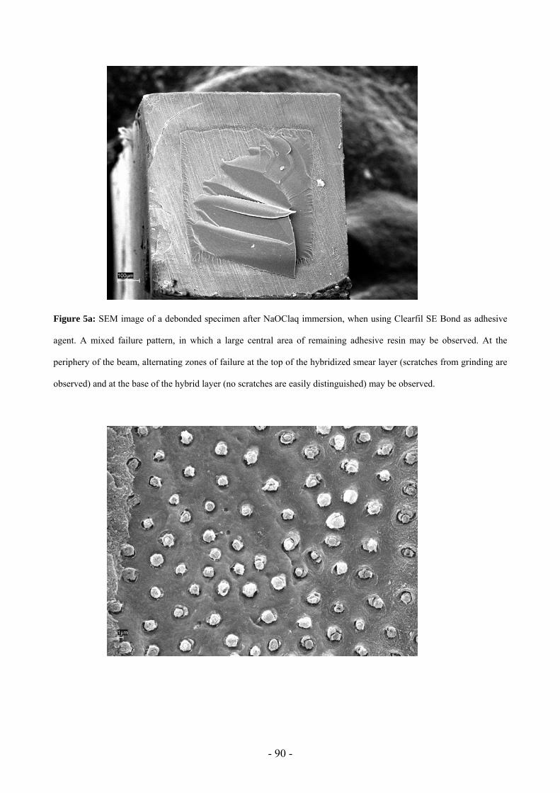
The infiltration of demineralized collagen fibers with resin permits formation of a hybrid
layer with resin tags and adhesive lateral branches, thus creating micromechanical retention of the
resin to the demineralized substrate. One of the first and most widely used tools to study the
mechanisms involved in the process of bonding has been the scanning electron microscope (SEM).
The retention of posts within root canals is affected by several factors involving the type of
the post, the luting agent and the post surface treatment. Fiber posts are becoming increasingly
popular for the restoration of endodontically treated teeth, however controversial results were
reported when different commercially available dentin adhesive and luting cement combinations
were employed for cementing fiber posts and when flowable materials and hybrid composites for
building up the core onto a fiber post were used.
Page 41
- 40 -
III.1. Effect of cyclic loading on microtensile bond strengths of total-etch and self-etch
adhesives
Authors:
Toledano M1, Osorio R1, Albaladejo A2, Aguilera FS1, Tay FR3, Ferrari M4.
1 Professor of Dental Materials, Faculty of Dentistry, University of Granada, Spain.
2 Research Fellow, Faculty of Dentistry, University of Granada, Spain.
3Honorary Clinical Professor, Faculty of Dentistry, University of Hong Kong, Hong Kong, China. 4 Professor of Dental Materials and Restorative Dentistry, Policlinico “Le Scotte”, University of
Siena, Italy.
Revista en la que está aceptado el artículo: Operative Dentistry 2005; (Aceptado para su
publicación).
Corresponding author: Manuel Toledano Avda. Fuerzas Armadas, nº1, 1º B. 18014, Granada. Spain Phone: 34 958 243788. Fax: 34 958 244085 E-mail: [email protected]
Page 42
- 41 -
ABSTRACT
Objective: To evaluate the effect of mechanical loading on the microtensile bond strength (MTBS)
of five adhesive systems to dentin. Methods: Flat dentin surfaces from 100 molars were divided into
five groups, and bonded with total-etch self-priming adhesives (Single Bond -SB-, Prime&Bond
NT –PNT- and Prime&Bond XP –PXP-), two-step self-etching primer (Clearfil SE Bond –SEB-),
and an all-in-one adhesive (Etch & Prime 3.0 –E&P-), according to the manufacturers´ instructions.
Composite build-ups were constructed incrementally with Tetric Ceram (TC). After 24 h of water
storage, half of the specimens were load cycled (5000 cycles, 90 N). The teeth were then sectioned
into beams of 1.0 mm2 cross-sectional area. Each beam was tested in tension in an Instron machine
at 0.5 mm/min. Data were analyzed by two way ANOVA and Student Newman Keuls multiple
comparisons tests (P<0.05). Results: SEB and SB attained higher MTBS than the other three
adhesives. PNT and PXP performed equally, and E&P resulted in the lowest MTBS. After
mechanical loading, MTBS decreased in all groups, except for PXP. SEB, SB and PXP obtained
higher MTBS than PNT. Specimens bonded with E&P resulted in premature failures and MTBS
could not be measured. The type of failure was predominantly mixed, except for E&P and PNT
after loading that exhibited predominantly adhesive failures. Clinical Relevance: All-in-one
adhesives do not provide a durable bond to dentin. If dentin is acid-etched, alcohol-based adhesive
systems showed the higher bond strength after mechanical loading.
Page 43
- 42 -
INTRODUCTION
Dentin bonding systems have been simplified and improved in order to provide increased
long-term strength and promote the durability and reliability of adhesive restorations (Nikaido &
others, 2002a). Two main strategies are used to create durable dentin bonding: 1) the total-etch self-
priming (i.e. single-bottle) bonding systems work by removing the smear layer with phosphoric
acid, followed by the application of a primer and an adhesive in the same step. With these systems,
incomplete expansion of the demineralized collagen matrix may impair resin infiltration and
compromise bonding (Van Meerbeek & others, 1994; Pashley & others, 2003); and 2) The self-
etching approach (SEB, E&P), in which increased concentrations of acid monomers enable the
primer or adhesive to etch and prime the dentin simultaneously. No discrepancy between the depth
of demineralisation and depth of resin infiltration is expected, since both processes occur
simultaneously (Tay & others, 2000).
In the clinical situation, dentin-resin bonds are not only subjected to immediate stresses, that
may disrupt the developing bonds, but also to cyclic loading during mastication that will induce
generation of cracks and subsequent crack growth that challenge the long-term survival of these
bonds. It has been shown that changes in the bonded interfaces in vivo may occur under occlusal
stresses, resulting in mechanical degradation of the bonds between the restoration and dentin (Sano
& others, 1999). Teeth are continuously subjected to stresses during mastication, swallowing and
parafunctional habits. Maximum biting force recorded on the first molar teeth is approximately 40-
90 Kg. Although masticatory loads recorded on a single molar are smaller (ca. 11-27 Kg) (Bates &
others, 1975; Anderson, 1956), they may still represent a challenge to the long-term durability of
resin-dentin bonds.
Page 44
- 43 -
Static bond strength tests may not adequately demonstrate the potential detrimental effects
that porosities and other internal defects within the adhesive layer may have on bonding durability
(Givan & others, 1995). After cyclic loading, the effect of these interfacial defects on long-term
bonding may be more readily apparent. It is anticipated that the combined use of mechanical
loading with microtensile bond strength (MTBS) testing permits the evaluation of the in vitro
durability of resin-dentin bonds under more clinically-relevant conditions than are usually
employed in static bond strength testing techniques.
Thus, the objective of this study was to compare the results of mechanical loading vs static
bond strength evaluation on the MTBSs of five total-etch and self-etch adhesives to human dentin.
The null hypothesis tested was that the incorporation of mechanical loading prior to bond strength
evaluation has no effect on the MTBSs of the adhesives to dentin.
Page 45
- 44 -
MATERIAL AND METHODS
One hundred caries-free extracted human third molars that were stored in 0.5% chloramine T
at 4 ºC and were used within one month after extraction. The specimens were sectioned below the
dentinoenamel junction and ground flat with 180-grit silicon carbide abrasive papers under running
water to provide uniform and clinically-relevant bonding surfaces. Three total-etch self-priming
adhesives (Single Bond -SB-, 3M ESPE, St. Paul, MN, USA; Prime&Bond NT –PNT-, Dentsply
DeTrey, Konstanz, Germany; and the experimental adhesive Prime&Bond XP –PXP-, Denstply
DeTrey), a two-step self-etching primer (Clearfil SE Bond –SEB-, Kuraray Medical Inc., Tokyo,
Japan), and an all-in-one self-etch adhesive (Etch&Prime 3.0 –E&P-, Dentsply Degussa AG,
Hanau, Germany) were examined (i.e. five experimental groups; N=20). The mode of application,
components and manufacturers of these adhesives are shown in Table 1. They were bonded to the
dentin surfaces according to the manufacturers´ instructions.
After bonding, composite build-ups, each 6 mm in height, were constructed incrementally
(1.5 mm) with a light-cured microhybrid resin composite (Tetric Ceram, Ivoclar-Vivadent, Schaan,
Liechtenstein). Each layer of the composite was light-activated for 40 s with a Translux EC halogen
light-curing unit (Heraeus-Kulzer GmbH, Hanau, Germany). Light intensity output was monitored
with a Demetron Curing Radiometer (Model 100 Demetron Research Corporation, Danbury, CT,
USA) to be at least 600 mW/sec.
The bonded specimens were stored in distilled water for 24 h at 37 ºC. For each
experimental group, half of the specimens (N=10) were mounted in plastic rings with dental stone
for load cycling under 90 N (5000 cycles, 3 cycles/sec) with the force applied longitudinally along
the center of the tooth. This compressive load was applied to the flat resin composite build-ups
using a spherical stainless steel plunger, 5mm in diameter, attached to a cyclic loading machine (S-
Page 46
- 45 -
MMT-250NB; Shimadzu, Tokyo, Japan). The rest of the specimens from each group (N=10) were
not subjected to cyclic loading and were stored in water until load-cycling for the other teeth was
completed. Each tooth was then sectioned vertically into serial slabs. The widest center slab from
each tooth was selected and further sectioned into beams with an approximate cross-sectional area
of 1 mm2, following the method described by Shono & others (1999). This resulted in the
generation of 32-39 beams for each experimental sub-group.
Each beam was tested for MTBS by attaching to a modified Bencor Multi-T testing
apparatus (Danville Engineering Co., Danville, CA) with a cyanoacrylate adhesive (Zapit, Dental
Venture of America Inc., Corona, CA, USA). The beams were stressed to failure in tension using a
universal testing machine (Instron 4411, Instron Corporation, Canton, MA, USA) at a crosshead
speed of 0.5 mm/min. The fractured beams were carefully removed from the apparatus and the
cross-sectional area at the site of failure was measured to the nearest 0.01 mm with a pair of digital
calipers (Sylvae Ultra-Call, Li, USA). The bond strength values were calculated in MPa and
analysed by two way ANOVA and Student Newman Keuls multiple comparison tests at α=0.05, to
examine the contribution of the two factors, adhesive type and cyclic loading, and their interactions
to the bond strength results. Fractured specimens were examined with a stereomicroscope (Olympus
SZ-CTV, Olympus, Tokyo, Japan) at 40X magnification to determine the mode of failure. Failure
modes were classified as adhesive, mixed, or cohesive in dentin or composite.
Representative fractured specimens from each of the ten subgroups were dehydrated for 48 h
in a desiccator (Sample Dry Keeper Simulate Corp., Japan) and then mounted on aluminum stubs
with carbon cement. They were then coated with gold by means of a sputter-coating unit (E500;
Polaron Equipment Ltd., Watford, England) and observed with a scanning electron microscope
(SEM) (Zeiss DSM-950, Karl-Zeiss, Germany) at an accelerating voltage of 20 kV, to examine the
morphology of the debonded interfaces.
Page 47
- 46 -
RESULTS
The mean MTBS values and failure modes obtained for the different groups are shown in
Table 2. Both the type of adhesive (F=25.02; P<0.0001) and the use of mechanical loading
(F=41.91; P<0.0001) influenced MTBS to dentin. No interaction existed between these two factors
(F=2.07; P=0.11). The power of the statistical analysis for MTBS was 0.78.
Multiple comparisons tests further revealed that SEB and SB exhibited greater MTBS to
dentin than the other three adhesives. PNT and PXP performed similarly, and E&P resulted in the
lowest MTBS. When specimens were subjected to mechanical loading, decreases in MTBS were
observed for all groups except for PXP. SEB, SB and PXP attained higher MTBS than PNT. All the
specimens bonded with E&P failed prematurely during laboratory beam preparation and MTBS
could not be obtained.
Most of the observed modes of failure were mixed except for specimens bonded with E&P,
and for those bonded with PNT after mechanical loading, in which the failure modes were
predominantly adhesive. Adhesive failures were associated with lower bond strengths. No cohesive
failure of dentin or resin composite was observed in any specimen.
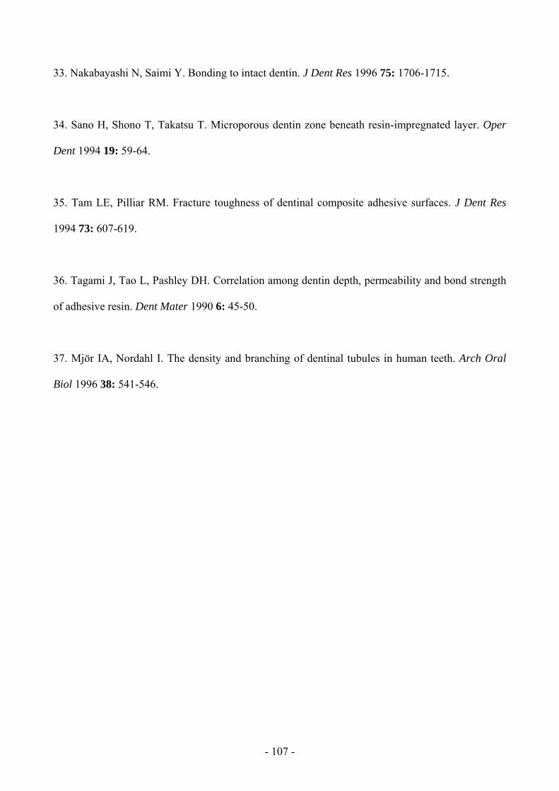
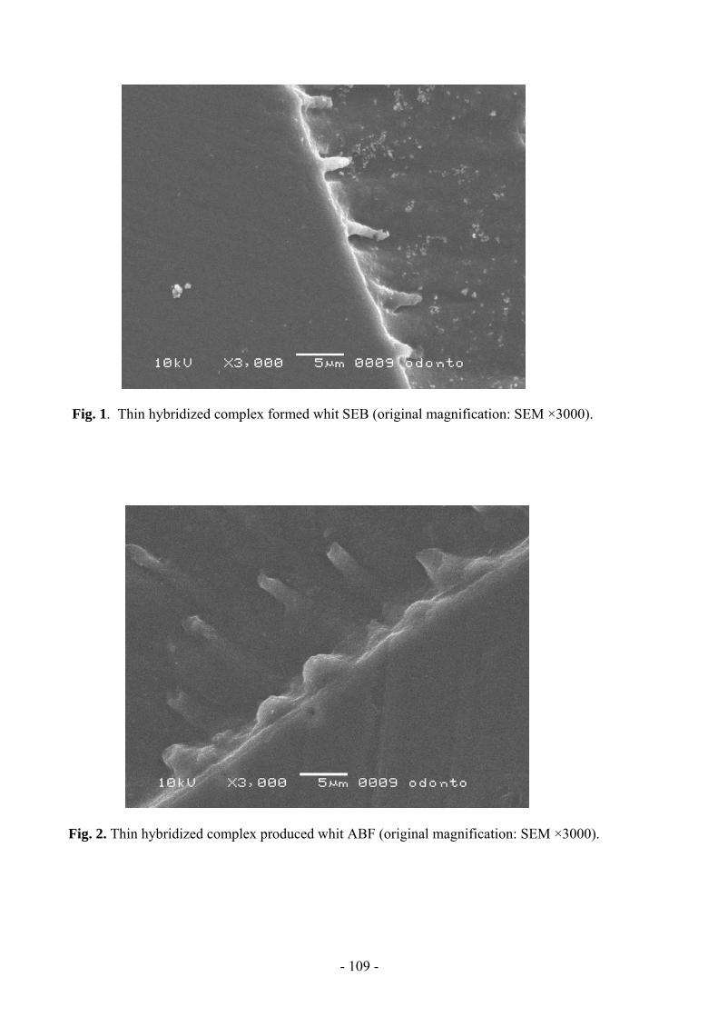
Fractured dentin surfaces after MTBS testing are shown in Figures 1 to 4. Mixed fracture
modes showed partially cohesive failures within the adhesive resin in all groups (Figures 1A and
2A). For the simplified total-etch adhesives (SB, PNT, PXP) failures were frequently observed at
either the top or the base of the hybrid layer, (Figures 1B and 2B). Partial cohesive fractures of
demineralized dentin just below the hybrid layer were sometimes observed. Specimens bonded with
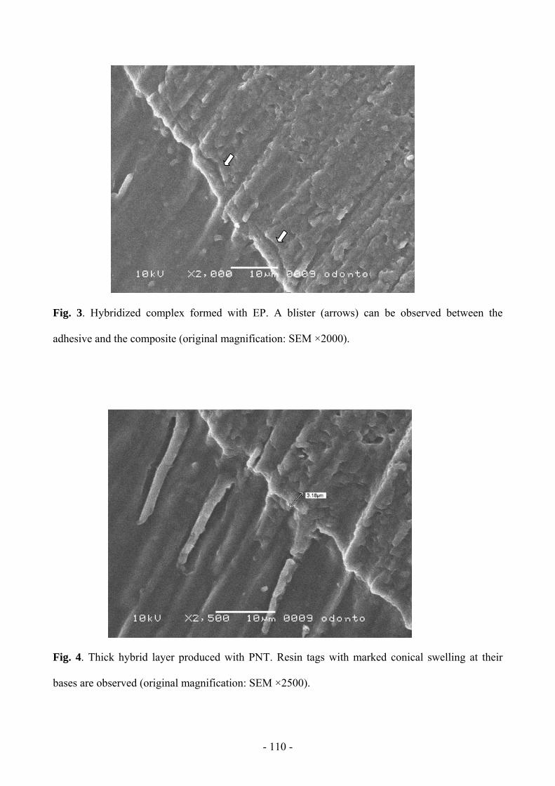
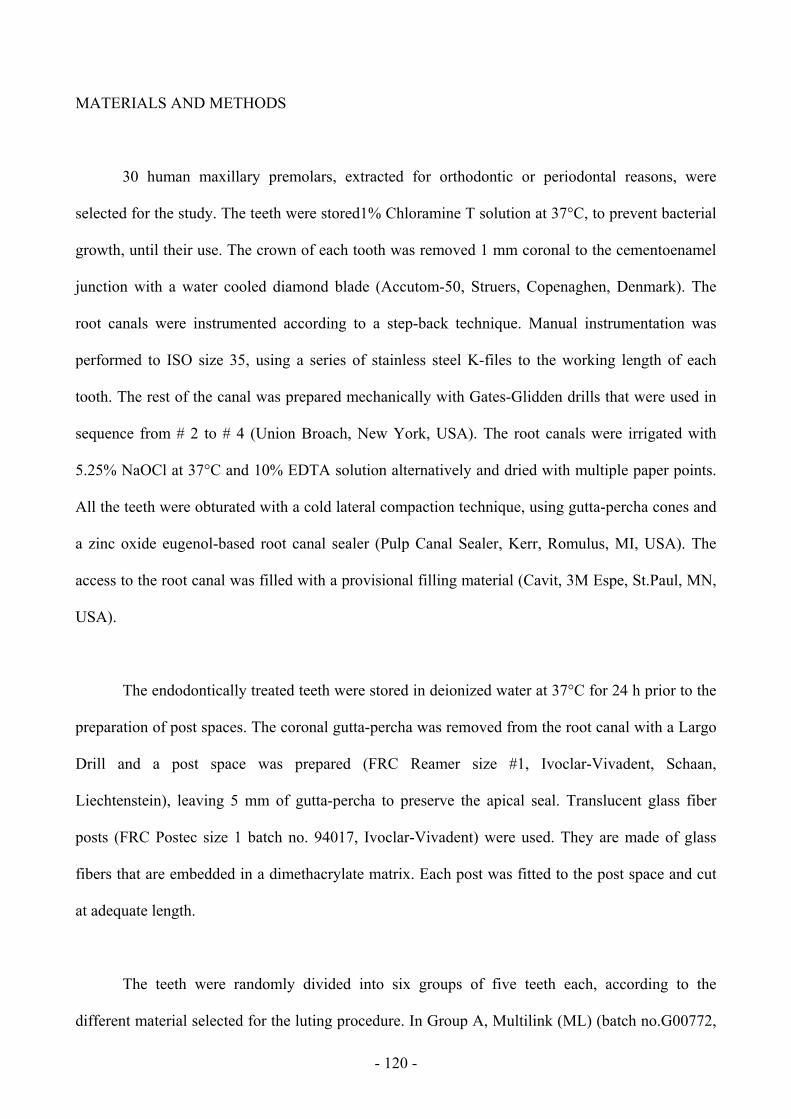
EP failed adhesively between both tooth substrate and bonding layer (Fig. 3A and 3B). Images from
Page 48
- 47 -
SEB specimens showed cohesive failures in both resin composite and adhesive with some fractures
seen at the base of the hybrid layer (Fig. 4) or even within the underlying dentin.
Page 49
- 48 -
DISCUSSION
Optimal dentin bonding is not always obtained in clinical practice, as normal daily
functioning, malocclusion and parafunctional habits such as bruxism impose additional stresses
upon the tooth and restorative system that may adversely affect the adhesive bond (Nikaido &
others, 2002a). A load of 90 N was used in this study, as it was considered to be within the normal
functional range (Anderson, 1956). In most of the studies 1,000 to 8,000 cycles are used; with 5,000
cycles being the median value (Abdalla & Davidson, 1996).
The one-step self-etching (all-in-one) adhesive E&P exhibited the lowest MTBS results and
frequent adhesive failures (Fig. 3A). The less than optimal result achieved with this adhesive was
further delineated after cyclic loading. Consensus exists in the literature that supports the poor
performance of such all-in-one adhesives in bond strength measurements (Fritz & Finger, 1999;
Inoue & others, 2000; Toledano & others, 2001; Toledano & others, 2003; Osorio & others 2003;
De Munck & others, 2003a), although they were able to completely dissolve the smear layer, and
formed a relatively thick hybridized complex (Haller & others, 2000; Cardoso, Placido & Moura,
2002; Toledano & others, 2003; Osorio & others 2003; Fritz & Finger, 1999) that incorporated the
smear layer (Santini, Plasschaert & Mitchell, 2001). Several reasons have been advocated to
account for the suboptimal performance of these all-in-one adhesive systems: (1) the combination
of acidic hydrophilic and hydrophobic monomers into a single step may compromise the
polymerization of the adhesive (De Munck & others, 2003a), (2) the stronger etching process may
destabilize the collagen, leading to a decrease in bond strength (Yoshiyama & others, 1995), (3) the
inherent weak strength of the adhesive polymer (Fritz & Finger, 1999; Haller & others, 2000; Inoue
& others, 2000), and (4) the lower degree of polymerization of the resin monomer, due to a major
solvent/oxygen inhibition effect in the photo-polymerization of these adhesives (Nunes & others
Page 50
- 49 -
2004). The lack of adequate polymerization may also account for the inability of the specimens to
withstand the occlusal loading forces, so that all specimens failed prematurely before testing.
PNT and the new experimental version of this simplified total-etch adhesive, PXP, showed
similar initial MTBS values. Both adhesive systems have similar composition, containing PENTA,
an acidic phosphonated monomer, which could have some kind of interaction with the calcium ions
left on dentin surface, or even with the underlying dentin (Inai & others, 1998). After load cycling,
MTBS values for PNT decreased but not those from PXP. Three main differences between these
adhesives may account for these results: 1) PXP contains TEGDMA, which lowers the initial
viscosity of the monomer mixture, enhancing its diffusion into the demineralised collagen matrix,
increasing the flexibility of the hybridized dentin, and improving the rate of polymerization of the
adhesive (Morgan & others, 2000; Nunes, Swift & Perdigão 2001; Nunes & others, 2004). 2)
Camphorquinone is included as a photosensitizer, increasing the polymerization of monomers and
bond strength to dentine (Miyazaki & others, 1995). 3) PXP contains t-butanol as solvent, (instead
of acetone, in PNT). After demineralization, the collagen fibrils adhere to one another via
intrafibrillar hydrogen bonding. A solvent with a solubility parameter for hydrogen bonding that
approximates that of the amino acid moieties of the collagen fibrils has a better capacity in breaking
up these intrafibrillar hydrogen bonds, and expanding the interfibrillar spaces to promote wetting
and infiltration of the adhesive monomers (Pashley & others, 2003). It has been demonstrated that
higher bond strengths were correlated with wider interfibrilar spaces and that such spaces should be
properly infiltrated with resin (Eddleston & others, 2003). Application of acetone produces little
solvation force affecting the further infiltration of resin monomers, while alcohols produces
progressively higher solvation pressures that develop at increasing rates (Pashley & others, 2002;
Reis & others, 2003). The total-etch alcohol-based adhesive systems used in the present
investigation (SB and PXP) are thought to be able to maintain the collagen fibrils in an expanded
condition after the evaporation of solvents, improving the monomers infiltration (Tay, Gwinnett &
Page 51
- 50 -
Pang, 1996; Perdigão, Van Meerbeek & Lopes, 1999). This may contribute to explain the lower
bond strengths of PNT after mechanical loading, because the decalcified non-infiltrated zone at the
base of the hybrid layer is susceptible to degradation during aging (Hashimoto & others, 2002a;
2002b; Pashley & others, 2002). Moreover, a low rate of polymerization of the bonding resin within
the hybrid layer has been shown for PNT (Hashimoto & others, 2002a), which also may lead to
rapid degradation of the resin-dentin bonds.
SB and SEB obtained the highest MTBS to dentin. SB is an adhesive based on a
HEMA/alcohol mixture and has been shown to obtain high bond strength values to dentin, when
compared to other total-etch adhesives (De Munck & others, 2003b). The results of SB were also
comparable to those of SEB (Toledano & others, 2003). A MTBS decrease is observed after
mechanical loading as well as previously reported, after water degradation (De Munck & others,
2003b).
SEB is a two-step self-etching primer containing a highly hydrophilic 10-MDP monomer,
which is believed to improve the wetting of the tooth surface (Van Meerbeeck & others, 1994). SEB
causes minimal dissolution of smear plugs and limited opening of tubules, which reduces dentin
permeability (Jackson & Söderholm, 2001) and facilitates penetration, impregnation,
polymerization and entanglement of monomers with the underlying dentin to form an hybrid layer
(Inoue & others, 2000; Toledano & others, 2002; Osorio & others, 2003). Moreover, 10-MDP has
two hydroxyl groups that may chelate with calcium ions of dentin (Kubo & others, 2001; Nunes &
others, 2003).
Within the limits of the study, we have to reject the null hypothesis as cyclic loading
lowered resin-dentin bond strengths of all the total-etch or self-etching adhesive systems examined.
Fatigue stress can expedite the degradation of bonds peripheral to the hybrid layer (Nikaido &
Page 52
- 51 -
others, 2002b; Sano & others, 1999; Qvist & others, 1983). When using SEB, the loading stress
seemed to have been concentrated mostly at the interface between the adhesive and the hybrid layer
and within the hybrid layer, whereas specimens bonded using a total-etch approach (SB, PNT and
PXP) mostly failed at the top of, or beneath the hybrid layer where demineralised collagen fibrils
were exposed and the adhesive failed to envelop the collagen network (Figures 1 and 2). Such
factors have been perceived to be the weakest link in achieving durable long-term bonding (Nikaido
& others, 2002b; Osorio & others, 2003; Pashley & others, 2002). Although the results obtained
from this study may not be directly extrapolated to the clinical situation, they provide some
information regarding the susceptibility of resin-dentin bonds to deteriorate after cyclic loading.
Long-term clinical data are still required to further evaluate the efficacy of these adhesives on
dentin.
CLINICAL SIGNIFICANCE
The resin-dentin bond is prone to deterioration after cyclic loading, and the all-in-one
adhesive examined is the least reliable system. After acid etching of dentin, alcohol/based adhesives
performed better than those containing acetone as solvent.
ACKNOWLEDGMENTS
This research project was supported by grants CICYT/FEDER MAT#2001-2843-C02-02,
MAT#2004-06872-C03-02 and Red CYTED VIII.J.
Page 53
- 52 -
REFERENCES
Abdalla A & Davidson C (1996) Effect of mechanical load cycling on the marginal integrity of
adhesive class I resin composite restorations Journal of Dentistry 24 87-90.
Anderson DJ (1956) Measurement of stress in mastication II Journal of Dental Research 35(5) 671-
673.
Bates JF, Stafford GD, Harrison A (1975) Masticatory function- a review of the literature. (II)
Speed of movement of the mandibule, rate of chewing and forces developed in chewing Journal of
Oral Rehabilitation 2 349-361.
Cardoso PEC, Placido E & Moura SK (2002) Microleakage of four simplified adhesive systems
under thermal and mechanical stresses American Journal of Dentistry 15(3) 164-168
De Munck J, Van Meerbeek B, Satoshi I, Vargas M, Yoshida Y, Armstrong S, Lambrechts P &
Vanherle G (2003a) Microtensile bond strengths of one- and two-step self-etch adhesives to bur-cut
enamel and dentin American Journal of Dentistry 16 414-420.
De Munck J, Van Meerbeek B, Yoshida Y, Inoue S, Vargas M, Suzuki K, Lambrechts P &
Vanherle G (2003b) Four-year of water degradation of total-etch adhesives bonded to dentin
Journal of Dental Research 82(2) 136-140.
Eddleston CL, Hindle AR, Agee KA, Carvalho RM, Tay FR, Rueggegerg FA, Pashley DH (2003)
Dimensional changes in demineralized dentin matrices following the use of HEMA-water versus
HEMA-alcohol primers Journal of Biomedical Materials Research 67A 900–907.
Page 54
- 53 -
Fritz UB & Finger WJ (1999) Bonding efficiency of single-bottle enamel/dentin adhesives
American Journal of Dentistry 12 277-282.
Givan DA, Fitchie JG, Anderson L, Zardiacks LD (1995) Tensile fatigue of 4-META cement
bonding three base metal alloys to enamel and comparison to other resin cements Journal of
Prosthetic Dentistry 73 377-385.
Haller B (2000) Recent developments in dentin bonding American Journal of Dentistry 13 44-50.
Hashimoto M, Ohno H, Kaga M, Sano H, Endo K & Oguchi H (2002a) The extent to which resin
can infiltrate dentin by acetone-based adhesives Journal of Dental Research 8(1) 74-78.
Hashimoto M, Ohno H, Sano H, Tay FR, Kaga M, Kudou Y, Oguchi H, Araki Y, Kubota M (2002)
Micromorphological changes in resin-dentin bonds after 1 year of water storage Journal of
Biomedical Materials Research 63(3) 306-311.
Inai N, Kannemura N, Tagami J, Watanable LG, Marshall SJ & Marshall GW (1998) Adhesion
between collagen depleted dentin and dentin adhesives American Journal of Dentistry 11 123–127.
Inoue S, Van Meerbeek B, Vargas M, Yoshida Y, Lambrechts P & Vanherle G (2000) Adhesion
mechanism of self-etching adhesives In: Tagami J, Toledano M, and Prati C. Advanced Adhesive
Dentistry Cirimido, Italia 131-148.
Jackson WD & Söderholm KJM (2001) Comparison of shear and flexural bond strength tests versus
failure modes of dentin bonding systems American Journal of Dentistry 14 297-303.
Page 55
- 54 -
Kubo S, Yokota H, Sata Y & Hayashi (2001) The effect of flexural load cycling on the
microleakage of cervical resin composites Operative Dentistry 26 451-459.
Miyazaki M, Hinoura K, Saito H, Onose H & Moore B (1995) Influence of light irradiation of
dentine primers on dentine-resin bond Journal of Dentistry 23(6) 329-376.
Morgan D, Kalachandra S, Shobha H, Gunduz N & Stejskal E (2000) Analysis of a dimethacrylate
copolymer (Bis-GMA and TEGDMA) network by DSC and C solution and solid-state NMR
spectroscopy Biomaterials 21 1897-1903.
Nikaido T, Kunzelmann KH, Ogata M, Harada N, Yamaguchi S, Cox CF, Hickel R & Tagami J
(2002a) The in vitro dentin bond strength of two adhesive systems in Class I cavities of human
molars Journal of Adhesive Dentistry 4 31-39.
Nikaido T, Kunzelmann KH, Chen H, Ogata M, Harada N, Yamaguchi S, Cox CF, Hickel R &
Tagami J (2002b) Evaluation of thermal cycling and mechanical loading on bond strength of a self-
etching primer system to dentin Dental Materials 18 269-275.
Nunes MF, Swift EJ & Perdigao J (2001) Effects of adhesive composition on microtensile bond
strength to human dentin American Journal of Dentistry 14 340-343.
Nunes TG, Polido M, Amorin A, Nunes S & Toledano M (2003) Liquid and solid state multinuclear
magnetic resonance studies on the interaction of self-etching adhesives with radicular and coronal
dentin Journal of Dental Research 82 192.
Page 56
- 55 -
Nunes TG, Ceballos L, Osorio R & Toledano M (2004) Spatially-resolved photopolymerization
kinetics and oxygen inhibition in dental adhesives Biomaterials (In press).
Osorio R, Toledano M, De Leonardi G & Tay F (2003) Microleakage and interfacial morphology of
self-etching adhesives in class V resin composite restorations Journal of Biomedical Materials
Research 66 B 399-409.
Pashley DH, Carvalho RM, Tay FR, Agee KA & Lee KW (2002) Solvation of dried dentin matrix
by water and other polar solvents American Journal of Dentistry 15 97-102.
Pashley DH, Agee KA, Carvalho RM, Lee KW, Tay FR & Callison TE (2003) Effects of water and
water-free polar solvents on the tensile properties of demineralised dentin Dental Materials 19 347-
352.
Perdigão J, Van Meerbeek B & Lopes MM (1999) The effect of a re-wetting agent on dentin
bonding Dental Materials 12 271-276.
Qvist V (1983) The effect of mastication on marginal adaptation of composite restoration in vivo
Journal of Dental Research 62 904-906.
Reis A, Loguercio AD, Azevedo CL, Crarvlaho RM, Singer JM & Grande RM (2003) Moisture
spectrum of demineralised dentin for adhesive systems with different solvent bases Journal of
Adhesive Dentistry 5 183-192.
Page 57
- 56 -
Sano H, Yoshikawa T, Pereira PNR, Kanemura N, Morigami M, Tagami J & Pashley DH (1999)
Long-term durability of dentin bonds made with a self-etching primer Journal of Dental Research
78 906-911.
Shono Y, Ogawa T, Terashita M, Carvalho RM, Pashley EL & Pashley DH (1999) Regional
measurement of resin-dentin bonding as an array Journal of Dental Research 78 699-705.
Santini A, Plasschaert AJM & Mitchell S (2001) Effect of composite resin placement techniques on
the microleakage of self-etching dentin-bonding agents American Journal of Dentistry 14 132-136.
Tay FR, Gwinnett AJ & Pang KM (1996) Resin permeation into acid-conditioned, moist, and dry
dentin: A paradigm using water-free adhesive primers Journal of Dental Research 75 1034-1044.
Tay FT, Sano H, Carvalho R, Pashley EL, Pashley DH (2000) An ultrastructural study of the
influence of acidity of self-etching primers and smear layer thickness on bonding to intact dentin
Journal of Adhesive Dentistry 2 83-98.
Toledano M, Osorio R, Leonardi de G, Rosales-Leal JI, Ceballos L & Cabrerizo-Vilchez MA
(2001) Influence of self-etching primer on the resin adhesion to enamel and dentin American
Journal of Dentistry 14 205-210.
Toledano M, Osorio R, Ceballos L, Fuentes V, Fernández CAO, Tay FR & Carvalho RM (2003)
Microtensile bond strength of several adhesive systems to different dentin depths American Journal
of Dentistry 16 292-298.
Page 58
- 57 -
Van Meerbeek BV, Peumans M, Verschueren M, Gladys S, Braen M, Lambrechts P & Vanherle G
(1994) Clinical status of ten dentin adhesive systems Journal of Dental Research 73 1690-1702.
Yoshiyama M, Carvalho R, Sano H, Horner J, Brewer P & Pashley DH (1995) Interfacial
morphology and strength of bonds made to superficial versus deep dentin American Journal of
Dentistry 8 297-302.
Page 59
- 58 -
Table 1: Mode of Application, compositions, and manufacturers of tested adhesives.
Materials Components
Mode/steps of application
Manufacturer
Single Bond 2-Hydroxyethylmethacrylate; water; ethanol; Bis-GMA;
dimethacrylates; amines; methacrylate-functional;
copolymer of polyacrylic and polyitaconic acids.
Etch for 15 seconds. Rinse with water spray for
10 seconds, leaving tooth moist. Apply two
consecutive coats of the adhesive with a fully
saturated brush tip. Dry gently for 2-5 seconds.
Light cure for 10 seconds.
3M, St.Paul, MN,
USA.
Lot. 4242.
Prime & Bond NT PENTA; UDMA resin; Resin R5-62-1; T-resin; D-resin;
nanofiller; initiators; stabilizer; cetylamine hydrofluoride;
acetone.
Etch for 15 seconds. Rinse with water spray for
15 seconds and remove water with a soft blow
of air. Leave a moist surface. Apply ample
amounts of the adhesive to saturate the surface,
reapply if it is necessary. Leave the surface
undisturbed for 20 seconds. Remove solvent by
blowing gently with air for at least 5 seconds.
Light cure for 10 seconds.
Dentsply / De Trey
GmbH, Konstanz,
Germany.
Lot. 0209000918.
Clearfil SE Bond Primer: 10-methacryloyloxydecyl dihydrogen phosphate; 2-
hydroxyethyl methacrylate; Hydrophilic dimethacrylate;
di-camphorquinone; N,N-diethanol-p-toudine, water.
Bond: 10-methacryloyloxydecyldihydrogen phosphate;
N,N-diethanol-p-toludine; 2-hydroxyethylmethacrylate;
Bis-phenol A diglycidylmethacrylate; silanated colloidal
silica; hydrophobic dimethacrylate; di-camphorquinone.
Apply Primer for 20 seconds. Mild air stream.
Apply Bond. Gentle air stream. Light cure for
10 seconds.
Kuraray Co, Osaka,
Japan.
Lot. 390.
Etch & Prime 3.0 Universal: 2hydroxyethylmethacrylate;Water; ethanol
Catalyst: Tetramethacrylocyethylpyrophophate.
Mix Etch & Prime 3.0 Universal and Catalyst.
Apply for 30 seconds. Air blow gently. Light
cure for 10 seconds. Repeat the above
mentioned steps.
Degussa AG, Hanau,
Germany.
Lot.019920.
Prime & Bond XP TCBresin; PENTA; UDMA; TEGDMA; BHT;
camphorquinone; functionalised amorphous silica
ethyl-4-dimethylaminobenzoate; t-butanol.
Etch for 15 seconds. Rinse with water spray for
15 seconds and remove water with a soft blow
of air. Leave a moist surface. Dispense directly
into a disposable brush. Apply ample amounts
of the adhesive to saturate the surface, reapply
if it is necessary. Leave the surface undisturbed
for 20 seconds. Remove solvent by blowing
gently with air for at least 5 seconds. Light cure
for 10 seconds.
Dentsply / De Trey
GmbH, Konstanz,
Germany.
Lot. 0304000987.
PENTA= penta-acrylate ester; TEGDMA= triethylene glycol-dimethacrylate; Bis-GMA= bysphenyl glycidyl
methacrylate, UDMA= urethane dimethacrylate; BHT= butylated hydroxyl toluene; TCB resin = carboxylic acid
modified dimethacrylate.
Page 60
- 59 -
Table 2: MTBS values and distribution of failure modes (A: Adhesive; M: Mixed) obtained with
the different adhesive systems with and without cyclic loading.
Without load cycling Load cycling
Mean (SD) A M Mean (SD) A M
SEB 46.07 (12.1) A 34.5 % 65.5 % 30.61 (5.3) B 44.5 % 55.5 % SB 43.34 (11.1) A 27.9 % 72.1 % 28.29 (8.4) B 40 % 60 % PXP 29.79 (4.5) B 20 % 80 % 25.15 (7.9) B 27.9 % 72.1 % PNT 29.08 (4.7) B 34 % 66 % 11.73 (3.2) D 77.8 % 22.2 % E&P 16.99 (7.7 ) C 72.7 % 27.3 % XX XX XX Values are means (standard deviation) in MPa, with the number of beams chosen as the statistical unit (n=32-39). Groups with the same letter are not statistically significant (P>0.05). XX: No MTBS data could be obtained due to premature failure of all the specimens during beam preparation.
Page 61
- 60 -
Figure 1a: SEM images of the fractured dentin surface of a specimen bonded with Single Bond after cyclic loading. A
mixed failure could be observed, with resin composite present at the right and left margins, and adhesive in the central
area.
Figure 1b: SEM images of the fractured dentin surface of a specimen bonded with Single Bond after cyclic loading. A
higher magnification view of the failure that occurred at the top of the hybrid layer.
Page 62
- 61 -
Figure 2a: SEM observations of the fractured surface along the dentin side of a specimen bonded with Prime&Bond
NT, after cyclic loading. An adhesive failure, mainly at the top of the hybrid layer is observed, but a small area (left and
inferior corner) failed at the bottom of the hybrid layer.
Figure 2b: SEM observations of the fractured surface along the dentin side of a specimen bonded with Prime&Bond
NT, after cyclic loading. At a higher magnification, resin filled dentinal tubules are shown, but non-infiltrated dentin
and porosity within the hybrid layer are also shown.
Page 63
- 62 -
Figure 3a: SEM observations of the fractured surface along the dentin side of a specimen bonded with Etch&Prime 3.0,
before cyclic loading. A general image of a typical adhesive failure. Scratches that remained from preparation of the
bonding dentin surface with silicon carbide papers confirmed that the interface failed adhesively at the level between
dentin and the adhesive.
Figure 3b: SEM observations of the fractured surface along the dentin side of a specimen bonded with Etch&Prime
3.0, before cyclic loading. The enlarged entrances of the dentinal tubules could be observed, and only some of them
were occluded by resin tags.
Page 64
- 63 -
Figure 4: SEM image of a specimen bonded with CSEB, showing cohesive fractures of the dentin just below the hybrid layer.
Page 66
- 65 -
III.2. Differential effect of in vitro degradation on resin-dentin bonds produced by self-etch vs.
total-etch adhesives
Authors:
Toledano M. MD. BDS. Ph.D1, Osorio R. DDS. Ph.D1, Albaladejo A. LDS1, Aguilera FS. DDS.
Ph. D1, Osorio E. DDS. Ph.D1
1 Department of Dental Materials, School of Dentistry, University of Granada, Granada E-18071
Spain.
Revista en la que está aceptado el artículo: Journal of Biomedical Materials Research, Applied
Biomaterials (Part A) 2005; (Aceptado para su publicación).
Short title: Differential degradation of resin-dentin bonds
Corresponding Author:
Raquel Osorio Av. de las Fuerzas Armadas nº1, 1ºB, 18014 Granada, Spain. Fax:34-958-240908 E-mail: [email protected]
Page 67
- 66 -
ABSTRACT
Objective: To evaluate the effect of an in vitro challenge (NaOCl immersion) on
microtensile bond strength (MTBS) of five adhesive systems to dentin. Methods: flat dentin
surfaces from forty molars were bonded with three total-etch adhesives (Single Bond, Prime&Bond
NT and the experimental Prime&Bond XP), and two self-etching agents (Clearfil SE Bond and
Etch&Prime 3.0). Composite build-ups were constructed with Tetric Ceram. Teeth were then
sectioned into beams of 1.0 mm2 cross-sectional area. Half of the beams were immersed in 10 %
NaOCl aqueous solution for 5 h. Each beam was tested in tension in an Instron machine at 0.5
mm/min. Data were analyzed by 2-way ANOVA and multiple comparisons tests (P<0.05). Results:
Clearfil SE Bond and Single Bond attained higher MTBS than the other three adhesives.
Prime&Bond NT and Prime&Bond XP performed equally, and Etch&Prime resulted in the lowest
MTBS. After NaOCl immersion, MTBS decreased in all groups. The highest MTBS values were
obtained for Clearfil SE Bond and Prime&Bond XP. Conclusions: The extent of the resin-dentin
bond degradation is material dependent. A high polymerization degree of the bonding resins, within
the hybrid layer, may be a very important factor to improve the long-term durability of resin-dentin
bonds.
Keywords: dentin, resin, degradation, bonding, sodium hypochlorite
Page 68
- 67 -
INTRODUCTION
The durability of bonds between adhesive resins and dentin is of critical importance [1,2]
and little is known regarding the stability of hybridized layers [3]. In general, reports show that
dentin bond strength decreased during water storage over time, due to degradation of the resin and
the collagen fibrils within the hybrid layer [2,4,5,6].
Dentin bonding systems have been simplified and improved in order to provide increased
long-term durability of adhesive restorations [7]. Two main strategies are used to create dentin
bonding: 1) the total-etch bonding systems (TE) work by removing the smear layer with phosphoric
acid, followed by the application of a primer and an adhesive in two different steps or in the same
one (Single Bond, Prime&Bond NT and Prime&Bond XP), but further incomplete expansion of
collagen may impair resin infiltration and compromise bonding [8,9]; and 2) the self-etching
approach (SE), in which the acid and the primer are combined in one solution to form an acidic
monomer [10] and a final bonding step is later applied (Clearfil SE Bond). Self-etch all-in-one
adhesives have also been introduced and contain all components in just one solution (Etch&Prime).
When using all these self-etching systems less discrepancy is expected between the depth of
demineralisation and depth of resin infiltration [10,11].
In vitro accelerated aging tests for resin-dentin bonds have been proposed, reducing
specimen size and immersing the bonded sticks in 10% sodium hypochlorite aqueous solution
(NaOClaq) for a short experimental time period, to determine the ability of resin monomers to
protect the collagen matrix of dentin from proteolytic activity [12,13].
Page 69
- 68 -
The null hypotheses to be tested are that there are not differences in dentin bond strength
when using different adhesive systems, and that NaOCl immersion of specimens does not affect
obtained bond strength to dentin.
Page 70
- 69 -
MATERIAL AND METHODS
Forty caries-free extracted human third molars that were stored (4 ºC) in 0.5% chloramine T
for up to one month were used. The specimens were sectioned below the dentin-enamel junction and
ground flat with 180-grit silicon carbide abrasive papers under running water to provide uniform and
smear-layer covered dentin surfaces. Single Bond, Prime&Bond NT, Prime&Bond XP, Clearfil SE
Bond and Etch&Prime adhesives were applied following manufacturers´ instructions. Table 1
displays mode of application, components and manufacturers of the tested adhesives. Resin build-
ups, each 6 mm in height, were constructed incrementally (1.5 mm) with Tetric Ceram resin
composite (Vivadent, Schänn, Liechtenstein). Each layer of the composite was light-activated for 40 s
with a Translux EC halogen light-curing unit (Kulzer GmbH, Bereich Dental, Wehrheim, Germany).
Light intensity output was monitored with a Demetron Curing Radiometer (Model 100 Demetron
Research Corporation, Danbury, CT, USA) to be, at least, 600 mW/cm2.
After storage in distilled water for 24 h at 37 ºC, specimens were vertically sectioned into
serial slabs, and further into beams giving a cross-sectional area of 1 mm2. Half of the beams in
each group were immersed in 10% NaOCl solution (Panreac Química SA, Barcelona, Spain) for 5
h, following by rinsing in water for 1 hour. Approximately 39-42 beams resulted from each
subgroup, they were attached to a modified Bencor Multi-T testing apparatus (Danville Engineering
Co., Danville, CA) with a cyanoacrylate adhesive (Zapit, Dental Venture of America Inc., Corona,
CA, USA) and stressed to failure in tension using in a universal testing machine (Instron 4411,
Instron Corporation, Canton, MA, USA) at a crosshead speed of 0.5 mm/min. The fractured beams
were carefully removed from the apparatus and the cross-sectional area at the site of failure was
measured to the nearest 0.01 mm with a pair of digital calipers (Sylvae Ultra-Call, Li, USA). The
bond strength values were calculated in MPa and analysed by ANOVA and Student Newman Keuls
multiple comparisons (P<0.05). Fractured specimens were examined with a stereomicroscope
Page 71
- 70 -
(Olympus SZ-CTV, Olympus, Tokyo, Japan) at 40X magnification to determine the mode of
failure. Failure modes were classified as adhesive or mixed. Representative specimens of each
group were maintained for 48 h in a dessicator (Sample Dry Keeper Simulate Corp., Japan) and
then mounted on aluminum stubs with carbon cement. They were then sputter-coated with pure
gold by means of a sputter-coating Unit E500 (Polaron Equipment Ltd., Watford, England) and
observed with a scanning electron microscopy (SEM) (Zeiss DSM-950, Karl-Zeiss, Germany) at an
accelerating voltage of 20 kV, so that microscopic fracture patterns and the morphology of the
debonded interface could be studied.
Page 72
- 71 -
RESULTS
Mean MTBS values and modes of failures obtained for the different groups are shown in
Table 2. The adhesive system (F=25.06; P<0.001) and NaOClaq immersion (F=150,79; P<0.001)
influenced MTBS to dentin. Interactions existed between both variables (F=6.68; P<0.001). The
power of the multiple ANOVA analysis for MTBS was about 0.82.
Clearfil SE Bond (SE) and Single Bond (TE) give greater MTBS to dentin than the other
three adhesives. Prime&Bond NT (TE) and Prime&Bond XP (TE) performed similarly, and
Etch&Prime (SE) resulted in the lowest MTBS. When specimens were subjected to NaOClaq
immersion, decreases in MTBS were observed for all groups. The SE system -Clearfil SE Bond-
and the TE adhesive –Prime&Bond XP- attained higher MTBS than the rest of the adhesives.
Specimens bonded with Etch&Prime (SE) produced pretesting failures and MTBS values could not
be obtained.
Most of the observed modes of failure were mixed except for specimens bonded with the
one-step self-etch adhesive (Etch&Prime) that failed predominantly adhesively. Major rates of
adhesive failures were associated with lower bond strengths. Specimens that undergo NaOClaq
immersion failed predominantly mixed, except for Prime&Bond NT (TE) that presented mostly
adhesive failures. No completely cohesive failure of dentin or resin composite was observed in any
specimen.
SEM of debonded dentin surfaces after MTBS testing are shown in Fig. 1 to 5. Mixed
fracture modes were frequent in all groups except for Etch&Prime (SE) that failed predominantely
adhesively at the top of the hybridized smear layer (Fig. 1a), and resin tags are observed occluding
Page 73
- 72 -
the enlarged tubule entrances (Fig. 1b). In the case of the total-etch adhesives failures were
frequently mixed found at the base of the hybrid layer, partial cohesive fractures of demineralized
dentin just below the hybrid layer were sometimes observed (not shown), usually associated with
high bond strength data. Images from Clearfil SE Bond (SE) specimens showed mixed failures
found either at the top, at the base of the hybrid layer or within the hibridized smear layer, and often
both within the same section. After NaOClaq immersion, specimens showed partially cohesive
failures within the adhesive resin (Fig. 2a to 5a). A gradual loss of adhesive from the periphery to
the center portion of the bonding area is observed in all groups, and resin remaining area is smaller
for Prime&Bond NT than for the rest of the adhesives (Fig. 2a to 5a). For Single Bond and
Prime&Bond NT (TE) groups, fractures used to be at the top of the hybrid layer (as scratching from
dentin grinding are clearly observed) (Fig. 2a and 3a), resin tags are maintained at the tubule
entrances (Fig. 2b and 3b). When Prime&Bond XP (TE) specimens are examined, most of the
fractures are located at the base of the hybrid layer and at the non-infiltrated underlying dentin (Fig.
4a), showing opened and enlarged dentin tubule entrances, without resin tags (Fig. 4b). Specimens
bonded with Clearfil SE Bond (SE) showed most of the failures at the top of the hybrid layer (Fig.
5a), and small areas of fractures located at the base of the hybrid layer, exposing the underlying
dentin, may also be observed (Fig. 5b). Tubule entrances may be observed, they are not enlarged
but opened and filled with resin tags (Fig. 5b).
Page 74
- 73 -
DISCUSSION
The total-etch self-priming adhesives Prime&Bond NT and Prime&Bond XP showed
similar initial MTBS values. Both adhesive systems contain PENTA, an acidic phosphonated
monomer, which could have some kind of interaction with the calcium ions left on dentin surface,
or even with the underlying dentin [14]. The TE system Single Bond attained higher values, it is
based on a HEMA/alcohol mixture that is able to better wet the etched dentin surface and maintain
the collagen fibers in an expanded condition after the evaporation of solvents, improving the
monomers infiltration, and has been shown to obtain high bond strength values to dentin, when
compared to other total-etch adhesives [5,15,16].
Bond strength of the TE Single Bond to dentin was similar to that of the SE Clearfil SE
Bond [17]. Clearfil SE Bond contains a highly hydrophilic 10-MDP monomer, which is believed to
improve the wetting of the tooth surface and chelate to calcium ions of dentin [18]. This SE
adhesive causes minimal dissolution of smear plugs and facilitates penetration, impregnation,
polymerization and entanglement of monomers with the underlying dentin to form a hybrid layer
[17,19,20].
Etch&Prime is a 1-step SE adhesive system (all-in-one), obtaining the lowest MTBS values
and frequent adhesive failures (Fig. 1). Consensus exists about the low bond strength of most of
these all-in-one adhesive systems [5,17,19,20,21]. Even though, studies have shown that the
bonding agent completely dissolved the smear layer, formed a relatively thick hybridized complex
[22,23,24,20,17], incorporating the smear layer and tubule entrances were not only opened but also
enlarged creating thick resin tags (Fig. 2) [17,20]. Some reasons have been advocated to explain the
worse bonding performance of these all-in-one adhesive systems as the inherent weak strength of
the adhesive polymer [19,22,24] and the low degree of polymerization of the resin monomer,
Page 75
- 74 -
probably due to a major solvent/oxygen inhibition effect in the photopolymerization of these
adhesives [20,25].
After storage in NaOClaq, the MTBS fell in all specimens. NaOClaq is a nonspecific
deproteinizing agent, in aqueous solution superoxide radicals O2- are formed and induce oxidations
that fragment long peptide chains of proteins [26]. Chlorination of protein terminal groups is also
produced and hypochlorous acid formation evidenced [27]. Some of these amino acid-derived
chloramines have also shown to increase the proteolytic susceptibility of this modified collagen
[28]. The decline in bond strength is the result of both an hydrolytic process on the resin and the
solubilization of unprotected collagen fibrils within the decalcified dentin [5,12,13,29,30,31].
Some studies have been conducted in order to evaluate the long-term durability of resin-
dentin bonds. In vivo studies showed that the bond strength of a three-step total-etch adhesive may
be reduced by 50-65% after 2-3 years [32], or even by 60 and 77% within 1 year when using a self-
priming total-etch adhesive and a two-step self-etching adhesive respectively [33]. However, in
vitro studies based on water storage of the specimens, attained smaller reductions in bond strength
that are between 23 and 55%, [4,5,6,34], even when the dentin-resin interfaces were directly
exposed to water up to six years [34]. The presented challenging method based upon 10% NaOClaq
immersion of specimens during a short period of time is much more reliable than in vitro studies
based on long-term water storage of specimens. Obtained reductions in bond strength in the present
study, and those previously reported after NaOClaq immersion (65% to 77%) [12,13] are similar to
the decline in bond strength obtained when in vivo degradation studies are performed [32,33]. In
vivo studies also reported that the microestructural aspect of the debonded surfaces suggested that
the exposed collagen was digested by proteolytic enzymes, which may be released from leukocytes,
salivary glands, and bacteria in plaque, explaining the attained very low bond strengths [32], and
Page 76
- 75 -
resinous materials seems to be degraded and extracted from the hybrid layer, increasing the
porosities at the interface [3,33].
A larger fall in bond strength was expected for the TE adhesives, when compared to the SE
systems, due to the existence of a discrepancy between the etching and the infiltrating processes
[8,9,12,13]. However, it is not observed at the present results. This may account for the reported
evidences supporting that for some self-etching systems an etched non-resin-infiltrated layer
remains after bonding [11,35]. It may also be that when using so highly hydrophilic resins, the
hydrolytic degradation of the resin is the most detrimental factor affecting long-term bonding
effectiveness.
The high hydrophilicity of the adhesive resin which is an advantage during dentin bonding,
may be a disadvantage in the long-term durability. As the higher water solubility of the resin and
the existence of residual water within the hybrid layer may lead to: voids formation, a lower degree
of cure of the adhesive resin [36,37,38], and poorly infiltrated collagen fibrils within the hybrid
layer that might accelerate the degradation effect [38].
Fractographic analysis of debonded sticks showed a gradual loss of adhesive at the top of the
hybrid layer or at the top of the hybridized smear layer (as grinding marks of polish may be
observed on the exposed dentin surface) (Fig. 2,3,5) with this loss of adhesive being from the
periphery to the center of the debonded area. The TE Prime&Bond XP and the SE Clearfil SE
Bond, that attained the better resistance to the challenging of NaOClaq immersion, showed the
smallest resin dissolution areas (Fig. 2 to 5). The resistance of these resins to the hydrolytic
degradation may be the chief reason of the lower reduction in bond strength attained by these
adhesives. The least susceptibility of these resins to hydrolysis is probably due to a higher degree of
Page 77
- 76 -
cure of the bonding resins. Prime&Bond XP contains TEGDMA, which brings down the initial
viscosity of the monomer mixture, enhancing diffusion of reactive groups, increasing the flexibility,
and the rates of polymerisation of the resin [25,39]. Camphorquinone is included in both adhesive
systems (Prime&Bond XP and Clearfil SE Bond) as sensitier. This activator is in charge to trigger
the cascade reaction of the curing, generating free radicals and increasing the polymerization of
monomers [37]. Moreover, a low rate of polymerization of the bonding resin has been previously
shown for Prime&Bond NT (Fig. 3) [40], and for Etch&Prime [25] leading to rapid degradation of
the dentin bonds.
According to previous observations the loss of resin was responsible for the loss of bond
strength [12,13]. The extent of resin dissolution is proportional to the bond strength decrease
[12,13] and is material dependent. However, the deterioration patterns depend on the used adhesive
approach (e.g. SE or TE). The test medium acted more aggressively with the intertubular dentin of
specimens bonded by a TE procedure compared to those bonded with the SE system. After
phosphoric acid etching collagen is highly susceptible to deproteinization processes [41]. When
bonding with a TE adhesive, the NaOClaq affected the resin-dentin bond structures following two
pathways 1) the etched and non-infiltrated layer (Fig. 4b) and 2) the collagen that was resin-
infiltrated but later exposed because of the bonding resin dissolution by the NaOClaq (Fig. 2b,3b).
Although the results obtained from this study may not be directly extrapolated to the clinical
situation, they provide some information with regard to the performance of dentin treatment
procedures. This in vitro testing data can elucidate some specific factors, that may be more
detrimental to long-term bonding effectiveness, as the hydrolytic degradation susceptibility of the
used adhesive resin, and future research can focus on improving it. Even so, long-term clinical data
are still required to further evaluate the efficacy of these adhesives on dentin.
Page 78
- 77 -
The null hypothesis has to be rejected as bond strength to dentin is different for the tested
adhesives and formed resin-dentin bonds are prone to in vitro degradation after NaOClaq immersion.
CLINICAL SIGNIFICANCE
The resin-dentin bond is prone to hydrolitic degradation, and the tested all-in-one adhesive,
E&P, provided the least durable bond strength. After the TE procedure, hydrolytic degradation not
only of the non-resin infiltrated but also of the resin-infiltrated collagen fibers does exist, and may
decline long-term bond strength of these systems to dentin. Resin dissolution rate is material
dependent and a high polymerization degree of the bonding resins, within the hybrid layer, may be a
very important factor to improve the long-term durability of resin-dentin bonds. The search for 2-
steps self-etch systems with a high polymerization degree of the adhesive resin is encouraged.
ACKNOWLEDGMENTS
This investigation was supported by Grants: CICYT/FEDER #MAT2001-2843-C02-02;
#MAT 2004-06872-C03-02 and RED CYTED VIII. J.
Page 79
- 78 -
REFERENCES
1. Okuda M, Pereira PN, Nakajima M, Tagami J, Pashley DH. Long-term durability of resin dentin
interface: nanoleakage and microtrensile vs. bons strength. Oper Dent 2002;27(3):289-296.
2. Nakabayashi N. Importance of mini-dumbbell specimen to access tensile strength of restored
dentine: historical background and the future perspective in dentistry. J Dent 2004;32:431-442.
3. Sano H, Yoshikawa T, Pereira PNR, Kanemura N, Morigami M, Tagami J, Pashley DH. Long-
term durability of dentin bonds made with a self-etching primer. J Dent Res 1999;78:906-11.
4. Okuda M, Pereira PNR, Nakajima M, Tagami J. Relationship between nanoleakage and log-term
durability of dentin bonds. Oper Dent 2001;26:482-490.
5. De Munck J, Van Meerbeek B,Yoshida Y, Inoue S, Vargas M, Suzuki K, Lambrechts P,
Vanherle G. Four year water degradation of total-etch adhesives bonded to dentin. J Dent Res 2003;
82(2):136-140.
6. Reis A, Loguercio AD, Carvalho RM, Grande RHM. Durability of resin dentin interfaces:
effects of surface moisture and adhesive solvent component. Dent Mat 2004; 20: 669-676.
7. Nikaido T, Kunzelmann KH, Ogata M, Harada N, Yamaguchi S, Cox CF, Hickel R, Tagami J.
The in vitro dentin bond strength of two adhesive systems in Class I cavities of human molars. J
Adhes Dent 2002;4:31-39.
Page 80
- 79 -
8. Van Meerbeek BV, Peumans M, Verschueren M, Gladys S, Braen M, Lambrechts P, Vanherle G.
Clinical status of ten dentin adhesive systems. J Dent Res 1994:73;1690-1702.
9. Pashley DH, Agee KA, Carvalho RM, Lee KW, Tay FR, Callison TE. Effects of water and
water-free polar solvents on the tensile properties of demineralised dentin Dent Mater 2003;19:347-
352.
10. Nakabayashi N, Pashley DH. Acid conditioning and hybridization of substrates. In:
Nakabayashi N, Pashley DH, editors. Hybridization of dental hard tissues. Tokyo: Quintessence
Publishing Co. Ltd; 1998. p 37-56.
11. Carvalho RM, Chersoni S, Frankenbarger R, Pashley DH, Prati C, Tay FR. A challenge to the
conventional wisdom that simultaneous etching and resin infiltration always occurs in self-etch
adhesives. Biomat 2005; 26(9):1035-1042.
12. Yamauti M, Hashimoto M, Sano H, Ohno H, Carvalho RM, Kaga M, Tagami J, Oguchi H,
Kubota M. Degradation of resin-dentin bonds using NaOCl storage. Dent Mater 2003;19:399-405.
13. Yoshida E, Hashimoto M, Hori M, Kaga M, Sano H, Oguchi H. Deproteinizing effects on resin-
tooth bond structures. J Biomed Mater Res Part B: Appl Biomat 2004; 68B (1): 29-35.
14. Inai N, Kannemura N, Tagami J, Watanable LG, Marshall SJ, Marshall GW. Adhesion between
collagen depleted dentin and dentin adhesives. Am J Dent 1998; 11:123–127.
15. Tay FR, Gwinnett AJ, Pang KM. Resin permeation into acid-conditioned, moist, and dry dentin:
A paradigm using water-free adhesive primers. J Dent Res 1996; 75:1034-1044.
Page 81
- 80 -
16. Toledano M, Osorio R, Albaladejo A, Aguilera FS, Tay FR, Ferrari M. Effect of cyclic loading
on microtensile bond strengths of total-etch and self-etch adhesives. Oper Dent 2005. In press.
17. Toledano M, Osorio R, Ceballos L, Fuentes V, Fernández CAO, Tay FR, Carvalho RM.
Microtensile bond strength of several adhesive systems to different dentin depths. Am J Dent
2003;16:292-298.
18. Kubo S, Yokota H, Sata Y, Hayashi H. The effect of flexural load cycling on the microleakage
of cervical resin composites. Oper Dent 2001;26:451-459.
19. Inoue S, Van Meerbeck B, Vargas M, Yoshida Y, Lambrechts P, Vanherle G. Adhesion
mechanism of self-etching adhesives. In: Tagami J, Toledano M, and Prati C, editors. Advanced
Adhesive Dentistry. Italia: Cirimido; 2000. p 131-148.
20. Osorio R, Toledano M, De Leonardi G, Tay F. Microleakage and interfacial morphology of self-
etching adhesives in class V resin composite restorations. J Biomed Mat Res Part B: Appl Biomat
2003; 66B:399-409.
21. Toledano M, Osorio R, Leonardi de G, Rosales-Leal JI, Ceballos L, Cabrerizo-Vilchez MA.
Influence of self-etching primer on the resin adhesion to enamel and dentin. Am J Dent
2001;14:205-210.
22. Haller B. Recent developments in dentin bonding. Am J Dent 2000;13:44-50.
23.Cardoso PEC, Placido E, Moura SK. Microleakage of four simplified adhesive systems under
thermal and mechanical stresses. Am J Dent 2002;15(3):164-168.
Page 82
- 81 -
24. Fritz UB & Finger WJ. Bonding efficiency of single-bottle enamel/dentin adhesives Am J Dent
1999;12:277-282.
25. Nunes TG, Ceballos L, Osorio R, Toledano M. Spatially-resolved photopolymerization kinetics
and oxygen inhibition in dental adhesives. Biomat 2005; 26:1809-1817.
26. Habelitz S, Balooch M, Marshall SJ, Balooch G, Marshall GW Jr. In situ atomic force
microscopy of partially demineralized human dentin collagen fibrils. J Struct Biol 2002;138:227-
236.
27. Weiss SJ, Slivka A, Wei M. Chlorination of taurine by human neutrophils. Evidence for
hypochlorous acid generation. J Clin Invest 1982;70:598-607.
28. Olszowski S, Mak P, Olszowska E, Marcinkiewicz J. Collagen type II modification by
hypochlorite. Acta Bioch Polon 2003;50:471-478.
29. Takahashi A, Sato Y, Uno S, Pereira PNR, Sano H. Effects of mechanical properties of adhesive
resins on bond strength to dentin. Dent Mater 2002;18:263-268.
30. Hashimoto M, Ohno H, Sano H, Kaga M, Oguchi H. Degradation patterns of different adhesives
and bonding procedures. J Biomed Mater Res Part B: Appl Biomater 2003; 66B:324-330.
31. Osorio R, Toledano M, Osorio E, Aguilera FS, Tay FR. Effect of load cycling and in vitro
degradation on resin-dentin bonds using a self-etching primer. J Biomed Mater Res Part A 2005. In
press.
Page 83
- 82 -
32. Hashimoto M, Ohno H, Kaga M, Endo K, Sano h, Oguchi H. In vivo de gradation of resin-
dentin bonds in humans over 1 to 3 years. J Dent Res 2000;79(6):1385-1391.
33. Koshiro K, Inoue S, Tanaka T, Koase K, Fujita M, Hashimoto M, Sano H. In vivo degradation
of resin-dentin bonds produced by self-etch vs. a total-etch adhesive system. Eur J Oral Sci
2004;112:368-375.
34. Frankenberger R, Strobel W O, Lohbauer U, Krämer N, Petschelt A. the effect of six years of
water storage on resin composite bonding to human dentin. J Biomed Mater Res Part B:Appl
Biomat 2004; 69B:25-32.
35. Oliveira SSA, Marshall SJ, Habeliz S, Gansky SA, Wilson RS, Marshall Jr GW. The effect of a
self-etching primer on the continuous demineralization of dentin. Eur J Oral Sci 2004;112:376-383.
36. Jacobsen T, Söderholm KJ. Some effects of water on dentin bonding. Dent Mater 1995;11:132-
136.
37. Miyazaki M, Onose H, Ida N, Kazama H. Determination of residual double bonds in resin-
dentin interface by Raman Spectroscopy. Dent Mat 2003;19:245-251.
38. Chersoni S, Suppa P, Breschi L, Ferrari M, Tay FR, Pashley DH, Prati C. Water movement in
the hybrid layer after different dentin treatments. Dent Mat 2004. In Press.
39.Morgan D, Kalachandra S, Shobha H, Gunduz N, Stejskal E. Analysis of a dimethacrylate
copolymer (Bis-GMA and TEGDMA) network by DSC and C solution and solid-state NMR
spectroscopy. Biomat 2000;21:1897-1903.
Page 84
- 83 -
40. Hashimoto M, Ohno H, Kaga M, Sano H, Endo K, Oguchi H. The extent to which resin can
infiltrate dentin by acetone-based adhesives. J Dent Res 2002;8(1):74-78.
41. Marshall GW, Yücel N, Balooch M, Kinney JH, Habelitz S, Marshall SJ. Sodium hypochlorite
alterations of dentin and dentin collagen. Surface Sci 2001;491:444-455.
Page 85
- 84 -
Table 1: Mode of Application, compositions, and manufacturers of tested adhesives.
Materials Components
Mode/steps of application
Manufacturer
Single Bond
Total-etch
Self-priming
2-Hydroxyethylmethacrylate; water; ethanol; Bis-GMA;
dimethacrylates; amines; methacrylate-functional;
copolymer of polyacrylic and polyitaconic acids.
Etch for 15 seconds. Rinse with water spray for
10 seconds, leaving tooth moist. Apply two
consecutive coats of the adhesive with a fully
saturated brush tip. Dry gently for 2-5 seconds.
Light cure for 10 seconds.
3M, St.Paul, MN,
USA.
Lot. 4242.
Prime & Bond NT
Total-etch
Self-priming
PENTA; UDMA resin; Resin R5-62-1; T-resin; D-resin;
nanofiller; initiators; stabilizer; cetylamine hydrofluoride;
acetone.
Etch for 15 seconds. Rinse with water spray for
15 seconds and remove water with a soft blow
of air. Leave a moist surface. Apply ample
amounts of the adhesive to saturate the surface,
reapply if it is necessary. Leave the surface
undisturbed for 20 seconds. Remove solvent by
blowing gently with air for at least 5 seconds.
Light cure for 10 seconds.
Dentsply / De Trey
GmbH, Konstanz,
Germany.
Lot. 0209000918.
Clearfil SE Bond
Self-etch
2-steps
Primer: 10-methacryloyloxydecyl dihydrogen phosphate; 2-
hydroxyethyl methacrylate; Hydrophilic dimethacrylate;
di-camphorquinone; N,N-diethanol-p-toudine, water.
Bond: 10-methacryloyloxydecyldihydrogen phosphate;
N,N-diethanol-p-toludine; 2-hydroxyethylmethacrylate;
Bis-phenol A diglycidylmethacrylate; silanated colloidal
silica; hydrophobic dimethacrylate; di-camphorquinone.
Apply Primer for 20 seconds. Mild air stream.
Apply Bond. Gentle air stream. Light cure for
10 seconds.
Kuraray Co, Osaka,
Japan.
Lot. 390.
Etch & Prime 3.0
Self-etch
1-step
Universal: 2hydroxyethylmethacrylate;Water; ethanol
Catalyst: Tetramethacrylocyethylpyrophophate.
Mix Etch & Prime 3.0 Universal and Catalyst.
Apply for 30 seconds. Air blow gently. Light
cure for 10 seconds. Repeat the above
mentioned steps.
Degussa AG, Hanau,
Germany.
Lot.019920.
Prime & Bond XP
Experimental
Total-etch
Self-priming
TCBresin; PENTA; UDMA; TEGDMA; BHT;
camphorquinone; functionalised amorphous silica
ethyl-4-dimethylaminobenzoate; t-butanol.
Etch for 15 seconds. Rinse with water spray for
15 seconds and remove water with a soft blow
of air. Leave a moist surface. Dispense directly
into a disposable brush. Apply ample amounts
of the adhesive to saturate the surface, reapply
if it is necessary. Leave the surface undisturbed
for 20 seconds. Remove solvent by blowing
gently with air for at least 5 seconds. Light cure
for 10 seconds.
Dentsply / De Trey
GmbH, Konstanz,
Germany.
Lot. 0304000987.
PENTA= penta-acrylate ester; TEGDMA= triethylene glycol-dimethacrylate; Bis-GMA= bysphenyl glycidyl
methacrylate, UDMA= urethane dimethacrylate; BHT= butylated hydroxyl toluene; TCB resin = carboxylic acid
modified dimethacrylate.
Page 86
- 85 -
Table 2: MTBS values and distribution of failure modes (A: Adhesive; M: Mixed) obtained with
the different adhesive systems with and without NaOCl challenge. Values are means (standard
deviation) in MPa, with the number of beams chosen as the statistical unit (n=39-42).
Within the same column, groups with the same letter are not statistically significant. * indicates differences between subgroups at 24 h evaluation and after NaOCl immersion. (α= 0.05). XX: No MTBS data could be obtained due to premature failure of all the specimens during beams preparation.
24 h evaluation NaOCl challenge
Mean (SD) A M Mean (SD) A M
Self-etch SEB 44.79 (10.1) A 33 % 67 % 23.53 (4.0) a * 0 % 100 % Total-etch SB 42.84 (9.1) A 25 % 75 % 16.22 (4.9) b * 0 % 100 % Total-etch PXP 30.01 (5.5) B 24 % 76 % 20.62 (5.8) a * 0 % 100 % Total-etch PNT 30.08 (5.7) B 30 % 70 % 13.26 (2.0) b * 90 % 10 % Self-etch E&P 15.01 (6.5 ) C 82 % 18 % XX XX XX
Page 87
- 86 -
Figure 1a: SEM observations of the fractured surface along the dentin side of a specimen bonded with Etch&Prime 3.0.
A general image of a typical adhesive failure. Existence of the scratches, produced by the preparation of the bonding
dentin surface with carbide papers, confirmed that the interface failed adhesively at the top of the hybridized smear
layer, and dentin remains covered by some adhesive resin.
Figure 1b: SEM observations of the fractured surface along the dentin side of a specimen bonded with Etch&Prime
3.0. Enlarged entrances of the dentinal tubules occluded by resin tags are observed.
Page 88
- 87 -
Figure 2a: SEM images of the fractured dentin surface of a specimen bonded with Single Bond and debonded after
NaOClaq immersion. A mixed failure may be observed, the main fracture occurred at the top of the hybrid layer
(because of the loss of the adhesive resin). Some adhesive resin is remaining at the central area.
Figure 2b: SEM images of the fractured dentin surface of a specimen bonded with Single Bond and debonded after
NaOClaq immersion. A higher magnification view of the failure that occurred at the top of the hybrid layer is observed,
showing resin tags at the tubule entrances. Intertubular dentin appearance is that of etched and NaOClaq-treated dentin,
showing no collagen and enlarged tubules with many interconnecting canals.
Page 89
- 88 -
Figure 3a: SEM observations of the fractured surface on the dentin side of a specimen bonded with Prime&Bond NT
and debonded after NaOClaq immersion. A mixed failure is shown to occur mainly at the top of the hybrid layer, and a
very small area of adhesive resin is remaining at the central part of the debonded area may be observed.
Figure 3b: SEM observations of the fractured surface on the dentin side of a specimen bonded with Prime&Bond NT
and debonded after NaOClaq immersion. At a higher magnification, resin filled dentinal tubules are shown, but there is
not resin remaining on the intertubular dentin. Texture and porosity of dentin are similar to those of etched and
NaOClaq-treated dentin.
Page 90
- 89 -
Figure 4a: SEM images of the fractured dentin surface of a specimen bonded with the experimental adhesive Prime &
Bond XP (after NaOClaq immersion). A mixed failure may be observed, the main fracture occurred below the hybrid
layer and a large adhesive remaining area is observed at the central part of the specimen.
Figure 4b: SEM images of the fractured dentin surface of a specimen bonded with the experimental adhesive Prime &
Bond XP (after NaOClaq immersion). A higher magnification view of the failure that occurred below the hybrid layer is
showing cohesive fractures of the etched and non-infiltrated dentin. A great microestructural alteration of dentin by the
test solution is evident.
Page 91
- 90 -
Figure 5a: SEM image of a debonded specimen after NaOClaq immersion, when using Clearfil SE Bond as adhesive
agent. A mixed failure pattern, in which a large central area of remaining adhesive resin may be observed. At the
periphery of the beam, alternating zones of failure at the top of the hybridized smear layer (scratches from grinding are
observed) and at the base of the hybrid layer (no scratches are easily distinguished) may be observed.
Page 93
- 92 -
III.3. Micromorphology of total etching versus self-etching adhesive systems: a SEM approach. Authors: Alberto Albaladejo a, Raquel Osorio a, Manuel Toledano a, Federica Papacchini b and Marco Ferrari b
a Department Dental Materials, University of Granada, Spain.
bDepartment of Restorative Dentistry and Dental Materials, University of Siena, Italy.
Revista a la que se ha enviado el artículo: International Dental Journal.
Corresponding author: Manuel Toledano Avda. Fuerzas Armadas, nº1, 1º B 18014, Granada. Spain Phone: 34 958 243788 Fax: 34 958 244085 E-mail: [email protected]
Page 94
- 93 -
ABSTRACT
Objective: To evaluate the effectiveness in the formation of resin tags, adhesive lateral
branches and the hybrid layer of five adhesive systems to dentin using scanning electron
microscope (SEM). Methods: Flat dentin surfaces from 25 molars were bonded with several
adhesive systems according to the manufacturers´ instructions. Composite build-ups were
constructed incrementally with Tetric Ceram. The Specimens were sectioned parallel to the long
axis. One section was gently decalcified and deproteinized in order to evaluate hybrid layer
formation. The other section was stored in 30% clorhidric acid to detect resin tags and adhesive
lateral branches formation. Results: The two total-etch self-priming adhesives exhibited thicker
hybrid layer than those found in self-etching adhesive systems. The resin tags formed with self-
priming adhesives were much longer than those found in samples whit self-etching adhesives.
Lateral branch formation was observed in total etch adhesives and Clearfil SE Bond (SEB).
Conclusion: The formed hybrid layer obtained with the two-step self-etching adhesive systems and
total etching adhesive systems were continuous and uniform in thickness. Resin tags obtained with
SEB and the total etch adhesive systems had marked conical swelling at their bases and showed
small lateral branches, a sign of proper resin infiltration and swelling.
Key words: Dentin, bonding, scanning electron microscope.
Page 95
- 94 -
INTRODUCTION
The infiltration of demineralized collagen fibers with resin permits formation of a hybrid
layer with resin tags and adhesive lateral branches, thus creating micromechanical retention of the
resin to the demineralized substrate.1,2,3 Van Meerbek et al4 indicates that micromechanical
retention is the chief mechanism for resin bonding to dentin with resin penetration of the
intertubular dentin being of major importance in bond strength.
Dentin bonding systems have been improved in order to promote the durability and
reliability of adhesive restorations.5 Two main strategies are used to create durable dentin bonding:
1) the total-etch self-priming bonding systems (Single Bond -SB-, Prime and Bond NT -PBNT-)
work by removing the smear layer with phosphoric acid, followed by the application of a primer
and an adhesive in the same step leading to the formation of a thick hybrid layer;6 and 2) the self-
etching approach, in which the acid and the primer are combined in one solution to form an acidic
monomer6 and a final bonding step is later applied (Clearfil SE Bond -SEB-, ABF). The etching
potential of self-etch bonding systems induces the formation of a hybridized complex comprising a
surface zone made up of the hybridized smear layer with a true hybrid layer underneath.4,7,8,9 Self-
etch all-in-one adhesives have also been introduced and contain all components in just one solution
(Etch & Prime -EP-).
One of the first and most widely used tools to study the mechanisms involved in the process
of bonding has been the scanning electron microscope (SEM).10 The principle of SEM is based
upon a pseudo three-dimensional image that is built up point-by-point and line-by-line from
secondary electrons.10
Page 96
- 95 -
Thus, the objective of this study was to evaluate the hybrid layer, resin tags and adhesive
lateral branches formations of five adhesive systems into dentin, describing morphological
characteristics and ascertaining probable relationships with the bonding performance of these
adhesive systems.
Page 97
- 96 -
MATERIALS AND METHODS
25 extracted human third molars that were stored in 0.5% chloramine T at 4 ºC and were used
within one month after extraction. The specimens were sectioned below the dentinoenamel junction
and ground flat with 180-grit silicon carbide abrasive papers under running water to provide uniform
and clinically relevant bonding surfaces. Two total-etch self-priming adhesives (Single Bond -SB-,
3M ESPE, St. Paul, MN, USA; Prime & Bond NT -PNT-, Dentsply DeTrey, Konstanz, Germany),
two two-step self-etching adhesives (Clearfil SE Bond -SEB-, Kuraray Medical Inc., Tokyo, Japan;
ABF Kuraray Medical Inc., Tokyo, Japan), and an all-in-one self-etch adhesive (Etch & Prime 3.0 -
EP-, Dentsply Degussa AG, Hanau, Germany) were used. They were bonded to the dentin surfaces
according to the manufacturers´ instructions (Table 1).
After bonding, composite build-ups, each 3 mm in height, were constructed incrementally
(1.5 mm) with a light-cured microhybrid resin composite (Tetric Ceram, Ivoclar-Vivadent, Schäan,
Liechtenstein). Each layer of the composite was light-activated for 40 s with a Translux EC halogen
light-curing unit (Heraeus-Kulzer GmbH, Hanau, Germany). Light intensity output was monitored
with a Demetron Curing Radiometer (Model 100 Demetron Research Corporation, Danbury, CT,
USA) to be at least 600 mW/sec.
The specimens were sectioned parallel to the long axis using a diamond saw wheel (Isomet,
Buehler, Lake Bluff, NY, USA) at slow speed under water coolant.
One section of each specimen was stored in water solution at room temperature. After 24
hours later the section was gently decalcified (32% phosphoric acid was applied for 30 seconds and
the specimen was then washed and gently air-dried) and 2% sodium hypochlorite was applied onto
the surface solution for 120 seconds in order to evaluate the hybrid layer and resin tags formation.
Page 98
- 97 -
After being extensively rinsed with water, the specimens were exposed to increasing
concentration of alcohol. Impressions were taken and prepared replica models from epoxy resin,
sputter-coated with gold (Edwards Ltd., London, UK) and observed with SEM (Philips 505,
Eindoven, The Nertherlands) at different standardized magnifications (X2000, X 2500, X 3000).
The other section of each sample was stored in 30% clorhidric acid for 48 hours and was
washed for 20 minutes in 2% sodium hypochlorite in order to completely dissolve the dental
substrate and to detect resin tag and adhesive lateral branch formation. The specimens were then
processed for SEM observation. SEM photomicrographs at X500, X800, X2000 and X3000 original
magnification were taken. The following aspects were evaluated by SEM: 1) the formation and
uniformity of the hybrid layer along the entire length of the adhesive interface; 2) resin tags
formation; 3) existence of lateral branches from the resin tags.
Page 99
- 98 -
RESULTS
Hybrid layer observation:
All the adhesive systems showed hybrid layer formation. Self-etching adhesive systems SEB
and ABF (experimental system) produced a hybridized complex of similar thickness (Fig. 1 and
Fig. 2). EP resulted in a thicker hybridized complex (Fig. 3). At the interface between the
hybridized complex and the restoration composite it was possible to see frequent blisters (Fig. 3).
PBNT and SB exhibited similar morphology under the SEM, showing thicker hybrid layers (Fig. 4)
than those found in self-etching adhesive systems.
Evaluation of resin tag formation:
SEB showed resin tags that were narrow and regularly shaped with marked conical swelling
at their bases (Fig. 5). Adhesive small lateral branches sporadically were observed on the sides of
the resin tags (Fig. 6). When ABF and EP were employed the tags were wide, short and funnel
shaped, with no lateral braches (Fig. 7-8). The resin tags formed with self-priming adhesives PBNT
and SB had marked conical swelling at their bases (Fig. 4). Both total-etching adhesives were much
frequent and longer than those found in samples bonded with self-etching adhesives (Fig. 9-10).
These tags showed numerous small lateral extensions of microtags branching off at right angles
from the main resin tags (Fig. 11).
Page 100
- 99 -
DISCUSSION
There are some morphological differences between the created bond structures when using a
total-etch bonding system (Fig. 4) and those formed by a self-etch adhesive system. The most
remarkable difference is the hybrid layer thickness.11 Hybrid layers created by the two total-etch
self-priming adhesives were thicker than those observed in the specimens bonded with self-etching
adhesive systems (Fig. 1-4). Despite the physical appearance of thin hybridized complex, high
immediate bond strength has been reported for these adhesive systems.2,12,13 This suggests the
absence of correlation between hybrid layer thickness and bonding efficacy as long as an uniform
demineralization front is created at the intertubular dentin14 and it is fully impregnated by resin.16
Most early self-etch bonding systems were hydrophobic, which did not allow them to adapt
to dentin properly.17 The adhesive must be able to diffuse and penetrate in an aqueous environment
and, therefore, be hydrophilic.18 The self-etching adhesives contain acid monomers, often mixed
with water, to make the adhesive systems sufficiently acid to cross the smear layer and form a bond
with the underlying dentin and to incorporate the smear layer into the hybrid layer.17,19 The latest
commercially available self-etching adhesives further incorporates all the resin monomers and
photoinitiator into a single bottle and eliminates an additional mixing step.
SEB and ABF (experimental system) produced hybridized complexes of similar thickness
and seems to interact with the underlying intertubular dentin (Fig. 1 and Fig. 2). The acidity of the
primer was sufficient to dissolve the smear layer and smear plugs from the dentin and to
demineralise the intact matrix to a depth of about 0.5 m.20 ABF bonding formulation is similar to
that of SEB. Both self-etching adhesive systems contain 10-MDP monomer (highly hydrophilic),
which interact chemically with hydroxyapatite that remains available at the partially demineralized
dentin surface.21 Moreover, MDP improves the wetting of the tooth surface,22 causes minimal
Page 101
- 100 -
dissolution of smear plugs and limited opening of tubules, which reduces dentin permeability23 and
facilitates penetration, impregnation, polymerization and entanglement of monomers with the
underlying dentin to form hybridized complexes.2,13,24
The self-etching adhesives vary in their acidity by virtue of the composition and
concentration of polymerizable acids and acidic resin monomers in these systems.25 However, it
seems that the pH value of self-etching adhesives does not influence the morphology of the dentin-
resin interfaces.18 The pH is not the determining factor conditioning the action of self-etching
adhesives,18 and the MTBS are neither affected by the adhesive’s acidity26 when a minimal pH of
approximately 1.8 is reached by the adhesive.27
EP showed a thick hybridized complex (Fig. 2). Previous studies have also shown that this
bonding agent is able to completely dissolve the smear layer, (pH 0.6) and to form a relatively thick
hybridized complex.28,29 When EP dentin interfaces were evaluated, many separation zones between
the adhesive and the resin composite were observed (Fig. 2). These separation zones, resembling
blisters, were found systematically on all the EP specimens but did not exist for the rest of the tested
adhesive systems. The same technique was used for all specimens, so that the hipothesis that ligth-
cured composite may not have been activated immediately, thus leading to formation of blisters, can
be set aside.18,30
The thickness of the hybrid layer produced by SB and PBNT was approximately 3 to 4 m
according to previous studies.31,32 The hybrid layer formed by the two total etch adhesive systems
(Fig. 4), was compact and homogenous in thickness.3 Bis-GMA, HEMA and polyalkenoic acid are
the main chemical components of the SB. An aqueous HEMA solution promotes the impregnation
of resin into the exposed collagen.33,34 On the other hand, PBNT contain PENTA, an acidic
Page 102
- 101 -
phosphonated monomer, which could have some kind of interaction within the calcium ions left on
dentin surface, or even with the underlying dentin.15
When SEB was applied, formed resin tags were much longer than those formed by EP or by
ABF (Fig. 5-7). In dentin-bonded interfaces, the contribution of the resin tags to the bond strength,
relative to the role of the intertubular dentin may vary depending on the used dentin bonding agent,
the orientation of the dentinal tubules and the tested dentin depth.35 The penetration of resin tags
into the dentinal tubules is believed to contribute little to the final bond strength.1,4,36 The adaptation
to the inner tubule walls probably contributes significantly much more dentin bonding.35 The resin
tags of the SEB (Fig. 5) were numerous and had marked conical swelling at their bases, which may
be considered as sign of a good seal.18 SEB showed adhesive small lateral branches (Fig. 6)
sporadically observed on the sides of the resin tags, indicating that resin attempted to fill lateral
canals. Mjör37 showed that supplementary retention and sealing were offered by the formation of
micro-tags in the lateral canal branch from the main tubules. EP showed short and funnel shaped
tags with no lateral braches (Fig. 8), even when it has been stated that produced a deeper etching
compared with SEB (pH 1.9), and tubule entrances were not only opened but also enlarged.2
The resin tags formed with the tested total-etch self-priming adhesives, PBNT and SB (Fig.
9 and Fig. 10) were much longer than those found in samples bonded with self-etching adhesives
and both had marked conical swelling at their bases, as a result of the removal of the peribubular
dentin by the previous acid etching of dentin (Fig. 4). PBNT showed resin infiltration of the
funnelled dentin tubules and the collagen fibrils from the overlying.3 PBNT and SB formed resin
tags revealed numerous small lateral extensions of microtags branching off at right angles from the
main resin tags (Fig. 11), which is probably a sign of proper adaptation and sealing.
Page 103
- 102 -
CONCLUSIONS
The formed hybrid layer obtained with the two-step self-etching adhesive systems (SEB and
ABF) and both total etching adhesive systems (SB and PBNT) were continuous and uniform in
thickness. All adhesive systems showed resin tags formation. Resin tags obtained with the two-step
self-etching adhesive system (SEB) and the total etch adhesive systems (SB and PBNT) had marked
conical swelling at their bases and showed small lateral branches on the sides of the main resin
tags, a sign of proper resin infiltration and swelling. EP showed blisters between the adhesive and
the resin composite which could affect negatively the bond durability.
Page 104
- 103 -
REFERENCES 1. Van Meerbeek B, Dhem A, Goret-Nicaise N, et al. Comparative SEM and TEM examination of
the ultrastructure of the resin dentin interdiffusion zone. J Dent Res 1993 72: 495-501.
2. Osorio R, Toledano M, De Leonardi G, et al. Microleakage and interfacial morphology of self-
etching adhesives in class V resin composite restorations. J Biomed Mat Res Part B: Appl Biomat
2003 66B: 399-409.
3. Breschi L, Prati C, Gobbi P, et al. Immunohistochemical analysis of collagen fibrils within the
hybrid layer: a FEISEM study. Oper Dent 2004 29: 538-546.
4.Van Meerbeek B, Inokoshi S, Braen M, et al. Morphological aspects of the resin-dentin
interdifussion zone with different dentin adhesive systems. J Dent Res 1992 71: 1530-1540.
5. Nikaido T, Kunzelmann KH, Chen H, et al. Evaluation of thermal cycling and mechanical
loading on bond strength of a self-etching primer system to dentin. Dent Mater 2002 18: 269-275.
6. Nakabayashi N, Pashley DH. Hybridization of dental hard tissues. 1st ed, pp 37-56. Tokyo:
Quintessence Publishing Co. Ltd, 1998.
7. Watanabe I, Nakabayashi N, Pashley DH. Bonding to ground dentin by a phenyl-P self-etching
primer. J Dent Res 1994 73: 1212-1220.
8. Tay FR, Carvalho R, Sano H, et al. Effect of smear layer on the bonding of self-etching primer to
dentin. J Adhes Dent 2000 2: 99-116.
Page 105
- 104 -
9. Toledano M, Osorio R, Aguilera FS, et al. Arte y Ciencia de los Materiales Dentales. 1st ed. Pp
181-216. Madrid: Ediciones Avances Medico-Dentales, S.L, 2003.
10. Van Meerbeek B, Vargas M, Inoue S, et al. Microscopy investigations: techniques, results,
limitations. Am J Dent 2000 13: 3-18.
11. Van Meerbeek B, Lambrechts P, Inokoshi S, et al. Factors affecting adhesion to mineralized
tissues. Oper Dent 2001 Suppl 5: 111-124.
12. Toledano M, Osorio R, Albaladejo A, et al. Effect of cyclic loading on microtensile bond
strengths of total-etch and self-etch adhesives. Oper Dent 2005, In press.
13. Toledano M, Osorio R, Albaladejo A, et al. Differential effect of in vitro degradation on resin-
dentin bonds produced by self-etch vs. total-etch adhesives. Biomed Mat Res Part A: Appl Biomat
2005, In press.
14. Vargas MA, Cobb DS, Denehy GE. Interfacial micromorphology and shear bond strength of
single-bottle primer/adhesives. Dent Mater 1997 13: 316-324.
15. Inai N, Kannemura N, Tagami J, et al. Adhesion between collagen depleted dentin and dentin
adhesives. Am J Dent 1998 11: 123-127.
16. Kenshima S, Reis A, Uceda-Gomez N, et al. Effect of smear layer thickness and pH of self-
etching adhesive systems on the bond strength and gap formation to dentin. J Adhes Dent 2005 7:
117-126.
Page 106
- 105 -
17. Van Meerbeek B, De Munck J, Yoshida Y, et al. Adhesion to enamel and dentin: current status
and future challenges. Oper Dent 2003 28: 215-235.
18. Grégoire G, Millas A. Microscopic evaluation of dentin interface obtained with 10
contemporary self-etching systems: correlation with their pH. Oper Dent 2005 30: 481-491.
19. Tay FR, Pashley DH. Aggressiveness of contemporary self-etching systems. I: Depth of
penetration beyond dentin smear layers. Dent Mater 2001 17: 137-140.
20. Lee KW, Son HH, Yoshiyama M, et al. Sealing properties of a self-etching primer system to
normal caries-affected and caries-infected dentin. Am J Dent 2003 16: 68-72.
21. Nunes TG, Polido M, Amorin A, et al. Liquid and solid state multinuclear magnetic resonance
studies on the interaction of self-etching adhesives with radicular and coronal dentin. J Dent Res
2003 82 (Sp Iss A): 192.
22. Van Meerbeek BV, Peumans M, Verschueren M, et al. Clinical status of ten dentin adhesive
systems. J Dent Res 1994 73: 1690-1702.
23. Jackson WD, Söderholm KJM. Comparison of shear and flexural bond strength tests versus
failure modes of dentin bonding systems. Am J Dent 2001 14: 297-303.
24. Tagami J, Toledano M, Prati C. Adhesion mechanism of self-etching adhesives. 1st ed. Pp 131-
148. Cirimido, Italia: Advanced Adhesive Dentistry, 2000.
Page 107
- 106 -
25. Pashley DH, Tay FR. Aggressiveness of contemporary self-etching adhesives. Part II: etching
effects on unground enamel. Dent Mater 2001 17: 430-444.
26. Kenshima S, Reis A, Uceda-Gomez N, et al. Effect of smear layer thickness and pH of self-
etching adhesive systems on the bond strength and gap formation to dentin. J Adhes Dent 2005 70:
117-126.
27. Tay FR, Sano H, Carvalho R, et al. An ultrastructural study of the influence of acidity of self-
etching primers and smear layer thickness on bonding to intact dentin. J Adhes Dent 2000 2: 83-98.
28. Toledano M, Osorio R, de Leonardi G, et al. Influence of self-etching primer on the resin
adhesion to enamel and dentin. Am J Dent 2001 14: 205-210.
29. Fritz UB, Finger WJ. Bonding efficiency of single-bottle enamel/dentin adhesives. Am J Dent
1999 12: 277-282.
30. Tay FR, Suh BI, Pashley DH, et al. Factors contributing to the incompatibility between
simplified-step adhesives and self-cured or dual-cured composites. Part II. Single-bottle, total-etch
adhesives. J Adhes Dent 2003 5: 91-105.
31. Frankenberger R, Perdigao J, Rosa B.T, et al. “No-bottle” vs “multi-bottle” dentin adhesives a
microtensile bond strength and morphological study. Dent Mater 2001 17: 373-380.
32. Ogata M, Okuda M, Nakajima M, et al. Influence of the direction of tubules on bond strength to
dentin. Oper Dent 2001 26: 27-35.
Page 108
- 107 -
33. Nakabayashi N, Saimi Y. Bonding to intact dentin. J Dent Res 1996 75: 1706-1715.
34. Sano H, Shono T, Takatsu T. Microporous dentin zone beneath resin-impregnated layer. Oper
Dent 1994 19: 59-64.
35. Tam LE, Pilliar RM. Fracture toughness of dentinal composite adhesive surfaces. J Dent Res
1994 73: 607-619.
36. Tagami J, Tao L, Pashley DH. Correlation among dentin depth, permeability and bond strength
of adhesive resin. Dent Mater 1990 6: 45-50.
37. Mjör IA, Nordahl I. The density and branching of dentinal tubules in human teeth. Arch Oral
Biol 1996 38: 541-546.
Page 109
- 108 -
Table 1: Mode of Application, compositions, and manufacturers of tested adhesives.
PENTA= penta-acrylate ester; TEGDMA= triethylene glycol-dimethacrylate; Bis-GMA= bysphenyl glycidyl
methacrylate, UDMA= urethane dimethacrylate; BHT= butylated hydroxyl toluene; TCB resin = carboxylic acid
modified dimethacrylate. MDPB: 12-methacryloyloxydodecylpyridium bromide.
Materials Components
Mode/steps of application
Manufacturer
Single Bond
Total-etch
Self-priming
2-hydroxyethylmethacrylate; water; ethanol; Bis-GMA;
dimethacrylates; amines; methacrylate-functional;
copolymer of polyacrylic and polyitaconic acids.
Etch for 15 seconds. Rinse with water spray for 10
seconds, leaving tooth moist. Apply two consecutive
coats of the adhesive with a fully saturated brush tip.
Dry gently for 2-5 seconds. Light cure for 10
seconds.
3M, St.Paul, MN, USA.
Lot. 4242.
Prime & Bond NT
Total-etch
Self-priming
PENTA; UDMA resin; resin R5-62-1; T-resin; D-resin;
nanofiller; initiators; stabilizer; cetylamine hydrofluoride;
acetone; hydroxyethylmethacrylate.
Etch for 15 seconds. Rinse with water spray for 15
seconds and remove water with a soft blow of air.
Leave a moist surface. Apply ample amounts of the
adhesive to saturate the surface, reapply if it is
necessary. Leave the surface undisturbed for 20
seconds. Remove solvent by blowing gently with air
for at least 5 seconds. Light cure for 10 seconds.
Dentsply / De Trey
GmbH, Konstanz,
Germany.
Lot. 0209000918.
Clearfil SE Bond
Self-etch
2-steps
Primer: 10-methacryloyloxydecyl dihydrogen phosphate; 2-
hydroxyethyl methacrylate; hydrophilic dimethacrylate;
camphorquinone; N,N-diethanol-p-toluidine, water.
Bond: 10-methacryloyloxydecyldihydrogen phosphate;
N,N-diethanol-p-toluidine; 2-hydroxyethylmethacrylate;
Bis-phenol A diglycidylmethacrylate; silanated colloidal
silica; hydrophobic dimethacrylate; camphorquinone.
Apply Primer for 20 seconds. Mild air stream. Apply
Bond. Gentle air stream. Light cure for 10 seconds.
Kuraray Co, Osaka,
Japan.
Lot. 390.
Etch & Prime 3.0
Self-etch
1-step
Universal: 2-hydroxyethylmethacrylate; water; ethanol
Catalyst: Tetramethacrylocyethylpyrophosphate.
Mix Etch & Prime 3.0 Universal and Catalyst. Apply
for 30 seconds. Air blow gently. Light cure for 10
seconds. Repeat the above mentioned steps.
Degussa AG, Hanau,
Germany.
Lot.019920.
ABF Experimental Primer: hydroxyethylmethacrylate; hydrophilic
dimethacrylate; 10-methacryloyloxydecyldihydrogen
phosphate; N,N-diethanol p-toluidine; camphorquinone;
water.
Bond: Silanated silica; BisGMA; hydroxyethylmethacrylate;
hydrophilic dimethacrylate; 10-
methacryloyloxydecyldihydrogen phosphate; toluidine;
camphorquinone; MDPB; sodium fluoride.
Apply Primer for 20 seconds. Mild air stream. Apply
Bond. Gentle air stream. Light cure for 10 seconds.
Kuraray Co, Osaka,
Japan.
Primer. Lot. 000001
Bond. Lot 000002
Page 110
- 109 -
Fig. 1. Thin hybridized complex formed whit SEB (original magnification: SEM ×3000).
Fig. 2. Thin hybridized complex produced whit ABF (original magnification: SEM ×3000).
Page 111
- 110 -
Fig. 3. Hybridized complex formed with EP. A blister (arrows) can be observed between the
adhesive and the composite (original magnification: SEM ×2000).
Fig. 4. Thick hybrid layer produced with PNT. Resin tags with marked conical swelling at their
bases are observed (original magnification: SEM ×2500).
Page 112
- 111 -
Fig. 5. Narrow and regularly shaped resin tags with marked conical swelling at their bases can be
observed when bonding a dentin specimen with SEB (original magnification: SEM ×800).
Fig. 6. Microphotograph shows adhesive small lateral branches sporadically observed on the sides
of the resin tags (SEB sample) (original magnification: SEM ×2000).
Page 113
- 112 -
Fig. 7. Wide, short, funnel shaped and no uniform resin tags are noted whit ABF (original
magnification: SEM ×500).
Fig. 8: Short and funnel shaped resin tags are noted with EP (original magnification: SEM ×500).
Page 114
- 113 -
Fig. 9. Long and uniformly formed resin tags are visible with PNT (original magnification: SEM
×500).
Fig. 10. Regular and very long formed resin tags are visible with SB; also adhesive lateral branches
are detectable (original magnification: SEM ×500).
Page 115
- 114 -
Fig. 11. Resin tag and numerous small lateral extensions of microtags branching off at angles from
the main resin tags are visible with PBNT (original magnification: SEM ×3000).
Page 117
- 116 -
IV.1. Effects of adhesive systems and luting agents on bonding of fiber posts to root canal
dentin.
Francesca Monticelli 1, Raquel Osorio 2, Alberto Abaladejo 2, Fatima S. Aguilera 2, Marco
Ferrari 1, Franklin R Tay 3, Manuel Toledano 2.
1 Department of Restorative Dentistry and Dental Materials, University of Siena, Italy.
2 Department of Dental Materials, School of Dentistry, University of Granada, Spain.
3 Pediatric Dentistry and Orthodontics, Faculty of Dentistry, The University of Hong Kong,
Pokfulam, Hong Kong SAR, China.
Revista en la que ha sido aceptado el artículo: Journal of Biomedical Materials Research, Applied
Biomaterials (Part B) 2005; (Aceptado para su publicación).
Corresponding author: Prof. Manuel Toledano, Avda. de las Fuerzas Armadas n.1, 1o B, 18014
Granada, Spain. e-mail address: [email protected]
Page 118
- 117 -
ABSTRACT
The study evaluated the influence of different luting materials on the microtensile bond
strength of glass fiber posts to root canal dentin. Thirty maxillary premolars were endodontically
treated and the roots were prepared for post cementation using the FRC Postec system (Vivadent).
Two luting materials (Multilink, Vivadent and Clearfil Photo Core, Kuraray) were used in
combination with three adhesive: Multilink Primer (Vivadent), Clearfil Photo Bond and Clearfil
New Bond (Kuraray). A composite build-up was performed around the root to provide adequate
gripping during testing. Specimens were cut to obtain beams with the post in the center and with the
radicular dentin overlaid by the composite build-up on each side. Microtensile testing was
performed with a universal testing machine at a cross-head speed of 0.5 mm/min. The failure mode
was classified under a stereomicroscope and four representative beams of each group were selected
for SEM analysis. Bond strength data that were analyzed with two-way ANOVA and Student-
Newman-Keuls multiple comparisons tests revealed that adhesive systems, luting materials and the
interaction between these two factors significantly influenced the bond strength results (p<0.01).
Multilink applied with its own adhesive system obtained the best results, while the lowest bond
strength was achieved with Clearfil Photo Core in combination with Multilink Primer.
Key Words: fiber post, microtensile test, self-etching adhesive, radicular dentin, curing mode.
Page 119
- 118 -
INTRODUCTION
Fiber posts are becoming increasingly popular for the restoration of endodontically treated
teeth. They provide retention for core restorations when the coronal portions of the teeth exhibited a
severe loss of tooth substances (1-4).
The retention of fiber posts within root canals is affected by several factors involving the
type of the post, its adaptation to the post space and the luting agent (5,6). Resin-based luting agents
in combination with dentin adhesives are commonly used for the cementation of fiber posts (7,8).
The rationale for using fiber posts for the rehabilitation of endodontically treated teeth is to create a
restoration with an elastic modulus that is close to dentin, and to produce stress fields that are
similar to those experienced by natural teeth during occlusal loading (3,9,10). The clinical results
are predictable when fiber posts are closely adapted to root canal spaces, with the canal walls being
surrounded by a thin and uniform film of cement (9, 11,12).
Controversial results were reported when different commercially available dentin adhesive
and luting cement combinations were employed for cementing fiber posts (8,13,14). These
materials may polymerize through a light-activated reaction, a chemical reaction or a combination
of both mechanisms (15). Recent investigations suggested the possibility of adverse interactions
between the polymerization modes of simplified dentin adhesives and chemical-cured resin
composites that may affect the bond integrity (16,17).
The observation of imperfectly round root canal cross sections after endodontic treatment is
not uncommon (18,19). The presence of a discrepancy between the post and the shaped canal is
considered an indication for using resin composites to lute or reline the post to improve its fit and
retention (9,11).
Page 120
- 119 -
The objectives of this investigation were: 1. to compare the performance of conventional
resin cement and a resin composite, and 2. to evaluate the influence of different combinations of
adhesive systems and luting agents when fiber posts were luted to prepared root canals. Evaluation
was performed using the microtensile bond strength test. The null hypothesis tested was that the
bond strength of fiber posts to intraradicular dentin is not affected by the different combinations of
dentin adhesives and luting cements.
Page 121
- 120 -
MATERIALS AND METHODS
30 human maxillary premolars, extracted for orthodontic or periodontal reasons, were
selected for the study. The teeth were stored1% Chloramine T solution at 37°C, to prevent bacterial
growth, until their use. The crown of each tooth was removed 1 mm coronal to the cementoenamel
junction with a water cooled diamond blade (Accutom-50, Struers, Copenaghen, Denmark). The
root canals were instrumented according to a step-back technique. Manual instrumentation was
performed to ISO size 35, using a series of stainless steel K-files to the working length of each
tooth. The rest of the canal was prepared mechanically with Gates-Glidden drills that were used in
sequence from # 2 to # 4 (Union Broach, New York, USA). The root canals were irrigated with
5.25% NaOCl at 37°C and 10% EDTA solution alternatively and dried with multiple paper points.
All the teeth were obturated with a cold lateral compaction technique, using gutta-percha cones and
a zinc oxide eugenol-based root canal sealer (Pulp Canal Sealer, Kerr, Romulus, MI, USA). The
access to the root canal was filled with a provisional filling material (Cavit, 3M Espe, St.Paul, MN,
USA).
The endodontically treated teeth were stored in deionized water at 37°C for 24 h prior to the
preparation of post spaces. The coronal gutta-percha was removed from the root canal with a Largo
Drill and a post space was prepared (FRC Reamer size #1, Ivoclar-Vivadent, Schaan,
Liechtenstein), leaving 5 mm of gutta-percha to preserve the apical seal. Translucent glass fiber
posts (FRC Postec size 1 batch no. 94017, Ivoclar-Vivadent) were used. They are made of glass
fibers that are embedded in a dimethacrylate matrix. Each post was fitted to the post space and cut
at adequate length.
The teeth were randomly divided into six groups of five teeth each, according to the
different material selected for the luting procedure. In Group A, Multilink (ML) (batch no.G00772,
Page 122
- 121 -
Ivoclar-Vivadent), a self-curing resin luting agent was used. For Group B, Clearfil Photo Core
(CPC) (batch no. 510A Kuraray Medical Inc., Tokyo, Japan), a light-curing composite material was
selected. Three adhesive systems were applied in combination with the two luting resin cements.
Six experimental subgroups were thus formed: 1A. Multilink + Multilink Primer (batch no.G00772,
Ivoclar-Vivadent); 2A. Multilink + Clearfil Photo Bond (batch no. 41152, Kuraray); 3A. Multilink
+ Clearfil New Bond (batch no. 0029A, Kuraray); 1B.Clearfil Photo Core + Multilink Primer; 2B.
Clearfil Photo Core + Clearfil Photo Bond; 3B. Clearfil Photo Core + Clearfil New Bond. The
composition of the tested materials are described in Table 1.
A pH meter was used for pH measurement of the adhesives employed (Micro pH 2000,
Crison Instruments, Alella, Spain). Before performing the luting procedure, the surface of each post
was silanized with Monobond-S (Batch no. F50602, Ivoclar-Vivadent) and gently air-dried after
60s. Monobond-S is a pre-hydrolyzed 3-methacryloxypropyl-trimethoxysilane (3-MPS) in a
water/ethanol solvent. The luting procedures were performed as described in Table 1. The resinous
material was inserted into the root canal with a lentulo drill.
After post insertion, composite core build-up was performed on each tooth at the coronal
level to avoid the risk of coronal leakage and to provide a sufficient bulk for handling. A light-
curing composite material was used for subgroups A (Tetric Ceram, Ivoclar-Vivadent), while the
same Clearfil Photocore was applied following an incremental technique on specimens of subgroup
B. Each increment was polymerized for 20 s (output 600 mW/cm2, Optilux, Demetron, Sybron-
Kerr, Orange, CA, USA).
Page 123
- 122 -
Microtensile bond strength test
Specimens were stored in deionized water at 37°C for 24 h before testing. In order to
provide an adequate gripping on the loading machine, additional composite build-up was made on
the external root surface, following a technique previously described by Boulliaguet et al. (15).
Briefly, the outer surface of each root was etched with 37% phosphoric acid for 15 s, rinsed with
water and gently air-dried. An adhesive system (Single Bond, 3M Espe) was applied, air-thinned
and light-cured for 20 s (Optilux, Demetron). The bulk of composite was performed with a light
curing composite following an incremental technique (Tetric Ceram). Each increment was
polymerized for 20 s.
Each specimen was sectioned perpendicularly to its longitudinal axis into 0.8 mm thick slabs
with a diamond blade under continuous water cooling (Accutom-50, Struers, Copenhagen,
Denmark). Each slab was then transversally sectioned at the outermost periphery of the post so as to
obtain beams of approximately 1 mm2 of area, the diameter of which was measured with a digital
caliper (Mituyoto, Tokyo, Japan).
Each stick was attached with Zapit (Dental Ventures of America, Corona USA) to the flat
grip of a Bencor Multi-T testing assembly (Danville Engineering, San Ramon, CA) and loaded in
tension at a cross-head speed of 0.5 mm/min until failure, using an universal testing machine
(Instron Model 4411, Instron, Canton MA, USA).
Fractured specimens were examined at 40x magnification under a stereomicroscope
(Olympus SZ-CTV, Olympus, Tokyo, Japan) to determine the failure mode. Failures were classified
as adhesive (at the post/cement or cement/dentin interface), cohesive (in the cement or dentin) or
mixed.
Page 124
- 123 -
The bond strength data were statistically analyzed with two-way ANOVA to evaluate the
performances of the different luting materials and the interaction between adhesive systems and the
luting agent. Multiple comparisons were performed with Student-Newman-Keuls test. Statistical
significance was set at α = 0.05.
Scanning Electron Microscopy analysis (SEM)
Four fractured beams of each group that were classified as mixed failures were prepared for
SEM examination. Specimens were rinsed with 96% ethanol (Sigma, Aldrich Chemic, GmbH,
Steinheim, Germany), air-dried, sputter-coated with gold (Polaron Equipment Ltd., Newhaven,
England) and observed under an SEM at different magnifications (DSM-950, Zeiss, Germany).
Page 125
- 124 -
RESULTS
Microtensile bond strength test
Mean microtensile bond strength values are shown in Table 2. Two-way ANOVA showed a
statistically significant influence of the adhesive system (p<0.0001), and the luting agent (p<0.01)
on the bond strength results. The interactions between these two variables was also significant
(p<0.01). Student-Newman-Keuls multiple comparisons test revealed that when using Clearfil
Photo Core, all adhesives performed similarly. Clearfil Photo Bond and Clearfil New Bond
exhibited lower bond strengths when applied in combination with Multilink cement. Multilink
Primer performed better when applied in combination with its own luting cement. Acidic pH values
were obtained from all the adhesive systems. Clearfil New Bond and Clearfil Photo Core had
similar pH values (2.55 and 2.52 respectively), while Multilink Primer recorded a lower value
(1.95).
Microscopic evaluation
The distribution and percentages of failures are described in Table 3. Most of the recorded
failures were adhesive in nature and occurred predominantly along the adhesive/dentin interface. In
some groups, a higher incidence of mixed failure was registered. SEM examination revealed
different fracture patterns in the tested groups. Some specimens showed a complete detachment of
the luting cement from the intraradicular dentin; residuals of adhesives were only present within the
tubules (Fig.1a). Others were characterized by the presence of a partial detachment of the adhesive
and overlying cement from dentin (Fig.1b). Remnant cement was evident on the post surface,
especially when Multilink was applied as the luting cement (Fig. 2).
Page 126
- 125 -
DISCUSSION
In this study, the microtensile bond strength test was used for evaluating the adhesive
strengths of glass fiber posts in the root canals (15), as more information could be obtained
compared with “push-out” or “pull-out” test, that were employed traditionally for assessing post
retention (20). As the microtensile bond strengths of the dentin adhesives were significantly
different when they were used in combination with the resin-based luting cements, we have to reject
the null hypothesis that the bond strength of fiber posts to intraradicular dentin is not affected by the
different combinations of dentin adhesives and luting cements.
The adhesive systems selected in this study were based on two different bonding strategies:
1. Clearfil Photo Bond and New Bond are simplified etch-and-rinse adhesives that require etching
and rinsing with phosphoric acid prior to bonding. Being an ionic resin monomer with acidic
functional groups, 10-MDP readily diffuses into the exposed collagen fibrils of the demineralized
intraradicular dentin in the absence of smear layer. Poor control of moisture and incomplete resin
infiltration may affect their efficacy (21); 2. Multilink primer is a simplified self-etch adhesive, it is
directly applied on the smear layer and, due to its acidic pH (1.93) it etched thorough the smear
layer, and partially demineralized the underlying intact dentin (22, 23).
When Clearfil Photo Bond was applied to the post space, tensile bond strength was not
affected by the choice of luting cements. Being a dual-curing adhesive system, the initiators that
catalyze its setting reaction may not have unfavourable interaction with both luting cements (24-
26). Conversely, the bond strengths of Clearfil New Bond were reduced when it was applied in
conjunction with the self-curing luting cement (ML). The adhesive polymerized through a
characteristic slow-setting chemical reaction; this aspect could be considered a favourable condition
for reducing stress at the bonding interface (15,27). Some chemical incompatibility probably exists
Page 127
- 126 -
and/or impurities (commonly water) may have penetrated the interface between the adhesive and
the resin cement, affecting polymerization (26,28). Recent studies showed that the bonding efficacy
of simplified etch-and-rinse adhesives to auto-cured composites/cements is hampered by the
intrinsic permeability of these adhesives to water as a result of their increased hydrophilicity (29-
31).
This phenomenon has been shown to occur in vivo in bonded vital crown dentin and recently
in endodontically treated teeth (32-34). The absence of differences in moisture content between a
vital and a non-vital tooth (35) and the reduction of dentin thickness due to the preparation of the
dowel space, may account for this intrinsic permeability (34,36). Rinsing with water during the
etching procedure, especially in narrow elliptic root canals, combined with the presence of
hydrophilic monomers in the adhesives, probably resulted in the retention of remnant water within
the dentinal tubules, which, in turn, may affect the bond quality (15,34). Chemically-cured
composites polymerize more slowly than light-cured composites, allowing sufficient time for water
to diffuse through the polymerized, simplified adhesives (28). This poor control of moisture may
have contributed to the occurrence of adhesive failure along the cement-dentin interface (17). When
Clearfil New Bond was applied in conjunction with Clearfil Photo Core, higher tensile bond
strengths were achieved. The command cure of the composite that was induced by light activation
may have prevented the diffusion of water through the adhesive layer, interfering with adhesion. In
general, the amount of water movement across resin-bonded dentin when etch-and-rinse adhesives
are used is greater than that with self-etching adhesives (37). The adhesive layer may have partially
reduced the shrinkage stress at dentin interface (15). As a result, a higher percentage of mixed
failures were recorded in this group.
Multilink Primer when applied in combination with Clearfil Photo Core achieved low bond
strength values. This simplified self-etching adhesive contains a high concentration of hydrophilic
Page 128
- 127 -
monomers. The presence of water in the solvent may affect the coupling between the adhesive and
the light-cured composite. Moreover, a recent investigation revealed that the high concentration of
acidic monomers in the adhesive systems negatively affects the polymerization rate of light curing
composites (38).
Multilink Primer achieved the best results when it was used with Multilink cement. The
technique of adhesive application on dry dentin, the maintenance of the smear plug within the
tubules (22,34) and the chemical compatibility of the products produced the same manufacturer
(39), could have contributed to this favourable result. Moreover, polymerization shrinkage stresses
that were generated because of the highly unfavourable cavity configuration factor of the post space
may be partially compensated by the use of slow-setting self-curing resin cements (15,40). This
probably accounted for the relatively higher percentage of mixed failure that was seen in this group
(25).
The chemical compatibility between the resinous matrix of the fiber posts and the cement
(both containing methacrylate resin), may be an additional factor for the low incidence of adhesive
failure along the post/cement interface (11.76 %). Moreover, the application of a silane coupling
agent on the post surface also contributed to the strengthening of this interface (41). The majority of
the failures showed cement remnants on the post surface (Fig. 2).
On the other hand, when the dual-cured adhesive system (Clearfil Photo Bond) was light-
activated and used in combination with the light-curing composite (Clearfil Photo Core), most of
the specimens failed at the cement-dentin interface (Fig.1a,1b). This could be the result of shrinkage
stresses that developed from the rapid curing of the composite (42). The use of a translucent fiber
post and the good polymerization rate of the selected light-cured composites probably accounted for
the good results achieved in the study.
Page 129
- 128 -
ACKNOWLEDGMENTS
The work was supported by grant CICYT/ FEDER MAT 2004-06872-C03-02 from the
University of Granada, Spain and by RGC CERG grant 10204604/07840/08004/324/01, Faculty of
Dentistry, University of Hong Kong.
Page 130
- 129 -
REFERENCES
1. Schwartz RS, Robbins JW. Post placement and restoration of endodontically treated teeth: a
literature review. J Endod 2004; 30: 289-301.
2. Mannocci F, Innocenti M, Ferrari M, Watson TF. Confocal and scanning electron
microscopy of teeth restored with fiber posts, metal posts and composite resin. J Endod
1999; 25: 789-794.
3. Aksornmuang J, Foxton RM, Nakajima M, Tagami J. Microtensile bond strength of a dual
cure resin core material to glass and quartz fibre posts. J Dent 2004; 32: 433-450.
4. Monticelli F, Grandini S, Goracci C, Ferrari M. Clinical behavior of translucent fiber posts
and luting and restorative materials: a 2-year report. Int J Prosthodont 2003; 16:593-596.
5. Prisco D, De Santis R, Mollica F, Ambrosio L, Rengo S, Nicolais L. Fiber Posts adhesion to
resin luting cements in the restoration of enedodontically treated teeth. Oper Dent 2003; 28:
515-521.
6. Scotti R, Ferrari M. Fiber Post characteristics clinical applications. Masson edn. Milano;
2002: 67-74.
7. Mannocci F, Sherrif M, Ferrari M, Watson TF. Microtensile bond strength and confocal
microscopy of dental adhesives bonded to root canal dentin. Am J Dent 2001; 14: 200-204.
Page 131
- 130 -
8. Ferrari M, Mannocci F. A “one –bottle” adhesive system for bonding a fibre post into root
canal: an SEM evaluation of the post-resin interface. Int Endod J 2000; 33:397-400.
9. Boschian L, Cavalli G, Bertani P, Gagliani G. Adhesive post-endodontic restorations with
fiber post: push-out tests and SEM observations. Dent Mat 2002; 18: 596-602.
10. Pegoretti A, Fambri L, Zappini G, Bianchetti M. Finite element analysis of a glass fibre
reinforced endodontic post. Biomat 2002; 23: 2667-2682.
11. Grandini S, Sapio S, Simonetti M. Use of Anatomic post’n core for reconstructing an
endodontically treated tooth: a case report. J Adhes Dent 2003; 5: 243-247.
12. Ferrari M, Vichi A, Grandini S, Goracci C. Efficacy of a self-curing adhesive/resin cement
system on luting glass-fiber posts into root canals: an SEM investigation. Int J Prosthod
2001; 14: 543-549.
13. Foxton RM, Nakajima M, Tagami J, Miura H. Adhesion to root canal dentine using two-
steps adhesives with dual-cure composite materials. J Oral Rehabil 2005; 32: 97-104.
14. Giachetti L, Scaminaci Russo D, Bertini F, Giuliani V. Translucent fiber post cementation
using a light curing adhesive/composite system: SEM analysis and pull-out test. J Dent
2004; 32: 629-32.
15. Bouillaguet S, Troesch S, Wataha JC, Krejci I, Meyer JM, Pashley DH. Microtensile bond
strength between adhesive cements and root canal dentin. Dent Mater 2003; 19:199-205.
Page 132
- 131 -
16. Swift EJ, May KN, Wilder AD. Effect of polymerization mode on bond strength of resin-
adhesive/cement systems. J Prosthod 1998; 7: 256-260.
17. Sanares AM, Itthagarum A, King NM, Tay FR, Pashley DH. Adverse surface interactions
between one-bottle light-cured adhesives and chemical cured composites. Dent Mater 2001;
17: 542-556.
18. Davis SR, Brayton SM, Goldman M. The morphology of the prepared root canal: a study
utilizing injectable silicone. Oral Surg 1972; 34: 642-648.
19. Walton R, Torabinejad M. Principles and practice of endodontics, 2nd edition. W. B.
Saunders Co; 1996: 212-214.
20. Mitchell CA, Off JF, Connor KN, Magill JPG, Maguire GR. Comparative study of four
glass ionomer luting cements during post pull-out test. Dent Mater 1994; 10: 88-91.
21. Tay FR, Gwinnet AJ, Pang KM, Wei SHY. Variability of microleakage observed in a total-
etch wet bonding technique under different handling conditions. J Dent Res 1995; 74: 1168-
1178.
22. Toledano M, Osorio R, Ceballos L, Fuentes MV, Fernandes CAO, Tay FR, Carvalho RM.
Microtensile bond strength of several different adhesive systems to different dentin depths.
Am J Dent 2003; 16: 292-298.
23. Tay FR, Pashley DH. Aggressiveness of contemporary self-etching systems. I: Depth of
penetration beyond dentin smear layers. Dent Mat 2001; 17:137-140.
Page 133
- 132 -
24. OooK S, Miyazaki M, Rikut A, Moore BK. Influence of polymerization mode of dual-
polymerized resin direct core foundation systems on bond strengths to bovine dentin. J
Prosthet Dent 2004; 92(3): 239-244.
25. Foxton RM, Nakajima M, Tagami J, Miura H. Bonding to photo and dual-cure adhesives to
root canal dentin. Oper Dent 2003; 28: 543-551.
26. Swift EJ, Perdigao J, Combe EC, Simpson CH, Nunes MF. Effects of restorative and
adhesive curing methods on dentin bond strength. Am J Dent 2001; 14: 137-140.
27. Goracci C, Tavares AU, Fabianelli A, Monticelli F, Raffaelli O, Cardoso PEC, Tay FR,
Ferrari M. The adhesion between fiber posts and root canal walls: comparison between
microtensile and push-out bond strength measurement. Eur J Oral Sci 2004; 112: 353-361.
28. Tay FR, Pashley DH. Have dentin adhesives become too hydrophilic? J Can Dent 2003; 69:
726-731.
29. Tay FR, Suh BI, Pashley DH, Prati C, Chuang SF, Li F. Factors contributing to the
incompatibility between simplified-step adhesives and self-cured or dual-cured composites.
Part II. Single-bottle, total-etch adhesives. J Adhes Dent 2003; 5: 91-105.
30. Carvalho RM, Pegoraro TA, Tay FR, Pegoraro LF, Silva NR, Pashley DH. Adhesive
permeability affects coupling of resin cements that utilize self etching primers to dentin. J
Dent 2004; 32: 55-65.
Page 134
- 133 -
31. Chersoni S, Suppa P, Breschi L, Ferrari M, Tay FR, Pashley DH, Prati C. Water movement
in the hybrid layer after different dentin treatment. Dent Mater 2004; 20:796-803.
32. Chersoni S, Suppa P, Grandini S, Goracci C, Monticelli F, Yiu C, Huang C, Prati C, Breschi
L , Ferrari M, Pashley DH, Tay FR. In vivo and in vitro permeability of 1-step self- etch
adhesives. J Dent Res 2004; 83: 459-464.
33. Tay FR, Frankemberger R, Krejci I, Bouillaguet S, Pashely DH, Carvalho RM, Lai CN.
Single-bottle adhesives behave as permeable membranes after polymerization. I. In vivo
evidence. J Dent 2004; 32: 611-621.
34. Chersoni S, Acquaviva GL, Prati C, Ferrari M, Grandini S, Pashely DH, Tay FR. In vivo
fluid movement through dentin adhesives in endodontically treated teeth. J Dent Res 2005;
84: 223-227.
35. Papa J, Cain C, Messer HH. Moisture content in vital vs endodontically treated teeth. Endod
Dent Traumatol 1994; 10:91-93.
36. Guignes P, Faure J, Maurette A. Relationship between endodontic preparations and human
dentin permeability measured in situ. J Endod 1996; 22: 60-67.
37. Hashimoto M, Ito S, Tay FR, Svizero NR, Sano H, Kaga M, Pashley DH. Fluid movement
across the resin-dentin interface during and after bonding. J Dent Res 2004; 83: 843-8.
Page 135
- 134 -
38. Suh BI, Feng L, Pashley DH, Tay FR. Factors contributing to the incompatibility between
simplified-step adhesives and chemically cured or dual cured composites. J Adhes Dent
2003; 5: 267-282.
39. Yamada M, Miyazaki M, Moore BK. Influence of interchanging adhesive resin and self-
etching primers on the mechanical properties of adhesive resins. Oper Dent 2004; 29: 532-
537.
40. Feilzer A, De Gee AJ, Davidson CL. Setting stress in composite resin in relation to
configuration of the restoration. J Dent Res 1987; 66: 1636-1639.
41. Goracci C, Raffaelli O, Monticelli F, Balleri P, Bertelli E, Ferrari M. The Adhesion between
fiber posts and composite resin cores: microtensile bond strength with and without post
silanization. Dental Materials 2004 in press.
42. Mannocci F, Bertelli E, Watson TF, Ford TP. Resin-dentin interfaces of endodontically
treated restored teeth. Am J Dent 2003; 16: 28-32.
Page 136
- 135 -
Table 1. Mode of application and composition of tested materials. Materials Components Mode/steps of application Multilink ( Ivoclar Vivadent)
Base and Catalyst: DMA HEMA Inorganic fillers Ytterbium trifluoride Initiators, stabilizers and pigments Primer A: Aqueous solution of initiators (sulfonate, amines) Primer B: Phosphonic acid acrylate HEMA TEDGMA Methacrylate modified polyacrylic acid
Mix Primer A and Primer B and apply for 15 s. Gently air dry. Mix the cement and apply on the teeth. Remove cement excess immediately. Wait for 120 sec.
Clearfil Photo Core (Kuraray)
Silanated Silica Silanated Barium Glass Bisphenol A Diglycidylmethacrylate CQ
Apply on the restoration. Light cure for 40 s.
Clearfil Photo Bond (Kuraray)
K-etchant gel Catalyst liquid: Bisphenol A diglycidyl. methacrylate 10- MDP HEMA Hydrophobic dimethacrylate Benzoyl Peroxide CQ Universal Liquid: N,N`Diethanol p-toluidine Sodium benzene sulfinate Ethyl alcohol
Etch for 15 s. Rinse with water spray and gently dry with air and paper points. Mix Catalyst and Universal liquid. Apply with a brush. Gently air dry for 2-3 s. Light cure for 10 s.
Clearfil New Bond (Kuraray )
K-etchant gel Catalyst liquid: Bisphenol A diglycidyl methacrylate 10- MDP HEMA Hydrophobic dimethacrylate Benzoyl Peroxide Universal Liquid: N,N`Diethanol p-toluidine Sodium benzene sulfinate Ethyl alcohol
Etch for 15 s. Rinse with water spray for 10 s. Mixing Catalyst and Universal liquid. Apply with disposable brush. Dry gently for 2 or 3 s.
DMA: Dimethacrylate; HEMA: Hydroxyethyl methacrylate; TEGDMA: Triethylene glycol-dimethacrylate; 10-MDP: 10- Methacryloyloxydecyl dihydrogen phosphate; CQ: Camphorquinone.
Page 137
- 136 -
Table 2. Mean (standard deviation) of microtensile bond strength values (MPa) obtained for each
tested group. Letters show differences within the same column and numbers within the same row
(p< 0.05).
Clearfil Photo Core
Multilink cement
Clearfil Photo Bond 11.89 (4.34) 1a
11.37 (5.20) 1b
Clearfil New Bond 13.02 (3.39) 1a
9.57 (1.34) 2b
Multilink Primer 10.75 (2.95) 1a
15.33 (1.95) 2a
Page 138
- 137 -
Table 3. Distribution of failure modes as observed with optical microscopy.
Clearfil PhotoCore Multilink
Mixed Post/Cement Dentin/Cement Mixed Post/Cement Dentin/CementClearfil Photo Bond
33.3% 25% 41.7%
25% 25% 50%
Clearfil New Bond
50% 20% 30% 23.08% 30.77% 46.15%
Multilink Primer
19.5% 32% 48.5% 47.06% 11.76% 41.18%
.
Page 139
- 138 -
Fig. 1A. Representative SEM images of the fracture pattern on dentin. A complete exposition of the
dentinal substrate with adhesive remnants at the tubular level (1000x, bar = 20 µm)
Fig. 1B. Representative SEM images of the fracture pattern on dentin A partial detachment of the
luting cement from dentin were registered in the tested groups (3000x, bar = 10 µm )
Page 140
- 139 -
Fig. 2. Microphotograph of the post surface after testing. A thin layer of residual resin cement
remained on the post surface (3000x, bar = 10 µm).
Page 142
- 141 -
IV. 2. Post silanization improves bond strength of translucent posts to the flowable composite
resins.
Authors:
Alberto Albaladejo, DDS*; Raquel Osorio, DDS, PhD*; Federeca Papacchini, DDS+; Cecilia
Goracci, MS, DDS, PhD+; Manuel Toledano, MD,BDS, PhD*; Marco Ferrari, MD, DDS,
PhD+
* Department of Dental Materials, University of Granada, Spain.
+ Department of Restorative Dentistry and Dental Materials, University of Siena, Italy.
Revista a la que se ha enviado el artículo: The International Journal of Prosthodontics.
Corresponding author: Manuel Toledano Avda. Fuerzas Armadas, nº1, 1º B. 18014, Granada. Spain Phone: 34 958 243788. Fax: 34 958 244085 E-mail: [email protected]
Page 143
- 142 -
ABSTRACT
Purpose: To evaluate the effect of post silanization on the microtensile bond strength
(MTBS) of translucent fiber posts to seven flowable composite resins used as core material.
Materials and Methods: A cylindrical plastic matrix was placed around the post and it was filled
with different resins. In half of the specimens silanization of the post surface was performed. Two
longitudinal cuts were made on two opposite sides of the post. From the slab, 1-mm thick beams
were serially sectioned. Each beam was tested in tension in an Instron machine at 0.5. mm/minute.
Results: The statistical analysis revealed that different resin composite materials used as core
materials (p<0.05) and the post silanization procedure (p<0.05) had a significant effect on MTBS.
X-Flow and Point 4 attained the highest MTBS regardless of the silane treatment. Conclusion:
High filler content and low viscosity of the flowable composite resin are the main characteristics
required to get high bond strength between flowables resins and the post surfaces. The application
of a silane-coupling agent considerably enhances the bonding of flowable composite resins to
translucent post.
Keywords: Silane, Core material, Microtensile bond strength, Flowable composite resin.
Page 144
- 143 -
INTRODUCTION
The use of fiber posts on restoring endodontically treated teeth has increased because of the
risk reduction of fracture since their modulus of elasticity can be compared to that dentin.1, 2, 3, 4, 5
When a significative loss of coronal structure is present, a fiber post and a build up of the
abutment around it are needed. For a successful build-up of a subsequent resin core, it is necessary
to establish a strong bond between resin and post as well as resin and dentin, although it has been
found that bond failures at the fiber post core material interface may occur.6, 7
The use of flowable materials and hybrid composites for building up the core onto a fiber
post has been proposed.8, 9 The flowable materials were purposed to offer high flow, better
adaptation, easier insertion and great elasticity than previously available products.10 Also, in order
to improve the interface resin-post, it has been speculate that the application of a silane coupling
agent to glass fiber could enhance the interfacial shear strength and flexural properties of fiber
reinforced composite.7, 11
Push-out test, has been used to remove posts from composite resin core materials.12, 13 The
development of the microtensile bond strength test has enabled the transverse force on small bonded
areas such as the post and resin interface.14 Some researches have reported microtensile bond
strength (MTBS) data of resin cement and core materials to post and root canal dentin.11, 15, 16, 17
Translucent fiber post has high fatigue and tensile strength.1 They should allow light to be
transmitted into the root canal. This would increase the conversion degree of composite resins with
Page 145
- 144 -
a consequent improvement of their mechanical properties such as modulus of elasticity and
hardness.18
The aim of this study was to evaluate the effect of post silanization on MTBS using different
flowable composites as core material bonded to translucent fiber posts.
The null hypothesis to be tested is that MTBS is not affected by post silanization or by the
different flowable composites used as core materials.
Page 146
- 145 -
MATERIALS AND METHODS
70 DT Light-Post size 2 posts with a 1.76 mm diameter in the coronal part (RTD, St Egève,
France) were used for testing. On half of the posts from each group the surface was treated with a
silane coupling agent (Monobond-S, Ivoclar-Vivadent, Schaan, Liechtenstein). Monobond-S
contains 3-methacryl-oxypropyltrimethoxysilane (MPS) as the effective silane (1% in weight), in a
solution of ethanol (52% in weight) and distilled water (47% in weight), and has a pH of 4 (15).
Following manufacturer's instructions, Monobond-S was applied on the post surface with a brush.
After being coated with the silane solution for 60 s at room temperature, the post surface was dried
with air, and the core portion was built up around the post with the flowable composite resins
reported in Table 1. On the other half of the posts, the same composite resins were used for core
build-ups but post silanization procedure was not performed.
For the core build-up procedure, each post was positioned upright on a glass slab, and
secured with a drop of sticky wax. Then, a cylindrical plastic matrix was placed around the post and
adjusted so that the post would be exactly in the middle. The matrix was 10 mm in diameter. The
flowable composite was applied in 1–2 mm thick increments, which were cured for 20 s with a
halogen curing light (Optilux 401, Kerr/Demetron, Orange, CA, USA) light-output 750 m W/cm2.
The composite resin was always irradiated directly from the open upper side of the matrix and
through the post. When the matrix was completely filled, the cylinder was taken off from the glass
slab, and a further 20 s irradiation was done on the side of the cylinder that had faced the glass slab,
in order to ensure complete polymerization of the core material. At this point, the plastic matrix was
cut-off.
Each cylinder of material was secured in the holding device of an Isomet machine (Buehler,
Lake Bluff, IL, USA). By means of a water-cooled diamond blade, two longitudinal cuts were then
Page 147
- 146 -
performed on two opposite sides of the post at its outermost periphery. As a result, a slab of
uniform thickness was created, that presented with the post in the center and the core build-up on
each side. From the slab, 1-mm thick beams were then serially sectioned. Thirty to 35 sticks were
obtained in each group. Each stick was glued with cyanoacrylate (Zapit, Dental Ventures of
America, CA, USA) to the two free sliding components of a jig, which was mounted on a universal
testing machine (Controls, Milano, Italy). This set-up was conceived to apply purely tensile forces
to the two opposite post–core interfaces. Each specimen was loaded at a cross-head speed of
0.5 mm/min until failure occurred at either one of the two stressed interfaces. Bond strength was
expressed in MPa. As the bonded interface was curved, the bonded area was calculated using a
mathematical formula previously applied by Bouillaguet et al.15
Kruskall-Wallis Non-Parametric Analysis of Variance was used, followed by the Mann-
Whitney U test for multiple comparisons. In all the tests the level of significance was set at p=0.05.
Kolmogorov-Smirnov test was used to prove normal data distribution. Statistical analysis was
processed by the SPSS 11.0 software (SPSS, Inc., Chicago, IL, USA).
The failure modes were determined by observation under a stereomicroscopy (Nikon type
102, Tokyo, Japan) with 30X magnification, and were classified as adhesive, mixed or cohesive
within the resin composite material. Selected debonded sticks of each group were evaluated by
scanning electron microscopy (SEM) analysis. Each stick was mounted on aluminum stubs, sputter-
coated with gold by means of the Polaron Range SC7620 device (Polaron Equipment Ltd.,
Newhaven, England), and observed under SEM (DSM-950, Zeiss, Germany).
Page 148
- 147 -
RESULTS
Microtensile bond strength mean values and standard deviation for the experimental groups
are reported in the Table 2.
When silane was applied on the post surface increases in MTBS were observed for all
groups. X-Flow and Point 4 attained greater MTBS than the rest of the flowable composite resins
regarding of the post silane treatment.
Premature failures of experimental groups are reported in Table 2. No premature failures
were found when silane was applied on the post surfaces. A highest percentage of premature failure
was obtained when using Venus Flow as core material without post silanization.
By SEM analysis was evidence that most of the failures were adhesives. X-flow (Fig. 1)
showed less microbubbles/voids than the other materials did (Fig. 2).
Page 149
- 148 -
DISCUSSION
The retention of the core portion around the post depends on the chemical and
micromechanical interaction between post and resin composite. According with Purton et al19 one
of the failure reason when fiber posts are used, is the lack of adhesion between the core material and
post.
No available published data exist about MTBS of fiber post and flowable composite resins
after post silanization. The results of the present study suggested that the additional application of a
silane-coupling agent considerably enhanced the bonding of flowable composite to the post. Some
recent data7, 11 did also demonstrate that a preliminary silanization of the post surface may improve
the bond strength between posts and other resin core materials. However, some uncertainly doubts
remains around the responsible mechanism for this enhancing effect.11 It has been suggested that
the coupling agent modifies the matrix polymer morphology, either weakening (deformable layer
theory) or strengthening the matrix (restrained layer theory).20 It seems that the silane would assist
substrate wetting due to its low coupling action, providing a physical adhesion by the intimate
contact between the interfacing materials and van der Waals’s forces which would become
effective.21
In this study seven flowable composite materials were compared. Wave mv composite resin
showed the lowest MTBS and revealed voids/bubbles wider than those noted with the other
composite resins (Fig. 2). In a recent study, Bonilla et al22 found that Wave mv exhibited the lowest
fracture toughness. The lower required energy to initiate and propagate a crack in this material
leading a catastrophic failure can justify the lowest value attained in the present study. X-Flow and
Point 4 attained greater MTBS than the rest of the composite resins. X-Flow is a composite resin
Page 150
- 149 -
combining high filler content and low viscosity that is able to achieve strong adhesion and good
adaptation to the post, and exhibit a satisfactory structural homogeneity within the abutment. Point
4 has an ultra-small particle filler that is 70% loaded and it’s highly filled with glass filler
(approximately 70% by weight), it could account for better mechanical properties. SEM analysis
revealed less presence of bubbles at the interface between flowable material and the post in X-Flow
compared with the other tested materials (Fig. 1). Some in vitro studies have shown that the use of
flowable composites reduces the occurrence of voids.23, 24 The presence of voids/bubbles within the
resin cores and the development of gaps along the interface with the post, negatively affects the
strength of the abutment.25
When silane was applied onto the post surfaces, UniFil Low Flow Plus showed greater
MTBS than UniFil Flow. UniFil Flow contains Fluoro-alumino silicate glass (50- 60%) and UniFil
Low Flow Plus Fluoro-alumino silicate glass (30-40%). Its lower filler content could increase the
wetting capacity of post surface. Previous studies9, 26 have demonstrated that resin cores with high
wettability show a good structural integrity and continuous adaptation around the fiber post.
Most of the failures were adhesives between the fiber post and the composite resin. DT
Light Posts produced by RTD contains silanized quartz fibers and an epoxy resin. During theirs
manufacturing process the fibbers are pre-stressed in tension and the soaked in resin, which is
finally polymerized. For this reason, when the post is subjected to forces, the tensile stresses which
are introduced can easily be absorbed, justifying the absence of cohesive failure within the fiber
post.2, 7, 8, 25
The different flowable composite resins used in this study and the surface treatments
significantly influenced the MTBS achieved, so the null hypothesis is rejected. In order to achieve
Page 151
- 150 -
high bond strength, a difficult combination of good post adaptation and high mechanical properties
is required. High filler content and low viscosity of the flowable composite resin are the main
characteristics required to get high bond strength between flowables resins and the post surfaces.
The application of a silane-coupling agent considerably enhances the bonding of flowable
composite resins to translucent post.
Page 152
- 151 -
References
1. Asmussen E, Peutzfeldt A, Heitmann T. Stiffness, elastic limit, and strength of newer types of
endodontics posts. J Dent 1999; 27:275-8.
2. Grandini S, Goracci C, Monticelli F, Tay F, Ferrari M. Fatigue resistance and structural
characteristics of fiber posts: three-point bending test and SEM evaluation. Dent Mater 2005; 21:75-
83.
3. Schwartz RS, Robbins JW. Post placement and restoration of endodontically treated teeth: a
literature review. J Endod 2004; 30:289-301.
4. Guignes P, Faure J, Maurette A. Relationship between endodontic preparations and human dentin
permeability measured in situ. J Endod 1996; 22:60-7.
5. Galhano GA, Valandro LF, de Melo RM, Scotti R, Bottino MA. Evaluation of the flexural
strength of carbon fiber-, quartz fiber-, and glass fiber-based posts. J Endod 2005; 31:209-11.
6. Mannocci F, Innocenti M, Ferrari M, Watson TF. Confocal and scanning electron microscopy of
teeth restored with fiber posts, metal posts and composite resin. J Endod 1999; 25:789-94.
7. Aksornmuang J, Foxton R, Nakajima M, Tagami J. Microtensile bond strength of a dual-cure
resin core material to glass and quartz fiber posts. J Dent 2004; 32:443-50.
Page 153
- 152 -
8. Monticelli F, Goracci C and Ferrari M. Micromorphology of the fiber post-resin core unit: a
scanning electron microscopy evaluation. Dent Mater 2004; 20:176-83.
9. Monticelli F, Grandini S, Goracci C and Ferrari M. Clinical behavior of translucent fiber posts: A
2-year prospective study. Int J Prosthod 2003; 16:593-6.
10. Payne JH. The marginal seal of class II restoration: flowable composite resin compared to
injectable glass ionomer. J Clin Pediatr Dent 1999; 23:123-30.
11. Goracci C, Raffaelli O, Monticelli F, Balleri B, Bertelli E, Ferrari M. The adhesion between
prefabricated FRC posts and composite resin cores: microtensile bond strength with and without
post-silanization. Dent Mater 2005; 21:437-44.
12. Patierno JM, Rueggeberg FA, Anderson RW, Weller RN, Pashley DH. Push-out and SEM
evaluation of resin composite bonded to internal cervical dentin. Endod Dent Traumatol 1996;
12:227-36.
13. Boschian Pest L, Cavalli G, Bertani P, Gagliani M. Adhesive post-endodontic restoration with
fiber post: push-out tests and SEM observations. Dent Mater 2002; 18:596-602.
14. Pashley DH, Carvalho RM, Sano H, Nakajima M, Yoshiyama M, Shono Y. The microtensile
bond test: a review. J Adhes Dent 1999; 129:96-7.
Page 154
- 153 -
15. Bouillaguet S, Troesch S, Wataha J.C, Krejci I, Meyer J.M, Pashley D.H. Microtensile bond
strength between adhesive cements and root canal dentin. Dent Mater 2003; 19:199-205.
16. Foxton R.M, Pereira P.N, Masatoshi, Tagami J, Miura H. Long-term durability of the dual-cure
resin cement/silicon oxide ceramic bond. J Adhes Dent 2002; 4:125-135.
17. Mannocci F, Sherriff M, Ferrari M, Watson TF. Microtensile bond strength and confocal
microscopy of dental adhesives bonded to root canal dentin. Am J Dent 2001; 14:200-204.
18. Giachetti L, Scaminaci R, Bertini F, Giuliani V. Translucent fiber post cementation using a
light-curing adhesive/composite system: SEM analysis and pull-out test. J Dent, in press.
19. Purton D.G, Payne J. Comparison of carbon fiber and stainless steel root canal posts.
Quintessence Int 1996; 27(2):93-7.
20. Debnath S, Wunder SL, Mc Cool JI, Baran GR. Silane treatment effects on glass/interfacial
shear strengths. Dent Mater 2003; 19:441-8.
21. Plueddemann EP. Silane coupling agents. New York: Plenum Press 1991.
22. Bonilla E, Yashar M, Caputo A. Fracture toughness of nine flowable resin composites. J
Prosthet Dent 2003; 89(3):261-7.
Page 155
- 154 -
23. Malmstrom H, Schlueter M, Roach T, Moss ME. Effect of thickness of flowable resins on
marginal leakage in class II composite restorations. Oper Dent 2002; 27:373-80.
24. Ferdianakis K. Microleakage reduction from newer esthetic restorative materials in permanent
molars. J Clin Pediatr Dent 1998; 22(3):221-9.
25. Ferrari M, Vichi A, Grandini S. Efficacy of different adhesive techniques on bonding to root
canal walls: an SEM investigation. Dent Mater 2001; 17:422-9.
26. Monticelli F, Goracci C, Grandini S, García-Godoy F, Ferrari M. Scanning electron microscopic
evaluation of fiber post-resin core units built up with different resin composite materials. Am J
Dent. 2005; 18:61-5.
Page 156
- 155 -
Table 1: Compositions and manufacturers of tested flowable composites.
Materials Components
Manufacturer
UniFil Flow 2-Methacryloxyethyl, 2,2,4-
trimethylhexamethylene
dicarbamate,
Triethylene glycol
dimethacrylate, Fluoro-alumino
silicate glass (50- 60%), Silica
powder (10-15%).
GC, Tokyo, Japan.
Lot. 0401061.
UniFil Low Flow Plus Urethane dimethacrylate,
Triethylene glycol
dimethacrylate, Fluoro-alumino
silicate glass (30-40%), Silica
powder (5-10%),
Camphorquinone.
GC, Tokyo, Japan.
Lot. 0405131.
Venus- Flow BIS-GMA, TEGDMA, low filler
(particle size of Ba glass is 0.7
microns).
Heraeus Kulzer, Hanau,
Germany.
Lot. 010100.
Revolution 2 Uncured methacrylate ester
monomers (38-53%), Inert
mineral fillers, activators and
stabilizers.
Kerr, Orange, Ca, USA.
Lot. 3-1323.
Point 4
Uncured methacrylate ester
monomers (20-35%), glass filler
Kerr, Orange, Ca, USA.
Lot. 202983.
Page 157
- 156 -
(approximately 70% by weight),
inert mineral fillers, activators
and stabilizers.
X-Flow Strontium alumino sodium
fluoro phosphate silicate glass,
Di- and multifunctional acrylate
and methacrylate resins ,
DGDMA, Highly dispersed
silicon dioxide, UV stabilizer,
Etyhyl –4-
dimethylaminobenzoate,
Camphorquinone, BHT, Iron
pigments, Titanium dioxide.
Dentsply, Konstanz, Germany.
Lot.0204001829.
Wave mv Dimethacrylate resin,
Camphorquinone, Tetramethyl
anilina, BHT, Inorganic filler
(Silica/glass powder).
Southern Dental Industries,
Cologne, Germany.
Lot. 030233.
Bis-GMA= bysphenyl glycidyl methacrylate; TEGDMA= triethylene glycol-dimethacrylate; DGDMA=diethylene
glycoldimethacrylate; BHT= butylated hydroxyl toluene.
Page 158
- 157 -
Table 2.
Mean (standard deviation) of microtensile bond strength values (MPa) and premature failures
obtained for each tested group. Similar groups (p>0.05) are labelled with the same letter.
No Silane Silane
MTBS Mean (SD) Premature failures
(%)
MTBS Mean (SD) Premature failures
(%)
UniFil Flow 4.83 (2.01) f 47.06 6.16 (1.71) d 0
UniFil Low Flow Plus 5.04 (1.73) f 29.17 8.54 (3.04) bc 0
Venus Flow 4.41 (1.31) g 61.29 7.57 (3.00) cd 0
Revolution Formula 2 4.67 (1.74) ef 11.53 7.50 (1.97) d 0
Point 4 Flowable 6.14 (2.19) de 0 9.48 (2.70) ab 0
X-Flow 5.22 (1.69) ef 5.56 12.24 (8.01) a 0
Wave mv 3.21 (2.29) g 44.4 5.96 (3.84) de 0
Page 159
- 158 -
Fig 1. Flowable composite material X-Flow after being tested. An adhesive failure along the surface
is detected. No voids/bubbles are noted (x80).
Fig 2. Flowable composite material Wave mv after being tested. An adhesive failure along the
surface is detected. Voids/bubbles are noted (x80).
Page 161
- 160 -
V. DISCUSSION
Different bond strength tests have been developed (Pashley et al., 1995). Currently the most
commonly used are the shear and micro-tensile bond strength (MTBS) test methods. It is important
to note that a bond strength value can not be considered as a material property (Van Noort et al.,
1989), as recorded data largely depend upon experimental factors: test method, stress rate, sample
size and geometry (Phurkkanon et al., 1998, Sudsangiam et al., 1999).
Microtensile bond strength test was used since MTBS testing seems most appropriate to
apply in vitro degradation (De Munck et al., 2004). There are a number of advantages to the use of
the microtensile testing method (Pashley et al., 1995): 1) More adhesive failures, fewer cohesive
failures. 2) Higher interfacial bond strengths can be measured. 3) Permits measurements of regional
bond strengths. 4) Means and variances can be calculated for single teeth. 5) Permits testing of
bonds made to irregular surfaces. 6) Permits testing of very small areas. 7) Facilitates SEM
examination of the failed bonds since the surface area is approximately 1mm2.
Microtensile bond strength test was used for evaluating the adhesive strengths of glass fiber
posts in the root canals (Bouillaguet et al., 2003), as more information could be obtained compared
with “push-out” or “pull-out” test, that were employed traditionally for assessing post retention
(Mitchell et al., 1994). The microtensile test may give a better evaluation of the local bonding
pattern inside the root canal when using adhesive cements (Pashley et al., 1999). Further, the
microtensile test allowed the use of relatively flat surfaces, which served as a control not subjected
to shrinkage stresses and accessibility problems, which dominate the intact canal. This type of
control may not be possible in a push-out test (Bouillaguet et al., 2003).
Page 162
- 161 -
To evaluate the MTBS to coronal dentin, five adhesive systems were selected: three total-
etch self-priming adhesives (Single Bond -SB-, Prime & Bond XP -PXP-, Prime & Bond NT -PNT-
), one self-etching adhesive (Clearfil SE Bond -SEB-), and an all-in-one adhesive (Etch & Prime -
EP-). Self-etching adhesive try to solve difficulties commonly associated with the clinical
application of total-etch adhesives (Toledano et al., 2001; De Munck et al., 2003a). Their
application procedure is considered less time-consuming, simplifying the bonding restauration to
tooth structures (Imazato et al., 2000) and less technique-sensitive, in particular with regard of
keeping the dentin surface in an adequate state of hydration (De Munck et al., 2003a). Self-etching
approach increase the concentrations of acid monomers enable the primer or adhesive to etch and
prime the dentin simultaneously. No discrepancy between the depth of demineralisation and depth
of resin infiltration is expected, since both processes occur simultaneously (Tay et al., 2000).
In chapter III.1 and III.2, SB and SEB obtained the highest MTBS to coronal dentin. SEB is
a two-step self-etching primer containing: 1) A highly hydrophilic 10-MDP monomer, which is
believed to improve the wetting of the tooth surface (Van Meerbeeck et al., 1994). SEB causes
minimal dissolution of smear plugs and limited opening of tubules, which reduces dentin
permeability (Jackson et al., 2001) and facilitates penetration, impregnation, polymerization and
entanglement of monomers with the underlying dentin to form an hybrid layer (Inoue et al., 2000;
Toledano et al., 2002; Osorio et al., 2003). Moreover, 10-MDP has two hydroxyl groups that may
chelate with calcium ions of dentin (Kubo et al., 2001; Nunes et al., 2003); 2) Camphorquinone as
sensitizer, which increases the polymerization of monomers and bond strength to dentine (Miyazaki
et al., 1995b); 3) Nanofiller that improve the viscosity of the resin, stabilizing and thickening the
adhesive layer and improving the elastic buffering capacity when shrinking during polymerisation
(Miyazaki et al., 1995a; Inoue et al., 2000; Gallo et al., 2001; Toledano et al., 2003); 4) Alcohol as
solvent which produces progressively higher solvation pressures that develop an increaser of the
infiltration of resin monomers rates (Pashley et al., 2002; Reis et al., 2003).
Page 163
- 162 -
The one-step self-etching (all-in-one) adhesive EP exhibited the lowest MTBS. Consensus
exists in the literature that supports the poor performance of such all-in-one adhesives in bond
strength measurements (Fritz et al., 1999; Inoue et al., 2000; Toledano et al., 2001; Toledano et al.,
2003b; Osorio et al., 2003; De Munck et al., 2003a), although they were able to completely dissolve
the smear layer, and formed a relatively thick hybridized complex (Haller et al., 2000; Cardoso et
al., 2002; Toledano et al., 2003; Osorio et al., 2003; Fritz et al., 1999) that incorporated the smear
layer (Santini et al., 2001).
Several reasons have been advocated to account for the suboptimal performance of these all-
in-one adhesive systems: (1) the combination of acidic hydrophilic and hydrophobic monomers into
a single step may compromise the polymerization of the adhesive (De Munck et al., 2003a), (2) the
stronger etching process may destabilize the collagen, leading to a decrease in bond strength
(Yoshiyama et al., 1995), (3) the inherent weak strength of the adhesive polymer (Fritz et al., 1999;
Haller et al., 2000; Inoue et al., 2000), and (4) the lower degree of polymerization of the resin
monomer, due to a major solvent/oxygen inhibition effect in the photo-polymerization of these
adhesives (Nunes et al., 2004).
There are some morphological differences between the created bond structures when
using a total-etch bonding system (Fig. 4. Chapter. III.3) and those formed by a self-etch adhesive
system (Fig. 1-3 Chapter III.3). The most remarkable difference is the hybrid layer thickness (Van
Meerbeek et al., 2001). The hybrid layers created by the two total-etch self-priming adhesives were
thicker than that observed in self-etching adhesive systems (Fig. 1-4. Chapter III.3). Despite the
physical appearance of thin hybridized complex, high immediate bond strength has been reported
for these adhesive systems (Osorio et al., 2003; Toledano et al., 2005a, Toledano et al, 2005b). This
suggests the absence of correlation between hybrid layer thickness and bonding efficacy as long as
an uniform demineralization front is created at the intertubular dentin (Gwinnet et al., 1996; Vargas
Page 164
- 163 -
et al., 1997; Inai et al., 1998) and it is fully impregnated by resin (Kenshima et al., 2005). Most
early self-etch bonding systems were hydrophobic, which did not allow them to adapt to dentin
properly (Van Meerbeek et al., 2003). The adhesive must be able to diffuse and penetrate in an
aqueous environment and, therefore, be hydrophilic (Grégoire et al., 2005). The non-rinsing self-
etching adhesives, designed to bond with dentin covered with a smear layer, contain acid
monomers, often mixed with water, to make the adhesive systems sufficiently acid to cross the
smear layer and form a bond with the underlying dentin and to incorporate the smear layer into the
hybrid layer (Tay et al., 2001; Van Meerbeek et al., 2003). The latest commercially available self-
etching adhesives further incorporates all the resin monomers and photoinitiator into a single bottle
and eliminates an additional mixing step.
SEB and ABF (experimental system) produced hybridized complexes of similar thickness
and seems to interact with the underlying intertubular dentin (Fig. 1 and Fig. 2. Chapter III.3). The
acidity of the primer was sufficient to dissolve the smear layer and smear plugs from the dentin and
to demineralise the intact matrix to a depth of about 0.5 m (Lee et al., 2003). ABF bonding
formulation is similar to that of SEB. Both self-etching adhesive systems contain 10-MDP
monomer, a highly hydrophilic, which interact chemically with hydroxyapatite that remains
available at the partially demineralized dentin surface (Nunes et al., 2003). Moreover, MDP
improve the wetting of the tooth surface (Van Meerbeek et al., 1994), causes minimal dissolution of
smear plugs and limited opening of tubules, which reduces dentin permeability (Jackson et al.,
2001) and facilitates penetration, impregnation, polymerization and entanglement of monomers with
the underlying dentin to form hybridized complex (Inoue et al., 2000; Toledano et al., 2003b;
Osorio et al., 2003).
The self-etching adhesives vary in their acidity by virtue of the composition and
concentration of polymerizable acids and acidic resin monomers in these systems (Pashley et al.,
Page 165
- 164 -
2001). However, it seems that the pH value of self-etching adhesives does not influence the
morphology of the dentin-resin interfaces (Grégoire et al., 2005). The pH is not the determining
factor conditioning the action of self-etching adhesives (Grégoire et al., 2005), and the MTBS are
neither affected by the adhesive’s acidity (Kenshima et al., 2005).
EP showed a thick hybridized complex (Fig. 2). Previous studies have also shown that this
bonding agent is able to completely dissolved the smear layer, (pH 0.6) and formed a relatively
thick hybridized complex (Toledano et al., 2001; Fritz et al., 1999). When EP hybridized complex
were evaluated, many avoid zones of separation between the adhesive and the resin composite were
observed (Fig. 2). These separation zones, resembling blisters, were found systematically on all the
EP specimens but did not exist for the rest of the tested adhesive systems. The same technique was
used for all specimens, so that the hipothesis that ligth-cured composite may not have been
activated immediately, thus leading to formation of blisters, can be set aside (Tay et al., 2003;
Grégoire et al., 2005).
Formed resin tags when SEB was applied were much longer than those formed by EP or by
ABF (Fig. 5-7. Chapter III.3). The contribution to the bond strength of the resin tags, relative to the
role of the intertubular dentin, in a dentin-bonded interface could vary tremendously, depending on
the dentin bonding agent used, the orientation of the dentinal tubules and the tested dentin depth
(Tam et al., 1994). The penetration of resin tags into the dentinal tubules is believed to contribute
little to the final bond strength (Tagami et al., 1990; Van Meerbeek et al., 1992; Van Meerbeek et
al., 1993). The adaptation to the inner tubule walls probably contribute significantly to dentin
bonding (Tam et al., 1994). The resin tags of the SEB (Fig. 5. Chapter III.3) were numerous and
had marked conical swelling at their bases, which may be considered as sign of a good seal
(Grégoier et al., 2005). SEB showed adhesive small lateral branches (Fig. 6. Chapter III.3)
sporadically observed on the sides of the resin tags, indicating that resin attempted to fill lateral
Page 166
- 165 -
canals. Mjör et al., (1996) showed that supplementary retention and sealing were offered by the
formation of micro-tags in the lateral canal branch from the main tubules
EP showed short and funnel shaped tags with no lateral braches (Fig. 8. Chapter III.3). EP
produced a deeper etching compared with SEB (pH 1.9), and tubule entrances were not only
opened but also enlarged (Osorio et al., 2003).
To evaluate the adhesive efficacy to root canal dentin, the adhesive systems used in chapter
IV.1 were based on two different bonding strategies: 1. Adhesives that require etching and rinsing
with phosphoric acid prior to bonding (Clearfil Photo Bond and Clearfil New Bond). 2. Adhesives
that are directly applied on the smear layer (Multilink). Multilink primer is a simplified self-etch
adhesive, it is directly applied on the smear layer and, due to its acidic pH (1.93) it etched thorough
the smear layer, and partially demineralized the underlying intact dentin (Toledano et al., 2003; Tay
et al., 2001).
Multilink Primer when applied in combination with Clearfil Photo Core achieved low bond
strength values. This simplified self-etching adhesive contains a high concentration of hydrophilic
monomers. The presence of water in the solvent may affect the coupling between the adhesive and
the light-cured composite. Moreover, a recent investigation revealed that the high concentration of
acidic monomers in the adhesive systems negatively affects the polymerization rate of light curing
composites (Suh et al., 2003).
Multilink Primer achieved the best results when it was used with Multilink cement. The
technique of adhesive application on dry dentin, the maintenance of the smear plug within the
tubules (Toledano et al., 2003; Chersoni et al., 2004) and the chemical compatibility of the products
Page 167
- 166 -
produced the same manufacturer (Yamada et al., 2004), could have contributed to this favourable
result. Moreover, polymerization shrinkage stresses that were generated because of the highly
unfavourable cavity configuration factor of the post space may be partially compensated by the use
of slow-setting self-curing resin cements (Bouillaguet et al., 2003; Feilzer et al., 1987). This
probably accounted for the relatively higher percentage of mixed failure that was seen in this group
(Foxton et al., 2003).
The chemical compatibility between the resinous matrix of the fiber posts and the cement
(both containing methacrylate resin), may be an additional factor for the low incidence of adhesive
failure along the post/cement interface (11.76 %). Moreover, the application of a silane coupling
agent on the post surface also contributed to the strengthening of this interface (Goracci et al.,
2004). The majority of the failures showed cement remnants on the post surface (Fig. 2. Chapter
IV.1).
Derived from “traditional” three-step total etch adhesives, simplified two-step total etch
adhesives have been developed that combine the primer and adhesive resin into one application
step. SB is a total etch adhesive based on a HEMA/alcohol mixture and has been shown to obtain
high bond strength values to dentin, when compared to other total-etch adhesives (De Munck et al.,
2003b). The results of SB were also comparable to those of SEB (Toledano et al., 2003). On the
other hand PNT and the new experimental version of this simplified total-etch adhesive, PXP,
showed similar initial MTBS values. Both adhesive systems have similar composition, containing
PENTA, an acidic phosphonated monomer, which could have some kind of interaction with the
calcium ions left on dentin surface, or even with the underlying dentin (Inai et al., 1998).
The resin tags formed with total-etch self-priming adhesives PBNT and SB (Fig. 9 and Fig.
10. Chapter III.3) were much longer than those found in samples whit self-etching adhesives and
Page 168
- 167 -
both had marked conical swelling at their bases as a result of the removal of peribubular dentin
using the previous acid etching of dentin (Fig. 4. Chapter III.3). PBNT showed resin infiltration of
the funnelled dentin tubule and the collagen fibrils from the overlying (Breschi et al., 2004). PBNT
and SB formed resin tags revealed numerous small lateral extensions of microtags branching off at
right angles from the main resin tags (Fig. 11. Chapter III.3). On the other hand, the thicknesses of
the hybrid layer produced by SB and PBNT was approximately 3 to 4 m thick according to
previous studies (Frankenberger et al., 2001; Ogata et al., 2001).
The hybrid layer formed by the two total etch adhesive systems (Fig. 4. Chapter III.3), was
compact and homogenous in thickness (Breschi et al., 2004). Bis-GMA, HEMA and polyalkenoic
acid are the main chemical components of the SB. An aqueous HEMA solution promotes the
impregnation of resin into the exposed collagen (Nakabayashi et al., 1996; Sano et al., 1994).Total
etch adhesives Clearfil Photo Bond and New Bond were used to evaluate the adhesive efficacy to
root canal dentin. Both are simplified etch-and-rinse adhesives that require etching and rinsing with
phosphoric acid prior to bonding. Being an ionic resin monomer with acidic functional groups, 10-
MDP readily diffuses into the exposed collagen fibrils of the demineralized intraradicular dentin in
the absence of smear layer. Poor control of moisture and incomplete resin infiltration may affect
their efficacy (Tay et al., 1995).
When Clearfil Photo Bond was applied to the post space, tensile bond strength was not
affected by the choice of luting cements. Being a dual-curing adhesive system, the initiators that
catalyze its setting reaction may not have unfavourable interaction with both luting cements (Oook
et al., 2004; Foxton et al., 2003; Swift et al., 2001). Conversely, the bond strengths of Clearfil New
Bond were reduced when it was applied in conjunction with the self-curing luting cement (ML).
Page 169
- 168 -
The adhesive polymerized through a characteristic slow-setting chemical reaction; this
aspect could be considered a favourable condition for reducing stress at the bonding interface
(Boullaguet et al., 2003; Goracci et al., 2004). Some chemical incompatibility probably exists
and/or impurities (commonly water) may have penetrated the interface between the adhesive and
the resin cement, affecting polymerization (Swift et al., 2001; Tay et al., 2003). Recent studies
showed that the bonding efficacy of simplified etch-and-rinse adhesives to auto-cured
composites/cements is hampered by the intrinsic permeability of these adhesives to water as a result
of their increased hydrophilicity (Tay et al., 2003; Carvalho et al., 2004; Chersoni et al., 2004a).
This phenomenon has been shown to occur in vivo in bonded vital crown dentin and recently
in endodontically treated teeth (Chersoni et al., 2004b; Tay et al., 2004; Chersoni et al., 2005). The
absence of differences in moisture content between a vital and a non-vital tooth (Papa et al., 1994)
and the reduction of dentin thickness due to the preparation of the dowel space, may account for this
intrinsic permeability (Chersoni et al., 2005; Guignes et al., 1996). Rinsing with water during the
etching procedure, especially in narrow eliptic root canals, combined with the presence of
hydrophilic monomers in the adhesives, probably resulted in the retention of remnant water within
the dentinal tubules, which, in turn, may affect the bond quality (Bouillaguet et al., 2003; Chersoni
et al., 2005). Chemically-cured composites polymerize more slowly than light-cured composites,
allowing sufficient time for water to diffuse through the polymerized, simplified adhesives (Tay et
al., 2003). This poor control of moisture may have contributed to the occurrence of adhesive failure
along the cement-dentin interface (Sanares et al., 2001). When Clearfil New Bond was applied in
conjunction with Clearfil Photo Core, higher tensile bond strengths were achieved. The command
cure of the composite that was induced by light activation may have prevented the diffusion of
water through the adhesive layer, interfering with adhesion. In general, the amount of water
movement across resin-bonded dentin when etch-and-rinse adhesives are used is greater than that
with self-etching adhesives (Hashimoto et al., 2004). The adhesive layer may have partially reduced
Page 170
- 169 -
the shrinkage stress at dentin interface (Bouillaguet et al., 2003). As a result, a higher percentage of
mixed failures were recorded in this group.
When the dual-cured adhesive system (Clearfil Photo Bond) was light-activated and used in
combination with the light-curing composite (Clearfil Photo Core), most of the specimens failed at
the cement-dentin interface (Fig.1a, 1b. Chapter IV.1). This could be the result of shrinkage stresses
that developed from the rapid curing of the composite (Manocci et al., 2003). The use of a
translucent fiber post and the good polymerization rate of the selected light-cured composites
probably accounted for the good results achieved in the study.
The retention of the core portion around the post depends on the chemical and
micromechanical interaction between post and resin composite. According with Purton et al.,
(1996) one of the failure reason when fiber posts are used, is the lack of adhesion between the core
material and post.
No available published data exist about MTBS of fiber post and flowable composite resins
after post silanization. The results of the chapter IV.2 suggested that the additional application of a
silane-coupling agent considerably enhanced the bonding of flowable composite to the post
according with some recent data (Goracci et al., 2005; Aksornmuang et al., 2004) that demonstrated
that a preliminary silanization of the post surface can improve this interaction. However, some
uncertainly remains around the mechanism actually responsible for the enhancing effect (Goracci et
al., 2005). It has been suggested that the coupling agent modifies the matrix polymer morphology,
either weakening (deformable layer theory) or strengthening the matrix (restrained layer theory)
(Debnath et al., 2003). The surface wetting theory is the most widely accepted. The silane would
assist substrate wetting due to its low coupling action, providing a physical adhesion by the intimate
Page 171
- 170 -
contact between the interfacing materials and van der Waals’s forces which would become effective
(Plueddemann, 1991).
In chapter IV.2 seven flowable composite materials were compared. Wave mv performance
showed the lowest MTBS and revealed voids/bubbles wider than those noted with the other
composite resins (Fig. 3, Chapter IV.2). In a recent study, Bonilla et al., (2003) found that Wave mv
exhibited the significantly lowest fracture toughness, a measure of the energy required to initiate
and propagate a crack in a material leading a catastrophic failure, which can justify its lowest value
in our study. X-Flow and Point 4 attained greater MTBS than the rest of the composite resins. X-
Flow is a composite resin combining high filler content and low viscosity that is able to achieve
strong adhesion and good adaptation to the post, and exhibit a satisfactory structural homogeneity
within the abutment (Ferrari et al., 2005). Point 4 has an ultra-small particle filler that is 70%
loaded and it’s highly filled with glass filler (approximately 70% by weight), it could account for
better mechanical properties. SEM analysis revealed less presence of bubbles at the interface
between flowable material and the post in X-Flow compared with the other tested materials (Fig. 2,
Chapter IV.2). Some in vitro studies have shown that use of flowable composites reduces the
occurrence of voids (Malmstrom H et al., 2002; Payne, 1999; Ferdianakis et al., 1998). The
presence of voids/bubbles within the resin cores and the development of gaps along the interface
with the post, negatively affects the strength of the abutment (Ferrari et al., 2001). UniFil Low Flow
Plus showed greater MTBS than UniFil Flow when silane was applied on the post surface. UniFil
Flow contains Fluoro-alumino silicate glass (50- 60%) and UniFil Low Flow Plus Fluoro-alumino
silicate glass (30-40%). Its low filler content could increase the wetting capacity of post surface.
Previous studies (Monticelli et al., 2003; Monticelli et al., in press) have demonstrated that resin
cores with high wettability show a good structural integrity and continuous adaptation around the
fiber post.
Page 172
- 171 -
One of the most commonly used artificial aging technique is long-term water storage. The
durability of bonds between adhesive resins and dentin is of critical importance (Okuda et al., 2002;
Nakabayashi, 2004) and little is known regarding the stability of hybridized layers (Sano et al.,
1999). In general, reports show that dentin bond strength decreased during water storage over time,
due to degradation of the resin and the collagen fibrils within the hybrid layer (Nakabayashi, 2004;
Okuda et al., 2001; De Munck et al., 2003; Reis et al., 2004).
Long-term water storage study attained smaller reductions in bond strength that are between
23 and 55% (Okuda et al., 2001; De Munck et al., 2003; Reis et al., 2004; Frankenberger et al.,
2004), even when the dentin-resin interfaces were directly exposed to water up to six years
(Frankenberger et al., 2004). The presented challenging method based upon 10% NaOClaq
immersion of specimens during a short period of time is much more reliable than in vitro studies
based on long-term water storage of specimens and those previously reported after NaOClaq
immersion (65% to 77%) (Yamauti et al., 2003; Yoshida et al., 2004) are similar to the decline in
bond strength obtained when in vivo degradation studies are performed (Hashimoto et al., 2000;
Koshiro et al., 2004).
In chapter III.2 after storage in NaOClaq, the MTBS fell in all specimens. NaOClaq is a
nonspecific deproteinizing agent, in aqueous solution superoxide radicals O2- are formed and induce
oxidations that fragment long peptide chains of proteins (Habelitz et al., 2002). Chlorination of
protein terminal groups is also produced and hypochlorous acid formation evidenced (Weiss et al.,
1982). Some of these amino acid-derived chloramines have also shown to increase the proteolytic
susceptibility of this modified collagen (Olszowski et al. 2003). The decline in bond strength is the
result of both an hydrolytic process on the resin and the solubilization of unprotected collagen
fibrils within the decalcified dentin (De Munck et al., 2003; Yamauti et al., 2003; Yoshida et
al.,2004; Takahashi et al., 2002; Hashimoto et al., 2003; Osorio et al., 2005).
Page 173
- 172 -
The total-etch PXP and the SEB, that attained the better resistance to the challenging of
NaOClaq immersion, showed the smallest resin dissolution areas (Fig. 2 to 5, Chapter III.2). The
resistance of these resins to the hydrolytic degradation may be the chief reason of the lower
reduction in bond strength attained by these adhesives. The least susceptibility of these resins to
hydrolysis is probably due to a higher degree of cure of the bonding resins. PXP contains
TEGDMA, which brings down the initial viscosity of the monomer mixture, enhancing diffusion of
reactive groups, increasing the flexibility, and the rates of polymerisation of the resin (Nunes et al.,
2005; Morgan et al., 2000). Camphorquinone is included in both adhesive systems (PXP and
CSEB) as sensitier. This activator is in charge to trigger the cascade reaction of the curing,
generating free radicals and increasing the polymerization of monomers (Miyazaki et al., 2003).
Moreover, a low rate of polymerization of the bonding resin has been previously shown for PNT
(Fig. 3. Chapter III.2) (Hashimoto et al., 2002), and for EP (Nunes et al., 2005) leading to rapid
degradation of the dentin bonds.
After phosphoric acid etching collagen is highly susceptible to deproteinization processes
(Marshall et al., 2001). When bonding with a total-etch adhesive, the NaOClaq affected the resin-
dentin bond structures following two pathways 1) the etched and non-infiltrated layer (Fig. 4b.
Chapter III.2) and 2) the collagen that was resin-infiltrated but later exposed because of the bonding
resin dissolution by the NaOClaq (Fig. 2b,3b. Chapter III.2).
Resin dissolution rate is material dependent and a high polymerization degree of the bonding
resins, within the hybrid layer, may be a very important factor to improve the long-term durability
of resin-dentin bonds. The search for 2-steps self-etch systems with a high polymerization degree of
the adhesive resin is encouraged.
Page 174
- 173 -
In the clinical situation, dentin-resin bonds are not only subjected to immediate stresses, that
may disrupt the developing bonds, but also to cyclic loading during mastication that will induce
generation of cracks and subsequent crack growth that challenge the long-term survival of these
bonds. It has been shown that changes in the bonded interfaces in vivo may occur under occlusal
stresses, resulting in mechanical degradation of the bonds between the restoration and dentin (Sano
et al., 1999). Teeth are continuously subjected to stresses during mastication, swallowing and
parafunctional habits. Maximum biting force recorded on the first molar teeth is approximately 40-
90 Kg. Although masticatory loads recorded on a single molar are smaller (ca. 11-27 Kg) (Bates et
al., 1975; Anderson, 1956), they may still represent a challenge to the long-term durability of resin-
dentin bonds.
Static bond strength tests may not adequately demonstrate the potential detrimental effects
that porosities and other internal defects within the adhesive layer may have on bonding durability
(Givan et al., 1995). After cyclic loading, the effect of these interfacial defects on long-term
bonding may be more readily apparent. It is anticipated that the combined use of mechanical
loading with microtensile bond strength (MTBS) testing permits the evaluation of the in vitro
durability of resin-dentin bonds under more clinically-relevant conditions than are usually
employed in static bond strength testing techniques.
A load of 90 N was used in this study (Chapter III.1), as it was considered to be within the
normal functional range (Anderson, 1956). In most of the studies 1,000 to 8,000 cycles are used;
with 5,000 cycles being the median value (Abdalla et al., 1996).
In chapter III.1 cyclic loading lowered resin-dentin bond strengths of all the total-etch or
self-etching adhesive systems examined The resin-dentin bond is prone to deterioration after cyclic
loading because fatigue stress can expedite the degradation of bonds peripheral to the hybrid layer
Page 175
- 174 -
(Nikaido et al., 2002b; Sano et al., 1999; Qvist et al., 1983). When using SEB, the loading stress
seemed to have been concentrated mostly at the interface between the adhesive and the hybrid layer
and within the hybrid layer, whereas specimens bonded using a total-etch approach (SB, PNT and
PXP) mostly failed at the top of, or beneath the hybrid layer where demineralised collagen fibrils
were exposed and the adhesive failed to envelop the collagen network. Such factors have been
perceived to be the weakest link in achieving durable long-term bonding (Nikaido et al., 2002b;
Osorio et al., 2003; Pashley et al., 2002).
The one-step self-etching (all-in-one) adhesive EP exhibited the lowest MTBS results and
frequent adhesive failures (Fig. 3A, Chapter III.1). The less than optimal result achieved with this
adhesive was further delineated after cyclic loading.
After load cycling, MTBS values for PNT decreased but not those from PXP. Three main
differences between these adhesives may account for these results: 1) PXP contains TEGDMA,
which lowers the initial viscosity of the monomer mixture, enhancing its diffusion into the
demineralised collagen matrix, increasing the flexibility of the hybridized dentin, and improving the
rate of polymerization of the adhesive (Morgan et al., 2000; Nunes, Swift et al., 2001; Nunes et al.,
2004). 2) Camphorquinone is included as a photosensitizer, increasing the polymerization of
monomers and bond strength to dentine (Miyazaki et al., 1995). 3) PXP contains t-butanol as
solvent, (instead of acetone, in PNT). After demineralization, the collagen fibrils adhere to one
another via intrafibrillar hydrogen bonding. A solvent with a solubility parameter for hydrogen
bonding that approximates that of the amino acid moieties of the collagen fibrils has a better
capacity in breaking up these intrafibrillar hydrogen bonds, and expanding the interfibrillar spaces
to promote wetting and infiltration of the adhesive monomers (Pashley et al., 2003). It has been
demonstrated that higher bond strengths were correlated with wider interfibrilar spaces and that
such spaces should be properly infiltrated with resin (Eddleston et al., 2003). Application of acetone
Page 176
- 175 -
produces little solvation force affecting the further infiltration of resin monomers; while alcohols
produces progressively higher solvation pressures that develop at increasing rates (Pashley et al.,
2002; Reis et al., 2003). The total-etch alcohol-based adhesive systems used in the chapter 1.1 (SB
and PXP) are thought to be able to maintain the collagen fibrils in an expanded condition after the
evaporation of solvents, improving the monomers infiltration (Tay et al., 1996; Perdigão, Van
Meerbeek et al., 1999). This may contribute to explain the lower bond strengths of PNT after
mechanical loading, because the decalcified non-infiltrated zone at the base of the hybrid layer is
susceptible to degradation during aging (Hashimoto et al., 2002a; 2002b; Pashley et al., 2002).
Moreover, a low rate of polymerization of the bonding resin within the hybrid layer has been shown
for PNT (Hashimoto et al., 2002a), which also may lead to rapid degradation of the resin-dentin
bonds.
Page 177
- 176 -
VI. CONCLUSIONES.
1. Las fuerzas adhesivas inmediatas obtenidas con el adhesivo autograbador de dos pasos, y el
sistema adhesivo de grabado total de dos pasos basado en ácido polialquenoico, fueron más altas
que las obtenidas con los otros sistemas adhesivos. El sistema adhesivo de un solo paso mostró la
menor fuerza de adhesión inmediata a dentina coronal.
2. Los valores de adhesión resina-dentina disminuyen tras el ciclado mecánico. Si la dentina se
graba con ácido ortofosfórico, los adhesivos que tienen alcohol como solvente se comportan mejor
que aquellos que contienen acetona. El adhesivo de un solo paso evaluado, mostró las fuerzas de
adhesión más bajas y menos duraderas, mientras que el autograbador de dos pasos obtuvo los
valores de adhesión más altos.
3. La interfaz resina-dentina puede degradarse químicamente. La disminución en las fuerzas de
adhesión de todos los sistemas adhesivos es el resultado de un proceso de solubilización de la resina
y de las fibras de colágeno desprotegidas en la dentina descalcificada. La extensión y velocidad de
la degradación de la interfaz resina-dentina depende del adhesivo usado. El adhesivo de un solo
paso evaluado, mostró las fuerzas de adhesión más bajas y menos duraderas.
4. Las capas híbridas obtenidas con los sistemas adhesivos autograbadores de dos pasos y con los de
grabado total fueron continuas y con el grosor más uniforme. Los tags de resina obtenidos con los
adhesivos autograbadores de dos pasos y los de grabado total mostraron unas bases amplias,
debidas probablemente al resultado de un buen sellado de la interfaz resina-dentina, y mostraron
pequeñas ramas laterales, un signo de una adecuada infiltración y humectabilidad de la dentina
descalcificada.
Page 178
- 177 -
5. Para dentina radicular, el adhesivo autograbador de dos pasos obtuvo los valores de adhesión más
altos debidos probablemente a la técnica de aplicación del adhesivo, que se realiza sobre la dentina
seca, al mantenimiento de los tapones de barrillo dentinario y a la compatibilidad química de los
productos utilizados para el cementado del perno.
6. La aplicación de un agente silano incrementa los valores de adhesión entre los composites fluidos
usados como agentes cementantes y la superficie de los pernos.
Page 179
- 178 -
VI. CONCLUSIONS.
1. The immediate resin-dentin bond values obtained by “mild” two-step self-etch and polyalkenoic
acid based two-step total etch adhesive systems are higher than those attained by the other tested
adhesive systems. The tested all-in-one adhesive system provided the lower immediate bond
strength.
2. The resin-dentin bonds are prone to deterioration after cyclic loading. If dentin is acid-etched,
alcohol/based adhesives performed better than those containing acetone as solvent. The tested all-
in-one adhesive, provided the least durable bond strength and the two-step self-etch obtained the
highest MTBS to dentin.
3. The resin-dentin bonds are prone to chemical degradation. The decline in bond strength in all
adhesive systems is the result of both solubilization of the resin and unprotected collagen fibrils
within the decalcified dentin. The extent of the resin-dentin bond degradation is material dependent.
The tested all-in-one adhesive provided the least durable bond strength.
4. The formed hybrid layer obtained with the two-step self-etching and the total etching adhesive
systems were continuous and the most uniform in thickness. Resin tags obtained with the two-step
self-etching and the two-step total etch adhesive systems had marked conical swelling at their bases
probably resulting in good sealing of resin-dentin interfaces and showed small lateral branches on
the sides of the main resin tags, a sign of proper resin infiltration and wetting of decalcified dentin.
5. When testing bonding to radicular dentin, the two-step self-etching adhesive system obtained the
highest MTBS probably due to the use of a dried technique and the maintenance of the smear plugs
Page 180
- 179 -
within the tubules. The chemical compatibility of the tested products may also be a reason to
explain the better performance of this adhesive system.
6. The application of a silane-coupling agent enhances the MTBS of flowable composite resin core
material to the post surface.
Page 181
- 180 -
VII. SUMMARY
The durability of resin-dentin bonds is of critical importance and little is known regarding
the stability of hybridized dentin layers. Static bond strength tests may not adequately demonstrate
the potential detrimental effects that porosities and other internal defects within the adhesive layer
may have on bond durability. The objectives of this thesis are: 1) to evaluate the immediate
microtensile bond strength (MTBS) of several adhesive systems to coronal human dentin; 2) To
evaluate by scanning electron microscopy (SEM) the histomorphology characteristics of the formed
resin tags, adhesive lateral branches and hybrid layer of several adhesive systems to coronal human
dentin; 3) to evaluate the effect of two in vitro challenges (mechanical loading and NaOClaq
immersion) on the MTBS of several adhesive systems to coronal human dentin; 4) to evaluate the
MTBS of several bonding agents to root canal dentin taking into account the effect of post
silanization and the used core material.
Resin-dentin bond strength was measured by means of a MTBS test. When bonding to
coronal human dentin, flat dentin surfaces from 140 molars were obtained and bonded with several
adhesive systems according to the manufacturers´ instructions. Composite build-ups were
constructed incrementally. After storage for 24 h in water at 37ºC, specimens were randomly
divided into three groups: 1) sectioned into beams with 1.0 mm2 cross-sectional area, 2) load cycled
for 5000 cycles (3 Hz, 90 N) and sectioned; 3) sectioned and beams stored in 10% NaOCl solution
during 5h. When bonding to root canal dentin, 100 maxillary premolars were endodontically treated
and the roots were prepared for post cementation using a fiber post. Different luting materials were
used in combination with several adhesives. Specimens were cut to obtain beams with the post in
the center. To translucent fiber posts, 70 posts were used for testing. A cylindrical plastic matrix
was placed around the post and it was filled with the different resins. In half of the specimens
Page 182
- 181 -
silanization of the post surface was performed. Two longitudinal cuts were made on two opposite
sides of the post at its outermost periphery. From the slab, 1 mm2 thick beams were then serially
sectioned. Microtensile testing for all the beams (posts, coronal and root canal dentin) was
performed in an Instron machine at 0.5 mm/min. The most representative beams of each group were
selected for SEM analysis. All results were analyzed by Multiple ANOVA and multiple
comparisons (P<0.05). To evaluate the histomorphology characteristics, flat dentin surfaces from 25
molars were bonded with several adhesive systems according to the manufacturers´ instructions.
Composite build-ups were constructed incrementally with composite. The specimens were
sectioned parallel to the long axis. One section of each specimen was gently decalcified and
deproteninzed in order to evaluate hybrid layer formation and the other section of each sample was
stored in 30% clorhidric acid and washed with 2% sodium hypochlorite to detect resin tag and
adhesive lateral branch formation. SEM photomicrographs at differents original magnification were
taken.
When bonding to coronal dentin, two-step self-etch and polyalkenoic acid based two-step
total etch adhesive systems attained higher MTBS than the other adhesives. The two-step total-etch
acetone and alcohol based adhesives performed equally, and the tested all-in-one obtained in the
lowest MTBS. After NaOCl immersion, MTBS decreased in all groups. The highest MTBS values
were obtained for two-step self-etch and the total-etch alcohol based two-step adhesive system.
After mechanical loading, MTBS decreased in all adhesives, except for the total-etch alcohol based
two-step adhesive system. Two-step self-etch, polyalkenoic acid based two-step total etch and total-
etch alcohol based two-step obtained higher MTBS than the total-etch acetone based two-step
adhesive system. Specimens bonded with the tested all-in-one resulted in premature failures and
MTBS could not be measured. All the adhesive systems showed hybrid layer formation. Total etch
adhesives showed thicker hybrid layers than those found in self-etching adhesive systems. The resin
tags formed with total etch adhesives were much longer than those found in samples bonded whit
Page 183
- 182 -
self-etching adhesives. Lateral branch formation was observed in total etch adhesives and in the
two-step self-etch. When bonding to root canal dentin luting cement applied with its own self etch
adhesive system obtained the highest MTBS while the lowest bond strength was achieved with the
light-cured composite in combination with self etch adhesive. When silane was applied on the post
surface increase in MTBS was observed for all groups.
In conclusion: 1) the immediate resin-dentin bond values obtained by “mild” two-step self-
etch and polyalkenoic acid based two-step total etch adhesive systems are higher than those attained
by the other tested adhesive systems. The tested all-in-one adhesive system provided the lower
immediate bond strength; 2) the resin-dentin bonds are prone to deterioration after cyclic loading. If
dentin is acid-etched, alcohol/based adhesives performed better than those containing acetone as
solvent. The tested all-in-one adhesive, provided the least durable bond strength and the two-step
self-etch obtained the highest MTBS to dentin; 3) the resin-dentin bonds are prone to chemical
degradation. The decline in bond strength in all adhesive systems is the result of both solubilization
of the resin and unprotected collagen fibrils within the decalcified dentin. The extent of the resin-
dentin bond degradation is material dependent. The tested all-in-one adhesive provided the least
durable bond strength; 4) the formed hybrid layer obtained with the two-step self-etching and the
total etching adhesive systems were continuous and the most uniform in thickness. Resin tags
obtained with the two-step self-etching and the two-step total etch adhesive systems had marked
conical swelling at their bases probably resulting in good sealing of resin-dentin interfaces and
showed small lateral branches on the sides of the main resin tags, a sign of proper resin infiltration
and wetting of decalcified dentin; 5) when testing bonding to radicular dentin, the two-step self-
etching adhesive system obtained the highest MTBS probably due to the use of a dried technique
and the maintenance of the smear plugs within the tubules. The chemical compatibility of the tested
products may also be a reason to explain the better performance of this adhesive system. The
Page 184
- 183 -
application of a silane-coupling agent enhance the MTBS of flowable composite resin core material
to the post surface.
Page 185
- 184 -
RESUMEN
La durabilidad de la adhesión resina-dentina es de crucial importancia y se sabe muy poco
acerca de la estabilidad de la capa híbrida. El test de fuerzas de adhesión inmediata no puede
demostrar adecuadamente los efectos que pueden tener en la durabilidad de la adhesión los poros y
otros defectos internos producidos en la capa híbrida. De esta manera, los objetivos de esta tesis
son: 1) evaluar las fuerzas adhesivas inmediatas de diferentes sistemas adhesivos a dentina coronal
humana a través de un test de microtensión; 2) evaluar el efecto de dos tests de degradación in vitro
(ciclado mecánico e inmersión en NaOClaq) en las fuerzas adhesivas de microtensión de diferentes
sistemas adhesivos; 3) Evaluar mediante microscopía electrónica de barrido (SEM) las
características histomorfológicas de la capa híbrida formada entre diversos sistemas adhesivos y
dentina coronal, y de los tags de resina principales y secundarios; 4) Evaluar las fuerzas de adhesión
de diferentes agentes adhesivos a dentina del canal radicular teniendo en cuenta el efecto del silano
aplicado sobre la superficie del perno y el material usado como agente cementante.
Las diferentes pruebas se realizaron usando el test de microtensión. Para determinar la
eficacia adhesiva a la dentina coronal humana, se adherieron a diferentes sistemas adhesivos las
superficies dentinarias de 140 molares siguiendo las normas del fabricante. Se construyó
incrementalmente una corona de composite. Después de almacenar los especímenes en agua a 37ºC
durante 24 horas, se dividieron en tres grupos: 1) cortados en barritas con 1 mm2 de área; 2)
ciclados mecánicamente con 5000 ciclos (3 Hz, 90 N) y cortados; 3) cortados y almacenados en
una solución de NaOClaq al 10% durante 5h. Para determinar la eficacia adhesiva a dentina
radicular, se endodonciaron 30 premolares maxilares y se prepararon las raíces para usar pernos de
fibra. Se utilizaron diversos materiales de cementado en combinación con diferentes adhesivos. Se
cortaron los especímenes para obtener barritas con el perno en el centro. Para los pernos de fibra
Page 186
- 185 -
translúcidos, se testaron 70 postes. Se colocó una matriz cilíndrica de plástico alrededor del perno y
se rellenó con las diferentes resinas. En la mitad de los especímenes, se silanizó la superficie de los
pernos. Se realizaron dos cortes longitudinales en los dos lados opuestos. El trozo obtenido se cortó
seriadamente en barritas de 1 mm2. Una vez obtenidas las barritas de los diversos especímenes
realizados (pernos de fibra translúcidos, dentina coronal y radicular), se realizó el test de
microtensión para todas ellas con una máquina Instron a 0.5 mm/min. Se seleccionaron las barritas
más representativas de cada grupo para un análisis con SEM. Todos los resultados se analizaron con
ANOVA y múltiples comparaciones (P<0.05). Para evaluar las características histomorfológicas, se
adhirieron las superficies dentinarias de 25 molares con diferentes sistemas adhesivos siguiendo las
normas del fabricante. Se construyó incrementalmente una corona con composite. Los especímenes
se seccionaron paralelos a su eje longitudinal. Una parte de cada especimen fue descalcificada y
desproteinizada para evaluar la formación de la capa híbrida, y la otra parte fue almacenada en
ácido clorhídrico y lavada con hipoclorito de sodio al 2% para observar los tags de resina
principales y secundarios. Se tomaron microfotografías con SEM a diferentes aumentos.
En la dentina coronal, las uniones obtenidas por el sistema autograbador de dos pasos y el de
grabado total basado en ácido polialquenoico de dos pasos, mostraron unas fuerzas de adhesión más
altas que el resto de los adhesivos. Los sistemas de grabado total de dos pasos basados en acetona y
alcohol actuaron de manera similar, mientras que el adhesivo de un solo paso testado obtuvo las
fuerzas de adhesión más bajas. Después de la inmersión en NaOClaq disminuyeron las fuerzas de
adhesión en todos los grupos. El sistema autograbador de dos pasos y el de grabado total basado en
alcohol de dos pasos obtuvieron las fuerzas de adhesión más altas. Después del ciclado mecánico,
se produjo una disminución en las fuerzas de adhesión de todos los adhesivos, excepto en el de
grabado total basado en alcohol de dos pasos. El adhesivo autograbador de dos pasos, el de grabado
total de dos pasos basado en ácido polialquenoico y el de grabado total basado en alcohol de dos
pasos obtuvieron unas fuerzas de adhesión más altas que el adhesivo de grabado total de dos pasos
Page 187
- 186 -
basado en acetona. Los especímenes adheridos con el adhesivo de un solo paso se fracturaron
prematuramente sin poder ser testados. Todos los adhesivos mostraron la formación de una capa
híbrida. Los sistemas de grabado total formaron una capa híbrida más gruesa que aquella obtenida
en los autograbadores. Los tags de resina creados con los adhesivos de grabado total fueron más
largos que los obtenidos con los autograbadores. Se observaron tags de resina secundarios en los
sistemas de grabado total y en el autograbador de dos pasos. En dentina radicular, el agente
cementante aplicado con su propio adhesivo autograbador obtuvo las fuerzas de adhesión más altas,
mientras que las más bajas se alcanzaron con el composite fotopolimerizable en combinación con el
adhesivo autograbador. Cuando el silano fue aplicado en la superficie del perno translúcido, se
observó un incremento de las fuerzas de adhesión en todos los grupos.
En conclusión: 1) las fuerzas adhesivas inmediatas obtenidas con el adhesivo autograbador
de dos pasos, y el sistema adhesivo de grabado total de dos pasos basado en ácido polialquenoico,
fueron más altas que las obtenidas con los otros sistemas adhesivos. El sistema adhesivo de un solo
paso mostró la menor fuerza de adhesión inmediata a dentina coronal; 2) los valores de adhesión
resina-dentina disminuyen tras el ciclado mecánico. Si la dentina se graba con ácido ortofosfórico,
los adhesivos que tienen alcohol como solvente se comportan mejor que aquellos que contienen
acetona. El adhesivo de un solo paso evaluado, mostró las fuerzas de adhesión más bajas y menos
duraderas, mientras que el autograbador de dos pasos obtuvo los valores de adhesión más altos; 3)
la interfaz resina-dentina puede degradarse químicamente. La disminución en las fuerzas de
adhesión en todos los sistemas adhesivos es el resultado de un proceso de solubilización de la resina
y de las fibras de colágeno desprotegidas en la dentina descalcificada. La extensión y velocidad de
la degradación de la interfaz resina-dentina depende del adhesivo usado. El adhesivo de un solo
paso evaluado, mostró las fuerzas de adhesión más bajas y menos duraderas; 4) las capas híbridas
obtenidas con los sistemas adhesivos autograbadores de dos pasos y con los de grabado total fueron
Page 188
- 187 -
continuas y con el grosor más uniforme. Los tags de resina obtenidos con los adhesivos
autograbadores de dos pasos y los de grabado total mostraron unas bases amplias, debidas
probablemente al resultado de un buen sellado de la interfaz resina-dentina, y mostraron pequeñas
ramas laterales, un signo de una adecuada infiltración y humectabilidad de la dentina descalcificada;
5) en dentina radicular, el adhesivo autograbador de dos pasos obtuvo los valores de adhesión más
altos debido, probablemente, a la técnica de aplicación del adhesivo, que se realiza sobre la dentina
seca, al mantenimiento de los tapones de barrillo dentinario y a la compatibilidad química de los
productos utilizados para el cementado del perno. La aplicación de un agente silano incrementa los
valores de adhesión entre los composites fluidos usados como cementos y la superficie de los
pernos.
Page 189
- 188 -
SOMMARIO
La longevità dei legami resina-dentina tra resina adesiva e dentina coronale è di
fondamentale importanza e poco si conosce riguardo la stabilità degli strati ibridi.I test statici di
forza di adesione possono non rappresentare adeguatamente i potenziali effetti negativi delle
porosità e degli altri difetti interni dello strato adesivo sulla durata dell’adesione. Di conseguenza,
gli obiettivi di questa tesi sono: 1) valutare la forza di adesione microtensile immediata di diversi
sistemi adesivi alla dentina coronale umana; 2) valutare gli effetti di due condizioni in vitro (carico
meccanico e immersione in NaOCl) sulla forza di adesione microtensile di diversi sistemi adesivi
alla dentina coronale umana; 3) valutare al microscopio elettronico a scansione (SEM) le
caratteristiche istomorfologiche degli zaffi resinosi presenti, degli zaffi adesivi laterali e dello strato
ibrido di diversi sistemi adesivi alla dentina coronale umana; 4) valutare la forza di adesione
microtensile di diversi materiali da cementazione alla dentina radicolare et valutare l’effetto del
silano sulla forza di adesione microtensile di perni in fibra translucenti a diverse resine composite
fluide usate come materiali da core.
Le valutazioni sono state eseguite con test di forza di adesione microtensile. Nello studio
sulla dentina coronale umana, diversi sistemi adesivi sono stati applicati su superfici piane di
dentina di 140 molari seguendo le istruzioni del produttore. Build-up di composito sono stati
costruiti con tecnica di stratificazione incrementale. Dopo conservazione in acqua per 24h a 37°C, i
campioni sono stati casualmente divisi in tre gruppi: 1) tagliati in campioni con sezione di area 1.0
mm2; 2) meccanociclati per 5000 cicli (3 Hz, 90 N) e sezionati; 3) sezionati e i campioni ottenuti
conservati in NaOCl al 10% per 5h. Nello studio su dentina radicolare, 30 premolari superiori sono
stati trattati endodonticamente e le radici sono state preparate per la cementazione di un perno in
Page 190
- 189 -
fibra. Differenti materiali da cementazione sono stati usati in combinazione con differenti sistemi
adesivi. I denti sono stati tagliati allo scopo di ottenere campioni con il perno al centro. Nello studio
su perni in fibra translucenti, 70 perni sono stati impiegati per il test. Una matrice cilindrica di
plastica è stata posta intorno al perno e riempita con diverse resine. Il silano è stato applicato sulla
superficie del perno in metà dei campioni. Due tagli longitudinali sono stati eseguiti in
corrispondenza dei due lati opposti del perno, alla sua periferia più esterna. Dalla fetta così ottenuta,
sono stati successivamente sezionati campioni spessi 1 mm. Per tutti i campioni (perni, dentina
coronale e radicolare) il test di adesione microtensile è stato eseguito con una macchina da carico
Instron alla velocità di 0.5 mm/min. I campioni più rappresentativi di ogni gruppo sono stati
selezionati per l’analisi SEM. Tutti i risultati sono stati analizzati con ANOVA a due vie e confronti
multipli (P<0.05). Per le valutazioni delle caratteristiche istomorfologiche, su superfici piatte di
dentina di 25 molari sono stati applicati diversi sistemi adesivi seguendo le istruzioni del produttore.
Build-up di composito sono stati ottenuti con tecnica di stratificazione incrementale. I campioni
sono stati sezionati parallelamente all’asse lungo. Una sezione di ogni campione è stata leggermente
decalcificata e deproteinizzata allo scopo di valutare la formazione dello strato ibrido, mentre l’altra
sezione di ogni campione è stata conservata in ClH al 30% e lavata con ipoclorito di sodio al 2% al
fine di valutare la formazione degli zaffi di resina principali e laterali. Quindi sono state eseguite
immagini SEM a diversi ingrandimenti.
Nello studio su dentina coronale, i sistemi adesivi self-etch a due passaggi e quelli total-etch
a due passaggi a base di acido polialchenoico, hanno ottenuto valori di forza di adesione
microtensile più alti di quelli degli altri sistemi adesivi. Gli adesivi total-etch a due passaggi a base
di acetone e quelli total-etch a due passaggi a base di alcool si sono comportati in modo simile,
mentre i sistemi all-in-one testati hanno dato i più bassi valori di forza di adesione microtensile.
Dopo immersione in NaOCl, la forza di adesione microtensile è risultata inferiore in tutti i gruppi. I
valori più alti sono stati ottenuti per i sistemi adesivi self-etch a due passaggi e per quelli total-etch
Page 191
- 190 -
a due passaggi a base di alcool. Dopo carico meccanico, la forza di adesione è risultata più bassa in
tutti i sistemi adesivi, fatta eccezione per quelli a due passaggi a base di alcool. I sistemi adesivi
self-etch a due passaggi, quelli total-etch a due passaggi a base di acido polialchenoico e quelli
total-etch a due passaggi a base di alcool hanno ottenuto valori di forza di adesione più alti rispetto
ai sistemi adesivi total-etch a due passaggi a base di acetone. I campioni in cui l’adesione era stata
eseguita con sistemi all-in-one hanno subito fratture premature e la forza di adesione microtensile
non è stata misurata. Tutti i sistemi adesivi hanno presentato la formazione di strato ibrido. I sistemi
adesivi total-etch hanno presentato strati ibridi più spessi di quelli riscontrati per i sistemi adesivi
self-etch. Gli zaffi resinosi formati con gli adesivi total-etch erano molto più lunghi di quelli
osservati nei campioni con adesivi self-etch. La formazione di zaffi laterali è stata osservata negli
adesivi total-etch e in quelli self-etch a due passaggi. Nello studio sulla dentina radicolare il
cemento applicato con il proprio sistema adesivo self-etch ha ottenuto il più alto valore di forza di
adesione microtensile mentre il valore più basso è stato ottenuto con il composito foto-
polimerizzato in combinazione con l’adesivo self-etch. Quando il silano è stato applicato sulla
superficie del perno, un aumento della forza di adesione è stata osservata in tutti i gruppi.
In conclusione 1)le forze di adesione resina-dentina ottenute con sistemi adesivi self-etch
“mild”a due passaggi e con sistemi adesivi total-etch a due passaggi a base di acido polialchenoico
sono più alte di quelle degli altri sistemi adesivi testati; 2) i legami resina-dentina tendono a
deteriorarsi dopo carico ciclico. Sulla dentina mordenzata, i sistemi adesivi a base di alcool
presentano una forza di adesione più alta dopo carico meccanico. Adesivi all-in-one non instaurano
legami duraturi con la dentina; 3) i legami resina-dentina tendono a deteriorarsi dopo immersione in
NaOClaq, L’entità della degradazione dei legami resina-dentina è materiale dipendente; 4) lo strato
ibrido e gli zaffi resinosi ottenuti con i sistemi adesivi self-etch a due passaggi erano più uniformi
rispetto agli altri adesivi. Adesivi all-in-one non instaurano legami duraturi con la dentina; 5) il
cemento applicato alla dentina radicolare con il proprio sistema adesivo self-etch ha ottenuto i
Page 192
- 191 -
risultati migliori. L’applicazione del silano è risultata migliorare in modo considerevole l’adesione
di resine composite fluide al perno.
Page 193
- 192 -
REFERENCES
Abdalla A, Davidson C. Effect of mechanical load cycling on the marginal integrity of adhesive
class I resin composite restorations. J Dent 1996; 24:87-90.
Abbott P, Heijkoop S, Cardaci S, Hume W. A SEM study of the effects of different irrigation
sequences and ultrasonics. Int Endod J 1991; 24:308-316.
Anderson DJ. Measurement of stress in mastication II. J Dent Res 1956; 35(5):671-673.
Armstrong SR, Keller JC, Boyer DB. The influence of water storage and C-factor on the dentin-
resin composite microtensile bond strength and debond pathway utilizing a filled and unfilled
adhesive resin. Dent Mater 2001; 17:268-276.
Asmussen E, Peutzfeldt A, Heitmann T. Stiffness, elastic limit, and strength of newer types of
endodontics posts. J Dent 1999; 27:275-278.
Aksornmuang J, Foxton R, Nakajima M, Tagami J. Microtensile bond strength of a dual-cure resin
core material to glass and quartz fiber posts. J Dent 2004; 32:443-450.
Bates JF, Stafford GD, Harrison A. Masticatory function- a review of the literature. (II) Speed of
movement of the mandibule, rate of chewing and forces developed in chewing. J Oral Rehabil
1975; 2:349-361.
Page 194
- 193 -
Besnault C, Attal JP. Simulated oral environment and microleakage of class II resin-based
composite and sandwich restorations. J Dent 2003; 16:186-190.
Bhaskar SN. Histología y embriología bucal de Orban. 1ª edición. México: Prado, 1993.
Bonilla E, Yashar M, Caputo A. Fracture toughness of nine flowable resin composites. J Prosthet
Dent 2003; 89(3):261-267.
Boschian L, Cavalli G, Bertani P, Gagliani G. Adhesive post-endodontic restorations with fiber
post: push-out tests and SEM observations. Dent Mater 2002; 18:596-602.
Bouillaguet S, Troesch S, Wataha JC, Krejci I, Meyer JM, Pashley DH. Microtensile bond strength
between adhesive cements and root canal dentin. Dent Mater 2003; 19:199-205.
Brannstrom M, Johnson G, Friskopp J. Microscopic observations of the dentin under caries lesions
excavated with the GK-101 technique. ASDC J Dent Child. 1980; 47(1):46-9.
Breschi L, Prati C, Gobbi P, Pashley D, Mazzotti G, Teti G, Perdigao J. Morphological study of
resin-dentin bonding TEM and in-lens FEISEM. Am J Dent 2003; 16(4):267-274.
Breschi L, Prati C, Gobbi P, Pashley D, Mazzotti G, Teti G, Perdigão J. Immunohistochemical
analysis of collagen fibrils within the hybrid layer: A FEISEM study. Oper Dent 2004; 29(5):538-
546.
Burrow MF, Tagami J, Hosoda H. The long term durability of bond strength to dentin. Bull Tokyo
Med Dent 1993; 40:173-191.
Page 195
- 194 -
Burrow MF, Satoh M, Tagami J. Dentin bod durability after three years using a dentin bonding
agent with and without priming. Dent Mater 1996; 12:302-307.
Burke FJT, Mc Caughey AD. The four generations of dentin bonding. Am J Dent 1995; 8:88-92.
Cal-Neto JO, Miranda MS, Dias KR. Comparative SEM evaluation of penetration of adhesive
systems in human dentin with a non-rinse conditioner and a self-etching primer. Braz Dent J. 2004;
15(1):19-25.
Calt , Serper A. Smear layer removal by EDTA. J Endod 2000; 26(8):459-461.
Carvalho RM, Pegoraro TA, Tay FR, Pegoraro LF, Silva NR, Pashley DH. Adhesive permeability
affects coupling of resin cements that utilize self etching primers to dentin. J Dent 2004; 32:55-65.
Carvalho RM, Chersoni S, Frankenbarger R, Pashley DH, Prati C, Tay FR. A challenge to the
conventional wisdom that simultaneous etching and resin infiltration always occurs in self-etch
adhesives. Biomat 2005; 26(9):1035-1042.
Cardoso PEC, Placido E, Moura SK. Microleakage of four simplified adhesive systems under
thermal and mechanical stresses. Am J Dent 2002; 15(3):164-168.
Chappell RP, Cobb CM, Spencer P, Eick JD. Dentinal tubule anastomosis: a potencial factor in
adhesive bonding?. J Prosthet Dent 1997; 72:183-188.
Chan AR, Titley KC, Chernecky R, Smith DC. A short and long term shear bond strength study
using acids of varying dilutions on bovine dentine. J Dent 1997; 24:145-152.
Page 196
- 195 -
Chersoni S, Suppa P, Breschi L, Ferrari M, Tay FR, Pashley DH, Prati C. Water movement in the
hybrid layer after different dentin treatment. Dent Mater 2004a; 20:796-803.
Chersoni S, Suppa P, Grandini S, Goracci C, Monticelli F, Yiu C, Huang C, Prati C, Breschi L,
Ferrari M, Pashley DH, Tay FR. In vivo and in vitro permeability of 1-step self- etch adhesives. J
Dent Res 2004b; 83:459-464.
Chersoni S, Acquaviva GL, Prati C, Ferrari M, Grandini S, Pashely DH, Tay FR. In vivo fluid
movement through dentin adhesives in endodontically treated teeth. J Dent Res 2005; 84: 223-227.
Chersoni S, Suppa P, Breschi L, Ferrari M, Tay FR, Pashley DH, Prati C. Water movement in the
hybrid layer after different dentin treatments. Dent Mater 2005. In Press.
Ciucchi B, Bouillaguet S, Holz J, Pashley D. Dentinal fluid dynamics in human teeth, in vivo. J
Endod 1995; 21:191-194.
Cohen S. Pathways of the pulp. Editorial Mosby, 8ª edicion.2002
Czonstkowsky M, Wilson E, Holstein F. Capa superficial en endodoncia. Dent Clin. Nort Am 1990;
1:11-21.
Davis SR, Brayton SM, Goldman M. The morphology of the prepared root canal: a study utilizing
injectable silicone. Oral Surg 1972; 34:642-648.
Davis WL. Histología y embriología bucal. México: Interamericana. McGraw-Hill, 1986.
Page 197
- 196 -
De Munck J, Van Meerbeek B, Satoshi I, Vargas M, Yoshida Y, Armstrong S, Lambrechts P,
Vanherle G. Microtensile bond strengths of one and two step self-etch adhesives to bur-cut enamel
and dentin. Am J Dent 2003a; 16:414-420.
De Munck J, Van Meerbeek B, Yoshida Y, Inoue S, Vargas M, Suzuki K, Lambrechts P, Vanherle
G. Four-year of water degradation of total-etch adhesives bonded to dentin. J Dent Res 2003b;
82(2):136-140.
De Munck. An in vitro and in vivo study on the durability of biomaterial-tooth bonds. Tesis
Doctoral, Leuven 2004.
Drummond JL. In vitro evaluation of endodontic posts. Am J Dent 2000; 33:397-400.
Duret B, Reynaud M, Duret F. Un nouveau concept de reconstitution corono-radiculaire : Le
composipost (1). Le Chir Dent de France 1990; 540:131-141.
Eddleston CL, Hindle AR, Agee KA, Carvalho RM, Tay FR, Rueggegerg FA, Pashley DH.
Dimensional changes in demineralized dentin matrices following the use of HEMA-water versus
HEMA-alcohol primers. J Biomed Mat Res Part A: Appl Biomat 2003; 67A:900–907.
Ellakwa A.E, Shortall A.C and Marquis P.M. Influence of fiber type and wetting agent on the
flexural properties of an indirect fiber reinforced composite. J Prosthet Dent 2002; 88:485-490.
Estafan AM, Estafan D. Microleakage study of flowable composite resin systems. Compend Contin
Educ Dent 2000; 21(9):705-708, 710, 712.
Page 198
- 197 -
Feilzer A, De Gee AJ, Davidson CL. Setting stress in composite resin in relation to configuration of
the restoration. J Dent Res 1987; 66:1636-1639.
Ferrari M, Manocci F, Kugel G. Standardized microscopic evaluation of the bonding mechanism of
NRC/Prime & Bond NT. Am J Dent 1999; 12:77-83.
Ferrari M, Vichi A, Manocci F, Mason PN. Retrospective study of the clinical performance of fiber
posts. Am J Dent 2000; 13:9B- 13B.
Ferrari M, Manocci F, Vichi A, Cagidiaco MC, Majör IA. Bonding to root canal: Structural
characteristics of the substrate. Am J Dent 2000; 13(5):255-260.
Ferrari M, Mannocci F. A “one –bottle” adhesive system for bonding a fiber post into root canal: an
SEM evaluation of the post-resin interface. Int Endod J 2000; 33:397-400.
Ferrari M, Vichi A, Grandini S, Goracci C. Efficacy of a self-curing adhesive/resin cement system
on luting glass-fiber posts into root canals: an SEM investigation. Int J Prosthod 2001a; 14:543-549.
Ferrari M, Vichi A, Grandini S. Efficacy of different adhesive techniques on bonding to root canal
walls: an SEM investigation. Dent Mater 2001b; 17:422-429.
Ferrari M, Scotti R. Pernos de fibra: Bases teoricas y aplicaciones clinicas. 1ª edición española,
2004, MASSON, S.A. Barcelona, España.
Ferdianakis K. Microleakage reduction from newer esthetic restorative materials in permanent
molars. J Clin Pediatr Dent 1998; 22(3):221-229.
Page 199
- 198 -
Foxton R.M, Pereira P.N, Masatoshi, Tagami J and Miura H. Long-term durability of the dual-cure
resin cement/silicon oxide ceramic bond. J Adhes Dent 2002; 4:125-135.
Foxton RM, Nakajima M, Tagami J, Miura H. Bonding to photo and dual-cure adhesives to root
canal dentin. Oper Dent 2003; 28:543-551.
Foxton RM, Nakajima M, Tagami J, Miura H. Adhesion to root canal dentine using two-steps
adhesives with dual-cure composite materials. J Oral Rehabil 2005; 32:97-104.
Fritz UB, Finger WJ. Bonding efficiency of single-bottle enamel/dentin adhesives. Am J Dent
1999; 12:277-282.
Frankenberger R, Strobel W O, Lohbauer U, Krämer N, Petschelt A. the effect of six years of water
storage on resin composite bonding to human dentin. J Biomed Mat Res Part B: Appl Biomat
2004; 69B:25-32.
Gaston BA, Pashley DH, West LA, Liewehr FR. Evaluation of regional bond strengths of resin
cement to endodontic surfaces. J Endod 1999; 25:284-289.
Giachetti L, Scaminaci Russo D, Bertini F, Giuliani V. Translucent fiber post cementation using a
light curing adhesive/composite system: SEM analysis and pull-out test. J Dent 2004; 32:629-632.
Givan DA, Fitchie JG, Anderson L, Zardiacks LD. Tensile fatigue of 4-META cement bonding
three base metal alloys to enamel and comparison to other resin cements. J Prosthet Dent 1995;
73:377-385.
Page 200
- 199 -
Gómez de Ferraris AE, Campos A. Histología y Embriología Bucodental. 2º edición, 2002,
Panamericana. Madrid.
Goracci G. Quality of bonding agents; a SEM study. En; Dondi dall’Orologio G, ed. Proceedings of
the international Symposium on Factors Influencing the Quality of Composite Restorations, Theory
and Practice. Bolonia, 22-23 de Noviembre, 1996: Como, Italia: Ariesdue, 1996.
Goracci C, Tavares AU, Fabianelli A, Monticelli F, Raffaelli O, Cardoso PEC, Tay FR, Ferrari M.
The adhesion between fiber posts and root canal walls: comparison between microtensile and push-
out bond strength measurement. Eur J Oral Sci 2004; 112: 353-361.
Goracci C, Raffaelli O, Monticelli F, Balleri B, Bertelli E, Ferrari M. The adhesion between
prefabricated FRC posts and composite resin cores: microtensile bond strength with and without
post-silanization. Dent Mater 2005; 21:437-444.
Gordan VV, Vargas MA, Cobb DS, Denehy GE. Evaluationof acidic primers in microleakage of
class V composite resin restorations. Oper Dent 1998; 23:244-249.
Grandini S, Sapio S, Simonetti M. Use of Anatomic post’n core for reconstructing an
endodontically treated tooth: a case report. J Adhes Dent 2003; 5: 243-247.
Grandini S, Goracci C, Monticelli F, Tay FR, Ferrari M. Fatigue resitance and structural
characteristics of fiber posts: three-point bending test and SEM evaluation. Dent Mater 2005; 21:75-
83.
Page 201
- 200 -
Grégoire G, Millas A. Microscopic evaluation of dentin interface obtained with 10 contemporary
self-etching systems: correlation with their pH. Oper Dent 2005; 30(4):481-491.
Guignes P, Faure J, Maurette A. Relationship between endodontic preparations and human dentin
permeability measured in situ. J Endod 1996; 22: 60-67.
Gwinnet AJ. Quantitative contribution of resin infiltration/hybridization to dentin bonding. Am J
Dent 1993; 6:7-9.
Habelitz S, Balooch M, Marshall SJ, Balooch G, Marshall GW Jr. In situ atomic force microscopy
of partially demineralized human dentin collagen fibrils. J Struct Biol 2002; 138:227-236.
Haller B. Recent developments in dentin bonding. Am J Dent 2000; 13:44-50.
Hashimoto M, Ohno H, Kaga M, Endo K, Sano h, Oguchi H. In vivo de gradation of resin-dentin
bonds in humans over 1 to 3 years. J Dent Res 2000; 79(6):1385-1391.
Hashimoto M, Ohno H, Kaga M, Endo K, Sano H, Oguchi H. Resin-tooth adhesive interfaces after
long-term fuction in humans. Am J Dent 2001; 14:211-5.
Hashimoto M, Ohno H, Kaga M, Endo K, Sano H, Oguchi H. Fractographical analysis of resin-
dentin bonds. Am J Dent 2001; 14(6):355-60.
Hashimoto M, Ohno H, Kaga M, Sano H, Endo K, Oguchi H. The extent to which resin can
infiltrate dentin by acetone-based adhesives. J Dent Res 2002a; 8(1):74-78.
Page 202
- 201 -
Hashimoto M, Ohno H, Sano H, Tay FR, Kaga M, Kudou Y, Oguchi H, Araki Y, Kubota M.
Micromorphological changes in resin-dentin bonds after 1 year of water storage. J Biomed Mat Res
Part B: Appl Biomat 2002b; 63B(3):306-311.
Hashimoto M, Ohno H, Sano H, Kaga M, Oguchi H. Degradation patterns of different adhesives
and bonding procedures. J Biomed Mater Res Part B: Appl Biomater 2003; 66B:324-330.
Hashimoto M, Ito S, Tay FR, Svizero NR, Sano H, Kaga M, Pashley DH. Fluid movement across
the resin-dentin interface during and after bonding. J Dent Res 2004; 83:843-848.
Inai N, Kannemura N, Tagami J, Watanable LG, Marshall SJ, Marshall GW. Adhesion between
collagen depleted dentin and dentin adhesives. Am J Dent 1998; 11:123–127.
Inoue S, Van Meerbeek B, Vargas M, Yoshida Y, Lambrechts P, Vanherle G. Adhesion mechanism
of self-etching adhesives In: Tagami J, Toledano M, and Prati C. Advanced Adhesive Dentistry
2000; Cirimido, Italia. 131-148.
International Organization for Standardization. ISO TR 11405 dental materials-guidance on testing
of adhesion tooth structure. Geneva Switzerland: WHOO, 1993.
Iwaku M, Nakamichi I, Nakamura K. Tags penetrating dentin of a new adhesive resin. Bull Tokyo
Med Dent Univ 1981; 28:45-51.
Jackson WD, Söderholm KJM. Comparison of shear and flexural bond strength tests versus failure
modes of dentin bonding systems. Am J Dent 2001; 14:297-303.
Page 203
- 202 -
Jacobsen T, Söderholm KJ. Some effects of water on dentin bonding. Dent Mater 1995;11:132-136.
Kato G, Nakabayashi N. The durability of adhesion to phosphoric acid etched, wet dentin
substrates. Dent Mater 1998;14:347-352.
Kenshima S, Reis A, Uceda-Gomez N, Tancredo de L, Filho LE, Nogueira FN, Loguercio AD.
Effect of smear layer thickness and pH of self-etching adhesive systems on the bond strength and
gap formation to dentin. J Adhes Dent 2005; 7(2):117-126.
Koshiro K, Inoue S, Tanaka T, Koase K, Fujita M, Hashimoto M, Sano H. In vivo degradation of
resin-dentin bonds produced by self-etch vs. a total-etch adhesive system. Eur J Oral Sci 2004;
112:368-375.
Kubo S, Yokota H, Sata Y, Hayashi. The effect of flexural load cycling on the microleakage of
cervical resin composites. Oper Dent 2001; 26:451-459.
Lee KW, Son HH, Yoshiyama M, Tay FR, Carvalho RM, Pashley DH. Sealing properties of a self-
etching primer system to normal caries-affected and caries-infected dentin. Am J Dent 2003; 16:68-
72.
Malmstrom H, Schlueter M, Roach T, Moss ME. Effect of thickness of flowable resins on marginal
leakage in class II composite restorations. Oper Dent 2002; 27(4):373-380.
Mannocci F, Innocenti M, Ferrari M, Watson T. Confocal and scanning electron microscopic study
of teeth restored with fiber posts, metal posts, and composite resins. J Endod 1999; 25:789-794.
Page 204
- 203 -
Mannocci F, Sherriff M, Ferrari M, Watson TF. Microtensile bond strength and confocal
microscopy of dental adhesives bonded to root canal dentin. Am J Dent 2001; 14:200-204.
Mannocci F, Bertelli E, Watson TF, Ford TP. Resin-dentin interfaces of endodontically treated
restored teeth. Am J Dent 2003; 16: 28-32.
Matinlinna J.P, Lassila L.V.J, Özcan M, Yli-Urpo A and Vallittu P. An introduction to silanes and
their clinical applications in dentistry. Int J Prosthod 2004; 17:155-164.
Marshall GW, Yücel N, Balooch M, Kinney JH, Habelitz S, Marshall SJ. Sodium hypochlorite
alterations of dentin and dentin collagen. Surface Sci 2001; 491:444-455.
Mc Comb K, Smith DC. A preliminary study of root canal after endodontic procedures. J Endod
1975; 1:228-241.
Mc Donough W.G, Antonucci J.M and Dunkers J.P. Interfacial shear strengths of dental resin–glass
fibers by the microbond test. Dent Mater 2001; 17:492-498.
Melfi RC. Permar’s oral embryology and microscopic anatomy. 9ª Ed. Malvern, Estados Unidos;
Lea & Febiger, 1994.
Mitchell CA, Off JF, Connor KN, Magill JPG, Maguire GR. Comparative study of four glass
ionomer luting cements during post pull-out test. Dent Mater 1994; 10:88-91.
Miyazaki M, Hinoura K, Saito H, Onose H, Moore B. Influence of light irradiation of dentine
primers on dentine-resin bond. J Dent1995; 23(6):329-376.
Page 205
- 204 -
Miyazaki M, Onose H, Ida N, Kazama H. Determination of residual double bonds in resin-dentin
interface by Raman Spectroscopy. Dent Mater 2003; 19:245-251.
Mjör IA, Fejerskov O. Embriología e histología oral humana. Barcelona, España: Salvat. 1989.
Mjör IA, Nordahl I. The density and branching of dentinal tubules in human teeth. Arch Oral Biol
1996; 38:541-546.
Monticelli F, Grandini S, Goracci C, Ferrari M. Clinical behavior of translucent fiber posts: A 2-
year prospective study. Int J Prosthod, 2003; 16:593-596.
Monticelli F, Goracci C, Ferrari M. Micromorphology of the fiber post-resin core unit: a scanning
electron microscopy evaluation. Dent Mater; 2004; 20: 176-183.
Monticelli F, Goracci C, Grandini S, García-Godoy F, Ferrari M. Scanning electron microscopic
evaluation of fiber post-resin core units built up with different resin composite materials. Am J Dent
(in press).
Morgan D, Kalachandra S, Shobha H, Gunduz N, Stejskal E. Analysis of a dimethacrylate
copolymer (Bis-GMA and TEGDMA) network by DSC and C solution and solid-state NMR
spectroscopy. Biomat 2000; 21:1897-1903.
Morgano S. Restoration of pulpless teeth: application of traditional principles in present and future
contest. J Prosthet Dent 1996; 75:375-380.
Page 206
- 205 -
Nathanson D, Moin K. Metal-reinforeced anterior tooth replacement using acid-etch-composite
resin technique. J Prosthet Dent 1980; 43:408-412.
Nakabayashi N, Nakamura M, Yasuda N. Hybrid layer as a dentin-bonding mechanism. J Esthetic
Dent 1991; 3:133-138.
Nakabayashi N, Ashizawa M, Nakamura M. Identification of a resin-dentin hybrid layer in vital
human dentin created in vivo: Durable bonding to dentin. Quintessence Int 1992; 23:135-141.
Nakabayashi N, Saimi Y. Bonding to intact dentin. J Dent Res 1996; 75:1706-1715.
Nakabayashi N, Pashley DH. Acid conditioning and hybridization of substrates. In: Nakabayashi N,
Pashley DH, editors. Hybridization of dental hard tissues. Tokyo: Quintessence Publishing Co. Ltd;
1998:37-56.
Nakabayashi N. Importance of mini-dumbbell specimen to access tensile strength of restored
dentine: historical background and the future perspective in dentistry. J Dent 2004; 32:431-442.
Nergiz I, Schmage P, Özcan M, Platzer U. Effect of length and diameter of tapered post on the
retention. J Oral Rehabil 2002; 29(1):28-34.
Nikaido T, Kunzelmann KH, Chen H, Ogata M, Harada N, Yamaguchi S, Cox CF, Hickel R,
Tagami J. Evaluation of thermal cycling and mechnical loading on bond strength of a self-etching
primer system to dentin. Dent Mater 2002a; 18:269-275.
Page 207
- 206 -
Nikaido T, Kunzelmann KH, Ogata M, Harada N, Yamaguchi S, Cox CF, Hickel R, Tagami J. The
in vitro dentin bond strength of two adhesive systems in Class I cavities of human molars. J Adhes
Dent 2002b; 4:31-39.
Nunes MF, Swift EJ, Perdigao J. Effects of adhesive composition on microtensile bond strength to
human dentin. Am J Dent 2001; 14:340-343.
Nunes TG, Polido M, Amorin A, Nunes S, Toledano M. Liquid and solid state multinuclear
magnetic resonance studies on the interaction of self-etching adhesives with radicular and coronal
dentin. J Dent Res 2003; 82:192.
Nunes TG, Ceballos L, Osorio R, Toledano M. Spatially-resolved photopolymerization kinetics and
oxygen inhibition in dental adhesives. Biomat 2005; 26(14):1809-1817.
Ogata M, Okuda M, Nakajima M, Pereira PN, Sano H, Tagami J. Influence of the direction of
tubules on bond strength to dentin. Oper Dent 2001; 26(1):27-35.
Okuda M, Pereira PNR, Nakajima M, Tagami J. Relationship between nanoleakage and log-term
durability of dentin bonds. Oper Dent 2001; 26:482-490.
Okuda M, Pereira PN, Nakajima M, Tagami J, Pashley DH. Long-term durability of resin dentin
interface: nanoleakage and microtrensile vs. bonds strength. Oper Dent 2002; 27(3):289-296.
Oliveira SSA, Marshall SJ, Habeliz S, Gansky SA, Wilson RS, Marshall Jr GW. The effect of a
self-etching primer on the continuous demineralization of dentin. Eur J Oral Sci 2004; 112:376-383.
Page 208
- 207 -
Olszowski S, Mak P, Olszowska E, Marcinkiewicz J. Collagen type II modification by
hypochlorite. Acta Bioch Polon 2003; 50:471-478.
OooK S, Miyazaki M, Rikut A, Moore BK. Influence of polymerization mode of dual-polymerized
resin direct core foundation systems on bond strengths to bovine dentin. J Prosthet Dent 2004;
92(3):239-244.
Osorio R, Toledano M, De Leonardi G, Tay F. Microleakage and interfacial morphology of self-
etching adhesives in class V resin composite restorations. J Biomed Mat Res Part B: Appl Biomat
2003; 66B:399-409.
Osorio R, Toledano M, Osorio E, Tay FR. Longevity of bonds made by composite and polyacid-
modified resins to dentin using a dual-cured adhesive system. Am J Dent 2005a; 18(1):19-22.
Osorio R, Toledano M, Osorio E, Aguilera FS, Tay FR. Effect of load cycling and in vitro
degradation on resin-dentin bonds using a self-etching primer. J Biomed Mat Res Part A: Appl
Biomat 2005b; 72A(4):399-408.
Papa J, Cain C, Messer HH. Moisture content in vital vs endodontically treated teeth. Endod Dent
Traumatol 1994; 10:91-93.
Pashley DH. Dentin-predentin complex and its permeability: physiologic overview. J Dent Res
1985; 64:613-620.
Pashley DH. Interactions of dental materials with dentin. Trans Acad Dent Mat 1990; 3:55-73.
Page 209
- 208 -
Pashley DH. Clinical correlations of dentin structure and fuction. J Prosthet Dent 1991a; 66:777-
781.
Pashley EL, Comer RW, Parry EE, Pashley DH. Permeability of normal versus carious dentin.
Endod Dent Traumatol 1991b; 7:207-211.
Pashley B, Ciucchi B, Sano H. Permeability of dentin to adhesive agents. Quintessence Int 1993;
24:618-631.
Pashley DH, Sano H, Ciucchi B, Yoshiyama M, Carvalho RM. Adhesion testing of dentin bonding
agents: A review. Dent Mater 1995; 11:117-125.
Pashley DH, Carvalho RM. Dentine permeability and dentine adhesion. J Dent 1997; 25:355-372.
Pashley DH, Carvalho RM, Sano H, Nakajima M, Yoshiyama M, Shono Y. The microtensile bond
test: a review. J Adhes Dent 1999a; 129:96-97.
Pashley DH, Carvalho RM, Sano H, Nakajima M, Yoshiyama M, Shono Y, Fernandes CA, Tay F.
The microtensile bond test: A review. J Adhes Dent, 1999b; 1:299-309.
Pashley DH, Tay FR. Aggressiveness of contemporary self-etching adhesives. Part II: etching
effects on unground enamel. Dent Mater 2001; 17(5):430-444.
Pashley DH, Carvalho RM, Tay FR, Agee KA, Lee KW. Solvation of dried dentin matrix by water
and other polar solvents. Am J Dent 2002; 15:97-102.
Page 210
- 209 -
Pashley DH, Agee KA, Carvalho RM, Lee KW, Tay FR, Callison TE. Effects of water and water-
free polar solvents on the tensile properties of demineralised dentin. Dent Mater 2003; 19:347-352.
Paterno JM, Rueggeberg FA, Anderson RW, Weller RN, Pashley DH. Push-out and SEM
evaluation of resin composite bonded to internal cervical dentin. Endod Dent Traumatol 1996;
12:227-236.
Payne JH. The marginal seal of class II restoration: flowable composite resin compared to injectable
glass ionomer. J Clin Pediatr Den 1999; 23(2):123-130.
Pegoretti A, Fambri L, Zappini G, Bianchetti M. Finite element analysis of a glass fibre reinforced
endodontic post. Biomat 2002; 23:2667-2682.
Perdigao J, Van Meerbeek B, Lopes MM. The effect of a re-wetting agent on dentin bonding. Dent
Mater 1999; 12:271-276.
Perdigao J. An ultra morphological study of human dentin exposed to adhesive systems. Tesis
doctoral. Lovaina, Bélgica: Universidad de Lovaina, 1995.
Perdigao J, Ramos J, Lambrechts P. In vitro interfacial relationship between human dentin and one-
bottle dental adhesive. Dent Mater 1997; 13:218-227.
Pest LB, Cavalli G, Bertani P, Gagliani M. Adhesive post-endodontic restorations with fiber post.
In: J Tagami, M Toledano and C Priati, Editors, Advanced adhesive dentistry. Third International
Kuraray Syposium (2000), pp. 49-58 ISBN 88-87961-00-X.
Page 211
- 210 -
Peutzfeldt A, Asmussen E. Composite restorations : influence of flowable and conventional resin
composites for posterior restorations. Oper Dent 1999; 24(1):20-25.
Pilo R, Ben-Amar A. Comparison of microleakage for three one-bottle and three multiple-step
dentin bonding agents. J Prosthet Dent 1999; 82:209-213.
Pilo R, Cardish HS, Levin E, Assif D. Effect of core stiffness on the in vitro fracture of crowned,
endodontically treated teeth. J Prosthet Dent 2002; 88:302-306.
Plueddemann EP. Silane coupling agents. New York: Plenum Press 1991.
Prisco D, De Santis R, Mollica F, Ambrosio L, Rengo S, Nicolais L. Fiber Posts adhesion to resin
luting cements in the restoration of enedodontically treated teeth. Oper Dent 2003; 28:515-521.
Phrukkanon S, Burrow MF, Tyas M. The influence of cross-sectional shape and surface area on the
microtensile bond test. Dent Mater 1998; 14:212-221.
Phurkkanon S, Burrow MF, Tyas MJ. The effect of dentin location and tubule orientation on the
bond strengths between resin and dentine. J Dent 1999; 27:265-274
Purton, D.G., Payne, J. A. Comparison of carbon fiber and stainless steel root canal posts.
Quintessence Int 1996; 27(2):93-97.
Qvist V. The effect of mastication on marginal adaptation of composite restoration in vivo. J Dent
Res 1983; 19:904-906.
Page 212
- 211 -
Reis A, Loguercio AD, Azevedo CL, Crarvlaho RM, Singer JM, Grande RM. Moisture spectrum of
demineralised dentin for adhesive systems with different solvent bases. J Adhes Dent 2003; 5:183-
192.
Reis A, Loguercio AD, Carvalho RM, Grande RHM. Durability of resin dentin interfaces: effects
of surface moisture and adhesive solvent component. Dent Mater 2004; 20:669-676.
Sainz de Robles FC. Ensayo de un diccionario español de sinónimos y antónimos, 8ª ed. Gredos,
Madrid, 1967.
Sanares AM, Itthagarum A, King NM, Tay FR, Pashley DH. Adverse surface interactions between
one-bottle light-cured adhesives and chemical cured composites. Dent Mater 2001; 17:542-556.
Sano H, Shono T, Sonoda H. Relationship between surface area for adhesion and tensile bond
strength- evaluation of a micro-tensile bond test. Dent Mater 1994; 10:236-240.
Sano H, Takatsu T, Ciucchi B, Horner JA, Matthews WG, Pashley DH. Nanoleakage: leakage
within the hybrid layer. Oper Dent 1995; 20:18-25.
Sano H, Yoshikawa T, Pereira PNR, Kanemura N, Morigami M, Tagami J, Pashley DH. Long-
term durability of dentin bonds made with a self-etching primer. J Dent Res 1999; 78:906-911.
Santini A, Plasschaert AJM, Mitchell S. Effect of composite resin placement techniques on the
microleakage of self-etching dentin-bonding agents. Am J Dent 2001; 14:132-136.
Schroeder HE. Oral structural biology. Nueva York, Estados Unidos: Medical Publishers, 1991.
Page 213
- 212 -
Schwartz RS, Robbins JW. Post placement and restoration of endodontically treated teeth: a
literature review. J Endod 2004; 30:289-301.
Scotti R, Ferrari M. Fiber Post characteristics clinical applications. Masson ed. Milano; 2002: 67-
74.
Sen B, wesselink P, Turkun M. The smear layer: phenomenon in root canal therapy. Int Endod
J.1995; 28:141-148.
Shono Y, Ogawa T, Terashita M, Carvalho RM, Pashley EL, Pashley DH. Regional measurement
of resin-dentin bonding as an array. J Dent Res1999; 78:699-705.
Spencer P, Swafford JR. Unprotected protein at the dentin-adhesive interface. Quintessence Int
1999; 30:501-507.
Somphone P, Pereira PN, Nikaido T, Tagami J. Enhanced bond strengths of compomers using two
dentin bonding systems. Am J Dent 2002; 15(5):325-9.
Suh BI, Feng L, Pashley DH, Tay FR. Factors contributing to the incompatibility between
simplified-step adhesives and chemically cured or dual cured composites. J Adhes Dent 2003;
5:267-282.
Swift EJ, May KN, Wilder AD. Effect of polymerization mode on bond strength of resin-
adhesive/cement systems. J Prosthod 1998; 7:256-260.
Page 214
- 213 -
Swift EJ, Perdigao J, Combe EC, Simpson CH, Nunes MF. Effects of restorative and adhesive
curing methods on dentin bond strength. Am J Dent 2001; 14:137-140.
Tagami J, Tao L, Pashley DH. Correlation among dentin depth, permeability and bond strength of
adhesive resin. Dent Mater 1990; 6:45-50.
Tam LE, Pilliar RM. Fracture toughness of dentinal composite adhesive surfaces. J Dent Res 1994;
73:607-619.
Tani C, Manabe A, Itoh K, Hisamitsu T, Wakumoto S. Contact angle of dentin bonding agents on
the dentin suface. Dent Mater 1996; 15:39-44.
Tay FR, Gwinnet AJ, Pang KM, Wei SHY. Variability of microleakage observed in a total-etch wet
bonding technique under different handling conditions. J Dent Res 1995; 74:1168-1178.
Tay FR, Gwinnett AJ, Pang KM. Resin permeation into acid-conditioned, moist, and dry dentin: A
paradigm using water-free adhesive primers. J Dent Res 1996; 75:1034-1044.
Tay FR, Carvalho R, Sano H, Pashley DH. Effect of smear layers on the bonding of self-etching
primer to dentin. J Adhes Dent 2000a; 2:99-116.
Tay FR, Sano H, Carvalho R, Pashley EL, Pashley DH. An ultrastructural study of the influence of
acidity of self-etching primers and smear layer thickness on bonding to intact dentin. J Adhes Dent
2000b; 2:83-98.
Page 215
- 214 -
Tay FR, Pashley DH. Aggressiveness of contemporary self-etching systems. I: Depth of penetration
beyond dentin smear layers. Dent Mater 2001; 17:137-140.
Tay FR, Pashley DH. Have dentin adhesives become too hydrophilic?. J Can Dent 2003a; 69:726-
731.
Tay FR, Suh BI, Pashley DH, Prati C, Chuang SF, Li F. Factors contributing to the incompatibility
between simplified-step adhesives and self-cured or dual-cured composites. Part II. Single-bottle,
total-etch adhesives. J Adhes Dent 2003b; 5: 91-105.
Tay FR, Frankemberger R, Krejci I, Bouillaguet S, Pashely DH, Carvalho RM, Lai CN. Single-
bottle adhesives behave as permeable membranes after polymerization. I. In vivo evidence. J Dent
2004; 32:611-621.
Takahashi A, Sato Y, Uno S, Pereira PNR, Sano H. Effects of mechanical properties of adhesive
resins on bond strength to dentin. Dent Mater 2002; 18:263-268.
Titley K, Chernecky R, Chan A, Smith D. The composition and ultrastructure of resin tags in etched
dentin. Am J Dent 1995; 8:224-230.
Tjan AHL, Catelnuovo J, Liu P. Bond strength of multi-step and simplified-step systems. Am J
Dent 1996; 9:269-272.
Toledano M, Perdigao J, Osorio R, Osorio E. Effect of dentin deproteinization on microleakage of
Class V composite restorations. Oper Dent 2000; 25(6):497-504.
Page 216
- 215 -
Toledano M, Osorio R, Leonardi de G, Rosales-Leal JI, Ceballos L, Cabrerizo-Vilchez MA.
Influence of self-etching primer on the resin adhesion to enamel and dentin. Am J Dent 2001;
14:205-210.
Toledano M, Osorio R, Sánchez F, Osorio E. Arte y Ciencia de los Materiales Dentales. 2003a,
Ediciones Avances Medico-Dentales, S.L. Madrid.
Toledano M, Osorio R, Ceballos L, Fuentes MV, Fernandes CAO, Tay FR, Carvalho RM.
Microtensile bond strength of several different adhesive systems to different dentin depths. Am J
Dent 2003b; 16:292-298.
Toledano M, Osorio R, Osorio E, Prai C, Carvalho. Microhardness of acid-treated and resin
infiltrated human dentine. J Dent 2004a; 33(4):349-354.
Toledano M, Osorio R, Moreira MA, Cabrerizo-Vilchez MA, Gea P, Tay FR, Pashley DH. Effect of
the hydration status of the smear layer on the wettability and bond strength of a self-etching primer
to dentin. Am J Dent 2004b; 17(5):310-314.
Toledano M, Osorio R, Albaladejo A, Aguilera FS, Tay FR, Ferrari M. Effect of cyclic loading on
microtensile bond strengths of total-etch and self-etch adhesives. Oper Dent 2005a. In press.
Toledano M, Osorio R, Albaladejo A, Aguilera FS, Osorio E. Differential effect of in vitro
degradation on resin-dentin bonds produced by self-etch vs. total-etch adhesives. Biomed Mat Res
Part A: Appl Biomat 2005b. In press.
Page 217
- 216 -
Utter JD, Wong BH, Miller BH. The effect of cementing procedures on retention of prefabricated
metal post. J American Dent Association, 1997; 128(8):1123-1127.
Van Meerbeek B, Inokoshi S, Braen M, Lambrechts P, Vanherle G. Morphological aspects of the
resin-dentin interdifussion zone with different dentin adhesive systems. J Dent Res 1992; 71:1530-
1540.
Van Meerbeek B, Dhem A, Goret-Nicaise N, Braem M, Lambrechts P, Vanherle G. Comparative
SEM and TEM examination of the ultrastructure of the resin dentin interdiffusion zone. J Dent Res
1993; 72(2):495-501.
Van Meerbeek BV, Peumans M, Verschueren M, Gladys S, Braen M, Lambrechts P, Vanherle G.
Clinical status of ten dentin adhesive systems. J Dent Res 1994; 73:1690-1702.
Van Meerbeek B, Robinson S, Eick JD. Epoxy-embedded versus non-embedded TEM examination
of the resin-dentin interface. Biomed Mat Res Part B: Appl Biomat 1998; 35B:191-187.
Van Meerbeek B, Vargas M, Inoue S, Yoshida Y, Perdigäo J, Lambrechts P, Vanherle G.
Microscopy investigations. Techniques, results, limitations. Am J Dent 2000; 13:3-18.
Van Meerbeek B, Lambrechts P, Inokoshi S, Braem M, Lambrechts P, Vanherle. Factors affecting
adhesion to mineralized tissues. Oper Dent 2001; Suppl 5:111-124.
Van Meerbeek B, De Munck J, Yoshida Y, Inoue S, Vargas M, Vijay P, Van Landuyt K,
Lambrechts P, Vanherle G. Adhesion to enamel and dentin: current status and future challenges.
Oper Dent 2003; 28(3):215-235.
Page 218
- 217 -
Van Noort R, Noroozi S, Howard IC, Cardew G. A critique of bond strength measurements. J Dent
1989; 17:61-67.
Vargas MA, Cobb DS, Denehy GE. Interfacial micromorphology and shear bond strength of single-
bottle primer/adhesives. Dent Mater 1997; 13(5):316-24.
Walton R, Torabinejad M. Principles and practice of endodontics, 2nd edition. W. B. Saunders Co;
1996:212-214.
Wang Y, Spencer P. Hybridization efficiency of the adhesive/dentin interface with wet bonding. J
Dent Res. 2003; 82(2):141-145.
Walshaw PR, McComb D. Microleakage in class 2 resin composite with low-modulus intermediate
materials. J Dent Res 1998; 77(Spec Issue A): Abstract 204.
Watanabe I, Nakabayashi N, Pashley DH. Bonding to ground dentin by a phenyl-P self-etching
primer. J Dent Res 1994; 73:1212-1220.
Weiss SJ, Slivka A, Wei M. Chlorination of taurine by human neutrophils. Evidence for
hypochlorous acid generation. J Clin Invest 1982; 70:598-607.
Yamada M, Miyazaki M, Moore BK. Influence of interchanging adhesive resin and self-etching
primers on the mechanical properties of adhesive resins. Oper Dent 2004; 29:532-537.
Yamauti M, Hashimoto M, Sano H, Ohno H, Carvalho R.M, Kaga M, Tagami J. Degradation of
resin-dentin bonds using NaOCl storage. Dent Mater 2003; 19:399-405.
Page 219
- 218 -
Yoshida E, Hashimoto M, Hori M, Kaga M, Sano H, Oguchi H. Deproteinizing effects on resin-
tooth bond structures. J Biomed Mater Res Part B: Appl Biomat 2004; 68B(1):29-35.
Yoshiyama M, Carvalho R, Sano H, Horner J, Brewer PD, Pashley D. Interfacial morphology and
strength of bonds made to superficial versus deep dentin. Am J Dent 1995; 8:297-302.