1 Programme of community action in the field of health (2008-2013) European registry and network for Intoxication type Metabolic Diseases (E-IMD) Contract no: 20101201 Deliverable 9: Consensus Care Protocols and Information Brochures June 2014
Transcript
1
Programme of community action in the field of health (2008-2013)
European registry and network for Intoxication type Metabolic Diseases (E-IMD)
Contract no: 20101201
Deliverable 9: Consensus Care Protocols and Information Brochures
June 2014
2
Description of the deliverable Consensus care protocols will be developed according to the methodology of SIGN. Meetings of the guideline development group will be synchronized to the annual meetings (M9,21,33). Based on this information brochures will be provided (M36).
Executive summary European proposed guidelines, using the SIGN methodology have been developed for OAD and UCD. The consortium was divided into four groups for UCD, GA1, MMA/PA and IVA. The SIGN methodology was strictly adhered to and included the establishment of a group of experts (this group included consortium members, external experts and patient representatives), identification of guideline topics (i.e. newborn screening, investigation, treatment, complications and so on), a systematic literature review of relevant publications, evaluation of published evidence, formal consensus finding, formulation of recommendations and specifying the strength of the recommendation, external review and publication. At least three meetings per group were held during the course of the activity. Full versions of the guidelines are published or in the process of publication. Short versions are in this report and can be downloaded from the website http://www.e-imd.org/en/healthcare-professionals/european-proposed-guidelines-using-sign-methodology/index.phtml or will be available in the next months. Full versions:
1. Häberle et al. “Suggested guidelines for the diagnosis and management of urea cycle disorders” Orphanet Journal of Rare Diseases 2012, 7:32 (http://www.ojrd.com/content/pdf/1750-1172-7-32.pdf)
2. Kölker et al. “Diagnosis and management of glutaric aciduria type I – revised recommendations” J Inherit Metab Dis. Jun 2011; 34(3): 677–694. (http://www.ncbi.nlm.nih.gov/pmc/articles/PMC3109243/
3. Baumgartner et al. “Suggested guidelines for the diagnosis and management of Methylmalonic and Propionic acidurias” submitted
4. Ensenauer et al “Suggested guidelines for the diagnosis and management of isovaleric aciduria” on-going
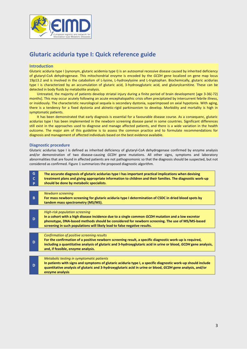
Introduction Glutaric aciduria type I (synonym, glutaric acidemia type I) is an autosomal recessive disease caused by inherited deficiency of glutaryl-CoA dehydrogenase. This mitochondrial enzyme is encoded by the GCDH gene localized on gene map locus 19p13.2 and is involved in the catabolism of L-lysine, L-hydroxylysine and L-tryptophan. Biochemically, glutaric acidurias type I is characterized by an accumulation of glutaric acid, 3-hydroxyglutaric acid, and glutarylcarnitine. These can be detected in body fluids by metabolite analysis.
Untreated, the majority of patients develop striatal injury during a finite period of brain development [age 3-36(-72) months]. This may occur acutely following an acute encephalopathic crisis often precipitated by intercurrent febrile illness, or insidiously. The characteristic neurological sequela is secondary dystonia, superimposed on axial hypotonia. With aging, there is a tendency for a fixed dystonia and akinetic-rigid parkinsonism to develop. Morbidity and mortality is high in symptomatic patients.
It has been demonstrated that early diagnosis is essential for a favourable disease course. As a consequenc, glutaric acidurias type I has been implemented in the newborn screening disease panel in some countries. Significant differences still exist in the approaches used to diagnose and manage affected patients, and there is a wide variation in the health outcome. The major aim of this guideline is to assess the common practice and to formulate recommendations for diagnosis and management of affected individuals based on the best evidence available.
Diagnostic procedure Glutaric acidurias type I is defined as inherited deficiency of glutaryl-CoA dehydrogenase confirmed by enzyme analysis and/or demonstration of two disease-causing GCDH gene mutations. All other signs, symptoms and laboratory abnormalities that are found in affected patients are not pathognomonic so that the diagnosis should be suspected, but not considered as confirmed. Figure 1 summarizes the proposed diagnostic algorithm.
GCP
The accurate diagnosis of glutaric acidurias type I has important practical implications when devising treatment plans and giving appropriate information to children and their families. The diagnostic work-up should be done by metabolic specialists.
B Newborn screening For mass newborn screening for glutaric aciduria type I determination of C5DC in dried blood spots by tandem mass spectrometry (MS/MS).
D
High-risk population screening In a cohort with a high disease incidence due to a single common GCDH mutation and a low excretor phenotype, DNA-based methods should be considered for newborn screening. The use of MS/MS-based screening in such populations will likely lead to false negative results.
D
Confirmation of positive screening results For the confirmation of a positive newborn screening result, a specific diagnostic work-up is required, including a quantitative analysis of glutaric and 3-hydroxyglutaric acid in urine or blood, GCDH gene analysis, and, if feasible, enzyme analysis.
D
Metabolic testing in symptomatic patients In patients with signs and symptoms of glutaric aciduria type I, a specific diagnostic work-up should include quantitative analysis of glutaric and 3-hydroxyglutaric acid in urine or blood, GCDH gene analysis, and/or enzyme analysis
4
Metabolic maintenance treatment Table 1 summarizes a proposed protocol for maintenance treatment.
C
Interdisciplinary team Metabolic treatment should be implemented by an interdisciplinary team that includes metabolic pediatricians, dietitians, and nurses. Parents and patients should have regular training and written treatment protocols.
C
Dietary treatment A low lysine diet (ie reducing lysine intake to a safe requirement) with or without additional administration of lysine-free, tryptophan-reduced amino acids supplements should be used for dietary treatment, in particular in asymptomatic patients up to age 6 years.
C Carnitine supplementation Carnitine should be supplemented in patients with glutaric aciduria type I and should be continued lifelong
Emergency treatment Routine treatment does not reliably protect against striatal injury, so it is crucial to use an intensified emergency treatment protocol during episodes that are likely to induce catabolic state. Tables 2-4 summarizes proposed protocols for emergency treatment.
C
Start of emergency treatment Emergency treatment should start without delay and should be performed aggressively during febrile illness, surgery and immunization within the vulnerable period for acute encephalopathic crises (up to age 6 years)
GCP
Emergency treatment after age 6 years Emergency treatment in children after age 6 years should be considered at least during severe illness. It should be performed similarly to that in the age group 0-6 years with adaptation to the individual.
Management of neurologic complications
GCP
Expert neurological evaluation In all symptomatic patients, expert neurological evaluation should be performed by a neuropediatrician and/or later on by a neurologist to identify clearly the kind and severity of movement disorder. In addition, dietitians, physicotherapists, occupational therapists, orthopedists, seating and speech specialists and providers of communication aids, should be consulted to provide multi-disciplinary support for children with movement disorders.
D
Antidystonic drug treatment Baclofen and benzodiazepines as monotherapy or in combination should be used as first line drug treatment for focal and generalized dystonia. Intrathecal baclofen should be considered as additional therapy for generalized dystonia and spasticity. Trihexyphenidyl should be considered as second line treatment for dystonia, particularly in adolescents and adults. Botulinum toxin A should be considered as additional therapy for severe focal dystonia.
GCP
Antiepileptic drug treatment Diagnosis, choice of antiepileptic drug therapy and management of seizures in GA-I should follow existing guidelines – except for the use of valproate which should be avoided in this condition. The diagnosis of epilepsy and choice of antiepileptic drugs should be made by a pediatric neurologist or pediatrician with expertise in childhood epilepsy. In adulthood, patients with epilepsy should be followed and antiepileptic therapy should be monitored by an adult neurologist.
D Subdural haemorrhage Children with subdural haemorrhage and/or bitemporal arachnoid cysts should be investigated for glutaric aciduria type I, in particular if occuring in combination with macrocephaly and/or a movement disorder.
D Shaken baby syndrome Glutaric aciduria type I should be excluded in children with suspected shaken baby syndrome using the
5
recommendations for selective screening.
D Neurosurgery Neurosurgical intervention for arachnoid cysts and subdural haemorrhages in affected patients should be undertaken very cautiously in close consultation with the pediatric neurosurgeon.
Therapy monitoring Table 5 provides a schedule for biochemical monitoring in glutaric aciduria type I.
GCP
General statement Therapy should be accompanied by regular professional monitoring. Monitoring should be intensified at any age if there are new complications (disease- or therapy-related) or non-adherence to treatment.
D Biochemical monitoring (organic acid analysis) Urine analysis of GA and 3-OH-GA is not informative for therapy monitoring.
D Biochemical monitoring (amino acid analysis) Amino acids in plasma (ideally 3-4 h postprandially) should be monitored during dietary treatment.
D Biochemical monitoring (carnitine status) Carnitine status in plasma should be monitored in all patients to detect secondary carnitine depletion.
GCP
Neuroradiologic investigations Neuroradiologic investigations should be performed in case of neurologic deteroriation but are not generally recommended for regular follow-up monitoring.
Legend
Grades of recommendations
A At least one meta-analysis, systematic review of RCTs, or RCT rated as 1++ and directly applicable to the target population; or A body of evidence consisting principally of studies rated as 1
+, directly applicable to the target population, and demonstrating overall consistency of
results.
B A body of evidence including studies rated as 2
++, directly applicable to the target population, and demonstrating overall consistency of results; or
Extrapolated evidence from studies rated as 1++
or 1+.
C A body of evidence including studies rated as 2+, directly applicable to the target population and demonstrating overall consistency of results; or Extrapolated evidence from studies rated as 2
++.
D Evidence level 3 and 4; or Extrapolated evidence from studies rated as 2+.
Good practice points G CP
Recommended best practice based on the clinical experience of the guideline development group.
6
ANNEXE
Fig 1. Diagnostic algorithm for glutaric aciduria type I A, Newborn screening for glutaric aciduria type I is performed using MS/MS. In low excretor cohorts with known mutations, GCDH gene mutation analysis should be considered as alternative method (dotted line). Treatment should be started after the identification of two disease-causing mutations (*). B, Selective screening should be initiated if the diagnosis of GA-I is suspected clinically or there is a positive family history. Note that a few patients with a low-excreting phenotype may show (intermittently) normal urinary excretion of 3-hydroxyglutaric acid (and glutaric acid). If an individual shows normal 3-hydroxyglutaric acid (and glutaric acid) concentrations in urine (or other body fluids) but presents with highly suspicious signs and symptoms for glutaric aciduria type I, further diagnostic studies should be considered (**).
7
Table 1. Metabolic maintenance treatment (protocol proposed by GDG) If normal growth and development are not achieved these recommendations should be modified according to individual needs.
Age
Treatment 0–6 months 7–12 months
1–3 years 4–6 years > 6 years
1. Low lysine diet Lysine (from natural protein)
a
mg/kg per day
100 90 80-60 60-50 Avoid excessive intake of natural protein; use natural protein with a low lysine content; according to ‘safe levels’ (Suppl. Table 6)
Amino acid mixtures (protein)
b
g/kg per day
1.3-0.8 1.0-0.8 0.8 0.8
Energy kcal/kg per day
115-80 95-80 95-80 90-80
2. Micronutrientsc % 100 100 100 100 100
3. Carnitine mg/kg per day
100 100 100 100-50 30-50
aThe lysine/protein ratio varies considerably in natural food and thus natural protein intake in children on a low
lysine diet is dependent on the natural protein source. The natural protein intake is relatively high if patients predominantly use sources of natural protein with a low lysine content.
bLysine-free, tryptophan-reduced amino acid mixtures should be supplemented with minerals and
micronutrients as required to maintain normal levels. Adequate intake of essential amino acids is provided from natural protein and lysine-free, tryptophan-reduced amino acid supplements. The amount of amino acid supplements is adjusted to reach at least the ‘safe levels’.
cAccording to international dietary recommendations.
8
Table 2. Strategies to prevent delayed start of emergency treatment
Topic Proposed strategies to avoid delay
Education and training of parents
Parents should be informed in detail about the natural history and the particular risks of glutaric aciduria type I, in particular the manifestation and neurological sequels of an acute encephalopathic crisis. They should be instructed precisely about the management of maintenance and emergency treatment and this knowledge should be reinforced during regular visits at a metabolic center.
Treatment protocols
Written protocols for maintenance and emergency treatment should be given to all who may be involved (parents, metabolic centers, local hospitals) and kept updated. Parents should also receive an emergency card (preferably laminated) summarizing the key information on glutaric aciduria type I and basic principles of treatment. The telephone number of the responsible metabolic center/physician should be written on the protocol and the emergency card. Parents should take their emergency instructions and supplies of maltodextran and AA supplements when going to hospital.
Supplies Parents should be advised always to maintain adequate supplies of specialized dietetic products (maltodextran, lysine-free, tryptophan-reduced amino acid mixtures) and drugs required for maintenance treatment and emergency treatment at home.
Local hospital or pediatrician
The closest hospital/pediatrician should be clearly instructed if glutaric aciduria type I has been newly diagnosed in a child. Key information (including written treatment protocols) should be provided to the local hospital/pediatrician without delay and before inpatient emergency treatment might be necessary. Inpatient emergency treatment should be started immediately in the closest hospital if necessary and follows the supervision of the responsible metabolic center which should be contacted without delay.
Holidays Metabolic specialists/centers in the vicinity of the holiday resort should be informed in writing about the disease and the recent treatment before the start of the holidays. The emergency card and treatment protocols should be translated before the start of the holidarys if necessary.
Infectious diseases During infectious disease the responsible metabolic center/metabolic specialists should be informed (by parents or local hospitals/pediatricians) without delay to allow supervision of the emergency management. Parents should be instructed to call their doctor and/or metabolic consultant as soon as a temperature of 38.5 °C is noted and an intercurrent illness is suspected either, an upper respiratory infection, gastrointestinal infection or if increased irritability develops.
Vomiting/ diarrhoea
Vomiting and diarrhoea is particularly dangerous – even in the absence of fever. Please follow the recommendations for “infectious diseases” (see above).
Surgery If a surgical intervention is planned, the responsible metabolic center/specialist should be informed before such interventions to discuss the specific risks of affected patients with surgeons and anaesthesiologists, to recommend a protocol for the postsurgical metabolic management and to allow supervision of this period. If possible, the postsurgical metabolic management should be performed in a metabolic center. In general, fasting should be avoided, glucose infusions applied, and carnitine dosage doubled.
9
Table 3. Emergency treatment at home (protocol proposed by GDG)
B. Protein intake Natural protein Stop for 24 to a maximum of 48 hours, then reintroduce and increase stepwise until
the amount of maintenance treatment is reached within 48 (-72) hours. Prolongation of inadequately low protein intake increases the risk of protein catabolism.
AA mixturesb If tolerated, amino acid mixtures should be administered according to maintenance
therapy (see also Table 1)
C. Pharmacotherapy L-Carnitine Double carnitine intake: eg 200 mg/kg per day p.o. in infants Antipyretics If body temperature raises above 38.5 °C (101 F), antipyretics, such as ibuprofen or
paracetamol (each 10-15 mg/kg per single dose, 3-4 doses daily, maximum daily dose 60 mg/kg body weight) should be administered.
aSolutions should be administered every 2 hours day and night.
bIf neonates and infants also receive a lysine-free, tryptophan-reduced amino acid supplement, this can be
continued but should be fortified by maltodextran. Patients should be re-assessed every 2 hours.
10
Table 4. Emergency treatment in hospital (protocol proposed by GDG)
A. Intravenous infusions Glucose Age (years) Glucose (g/kg per day IV)
0-1 (12-) 15
1-3 (10-) 12
3-6 (8-) 10
6-10 (6-) 8
>10 3-6
Insulin If persistent hyperglycemia > 150 mg/dL (> 8 mmol/L) and/or glucosuria occurs, start with 0.05 IE insulin/kg per h IV and adjust the infusion rate according to serum glucose
B. Protein intake Natural protein Stop for 24 to a maximum of 48 hours, then reintroduce and increase stepwise until
the amount of maintenance treatment is reached within 48 (-72) hours. Prolongation of inadequately low protein intake increases the risk of protein catabolism.
AA mixturesa If tolerated, lysine-free and tryptophan-reduced amino acid mixtures should be
administered orally or by nasogastric tube according to maintenance therapy (see also Table 1).
C. Pharmacotherapy L-Carnitine 100 (-200) mg/kg per day i.v. Antipyretics If body temperature rises above 38.5 °C (101 F), antipyretics, such as ibuprofen or
paracetamol (each 10-15 mg/kg per single dose, 3-4 doses daily, maximum daily dose 60 mg/kg body weight) should be administered.
Sodium bicarbonate If acidosis; alkalination of urine also facilitates urinary excretion of organic acids D. Monitoring Blood Glucose, blood gases, electrolytes, calcium, phosphate, complete blood cell count,
Urine Ketone bodies, pH Vital signs Heart rate, blood pressure, temperature, diuresis; Glasgow Coma Scale if reduced
consciousness; assessment for neurological signs (hypotonia, irritability, rigor, dystonia)
aLysine-free, tryptophan-reduced amino acid mixtures should be supplemented with minerals and
micronutrients.
bDuring the recovery phase.
cIn severe illness to detect pancreatitis (amylase/lipase) or rhabdomyolysis (creatine kinase).
11
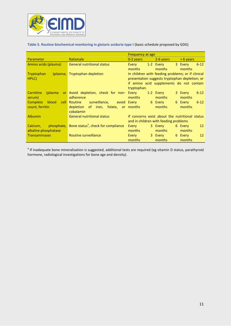
Table 5. Routine biochemical monitoring in glutaric aciduria type I (basic schedule proposed by GDG)
Frequency at age
Parameter Rationale 0-2 years 2-6 years > 6 years
Amino acids (plasma) General nutritional status Every 1-2 months
Every 3 months
Every 6-12 months
Tryptophan (plasma; HPLC)
Tryptophan depletion In children with feeding problems; or if clinical presentation suggests tryptophan depletion; or if amino acid supplements do not contain tryptophan.
Carnitine (plasma or serum)
Avoid depletion, check for non-adherence
Every 1-2 months
Every 3 months
Every 6-12 months
Complete blood cell count, ferritin
Routine surveillance, avoid depletion of iron, folate, or cobalamin
Every 6 months
Every 6 months
Every 6-12 months
Albumin General nutritional status If concerns exist about the nutritional status and in children with feeding problems
Calcium, phosphate, alkaline phosphatase
Bone statusa, check for compliance Every 3
months Every 6 months
Every 12 months
Transaminases Routine surveillance Every 3 months
Every 6 months
Every 12 months
a If inadequate bone mineralisation is suggested, additional tests are required (eg vitamin D status, parathyroid
hormone, radiological investigations for bone age and density).
12
According to: Stefan Kölker, Ernst Christensen, James V. Leonard, Cheryl R. Greenberg, Avihu Boneh, Alberto B. Burlina, Alessandro P. Burlina, Marjorie Dixon, Marinus Duran, Angels García Cazorla, Stephen I. Goodman, David M. Koeller, Mårten Kyllerman, Chris Mühlhausen, Edith Müller, Jürgen G. Okun, Bridget Wilcken, Georg F. Hoffmann, and Peter Burgard. Diagnosis and management of glutaric aciduria type I – revised recommendations. Journal of Inherited Metabolic Diseases 2011; 34: 677 – 694.
13
Methylmalonic and Propionic acidurias: Quick reference guide
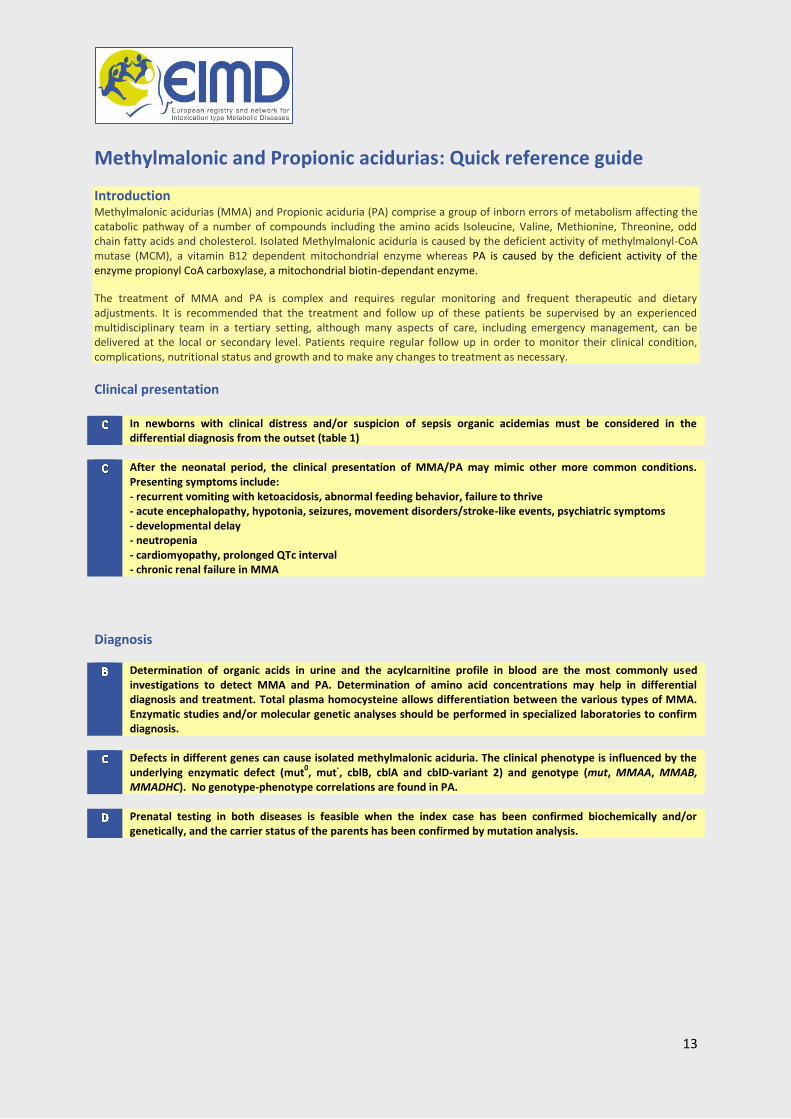
Introduction Methylmalonic acidurias (MMA) and Propionic aciduria (PA) comprise a group of inborn errors of metabolism affecting the catabolic pathway of a number of compounds including the amino acids Isoleucine, Valine, Methionine, Threonine, odd chain fatty acids and cholesterol. Isolated Methylmalonic aciduria is caused by the deficient activity of methylmalonyl-CoA mutase (MCM), a vitamin B12 dependent mitochondrial enzyme whereas PA is caused by the deficient activity of the enzyme propionyl CoA carboxylase, a mitochondrial biotin-dependant enzyme.
The treatment of MMA and PA is complex and requires regular monitoring and frequent therapeutic and dietary adjustments. It is recommended that the treatment and follow up of these patients be supervised by an experienced multidisciplinary team in a tertiary setting, although many aspects of care, including emergency management, can be delivered at the local or secondary level. Patients require regular follow up in order to monitor their clinical condition, complications, nutritional status and growth and to make any changes to treatment as necessary.
Clinical presentation
In newborns with clinical distress and/or suspicion of sepsis organic acidemias must be considered in the differential diagnosis from the outset (table 1)
After the neonatal period, the clinical presentation of MMA/PA may mimic other more common conditions. Presenting symptoms include: - recurrent vomiting with ketoacidosis, abnormal feeding behavior, failure to thrive - acute encephalopathy, hypotonia, seizures, movement disorders/stroke-like events, psychiatric symptoms - developmental delay - neutropenia - cardiomyopathy, prolonged QTc interval - chronic renal failure in MMA
Diagnosis
Determination of organic acids in urine and the acylcarnitine profile in blood are the most commonly used investigations to detect MMA and PA. Determination of amino acid concentrations may help in differential diagnosis and treatment. Total plasma homocysteine allows differentiation between the various types of MMA. Enzymatic studies and/or molecular genetic analyses should be performed in specialized laboratories to confirm diagnosis.
Defects in different genes can cause isolated methylmalonic aciduria. The clinical phenotype is influenced by the underlying enzymatic defect (mut
0, mut
-, cblB, cblA and cblD-variant 2) and genotype (mut, MMAA, MMAB,
MMADHC). No genotype-phenotype correlations are found in PA.
Prenatal testing in both diseases is feasible when the index case has been confirmed biochemically and/or genetically, and the carrier status of the parents has been confirmed by mutation analysis.
14
Acute management
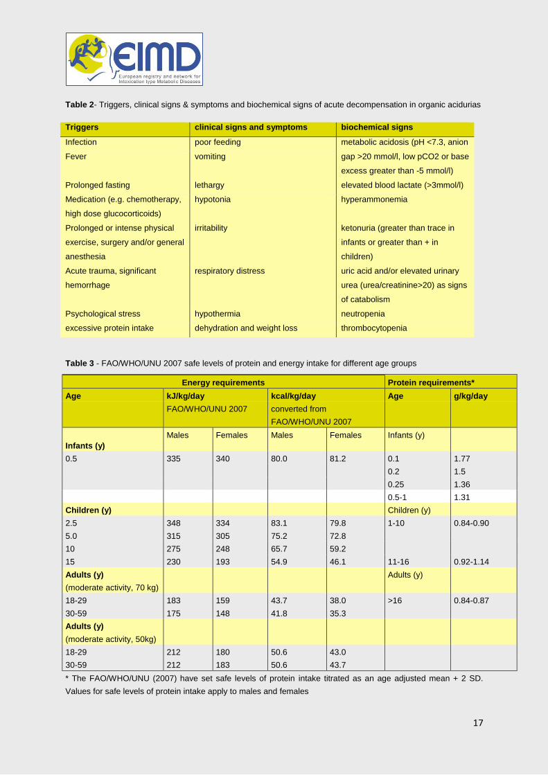
The presence of any one or more of certain clinical or biochemical signs (Table 1) compared to the individual patient’s baseline should trigger further evaluation and potential adjustment of therapy and monitoring in order to prevent complications (Table 2). Any acute illness warrants closer monitoring and follow up.
During mild illness and without gastrointestinal symptoms, home enteral emergency feeding management is appropriate. There should be provision of adequate energy to meet increased metabolic demands and prevent endogenous protein catabolism.
Intolerance or refusal of emergency feeds warrants hospitalization for intravenous therapy. Intravenous fluids containing glucose should be infused and insulin may be used to promote anabolism; lipid emulsion should be commenced early to provide additional calories. Following improvement of metabolic and clinical abnormalities, protein should be rapidly reintroduced. Enteral feeding should be started as soon as possible. Parenteral nutrition is indicated, when enteral feeding cannot be established within 24-48h.
Extracorporeal detoxification may be required in severely decompensated patients
Long-term management
The most common medical treatments besides the diet used in long-term treatment of MMA/PA are L-carnitine, antibiotics to reduce intestinal flora and vitamin B12.
Response to vitamin B12 should be assessed in every MMA patient. For responders hydroxocobalamin should be used as long-term treatment.
Chronic hyperammonaemia indicates metabolic imbalance and requires investigation and treatment of the underlying cause. Sodium benzoate has been used to treat chronic hyperammonaemia in MMA/PA patients.
Drugs containing propionate, valproate, pivalic acid, nephrotoxic drugs and chemotherapy agents should be avoided or used with great caution in patients with MMA/PA. Medications known to prolong the QTc-interval (such as prokinetic drugs) should be avoided if possible. Steroids administered by a systemic route should be avoided if possible, or if unavoidable, should be used with caution. Inhaled steroids seem safe.
Dietary management of MMA/PA aims at metabolic stability and normal growth. It is based on adequate energy supply combined with avoidance of prolonged fasting and reduced intake of precursor amino acids through a restricted natural protein diet, commonly supplemented with precursor-free synthetic amino acids. The WHO/FAO/ UNU (2007) safe levels of protein intake provide a useful guide for protein prescription (table 3).
MMA/PA precursors free amino acid supplements should form part of the total protein intake if natural protein tolerance is below WHO/UNU/FAO (2007) safe levels of protein intake.
Tube feeding may be necessary to avoid catabolism/prolonged fasting, achieve nutritional adequacy, administer medications and supplements and maintain metabolic stability.
Liver and/or kidney transplantation has been used as an alternative therapy to conventional medical treatment in MMA and PA patients. Transplantation should be considered in patients with frequent metabolic decompensations where the clinical condition is difficult to stabilize. Transplantation only partially corrects the enzymatic defect; renal and neurological complications may still occur afterwards.
The management of the long-term renal complications is based on adequate hydration, drug therapy, hemo- or peritoneal dialysis and ultimately kidney transplantation.
15
Follow up
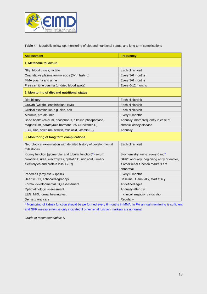
Regular follow up with clinical, biochemical and dietary monitoring is essential to prevent nutritional imbalance and to detect and treat long-term complications (Table 4).
Despite intensive medical treatment, MMA and PA are associated with a high frequency of intellectual disability. Intellectual abilities and cognitive development should be assessed early and reliably to allow timely referral for appropriate intervention.
Neurological examination with detailed history of developmental milestones should be a routine part of evaluation in every visit to the metabolic clinic. Input from a pediatric neurologist should be considered in case of acute neurological presentation (encephalopathy/coma, seizures), any suspicion of developmental delay, spasticity/dystonia and movement disorder, epilepsy, hearing or visual field defects..
Ophthalmologic assessment (fundoscopy, visual acuity and visual field) and a formal examination by an ophthalmologist to exclude optic neuropathy should be a part of routine evaluation at baseline, at any time of concern and yearly after 6 years of age.
One of the most severe long-term complications in MMA is chronic kidney disease (CKD). CKD is characterized by progressive functional abnormality leading to chronic renal failure. Regular measurements of glomerular filtration rate and plasma and urinary MMA are recommended as parameters in the follow-up of patients with MMA. With declining kidney function urinary MMA ceases to be a reliable marker and should be replaced by plasma MMA. Plasma creatinine levels may be less reliable in these patients due to the poor muscle mass and the low protein intake.
Cardiac complications include cardiomyopathy and prolonged QTc interval which may be life-threatening and may occur in MMA and PA patients with increasing frequency with age. Therefore ECG and heart ultrasound (echocardiography) are recommended every year. If cardiomyopathy or long QTc is present standard cardiac therapy should be undertaken and metabolic treatment and monitoring should be optimized. .
Acute, recurrent acute and chronic pancreatitis may develop in MMA and PA, independently from metabolic decompensations and metabolic control. Diagnosis and management of acute or chronic pancreatitis follows the same principles as in any other case of pancreatitis..
Pancytopenia (especially neutropenia) is frequent in MMA and PA and may respond to improved metabolic control. Evaluation with a low threshold to treat infections as well as infection control practices should be performed according to institutional guidelines early in neutropenic patients. .
16
Table 1 – Bedside differential diagnostics of inborn errors of metabolism presenting with acute encephalopathy
Heart (ECG, echocardiography) Baseline annually, start at 6 y
Formal developmental / IQ assessment At defined ages
Ophthalmologic assessment Annually after 6 y
EEG, MRI, formal hearing test If clinical suspicion / indication
Dentist / oral care Regularly
* Monitoring of kidney function should be performed every 6 months in MMA; in PA annual monitoring is sufficient
and GFR measurement is only indicated if other renal function markers are abnormal
Grade of recommendation: D
19
Legend
Grades of recommendations
A At least one meta-analysis, systematic review of RCTs, or RCT rated as 1++ and directly applicable to the target population; or A body of evidence consisting principally of studies rated as 1
+, directly applicable to the target population, and demonstrating overall consistency of
results.
B A body of evidence including studies rated as 2
++, directly applicable to the target population, and demonstrating overall consistency of results; or
Extrapolated evidence from studies rated as 1++ or 1+.
C A body of evidence including studies rated as 2+, directly applicable to the target population and demonstrating overall consistency of results; or Extrapolated evidence from studies rated as 2++.
D Evidence level 3 and 4; or Extrapolated evidence from studies rated as 2
+.
Good practice points G CP
Recommended best practice based on the clinical experience of the guideline development group.
Guideline development group: Matthias R. Baumgartner (lead), Friederike Hörster, Carlo Dionisi-Vici, Goknur Haliloglu, Daniela Karall, Kimberly A. Chapman, Martina Huemer, Michel Hochuli, Murielle Assoun, Diana Ballhausen, Alberto Burlina, Brian Fowler, Sarah C. Grünert, Stephanie Grünewald, Tomas Honzik, Begoña Merinero, Celia Pérez-Cerdá, Sabine Scholl-Bürgi, Flemming Skovby, Frits Wijburg, Anita MacDonald, Diego Martinelli, Jörn Oliver Sass, Vassili Valayannopoulos, and Anupam Chakrapani
20
Urea cycle disorders: Quick reference guide
Introduction Urea cycle disorders (UCDs) are a group of inborn errors of metabolism affecting the detoxification of nitrogen and the endogenous synthesis of arginine. The incidence of UCDs is about 1 in 35.000. Patients with a complete enzyme deficiency often present during the first days of life with hyperammonemic coma with about 50% mortality despite early and aggressive treatment. The majority of survivors of a neonatal presentation suffer from severe developmental delay and a high risk of recurrent crises. Late-onset patients may present at any age after the neonatal period and their risk of death is also high (up to about 45%). Brain damage correlates with duration and severity of acute hyperammonemia, especially in neonatal patients. Therefore, at all ages patients must be identified as soon as possible and should be transferred to a metabolic centre very early during the course. These guidelines are a consensus from several European countries on how to diagnose and treat patients with a suspicion of UCD or confirmed UCD.
Clinical presentation*
Table 1 (below) provides clinical signs and symptoms of acute and chronic manifestations of UCDs irrespective of patient age D Clinical signs and symptoms of UCDs are non-specific and a high degree of awareness is required. Key questions
have to be asked and a detailed family history (pedigree) is mandatory. D UCDs may cause acute or chronic symptoms at any age. Most common signs are neurological and psychiatric,
others are uncommon and few are only described in single patients.
D UCDs must be included in the differential diagnosis of any acute unexplained encephalopathy or acute psychiatric
illness in patients of any age. This should always prompt immediate determination of plasma ammonia.
D Any cause of protein catabolism, protein load and some drugs may trigger hyperammonemia in UCD patients.
Diagnosis*
Figure 1 (below) provides a diagnostic algorithm for neonatal hyperammonemia Table 2 (below) summarizes the bedside differential diagnosis of IEMs presenting with hyperammonemia
D Ammonia determination is an emergency procedure and the result must be available within 30 minutes. If ammonia is increased, further metabolic investigations should be performed immediately but specific treatment must not be delayed. Ammonia measurement is recommended in patients of any age presenting with unusual or unexplained neurological illness or unexplained liver failure. Moreover, as soon as the clinician suspects any intoxication, an inborn error of metabolism should be considered.
C Mutation analysis is the method of choice to establish the diagnosis of a UCD and to offer genetic counseling.
D Enzyme analysis of UCDs is feasible but not the method of first choice if genetic testing is available. Liver tissue
and some other tissues can be used for enzyme analysis of UCDs. In deceased patients with a suspicion of UCD, fibroblasts and/or liver tissue should be preserved.
D Prenatal testing requires careful counselling by human geneticists and metabolic specialists jointly. Various
methods for prenatal testing are available comprising investigations of metabolites as well as enzyme and mutation analyses. The preferred method for all disorders is molecular genetic analysis.
C Newborn screening (NBS) for deficiencies of ASS, ASL, and arginase should be seriously considered. Currently NBS
cannot be recommended for deficiencies of NAGS, CPS1 and OTC.
Acute management*
Table 3 (below) lists levels of hyperammonemia and suggested actions in case of symptomatic patients Table 4 (below) gives dosages of drugs to be used in acute hyperammonemia and acute decompensations of UCDs Table 5 (below) provides an emergency regime for protein-free feeding in infants and children
D The early clinical suspicion and diagnosis of hyperammonemia is crucial for a favourable outcome. The start of ammonia detoxification and measures to reverse catabolism must not be delayed unless a decision for only palliative care is made.
21
C Total coma duration and peak ammonia levels are the most relevant factors for the prognosis of
hyperammonemic decompensations. To better understand all contributing factors, more studies on the outcome of hyperammonemia are needed.
C In neonates and children with symptomatic hyperammonemia, dialysis should be started if there is no response
within four hours after start of medical treatment. The method of choice for ammonia detoxification is hemodiafiltration. Peritoneal dialysis is the least effective method and should only be used as a last resort. Exchange transfusion should not be used. Extracorporeal detoxification is the first line treatment in acute hyperammonemic decompensations in adults.
D For treatment of acute hyperammonemia, it is crucial to promote and maintain anabolism. This is best achieved
by high dose glucose plus, if a fatty acid oxidation disorder has been excluded, lipids. Protein should be (re)started after an acute hyperammonemic decompensation when ammonia levels fall to < 100 μmol/L. The period of protein free nutrition should aim not to exceed 24-48 hours.
Long-term management*
Table 6 (below) gives dosages of drugs to be used perorally for long-term treatment of UCDs D UCD patients usually need restriction of protein intake. This needs to be individually determined, based on
tolerance. The FAO/WHO recommendations can be used as a guide for protein prescription. C Essential amino acids or branches chain amino acids supplements may form part of the dietary treatment.
D Dietary treatment of UCD patients is one of the cornerstones of therapy and needs to be largely individualised.
The fine balance between provision of nutritional requirements and metabolic stability warrants a particular expertise and a specialist metabolic dietician should always be involved.
C Use of nitrogen scavengers seems to be safe at recommended doses but there is a need for more controlled
studies on the adverse effects of sodium benzoate and sodium phenylbutyrate.
C All UCD patients should be monitored for plasma arginine levels. Most UCD patients (apart from
hyperargininemia) will need a supplementation of L-arginine. D N-carbamylglutamate is the first line medication for treatment of N-acetylglutamate synthase deficiency and
might also be used as an emergency drug during acute neonatal hyperammonemia of unknown etiology.
C Liver transplantation is the only cure of a UCD, it allows a normal diet and avoids the need for alternative
pathway therapy. Liver transplantation is recommended for patients with severe neonatal onset UCDs and may be considered as first-line treatment. Liver transplantation is indicated also for patients with progressive liver disease, e.g. in argininosuccinate lyase deficiency and for UCD patients suffering from recurrent metabolic decompensations and hospitalizations despite medical therapy. Liver transplantation should ideally be sought for before the onset of irreversible neurological damage and/or repeated crises. Generally, it should be performed between 3 and 12 months of age.
Follow up* D Clinical, biochemical and nutritional monitoring are essential and should follow an individualised plan. D Regular testing for IQ, development and specific abilities/weaknesses is recommended. Health related quality of
life, anxiety, stress and psychosocial factors are meaningful outcome parameters. Psychological management is an important additional task in caring for patients with UCDs and their families.
*Legend: Grades of recommendations
A At least one meta-analysis, systematic review of RCTs, or RCT rated as 1++ and directly applicable to the target population; or A body of evidence consisting principally of studies rated as 1
+, directly applicable to the target population, and demonstrating overall consistency of results.
B A body of evidence including studies rated as 2++, directly applicable to the target population, and demonstrating overall consistency of results; or Extrapolated evidence from studies rated as 1++ or 1+.
C A body of evidence including studies rated as 2+, directly applicable to the target population and demonstrating overall consistency of results; or Extrapolated evidence from studies rated as 2++.
D Evidence level 3 and 4; or Extrapolated evidence from studies rated as 2+.
22
Figure 1: Diagnostic algorithm for neonatal hyperammonemia
Investigations in plasma if not stated otherwise: U = urine
Table 1: Clinical signs and symptoms of acute and chronic manifestations of UCDs irrespective of patient age
Acute presentation Chronic presentation
Altered level of consciousness (from lethargy and somnolence to coma) mimicking encephalitis or drug intoxication
Acute encephalopathy (see below)
Seizures (in general seizures do not present isolated but within the context of altered level of consciousness)
Ataxia: in general associated with altered level of consciousness
Stroke-like episodes
Transient visual loss
Vomiting and progressive poor appetite
Liver failure
Multiorgan failure
Peripheral circulatory failure
“Post-partum psychosis”
Psychiatric symptoms (hallucinations, paranoia, mania, emotional or personality changes)
In neonates: sepsis-like picture, temperature instability, respiratory distress, hyperventilation
Specific neuropsychological phenotype in heterozygous OTC females
Episodic character of signs and symptoms
bold: typical signs and symptoms; standard: uncommon signs and symptoms; italics: signs and symptoms only reported in single patients
Hyperammonemia
Glutamine
Low-normal
Glutamine
Elevated
Preterm Full TermCitrulline
Citrulline
Citrulline
Citrulline
THAN
Normal AA
profile
Organic acid
Acylcarnitines
Citrulline
Organic acidurias
MMA – PA
Acidosis
FAO defectsMCAD – VLCAD - TPD
HypoglycemiaCIT 2
Met – Tyr
A-FP- galactose
PC deficiency
Lactic acidosis
U Orotic acid
normal
U Orotic acid
elevated
NAGS D
CPS D
P5CS D
Orn, Pro, Arg
starved
hyper-NH3
Ornithine
Elevated
Ornithine
Normal
HHH s
Homocit
Arg
Ala
OTC D
Arg
ARGININEMIA
Orn only
transiently normal
OAT D
Arg, Lys, Orn
U Arg, Lys, Orn
U Orotic acid
LPI
ASA
U Orotic acid
ASL D
U Orotic acid
ASS D
Hypoglycemia
HI-HA
HMG
23
Table 2: Bedside differential diagnosis of IEMs presenting with hyperammonemia
Parameter
Condition
UCDs Organic acidurias
-Oxidation defects
Hyperinsulinism/ hyperammonemia syndrome
Pyruvate carboxylase deficiency
g
Acidosis /– e /– –
Ketonuriaa – – –
Hypoglycemiab – /–
Lactic acidc – /– –
AST/ALT ()d – – /–
CPK – – – –
Uric acid – – –
WBC/RBC/Plt – – – –
Weight loss – f – –
a In neonates ketonuria (++ - +++) suggests organic aciduria. b Hypoglycemia and hyperammonemia (“pseudo-Reye”) can be predominant manifestations of the organic aciduria 3-HMG-CoA-lyase deficiency. c Blood lactate >6 mmol/L, since lower high lactate levels (2-6 mM) may be due to violent crying or to extensive muscle activity. d AST/ALT elevations can be found but are not constant in UCDs. e Can be absent in neonates. f Occurrence only in neonates. g Only type B associated with hyperammonemia but not types A and C.
Table 3 Levels of hyperammonemia and suggested actions in case of symptomatic patients
Ammonia level (µmol/L)
Action in undiagnosed patient Action in known UCD patient Comments
Increased > upper limit of normal
Stop protein intake Give IV glucose at an appropriate
dosage to prevent catabolism (10 mg/kg/min in a neonate) ± insulin$
Monitor ammonia blood levels every 3 hours
Stop protein intake Give IV glucose at an appropriate dosage
to prevent catabolism (10 mg/kg/min in a neonate) ± insulin$
Monitor ammonia blood levels every 3 hours
Stop protein for max 24 – 48 h Avoid exchange transfusions as
cause of catabolism Hyperglycemia can be
extremely dangerous (hyperosmolarity)
If major hyperglycemia occurs with high lactate (>3 mmol/L) reduce glucose infusion rate rather than increase insulin
Avoid hypotonic solutions Add sodium and potassium
according to the electrolyte results
Take into account the sodium intake if sodium benzoate or sodium PBA are used§
L-arginine not to be given in ARG1D
Some concerns of sodium benzoate use in OAs
Avoid repetitive drug boluses Monitor phosphate levels and
supplement early especially during hemodialysis
In addition if >100 and <250 #
Start drug treatment with IV L-arginine and sodium benzoate (see Table 4)
Start carbamylglutamate, carnitine, vitamin B12, biotin (see Table 4 and its legend)
Continue drug treatment with L-arginine (plus continue or add L-citrulline for mitochondrial UCDs) and sodium benzoate ± sodium PBA/phenylacetate* (see Table 4), increase dose or give IV
Consider NG carbohydrate and lipid emulsions unless the child is vomiting (enables higher energy intake)
In addition if 250 to 500
As above Prepare hemo(dia)filtration if
significant encephalopathy and/or early high blood ammonia level or very early onset of disease (day 1 or 2)
Begin hemo(dia)filtration if no rapid drop of ammonia within 3-6 hours
As above, but all drugs per IV Prepare hemo(dia)filtration if significant
encephalopathy and/or early high blood ammonia level or very early onset of disease (day 1 or 2)
Begin hemo(dia)filtration if no rapid drop of ammonia within 3-6 hours
In addition if 500 to 1000
As above Start hemo(dia)filtration immediately
As above Start hemo(dia)filtration as fast as possible
In addition if >1000
Evaluate whether to continue specific treatment or to start palliative care
Evaluate whether to aim at curative treatment or palliative care
*If available, an IV equimolar solution of sodium benzoate and sodium phenylacetate can be used: 250 mg/kg as bolus IV/90-120 min, then 250 mg/kg as continuous IV infusion over 24h. The combination of sodium-benzoate and sodium phenylacetate is available as a drug, registered by the FDA (available in the EU on Named Patient Basis) and indicated as adjunctive therapy for the treatment of acute hyperammonemia and associated encephalopathy in patients with deficiencies in enzymes of the urea cycle. # This limit of action applies for patients outside the neonatal period; for neonates use >150 and <250. $Monitor blood glucose after 30 min and subsequently every hour, because some neonates are very sensitive to insulin.
24
§1g sodium benzoate and sodium PBA contain 7 mmol Na and 5.4 mmol Na, respectively.
Table 4: Dosages of drugs to be used in acute hyperammonemia and acute decompensations of UCDs
Disorder
Sodium benzoate (to be given IV in glucose 10%)
Sodium PBA/Sodium phenylacetate (to be given IV in glucose 10%)
L-arginine hydrochloride (to be given IV in glucose 10%)
N-carbamylglutamate (only available as oral/enteral drug)
Undiagnosed patient°
250 mg/kg as bolus in 90-120 min, then maintenance 250-500 mg/kg/d$
> 20 kg bw: 5.5 g/m2/d
250 mg/kg as bolus in 90-120 min, then maintenance: 250-500mg/kg/d$
250(-400) mg/kg (1-2 mmol/kg) as bolus in 90-120 min, then maintenance 250 mg/kg/d (1.2 mmol/kg/d)
100 mg/kg bolus per NG tube then 25-62.5 mg/kg every 6h
NAGSD same -
250 mg/kg (1.2 mmol/kg) as bolus in 90-120 min, then maintenance 250 mg/kg/d (1.2 mmol/kg/d)
same
CPS1D & OTCD same 250 mg/kg as bolus in 90-120 min, then maintenance: 250(-500) mg/kg/d$
same -
ASSD same same same -
ASLD‡ same 250 mg/kg as bolus in 90-120 min, then maintenance: 250 mg/kg/d$
200-400 mg/kg (1-2 mmol/kg) as bolus in 90-120 min, then maintenance 200-400 mg/kg/d (1-2 mmol/kg/d)
-
ARG1D* same - AVOID -
HHH syndrome same - - -
°In undiagnosed patients, consider additional use of carnitine 100 mg/kg IV, hydroxycobalamin 1 mg IM/IV, and biotin 10 mg IV/PO *The risk for acute hyperammonemic decompensation is low in ARG1 deficiency §If citrulline is given, there is usually no need for concomitant use of L-arginine $If on hemodialysis/hemodiafiltration doses should be increased to 350 mg/kg/d (maintenance dose) ‡In ASL deficiency, L-arginine therapy for acute decompensations might be sufficient for some patients Maximal daily drug dosages: sodium benzoate 12 g/d, sodium PBA 12 g/d, L-arginine 12 g/day Cave: The doses indicated in Table 4 can be used at the start of treatment but must be adapted depending on plasma ammonia and amino acids. Sodium benzoate and sodium PBA/phenylacetate should be given in parallel in severe acute decompensation. In less severe cases, a step-wise approach with initial sodium benzoate and if hyperammonemia persists or worsens, the addition of sodium PBA/phenylacetate can be chosen.
Table 5: Emergency regime for protein-free feeding in infants and children
Age Glucose polymer concentration % CHO
Energy/100ml Kcal kJ
Suggested daily fluid volume ml/kg
Feeding frequency
up to 6 m 10 40 167 150 ml/kg 2 to 3 hourly oral/bolus day and night or continuous tube feeds using enteral feeding pump
7-12 m 10-15 48 202 120 ml/kg
1 y 15 60 250 1200 ml
2-9 y 20 80 334 *
>10 y 25 100 418 *
* For children > 10 kg normal fluid requirements can be calculated as: 11-20 kg: 100 ml/kg for the first 10 kg, plus 50 ml/kg for the next 10 kg 20 kg and above: 100 ml/kg for the first 10 kg, plus 50 ml/kg for the next 10 kg, plus 25 ml/kg thereafter up to a maximum of 2500 ml/day
25
Table 6: Dosages of drugs to be used perorally for long-term treatment of UCDs
HHH syndrome same same <20 kg: 100-200* mg/kg/d >20 kg: 2.5-6 g/m2/d max. 6 g/d
100-250§ max. 6 g/d
-
All medications should be divided into three to four doses daily taken with meals and distributed as far as possible throughout the day. ° PBA is the second choice drug for long-term treatment and should be given in patients not responsive to benzoate alone * serum/plasma levels of benzoate/PBA and plasma levels of arginine should be monitored # in some patients higher doses are needed (the US FDA studies consider doses up to 13 g/m2/d), according to expert advice § if citrulline is given, there is usually no need for concomitant use of L-arginine $ 100 mg equal 0.694 mmol sodium benzoate; 0.537 mmol sodium PBA; 0.475 mmol arginine hydrochloride; 0.574 mmol arginine base; 0.571 mmol citrulline; 0.532 mmol carbamylglutamate, respectively
1
Isovaleric Acidemia: Quick reference guide
Introduction
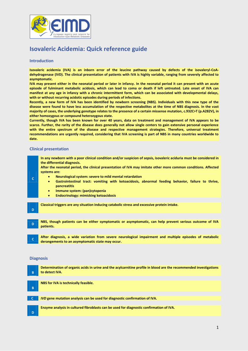
Isovaleric acidemia (IVA) is an inborn error of the leucine pathway caused by defects of the isovaleryl-CoA-dehydrogenase (IVD). The clinical presentation of patients with IVA is highly variable, ranging from severely affected to asymptomatic. IVA may present either in the neonatal period or later in infancy. In the neonatal period it can present with an acute episode of fulminant metabolic acidosis, which can lead to coma or death if left untreated. Late onset of IVA can manifest at any age in infancy with a chronic intermittent form, which can be associated with developmental delays, with or without recurring acidotic episodes during periods of infections. Recently, a new form of IVA has been identified by newborn screening (NBS). Individuals with this new type of the disease were found to have less accumulation of the respective metabolites at the time of NBS diagnosis. In the vast majority of cases, the underlying genotype relates to the presence of a certain missense mutation, c.932C>T (p.A282V), in either homozygous or compound heterozygous state. Currently, though IVA has been known for over 40 years, data on treatment and management of IVA appears to be scarce. Further, the rarity of the disease does generally not allow single centers to gain extensive personal experience with the entire spectrum of the disease and respective management strategies. Therefore, universal treatment recommendations are urgently required, considering that IVA screening is part of NBS in many countries worldwide to date.
Clinical presentation
C
In any newborn with a poor clinical condition and/or suspicion of sepsis, isovaleric aciduria must be considered in the differential diagnosis. After the neonatal period, the clinical presentation of IVA may imitate other more common conditions. Affected systems are:
Neurological system: severe to mild mental retardation
Gastrointestinal tract: vomiting with ketoacidosis, abnormal feeding behavior, failure to thrive, pancreatitis
Immune system: (pan)cytopenia
Endocrinology: mimicking ketoacidosis
D
Classical triggers are any situation inducing catabolic stress and excessive protein intake.
D NBS, though patients can be either symptomatic or asymptomatic, can help prevent serious outcome of IVA patients.
C After diagnosis, a wide variation from severe neurological impairment and multiple episodes of metabolic derangements to an asymptomatic state may occur.
Diagnosis
B
Determination of organic acids in urine and the acylcarnitine profile in blood are the recommended investigations to detect IVA.
B
NBS for IVA is technically feasible.
C IVD gene mutation analysis can be used for diagnostic confirmation of IVA.
D
Enzyme analysis in cultured fibroblasts can be used for diagnostic confirmation of IVA.
2
C
Metabolic acidosis (with elevated anion gap), elevated lactate, hyperammonemia, elevated urinary ketone bodies (in particular in newborns) are laboratory hallmarks of organic acidurias, including IVA, and therefore should be investigated in any critically ill patient or unexplained condition.
Acute management
D
In case of severe metabolic decompensation of an IVA patient, therapy must not be delayed and emergency treatment should be started as follows:
Rehydration of dehydrated patients
Supplementation/ increase of carnitine and/or glycine
Decrease of protein intake, or total stop of protein intake in severe cases
Supply of adequate calories
D
Measurement of ammonia and lactate in blood and blood gas analysis should be performed, as they are valuable markers for acute treatment.
D
During minor illnesses (e.g. rhinitis without fever), the metabolic physician should be contacted by the family and, in accordance with his/her judgement, the management may be initiated at home. This should include a decrease in protein intake, increase in caloric intake as well as increase in glycine or carnitine dosages. If symptoms persist or deteriorate over the following hours, the patient needs to be presented to the hospital for intravenous glucose and possibly also insulin and lipids. If the child is less than 1 year old, it should always be presented to the hospital without any delay.
Long-term management
D
A moderate restriction of dietary protein provides the basis for optimal long-term therapy in patients with IVA.
D
Natural Protein intake should be restricted to reduce the isovaleric acid burden but should supply at least the safe levels of intake advocated by FAO/WHO/UNU 2007. Over-restriction of natural protein could lead to catabolism, compromised growth and metabolic instability (Table 1 and 2).
D
Leucine intake should supply at least the safe levels of intake advocated by FAO/WHO/UNU 2007 and any restriction should be supported by age appropriate energy requirements being met. Over-restriction of leucine could lead to catabolism, weight loss and metabolic instability (Table 3).
D
L-Carnitine is recommended in long-term treatment of IVA patients for acid conjugation. Doses should be adjusted to keep an adequate free carnitine level in blood. Metabolically severe types of IVA should be treated with both L-carnitine and L-glycine.
D
Medication is well tolerated.
Follow up
D
Amino acids and carnitine in plasma should be monitored during treatment. Monitoring and interpretation of urinary isovalerylglycine and plasma isovalerylcarnitine levels should be tailored on an individual basis.
D
Most of the IVA patients who have been diagnosed early (< 6 weeks of life) and fewer patients with a late diagnosis develop normally. If diagnosed early, IVA outcome is much better than if diagnosed late (Figure 1).
D
Death most commonly occurs in the newborn period. Acute forms of metabolic decompensation are the most common causes of death in the newborn period.
3
Table 1 - FAO/WHO/UNU 2007 safe levels of protein intake in different age groups
* The FAO/WHO/UNU (2007) have set safe levels of protein intake titrated as an age adjusted mean + 2 SD
Table 2 - Amount of natural protein for age as a result of the literature review performed by the IVA guideline group
Age 1-12 months 1-10 years 11-16 years > 16 years
Natural protein
(g/kg/d)
0.9-3.0 a 0.8-2.0 b 1.0 c 0.6-0.8 d
a Levy et al. 1973, Cohn et al. 1978, Bakkeren et al. 1982, Wolff et al. 1985, Berry et al. 1988, Pesce et al. 1991,
Orban et al. 1994, Gilbert-Barness and Barness 1999, Loots et al. 2007 b
Budd et al. 1967, Guibaud et al. 1973, Levy et al. 1973, Krieger and Tanaka 1976, Cohn et al. 1978, Duran et al. 1979, Chalmers et al. 1985, Berry et al. 1988, Elsas and Naglak 1988, Hou and Wang 1990, Mayatepek et al. 1991, Ito et al. 1995, Fries et al. 1996, Erdem et al. 2010 c Lee, P. J. et al. 1998
d Martin-Hernandez et al. 2009
Table 3 - Leucine requirements for the normal healthy population and reported leucine intakes in IVA
Leucine
WHO/UNU/FAO 2007 for
normal healthy population
mg/kg/day
Reported leucine
intake from reported
case studies
Marriage et al.,
2010 (USA)
mg/kg/d
Tissue amino acid pattern 75
Maintenance amino acid
pattern
59
0-1y 73 (0.5y of age) 50-155 mg/kg/day 0-6 m : 65-120
7-12m : 50-90
1-2y 54 45-185 mg/kg/day
1-3y: 40-90
3-10y 44 55-185 mg/kg/day 40-60
11-14y 44 Unreported 11-12y: 40-60
13-14y: 30-60
15-18y 42 Unreported 30-60
4
>18y 39 30 mg/kg/d 30-60
Marriage B (2010): "Nutrition management of patients with inherited disorders of branched-chain amino acid
metabolism" Acosta pb (ed): Nutrition management of patients with inherited metabolic disorders, Massachusetts: Jones and Bartlett: 175-236
5
Figure 1 - Outcome of patients with IVA according to the literature review performed by the IVA guideline group
Grades of recommendations
A
At least one meta-analysis, systematic review of RCTs, or RCT rated as 1++
and directly applicable to the target population; or A body of evidence consisting principally of studies rated as 1
+, directly applicable to the target population, and
demonstrating overall consistency of results.
B A body of evidence including studies rated as 2
++, directly applicable to the target population, and demonstrating
overall consistency of results; or Extrapolated evidence from studies rated as 1
++ or 1
+.
C A body of evidence including studies rated as 2
+, directly applicable to the target population and demonstrating overall
consistency of results; or Extrapolated evidence from studies rated as 2
++.
D Evidence level 3 and 4; or Extrapolated evidence from studies rated as 2
+.
Good practice points
G CP
Recommended best practice based on the clinical experience of the guideline development group.