Evaluation of chemiluminescence, toluidine blueand histopathology for detection of high risk oralprecancerous lesions: A cross-sectional studyShweta Ujaoney1,2, Mukta B Motwani2, Shirish Degwekar2, Vijay Wadhwan2, Prajakta Zade3, Minal Chaudhary2,Vinay Hazarey3, Tushar P Thakre1 and Manju Mamtani1,4*
Abstract
Background: Early detection holds the key to an effective control of cancers in general and of oral cancers inparticular. However, screening procedures for oral cancer are not straightforward due to procedural requirementsas well as feasibility issues, especially in resource-limited countries.
Methods: We conducted a cross-sectional study to compare the performance of chemiluminescence, toluidineblue and histopathology for detection of high-risk precancerous oral lesions. We evaluated 99 lesions from 55patients who underwent chemiluminescence and toluidine blue tests along with biopsy and histopathologicalexamination. We studied inter-as well as intra-rater agreement in the histopathological evaluation and then usinglatent class modeling, we estimated the operating characteristics of these tests in the absence of a referencestandard test.
Results: There was a weak inter-rater agreement (kappa < 0.15) as well as a weak intra-rater reproducibility(Pearson’s r = 0.28, intra-class correlation rho = 0.03) in the histopathological evaluation of potentially high-riskprecancerous lesions. When compared to histopathology, chemiluminescence and toluidine blue retention had asensitivity of 1.00 and 0.59, respectively and a specificity of 0.01 and 0.79, respectively. However, latent class analysisindicated a low sensitivity (0.37) and high specificity (0.90) of histopathological evaluation. Toluidine blue had anear perfect high sensitivity and specificity for detection of high-risk lesions.
Conclusion: In our study, there was variability in the histopathological evaluation of oral precancerous lesions. Ourresults indicate that toluidine blue retention test may be better suited than chemiluminescence to detect high-riskoral precancerous lesions in a high-prevalence and low-resource setting like India.
BackgroundOral malignancies continue to burden the clinical andeconomic dimensions of health care around the world[1,2]. In India, for example, oral cancers constitute 40%of all cancers and rank as the most common cancer inmen and third most common cancer in women [3,4].The reason why oral cavity cancers occupy a strategicposition in the health care systems is that an earlydetection of these lesions is theoretically possible andpractically useful [5-8]. Such early detection is generally
associated with a high expectation of prevention ofdeformity, relapse and mortality [3,9].Early detection of oral cavity carcinoma is, however,
far from straightforward. Presence of precancerouslesions is not easy to detect due to a high likelihood offalse-positivity. Histopathology continues to be used asthe reference standard test [10]. However the difficultiesin detecting early lesions with confidence [11] combinedwith the possible interrater variations of histopathologi-cal evaluations [12] compound the diagnostic challenges.For this reason, light-based methods [9,13,14] thatvisually highlight lesions are becoming popular as anadjunct for detection of precancerous lesions. Despite
* Correspondence: [email protected] Medical Research Foundation, Nagpur, IndiaFull list of author information is available at the end of the article
Ujaoney et al. BMC Clinical Pathology 2012, 12:6http://www.biomedcentral.com/1472-6890/12/6
the expected theoretical benefit of these tests, Mehrotraet al [3] recently reported that these measures may notadd a meaningful value to the simple diagnostic protocolof a detailed visual examination in a high prevalence set-ting. It has been argued [15,16] that the light-basedmethods are designed for screening rather than as adiagnostic aid in a tertiary care setting. However, in ourexperience and in conjunction with those reported byMehrotra et al [3], these tests are currently used as diag-nostic aids in tertiary care centers in India.A possible explanation to the contested use of the
light-based protocols for the diagnosis of precancerouslesions in high prevalence settings could be the variabil-ity in the histopathological evaluation. Current evalua-tion of the diagnostic/screening utility of these tests iscontingent upon the assumption that histopathologicalevaluation is the reference standard. Arguably, however,if the histopathological evaluation is itself subject toerrors then the estimates of the sensitivity and specifi-city of the light-based protocols can be expected to bebiased. In this study, we considered the diagnostic per-formance of the light-based protocols without treatinghistopathological evaluation as a gold standard.
MethodsStudy subjectsThis study was conducted at the Oral Diagnosis, Medi-cine and Radiology Department of the Sharad PawarDental College, Sawangi, Maharashtra, India. Consecu-tive outpatients who visited the study center and whoclinically presented with at least one precancerous lesionwere recruited into this study. The exclusion criteriawere: presence of frank malignancy (class I lesions basedon Sciubba’s [11] definition); known hypersensitivity toany ingredient or their analogues used during chemilu-minescent light examination; any systemic disease thatcould obscure the true clinical presentation and inter-fere with or are contraindications to biopsy procedure;and any dental conditions such as orthodontic appli-ances or prostheses that may interfere with theexamination.
Study protocolA pre-enrolment screening questionnaire was used torecord the history regarding the patients’ complaints.After obtaining written informed consent the patientswere enrolled in the study. The study was approved bythe Ethical Committee of the Datta Meghe Institute ofMedical Science, Wardha (Sawangi), India. Suspiciouslesions were first identified with conventional visualexamination under incandescent projected light anddata including lesion characteristics like the location ofthe lesion, the type of lesion, the size, and the presenceor absence of any adjacent satellite lesions were
obtained. This was followed by an oral rinse with 1%acetic acid solution which was given to the patient tohold in the mouth for 30-60 seconds before expectorat-ing. The oral cavity was then examined under conven-tional incandescent light for any new lesions thatbecame visible or accentuated after the use of aceticacid (Figure 1A).We then conducted two diagnostic tests and docu-
mented the results using one of the following threediagnostic protocols: chemiluminescent illumination sys-tem (CHEM, obtained from Vizilite®, Zila, Inc. Fort Col-lins, CO), toluidine blue retention test (TBLU) and acombination of chemiluminescence and toluidine blueretention test (CHTB, obtained from Vizilite PLus®,Zila, Inc. Fort Collins, CO). For CHEM protocol, weused The Vizilite® light stick comprising an outer flex-ible capsule and a retractor (Figure 1B). Upon activation,the emanating light radiation (wavelength 430-580nm)was used to examine the oral cavity after dimming theroom lights. The lesions that reflected the blue-whitelight were considered CHEM-positive. Any new lesion,not visible during conventional visual examinationunder incandescent light, but visible after chemilumines-cent illumination test was noted and documented.For TBLU protocol, the entire oral cavity was swabbed
with 1% acetic acid solution and a pre-soaked swab ofpharmaceutical grade toluidine blue was applied. Excesstoluidine blue was removed using 1% acetic acid. Visualexamination was then repeated under standard incan-descent light to identify toluidine blue retention (Figure1C) for each previously identified lesion and/or any newlesions subsequently found. Dark staining lesions wereconsidered positive; faint lesions were considered equi-vocal; and those which did not take up the stain wereconsidered negative. Using these categories, lesions wereclassified as TBLU-positive if it was observed to be posi-tive and TBLU-negative if the result was either equivo-cal or negative. Finally, to classify using the CHTBprotocol (Figure 1D), we considered a lesion to beCHTB-positive if it was both CHEM-positive andTBLU-positive; otherwise the lesion was considered tobe CHTB-negative. Finally, incisional biopsy was per-formed on all lesions. All procedures were conductedduring a single patient visit.
Histopathological evaluationBiopsy specimens were collected in 10% formalin solu-tion and processed. Histopathologic evaluation wasdone by two senior Oral Pathologists blinded to theclinical findings. The first pathologist evaluated eachspecimen at two time points. The average intervalbetween the two evaluations was 3 months. For allevaluations, the histopathologists used Smith and Pind-borg ’s [17] scoring system which was based on 13
Ujaoney et al. BMC Clinical Pathology 2012, 12:6http://www.biomedcentral.com/1472-6890/12/6
Page 2 of 7
histopathological features. The total score ranged from0 to 75 and, based on this total score, the histopatho-logical grading was given as follows: no dysplasia(score 0-10, Figure 1E), mild dysplasia (score 11-25,Figure 1F), moderate dysplasia (score 26-45, Figure1G) and severe dysplasia (score > 45, Figure 1H). Wefurther reduced these evaluations to a binary classifica-tion scheme as high risk/low risk in accordance withthe criteria set by the World Health Organization(WHO) classification [18].
Statistical analysesWe studied the intra-and inter-rater agreement usingSiegel and Castellan’s fixed-marginal multi-rater kappastatistic, Bland-Altman plot and Pitman’s variance ratiotest for paired observations. The Siegel and Castellan’smethod of kappa estimation permits the estimation ofper category kappa statistic (using the kap command inthe Stata software package). To estimate the diagnosticperformance of histopathological evaluations along withthe three test protocols (CHEM, TBLU and CHTB) wedid not make any a priori assumption about the refer-ence standard. Such a representation of the data isamenable to latent class analysis (LCA) [19-22]. Weused Hui and Walter’s multinomial latent class model,the details of which are described elsewhere [23]. Briefly,
if there are n dichotomous diagnostic tests, then thereexist 2n + 1 unknown parameters to be estimated (nsensitivities, n specificities and prevalence) from a totalof 2n diagnostic combinations. The degrees of freedomfor estimation of the parameters are, thus, 2n-1. There-fore this model can be used only if there are at leastthree tests (number of parameters to be estimated = 7and degrees of freedom = 7). When the degrees of free-dom exceed the number of parameters to be estimatedthe excess degrees of freedom can be used to test thegoodness-of-fit of the latent class model. For latent classanalyses, we used the latent1.exe program (Walter andCook, personal communication). Other statistical ana-lyses were conducted using the Stata 10.0 (Stata Corp,College Station, TX) statistical software package. Statisti-cal significance was assessed at a type I error rate of0.05.
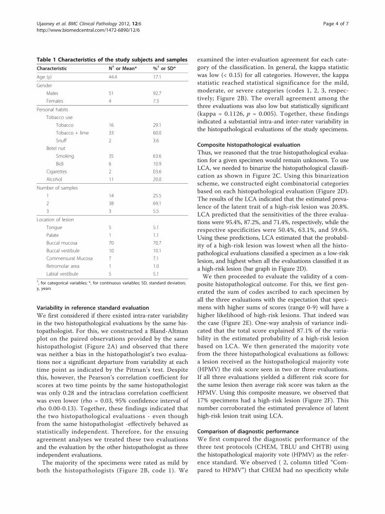
ResultsWe recruited 55 patients with 99 lesions. The character-istics of the study subjects and the lesions are describedin Table 1. The majority of the study subjects were maleand indulged in chronic tobacco use and/or betel nutchewing. There were ~70% subjects with two lesions.Also, 71% of the lesions involved the buccal mucosa(Table 1).
Figure 1 Study protocol (A-D) and histopathological classification method (E-H). The study protocol included first (A) a regular visualinspection using standard operating procedures and regular light followed by examination using chemiluminescence (B). This was followed byapplication of toluidine blue. The results of toluidine blue retention were seen as dark royal blue coloration (C) or faintly stained blue coloration(D). Photomicrographs demonstrating the histopathological grading system which classified a lesion as ‘no dysplasia’ (E) if there was no cellatypia and no changes in architecture; ‘mild dysplasia’ (F) is there was keratosis, mild cellular atypia and architectural changes in the lower thirdof epithelium; ‘moderate dysplasia’ (G) if the architectural changes extended to the middle third of the epithelium; and ‘severe dysplasia’ (H) ifthere was a marked cellular atypia associated with architectural changes extending through the entire thickness of epithelium. Allphotomicrographs show hematoxilin and eosin staining and are depicted at 10× magnification.
Ujaoney et al. BMC Clinical Pathology 2012, 12:6http://www.biomedcentral.com/1472-6890/12/6
Page 3 of 7
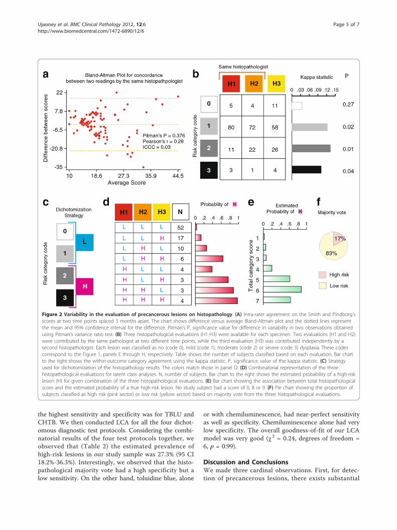
Variability in reference standard evaluationWe first considered if there existed intra-rater variabilityin the two histopathological evaluations by the same his-topathologist. For this, we constructed a Bland-Altmanplot on the paired observations provided by the samehistopathologist (Figure 2A) and observed that therewas neither a bias in the histopathologist’s two evalua-tions nor a significant departure from variability at eachtime point as indicated by the Pitman’s test. Despitethis, however, the Pearson’s correlation coefficient forscores at two time points by the same histopathologistwas only 0.28 and the intraclass correlation coefficientwas even lower (rho = 0.03, 95% confidence interval ofrho 0.00-0.13). Together, these findings indicated thatthe two histopathological evaluations - even thoughfrom the same histopathologist -effectively behaved asstatistically independent. Therefore, for the ensuingagreement analyses we treated these two evaluationsand the evaluation by the other histopathologist as threeindependent evaluations.The majority of the specimens were rated as mild by
both the histopathologists (Figure 2B, code 1). We
examined the inter-evaluation agreement for each cate-gory of the classification. In general, the kappa statisticwas low (< 0.15) for all categories. However, the kappastatistic reached statistical significance for the mild,moderate, or severe categories (codes 1, 2, 3, respec-tively; Figure 2B). The overall agreement among thethree evaluations was also low but statistically significant(kappa = 0.1126, p = 0.005). Together, these findingsindicated a substantial intra-and inter-rater variability inthe histopathological evaluations of the study specimens.
Composite histopathological evaluationThus, we reasoned that the true histopathological evalua-tion for a given specimen would remain unknown. To useLCA, we needed to binarize the histopathological classifi-cation as shown in Figure 2C. Using this binarizationscheme, we constructed eight combinatorial categoriesbased on each histopathological evaluation (Figure 2D).The results of the LCA indicated that the estimated preva-lence of the latent trait of a high-risk lesion was 20.8%.LCA predicted that the sensitivities of the three evalua-tions were 95.4%, 87.2%, and 71.4%, respectively, while therespective specificities were 50.4%, 63.1%, and 59.6%.Using these predictions, LCA estimated that the probabil-ity of a high-risk lesion was lowest when all the histo-pathological evaluations classified a specimen as a low-risklesion, and highest when all the evaluations classified it asa high-risk lesion (bar graph in Figure 2D).We then proceeded to evaluate the validity of a com-
posite histopathological outcome. For this, we first gen-erated the sum of codes ascribed to each specimen byall the three evaluations with the expectation that speci-mens with higher sums of scores (range 0-9) will have ahigher likelihood of high-risk lesions. That indeed wasthe case (Figure 2E). One-way analysis of variance indi-cated that the total score explained 87.1% of the varia-bility in the estimated probability of a high-risk lesionbased on LCA. We then generated the majority votefrom the three histopathological evaluations as follows:a lesion received as the histopathological majority vote(HPMV) the risk score seen in two or three evaluations.If all three evaluations yielded a different risk score forthe same lesion then average risk score was taken as theHPMV. Using this composite measure, we observed that17% specimens had a high-risk lesion (Figure 2F). Thisnumber corroborated the estimated prevalence of latenthigh-risk lesion trait using LCA.
Comparison of diagnostic performanceWe first compared the diagnostic performance of thethree test protocols (CHEM, TBLU and CHTB) usingthe histopathological majority vote (HPMV) as the refer-ence standard. We observed ( 2, column titled “Com-pared to HPMV”) that CHEM had no specificity while
Table 1 Characteristics of the study subjects and samples
Characteristic N† or Mean* %† or SD*
Age (y) 44.4 17.1
Gender
Males 51 92.7
Females 4 7.3
Personal habits
Tobacco use
Tobacco 16 29.1
Tobacco + lime 33 60.0
Snuff 2 3.6
Betel nut
Smoking 35 63.6
Bidi 6 10.9
Cigarettes 2 03.6
Alcohol 11 20.0
Number of samples
1 14 25.5
2 38 69.1
3 3 5.5
Location of lesion
Tongue 5 5.1
Palate 1 1.1
Buccal mucosa 70 70.7
Buccal vestibule 10 10.1
Commensural Mucosa 7 7.1
Retromolar area 1 1.0
Labial vestibule 5 5.1†, for categorical variables; *, for continuous variables; SD, standard deviation;y, years
Ujaoney et al. BMC Clinical Pathology 2012, 12:6http://www.biomedcentral.com/1472-6890/12/6
Page 4 of 7
the highest sensitivity and specificity was for TBLU andCHTB. We then conducted LCA for all the four dichot-omous diagnostic test protocols. Considering the combi-natorial results of the four test protocols together, weobserved that (Table 2) the estimated prevalence ofhigh-risk lesions in our study sample was 27.3% (95 CI18.2%-36.3%). Interestingly, we observed that the histo-pathological majority vote had a high specificity but alow sensitivity. On the other hand, toluidine blue, alone
or with chemiluminescence, had near-perfect sensitivityas well as specificity. Chemiluminescence alone had verylow specificity. The overall goodness-of-fit of our LCAmodel was very good (c2 = 0.24, degrees of freedom =6, p = 0.99).
Discussion and ConclusionsWe made three cardinal observations. First, for detec-tion of precancerous lesions, there exists substantial
Figure 2 Variability in the evaluation of precancerous lesions on histopathology. (A) Intra-rater agreement on the Smith and Pindborg’sscores at two time points spaced 3 months apart. The chart shows difference versus average Bland-Altman plot and the dotted lines representthe mean and 95% confidence interval for the difference. Pitman’s P, significance value for difference in variability in two observations obtainedusing Pitman’s variance ratio test. (B) Three histopathological evaluations (H1-H3) were available for each specimen. Two evaluations (H1 and H2)were contributed by the same pathologist at two different time points, while the third evaluation (H3) was contributed independently by asecond histopathologist. Each lesion was classified as no (code 0), mild (code 1), moderate (code 2) or severe (code 3) dysplasia. These codescorrespond to the Figure 1, panels E through H, respectively. Table shows the number of subjects classified based on each evaluation. Bar chartto the right shows the within-outcome category agreement using the kappa statistic. P, significance value of the kappa statistic. (C) Strategyused for dichotomization of the histopathology results. The colors match those in panel D. (D) Combinatorial representation of the threehistopathological evaluations for latent class analyses. N, number of subjects. Bar chart to the right shows the estimated probability of a high-risklesion (H) for given combination of the three histopathological evaluations. (E) Bar chart showing the association between total histopathologicalscore and the estimated probability of a true high-risk lesion. No study subject had a score of 0, 8 or 9. (F) Pie chart showing the proportion ofsubjects classified as high risk (pink sector) or low risk (yellow sector) based on majority vote from the three histopathological evaluations.
Ujaoney et al. BMC Clinical Pathology 2012, 12:6http://www.biomedcentral.com/1472-6890/12/6
Page 5 of 7
intra-rater and inter-rater variation in the histopatholo-gical evaluation. Our results suggest that histopathologymay be useful as a diagnostic test in demonstrably highdegree of dysplasia or frank neoplasia but its value as areference standard for diagnosis of low-risk precancer-ous lesions is questionable. Consequently, the use of his-topathology as a reference standard against light-basedassistance for diagnosis of high-risk lesions may lead tobiased estimates of the diagnostic performance of thesemeasures.Second, we observed widely differing estimates of the
sensitivity and specificity of the studied diagnostic pro-tocols. However, caution needs to be exercised whenreading and interpreting the results of latent class mod-eling [24-27]. A substantially different estimate of sensi-tivity (or specificity) for a test from that for the othertests can result from two scenarios: a) if the test is diag-nostically inferior as compared to the rest; and b) if thetest is using different criteria for classification of the dis-ease state. In our case, the results do not necessarilyimply that TBLU and CHTB are diagnostically superiorto histopathology - rather it is possible that these testsuse totally different criteria that do not compare withthose used by histopathology. Nevertheless, our resultsclearly demonstrate (Table 2) that one of the main rea-sons for the controversial estimates of the diagnosticperformance of light-based aids may be the classificationmethod employed for the reference standard.Third, a comparison of the diagnostic performance of
TBLU and CHTB consistently indicated that use ofCHEM may be somewhat redundant. From a primaryhealth care perspective this finding is important since itwill reduce the cost of diagnostic evaluation consider-ably by restricting the use of the more expensive com-ponent. Indeed the estimates of sensitivity andspecificity of TBLU observed in this study are compar-able with or better than those of other more expensive
protocols like autofluorescence [28,29], photodynamicdiagnosis [30], and chemiluminescence [31]. Our resultsare in agreement with the findings of Epstein et alwhich show that toluidine blue retention test holds pro-mise as a screening tool for high-risk oral precancerouslesions since it can reduce a large number of unneces-sary biopsies [32]. Concurring with other studies [33,34],our results encourage consideration of TBLU as a viableand feasible screening method in high-prevalence andlow-resource scenarios like India.There are important limitations of this study. First, as
with the Mehrotra et al [3] study, our study recruitedpatients with a suspicion of a precancerous lesion forthe reasons of feasibility as observed elsewhere [35].However, the protocol did preclude visually negativepatients that could have been later detected by at leastone of the diagnostic methods. Our estimates of highsensitivity may also partially reflect this spectrum biasthereby limiting a ready generalization of the results.Second, the study sample had an a priori high likelihoodof a precancerous lesion. Therefore our study designdoes not permit a full evaluation of the screening per-formance of these tests but rather considers them in themore practical scenario of a tertiary care setting as adiagnostic aid.In summary, our findings support those of Mehrotra
et al [3] and demonstrate that improvements are neededfor histopathological evaluation of precancerous lesions- especially, low risk lesions. Our findings also suggestthat toluidine blue retention may be considered as adiagnostic strategy for oral cancers in countries likeIndia. More robust and larger studies are required toassertively and definitively answer questions related tothe screening use of these tools in high prevalencesettings.
Author details1Lata Medical Research Foundation, Nagpur, India. 2Sharad Pawar DentalCollege & Hospital, Sawangi (Meghe), Wardha, India. 3Government DentalCollege & Hospital, Nagpur, India. 412023 Waterway Rdg, San Antonio, TX78249, USA.
Authors’ contributionsSU conceptualized the study, collected the data, conducted analyses andwrote manuscript. MBM conceptualized the study and reviewed manuscript.SD helped in conceptualizing the study, supported the administrativeconduct of the study and reviewed the manuscript. VW and PZ conductedthe histopathlogical assessments and reviewed the manuscript. MCcontributed to the histopathological quality assessment and reviewed themanuscript. VH provided academic, administrative and conceptual supportto the manuscript. TPT wrote and reviewed the manuscript. MM conductedthe statistical analyses and prepared the first draft and subsequent revisionsof the manuscript. All authors read and approved the final draft.
Competing interestsThe authors declare that they have no competing interests.
Received: 8 July 2011 Accepted: 12 March 2012Published: 12 March 2012
Table 2 Diagnostic performance of the tests for high-risklesions
Prevalence of HRL 0.17 (0.10-0.24) 0.27 (0.18-0.36)
CHEM, chemiluminescence; TBLU, toluidine blue retention; CHTB, toluidineblue retention and chemiluminescence; HPMV, histopathology majority vote;LCA, latent class analysis; HRL, high risk lesion
Ujaoney et al. BMC Clinical Pathology 2012, 12:6http://www.biomedcentral.com/1472-6890/12/6
Page 6 of 7
References1. Mignogna MD, Fedele S, Lo Russo L: The World Cancer Report and the
burden of oral cancer. Eur J Cancer Prev 2004, 13(2):139-142.2. Petersen PE: Oral cancer prevention and control-the approach of the
World Health Organization. Oral Oncol 2009, 45(4-5):454-460.3. Mehrotra R, Singh M, Thomas S, Nair P, Pandya S, Nigam NS, Shukla P: A
cross-sectional study evaluating chemiluminescence andautofluorescence in the detection of clinically innocuous precancerousand cancerous oral lesions. J Am Dent Assoc 2010, 141(2):151-156.
4. Yeole BB, Sankaranarayanan R, Sunny MSL, Swaminathan R, Parkin DM:Survival from head and neck cancer in Mumbai (Bombay), India. Cancer2000, 89(2):437-444.
5. Sankaranarayanan R: Screening for cervical and oral cancers in India isfeasible and effective. Natl Med J India 2005, 18(6):281-284.
6. Sankaranarayanan R, Boffetta P: Research on cancer prevention, detectionand management in low- and medium-income countries. Ann Oncol2010, 21(10):1935-1943.
7. Sankaranarayanan R, Dinshaw K, Nene BM, Ramadas K, Esmy PO, Jayant K,Somanathan T, Shastri S: Cervical and oral cancer screening in India. JMed Screen 2006, 13(Suppl 1):S35-S38.
8. Sankaranarayanan R, Mathew B, Jacob BJ, Thomas G, Somanathan T,Pisani P, Pandey M, Ramadas K, Najeeb K, Abraham E: Early findings from acommunity-based, cluster-randomized, controlled oral cancer screeningtrial in Kerala, India. The Trivandrum Oral Cancer Screening StudyGroup. Cancer 2000, 88(3):664-673.
9. Trullenque-Eriksson A, Munoz-Corcuera M, Campo-Trapero J, Cano-Sanchez J, Bascones-Martinez A: Analysis of new diagnostic methods insuspicious lesions of the oral mucosa. Med Oral Patol Oral Cir Bucal 2009,14(5):E210-E216.
10. Patton LL, Epstein JB, Kerr AR: Adjunctive techniques for oral cancerexamination and lesion diagnosis: a systematic review of the literature. JAm Dent Assoc 2008, 139(7):896-905, quiz 993-894.
11. Sciubba JJ: Improving detection of precancerous and cancerous orallesions. Computer-assisted analysis of the oral brush biopsy. U.S.Collaborative OralCDx Study Group. J Am Dent Assoc 1999,130(10):1445-1457.
12. Brandwein-Gensler M, Smith RV, Wang B, Penner C, Theilken A, Broughel D,Schiff B, Owen RP, Smith J, Sarta C, et al: Validation of the histologic riskmodel in a new cohort of patients with head and neck squamous cellcarcinoma. Am J Surg Pathol 2010, 34(5):676-688.
13. Kerr AR, Sirois DA, Epstein JB: Clinical evaluation of chemiluminescentlighting: an adjunct for oral mucosal examinations. J Clin Dent 2006,17(3):59-63.
14. Ram S, Siar CH: Chemiluminescence as a diagnostic aid in the detectionof oral cancer and potentially malignant epithelial lesions. Int J OralMaxillofac Surg 2005, 34(5):521-527.
15. Huff KD: More about cancer detection. J Am Dent Assoc 2010,141(6):626-628, author reply 628, 630.
19. Dendukuri N, Hadgu A, Wang L: Modeling conditional dependencebetween diagnostic tests: a multiple latent variable model. Stat Med2009, 28(3):441-461.
20. Ihorst G, Forster J, Petersen G, Werchau H, Rohwedder A, Schumacher M:The use of imperfect diagnostic tests had an impact on prevalenceestimation. J Clin Epidemiol 2007, 60(9):902-910.
21. Koukounari A, Webster JP, Donnelly CA, Bray BC, Naples J, Bosompem K,Shiff C: Sensitivities and specificities of diagnostic tests and infectionprevalence of Schistosoma haematobium estimated from data on adultsin villages northwest of Accra, Ghana. Am J Trop Med Hyg 2009,80(3):435-441.
22. Yang I, Becker MP: Latent variable modeling of diagnostic accuracy.Biometrics 1997, 53(3):948-958.
23. Bertrand P, Benichou J, Grenier P, Chastang C: Hui and Walter’s latent-classreference-free approach may be more useful in assessing agreementthan diagnostic performance. J Clin Epidemiol 2005, 58(7):688-700.
24. Albert PS, Dodd LE: On Estimating Diagnostic Accuracy From StudiesWith Multiple Raters and Partial Gold Standard Evaluation. J Am StatAssoc 2008, 103(481):61-73.
25. Baughman AL, Bisgard KM, Cortese MM, Thompson WW, Sanden GN,Strebel PM: Utility of composite reference standards and latent classanalysis in evaluating the clinical accuracy of diagnostic tests forpertussis. Clin Vaccine Immunol 2008, 15(1):106-114.
26. Chu H, Zhou Y, Cole SR, Ibrahim JG: On the estimation of diseaseprevalence by latent class models for screening studies using twoscreening tests with categorical disease status verified in test positivesonly. Stat Med 2010, 29(11):1206-1218.
27. Toft N, Jorgensen E, Hojsgaard S: Diagnosing diagnostic tests: evaluatingthe assumptions underlying the estimation of sensitivity and specificityin the absence of a gold standard. Prev Vet Med 2005, 68(1):19-33.
28. Rana M, Zapf A, Kuehle M, Gellrich NC, Eckardt AM: Clinical evaluation ofan autofluorescence diagnostic device for oral cancer detection: aprospective randomized diagnostic study. Eur J Cancer Prev 2012, doi:10.1097/CEJ.0b013e32834fdb6d.
29. Awan KH, Morgan PR, Warnakulasuriya S: Evaluation of anautofluorescence based imaging system (VELscope) in the detection oforal potentially malignant disorders and benign keratoses. Oral Oncol2011, 47(4):274-277.
30. Driemel O, Kunkel M, Hullmann M, von Eggeling F, Muller-Richter U,Kosmehl H, Reichert TE: Diagnosis of oral squamous cell carcinoma andits precursor lesions. Journal der Deutschen Dermatologischen Gesellschaft =Journal of the German Society of Dermatology: JDDG 2007, 5(12):1095-1100.
31. Seoane Leston J, Diz Dios P: Diagnostic clinical aids in oral cancer. OralOncol 2010, 46(6):418-422.
32. Epstein JB, Silverman S Jr, Epstein JD, Lonky SA, Bride MA: Analysis of orallesion biopsies identified and evaluated by visual examination,chemiluminescence and toluidine blue. Oral Oncol 2008, 44(6):538-544.
33. Guneri P, Epstein JB, Kaya A, Veral A, Kazandi A, Boyacioglu H: The utility oftoluidine blue staining and brush cytology as adjuncts in clinicalexamination of suspicious oral mucosal lesions. Int J Oral Maxillofac Surg2011, 40(2):155-161.
34. Epstein JB, Guneri P: The adjunctive role of toluidine blue in detection oforal premalignant and malignant lesions. Current opinion in otolaryngology& head and neck surgery 2009, 17(2):79-87.
35. Patton LL: The effectiveness of community-based visual screening andutility of adjunctive diagnostic aids in the early detection of oral cancer.Oral Oncol 2003, 39(7):708-723.
Pre-publication historyThe pre-publication history for this paper can be accessed here:http://www.biomedcentral.com/1472-6890/12/6/prepub
doi:10.1186/1472-6890-12-6Cite this article as: Ujaoney et al.: Evaluation of chemiluminescence,toluidine blue and histopathology for detection of high risk oralprecancerous lesions: A cross-sectional study. BMC Clinical Pathology2012 12:6.
Submit your next manuscript to BioMed Centraland take full advantage of:
• Convenient online submission
• Thorough peer review
• No space constraints or color figure charges
• Immediate publication on acceptance
• Inclusion in PubMed, CAS, Scopus and Google Scholar
• Research which is freely available for redistribution
Submit your manuscript at www.biomedcentral.com/submit
Ujaoney et al. BMC Clinical Pathology 2012, 12:6http://www.biomedcentral.com/1472-6890/12/6