Working Report No. 43, 2003 Arbejdsrapport fra Miljøstyrelsen Evaluation of in vitro assays for determination of estrogenic activity in the environment Karin Kinnberg University of Southern Denmark
Transcript
Working Report No. 43, 2003Arbejdsrapport fra Miljøstyrelsen
Evaluation of in vitro assays fordetermination of estrogenic activityin the environment
Karin KinnbergUniversity of Southern Denmark
The Danish Environmental Protection Agency will, when opportunityoffers, publish reports and contributions relating to environmental
research and development projects financed via the Danish EPA.
Please note that publication does not signify that the contents of the
reports necessarily reflect the views of the Danish EPA.
The reports are, however, published because the Danish EPA finds that
the studies represent a valuable contribution to the debate on
environmental policy in Denmark.
3
Contents
CONTENTS 3
PREFACE 5
1 SUMMARY (ENGLISH) 7
2 RESUMÉ (DANSK) 11
3 DESCRIPTION OF THE USED METHODS AND CELLCULTURES 15
3.1 ER BINDING ASSAYS 153.2 REPORTER GENE ASSAYS 16
5.3.1 Detection limits and EC50 values for the various in vitro assays 295.3.2 Detection limit and limit of quantification for the whole method 31
5.4 TIME AND COST CONSIDERATIONS 325.5 ROBUSTNESS 335.6 UTILITY IN VARIOUS MATRICES 345.7 ADVANTAGES AND LIMITATIONS COMPARED TO CHEMICALANALYSES 365.8 LIMITATIONS COMPARED TO IN VIVO ASSAYS 37
6 DISCUSSION AND RECOMMENDATIONS 41
7 CONCLUSIONS 45
8 KONKLUSIONER 47
9 REFERENCE LIST 49
4
5
Preface
The aquatic environment is particularly susceptible to the effects ofcontaminants. Effluents from municipal and industrial wastewater treatmentplants, and agricultural run-off and drainage add numerous exogenouscompounds to the aquatic system. Among these compounds are substanceswith estrogenic activity. Recent studies in a number of countries have shownthat the aquatic environment can possess estrogenic activity capable ofinfluencing the fauna. Examples of this are vitellogenin induction andfeminised reproductive organs in male fish (Christiansen et al. 2002). Thishas led to efforts of finding simple, sensitive and specific in vitro tests forrapid screening of samples from wastewater and surface waters for theirestrogenic activity. Already existing in vitro assays for screening of theestrogenic activity of single compounds have therefore been applied toenvironmental samples. This report gives an evaluation of the existing in vitromethods for determination of estrogenic activity in various environmentalmatrices. The existing knowledge on the potentials and limitations of thesemethods will be presented with the aim of finding the optimal method(s) formonitoring wastewater and surface water, and with a view to assessing thepossibilities for monitoring agricultural drain water and animal manure slurry.Parallel to this report a report has been written assessing existing chemicalmethods for detection of estrogens in the environment.
6
7
1 Summary (English)
Recent studies in a number of countries have shown that the aquaticenvironment can possess estrogenic activity capable of influencing the fauna.(Xeno)estrogens are believed to reach the aquatic environment mainly bymeans of municipal and industrial sewage outfalls. However, agriculturaldrainage may also be a route for (xeno)estrogens to enter the aquatic system.Numerous natural and anthropogenic substances are known to exhibitestrogenic activity. In the aquatic environment, estrogenic activity hasprimarily been ascribed to the natural steroids, 17β-estradiol (E2), estrone(E1) and estriol (E3), and the synthetic estrogen, ethinylestradiol (EE2), usedin contraceptives. To a lesser extent xenoestrogenic chemicals, such asalkylphenols and bisphenol A, may also contribute to the estrogenic activity inthe aquatic environment.In vitro assays measure the total estrogenic activity of an environmental watersample, regardless of which compounds are responsible for the activity. Thetotal estrogenic activity in the sample is then compared to the activity of thenatural estrogen, E2, and expressed as estradiol equivalents (EEQ). A numberof studies employing in vitro assays have demonstrated the estrogenic activityof wastewater and surface water in various countries. Total estrogenic activity(expressed as EEQ values) of sewage treatment plant influents have beenreported to be 0.6-153 nanograms per litre. In the effluents, EEQ values areusually below 25 nanograms per litre, although values of up to about 150nanograms per litre have been reported in the USA. In surface water, theEEQ values found are generally from below 1 nanogram up to 15 nanogramsper litre, although values of up to about 80 nanograms per litre have beenreported in one study. The EEQ levels found in some aquatic systems aresufficient to cause estrogenic effects in fish in laboratory experiments.
Several in vitro assays have been developed to assess the estrogenic activity ofsingle compounds or complex mixtures. Each assay measures different endpoints at different levels of biological complexity of estrogen action. Mostassays fall into one of three categories: 1) estrogen receptor (ER) competitiveligand binding assays that measure the binding affinity of a chemical for theER; 2) cell proliferation assays that measure the increase in cell number ofestrogen sensitive cells (E-screen); and 3) reporter gene assays that measureER binding-dependent transcriptional and translational activity. No single invitro assay can be regarded as ideal for assessing the estrogenic activity ofwastewater and surface water. They all have their advantages and limitations.
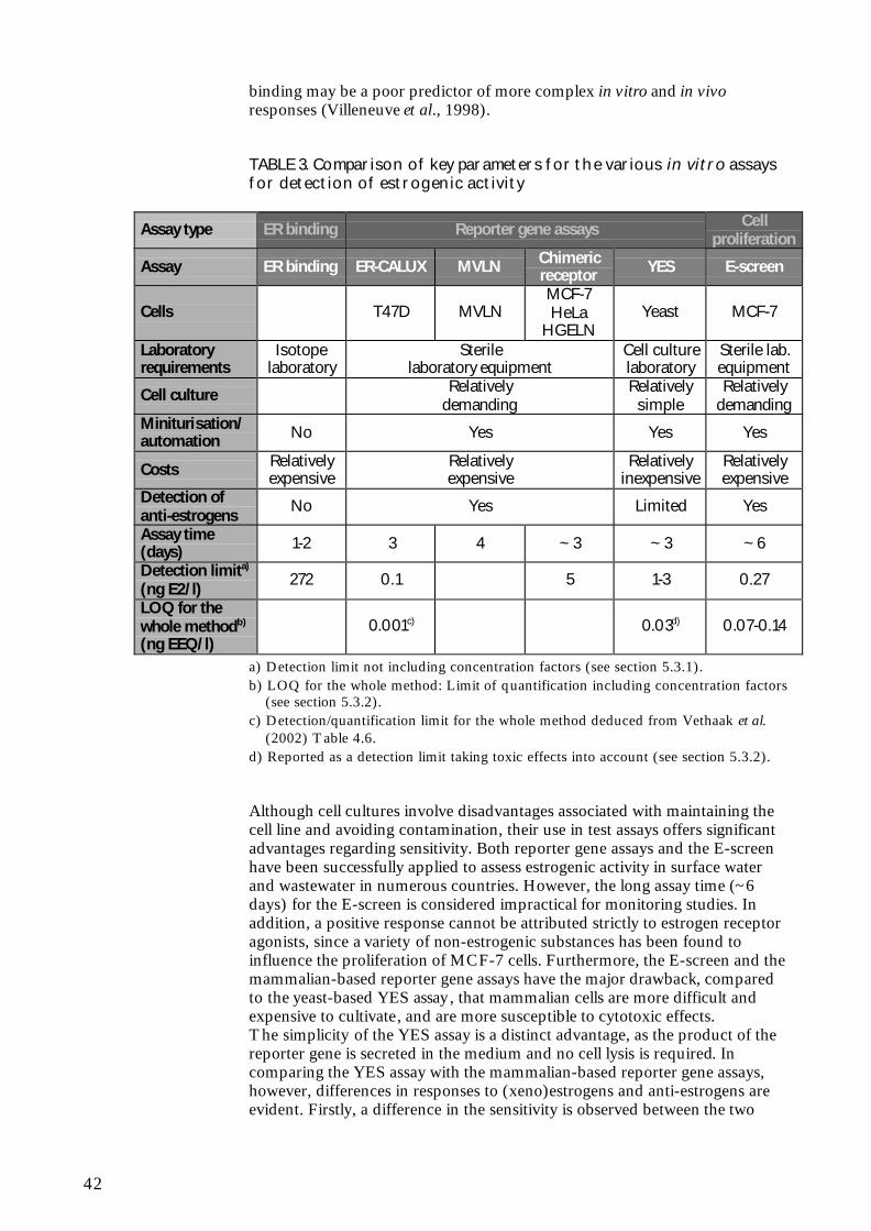
Most ER binding assays quantifies the ability of a test compound to competewith radiolabelled E2 for binding to the ER. The sample is added along withan excess of radiolabelled E2 to isolated ERs whereupon the amount ofunbound radioactivity is measured. ER binding assays are fast. However, theyare significantly less sensitive than the other in vitro assays. In addition,binding assays are not easily amenable to automation, thereby limiting theirutility as a screening tool. Furthermore, ER binding assays require specialisedlaboratory facilities because of the radioactive substances. Finally, the bindingof a substance to the ER is only indicative that it may act as a xenoestrogen;ER binding may be a poor predictor of more complex in vitro and in vivoresponses.
8
In the E-screen assay, proliferation of human MCF-7 breast cancer cells as aresponse to estrogen is measured. The E-screen is based on the followingthree premises: (i) factors in serum added to the medium inhibit theproliferation of MCF-7 cells, (ii) estrogens induce cell proliferation bynegating this inhibitory effect, and (iii) non-estrogenic substances do notneutralize the inhibitory signal present in serum. However, it has been shownthat the E-screen may not be as estrogen specific as assumed, since a range ofnon-estrogenic substances has been found to influence the proliferation ofMCF-7 cells, at least in some cell lines. In addition, considerable inter-laboratory variability has been observed in test results from the E-screen.Furthermore, the E-screen is more time consuming than the other assays andis thus considered impractical for extensive monitoring studies.
Reporter gene assays are based on the ability of a compound to stimulate ER-dependent transcriptional activity. Reporter gene assays are carried out withgenetically engineered human cancer cells or yeast cells tranfected withestrogen response elements (ERE) linked to a reporter gene. In the human-based reporter gene assays (ER-CALUX, MVLN cell assay and chimericreceptor/reporter gene assays) the reporter gene codes for luciferase and in theyeast-based reporter gene assay (YES) the reporter gene codes for β-galactosidase. Yeast cells are further transfected with the DNA sequence forthe human ER, since yeast cells do not possess endogenous ER.In reporter gene assays, the sample is added to the transfected cells.Estrogenic substances that enter the cells binds to the ER, which becomesactivated and binds to the EREs. This biding initiates the expression of thereporter gene and thereby the synthesis of the enzyme. An appropriatesubstrate in the incubation medium is metabolized by the newly synthesizedenzyme, resulting in the production of an easily detected product.The mammalian-based reporter gene assays have the major drawback,compared to the yeast-based assay, that mammalian cells are more difficultand expensive to cultivate, and are more susceptible to cytotoxic effects. Thesimplicity of the YES assay is a distinct advantage, as the product of thereporter gene is secreted in the medium and no cell lysis is required. Incomparing the YES assay with the mammalian-based reporter gene assays,however, differences in responses to (xeno)estrogens and anti-estrogens areevident. Firstly, a difference in the sensitivity is observed between the twomammalian-based endogenous receptor/reporter gene assays (ER-CALUXand MVLN cell assay) and the YES assay, demonstrating that the former candetect (xeno)estrogens at lower concentrations. Secondly, a difference inresponse to anti-estrogens is found between the mammalian-based reportergene assays and the YES assay , as the latter does not consistently detect anti-estrogenic activity, but sometimes identifies it as agonistic. This “limitation”,which the YES assay has in common with ER binding assays, could beconsidered an advantage if all one is interested in is detecting compounds thatinteracts with the ER and elicit a response, thus having potential endocrinedisrupting effects. From this point of view, the mammalian-based reportergene assays may actually underestimate the actual estrogenic potential of acomplex water sample.A main problem in the utilization of in vitro assays to analyse aquaticenvironmental samples is the presence of inhibitory/cytotoxic compounds.Yeast assays may perform better for monitoring of environmental samples, asthese samples are frequently contaminated with substances other than(xeno)estrogens interfering with the growth and viability of animal cells, butnot with yeast cells.
9
Reporter gene assays seem to be a suitable choice for monitoringenvironmental matrices for estrogenic activity. The final choice of whichreporter gene assay to employ (mammalian-based or yeast-based) depends onthe importance of lower detection limit versus the importance of ease of useand lower costs.
Significant advantages of in vitro assays over chemical analyses are that nounknown components with estrogenic activity are overlooked and that anycombination effects are taken into account in the analysis. Chemical analysisof all compounds with potential estrogenic activity would be very costly andunknown estrogenic compounds, including metabolites, may still be present inenvironmental matrices. By a combination of the two types of analysis it ispossible both to assess the estrogenic activity in a sample and to (partly)identify and quantify the compounds responsible for the estrogenic activity.
The advantages of in vitro assays over in vivo assays include lower costs andtime consumption as well as sparing of experimental animals. However, invitro assays do not always reliably predict the results of in vivo assays andshould not be used alone for full assessment of potential estrogenic hazards inthe aquatic system. In vitro assays usually possess minimal metaboliccapabilities. As a result, extrapolation from in vitro to in vivo systems can leadto false negatives for compounds that are bioactivated, and overestimates ofpotency for compounds readily degraded in vivo. In addition, bioavailability,cross talk between biological pathways and the complex processes of uptake,binding to carrier proteins, transport, targeting, disposition and excretion ofcompounds in whole animals are not taken into account in the in vitro assays.Furthermore, it should be kept in mind that there are estrogenic effects thatare based on mechanisms different from receptor binding, e.g. interferenceswith hormone synthesis and metabolism. Environmental samples shouldtherefore also be tested for their estrogenic activity in relevant in vivo tests,such as vitellogenin induction or gonadal effects in fish.
10
11
2 Resumé (dansk)
Det er igennem de seneste år konstateret, ved undersøgelser i en rækkeforskellige lande, at der i nogle tilfælde kan registreres østrogen aktivitet i detakvatiske miljø, der er i stand til at påvirke faunaen.(Xeno)østrogener menes at ende i det akvatiske miljø primært via kommunaltog industrielt spildevand. Desuden kan drænvand fra marker være enyderligere kilde til (xeno)østrogener i det akvatiske miljø.En vifte af naturlige og menneskeskabte stoffer vides at besidde østrogenaktivitet. I det akvatiske miljø er den østrogene aktivitet primært blevettilskrevet de naturlige østrogener, 17β-østradiol (E2), østron (E1) og østriol(E3), og det syntetiske østrogen, ethinyløstradiol (EE2), der anvendes i p-piller. Desuden kan xenoøstrogener som alkylfenoler og bisfenol A i mindregrad også bidrage til den østrogene aktivitet i det akvatiske miljø.In vitro assays måler den totale østrogene aktivitet i en vandprøve fra miljøetuanset hvilke stoffer, der er ansvarlige for aktiviteten. Den totale østrogeneaktivitet i en prøve sammenlignes så med aktiviteten af det naturlige østrogen,E2, og udtrykkes som østradiolækvivalenter (EEQ). En række studier, hvorder har været anvendt in vitro assays, har demonstreret østrogen aktivitet ispildevand og overfladevand i forskellige lande. Den totale østrogene aktivitet(udtrykt som EEQ-værdier) målt i urenset spildevand er 0,6-153 nanogrampr. liter. I renset spildevand er EEQ-værdierne som regel under 25 nanogrampr. liter. Der er dog i USA målt værdier helt op til omkring 150 nanogram pr.liter. I overfladevand er der generelt fundet EEQ-værdier fra under 1nanogram pr. liter og op til 15 nanogram pr. liter. Der er dog i énundersøgelse fundet værdier på op til omkring 80 nanogram pr. liter. DeEEQ-niveauer, der er fundet i nogle akvatiske systemer, er høje nok til atinducere østrogene effekter i laboratorieforsøg med fisk.
Adskillige in vitro assays er blevet udviklet til at måle den østrogene aktivitet afenkeltstoffer eller komplekse blandinger. Hvert assay måler forskellige end-points på forskellige niveauer af den biologiske kompleksitet af østrogenvirkning. De fleste assays tilhører én af tre kategorier: 1) østrogenreceptor-bindingassays som måler et stofs bindingsaffinitet for østrogenreceptoren; 2)celledelingsassays som måler stigningen i antallet af østrogensensitive celler(E-screen), og 3) reportergenassays som måler østrogenreceptorafhængigtranskriptionel og translationel aktivitet. Intet enkelt in vitro assay kan ansessom ideelt for vurdering af den østrogene aktivitet i spildevand ogoverfladevand. De har alle deres fordele og begrænsninger.
De fleste østrogenreceptorbindingsassays kvantificerer et teststofs evne til atkonkurrere med radioaktivt mærket E2 om binding til østrogenreceptoren.Prøven tilsættes sammen med en overskud af radioaktivt mærket E2 tilisolerede østrogenreceptorer, hvorefter mængden af ubunden radioaktivitetmåles. Østrogenreceptorbindingsassays er hurtige, men er betydeligt mindrefølsomme end de andre in vitro assays. Desuden er de vanskelige atautomatisere og har derfor begrænset anvendelighed som screeningsværktøj.Yderligere kræver østrogenreceptorbindingsassays specialiseredelaboratoriefaciliteter på grund af de radioaktive stoffer. Endelig giverbindingen af et stof til østrogenreceptoren kun en indikation af, at stoffetmåske har østrogen virkning. Østrogenreceptorbinding medfører ikke
12
nødvendigvis de efterfølgende komplekse reaktioner, der er involveret iøstrogen virkning.
I E-screen assayet måles delingen af humane MCF-7 brystkræftceller somsvar på østrogen. E-screen assayet er baseret på følgende tre forudsætninger:(i) faktorer i serum tilsat dyrkningsmediet hæmmer delingen af MCF-7 celler,(ii) østrogener inducerer celledeling ved af ophæve denne hæmmende effekt,og (iii) ikke-østrogene stoffer ophæver ikke det hæmmende signal der er tilstede i serum. Det har dog vist sig, at E-screen assayet måske ikke er såøstrogenspecifikt som antaget, da en række ikke-østrogene stoffer har vist sigat influere på celledelingen i MCF-7 celler, i hvert fald hos nogle cellelinier.Desuden er der observeret betydelige forskelle i resultater opnået med E-screen i forskellige laboratorier. Endelig er E-screen assayet merelangsommeligt end de andre assays og må derfor betragtes som upraktisk forekstensive moniteringsstudier.
Reportergenassays er baseret på et stofs evne til at stimulereøstrogenreceptorafhængig transkriptionel aktivitet. Reportergenassays gørbrug af genmanipulerede humane cancerceller eller gærceller transfekteretmed østrogenresponselementer forbundet med et reportergen. I de humant-baserede reportergenassays (ER-CALUX, MVLN celle assay og chimeriskreceptor/reportergenassays) koder reportergenet for luciferase og i det gær-baserede reportergenassay (YES) koder reportergenet for β-galactosidase.Gærceller er yderligere transfekteret med DNA-sekvensen for den humaneøstrogenreceptor, da gærceller ikke besidder endogene østrogenreceptorer.I reportergenassays tilsættes prøven til de transfekterede celler. De østrogenestoffer i prøven binder til østrogenreceptoren, som aktiveres og binder tiløstrogenresponselementerne. Denne binding initierer ekspressionen afreportergenet og derved syntesen af enzymet. Et passende substrat iinkuberingsmediet metaboliseres af det nyligt syntetiserede enzym, hvilketresulterer i dannelsen af et nemt målbart produkt.De pattedyr-baserede reportergenassays har den store ulempe sammenlignetmed det gær-baserede assay, at pattedyrceller er vanskeligere og dyrere atdyrke. Desuden er de mere sårbare over for cytotoksiske effekter. En andenfordel ved YES-assayet er, at det er en mere simpel metode, der ikke krævercellelysis, da produktet fra reportergenet frigives til mediet. Vedsammenligning af YES-assayet med de pattedyr-baserede reportergenassaysses dog klare forskelle i respons på (xeno)østrogener og anti-østrogener. Fordet første er der forskelle i følsomheden mellem de to pattedyr-baseredeendogen receptor/reportergenassays (ER-CALUX og MVLN celle assay) ogYES-assayet, hvilket afspejler sig i, at de førstnævnte kan detektere laverekoncentrationer af (xeno)østrogener. For det andet ses forskelle på responsetpå anti-østrogener hos pattedyr-baserede reportergenassays og YES-assayet,idet det sidstnævnte ikke altid kan bestemme anti-østrogen aktivitet, men af ogtil registrerer denne som agonistisk. Denne ”begrænsning”, som YES-assayethar tilfælles med østrogenreceptorbindingsassays, kan betragtes som en fordel,hvis dét, man er interesseret i, er at registrere stoffer som interagerer medøstrogenreceptoren og udviser er respons og således har potentiellehormonforstyrrende effekter. Ud fra dette synspunkt kan pattedyr-baseredereportergenassays faktisk underestimere det østrogene potentiale i enkompleks vandprøve.Et vigtigt problem ved anvendelsen af in vitro assays til at teste prøver fra detakvatiske miljø er tilstedeværelsen af hæmmende/cytotoksiske stoffer. Gær-assays er muligvis bedre til monitering af prøver fra miljøet, da disse prøver
13
ofte er kontaminerede med andre stoffer, der interfererer med væksten oglevedygtigheden af dyreceller men ikke gærceller.Reportergenassays synes at være et passende valg for monitering af østrogenaktivitet i forskellige miljømatricer. Det endelige valg af hvilketreportergeneassay der skal benyttes (pattedyr-baseret eller gær-baseret)afhænger af vigtigheden af en lavere detektionsgrænse holdt op imodvigtigheden af simpel udførelse og lavere omkostninger.
Betydelige fordele ved in vitro assays i forhold til kemiske analyser er, at ingenukendte stoffer med østrogen aktivitet overses, og at der tages hensyn tilkombinationseffekter i analysen. Kemisk analyse af alle stoffer med potentieløstrogen aktivitet ville være meget dyrt og ukendte østrogene stoffer, inklusivemetabolitter, kan stadig være til stede i prøver fra miljøet. Ved en kombinationaf de to typer af analyse, kan man både få et mål for den østrogene aktivitet ien prøve, samt (til dels) identificere og kvantificere de stoffer, der er årsag tilden østrogene aktivitet.
Fordelene ved in vitro assays frem for in vivo assays er blandt andet lavereomkostninger og tidsforbrug såvel som at man undgår brug af forsøgsdyr. Invitro assays er dog ikke altid pålidelige i deres forudsigelser for udfaldet i invivo assays og bør aldrig bruges alene ved vurdering at potentielt skadeligeøstrogene effekter i det akvatiske system. In vitro assays besidder som regelminimale metaboliske evner. Som et resultat heraf kan ekstrapolering fra invitro til in vivo systemer føre til falske negative resultater for stoffer, derbioaktiveres, og overestimeringer for stoffer, der hurtigt nedbrydes in vivo.Desuden afspejler in vitro assays ikke biotilgængelighed, interaktion mellembiologiske systemer og de komplekse processer som optagelse, binding tilproteiner, transport, fordeling og udskillelse af stoffer, som spiller en rolle invivo. Ydermere skal man være opmærksom på, at der eksisterer østrogeneeffekter, som er baserede på andre mekanismer end receptorbinding, f.eks.interferenser med hormonsyntese og -metabolisme. Prøver fra miljøet børderfor også testes for deres østrogene aktivitet i relevante in vivo tests, såsomvitellogenininduktion eller gonadeeffekter i fisk.
14
15
3 Description of the used methodsand cell cultures
In vitro assays are useful techniques for the determination of estrogenicactivity in environmental samples containing complex mixtures ofcontaminants. They enable estimation of total biological activity of allcompounds that act through the same mode of action present in extracts ofany environmental media.The molecular mechanisms of estrogen action are the basis for thedevelopment of in vitro test systems. Therefore, a short description of thesemechanisms is given here. The effects of estrogens are mediated by theestrogen receptor (ER), a member of the nuclear receptor superfamily (Ingand O’Malley, 1995). Inactive ERs exist in large complexes associated withheat shock proteins. Upon binding of an estrogenic compound to the ER, theheat shock proteins disassociate, inducing a conformational change thatactivates the receptor, and causes dimerization. The resulting homodimercomplex exhibits high affinity for specific DNA sequences referred to asestrogen response elements (EREs) positioned in the regulatory region ofestrogen-inducible genes in the nucleus. After binding to the ERE, thehomodimer complex recruits transcription factors to the target gene promoter,which leads to gene activation and transcription. Following transcription,mRNA is then translated into proteins that are the ultimate effectors of theobserved responses. By inducing the synthesis of new proteins that altercellular functions, estrogens can have profound effects on cell function andphysiology. Xenoestrogens can act as ER ligands that bind to the receptor,thus modulating endocrine pathways via a receptor-mediated process.Several in vitro assays have been developed to assess the estrogenic activity ofindividual compounds or complex mixtures (Zacharewski, 1997). Most ofthese assays fall into one of three categories: 1) estrogen receptor (ER)competitive ligand binding assays that measure the binding affinity of achemical for ER; 2) reporter gene assays that measure ER binding-dependenttranscriptional and translational activity; and 3) cell proliferation assays thatmeasure the increase in cell number of target cells during the exponentialphase of proliferation. In the following, the in vitro assays most widely used toassess estrogenic activity in wastewater and surface water are described.
3.1 ER binding assays
Competitive ligand binding assays are based on the primary mode of action of(xeno)estrogens, which is binding to the ER. In vitro competitive bindingassays for the ER are well established and have been extensively used toinvestigate ER-ligand interactions. ER binding assays can be performed withreceptors obtained from cytosolic or nuclear extracts of various mammalianand other vertebrate tissues (Ankley et al., 1998). Most ER binding assaysquantifies the ability of a test compound to compete with radiolabelled 17β-estradiol for binding to the ER. In a typical competitive hormone bindingassay, a high-speed centrifugal fraction of rat uterine cytosol or cell extract isincubated with excess radiolabelled 17β-estradiol ([3H]E2) and various
16
concentrations of unlabelled test compounds. If the compounds compete withthe [3H]E2 for receptor binding they will displace a fraction of the [3H]E2from the receptor in a concentration dependent manner. The greater theconcentration of the unlabelled competitor, the more [3H]E2 is displaced fromthe ER, and the less bound activity. The free [3H]E2 is separated from thebound [3H]E2 by filtration, hydroxyapatite extraction, or other methods andquantified by liquid scintillation counting (Gray et al., 1997). Non-specificbinding is measured by addition of excesses of radioinert diethylstilbestrol(DES) or 17β-estradiol (E2). Total specific binding of [H3]E2 to the ER iscalculated by subtracting the amount of [H3]E2 bound in the presence of DESor E2 from the amount of [H3]E2 bound in the absence of a competitor.Decreased specific binding of the [H3]E2 in the presence of a test samplesuggests that the sample contains compounds, which can competitively bindto the ER ligand-binding site. In this assay, the compounds can reach the ERwithout having to pass a cell membrane.Non-radioactive methods employing fluorescent polarization (Bolger et al.,1998) or enzyme-linked receptor assays (Seifert et al., 1999) have also beenreported. However, these methods have not been widely used forenvironmental samples.The concentration at which the tested compound results in a 50% decrease ofthe binding of [H3]E2 to the receptor is denoted as the IC50. Results areexpressed as IC50 or as a relative binding affinity, which is the ratio betweenthe IC50 of the test compound and that of unlabelled E2 (Soto et al., 1998).
ER binding assays are essential for the characterization of a compound as aligand for the ER. However, ER binding determinations do not classify theligand as agonist or antagonist. Moreover, the ability of a substance to initiatethe molecular cascade of events implicated in gene transcription and proteinsynthesis associated with adverse effects is not determined in this assay.Furthermore, high concentrations of competitor ligand may result in non-competitive displacement (Zacharewski, 1997; Jobling, 1998). Finally, thecell-free nature of ER binding assays may lead to positive results forcompounds, which have physical characteristics that would make it unlikelythat they would normally enter the cell.
3.2 Reporter gene assays
The ER functions by modulating the rate of transcription of its target cellgenes. Reporter gene assays are based on the ability of a compound tostimulate ER-dependent transcriptional activity. Thus, reporter geneexpression is a result of the molecular cascade of events implicated in receptoractivation, and as such provides a more integral indication of the estrogenicactivity of a compound.
Reporter gene assays are carried out with genetically engineered mammaliancells or strains of yeast, with cells transformed (tranfected) by introducingvectors containing DNA sequences for the receptor, along with EREs linkedto a reporter gene, and the reporter gene itself. A number of assays areavailable using cell lines with an endogenous ER (T47D cells or MCF-7 cells)or cell lines without an endogenous ER (e.g. yeast cells or HeLa cells). Thereporter gene used in human cancer cells usually codes for luciferase and thereporter gene used in yeast cells usually codes for β-galactosidase.
17
Reporter genes can be introduced into cells for the duration of the experimentonly (transient transfection) or permanently, generating a genetically alteredsubline (stable transfection). Regardless of whether transient or stablytransfected cells are utilized in the assays, test substances that enter the cellsinteract with the ER, which becomes activated by a change in itsconformation. The activated ER then binds with soluble cell factors, and theresulting complex binds to the ERE on the reporter plasmid. This bidinginitiates the expression of the reporter gene and thereby the production of theenzyme. An appropriate substrate in the incubation mixture is metabolized bythe newly synthesized enzyme, resulting in the production of an easilydetected product.
In agonism studies, the cells are treated with a test substance and theinduction of the reporter gene product is utilized to measure the response. Foran assessment of relative potency, the induction can be compared to theinduction by a reference estrogen. Alternatively, when dose-response data aregenerated, the EC50 for the test substance can be determined and comparedwith that for the reference estrogen.For antagonism studies, the cells are exposed simultaneously to the referenceestrogen and the test substance, while control cells are exposed to thereference estrogen only. The difference in induction of the reporter geneproduct in the presence and absence of the test substance is used as a measureof estrogen antagonism.
3.2.1 Mammalian-based reporter gene assays
3.2.1.1 ER-mediated chemical activated luciferase gene expression (ER-CALUX)assay
The ER-CALUX assay is a relatively new method developed in theNetherlands and is not yet widely used. The assay uses T47D human breastadenocarcinoma cells expressing endogenous ER and stably transfected withan estrogen-responsive luciferase reporter gene containing three EREs. In theER-CALUX assay, exposure of cells to xenoestrogens results in binding toendogenous ER, activation of the receptor, and consequently, binding of theligand-receptor complex to the EREs present in the promoter region of thestably integrated luciferase gene. This leads to expression of the luciferasegene, which is assayed by lysing cells, adding the substrate luciferin andmeasuring light output in a luminometer (Legler et al., 1999, 2003).
3.2.1.2 MVLN cell assay
The principles of this assay are similar to those of the ER-CALUX assay.However, the MVLN cell assay utilizes a derivate of the MCF-7 breast cancercell line (MVLN) expressing endogenous ER and stably transfected with anestrogen-responsive luciferase reporter gene (Pons et al., 1990; Demirpence etal., 1993). Like in the ER-CALUX assay, the estrogen specific transcriptionactivity of a test compound is directly related to the luciferase activitymeasured in the lysate of treated MVLN cells.
3.2.1.3 Chimeric receptor/reporter gene assays
Chimeric receptor/reporter gene constructs have also been proven to haveutility in screening compounds for estrogenic activity. For example, the E2Bioassay (Zacharewski et al., 1995) consists of a chimeric receptor (with theligand binding domain of the ER and the DNA binding domain of the yeast
18
transcription factor Gal4) and a Gal4-regulated reporter gene consisting of aluciferase gene regulated by a basal promoter and five tandem Gal4 responseelements. Both of these constructs have been transiently or stably tranfectedinto recipient MCF-7 cells or HeLa cells (human cervical cancer cells).HGELN cells are stably transfected HeLa cells (Gutendorf and Westendorf,2001). The transfected cells are treated with the test compounds. Estrogeniccompounds will bind to the ER ligand-binding domain of the chimericreceptor and transform the construct into an activated high affinity DNAbinding receptor complex, which binds to the Gal4 response element on theluciferase reporter gene. Binding of this activated complex will then initiateexpression of the luciferase gene, which results in the induction of luciferaseactivity. Thus, luciferase activity is a direct measure of estrogenic activity.
3.2.2 Yeast-based reporter gene assay
3.2.2.1 Yeast estrogen screen (YES)
Yeast cells do not contain endogenous steroid hormone receptors. However,Metzger et al. (1988) showed that the human ER functions in yeast. The yeaststrain Saccharomyces cerevisiae has been extensively used to investigatereceptor structure and function as well as the activity of selected ligands(Zacharewski, 1997). The recombinant yeast estrogen screen (YES)developed by Glaxo, U.K. and first published by Routledge and Sumpter(1996) has been widely used to rapidly screen various estrogenic compounds.In this assay, yeast cells Saccharomyces cerevisiae have been stably transfectedwith the gene for the human ER (which has essentially the same specificity asthe trout estrogen receptor (Le Dréan et al., 1995)) and a plasmid containingEREs and the lac-Z gene as a reporter gene coding for the enzyme β-galactosidase. The stably transfected yeast is incubated with the testcompound for about 3 days. Activation of the receptor, by binding of aligand, causes expression of lacZ, which produces β-galactosidase. Thisenzyme is secreted into the culture medium where it metabolizes thechromogenic substrate chlorophenol red-β-d-galactopyranoside, thusinducing a change in colour from yellow to red. The intensity of the redcolour can be readily measured spectrophotometrically (Routledge andSumpter, 1996). A dilution series of E2 as an estrogenic reference is assayedalongside the samples. The estrogenic activity for each sample is thencompared to the E2 standard.To determine whether compounds possess anti-estrogenic activity, E2 isadded to the medium at a concentration that produces a sub-maximalresponse. The ability of the compounds to inhibit the colour change inducedby E2 is then determined (Routledge and Sumpter, 1997; Sohoni andSumpter, 1998).
Yeast has a number of advantages over other systems, including the absenceof endogenous steroid hormone receptors and consequent lack of complexinteractions between the ER and other receptors (Routledge and Sumpter,1996). In addition, since the ER is transfected into the cell there is no concernabout the effect of mutant or variant receptors, which are known to be presentin receptor-positive cell lines such as MCF-7 cells (Sluyser, 1992; Pfeffer etal., 1996). Furthermore, the yeast cells grow in a medium devoid of steroidhormones, thereby ensuring low background levels. A disadvantage of theyeast-based assay is the presence of a yeast cell wall and active transportmechanisms that may differ from those found in mammalian cells and mayaffect the activity of some test compounds (Legler et al., 2002a).
19
Furthermore, the YES assay cannot detect all anti-estrogens (Beresford et al.,2000; Graumann and Jungbauer, 2000).
Yeast-based reporter gene assays other than the YES assay employed byRoutledge and Sumpter exist. Among these are a similar assay employed byGaido et al. (1997) and a yeast two-hybrid assay employed by Nishikawa et al.(1999). However, these assays are more sensitive to toxic effects than the YESassay (Saito et al., 2002). In a comparative study of the three yeast-basedassays, the YES assay measured estrogenic activity in each of 13 samples ofinfluent sewage and final discharge. However, the assay employed by Gaido etal. and the yeast two-hybrid assay did not detect estrogenic activity in 5 or 9of the 13 samples, respectively, because the yeast growth was inhibited (Saitoet al., 2002).
3.3 Cell proliferation assays
3.3.1 E-screen assay
The MCF-7 cell line, which was developed at the Michigan CancerFoundation in the early 1970s, derives from a woman in the late stages ofmetastatic mammary carcinoma (Soule et al., 1973). The MCF-7 cell line hasbeen widely utilized in studies of cancer, steroid hormone biochemistry andtoxicology. One of the most common applications of MCF-7 cells is for thestudy of estrogenic compounds. The estrogen-responsive cell growth ofMCF-7 cells was discovered in 1976 by Lippman et al. In the E-screen assaydeveloped by Soto et al. (1992), proliferation of MCF-7 cells as a response toestrogen is measured. The E-screen is based on the following three premises:(i) factors in human serum inhibit the proliferation of MCF-7 cells, (ii)estrogens induce cell proliferation by negating this inhibitory effect, and (iii)non-estrogenic steroids and growth factors do not neutralize the inhibitorysignal present in human serum (Soto et al., 1992, 1995; Sonnenschein et al.,1996; Zacharewski, 1997). A similar number of MCF-7 cells are seeded ineach well, they are allowed to attach for 24 hours, and then the medium ischanged. Cells are then allowed to proliferate for 4-6 days in the presence ofmedium containing serum rendered estrogenless by charcoal-dextranadsorption, along with a range of concentrations of the compound beingtested. After incubation, the cells are lysed and nuclei counted on a Coultercounter. The E-screen then compares the number of cells present followingincubation in the presence or absence of the test substance (Soto et al., 1992,1998). The end point of the E-screen has been modified by Körner et al.(1998), who, rather than counting cells or nuclei, utilize a colorimetric endpoint.Antagonists are identified in a two-step test by a modification of the E-screenassay. In the first step the ability of the compound to inhibit estrogen action istested. A range of concentrations of the presumptive antagonist is added tothe medium containing the minimal dose of E2 that induces maximalproliferation. If it is established that a compound inhibits estrogen action, itshould be verified that this is a receptor-mediated phenomenon; that is,increasing the concentration of E2 can reverse it. In this second step, theminimal dose of the antagonist needed for maximal inhibition is tested in thepresence of a range of doses of E2 (Soto et al., 1998).
One potential disadvantage of the E-screen is its lack of estrogen specificity, asstudies have shown that the MCF-7 cells proliferate in response to a range of
20
mitogens, cytokines, growth factors, nutrients and hormones other thanestrogens (Osborne et al., 1990; van der Burg et al., 1992; Dickson andLippman, 1995; Jones et al., 1998; Diel et al., 1999; Andò et al., 2002). Thus,the E-screen assay could lead to false positive determinations of estrogeniccompounds. Conversely, cytotoxic substances and general growth inhibitorscould lead to identification of false negatives.
21
4 Estrogenic activity in aquaticenvironmental samples assessed byin vitro assays
By utilization of in vitro assays, a variety of substances have beendemonstrated to possess estrogenic activity, and many of these have beenidentified in the environment using chemical analysis. Estrogenic activity inthe aquatic environment has primarily been ascribed to the natural steroids,17β-estradiol (E2), estrone (E1) and estriol (E3), and the synthetic estrogen,17α-ethinylestradiol (EE2), used in contraceptives and all being excreted bywomen and ending up in domestic sewage. To a lesser extent xenoestrogenicchemicals, such as alkylphenols and bisphenol A, may also contribute to theestrogenic activity in the aquatic environment. Environmental water samplesthus represent a complex mixture of compounds, including low-potencyestrogenic substances (like alkylphenols), which may be present in largequantities, whereas some compounds with very high estrogenic activity (likesynthetic or natural estrogens) may be present in trace amounts. The additivebehaviour of the estrogenic activity of single substances in a mixture has beendemonstrated and this is the basis for quantitatively assessing the total contentof estrogenic activity in an environmental sample by use of in vitro assays.The total estrogenicity in the sample is then compared to the magnitude ofresponse elicited by the natural estrogen, E2, and expressed as estradiolequivalents (EEQ). For single compounds, the EEQ value is calculated as thequotient of the EC50 values of E2 and the sample: EEQ =EC50[E2]/EC50[sample]. For extracts of liquid samples, the EC50 is not aconcentration but the dilution volume at which 50% of the maximal effect isachieved. The EEQ value is the product of the dilution factor and the EC50of E2. The EEQ value allows for the quantification of estrogenic activity inthe sample without having to know the chemical nature of all estrogenicsubstances involved.
The use of in vitro assays has demonstrated estrogenic activity of wastewaterand surface water in numerous countries. In this context, it should be borne inmind that a concentration of 10 ng EEQ/l has been shown to induce intersex(Metcalfe et al., 2001) and a concentration of 5 ng EEQ/l has been shown toinduce vitellogenin synthesis in male fish (Thorpe et al., 2001).
Körner et al. (1999, 2000, 2001) have used the E-screen assay to assess theestrogenic activity of sewage effluent and sludge from German sewagetreatment plants. Analysis of the sewage effluent from five different municipalsewage treatment plants in South Germany showed EEQ values between 2and 25 ng/l (Körner et al., 1999). In another study EEQ values between 0.2and 7.8 ng/l were detected (median 1.6 ng/l) in effluents from 16 municipaland two industrial sewage treatment plants in the state of Baden-Wüttemberg,Germany (Körner et al., 2001). Effluent from a modern municipal sewagetreatment plant in Germany with a technical standard reported to be very highstill contained 6 ng/l EEQ (Körner et al., 2000).
22
Estrogenic potency in wastewater and surface water in the Netherlands wasevaluated with three in vitro assays (Murk et al., 2002). The three assaysapplied were an ER binding assay and two reporter gene assays: YES and ER-CALUX. The EEQ values found in wastewater treatment plants with the ER-CALUX assay were below 120 and 16 ng/l for influent and effluent,respectively. Water extracts from four large rivers all had EEQ levels below0.5 ng/l.In the Dutch national survey (Vethaak et al., 2002) the YES assay and theER-CALUX assay were applied to measure estrogenic activity in wastewaterand surface water samples. In nearly half of the surface water samples, theresponse of the YES assay was below the limit of detection. Most sewagetreatment plant effluents likewise exhibited estrogenic activity below thedetection limit, as measured by the YES assay. However it appeared that theestrogenic activity in the effluent samples was higher than in the receivingwater. In the ER-CALUX assay the estrogenic activity of untreated municipalwastewater samples were in the range of 0.6 ng/l to 75 ng/l, with a median of 7ng EEQ/l. In general, estrogenic activity was intensely reduced (88-99.9%removal) by wastewater treatment, but most sites still had EEQ values ineffluents exceeding median surface water EEQ values (above 0.02 ng EEQ/l).Concentrations varied widely from 1.6 ng/l to 153 ng/l EEQ for untreatedindustrial wastewater and from 0.05 ng/l to 2.6 ng/l EEQ for biologicallytreated industrial water. The highest estrogenic activity in surface waters wasfound in the river Meuse and averaged 0.04 ng EEQ/l over three samplingseasons. In the river Rhine, lower mean EEQ values were found (0.02 ng/lEEQ). Estrogenic activity in water collected from ditches located in areas withintensive cattle farming was similar to that in other surface waters. Two cattlemanure samples tested for estrogenic activity showed the highest EEQ levelsof all compartments tested (16 and 368 ng EEQ/l).
A study of samples from rivers and effluents from municipal wastewatertreatment plants in Belgium surprisingly showed the highest estrogenicactivity in the surface water compared to the effluent (Witters et al., 2001). 16water samples were analysed in the YES assay. The estrogenic activity of thewater samples ranged from below detection (~2.75 ng EEQ/l) to 81 ng EEQ/l.More than 10 ng EEQ/l were found in 7 of the samples.
Thomas et al. (2001) employed the YES assay to assess in vitro estrogenicactivity in wastewater and estuarine surface waters in the United Kingdomand found a maximum of 24 ng EEQ/l.
Wastewater from four New York and one Texas municipal wastewaterfacilities was evaluated for estrogenicity using the YES assay (Huggett et al.,2003). Estrogenicity was observed in effluent from two of the New Yorktreatment facilities and from the Texas facility. EEQ values ranged from ≤1 to15 ng/l.In effluents from three municipal wastewater treatment plants in Michigan,four point source locations and five locations in Lake Mead, EEQ values of1.9-14.90 ng/l, 3.64-5.30 ng/l and 0.86-10.9 ng/l, respectively have beenfound with the MVLN cell assay (Snyder et al., 2001).Evaluation of estrogenic activity in effluents from two municipal wastewatertreatment plants located in Mississippi indicated the presence of 21 to 147 ngEEQ/l as assessed by the YES assay (Tilton et al., 2002).
23
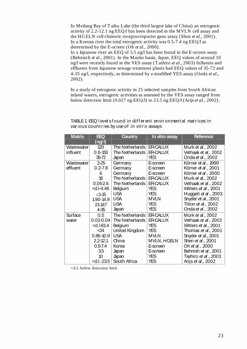
In Meilang Bay of Taihu Lake (the third largest lake of China) an estrogenicactivity of 2.2-12.1 ng EEQ/l has been detected in the MVLN cell assay andthe HGELN cell chimeric receptor/reporter gene assay (Shen et al., 2001).In a Korean river the total estrogenic activity was 0.5-7.4 ng EEQ/l asdetermined by the E-screen (Oh et al., 2000).In a Japanese river an EEQ of 3.5 ng/l has been found in the E-screen assay(Behnisch et al., 2001). In the Manko basin, Japan, EEQ values of around 10ng/l were recently found in the YES assay (Tashiro et al., 2003) Influents andeffluents from Japanese sewage treatment plants had EEQ values of 35-72 and4-35 ng/l, respectively, as determined by a modified YES assay (Onda et al.,2002).
In a study of estrogenic activity in 25 selected samples from South Africaninland waters, estrogenic activities as assessed by the YES assay ranged frombelow detection limit (0.027 ng EEQ/l) to 23.5 ng EEQ/l (Arijs et al., 2002).
TABLE 1. EEQ levels found in different environmental matrices invarious countries by use of in vitro assays
Matrix EEQ(ng/l)
Country In vitro assay Reference
Wastewaterinfluent
1200.6-15335-72
The NetherlandsThe NetherlandsJapan
ER-CALUXER-CALUXYES
Murk et al., 2002Vethaak et al., 2002Onda et al ., 2002
Körner et al ., 1999Körner et al ., 2001Körner et al ., 2000Murk et al., 2002Vethaak et al., 2002Witters et al., 2001Huggett et al ., 2003Snyder et al., 2001Tilton et al., 2002Onda et al ., 2002
Surfacewater
0.50.02-0.04<d.l-81.4
<240.86-10.92.2-12.10.5-7.4
3.510
<d.l.-23.5
The NetherlandsThe NetherlandsBelgiumUnited KingdomUSAChinaKoreaJapanJapanSouth Africa
Murk et al., 2002Vethaak et al., 2002Witters et al., 2001Thomas et al., 2001Snyder et al., 2001Shen et al., 2001Oh et al ., 2000Behnish et al., 2001Tashiro et al., 2003Arijs et al., 2002
<d.l: below detection limit
24
25
5 Evaluation of the various methods
5.1 Sample preparation
5.1.1 Collection and storage
The assessment of estrogenic activity in environmental water samples beginswith sample collection and some sort of storage until analysis. However,detailed descriptions of the modes of water sample collection and storage forin vitro investigations are lacking in most papers.Obtaining representative samples is an important requirement and is mademore problematic when working with raw sewage and other samples that arenot homogenous. Sampling periods of 7 (Murk et al., 2002) or 24 h(Desbrow et al., 1998; Körner et al., 1999, 2000, 2001) have been used tocollect composite, representative water samples in some studies, whereasdiscrete water samples have been studied in other cases. In most studies thesamples have been collected in glass bottles. The sample volume varies from 1to 25 L.The water samples were usually stored, from the time of collection untilextraction, which was usually carried out within 48 h of collection, at 4°Cwithout preservation. One study reported storage of unpreserved samples forup to 10 days (Murk et al., 2002). Other authors added methanol to the watersamples to minimize bacterial activity (Desbrow et al., 1998; Kirk et al.,2002).
Kelly (2000) reported that storage of water samples for more than a weekresulted in degradation of E2 to E1.Baronti et al. (2000) performed a stability study to evaluate estrogendegradation during storage of river water samples. According to their studythe best sample storage scheme consists in passing the field sample throughthe extraction cartridge, washing the cartridge with methanol, and storing it at−18°C. Under these conditions, which facilitate the storage of many samplesin extensive monitoring, no significant loss of estrogens was detectedfollowing storage for 60 days.
5.1.2 Filtration
Sample preparation usually begins with filtration. This step is especiallycrucial when subsequent extraction of the sample is based on the use of solid-phase extraction, since suspended solids could easily clog the adsorbent bed.The majority of the studies reviewed utilized glass fibre filters (pore sizebetween 0.45 and 1.2 µm). To elucidate whether or not the (xeno)estrogensare retained by the filter material, Desbrow et al. (1998) extractedsequentially, with a series of solvents of increasing polarity, the materialremoved from wastewater treatment plant effluent by filtration through glassfibre. The estrogenic activity of the solvent extracts, determined by means ofthe YES assay, indicated that the estrogenic activity was not retained by thefilters but was present in the dissolved phase of the effluent samples. It isquestionable, however, whether merely rinsing particulates with organic
26
solvents will quantitatively desorb the analytes from these particulates (Xiao etal., 2001).Huang and Sedlak (2001) performed recovery experiments to assess thepotential for adsorption of dissolved E2 onto filters. E2 was added towastewater effluent. After filtration, extraction and cleanup, a recovery of 99%was obtained, indication that sorption onto filters was negligible.Mol et al. (2000) reported that alkylphenols are prone to losses duringfiltration of water samples, but that these compounds, however, can readily beextracted from the filter again using a combined filtration/solid-phase set up.In one study (Kirk et al., 2002), as a supplement to filtration, centrifugationwas employed with the same purpose of eliminating suspended materials.
5.1.3 Extraction
In general, crude water samples, with no pH adjustment or addition ofmodifiers, were extracted and analysed. Exceptions to this were addition of0.5% (v/v) methanol to the sample, to facilitate solid-phase extraction(Desbrow et al., 1998; Körner et al., 1999, 2000; Kirk et al., 2002), andadjustment of the pH of the sample (to 2-3), as made by Körner et al. (1999,2000) who found that extraction of untreated wastewater at neutral pH wasincomplete. The pH adjustment step may deconjugate the steroid metabolitespresent in the water samples (Desbrow et al., 1998).Extraction of (xeno)estrogens from water samples is usually carried out bysolid-phase extraction. Both disks and, most commonly, cartridges have beenutilized for the solid-phase extraction of (xeno)estrogens from water samples,although the latter have been described as disadvantageous compared with theformer. Disks are not clogged as easily as cartridges by the suspended materialpresent in the samples, have a comparatively larger water/extractant surfacearea (which results in higher extraction rates), and eliminate the risk of samplecontamination as a consequence of leaching of plasticizers from the cartridgesupport material during elution (Schülein et al., 1995; Kelly, 2000). Otherconsiderations suggest, however, that these disadvantages of cartridges are nota great problem. Thus, filtration usually prevents clogging of cartridges.Furthermore, appropriate cleaning of the cartridge, before its conditioningand the sample loading, with the solvents which will subsequently be used forelution should eliminate, or at least minimize, leaching from the plasticholders of the cartridges (López de Alda and Barceló, 2001). Cartridges,compared with disks, have the advantage of being amenable to systemautomation, because specific devices are available for unattended washing,conditioning, sample loading, eventual drying, and final elution of a largenumber of samples (López de Alda and Barceló, 2001).Xiao et al. (2001) found extraction discs suitable for river water samples,giving good recoveries of the major estrogens E1, E2, E3 and EE2. However,the recoveries of estrogens extracted from samples of treated sewage effluentwere found to be low, possibly due to overloading by the large amount oforganic material present. For these samples, extraction using large volumeC18 cartridges provided an alternative procedure.Octadecyl (C18)-bonded silica has been the solid-phase extraction adsorbentmost commonly utilized (Desbrow et al., 1998; Körner et al., 1999, 2001;Balaguer et al., 2000; Kirk et al., 2002; Fenet et al., 2003; Kawagoshi et al.,2003; Tashiro et al., 2003). However, styrenedivinylbenzene (SDB), availableas ENV+ cartridges (Körner et al., 2000) or SBD-XC discs (Witters et al.,2001; Murk et al., 2002; Huggett et al., 2003) has also been employed. Astudy comparing the behaviour of C18 and SDB however showed that thepolymeric adsorbent (SDB) was unsuitable for quantitative extraction of E3
27
from water (López de Alda and Barceló, 2000). Körner et al. (1999) foundthat after solid-phase extraction with C18 or ENV+ cartridges, analysis of theextracts in the E-screen assay gave practically the same quantitative results.Using the C18 phase the extraction procedure generally required more timeand higher vacuum, especially when the sample contained larger amounts ofsuspended matter (raw sewage). The ENV+ phase was therefore preferred forextraction of sewage samples.The XAD-2 resin columns utilized by Shen et al. (2001) has been shown tobe inadequate for the preconcentration of estrogens from water. Kuch andBallschmiter (2000) compared the efficiencies of XAD-2 and a mixture ofLiChrolut EN and Bondesil C18 for extraction of the recovery standardcholesteryl acetate from sewage treatment plant effluent samples. Therecovery obtained with these adsorbents varied between 8 and 39% (mean:23%) and between 61 and 94% (mean: 78%), respectively. Where stated, the sample-loading flow rate in the reviewed studies variedbetween 7 and 100 ml/min (Desbrow et al., 1998; Körner et al., 1999, 2000;Shen et al., 2001; Witters et al., 2001; Tashiro et al., 2003).Elution of the retained compounds from C18 is typically carried out withmethanol (Desbrow et al., 1998; Kirk et al., 2002; Vethaak et al., 2002;Tashiro et al., 2003), with total elution volumes varying between 5 and 45 ml.Elution from SDB adsorbents has been achieved with various solvents such asacetone, methanol, methylene chloride and hexane (Witters et al., 2001;Körner et al., 2000; Murk et al., 2002; Huggett et al., 2003).Subsequent drying of the cartridge with either nitrogen or air is a commonprocedure.Ultimately extracts are taken up in methanol, dimethylsulfoxide (DMSO),hexane or ethanol for exposure in the in vitro assays (Desbrow et al., 1998;Körner et al., 1999, 2000, 2001; Balaguer et al., 2000; Shen et al., 2001;Witters et al., 2001; Kirk et al., 2002; Koh et al., 2002; Murk et al., 2002;Fenet et al., 2003; Huggett et al., 2003; Kawagoshi et al., 2003). Beresford etal. (2000) found a slightly increased sensitivity of the YES assay with DMSOcompared to ethanol.
To the knowledge of the author, the only published study of estrogenicactivity in liquid manure using in vitro assays is from the Dutch nationalinvestigation into the occurrence and effects of estrogenic compounds in theaquatic environment (Vethaak et al., 2002). In this investigation, the samplepreparation method for cattle manure samples for the ER-CALUX assay wasthe same as described for wastewater with a large load of suspended solids(personal communication, Gerard Rijs, RIZA, the Netherlands). Thisextraction procedure included soxhlet extraction of the solid material in thesamples.
To the knowledge of the author, the only published study of estrogenicactivity in agricultural drain water using in vitro assays is from the CentralValley of California (Johnson et al., 1998). In this study, water samples wereextracted with chloroform. The solvent was extracted under nitrogen anddried samples resuspended with dioxane and diluted with DMSO. Theestrogenic activity in the water samples was then assessed with an ER bindingassay.
Estrogenic activity and estrogenic chemicals in landfill leachate wereinvestigated by use of an in vitro yeast assay and chemical analysis(Kawagoshi et al., 2003). Leachate samples extracted by liquid-liquid
28
extraction with dichloromethane showed a higher in vitro estrogenic activitythan samples extracted by solid-phase extraction.In the Dutch national survey, results from solid-phase and liquid-liquidextraction were also compared (Vethaak et al., 2002). In addition to thewastewater samples prepared with standard solid-phase extraction (includingfiltration for some of the samples), some of the samples were extracted byliquid-liquid extraction (3 ml wastewater with 3x4 ml diethyl ether). Theextracts were then tested with the ER-CALUX assay. Comparison of theresults indicated a considerable difference in ER-CALUX activity measured.The EEQ values of the samples extracted with the standard solid-phaseextraction procedure were on average only 17% of the EEQ values obtainedvia the liquid-liquid extraction procedure. This indicates that a relatively largeportion of the estrogenic activity in water may be lost via filtration and solid-phase extraction. However, for surface water samples, Mol et al. (2000) found both liquid-liquid extraction (at pH 5-6) and a combined filtration/solid-phase extraction(at pH 4) to be suited for extraction of (xeno)estrogens. With the lattermethod, recoveries between 58% (for bisphenol A) and 106% (for EE2) werefound. For the liquid-liquid extraction method, recoveries of 109-117% werefound.
In the comparative study with an ER binding assay, the YES assay and theER-CALUX assay for detection of estrogenic potency in wastewater andsurface water, the same sample preparation was used for all three in vitro tests(Murk et al., 2002).The amounts of material needed to determine the estrogenic potency of thethree in vitro assays, however, differed greatly: ER binding assay > YES >ER-CALUX. In the ER-CALUX assay 6-30 ml surface water; 0.2-2.5 mlwastewater influent; and 0.9-9 ml wastewater treatment plant effluent wasneeded. In the YES assay the corresponding amounts were 60-250 ml, 4-100ml, and 10-100 ml, respectively. In the ER binding assay the needed amountswere approximately 950 ml for surface water and 400 ml for wastewaterinfluents or effluents.
Körner et al. (1999, 2000, 2001) used the E-screen for analysis of estrogenicactivity in sewage plant effluents in Germany. Following sample extraction,stock solutions of the extracts were prepared with steroid-free experimentalmedium. This medium consisted of phenol red-free Dulbecco´s modificationof Eagle´s medium supplemented with charcoal dextran-treated fetal calfserum (CD-FCS), HEPES, glutamine, amino acids and apenicillin/streptomycin/amphotericin solution. CD-FCS was prepared bytreatment of fetal calf serum (FSC) with charcoal-dextran (CD) followed bystirring for 24 h, centrifugation and filtration.
5.2 Laboratory facility requirements
ER binding assays include the use of radiolabels, which involves potentialhealth hazards and the requirement for special licenses, equipment andprecautions for handling and disposing radioactive material. The majorequipment required is a liquid scintillation counter.
Unlike the ER-binding assays, reporter gene assays and the E-screen involvecell cultures. Compared with yeast cells, mammalian cells are more expensiveand difficult to cultivate. Mammalian cells require more constant care and
29
fresh medium; and are vulnerable to the risks of contamination. Culturedmammalian cells are particularly sensitive to variations in temperature, pH,dissolved oxygen and certain metabolites, which makes it necessary to controlculture conditions carefully. Yeast cells are more resilient and highly resistantto adverse environmental conditions, making them relatively easy to maintainand to grow.
The specific needs as related to the various in vitro procedures utilizingreporter genes, whether transiently or stably transfected, are essentially thesame. A standard cellular or molecular biology laboratory with cell culturecapabilities is required.The major equipment required for mammalian-based studies is a cellincubator with temperature, CO2, and humidity controls; sterile laminarcabinets; and a luminometer for assays requiring luciferase detection.The YES assay with yeast cells does no require that the researchers work tothe same standards of sterility as for the mammalian cell assays (personalcommunication, Dr. Juliette Legler, IVM, Vrije Universiteit Amsterdam, theNetherlands). A sterile laminar cabinet may not be required for the YES assayas the yeast cells are less susceptible to effects by bacterial and fungalinfections. A specialized incubator is also not necessary for the yeast cells.
The E-screen has the same facility and equipment requirements asmammalian-based reporter gene assays, except that cell-counting equipment(Coulter counter) would be an additional requirement if the method of Soto etal. is followed.
5.3 Detection limits and EC50 values
5.3.1 Detection limits and EC50 values for the various in vitro assays
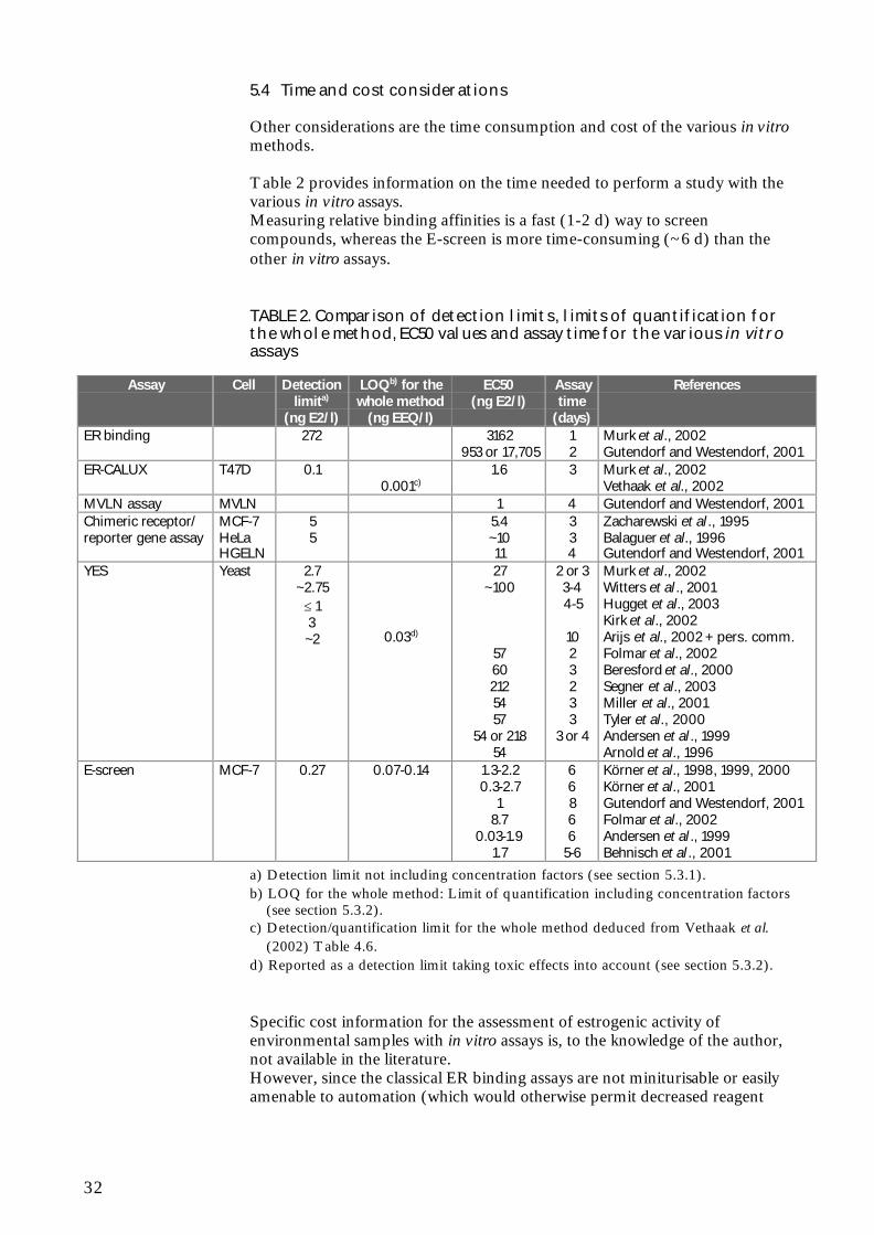
This section deals with the detection limits and EC50 values for E2, notincluding the concentration factors employed when assessing environmentalsamples. The EC50 is the concentration at which half-maximal activity isinduced. The definition of the detection limit is often not stated in thepublished studies. However, Witters et al. (2001) calculated the detectionlimit of the YES assay as absorbance elicited by the solvent control plus threetimes the standard deviation. Körner et al. (1999) defined the detection limitof the E-screen as the concentration of a single compound or anenvironmental sample inducing a cell proliferation significantly higher thanthat of the hormone-free negative control.An overview of the detection limits and EC50 values for the various in vitroassays is found in Table 2.
Detection of estrogenic potency in wastewater and surface water with three invitro assays was studied by Murk et al. (2002). The three assays applied werean ER binding assay and two reporter gene assays: ER-CALUX and YES. Allassays were able to detect estrogenicity in wastewater and surface water.However, the detection limits differed greatly between the three assays: ERbinding assay >> YES > ER-CALUX. The detection limit for the ER-CALUX assay was 0.1 ng/l E2; for the YES assay it was 2.7 ng/l E2; and forthe ER binding assay it was 272 ng/l E2. The EC50 values for the ER-CALUX, the YES and the ER binding assay were 1.6 ng/l E2, 27 ng/l E2 and3162 ng/l E2, respectively.
30
For E2, nonylphenol and o,p’-DDT, a difference of approximately 6- to 20-fold was found between the EC50 values in the ER-CALUX and the YESassay (Legler et al., 2002a).
In order to assess the (anti)estrogenic potential of pure compounds andcomplex environmental samples Gutendorf and Westendorf (2001)compared an array of in vitro test systems, (i) two luciferase reporter geneassays: the MVLN cell assay and the HGELN cell chimeric receptor/reportergene assay; (ii) competitive binding assays with recombinant human ER αand β; and (iii) the E-screen. The sensitivity of the assays for E2 decreased inthe order: MVLN cell assay = E-screen > HGELN cells > binding to ER-α >binding to ER-β. The EC50 for the MVLN cells and the E-screen was 1 ng/lE2. For the HGELN cells, binding to the ER-α and binding to the ER-β theEC50 values were 11 ng/l, 953 ng/l and 17,705 ng/l, respectively.
A study using the E2 Bioassay for detection of estrogenic activity in pulp andpaper mill black liquor and effluent showed a detection limit of approximately5 ng/l and an EC50 of 5.4 ng/l for E2 (Zacharewski et al., 1995).Another study using the E2 Bioassay for assessment the estrogenic activities ofchemicals and complex mixtures likewise showed a detection limit ofapproximately 5 ng/l E2. The EC50 was 11 ng/l E2 (Balaguer et al., 1996).
A comparison of the estrogenic potencies of E2, EE2, diethylstilbestrol,nonylphenol and methoxychlor in vivo and in vivo showed that the EC50values for all five chemicals were approximately one order of magnitudehigher in the YES assay than in the E-screen assay. In the YES assay theEC50 for E2 was 57 ng/l whereas in the E-screen the EC50 for E2 was 8.7ng/l (Folmar et al., 2002).
A recent study compared the potencies of estrogenic compounds in the YESassay (Segner et al., 2003). The EC50 values for EE2, E2, 4-tert-octylphenoland bisphenol A were 220 ng/l, 212 ng/l, 436 µg/l and 2615 µg/l, respectively.
In a comparative study of in vitro and in vivo assays for estrogenicity ineffluent from North American municipal wastewater facilities Hugget et al.(2003) reported the detection limit of the YES assay as ≤ 1 ng/l E2.A study using the YES assay for detection of estrogenic activity in Flemishsurface waters showed a detection limit of ~2.75 ng/l E2 and EC50 values of~100 ng/l E2 (Witters et al., 2001). Similar detection limits and EC50 valueswere reported in other studies (Beresford et al., 2000; Kirk et al., 2002).Comparison of short-term estrogenicity tests for identification of hormone-disrupting chemicals revealed EC50 values of 54 ng/l or 218 ng/l E2 for theYES assay (Andersen et al., 1999). Arnold et al. (1996), Tyler et al. (2000)and Miller et al. (2001) likewise reported EC50 values around 54 ng/l for theYES assay.
In a number of papers dealing with the E-screen and estrogenic activecompounds in sewage treatment plants in Germany, EC50 values between0.3-2.7 ng/l E2 were recorded (Körner et al., 1998, 1999, 2000, 2001). Thedetection limit of the E-screen method was 0.27 ng/l E2.A comparison of short-term estrogenicity tests for identification of hormone-disrupting chemicals revealed EC50 values of 0.03-1.9 ng/l E2 for the E-screen (Andersen et al., 1999).
31
A study of the estrogenic potency in each step of a controlled landfill leachatetreatment plant in Japan showed an EC50 of 1.7 ng/l E2 for the E-screen(Behnisch et al., 2001).
The above studies show that the yeast-based YES assay is less sensitive thanthe mammalian-based MVLN cell assay, ER-CALUX and E-screen. Theremay be a number of explanations for this difference in sensitivity between theassays. One explanation could be differences in uptake of compounds throughthe yeast cell wall relative to mammalian cell membranes (Zysk et al., 1995)and the ability of yeast cells to actively transport specific compounds out ofthe cell (Kralli et al., 1995). Other mechanisms that may be involved in thedifference in sensitivity between the yeast- and mammalian-based assaysinclude differences in cellular transcription factors (Halamachi et al., 1994),multiple drug resistance (Dexter et al., 1994), and endogenous yeast estrogenbinding proteins (Feldman et al., 1982). For the reporter gene assays, the typeof reporter protein used may also have a major impact on the sensitivity of theassay, because it determines the type of instruments or analytical methods thatcan be used to detect it (Villeneuve et al., 1998). Because of the availability ofsensitive detectors for light and the high quantum efficiency of the luciferasereaction, the light-producing endpoint for luciferase-based reporter geneassays can be very sensitively detected using a luminometer. Colorimetricendpoints, such as the β-galactosidase endpoint used in the YES assay tend tobe less sensitive.
The YES assay can be made more sensitive by using longer incubationperiods (UK Environment Agency, 1997; Beresford et al., 2000).
5.3.2 Detection limit and limit of quantification for the whole method
This section deals with the detection limit and limit of quantification for thewhole method, i.e. including the concentration factors employed whenassessing environmental samples.In calculating a detection limit for the whole method for analysing a liquidsample the maximally achievable concentration factor has to be taken intoaccount. However, few studies report this concentration factor, the detectionlimit or the limit of quantification for the whole method.Körner et al. (1999) used a maximal concentration factor of 20, resulting in adetection limit of 0.014 ng/l EEQ for the whole method. As the highestconcentration of the effluent samples showed some cytotoxic effects in the E-screen but not anymore in the 5-10-fold dilution, the limit of quantificationwas 0.07-0.14 ng EEQ/l.Witters et al. (2001) used a maximal concentration factor of 100 for the YESassay, which would result in a detection limit for the whole method of 0.028ng EEQ/l. Although they observed cytotoxic effects at the highest testconcentrations they did not report at which dilution cytotoxic effects were nolonger observed. Thus, the limit of quantification is not known but is >0.028ng EEQ/l.Arijs et al. (2002) reported a detection limit of 0.027 ng EEQ/l for the YESassay. The detection limit was calculated as EC50[E2] divided by the max.extract concentration that was not toxic to the yeast (personalcommunication, Katrien Arijs, Ghent University, Belgium).
32
5.4 Time and cost considerations
Other considerations are the time consumption and cost of the various in vitromethods.
Table 2 provides information on the time needed to perform a study with thevarious in vitro assays.Measuring relative binding affinities is a fast (1-2 d) way to screencompounds, whereas the E-screen is more time-consuming (~6 d) than theother in vitro assays.
TABLE 2. Comparison of detection limits, limits of quantification forthe whole method, EC50 values and assay time for the various in vitroassays
Zacharewski et al ., 1995Balaguer et al., 1996Gutendorf and Westendorf, 2001
YES Yeast 2.7~2.75
≤ 13~2 0.03d)
27~100
57602125457
54 or 21854
2 or 33-44-5
1023233
3 or 4
Murk et al., 2002Witters et al ., 2001Hugget et al., 2003Kirk et al., 2002Arijs et al., 2002 + pers. comm.Folmar et al., 2002Beresford et al., 2000Segner et al., 2003Miller et al., 2001Tyler et al., 2000Andersen et al ., 1999Arnold et al., 1996
E-screen MCF-7 0.27 0.07-0.14 1.3-2.20.3-2.7
18.7
0.03-1.91.7
66866
5-6
Körner et al., 1998, 1999, 2000Körner et al., 2001Gutendorf and Westendorf, 2001Folmar et al., 2002Andersen et al ., 1999Behnisch et al ., 2001
a) Detection limit not including concentration factors (see section 5.3.1).b) LOQ for the whole method: Limit of quantification including concentration factors
(see section 5.3.2).c) Detection/quantification limit for the whole method deduced from Vethaak et al.
(2002) Table 4.6.d) Reported as a detection limit taking toxic effects into account (see section 5.3.2).
Specific cost information for the assessment of estrogenic activity ofenvironmental samples with in vitro assays is, to the knowledge of the author,not available in the literature.However, since the classical ER binding assays are not miniturisable or easilyamenable to automation (which would otherwise permit decreased reagent
33
cost and increased throughput), they must be expected to be relativelyexpensive assays.The reporter gene assays and the E-screen are applicable to multiwelltechnology, thus reducing time consumption and cost. However, cultivationof mammalian cells is more demanding than cultivation of yeast cells in termsof growth medium reagents and time consumption. The mammalian-basedreporter gene assays and the E-screen must therefore also be expected to berelatively expensive, whereas the YES assay must be expected to be relativelyinexpensive.
5.5 Robustness
The success of assessments of estrogenic activity is dependent on therobustness of the in vitro assay in providing reproducible data with relativelysmall variations.
Despite its widespread use, considerable inter-laboratory variability has beenobserved in test results from the E-screen. Numerous cell lines and widelyvarying test procedures have been employed, which may account for much ofthe variability in results (Zacharewski, 1997). Since the establishment of theMCF-7 cell line 30 years ago, the cell line has undergone several changes, andstudies have demonstrated that MCF-7 cell line variants exhibit fundamentaldivergences in characteristics such as (xeno)estrogen-dependent proliferationrate, population doubling time (Villalobos et al., 1995), and susceptibility toapoptosis (Burow et al., 1998). In addition there are differences betweendifferent MCF-7 stocks in regard to their ability to detect antagonists (Diel etal., 1999). Villalobos et al. (1995) have shown the influence of differentMCF-7 cell sublines on test results. Likewise, variations in culture conditions,such as the number of cells plated and the duration of incubation in estrogen-free medium prior to treatment with test compounds, have been shown tohave prominent effects on the responses to E2 (Jones et al., 1997; Rasmussenand Nielsen, 2002). In addition, only some sera support estrogen-specificgrowth of MCF-7 cells (Wiese et al., 1992). Furthermore, drift inresponsiveness of MCF-7 cells during culture may confound their consistentuse in proliferation assays (Desaulniers et al., 1998; Jones et al., 1998; Odumet al., 1998). Payne et al. (2000) have demonstrated the importance of choiceof cell line and culture conditions in determining test results. In a large inter-laboratory study, Andersen et al. (1999) reported that by using a standardisedcell line (MCF-7/BUS) with similar protocols, good agreement could beachieved with most test compounds. However, they found a lack ofconsistence with chemicals such as benzyl butyl phthalate and p,p’-DDE.It must also be noted that a range of non-estrogenic substances, includingprogesterone, androstenediol, insulin-like growth factors, epidermal growthfactor, caffeine and ethanol have been found to influence the proliferation ofhuman breast cancer cells (Osborne et al., 1990; van der Burg et al., 1992;Dickson and Lippman, 1995; Jones et al., 1998; Diel et al., 1999; Andò et al.,2002). Furthermore, the E-screen assay might be extremely sensitive to smallchanges in the physical or chemical properties of culture conditions inducedby test substances, leading to non-specific increases or decreases inproliferation independent of ER-binding (Desaulniers et al., 1998). Thus, apositive response cannot be attributed strictly to estrogen receptor agonists.The use of anti-estrogens (e.g. ICI 182,780) could help to distinguish
34
estrogenic from non-estrogenic activity. However, this increases thecomplexity of the assay.
In the E2 Bioassay responsiveness has been observed to be sensitive to thenumber of passages and the type of cells utilized (Zacharewski, 1997). MCF-7 cells recently taken from frozen stocks (i.e., within the first three passages)and those beyond 10 passages after removal from frozen stocks show loweroverall E2-induced luciferase activity. Consequently the assay is mostconsistent when the cells utilized are between 3 and 10 passages. Differencesin responsiveness have also been observed when the constructs are transfectedinto different cell lines. For example, maximum induction in stably transfectedHeLa cells ranges from 8- to 12-fold while maximum induction in transientlytransfected MCF-7 cells averages between 40- and 50-fold. Differences in thelevel of responsiveness within experiments and between cell types may be dueto variations in ER levels and in the presence of appropriate transcriptionfactors.
When MVLN cells are exposed to hydroxytamoxifen or tamoxifen theirluciferase reporter gene can irreversibly no longer respond to estrogens (Badiaet al., 1994). This raises the issue of instability of the MVLN cell assay due toexposure to inhibiting chemicals during cell culture or assay performance.
Beresford et al. (2000) investigated the effect of alterations in assaymethodology for the YES assay on the response to certain xenoestrogens.None of the four parameters examined (incubation time, whether the solventwas allowed to evaporate or not, the type of solvent, and initial yeast cellnumber) had any appreciable effect on the relative potencies of nonylphenolor bisphenol A. However altering these criteria did affect both the dose-response curves produced by butyl benzyl phthalate and o,p’-DDT. Inaddition other factors, such as incubation temperature and growth stage of theyeast, may also alter the response in the YES assay.The YES assay does not consistently differentiate between estrogen agonistsand antagonists. Tamoxifen and hydroxytamoxifen have shown both agonisticand antagonistic activity in the YES assay (Routledge and Sumpter, 1997;Sohoni and Sumpter, 1998; Legler et al., 2002a). The anti-estrogen ICI182,780 produces a purely agonistic response in the YES assay (Beresford etal., 2000, Legler et al., 2002a).
Legler et al. have used both the ER-CALUX and the YES assay fordetermination of estrogenic activity in environmental matrices. They do notculture the ER-CALUX T47D cells higher than 30 passages (personalcommunication, Dr. Juliette Legler, IVM, Vrije Universiteit Amsterdam, theNetherlands), as some studies indicate that the responsiveness of the cellsdecreases, as they get older. Despite the “fussiness” of the cells, they findthem to give more reproducible results (ER-CALUX %CV: 5-10) than theYES assay (%CV: 10-25) and would say that the ER-CALUX is more robust.
5.6 Utility in various matrices
In vitro assays have been employed to assess estrogenic activity in a range ofenvironmental matrices. Various aquatic samples, such as wastewatertreatment plant influents and effluents (Körner et al., 1999, 2001; Kirk et al.,2002; Murk et al., 2002; Onda et al., 2002; Tilton et al., 2002; Vethaak et al.,2002; Huggett et al., 2003), surface water (Oh et al., 2000; Khim et al., 2001;
35
Thomas et al., 2001; Witters et al., 2001; Murk et al., 2002; Vethaak et al.,2002; Fenet et al., 2003) or agricultural drain water (Johnson et al., 1998)have been analysed by in vitro assays.For wastewater and surface water both ER binding assays, ER-CALUX,MVLN cell assays, YES and E-screen have been utilized (Körner et al., 1999,2000, 2001; Oh et al., 2000; Behnish et al., 2001 Shen et al., 2001; Snyder etal., 2001; Witters et al., 2001; Murk et al., 2002; Onda et al., 2002; Vethaak etal., 2002; Huggett et al., 2003). To the knowledge of the author, the onlypublished study utilizing an in vitro assay for assessment of estrogenic activityin agricultural drain water was the ER binding study performed by Johnson etal. (1998). Likewise, to the knowledge of the author, only a single study hasbeen published on the employment of in vitro assays on manure samples(Vethaak et al., 2002). In this study the ER-CALUX assay was used to assessestrogenic activity in two liquid cattle manure samples.
In vitro assays may be subject to interferences owing to the complexity ofenvironmental samples. Toxicity of the samples to the yeast or mammaliancells is a potential problem in assessing estrogenic activity in an environmentalsample. Cytotoxic effects at high sample concentrations have been observedin both the ER-CALUX assay (Legler et al., 2002a), the MVLN cell assay(Khim et al., 1999, 2001), the E-screen (Körner et al., 2001) and the YESassay (Andersen et al., 1999; Payne et al., 2000; Witters et al., 2001). Toxiceffects cause irregularities in the concentration-response curves for estrogenicactivity. Consequently, toxic effects should be carefully distinguished fromestrogenic responses, and the assays should not be run with sampleconcentrations that affect cell viability. Dilution of the samples may reducetoxicity to the cells but also reduces the possibility of detecting estrogenicactivity.Yeast assays may perform better for monitoring of environmental samples, asthese samples are frequently contaminated with substances other than(xeno)estrogens interfering with the growth and viability of animal cells, butnot with yeast cells (Graumann et al., 1999). Yeast is more resistant toenvironmental contaminants such as heavy metals and bacterial endotoxinscompared to mammalian cells (Breihofer et al., 1998). In a survey of watersamples from Flemish freshwater systems the MVLN cell assay and the YESassay were compared (Witters et al., 2001). Significant cytotoxicity for anumber of samples was found in the MVLN cell assay compared to the YESassay.
Körner et al. (1999, 2000) used the E-screen for the quantitativedetermination of total estrogenic activity in extracts from municipal sewageplants. As no cytotoxic effects occurred in the range of dilutions where a dose-response relationship of the estrogenic activity was observable, it was possibleto analyse the samples quantitatively in the E-screen assay without any clean-up step. To exclude the theoretical possibility that a sample may stimulateproliferation of MCF-7 cells by mechanisms that are separate from aninteraction with the ER an active concentration of each sample was analysedtogether with the ER antagonist ICI 182,780. Cell proliferation induced bythe sewage samples was completely inhibited by coincubation with ICI182,780. This verified that the cell proliferation caused by the samples wasER-mediated.
36
5.7 Advantages and limitations compared to chemical analyses
Significant advantages of in vitro assays over chemical analyses are that nounknown components with estrogenic activity are overlooked and that anycombination effects are taken into account in the analysis. Although chemicalanalyses are important for the identification and quantification of compounds,they give no information about the estrogenic potency of those compounds. Invitro assays provide an integrated measure of the combined potency of allcompounds in a sample.The wide structural diversity of (xeno)estrogens sets a limit to singlecompound chemical analysis of environmental matrices regarding time andlabour. Therefore, in vitro assays are essential for monitoring environmentalsamples for their content of substances with estrogenic activity regardless oftheir chemical nature. Moreover, in vitro assays can detect compounds forwhich there are no analytical methods available.
If both chemical analyses and in vitro assays are performed on anenvironmental sample, and if the in vitro estrogenic activity of the chemicalsmeasured is known, it is possible to determine which proportion of the totalestrogenic activity can be ascribed to the (xeno)estrogenic chemicals analysed.In order to compare the in vitro estrogenic activity measured in variousenvironmental samples with the chemically detected concentrations of known(xeno)estrogenic compounds, results of the chemical analyses of a sample canbe expressed as theoretical EEQ values. By summing all the EEQ values forsingle compounds in a sample, the total calculated EEQ in that sample can bedetermined assuming concentration additivity.If the EEQ levels calculated are lower than the measured in vitro EEQ levels,other compounds, that were not chemically analysed in the study may bepresent in the extracts and also contribute to estrogenic activity.If the calculated EEQ values are higher than in vitro EEQ values, there may beseveral explanations: (i) calculation of EEQ values based on detection limitscauses an overestimation of the EEQ (Murk et al., 2002; Vethaak et al., 2002);(ii) differences in extraction methods; and (iii) unidentified compounds mayantagonise estrogenic compounds in the in vitro assay.
In the published studies using this comparative approach, the in vitroestrogenic activity is often found to be lower than the chemically measuredestrogenic activity.The EEQ values of effluents of sewage treatment plants in SouthwesternGermany as determined by the E-screen were compared to those calculatedfrom the results of chemical analysis of 13 natural and synthetic estrogenicsubstances (Körner et al., 2001; Spengler et al., 2001). For the majority of thesamples, the EEQ calculated from the GC-MS results was higher than thatdetermined in the E-screen assay by a factor 2 to 4, whereas on the otherhand, no sample had an EEQ in the cell culture test higher than that derivedfrom chemical analysis. Murk et al. (2002) demonstrated that the calculatedEEQ values in the surface waters in the Netherlands were 5 to 10 times higherthan EEQ values measured in the ER-CALUX assay. Similar results werefound by utilizing the YES assay on effluents from North American municipalwastewater (Huggett et al., 2003). Likewise, lower in vitro activities in theMVLN cell assay versus measured concentrations were observed in Nevadawastewater evaluations (Snyder et al., 2001) and in Taihu Lake, China (Shenet al., 2001). The Dutch national survey (Vethaak et al., 2002) and pilot study(Belfroid et al., 1999) compared the calculated estrogenic activity and in vitro
37
estrogenic activity measured in the ER-CALUX assay . In the pilot study, thecompounds studied accounted for 70% of the estrogenic activity of untreatedwastewater. The corresponding value for wastewater treatment plant effluentswas only 20%. For surface water, on the other hand, the measured activitieswere much lower than the calculated activities. In the national survey, some ofthe sewage water plant effluents and surface water samples had calculatedEEQ levels below the measured in vitro EEQ levels. However, calculated EEQvalues were higher than in vitro EEQ values by one order of magnitude formost of the extracts.
In vitro assays facilitate identification of environmental samples that warrantadditional chemical analysis. If no significant response is detected in vitro,there may be no need to conduct expensive and time consuming chemicalanalyses. Since the method detection limit is known for the in vitro assay, anupper limit of EEQ in the sample can be defined. However, if a sampleproduces a significant response in an in vitro assay, chemical analysis can beemployed to uncover the cause. Fractionation of sample extracts showingsignificant activities, and subsequent reanalysis with the in vitro assays canidentify important classes of compounds that are responsible for the observedactivity. Chemical analysis is utilized to determine the compounds likelyresponsible for the activity observed in each fraction. Once candidatecompounds have been identified, a theoretical estrogenic activity can beestimated as the summation of the potency of the single compounds based onidentification and quantification by chemical analysis. This theoreticalestrogenic activity may be compared to the actual estrogenic activity detectedin the in vitro assay to find out if all of the estrogenic activity has beenidentified.
A combination of in vitro assays and chemical techniques has been utilized instudies on the effluents from sewage treatment works to both confirm thatthey exhibit estrogenic activity and to identify the compounds that wereresponsible. Such a study was carried out in the United Kingdom, followingthe observation that sewage effluent was estrogenic to fish, in an attempt toidentify the causative compounds (Desbrow et al., 1998). Samples werefractioned and the YES assay was employed to identify the fractions thatcontained estrogenic activity. The active fractions were then analyzed usingGC-MS to enable elucidation of the compounds present. A similar approachhas been used to identify estrogenic compounds in effluents from a sewagetreatment works in Michigan (Snyder et al., 2001).
5.8 Limitations compared to in vivo assays
Although in vitro assays are attractive for monitoring studies, one of theirmain disadvantages is their simplification of the in vivo situation. In vitroassays are mechanism specific, whereas in vivo assays permit the detection ofeffects resulting from multiple mechanisms. In vitro assays do not completelyaccount for complex in vivo events, such as bioavailability, toxicokinetics,metabolism and cross talk between biological pathways (Zacharewski, 1997).For example, in vitro assays are limited in their ability to mimic whole animalmetabolism. As a result, extrapolation from in vitro to in vivo assays can leadto false negatives for compounds that are bioactivated, and overestimates ofpotency for compounds readily inactivated in vivo. Though some metabolic
38
capacity has been observed in some in vitro assays (Soto et al., 1995; Sohoniand Sumpter, 1998; Andersen et al., 1999; Legler et al., 1999; Tyler et al.,2000; Layton et al., 2002), the complex processes of uptake, binding to carrierproteins, transport, targeting and disposition of compounds in whole animalsare not taken into account in the in vitro systems (Zacharewski, 1997). Inaddition, bioaccumulation and homeostatic controls generally are notsimulated by in vitro testing systems. In vitro assays would not revealcompounds that exert (anti-)estrogenic responses in vivo by modifyingendogenous E2 levels via effects on the hypothalamus or pituitary. In vivoassays can integrate estrogenic effects that may occur by different mechanismsin multiple tissues simultaneously. Fish can serve as integrators of responsesto mixtures of toxicants that occur in the aquatic environment and are thususeful in vivo models for studying effects of estrogenic compounds in water.In addition, effects related to growth and reproduction in fish are more easilyrelated to population level and ecological effects than are effects in in vitrosystems. Thus, in vivo studies in both the field and laboratory are essential forlinking exposure to biologically relevant effects. They are, however,impractical for routine, high throughput monitoring of environmentalsamples. In vivo studies are in general more laborious, time consuming andcostly compared to in vitro assays, and may involve large amounts of animals.Furthermore, considerable inter-individual variations often exist in responses.Utilization of in vitro assays circumvents much of the inter-individual,seasonal, and temporal variability, which can complicate interpretation of invivo responses. Additionally, in vitro assays avoid many of the ethical issuesassociated with whole animal studies. However, due to the clear drawbacks ofin vitro assays that might result in unreliable predictions, a combination of testmethods, including in vivo assays, which assess both the ER- and non-ER-mediated mechanisms of action is suggested as most appropriate to determinethe estrogenic activity of environmental matrices.
It is not uncommon for in vitro estrogenic activity to differ from in vivoactivity.An investigation was made to determine how accurately two in vitro assays(YES and E-screen) predicted responses of estrogenic compounds ininducing vitellogenin synthesis in sheepshead minnow (Folmar et al., 2002).The results showed major discrepancies between the relative estrogenicactivities of the xenoestrogens methoxychlor and nonylphenol in the in vitroassays relative to the in vivo assay. The xenoestrogens were clearly morepotent in vivo than in the in vitro assays.Likewise, another study showed that the relative potency of (xeno)estrogensin the YES assay did not reflect potency in a medaka assay (Metcalfe et al.,2000).Segner et al. (2003) compared potencies of estrogenic compounds in in vitroassays (YES, ER-receptor binding and vitellogenin induction in cultured fishhepatocytes) and in life cycle tests with zebrafish in vivo. The in vivo activityof the test compounds was not accurately predicted by the in vitro assays, withrespect to neither sensitivity nor ranking. The in vitro assays tended tooverestimate the relative potency of the xenoestrogens; i.e. the ratio betweenthe EC50 of the reference compound, EE2, and that of the test compound.The best prediction of the in vivo fish test results was obtained from the YESassay. Although still being clearly less sensitive than the in vivo test, the YESassay showed the highest absolute sensitivity of the in vitro test systems. Thesensitivity difference between the YES and zebrafish assay was mostpronounced for EE2 (about two orders of magnitude) while it was smaller forthe xenoestrogens (about one order of magnitude).
39
To compare in vitro and in vivo assays, wastewaters from North Americanmunicipal wastewater facilities were evaluated for estrogenic activity using theYES assay and an in vivo fish vitellogenin assay (Huggett et al., 2003). In vivoestrogenic activity was nearly 10-fold greater than YES activity.In addition to the already mentioned risks of cytotoxic effects in in vitroassays, other potential explanations for enhanced in vivo activity are enhancedbioavailability/absorption of compounds by the fish relative to in vitro cells,bioaccumulation in vivo, and/or the occurrence of non-ER ligands, whichelicit estrogenic effects in fish through indirect mechanisms (Folmar et al.,2002; Huggett et al., 2003).
Compounds with similar estrogenic activities in vitro sometimes possess verydifferent activities in vivo. Several in vitro assays (ER binding assays, ER-CALUX, MVLN cell assay, HGELN cell chimeric receptor/reporter geneassay, YES and E-screen) have displayed approximately the same estrogenicactivity of E2 and EE2 (Gutendorf and Westendorf, 2001; Shen et al., 2001;Murk et al., 2002) while EE2 in vivo has been found to have approximately10-100 times higher estrogenic activity than E2. For example, EE2 has beenreported to be 16-fold more potent than E2 in inducing vitellogenin synthesisin male zebrafish (Rose et al., 2002), 11- to 27-fold fold more potent than E2in inducing vitellogenin synthesis in juvenile rainbow trout (Thorpe et al.,2003) and 100 times more potent than E2 in inducing testis-ova in Japanesemedaka following exposure during early life-stages (Metcalfe et al., 2001).Legler et al. (2002b) likewise found considerable differences in the relativepotencies of (xeno)estrogens in vitro and in vivo. EE2 was the most potent ofthe (xeno)estrogens tested and was 100 times more potent than E2 in an invivo transgenic zebrafish assay whereas in the ER-CALUX assay, EE2 wasonly slightly (1.2 times) more potent than E2. For wastewater treatment planteffluent extracts the average measured EEQ value in the ER-CALUX assaywas 3 ng/l whereas for the transgenic fish assay the corresponding value was51 ng/l (Legler, 2001). Thus it was demonstrated that the effluent sampleswere more estrogenically potent in vivo than would be expected based on theirin vitro estrogenic activity. Similarly, Huggett et al. (2003) found that in vivoestrogenic activity of effluent from North American municipal wastewaterfacilities was nearly 10-fold greater than in vitro estrogenic activity assessed bythe YES assay .The results from the above studies suggest that the sole use of in vitro assaysto monitor for estrogenic activity may severely underestimate in vivoestrogenic potential of environmental samples and thus could potentiallyresult in false negative evaluations.
40
41
6 Discussion and recommendations
Numerous substances present in the aquatic environment have beendemonstrated to possess estrogenic activity. Chemical analysis of allcompounds with potential estrogenic activity would be very costly andunknown estrogenic compounds, including metabolites, may still be present inenvironmental matrices. In vitro assays offer an integrated measure of theestrogenic potencies of environmental matrices without knowing all relevantcompounds beforehand. In vitro assays can be utilized to examineconsiderable numbers of samples for their estrogenic activity.
The sample preparation procedure often is the most tedious and timeconsuming step in monitoring using in vitro assays. However, samplepreparation may be considered the most crucial part of the assessmentprocess. Extensive environmental surveys involve the analysis of a largenumber of samples that cannot be immediately analyzed. However, extractionshould be carried out as quickly as possible to avoid addition of chemicalpreservatives. It is important to note that when working with extracts ofenvironmental matrices there may be loss of compounds during the extractionprocess. (Xeno)estrogens can be extracted by both solid-phase extraction andliquid-liquid extraction. Liquid-liquid extraction has been shown to be moreefficient than solid-phase extraction. This may be due to the requiredfiltration step preceding solid-phase extraction, which may result in loss of(xeno)estrogens adsorbed to suspended particulate matter. However, theconventional liquid-liquid extraction often requires considerable amounts oftoxic organic solvents, and time-consuming procedures. Thus, solid-phaseextraction could be preferred over liquid-liquid extraction. A large number ofsamples require a high sample throughput, which can be achieved by using avacuum manifold system for solid-phase extraction cartridges. The filtrationand extraction procedures for environmental samples should be properlyvalidated and optimised before the onset of extensive monitoring studies. Byusing the appropriate extraction method it should be possible to assessestrogenic activity in different environmental matrices, such as wastewater,surface water, drain water and liquid manure slurry.
A wide variety of in vitro assays has been developed to measure the estrogenicactivity of single compounds or complex environmental samples. Each assaymeasures different end points at different levels of biological complexity ofestrogen action (i.e., receptor binding, cell proliferation, and expression of areporter gene). No single in vitro assay can be regarded as ideal for assessingthe estrogenic activity of wastewater and surface water. They all have theiradvantages and limitations. A comparison of some of the key parameters forthe various in vitro assays is shown in Table 3.
Although they are fast, ER binding assays are significantly less sensitive thanthe other in vitro assays. In addition, binding assays are not easily amenable toautomation, thereby limiting their utility as a screening tool. Furthermore, ERbinding assays require specialised laboratory facilities because of theradioactive substances. Finally, the binding of a substance to the ER is onlyindicative that it may act as a xenoestrogen and some studies suggest, that ER
42
binding may be a poor predictor of more complex in vitro and in vivoresponses (Villeneuve et al., 1998).
TABLE 3. Comparison of key parameters for the various in vitro assaysfor detection of estrogenic activity
Assay type ER binding Reporter gene assays Cellproliferation
Assay ER binding ER-CALUX MVLN Chimericreceptor YES E-screen
Cells T47D MVLNMCF-7HeLa
HGELNYeast MCF-7
Laboratoryrequirements
Isotopelaboratory
Sterilelaboratory equipment
Cell culturelaboratory
Sterile lab.equipment
Cell culture Relativelydemanding
Relativelysimple
Relativelydemanding
Miniturisation/automation No Yes Yes Yes
Costs Relativelyexpensive
Relativelyexpensive
Relativelyinexpensive
Relativelyexpensive
Detection ofanti-estrogens No Yes Limited Yes
Assay time(days) 1-2 3 4 ~ 3 ~ 3 ~ 6
Detection limita)
(ng E2/l) 272 0.1 5 1-3 0.27
LOQ for thewhole methodb)
(ng EEQ/l)0.001c) 0.03d) 0.07-0.14
a) Detection limit not including concentration factors (see section 5.3.1).b) LOQ for the whole method: Limit of quantification including concentration factors
(see section 5.3.2).c) Detection/quantification limit for the whole method deduced from Vethaak et al.
(2002) Table 4.6.d) Reported as a detection limit taking toxic effects into account (see section 5.3.2).
Although cell cultures involve disadvantages associated with maintaining thecell line and avoiding contamination, their use in test assays offers significantadvantages regarding sensitivity. Both reporter gene assays and the E-screenhave been successfully applied to assess estrogenic activity in surface waterand wastewater in numerous countries. However, the long assay time (~6days) for the E-screen is considered impractical for monitoring studies. Inaddition, a positive response cannot be attributed strictly to estrogen receptoragonists, since a variety of non-estrogenic substances has been found toinfluence the proliferation of MCF-7 cells. Furthermore, the E-screen and themammalian-based reporter gene assays have the major drawback, comparedto the yeast-based YES assay, that mammalian cells are more difficult andexpensive to cultivate, and are more susceptible to cytotoxic effects.The simplicity of the YES assay is a distinct advantage, as the product of thereporter gene is secreted in the medium and no cell lysis is required. Incomparing the YES assay with the mammalian-based reporter gene assays,however, differences in responses to (xeno)estrogens and anti-estrogens areevident. Firstly, a difference in the sensitivity is observed between the two
43
mammalian-based endogenous receptor/reporter gene assays (ER-CALUXand MVLN cell assay) and the YES assay, demonstrating that the former candetect estrogens at lower concentrations. The difference in response to anti-estrogens in the mammalian-based reporter gene assays and the YES assayforms a further significant distinction. While the anti-estrogen ICI 182 780 isa potent inhibitor of E2-mediated reporter gene induction in the mammalian-based assays, this compound shows agonistic, rather than antagonistic, effectsin the YES assay. The difference in capacity to distinguish estrogen agonistsand antagonists between the mammalian-based reporter gene assays and thewidely used YES assay should be kept in mind when testing complexenvironmental samples. This “limitation”, which the YES assay has incommon with ER binding assays, could be considered an advantage if all oneis interested in is detecting compounds that interact with the ER and thushave potential endocrine disrupting effects. From this point of view, themammalian-based reporter gene assays may actually underestimate the actualestrogenic potential of a complex water sample.A main problem in the utilization of in vitro assays to analyse complex aquaticenvironmental samples is the presence of inhibitory/cytotoxic compounds,which may cause considerable interferences with the results. Yeast assays mayperform better for monitoring of environmental samples, as these samples arefrequently contaminated with substances other than (xeno)estrogensinterfering with the growth and viability of animal cells, but not with yeastcells. Thus, a major advantage of the YES assay is that there may be fewersamples whose estrogenic activity cannot be quantified when assessing highlycontaminated environmental samples, such as influent sewage, containingtoxic compounds.
The advantages of in vitro assays over in vivo assays include lower costs andtime consumption as well as sparing of experimental animals. However, invitro assays do not always reliably predict the results of in vivo assays andshould not be used alone for full assessment of potential estrogenic hazards inthe aquatic system. In vitro the exposure of the cells is very direct withoutinteraction with environmental factors influencing bioavailability andaccumulation and without the toxicokinetics occurring in vivo. In vitro assaysusually possess minimal metabolic capabilities. As a result, extrapolation fromin vitro to in vivo systems can lead to false negatives for compounds that arebioactivated, and false positives for compounds that elicit an estrogenicresponse in vitro but are inactive in vivo as a result of rapid metabolicclearance. In addition, estrogenic effects in vivo include processes beyond theearly simple events measured in vitro, and may involve complex interactionsbetween different hormone systems, target tissues, and feedback loops.Furthermore, it should be kept in mind that there are estrogenic effects thatare based on mechanisms different from receptor binding, e.g. interferenceswith hormone synthesis and metabolism.The difference in estrogenic activity in vitro and in vivo is illustrated by EE2,which in vitro has approximately the same estrogenic activity as E2 while EE2in vivo has been found to have approximately 10-100 times higher estrogenicactivity than E2. Estrogenic potency in wastewater treatment plant effluentshas likewise been shown to be higher in vivo than in vitro. This may be due tothe presence of specific estrogens, such as EE2, which are more potentestrogens in fish than in in vitro assays. Environmental samples shouldtherefore also be tested for their estrogenic activity in relevant in vivo tests,such as vitellogenin induction or gonadal effects in fish.
44
45
7 Conclusions
Background• Recent studies in a number of countries have shown that the aquatic
environment can possess estrogenic activity capable of influencing thefauna.
• (Xeno)estrogens are believed to reach the aquatic environment mainly bymeans of municipal and industrial sewage outfalls. However, agriculturaldrainage may also be a route for (xeno)estrogens to enter the aquaticsystem.
• In the aquatic environment, estrogenic activity has primarily been ascribedto the natural steroids, 17β-estradiol (E2), estrone (E1) and estriol (E3),and the synthetic estrogen, ethinylestradiol (EE2). To a lesser extent,xenoestrogenic chemicals, such as alkylphenols and bisphenol A, may alsocontribute to the estrogenic activity in the aquatic environment.
• In vitro assays measure the total estrogenic activity of an environmentalwater sample. The total estrogenic activity in the sample is then comparedto the activity of E2 and expressed as estradiol equivalents (EEQ).
• Total estrogenic activity (expressed as EEQ values) of sewage treatmentplant influents have been reported to be 0.6-153 nanograms per litre. Inthe effluents, EEQ values are usually below 25 nanograms per litre,although values of up to about 150 nanograms per litre have beenreported in the USA.
• In surface water, the EEQ values found are generally from below 1nanogram up to 15 nanograms per litre, although values of up to about 80nanograms per litre have been reported in one study .
• The EEQ levels found in some aquatic systems are sufficient to causeestrogenic effects in fish in laboratory experiments.
Sample preparation for in vitro analysis• The assessment of estrogenic activity in environmental water samples
begins with sample collection and storage until analysis. However, detaileddescriptions of the modes of water sample collection and storage for invitro investigations are lacking in most papers.
• Some studies suggest that there may be loss of compounds during theextraction procedure, particularly when employing solid-phase extraction.
• The filtration and extraction procedures for environmental samplesshould be properly validated and optimised before the onset of extensivemonitoring studies.
46
Evaluation of in vitro assays• Several in vitro assays have been developed to screen for estrogenic
activity, including estrogen receptor binding assays, MCF-7 cellproliferation assays (E-screen) and reporter gene assays.
• The detection limits for the various assays differ:− ER binding: 272 ng E2/l− E-screen: 0.27 ng E2/l− Mammalian-based reporter gene assay (ER-CALUX): 0.1 ng E2/l− YES assay (yeast-based reporter gene assay): 1-3 ng E2/lBy including the concentration factors employed when assayingenvironmental samples, the detection/quantification limit for the wholemethod can be determined:− E-screen: 0.07-0.14 ng EEQ/l− ER-CALUX: 0.001 ng EEQ/l− YES assay: 0.03 ng EEQ/l.
• Estrogen receptor binding assays have poor sensitivity and are not easilyamenable to automation, thereby limiting their utility for monitoringstudies of environmental samples.
• The E-screen has the limitation, that a positive response cannot beattributed strictly to estrogen receptor agonists, since a range of non-estrogenic substances has been found to influence the proliferation ofMCF-7 cells, at least in some cell lines. In addition, considerable inter-laboratory variability has been observed in test results from the E-screen.Finally, the long assay time is considered impractical for extensivemonitoring studies.
• Reporter gene assays seem to be a suitable choice for monitoringenvironmental matrices for estrogenic activity. Reporter gene assays aredivided in the mammalian-based and the yeast-based assays. Some mammalian-based reporter gene assays are highly sensitive.However, mammalian cells are more difficult and expensive to cultivate.The yeast-based reporter gene assay (YES) is less sensitive and does notalways detect antagonistic activity. However, it is more simple andinexpensive to perform.The final choice of which reporter gene assay to employ (mammalian-based or yeast-based) depends on the importance of a lower detectionlimit versus the importance of ease of use and lower costs.
• Reporter gene assays could be proposed as a first step for identifyingenvironmental samples with estrogenic activity. However, they should becomplemented by in vivo assays, taking into account the complexity ofprocesses occurring in whole organisms, for assessment of potentialadverse effects on the fauna.
47
8 Konklusioner
Baggrund• Det er igennem de seneste år konstateret, ved undersøgelser i en række
forskellige lande, at der i nogle tilfælde kan registreres østrogen aktivitet idet akvatiske miljø, der er i stand til at påvirke faunaen.
• (Xeno)østrogener menes at ende i det akvatiske miljø primært viakommunalt og industrielt spildevand. Desuden kan drænvand fra markervære en yderligere kilde til (xeno)østrogener i det akvatiske miljø.
• I det akvatiske miljø er den østrogene aktivitet primært blevet tilskrevet denaturlige østrogener, 17β-østradiol (E2), østron (E1) og østriol (E3), ogdet syntetiske østrogen, ethinyløstradiol (EE2). Desuden kanxenoøstrogener som alkylfenoler og bisfenol A i mindre grad også bidragetil den østrogene aktivitet i det akvatiske miljø.
• In vitro assays måler den totale østrogene aktivitet i en vandprøve framiljøet uanset hvilke stoffer, der er ansvarlige for aktiviteten. Det totaleøstrogene respons i en prøve sammenlignes så med størrelsen af detrespons E2 fremkalder og udtrykkes som østradiolækvivalenter (EEQ).
• Den totale østrogene aktivitet (udtrykt som EEQ-værdier) målt i urensetspildevand er 0,6-153 nanogram pr. liter. I renset spildevand er EEQ-værdierne som regel under 25 nanogram pr. liter. Der er dog i USA måltværdier helt op til omkring 150 nanogram pr. liter.
• I overfladevand er der generelt fundet EEQ-værdier fra under 1 nanogrampr. liter og op til 15 nanogram pr. liter. Der er dog i én undersøgelsefundet værdier op til omkring 80 nanogram pr. liter.
• De EEQ-niveauer, der er fundet i nogle akvatiske systemer, er høje nok tilat inducere østrogene effekter i laboratorieforsøg med fisk.
Prøveforberedelse til in vitro analyse• Bestemmelse af østrogen aktivitet i prøver fra miljøet begynder med
prøvetagning og opbevaring af prøverne indtil videre analyse. Dermangler dog i de fleste publikationer en detaljeret beskrivelse af deanvendte metoder til udtagning og opbevaring af prøver til in vitroundersøgelser.
• Nogle studier indikerer, at der kan være tab af stoffer underekstraktionsproceduren, specielt ved anvendelse af fastfase-ekstraktion.
• Filtrerings- og ekstraktionsprocedurerne for prøver fra miljøet børordentligt valideres og optimeres før igangsættelse af ekstensivemoniteringsstudier.
48
Evaluering af in vitro assays• Der er udviklet adskillige in vitro assays til screening for østrogen aktivitet.
Testmetoderne er baseret på forskellige principper: direkte binding tiløstrogenreceptoren, proliferation af østrogenafhængige MCF-7 celler (E-screen) og reportergenekspression.
• Detektionsgrænserne for de forskellige assays varierer:− Østrogenreceptorbinding: 272 ng E2/l− E-screen: 0,27 ng E2/l− Pattedyr-baseret reportergenassay (ER-CALUX): 0,1 ng E2/l− YES-assay (gær-baseret reportergenassay): 1-3 ng E2/l.Når man inkluderer de opkoncentrationsfaktorer, man anvender vedanalyse af prøver fra miljøet, kan man bestemme en detektions-/kvantifikationsgrænse for hele metoden:− E-screen: 0,07-0,14 ng EEQ/l− ER-CALUX: 0,001 ng EEQ/l− YES-assay: 0,03 ng EEQ/l.
• Østrogenreceptorbindingsassays har lav følsomhed og er vanskelige atautomatisere, hvilket begrænser deres anvendelighed i moniteringsstudieraf prøver fra miljøet.
• E-screen assayet har den begrænsning, at et positivt respons ikke medsikkerhed kan tilskrives østrogenreceptoragonister, da en række ikke-østrogene stoffer har vist sig at influere på prolifereringen af MCF-7celler, i hvert fald hos nogle cellelinier. Desuden er der observeretbetydelige forskelle i resultater opnået med E-screen i forskelligelaboratorier. Endelig må den lange analysetid anses for upraktisk iekstensive moniteringsstudier.
• Reportergenassays synes at være et passende valg for monitering aføstrogen aktivitet i forskellige miljømatricer. Reportergenassays opdeles ide pattedyr-baserede og de gær-baserede assays.Nogle pattedyr-baserede reportergenassays er højst følsomme. Tilgengæld er pattedyrceller sværere og dyrere at dyrke.Det gær-baserede reportergenassay (YES) er mindre følsomt ogdetekterer ikke altid antagonistisk aktivitet. Til gengæld er det meresimpelt og billigere at udføre.Det endelige valg af hvilket reportergeneassay der skal benyttes (pattedyr-baseret eller gær-baseret) afhænger af vigtigheden af en laveredetektionsgrænse holdt op imod vigtigheden af simpel udførelse og lavereomkostninger.
• Reportergenassays kunne foreslås som det første trin i at identificere hvilkeprøver fra miljøet, der besidder østrogen aktivitet. Reportergenassays børdog suppleres af in vivo assays (der tager højde for de komplekseprocesser, der foregår i hele dyr) for vurdering af potentielt skadeligeeffekter på faunaen.
49
9 Reference list
Andersen HR, Andersson A-M, Arnold SF, Autrup H, Barfoed M, BeresfordNA, Bjerregaard P, Christiansen LB, Gissel B, Hummel R, BonefeldJørgensen E, Korsgaard B, Le Guevel R, Leffers H, McLachlan J, Møller A,Nielsen JB, Oles N, Oles-Karasko A, Pakdel F, Pedersen KL, Perez P,Skakkebæk NE, Sonnenschein C, Soto AM, Sumpter JP, Thorpe SM,Grandjean P (1999). Comparison of short-term estrogenicity tests foridentification of hormone-disrupting chemicals. Environmental HealthPerspectives 107 (Suppl 1), 89-108.
Andò S, De Amicis F, Rago V, Carpino A, Maggiolini M, Panno ML,Lanzino M (2002). Breast cancer: from estrogen to androgen receptor.Molecular and Cellular Endocrinology 193, 121-128.
Ankley G, Mihaich E, Stahl R, Tillitt D, Colborn T, McMaster S, Miller R,Bantle J, Campbell P, Denslow N, Dickerson R, Folmar L, Fry M, Giesy J,Gray LE, Guiney P, Hutchinson T, Kennedy S, Kramer V, LeBlanc G,Mayes M, Nimrod A, Patino R, Peterson R, Purdy R, Ringer R, Thomas P,Touart L, Van Der Kraak G, Zacharewski T (1998). Overview of a workshopon screening methods for detecting potential (anti-) estrogenic/androgenicchemicals in wildlife. Environmental Toxicology and Chemistry 17, 68-87.
Arijs K, Verslycke T, Vandenbergh GF, Slabbert JL, Janssen CR (2002).Estrogenic activity in South African inland waters assessed by a yeastestrogenic screen. Poster presentation at the SETAC Europe 12th AnnualMeeting. 12-16 May 2002, Vienna, Austria.
Arnold SF, Robinson MK, Notides AC, Guillette LJ Jr, McLachlan JA(1996). A yeast estrogen screen for examining the relative exposure of cells tonatural and xenoestrogens. Environmental Health Perspectives 104, 544-548.
Badia E, Duchesne M-J, Fournier-Bidoz S, Simar-Blanchet A-E, TerouanneB, Nicolas J-C, Pons M (1994). Hydroxytamoxifen induces a rapid andirreversible inactivation of an estrogenic response in an MCF-7-derived cellline. Cancer Research 54, 5860-5866.
Balaguer P, Joyeux A, Denison MS, Vincent R, Gillesby BE, Zacharewski T(1996). Assessing the estrogenic and dioxin-like activities of chemicals andcomplex mixtures using in vitro recombinant receptor-reporter gene assays.Canadian Journal of Physiology and Pharmacology. 74, 216-222.
Balaguer P, Fenet H, Goerget V, Comunale F, Térouanne B, Gilbin R,Gomez E, Boussioux A-M, Sultan C, Pons M, Nicolas J-C, Casellas C(2000). Reporter cell lines to monitor steroid and antisteroid potential ofenvironmental samples. Ecotoxicology 9, 105-114.
Baronti C, Curini R, D’Ascenzo G, Di Corcia A, Gentili A, Samperi R(2000). Monitoring natural and synthetic estrogens at activated sludge sewagetreatment plants and in a receiving river water. Environmental Science &Technology 34, 5059-5066.
50
Behnish PA, Fujii K, Shiozaki K, Kawakami I, Sakai S-I (2001). Estrogenicand dioxin-like potency in each step of a controlled landfill leachate treatmentplant in Japan. Chemosphere 43, 977-984.
Belfroid AC, Murk AJ, de Voogt P, Schäter AJ, Rijs GBJ, Vethaak AD(1999). Endocrine-disrupting compounds in water systems: A pilot study ofthe occurrence of estrogenic compounds in surface and wastewater in theNetherlands. RIZA/RIKZ report no. 99.007/99.024, Lelystad/Middelburg.109 pp (in Dutch with extended English summary).
Beresford N, Routledge EJ, Harris CA, Sumpter JP (2000). Issues arisingwhen interpreting results from an in vitro assay for estrogenic activity.Toxicology and Applied Pharmacology 162, 22-33.
Bolger R, Wiese TE, Ervin K, Nestich S, Cheovich W (1998). Rapidscreening of environmental chemicals for estrogen receptor binding capacity.Environmental Health Perspectives 106, 551-557.
Breithofer A, Graumann K, Scicchitano MS, Karathanasis SK, Butt TR,Jungbauer A (1998). Regulation of human estrogen receptor byphytoestrogens in yeast and human cells. Journal of Steroid Biochemistry andMolecular Biology 67, 421-429.
Burow ME, Weldon CB, Tang Y, Navar GL, Krajewski S, Reed JC,Hammond TG, Clejan S, Beckman BS (1998). Differences in susceptibilityto tumor necrosis factor α-induced apoptosis among MCF-7 breast cancercell variants. Cancer Research 58, 4940-4946.
Christiansen LB, Winther-Nielsen M, Helweg C (2002). Feminisation offish - The effect of estrogenic compounds and their fate in sewage treatmentplants and nature. Environmental Project No. 729, The DanishEnvironmental Protection Agency. 184 pp.
Demirpence E, Duchesne M-J, Badia E, Gagne D, Pons M (1993). MVLNcells: A bioluminescent MCF-7-derived cell line to study the modulation ofestrogenic activity. Journal of Steroid Biochemistry and Molecular Biology 46,355-364.
Desaulniers D, Leingartner K, Zacharewski T, Foster WG (1998).Optimization of an MCF7-E3 cell proliferation assay and effects ofenvironmental pollutants and industrial chemicals. Toxicology in Vitro 12,409-422.
Desbrow C, Routledge EJ, Brighty GC, Sumpter JP, Waldock M (1998).Identification of estrogenic chemicals in STW effluent. 1. Chemicalfractionation and in vitro biological screening. Environmental Science &Technology 32, 1549-1558.
Dexter D, Moye-Rowley WS, Wu A-L, Golin J (1994). Mutations in theyeast PDR3, PDR4, PDR7 and PDR9 pleiotropic (multiple) drug resistanceloci affect the transcript level of an ATP binding cassette transporter encodinggene, PDR5. Genetics 136, 505-515.
Dickson RB, Lippman ME (1995). Growth factors in breast cancer.Endocrine Reviews 16, 559-589.
51
Diel P, Smolnikar K, Michna H (1999). In vitro test systems for theevaluation of the estrogenic activity of natural products. Planta Medica 65,197-203.
Feldman D, Do Y, Burshell A, Stathis P, Loose DS (1982). An estrogen-binding protein and endogenous ligand in Saccharomyces cerevisiae: possiblehormone receptor system. Science 218, 297-298.
Fenet H, Gomez E, Pillon A, Rosain D, Nicolas J-C, Casellas C, Balaguer P(2003). Estrogenic activity in water and sediments of a French river:Contribution of alkylphenols. Archives of Environmental Contamination andToxicology 44, 1-6.
Folmar LC, Hemmer MJ, Denslow ND, Kroll K, Chen J, Cheek A, RichmanH, Meredith H, Grau EG (2002). A comparison of the estrogenic potenciesof estradiol, ethynylestradiol, diethylstilbestrol, nonylphenol and methoxychlorin vivo and in vitro. Aquatic Toxicology 60, 101-110.
Gaido KW, Leonard LS, Lovell S, Gould JC, Babaï D, Portier CJ,McDonnell DP (1997). Evaluation of chemicals with endocrine modulatingactivity in a yeast-based steroid hormone receptor gene transcription assay.Toxicology and Applied Pharmacology 143, 205-212.
Graumann K, Breithofer A, Jungbauer A (1999). Monitoring of estrogenmimics by a recombinant yeast assay: synergy between natural and syntheticcompounds? The Science of the Total Environment 225, 69-79.
Graumann K, Jungbauer A (2000). Agonistic and synergistic activity oftamoxifen in a yeast model system. Biochemical Pharmacology 59, 177-185.
Gray LE, Kelce WR, Wiese T, Tyl R, Gaido K, Cook J, Klinefelter G,Desaulniers D, Wilson E, Zacharewski T, Waller C, Foster P, Laskey J, ReelJ, Giesy J, Laws S, McLachlan J, Breslin W, Cooper R, Di Giulio R, JohnsonR, Purdy R, Mihaich E, Safe S, Sonnenschein C, Welshons W, Miller R,McMaster S, Colborn T (1997). Endocrine screening methods workshopreport: Detection of estrogenic and androgenic hormonal and antihormonalactivity for chemicals that act via receptor or steroidogenic enzymemechanisms. Reproductive toxicology 11, 719-750.
Gutendorf B, Westendorf J (2001). Comparison of an array of in vitro assaysfor the assessment of the estrogenic potential of natural and syntheticestrogens, phytoestrogens and xenoestrogens. Toxicology 166, 79-89.
Halachmi S, Marden E, Martin G, MacKay H, Abbondanza C, Brown M(1994). Estrogen receptor-associated proteins: possible mediators ofhormone-induced transcription. Science 264, 1455-1458.
Huang C-H, Sedlak DL (2001). Analysis of estrogenic hormones inmunicipal wastewater effluent and surface water using enzyme-linkedimmunosorbent assay and gas chromatography/tandem mass spectrometry.Environmental Toxicology and Chemistry 20, 133-139.
Huggett DB, Foran CM, Brooks BW, Weston J, Peterson B, Marsh KE, LaPoint TW, Schlenk D (2003). Comparison of in vitro and in vivo bioassaysfor estrogenicity in effluent from North American municipal wastewaterfacilities. Toxicological Sciences 72, 77-83.
52
Ing NH, O’Malley BW (1995). The steroid hormone receptor superfamily:Molecular mechanisms of action. In: Molecular Endocrinology: BasicConcepts and Clinical Correlations. Ed.: Weintraub BD. Raven Press, Ltd.,New York. pp. 195-215.
Jobling S (1998). Review of suggested testing methods for endocrine-disrupting chemicals. Pure & Applied Chemistry 70, 1805-1827.
Johnson ML, Salveson L, Holmes S, Denison D, Fry M (1998).Environmental estrogens in agricultural drain water from the Central Valley ofCalifornia. Bulletin of Environmental Contamination and Toxicology 60,609-614.
Jones PA, Baker VA, Irwin A, Earl LK (1997). Modulation of MCF-7 cellproliferative responses by manipulation of assay conditions. Toxicology inVitro 11, 769-773.
Jones PA, Baker VA, Irwin AJE, Earl LK (1998). Interpretation of the in vitroproliferation response of MCF-7 cells to potential oestrogens and non-oestrogenic substances. Toxicology in Vitro 12, 373-382.
Kawagoshi Y, Fulita Y, Kishi I, Fukunga I (2003). Estrogenic chemicals andestrogenic activity in leachate from municipal waste landfill determined byyeast two-hybrid assay. Journal of Environmental Monitoring 5, 269-274.
Kelly C (2000). Analysis of steroids in environmental water samples usingsolid-phase extraction and ion-trap gas chromatography-mass spectrometryand gas chromatography-tandem mass spectrometry. Journal ofChromatography A 872, 309-314.
Khim JS, Villeneuve DL, Kannan K, Koh CH, Giesy JP (1999).Characterization and distribution of trace organic contaminants in sedimentfrom Masan Bay, Korea. 2. In vitro gene expression assays. EnvironmentalScience & Technology 33, 4206-4211.
Khim JS, Lee KT, Villeneuve DL, Kannan K, Giesy JP, Koh CH (2001). Invitro bioassay determination of dioxin-like and estrogenic activity in sedimentand water from Ulsan Bay and its vicinity, Korea. Archives of EnvironmentalContamination and Toxicology 40, 151-160.
Kirk LA, Tyler CR, Lye C, Sumpter JP (2002). Changes in estrogenic andandrogenic activities at different stages of treatment in wastewater treatmentworks. Environmental Toxicology and Chemistry 21, 972-979.
Koh C-H, Khim JS, Villeneuve D, Kannan K, Giesy JP (2002). Analysis oftrace organic contaminants in sediment, pore water, and water samples fromOnsan Bay, Korea: Instrumental analysis and in vitro gene expression assay.Environmental Toxicology and Chemistry 21, 1796-1803.
Körner W, Hanf V, Schuller W, Bartsch H, Zwirner M, Hagenmaier H(1998). Validation and application of a rapid in vitro assay for assessing theestrogenic potency of halogenated phenolic chemicals. Chemosphere 37,2395-2407.
Körner W, Hanf V, Schuller W, Kempter C, Metzger J, Hagenmaier H(1999). Development of a sensitive E-screen assay for quantitative analysis of
53
estrogenic activity in municipal sewage plant effluents. The Science of theTotal Environment 225, 33-48.
Körner W, Bolz U, Süßmuth W, Hiller G, Schuller W, Hanf V, HagenmaierH (2000). Input/output balance of estrogenic active compounds in a majormunicipal sewage plant in Germany. Chemosphere 40, 1131-1142.
Körner W, Spengler P, Boltz U, Schuller W, Hanf V, Metzger JW (2001).Substances with estrogenic activity in effluents of sewage treatment plants inSouthwestern Germany. 2. Biological analysis. Environmental Toxicology andChemistry 20, 2142-2151.
Kralli A, Bohen SP, Yamamoto KR (1995). LEM1, an ATP-binding-cassettetransporter, selectively modulates the biological potency of steroid hormones.Proceedings of the National Academy of Sciences of the United States ofAmerica 92, 4701-4705.
Kuch HM, Ballschmiter K (2000). Determination of endogenous andexogenous estrogens in effluents from sewage treatment plants at the ng/L-level. Fresenius Journal of Analytical Chemistry 366, 392-395.
Layton AC, Sanseverino J, Gregory BW, Easter JP, Sayler GS, Schultz TW(2002). In vitro estrogen receptor binding of PCBs: Measured activity anddetection of hydroxylated metabolites in a recombinant yeast assay.Toxicology and Applied Pharmacology 180, 157-163.
Le Dréan Y, Kern L, Pakdel F, Valotaire Y (1995). Rainbow trout estrogenreceptor presents an equal specificity but a differential sensitivity for estrogensthan human estrogen receptor. Molecular and Cellular Endocrinology 109,27-35.
Legler, J (2001). Development and application of in vitro and in vivo reportergene assays for the assessment of (xeno-)estrogenic compounds in the aquaticenvironment. PhD thesis. Wageningen University, the Netherlands. 132 pp.
Legler J, van den Brink C, Brouwer A, Murk AJ, van der Saag PT, VethaakAD, van den Burg B (1999). Development of a stably transfected estrogenreceptor-mediated luciferase reporter gene assay in the human T47D breastcancer cell line. Toxicological Sciences 48, 55-66.
Legler J, Dennekamp M, Vethaak AD, Brouwer A, Koemann JH, van derBurg B, Murk AJ (2002a). Detection of estrogenic activity in sediment-associated compounds using in vitro reporter gene assays. The Science of theTotal Environment 293, 69-83.
Legler J, Zeinstra LM, Schuitemaker F, Lanser PH, Bogerd J, Brouwer A,Vethaak AD, de Voogt P, Murk AJ, van der Burg B (2002b). Comparison ofin vivo and in vitro reporter gene assays for short-term screening of estrogenicactivity. Environmental Science & Technology 36, 4410-4415.
Legler J, Leonards P, Spenkelink A, Murk A (2003). In vitro biomonitoring inpolar extracts of solid phase matrices reveals the presence of unknowncompounds with estrogenic activity. Ecotoxicology 12, 239-249.
Lippman M, Bolan G, Huff K (1976). The effects of estrogens andantiestrogens on hormone-responsive human breast cancer cells. BreastCancer Research 36, 4595-4601.
54
López de Alda MJ, Barceló D (2000). Determination of steroid sex hormonesand related synthetic compounds considered as endocrine disrupters in waterby liquid chromatography-diode array detection-mass spectrometry. Journalof Chromatography A 892, 391-406.
López de Alda MJ, Barceló D (2001). Review of analytical methods for thedetermination of estrogens and progestogens in waste waters. FreseniusJournal of Analytical Chemistry 371, 437-447.
Metcalfe TL, Metcalfe CD, Kiparissis Y, Niimi AJ, Foran CM, Benson WH(2000). Gonadal development and endocrine responses in Japanese medaka(Oryzias latipes) exposed to o,p’-DDT in water or through maternal transfer.Environmental Toxicology and Chemistry 19, 1893-1900.
Metcalfe CD, Metcalfe TL, Kiparissis Y, Koenig BG, Khan C, Hughes RJ,Croley TR, March RE, Potter T (2001). Estrogenic potency of chemicalsdetected in sewage treatment plant effluents as determined by in vivo assayswith Japanese medaka (Oryzias latipes). Environmental Toxicology andChemistry 20, 297-308.
Metzger D, White JH, Chambon P (1988). The human estrogen receptorfunctions in yeast. Nature 334, 31-36.
Miller D, Wheals BB, Beresford N, Sumpter JP (2001). Estrogenic activity ofphenolic additives determined by an in vitro yeast bioassay. EnvironmentalHealth Perspectives 109, 133-138.
Mol HGJ, Sunarto S, Steijger OM (2000). Determination of endocrinedisruptors in water after derivatization with N-methyl-N-(tert.-butyldimethyltrifluoroacetamide) using gas chromatography with massspectrometric detection. Journal of Chromatography A 879, 97-112.
Murk AJ, Legler J, van Lipzig MMH, Meerman JHN, Belfroid AC,Spenkelink A, van der Burg B, Rijs GBJ, Vethaak D (2002). Detection ofestrogenic potency in wastewater and surface water with three in vitrobioassays. Environmental Toxicology and Chemistry 21, 16-23.
Nishikawa J-I, Saito K, Goto J, Dekeyama F, Matsuo M, Nishihara T (1999).New screening methods for chemicals with hormonal activities usinginteraction of nuclear hormone receptor with coactivator. Toxicology andApplied Pharmacology 154, 76-83.
Odum J, Tittensor S, Ashby J (1998), Limitations of the MCF-7 cellproliferation assay for detecting xenobiotic oestrogens. Toxicology in Vitro12, 273-278.
Oh S-M, Choung S-Y, Sheen Y-Y, Chung K-H (2000). Quantitativeassessment of estrogenic activity in the water environment of Korea by the E-screen assay. The Science of the Total Environment 263, 161-169.
Onda K, Yang S-Y, Miya A, Tanaka T (2002). Evaluation of estrogen-likeactivity on sewage treatment processes using recombinant yeast. WaterScience and Technology 46, 367-373.
55
Osborne CK, Clemmons DR, Arteaga CL (1990). Regulation of breastcancer growth by insulin-like growth factors. The Journal of SteroidBiochemistry and Molecular Biology 37, 805-809.
Payne J, Jones C, Lakhani S, Kortenkamp A (2000). Improving thereproducibility of the MCF-7 cell proliferation assay for the detection ofxenoestrogens. The Science of the Total Environment 248, 51-62.
Pfeffer U, Fecarotta E, Arena G, Forlani A, Vidali G (1996). Alternativesplicing of the estrogen receptor primary transcript normally occurs inestrogen receptor positive tissues and cell lines. Journal of SteroidBiochemistry and Molecular Biology 56, 99-105.
Pons M, Gagne D, Nicolas JC, Mehtali M (1990). A new cellular model ofresponse to estrogens: A bioluminescent test to characterize (anti)estrogenmolecules. Biotechniques 9, 450-459.
Rasmussen TH, Nielsen JB (2002). Critical parameters in the MCF-7 cellproliferation bioassay (E-Screen). Biomarkers 7, 322-336.
Rose J, Holbech H, Lindholst C, Nørum U, Povlsen A, Korsgaard B,Bjerregaard P (2002). Vitellogenin induction by 17β-estradiol and 17α-ethinylestradiol in male zebrafish (Danio rerio). Comparative Biochemistryand Physiology C 131, 531-539.
Routledge EJ, Sumpter JP (1996). Estrogenic activity of surfactants and someof their degradation products assessed using a recombinant yeast screen.Environmental Toxicology and Chemistry 15, 241-248.
Routledge EJ, Sumpter JP (1997). Structural features of alkylphenolicchemicals associated with estrogenic features. The Journal of BiologicalChemistry 272, 3280-3288.
Saito M, Tanaka H, Takahashi A, Yakou Y (2002). Comparison of yeast-based estrogen receptor assays. Water Science and Technology 46, 349-354.
Schülein J, Martens D, Spitzauer P, Kettrup A (1995). Comparisons ofdifferent solid phase extraction techniques by application of multiresiduemethods for the determination of pesticides in water by high-performanceliquid chromatography (HPLC). Fresenius Journal of Analytical Chemistry352, 565-571.
Segner H, Navas JM, Schäfers C, Wenzel A (2003). Potencies of estrogeniccompounds in in vitro screening assays and in life cycle tests with zebrafish invivo. Ecotoxicology and Environmental Safety 54, 315-322.
Seifert M, Haindl S, Hock B (1999). Development of an enzyme linkedreceptor assay (ELRA) for estrogens and xenoestrogens. Analytica ChimicaActa 386, 191-199.
Shen JH, Gutendorf B, Vahl HH, Shen L, Westendorf J (2001). Toxicologicalprofile of pollutants in surface water from an area in Taihu Lake, YangtzeDelta. Toxicology 166, 71-78.
Sluyser M (1992). Role of estrogen receptor variants in the development ofhormone resistance in breast cancer. Clinical Biochemistry 25, 407-414.
56
Snyder SA, Villeneuve DL, Snyder EM, Giesy JP (2001). Identification andquantification of estrogen receptor agonists in wastewater effluents.Environmental Science & Technology 35, 3620-3625.
Sohoni P, Sumpter JP (1998). Several environmental oestrogens are also anti-androgens. Journal of Endocrinology 158, 327-339.
Sonnenschein C, Soto AM, Michaelson, CL (1996). Human serum albuminshares the properties of estrocolyone-I, the inhibitor of the proliferation ofestrogen-target cells. Journal of Steroid Biochemistry and Molecular Biology59, 147-154.
Soto AM, Lin T-M, Justicia H, Silvis RM, Sonnenschein C (1992). An “inculture“ bioassay to assess the estrogenicity of xenobiotics (E-screen).Advances in Modern Environmental Toxicology 21, 295-309.
Soto AM, Sonnenschein C, Chung KL, Fernandez MF, Olea N, Serrano O(1995). The E-screen assay as a tool to identify estrogens: An update onestrogenic environmental pollutants. Environmental Health Perspectives 103(Suppl 7), 113-122.
Soto AM, Michaelson CL, Prechtl NV, Weill BC, Sonnenshein C, Olea-Serrano F, Olea N (1998). Assays to measure estrogen and androgen agonistsand antagonists. Reproductive Toxicology. Advances in ExperimentalMedicine and Biology 444, 9-28.
Soule HD, Vazques J, Long A, Albert S, Brennan M (1973). A human cellline from a pleural effusion derived from a breast carcinoma. Journal of theNational Cancer Institute 51, 1409-1416.
Spengler P, Körner W, Metzger JW (2001). Substances with estrogenicactivity in effluents of sewage treatment plants in Southwestern Germany. 1.Chemical analysis. Environmental Toxicology and Chemistry 20, 2133-2141.
Tashiro Y, Takemura A, Fujii H, Takahira K, Nakanishi Y (2003). Livestockwastes as a source of estrogens and their effects on wildlife of Manko tidal flat,Okinawa. Marine Pollution Bulletin 47, 143-147.
Thomas KV, Hurst MR, Matthiessen P, Waldock MJ (2001).Characterization of estrogenic compounds in water samples collected fromUnited Kingdom estuaries. Environmental Toxicology and Chemistry 20,2165-2170.
Thorpe KL, Hutchinson TH, Hetheridge MJ, Scholze M, Sumpter JP, TylerCR (2001). Assessing the biological potency of binary mixtures ofenvironmental estrogens using vitellogenin induction in juvenile rainbow trout(Onchorhynchus mykiss). Environmental Science & Technology 35, 2476-2481.
Thorpe KL, Cummings RI, Hutchinson TH, Scholze M, Brighty J, SumpterJP, Tyler CR (2003). Relative potencies and combination effects of steroidalestrogens in fish. Environmental Science & Technology 37, 1142-1149.
Tilton F, Benson WH, Schlenk D (2002). Evaluation of estrogenic activityfrom a municipal wastewater plant with predominantly domestic input.Aquatic Toxicology 61, 211-224.
57
Tyler CR, Beresford N, van der Woning M, Sumpter JP, Thorpe K (2000).Metabolism and environmental degradation of pyrethroid insecticidesproduce compounds with endocrine activities. Environmental Toxicology andChemistry 19, 801-809.
UK Environment Agency (1997). The identification and assessment ofoestrogenic substances in sewage treatment works effluents. R&D Project490, Environment Agency, The Stationery Office, London. 55 pp.
van der Burg B, de Groot RP, Isbrücker L, Kruijer W, de Laat SW (1992).Direct stimulation by estrogen of growth factor signal transduction pathwaysin human breast cancer cells. Journal of Steroid Biochemistry and MolecularBiology 43, 111-115.
Vethaak AD, Rijs GBJ, Schrap SM, Ruiter H, Gerritsen A, Lahr GJ, (BelfroidAC, de Boer J, Bulder AS, Kuiper RV, Legler J, Murk AJ, de Voogt P, van derVelde EG, Verhaar HJM) (2002). Estrogens and xeno-estrogens in theaquatic environment of the Netherlands. RIZA/RIKS-report no. 2002.001,Lelystad/Den Haag, 293 pp.
Villalobos M, Olea N, Brotons JA, Olea-Serrano MF, Ruiz de Almodovar JM,Pedraza V (1995). The E-screen assay: A comparison of different MCF7 cellstocks. Environmental Health Perspectives 103, 844-850.
Villeneuve DL, Blankenship AL, Giesy JP (1998). Interactions betweenenvironmental xenobiotics and estrogen receptor-mediated responses. In:Toxicant-Receptor Interactions. Eds. Denison MS, Helferich WG. Taylor &Francis, USA. pp. 69-99.
Wiese TE, Kral LG, Dennis KE, Butler WB, Brooks SC (1992).Optimization of estrogen growth response in MCF-7 cells. In Vitro Cellular &Developmental Biology – Animal 28A, 595-602.
Witters HE, Vangenechten C, Berckmans P (2001). Detection of estrogenicactivity in Flemish surface waters using an in vitro recombinant assay withyeast cells. Water Science and Technology 43, 117-123.
Xiao X-Y, McCalley DV, McEvoy J (2001). Analysis of estrogens in riverwater and effluents using solid-phase extraction and gas chromatography-negative chemical ionisation mass spectrometry of the pentafluorobenzoylderivatives. Journal of Chromatography A 923, 195-204.
Zacharewski TR, Berhane K, Gillesby BE, Burnison BK (1995). Detection ofestrogen- and dioxin-like activity in pulp and paper mill black liquor andeffluent using in vitro recombinant receptor/reporter gene assays.Environmental Science & Technology 29, 2140-2146.
Zacharewski TR (1997). In vitro bioassays for assessing estrogenicsubstances. Environmental Science & Technology 31, 613-623.
Zysk JR, Johnson B, Ozenberger BA, Bingham B, Gorski J (1995). Selectiveuptake of estrogenic compounds by Saccharomyces cerevisiae: A mechanism forantiestrogen resistance in yeast expressing the mammalian estrogen receptor.Endocrinology 136, 1323-1326.