Page 1
EVALUATION OF LEPTIN LEVELS IN GINGIVAL
CREVICULAR FLUID DURING ORTHODONTIC TOOTH
MOVEMENT
Dissertation Submitted To
THE TAMIL NADU DR. M.G.R. MEDICAL UNIVERSITY
In Partial Fulfillment for the Degree of
MASTER OF DENTAL SURGERY
BRANCH V
ORTHODONTICS AND DENTOFACIAL ORTHOPEDICS
APRIL 2011
Page 2
CERTIFICATE
This is to certify that the dissertation entitled “ Evaluation of
Leptin Levels In Gingival Crevicular Fluid During Orthodontic
Tooth Movement” done by Dr. Joseph. A, post graduate student
(M.D.S), Orthodontics (Branch V), Tamil Nadu Govt. Dental
College and Hospital, Chennai, submitted to the Tamil Nadu
Dr.M.G.R.Medical University in partial fulfilment for the M.D.S.
degree examination (April 2011) is a bonafide research work
carried out by him under my supervision and guidance.
Guided By
Dr. M.C.Sainath MDS
Professor,
Dept. of Orthodontics,
Tamil Nadu Govt Dental College
& Hospital, Chennai- 3
Dr. W.S.MANJULA M.D.S., Dr.K.S.G.A. NASSER, M.D.S.,
Professor and Head of Department Principal, Dept. of Orthodontics,
Tamil Nadu Govt Dental College Tamil Nadu Govt Dental College
&Hospital, & Hospital,
Chennai-3 Chennai- 3
Page 3
DECLARATION
I, Dr. Joseph.A, do hereby declare that the dissertation titled
“Evaluation of Leptin Levels In Gingival Crevicular Fluid During
Orthodontic Tooth Movement” was done in the Department of
Orthodontics, Tamil Nadu Government Dental College & Hospital,
Chennai 600 003. I have utilized the facilities provided in the Government
Dental College for the study in partial fulfilment of the requirements for
the degree of Master of Dental Surgery in the specialty of Orthodontics
and Dentofacial Orthopaedics (Branch V) during the course period 2008-
2011 under the conceptualization and guidance of my dissertation guide,
Professor Dr.M.C. Sainath, MDS.
I declare that no part of the dissertation will be utilized for gaining
financial assistance for research or other promotions without obtaining
prior permission from the Tamil Nadu Government Dental College &
Hospital.
I also declare that no part of this work will be published either in
the print or electronic media except with those who have been actively
involved in this dissertation work and I firmly affirm that the right to
preserve or publish this work rests solely with the prior permission of the
Principal, Tamil Nadu Government Dental College & Hospital, Chennai
600 003, but with the vested right that I shall be cited as the author(s).
Signature of the PG student Signature of the HOD
Signature of the Head of the Institution
Page 4
ACKNOWLEDGMENT
My sincere thanks to Dr.K.S.G.A.Nasser, M.D.S., Principal,
Tamil Nadu Government Dental College and Hospital, Chennai-3, for his
kind support and encouragement.
I express my deep sense of gratitude and great honour to respected
Professor Dr.W.S.Manjula M.D.S, Head of the Department,
Department of Orthodontics and Dentofacial Orthopaedics, Tamilnadu
Govt. Dental College and Hospital, Chennai-3, for her inspiration and
encouragement throughout the study and the entire course.
I owe my thanks and great honour to Dr.C karunanithi M.D.S,
Professor, Dept of Orthodontics and Dentofacial Orthopaedics,
Tamilnadu Govt. Dental College and Hospital, Chennai-3, for helping me
with his valuable and timely suggestions and encouragement.
I consider as my privilege and a great honour to express my
gratitude to Dr.M.C.Sainath M.D.S., Professor Department of
Orthodontics and Dentofacial orthopaedics, Tamilnadu Govt. Dental
College and Hospital, Chennai-3, for his patient guidance, support and
encouragement throughout the study.
Page 5
I am grateful to Dr. S. Prem Kumar., M.D.S., civil surgeon /
Assistant Professor, of Department of Orthodontics, Tamil Nadu
Government Dental College and Hospital, Chennai – 600 003 for his
support and encouragement.
I thank Dr.B. Balashanmugam M.D.S Assistant Professor, of
Department of Orthodontics, Tamil Nadu Government Dental College and
Hospital, Chennai – 600 003 for his support and encouragement
I am grateful to Dr. Usha Rawat, M.D.S. Assistant Professor, of
Department of Orthodontics, Tamil Nadu Government Dental College and
Hospital, Chennai – 600 003 for her support and encouragement
I am indebted to Dean, Prof. and HOD Dr. Suresh M.D.S.
Department Of Periodontology Sri Ramachandra Medical College And
Hospital, Chennai for sparing his precise time and offering valuable
support and suggestions throughout the study.
I thank the Dept of Herbal and Indian Medicine Research
Laboratory, Department Of Biochemistry, Sri Ramachandra University,
Porur, Chennai for their help in biochemical analysis
Page 6
I thank, Dr.G. Ravanan. M.Sc., M.Phil., Ph.D., Professor of
Statistics, Presidency College for helping me with the Statistics in the
study.
A special mention of thanks to all my study subjects for their
consent, cooperation and participation.
I take this opportunity to express my gratitude to my friends and
colleagues for their valuable help and suggestions throughout this study.
I offer my heartiest gratitude to my family members for their
selfless blessings.
I seek the blessings of the Almighty God without whose
benevolence; the study would not have been possible.
Page 7
CONTENTS
S. NO. TITLE PAGE NO
1. INTRODUCTION 1
2. AIMS AND OBJECTIVES 5
3. REVIEW OF LITERATURE 6
4.
MATERIALS AND METHODS 44
5. RESULTS 61
6. STATISTICS 66
7. DISCUSSION 67
8. SUMMARY & CONCLUSION 74
9. BIBLIOGRAPHY 76
10 ANNEXURE 93
Page 8
LIST OF TABLES
SL
NO. TITLE
PAGE
NO.
I GCF Volume In all subjects
63
II Concentration of Leptin In GCF of all subjects
64
III Mean And Standard Deviation Of GCF Volume 65
IV Mean And Standard Deviation Of GCF Leptin
Concentration 65
LIST OF CHARTS
CHART
NO. TITLE
I. GCF Volume
II. Concentration of Leptin In GCF
Page 9
LIST OF PHOTOPLATES
SL.NO. TITLE PAGE
NO.
I. Evaluation of Probing Depth And Indexing 52
II. Gingival Fluid Collection Strips 53
III. Collection of Gingival Fluid 54
IV. Periotron Instrument, 55
V.
HBSS Solution, Transferring HBSS Solution Into
Micro Centrifuge Tubes 56
VI.
Cyclotron, Centrifuge, Deep Freezer , Eliza
Reader 57
VII. Deep freezer, storage box 58
VIII Eliza Kit 59
IX
Detection of Leptin in The Sample By The
Formation of Coloured Products 60
Page 10
LIST OF ANNEXURES
SL. NO. TITLE PAGE
NO.
I. Case Sheet 93
II. Information Sheet
94
III. Informed Consent Form.
95
Page 11
Introduction
1
INTRODUCTION
Orthodontic tooth movement is based on force induced
periodontal ligament and alveolar bone remodeling.
Mechanical stimuli exerted on a tooth causes inflammatory
response in periodontium. Inflammatory mediators are
released that trigger the biological processes associated
with alveolar bone resorption and deposition. The
knowledge of two possible control elements namely
bioelectric signal and chemical signal are mandatory for
better understanding of the physiologic response of the
teeth against sustained pressure. Hence it is necessary to
consider the biologic control mechanism that leads from
mechanical stimulus of sustained force application to the
response of orthodontic tooth movement.
Cytokines are one among the local biochemical
mediators of tooth movement and are secreted mainly by
adipocytes2 and also by mononuclear cells and leukocytes.
17
Cytokines can provoke the synthesis and secretion of
numerous substances that form the molecular basis for cell -
to-cell communication, including prostaglandins (PGs) and
Page 12
Introduction
2
growth factors, thus interacting directly or indirectly with
bone cells 17
Cytokines are extracellular signaling proteins
that act on nearby target cells in low concentration.
Cytokines are involved in initiating, amplifying,
perpetuating and resolving inflammatory responses.
Cytokines have multiple biologic activities and they are
also involved in bone remodeling, resorption and or new
bone deposition and thus they play an important role in
tooth movement 2. Leptin is a 16 KDa non –glycosylated
polypeptide hormone and has been classified as pro
inflammatory cytokine. It could be easily defined as
cytokine like hormone with pleiotropic actions 65
. It was
named leptin after the Greek God Leptos which means
“THIN”69
. Leptin is chiefly synthesized and secreted by
adipocytes. Leptin has been reported to influence various
biological mechanisms including the immune and
inflammatory response, hematopoeisis, angiogenesis, bone
formation and wound healing.86
Acute infection, sepsis and
wide range of inflammatory mediators increase leptin
synthesis. However chronic stimulation induces a
suppression of leptin synthesis.65
Leptin orchestrates the
host response to inflammatory and infectious stimuli as it
Page 13
Introduction
3
stimulates the immune response by enhancing cytokine
production and phagocytosis of macrophages22
Thus overall
increase in leptin during inflammation and infection
indicates leptin is a part of immune response and defense
mechanism4. Recently it has been suggested that leptin
plays a significant role in bone formation by virtue of its
direct effect on osteoblast proliferation, differentiation and
in prolonging the life span of human primary osteoblasts by
inhibiting apoptosis.90
Thus leptin at high concentration
protects the host from inflammation and infections and
maintains the bone level that is very crucial for orthodontic
tooth movement.34
Gingival crevice fluid (GCF) is an inflammatory
exudate that seeps into gingival crevices or periodontal
pockets around teeth with inflamed gingiva 15
Since 1960,
when it was first suggested that analysis of GCF might be a
way to quantitatively evaluate the inflammatory status of
gingival and periodontal tissues11
, there has been intense
interest in the diagnostic potential of GCF. Recently, a
number of GCF constituents have been shown to be
diagnostic markers of active tissue destruction in
Page 14
Introduction
4
periodontal diseases 38
Therefore biochemical analysis of
GCF provides a non- invasive model for investigating the
cellular response of underlying PDL during orthodontic
tooth movement.83
The purpose of this study was to test the levels of
leptin in GCF around a moving tooth and to find if any
changes in leptin level occur during Orthodontic tooth
movement after applying constant continuous force.
Page 15
Aim and Objectives
5
AIM & OBJECTIVES
AIM
To evaluate the levels of leptin in gingival crevicular
fluid during orthodontic tooth movement
OBJECTIVES
To find the leptin levels in gingival crevicular fluid
in normal healthy patients
To find the leptin levels during orthodontic tooth
movement in the same sample without applying
retractive force
To find the leptin levels in orthodontic during
orthodontic tooth movement after applying retractive
force
To compare the above two values and to determine
whether any changes in leptin level occur during
orthodontic tooth movement
To find the role of leptin as a mediator for tooth
movement
Page 16
Review of Literature
6
REVIEW OF LITERATURE
Orthodontic tooth movement is the result of alveolar
bone remodeling due to mechanical stimulus at the interface
with periodontal ligament. But the important factors for
effective tooth movement include patients‟ cell biology at
bio molecular level. A perusal of literature pertaining to
tooth movement indicate that researchers have investigated
several factors involved in bone remodeling and the role of
cytokines in gingival crevicular fluid that are said to be
one of the bio-mediators of tooth movement. Very few
studies have been undertaken to implicate the pivotal role
of leptin as a biomarker in gingival crevicular fluid during
orthodontic tooth movement.
STUDIES RELATED TO ORTHODONTIC TOOTH
MOVEMENT AND BIOMARKERS IN GCF
Sandstedt (1904)77
showed histologicaly that bone gets
resorbed in area of pressure and gets deposited in areas of
tension
Page 17
Review of Literature
7
Oppenheim (1911)55
reported necrotic areas produced in
PDL on pressure side when heavy forces were used.
Luz. C. Macapanpan, Joseph (1954)44
studied rat dentition
to explore tissue changes following tooth movement. They
concentrated on early changes from (1-72 hrs) following
tooth movement. They stated that not only osteoblasts and
osteoclasts but also fibroblasts play an important role in
repair following tooth movement.
Kaare Reitan (1964)33
stated that light interrupted force
results in direct resorption on pressure side. On the other
hand when a strong continuous force is exerted root
resorption occurs.
Theodre M. Trick (1967)79
did a study to evaluate the PDL
tissue response to orthodontic tooth movement by
panoromix. Eventhough some Periodontists have indicated
that one of the etiological factors in developing PDL
disease is orthodontic treatment and orthodontic treatment
is destructive to investing tissues of teeth orthodontists
Page 18
Review of Literature
8
have held that physiologic tooth movements with
orthodontic appliance is possible.
Erickson et al (1978)21
demonstrated that in absence of
plaque orthodontic forces moving individual teeth bodily in
dog not induce gingivitis. In presence of plaque similar
forces are not capable of converting gingival inflammation
into a destructive and progressive periodontal disease
Shanfeld, JoLynda Davidovitch (1986)74
Conducted a
study in which the objective was to extract and assay cyclic
nucleotides and prostaglandins from tissues surrounding
orthodontically treated canines in cats. The results
demonstrated that alterations in the levels o f each of these
substances in tissues surrounding teeth may be brought
about by long-term applications of orthodontic force in
vivo.
Samuel J. Burrow, Patrick J. et al (1986)67
did a study
on the
Effects of diazepam on orthodontic tooth
movement and alveolar bone cAMP levels in cats . Cyclic
AMP has been suggested as a possible intracellular
Page 19
Review of Literature
9
mediator in bone remodeling during tooth movement
interestingly, although diazepam had no effect on
undisturbed tissues, it lowered the cAMP levels in the
periodontal tissues of orthodontically moved teeth. . On the
basis of these results, it was concluded tha t the
concentration of cAMP did not correlate with bone
remodeling in this model and perhaps should not be used as
an index of periodontal-tissue response during orthodontic
tooth movement.
Christer Engström, Göste Granström, Birgit Thilander
(1988)13
conducted a study in which the aim was to, by
histologic and new biochemical methods, to investigate the
effect of orthodontic forces on the periodontal tissues in the
normal and the hypocalcemic situation with secondary
hyperparathyroidism. This study has shown that root
resorptions were clearly related to the degradation process
occurring in the vicinity of the hyaline zone and that in the
hypocalcemic situation, the increase in root resorptions was
related to an enhanced alveolar bone resorption.
Page 20
Review of Literature
10
Abbas H. Mohammed, Dimitris N. Tatakis, Rosemary
Dziak (1989)1
did a study on the role of Luekotrienes in
modulating or mediating orthodontic tooth movement. They
concluded that significant inhibition of tooth
movement occurred beginning on day 7 in the
indomethacin.
R. L. Boyd, P. J. Leggott, R. S. Quinn, W. S. Eakle,
D.Chambers (1989)60
conducted a longitudinal study and
monitored periodontal status in 20 adults and 20
adolescents undergoing fixed orthodontic treatment. They
also received periodontal maintenance at 3-month intervals
during orthodontic treatment. Periodontal status was
determined (1) at six standard sites before fixed appliances
were placed (baseline), (2) at 1, 3, 6, 9, 12, and 18 months
after appliances had been place, and (3) 1, 3, 6 , and 12
months after appliances had been removed.
During orthodontic treatment the adolescent group showed
significantly more periodontal inflammation and
supragingival plaque than the adults; after appliances were
removed, this pattern was no longer stat istically significant.
For loss of attachment, there were no significant differences
Page 21
Review of Literature
11
among adolescents, adults with normal periodontal tissues,
or adults with reduced but healthy periodontal tissues who
had undergone treatment for periodontal disease.
Jan L. Wennström et al (1993)30
did a study to evaluate
the
Periodontal tissue response to orthodontic
movement of teeth with infrabony pockets . It was
concluded that orthodontic therapy involving bodily tooth
movement may enhance the rate of destruction of the
connective tissue attachment at teeth with inflamed,
infrabony pockets and that the risk for additional
attachment loss is particularly evident when the tooth is
moved into the infrabony pocket.
Lu et al (1993)37
stated that elastomeric chains from
different manufacturers vary in their properties. Longer the
stretch of the elastomeric chains more the rate of decay of
its force. None of the chains produce more than 180 gm of
forces for more than 3 weeks. and several of the chains for
less than 1 week. Starting force of these elastomeric chains
were approximately around 400gm. Moreover 50% -70% of
their force had decayed by 21 days.
Page 22
Review of Literature
12
Baty et al (1994)9 in a comprehensive review of
elastomeric chains concluded that most studies indicate a
loss of 50% - 70% of force in the 1s t
day with only 30%-
40% remaining at 3 weeks. He also reported that pre
stretching of elastomeric chains in order to reduce rapid
decay of force only increased the residual force at 3 weeks
by 5% which in clinically insignificant.
J. Okasaki et al (1995)29
found that chondroitin sulphate is
found in all GCF samples with greater amount in
periodontal disease that at control sites with a relatively
healthy periodontium.
Setsuko Uematsu Et Al (1996)73
did a study to identify
and quantify Transforming Growth Factor - B1 (TGF-B1) in
human gingival crevicular fluid and to investigate changes
occurring during orthodontic tooth movement. The
concentration of TGF_B1 was significantly higher in
experimental group than the control. Results suggested that
TGF_B1 is associated with bone remodeling that occurs
during orthodontic tooth movement
Page 23
Review of Literature
13
Michael, Gregory J. King, (1996)49
examined acid and
alkaline phosphatase activities in gingival crevicular fluid
(GCF) to learn whether bone turnover dynamics can be
monitored in human subjects during orthodontic tooth
movement. Alkaline phosphatase peaked between the first
and third weeks, followed by an increase in acid
phosphatase between the third and sixth weeks. The GCF
phosphatase activities were assessed as functions of
location on the tooth, treatment modality, duration of
treatment, gingival inflammation, and plaque accumulation.
The plaque index did not show a relationship to either acid
or alkaline phosphatase activity on the mesial or distal in
the treated groups. However, alkaline phosphatase increased
with inflammation on the distal in treated groups and acid
phosphatase was consistently higher on the mesial than on
the distal in the treatment groups. Alternating peaks of acid
and alkaline phosphatase were found in GCF of treated
teeth as functions of treatment duration. It was concluded
that phosphatase activities in GCF may be a useful mean s
for monitoring tissue responses to orthodontic treatment.
Page 24
Review of Literature
14
Samuel et al (
1998)66
stated that existing evidence support
that a nominal of 150 gm NiTi coil springs usually works
well clinically.
C.-C. Tsai, Y. C. Hong, C. C. Chen (1998)12
stated that
the arachidonic acid metabolites prostaglandin E, (PGE,)
and leukotriene B, (LTB,) are inflammatory mediators
which are likely to be involved in the pathogenesis of
periodontal disease. The objectives of this study were to
measure gingival crevicular fluid (GCF) levels of PGE,
LTB, and periodontal health. Results showed significant
differences in the levels of PGE, and LTB, were found
between patients with periodontitis, and non-periodontitis
individuals. The PGE, LTB, levels were positively
correlated with the clinical parameters and reduced
markedly after phase1 of the periodontal treatment . The
total amount and concentration of LTB, was positively
correlated with the gingival index. These results indicate
that the levels of PGE, correlated with the severity of the
periodontal status, and the levels of LTB, correlated with
gingival inflammation . Thus the data suggest that the total
Page 25
Review of Literature
15
amounts of PGE and LTB, may be good indicators for
periodontal inflammation,
G. J. King, L. Archer, (1998)23
stated that delays in the
appearance of osteoclasts at compression sites occur after
orthodontic appliance reactivation, when this is done during
both the period of osteoclast recruitment and the peak
expansion in the osteoclast population. This experiment
examined osteoclasts and tooth movement in alveolar bone
after appliance reactivation coinciding with alveolar bone
formation and the time when reactivation osteoclasts first
appear (ie, 10 days after initial appliance activation).
Results showed that teeth in the reactivated group (Group I)
displayed linear tooth movement (62.6 mm/day), and 0.9
mm tooth movement by day 10. Significant increases in
osteoclast numbers, osteoclast surface percentage, and
surface per individual osteoclast were evident in these
animals by 1 day post reactivation (P < .01). These findings
indicate that, after appliance reactivation during the time
when reactivation osteoclasts appear, a second cohort of
osteoclasts can be recruited immediately, along with
Page 26
Review of Literature
16
immediate and substantial tooth movement and no greater
risk of root resorption.
Sappho Tzannetou, Stella Efstratiadis, (1998)68
examined whether the inflammatory mediators interleukin
(IL- -
gingival crevicular fluid (GCF) of children undergo ing
rapid palatal expansion and whether their levels vary upon
activation of the appliance and movement of the maxillary
-
their levels decrease following a strict regimen of plaque
control, (3) orthodontic/orthopedic forces evoke changes in
the levels of the inflammatory mediators IL-
the periodontal tissues that can be detected in GCF. The
results of this study support the hypothesis that mechanical
stimulus causes an inflammatory reaction within the
periodontal tissues, which in turn may trigger the biological
processes associated with bone remodeling.
R.B.Johnson And F.G.Serio (2001)59,31
did a study on
leptin within healthy and diseased human gingiva. They
Page 27
Review of Literature
17
concluded that human leptin present within healthy and
marginally inflamed gingiva and decreases in concentration
as the adjacent probing depth increases. When leptin
concentrations decreased vascular endothelial growth factor
(VEGF) concentrations increased, suggesting that leptin
could be released from gingiva coincident to vascular
expansion. Thus gingiva in addition to adipose tissue could
be a source of circulating leptin in patients with periodontal
disease
Emanuela Serra, Giuseppe Perinetti, et al (2003)19
examined the lactate dehydrogenase (LDH) activity in GCF
to assess whether GCF LDH can be proposed as a sensitive
marker for periodontal tissue modifications during
orthodontic tooth movement. The results showed that no
differences in clinical conditions and GCF volume occurred
between the experimental teeth. On the contrary, GCF LDH
activity in the test teeth was significantly greater than that
of the control teeth (P _ .01). Moreover, no differences
were found in the enzymatic activity between the sexes by
experimental tooth, and no significant correlation was
present between GCF LDH activity and patients‟ ages
Page 28
Review of Literature
18
within experimental teeth. Results indicated a possible role
of GCF LDH during the early phases of orthodontic
treatment
Kee-Joon Lee, Young-Chel Park, (2004)36
did a study to
evaluate the effects of a light continuous force and an
interrupted force with weekly reactivation on interleukin -1_
(IL-1_) and prostaglandin E2 (PGE2); possible interactions
between these 2 potent mediators of the bone resorption
process were assessed in vivo. In each subject, 1 maxillary
canine (E1) received continuous force with a nickel -
titanium coil spring. The opposite canine (E2) received an
interrupted force with a screw-attached retractor; An
antagonistic canine was used as a control. The PGE2 level
showed a significant elevation at 24 hours and then
decreased. For E2, a significant elevation of IL-1_ level
was observed at 24 hours and a greater significant elevation
at 24 hours after the first reactivation, compared with the
control sites. The PGE2 level increased significantly at 24
hours and remained high for 1 week. The synergistic up -
regulation of PGE2 by appliance reactivation and secreted
IL-1_ was not evident with either type of force after 1
Page 29
Review of Literature
19
week. Both experimental sites showed signif icant tooth
movement compared with the control sites at 3 weeks;
however, there was no significant difference between the 2
experimental sites. A well-controlled mechanical stress
with timely reactivation cans effectively upregulate IL-1_
secretion.
Emel Sarı, Hu¨ seyin O¨ lmez, (2004)20
conducted a
study to examine the effects of 2 different anti -
inflammatory drugs on gingival crevicular fluid (GCF)
volume and on prostaglandin E2 (PGE2) levels of the GCF
during orthodontic tooth movement. A total of 36 extraction
patients, were divided into 3 groups. Acetylsalicylic acid
(aspirin) and rofecoxib were used for pain control in the
first and second groups; the third group was used as a
control. Gingival crevicular fluid was sampled at the
beginning of tooth movement and at 24, 48, and 168 hours.
Depending on the variations of fibroblast activation, PGE2
levels of all the groups increased at 24 and 48 hours and
decreased at 168 hours. When the drugs were compared, it
was found that the inhibition effect of aspirin on PGE2 was
more than that of rofecoxib. The results suggest that
Page 30
Review of Literature
20
rofecoxib can be used during orthodontic treatment, but
further study is recommended.
Selin Kale, I˙ lken Kocadereli (2004)71
compared the
effects of local administrations of prostaglandin E2 (PGE2)
and 1,25-dihydroxycholecalciferol (1,25-DHCC) on
orthodontic tooth movement in rats.. There was no
significant difference in tooth movement between the PGE2
and the 1,25-DHCC groups. Both PGE2 and 1, 25-DHCC
enhanced the amount of tooth movement significantly when
compared with the control group but , 1,25-DHCC was
found to be more effective in modulating bone turnover
during orthodontic tooth movement, because its effects on
bone formation and bone resorption were well balanced.
Laura R. Iwasaki, Larry D. Crouch (2005)40
did a
research to test 3 hypotheses: (1) the velocity of tooth
translation (vt) is related to applied stress and growth
status, (2) a threshold of stress accounts for the lag phase,
and (3) vt is correlated with the ratio (AI) of 2 cytokines
(IL-1_, IL-1RA) measured in gingival crevicular fluid
(GCF) and stimulated whole blood (SWB). 0.78). It was
Page 31
Review of Literature
21
concluded that Vt varied with growth status and stresses _
52 kPa; stresses of _ 52 kPa showed no lag phase; and
equivalent stresses yielded subject-dependent differences in
vt, which correlated with cytokines in GCF .
Vinod Krishnan and Ze’ev Davidovitch (2006)84
stated
that remodeling changes in paradental tissues are
considered essential in effecting orthodontic tooth
movement. The force-induced tissue strain produces local
alterations in vascularity, as well as cellular and
extracellular matrix reorganization, leading to the synthesis
and release of various neurotransmitters, cytokines, grow th
factors, colony-stimulating factors, and metabolites of
arachidonic acid. Their review aims to achieve this goal
and is organized to include all major findings from the
beginning of research in the biology of tooth movement. It
highlights recent developments in cellular, molecular,
tissue, and genetic reactions in response to orthodontic
force application. It reviews briefly the processes of bone,
periodontal ligament, and gingival remodeling in response
to orthodontic force. This review also provides in sight into
Page 32
Review of Literature
22
the biological background of various deleterious effects of
orthodontic forces.
Richard S. Masellaa and Malcolm Meisterb (2006)64
stated that five micro-environments are altered by
orthodontic force: extracellular matrix, cell membrane,
cytoskeleton, nuclear protein matrix, and genome. Gene
activation (or suppression) is the point at which input
becomes output, and further changes occur in all 5
environments. Gene-directed protein synthesis,
modification, and integration form the essence of all life
processes, including OTM. Cell membrane receptor -ligand
docking is an important initiator of signal transduction and
a discovery target for new bone-enhancing drugs.
Interpatient variation in mechanobiological response is
most likely due to differences in periodontal ligament and
bone cell populations, genomes, and protein expression
patterns. Discovery of mutations in OTM-associated genes
of orthodontic patients, including those regulating
osteoclast bone-matrix acidification, chloride channel
function, and osteoblast-derived mineral and protein
matrices, will permit gene therapy to restore normal matrix
Page 33
Review of Literature
23
and protein synthesis and function. Achieving selectivity in
targeting abnormal genes, cells, and tissues is a major
obstacle to safe and effective cl inical application of gene
engineering and stem-cell mediated tissue growth.
Melih Y. Sueri, Tamer Turk (2006)48
did a study to
evaluate the effects of lace back ligatures on canine
distalisation during the leveling and aligning stage and to
compare the effectiveness of lace backs ligatures with that
of super elastic NiTi coil springs. For canine distalization
super elastic NiTi coil springs generating 150 gm of force
were use on one side. Lace backs made from 0.010 inch
ligature wires were applied on contralateral side. Results
showed that canine and molar movements were greater for
the coil group than for the laceback group and the
differences were significant.
Oscar R. Ariasa and Maria C. Marquez-Orozcob
(2006)56
conducted a study to determine by direct
measurement the effects that acetylsalicylic acid, ibuprofen,
and acetaminophen have on orthodontic tooth movement in
rats and to evaluate histologically the differences in bone
Page 34
Review of Literature
24
resorption in the pressure area in rats treated with these
analgesics. There was no significant difference between the
acetaminophen group and the control group, or between the
aspirin and ibuprofen groups. Tooth movement was similar
in the groups. The results indicate that nonsteroidal anti -
inflammatory analgesics such as aspirin and ibuprofen
diminish the number of osteoclasts, probably by inhibiting
the secretion of prostaglandins, thereby reducing
orthodontic tooth movement. Acetaminophen did not affect
orthodontic tooth movement in rats, and it might be the
analgesic of choice for treating pain associated with
orthodontic treatment.
Giuseppina Cantarella, Rosita Cantarella, (2006)
26 did
a. investigation to evaluate matrix metalloproteinase
(MMP)-1 and MMP-2 in the GCF of human teeth exposed to
orthodontic force on both the tension and compression sides
in the initial phase of orthodontic tooth movement.
Orthodontic force was applied by us ing a Sentalloy coil-
spring) of 150 g. The GCF sampling on the mesiobuccal and
distobuccal aspects of each experimental and control tooth
was performed at specific times up to 8 hours with paper
Page 35
Review of Literature
25
strips. Results showed that compression force induced a
significant increase of MMP-1 protein after 1 hour; the
increase lasted until the third hour of force application and
disappeared thereafter. The tension force induced
significantly increased levels of the MMP-1 protein after
just 1 hour of force application. MMP-2 protein was
induced by compression and increased significantly in a
time-dependent fashion, reaching a peak after 8 hours of
force application. On the tension side, MMP-2 was
significantly increased after 1 hour but gradually returned
to basal levels within 8 hours. It was concluded that
Orthodontic forces affect both MMP-1 and MMP-2 protein
levels on the compression and the tension sides, although to
different extents, whereas MMP-1 and MMP-2 protein
levels change in a time-dependent fashion.
Yesim.Bozkurt Et Al (2006)90
did a study on leptin levels
in GCF in periodontitis patients with long term and heavy
smoking .They concluded that higher leptin GCF levels in
healthy sites in periodontitis patients may play a protective
role in periodontal disease.
Page 36
Review of Literature
26
Güvenç Bas¸ aran, Törün Özer,et al (2006)28
did a study
in which the aims were to determine levels of interleukins
2, 6, and 8 during tooth movement, and test whether they
differ from each other with leveling and distalization forces
used in various treatment stages of standard orthodontic
therapy. Results showed that increases were seen in the
volume of gingival crevicular fluid and the concentrations
of interleukins 2, 6, and 8. Hence it was concluded that
Leveling and distalization of the teeth evoke increases in
interleukins 2, 6, and 8 levels in the periodontal tissues that
can be detected in gingival crevicular fluid
Karthikeyan, B. V. and Pradeep, A. R. (2007)34
concluded
that as periodontal tissue destruction increased, there was a
substantial decrease in gingival crevicular fluid leptin
concentration. This observation extends our knowledge of
the protective role of leptin in periodontal health.
Levent Kardeşler , Nurcan Buduneli (2008)41
did a study
to evaluate if type 2 diabetes mellitus increase gingival
crevicular fluid (GCF) levels of prostaglandin E2 (PGE2),
interleukin- 1beta (IL-1ß), tissue-type plasminogen
Page 37
Review of Literature
27
activator (t-PA), and plasminogen activator inhibitor -2
(PAI-2). Results showed that DM group revealed lower IL-
1ß levels than PD group. PGE2, t-PA and PAI-2 levels were
similar in DM and PD groups. PGE2, t -PA levels were
higher in DM and PD groups than H group. PAI-2 level was
higher in DM group than H group. GCF total amount of
PGE2 in DM group exhibited significant correlations with
all clinical periodontal measurements. It was concluded that
Type 2 diabetes in this study seems not to increase GCF
levels of the evaluated inflammatory mediators 2
Masako Yoshimatsu, Masataka Uehara (2008)46
stated
that Heat shock protein 47 (HSP47) is a molecular
chaperone specifically involved in the processing and
quality control of collagen molecules. HSP47 is expressed
in the endoplasmic reticulum of cells producing type I
collagen and in the intercellular collagenous matrices, and
it is actively involved in type I collagen biosynthesis. It
was therefore considered of value to investigate HSP47
expression in the PDL during tooth movement. The aim of
his study was to investigate the kinetics of heat shock
protein 47 (HSP47) and proliferating cell nuclear antigen
Page 38
Review of Literature
28
(PCNA) immunohistochemistry in periodontal ligament
(PDL) cells during orthodontic tooth movement in a mouse
model. HSP47 expression was significantly higher on the
tension side 2 days after application of the appliance,
whereas no significant change was observed on the pressure
side at any time point. Furthermore, the PCNA labelling
indices of PDL cells were increased significantly on the
tension side 6 and 10 days after application of the
appliance, and on the pressure side 2, 6 and 10 days after
application of the appliance. These data suggest that
collagen is metabolized predominantly on the tension side,
and that PDL cells actively proliferate on both the tension
and pressure sides during orthodontic tooth movement. l
Theodosia Bartzela, Jens C. Türp, Edith Motschall
(2009)78
published
a systematic literature review on the
effects of medications and dietary supplements on the rate
of experimental tooth movement . Forty-nine articles were
included in the review, but their interpretation was hindered
by the variability in experimental design, magnitude of
force applied during tooth movement, and medication
regimens. Therapeutic administration of eicosanoids
Page 39
Review of Literature
29
resulted in increased tooth movement, whereas their
blocking led to a decrease. Nonsteroidal anti -inflammatory
drugs (NSAIDs) decreased tooth movement, but non -NSAID
analgesics, such as paracetamol (acetaminophen), had no
effect. Corticosteroid hormones, parathyroid hormone, and
thyroxin have all been shown to increase tooth movement.
Estrogens probably reduce tooth movement, although no
direct evidence is available. Vitamin D3 stimulates tooth
movement, and dietary calcium seemed to reduce it.
Bisphosphonates had a strong inhibitory effect. I t was
concluded that medications might have an important
influence on the rate of tooth movement, and information
on their consumption is essential to adequately discuss
treatment planning with patients.
Patricia Joyce Brooksa; Dorrin Nilforoushanb (2009)57
conducted a study to understand the molecular basis of
early orthodontic tooth movement by looking at the
expression of KI-67, runt-related transcription factor 2
(Runx2), and tumor necrosis factor ligand superfamily
member 11 (RANKL) proteins. Results showed increased
expression of KI-67, a proliferation marker, and RANKL, a
Page 40
Review of Literature
30
molecule associated with osteoclastic differentiation, in the
compression sites of the periodontal ligament subjected to 3
hours of force. In contrast, there was increased expression
of KI-67 and Runx2, a marker of osteoblast precursors, in
tension areas after 24 hours of force. Decreased KI -67
expression in the mesial and distal regions of the
periodontal ligament was observed at the midpoint of the
tooth root. Thereby it was concluded that the early RANKL
expression indicates that at this early stage cells are
involved in osteoclast precursor signaling. Also, decreased
KI-67 expression found near the midpoint of the tooth root
is believed to represent the center of rotation, providing a
molecular means of visualizing mechanical loading patterns
Yamaguchi, M. (2009)88 ,35
found that concentrations of
RANKL in GCF is increased during orthodontic tooth
movement, and the ratio of concentration of RANKL to that
of OPG in the GCF. In vivo studies have shown the
presence of RANKL and RANK in periodontal tissues
during experimental tooth movement of rat molars, and that
PDL cells under mechanical stress may induce
osteoclastogenesis through upregulation of RANKL
Page 41
Review of Literature
31
expression during orthodontic tooth movement. Hence it is
concluded that the RANKL, and OPG are important in
physiologic osteoclast formation, it is reasonable to propose
that the RANKL/RANK/OPG system plays an important
role in orthodontic tooth movement
Andrea M. Marcaccini, Patricia A.F. Amato, Fernanda
(2010)5
conducted a study in which the aim was to
determine MPO activity in the GCF and saliva (whole
stimulated saliva) of orthodontic patients at different time
points after fixed appliance activation. GCF and saliva
samples were collected at baseline, 2 hours, and 7 and 14
days after application of the orthodontic force. Results
showed mean MPO activity was increased in both the GCF
and saliva of orthodontic patients at 2 hours after appliance
activation. At 2 hours, PMN infiltration into the periodontal
ligament from the orthodontic force probably results in the
increased MPO level observed at this time point. Hence it
was concluded that MPO might be a good marker to assess
inflammation in orthodontic movement; it deserves further
studies in orthodontic therapy.
Page 42
Review of Literature
32
Andrea Wichelhaus, Lorenz Brauchli, (2010)6 stated that
the main advantage of superelastic nickel -titanium (NiTi)
products is their unique characteristic of force plateaus,
which allow for clinically precise control of the force. The
aims of their study were to define the mechanical
characteristics of several currently available closed-coil
retraction springs and to compare these products. It was
concluded that in sliding mechanics, the strongly
superelastic closed-coil springs with preactivation are
recommended. In addition, it was found that the ora l
environment seems to have only a minor influence on their
mechanical properties.
Page 43
Review of Literature
33
STUDIES RELATED TO LEPTIN
Piotr C. Konturekb, Stanislaw J. Kontureka (2001)58
have stated that Leptin, encoded by the ob gene, is known
mainly for its role in the regulation of food intake, body
composition and energy expenditure through a central
feedback mechanism. Initially leptin was considered as an
ob gene product of adipocytes but recently the presence of
leptin and its receptors have been revealed in other organs
including gastric mucosa and the pancreas and found to be
released from these organs by cholecystokinin (CCK),
gastrin and ordinary feeding. Furthermore, leptin was found
to mimic the action of CCK on gastric and pancreatic
integrity, while reducing the food intake and to affect
gastric and pancreatic secretion. This report emphasizes the
role of leptin originating from the gastrointestinal tract
acting synergistically with CCK at the hypothalamus level
on the mechanism of food intake and locally on the
protection of gastric mucosa and the pancreas against
noxious agents and to maintain tissue integrity.
M. Tena-Sempere (2002)45
has stated strongly suggested
that leptin is able to act at different levels of t he
Page 44
Review of Literature
34
hypothalamic-pituitary-testicular axis. Leptin appears to act
as a direct inhibitory signal for testicular steroidogenesis,
which may be relevant to explain the link between
decreased testosterone secretion and hyperleptinaemia in
obese men. Analysis of the molecular basis for leptin-
induced inhibition of testosterone secretion revealed the
potential involvement of decreased gene expression of
several up-stream factors in the steroidogenic pathway.
Overall, data indicate that the testis is a direct targe t for
leptin actions. Furthermore, the available evidence is
suggestive of a tightly regulated, complex mode of action
of leptin at different levels of the male gonadal axis that
involves not only stimulatory but also inhibitory effects
Jean L. Chan et al (2002) stated that even after correcting
for body weight and fat mass, women have higher serum
leptin levels than men. This sexual dimorphism in serum
leptin concentrations has been associated with or is causally
related to a number of factors. First, the pulse amplitude,
but not the pulse frequency, of leptin secretion from
adipose tissue is twofold to threefold higher in females than
in males. Second, fat mass is increased in females, and
Page 45
Review of Literature
35
there is differential fat distribution with a higher
subcutaneous/visceral fat ratio in women than men. Leptin
mRNA expression is known to be higher in subcutaneous
than visceral fat depots .Third, women have higher total
serum leptin levels but lower leptin-binding protein levels
than men, indicating higher free leptin levels Finally,
female adipose tissue may be more sensitive to hormones
(i.e., insulin and glucocorticoids) or other substances that
stimulate leptin production. It is known that sex steroids
such as estrogens increase leptin levels whereas androgens
decrease leptin levels
Darleen A. Sandoval, Stephen N. Davis (2003)16
did a
study in which the purpose was to critically analyze the
literature regarding the impact of different types of stress
on leptin secretion, the function of leptin during stress, and
the role of leptin in the pathophysiology of diabetes. While
it is clearly evident that leptin is decreased during calor ic
restriction, the response of leptin to other types of stress
has been plagued by conflicting data. With hypoglycemia
stress, the literature may conflict because experimentally
hypoglycemia is induced with infusion of insulin, an
Page 46
Review of Literature
36
endocrine factor that can increase leptin levels. With
exercise, leptin‟s response may depend on duration and
intensity of exercise. While it has been clearly shown that
the sympathetic nervous system (SNS) inhibits leptin
secretion in a variety of experimental modes, the
hypothalamic–pituitary–adrenal (HPA) axis may stimulate
leptin secretion. This creates a paradox of leptin regulation
during stress since both systems are activated with stress.
In type 1 diabetes mellitus, autonomic dysfunction may
prevent the fall in leptin dur ing stress. Although obesity is
associated with type 2 diabetes mellitus, patients may have
decreased leptin levels, especially when glucose is poorly
controlled. This may contribute to further obesity and
worsening of the disease.
Berna Binnur Kıvırcıka,, Ko¨ksal Alptekina (2003)10
,
did a study in which the aim was to investigate the
influence of clozapine on hormones leptin and insulin in
relation to body weight and composition measures to
determine their contribution to clozapine-induced weight
gain. The results showed that Leptin and insulin levels did
not show any significant alterations across time. The use of
Page 47
Review of Literature
37
clozapine was associated with significant increases in BMI
significantly decreased. . The change in leptin levels was
correlated to change in body fat mass. It was concluded that
the role of leptin in weight gain induced by clozapine might
be a regulatory mechanism rather than being etiologic
Julie A. Meyers, Anne McTiernana (2005)32
in their
review presented a summary of published research relating
serum leptin concentrations to measures of inflammation
and immune function. In vitro and animal studies suggest a
multifunctional role of leptin in immune function, including
associations with the proinflammatory TH1 response,
natural killer cell cytotoxicity, C-reactive protein, IL-6,
tumor necrosis factor a, and, possibly, with serum amyloid
A. It is difficult to discern whether there are also direct
effects of cytokines on leptin; yet, at least with respect to
tumor necrosis factor a, some studies suggest such a link.
Susan A. Farr William A. Banks et al (2005)77
have stated
that Leptin also acts in the hippocampus where it facilitates
the induction of long-term potentiation and enhances
NMDA receptor-mediated transmission. This suggests that
Page 48
Review of Literature
38
leptin plays a role in learning and memory. Obese mice and
rats, which have leptin receptor deficiency, have impaired
spatial learning. In disease states such as diabetes, humans
and animals develop leptin resistance at the BBB. This
suggests that low leptin levels in the brain may be involved
in cognitive deficits associated with diabetes. The ir results
indicated that leptin in the hippocampus is involved in
memory processing and suggests that low levels of leptin
may be involved in cognitive deficits seen in disease states
where leptin transport into the CNS is compromised.
Seth S. Martin, , Atif Qasim, , Muredach P. Reilly
(2008)72
, have discussed about increased circulating leptin,
a marker of leptin resistance, that is common in obesity and
independently associated with insulin resistance and
cardiovascular disease (CVD) in humans. The mechanisms
of leptin resistance include genetic mutation, leptin self -
regulation, limited tissue access, and cellular or circulating
molecular regulation. Evidence suggests that central leptin
resistance causes obesity and that obesity-induced leptin
resistance injures numerous peripheral tissues, including
liver, pancreas, platelets, vasculature, and myocardium.
Page 49
Review of Literature
39
This metabolic- and inflammatory-mediated injury may
result from either resistance to leptin‟s action in selective
tissues, or excess leptin action from adiposity associated
hyperleptinemia. In this sense, the term “leptin resistance”
encompasses a complex pathophysiological phenomenon.
Leptin is even purported to physically interact with C -
reactive protein, resulting in leptin resistance, which is
particularly intriguing, given C-reactive protein‟s well-
studied relationship to cardiovascular disease. Given that
plasma levels of leptin and inflammatory markers are
correlated and also predict cardiovascular risk, it is
conceivable that part of this risk may be mediated through
leptin resistance-related insulin resistance, chronic
inflammation, type II diabetes, hypertension,
atherothrombosis, and myocardial injury. Leptin resistance
and its interactions with metabolic and inflammatory
factors, therefore, represent potential novel diagnostic and
therapeutic targets in obesity-related cardiovascular
disease.
Rocı´o Lago , Rodolfo Go´mez a, Francisca Lago
(2008)65
have stated that Leptin, a 16 kDa non-glycosylated
Page 50
Review of Literature
40
polypeptide produced primarily by adipocytes and released
into the systemic circulation, exerts a multitude of
regulatory functions including energy utilization and
storage, regulation of various endocrine axes, bone
metabolism, and thermoregulation. In addition to leptin‟s
best known role as regulator of energy homeostasis, several
studies indicate that leptin plays a pivotal role in immune
and inflammatory response. Because of its dual nature as a
hormone and cytokine, leptin can be nowadays considered
the link between neuroendocrine and immune system. The
increase in leptin production that occurs during infections
and inflammatory processes strongly suggests that this
adipokine is a part of the cytokines network which governs
Inflammatory/immune response and host defence
mechanisms. Indeed, leptin plays a relevant role in
inflammatory processes involving either innate or adaptive
immune responses.
Chung-Hua Hsu, Su-Ching Lin, Kung-Chang Hwang
(2008)14
did a study in which the aim was to examine the
gender differences in leptin level in a homogeneous Type 2
diabetic cohort and the factors contributing to such a
Page 51
Review of Literature
41
difference. Results of the study demonstrated that Type 2
diabetic women had higher plasma leptin concentrations
than their male counterparts (p < 0.001). It was concluded
that men had lower leptin levels than women, and seem to
indicate that insulin concentration is the main predictor of
leptin level in both Type 2 diabetic men and women.
Giamila Fantuzzi (2008) 25
has discussed 3 issues namely
(1) Where am I (leptin) going, or what is the cellular target
of leptin for modulation of immune responses? (2) Where
am I coming from, or Is the cellular source important in
determining leptin‟s effects on immune responses? and (3)
What am I doing, or What are leptin‟s effects on immune
and inflammatory responses?
Satya P. Kalra (2008)70
in his review has stated about the
recent scientific evidence concerning central leptin
insufficiency versus leptin resistance formulations to
explain metabolic and neural disorders resulting from
subnormal or defective leptin signaling in various sites in
the brain. The cumulative new knowledge favors a unified
central leptin insufficiency syndrome over the, in vogue,
Page 52
Review of Literature
42
central resistance hypothesis to explain the global adverse
impact of deficient leptin signaling in the brain.
Furthermore, the leptin insufficiency syndrome delineates a
novel role of leptin in the hypothalamus in restraining
rhythmic pancreatic insulin secretion while concomitantly
enhancing glucose metabolism and non-shivering
thermogenic energy expenditure, sequelae that would
otherwise promote fat accrual to store excess energy
resulting from consumption of energy-enriched diets.
Tina A. Dardeno , Sharon H. Chou et al (2010) 81
in their
review have stated that the role of leptin in human
physiology and review evidence from recent „„proof of
concept” clinical trials using recombinant human leptin in
subjects with congenital leptin deficiency, hypoleptinemia
associated with energy-deficient states, and hyperleptinemia
associated with garden-variety obesity. Since most obese
individuals are largely leptin-tolerant or -resistant,
therapeutic uses of leptin are currently limited to patients
with complete or partial leptin deficiency, including
hypothalamic amenorrhea and lipoatrophy. Leptin
administration in these energy-deficient states may help
Page 53
Review of Literature
43
restore associated neuroendocrine, metabolic, and immune
function and bone metabolism. Leptin treatment is currently
available for individuals with congenital leptin deficiency
and congenital lipoatrophy. The long-term efficacy and
safety of leptin treatment in hypothalamic amenorrhea and
acquired lipoatrophy are currently under investigation.
Whether combination therapy with leptin and potential
leptin sensitizers will prove effective in the treatment of
garden-variety obesity and whether leptin may have a role
in weight loss maintenance is being greatly anticipated .
Page 54
Materials and Methods
44
MATERIALS AND METHODS
25 orthodontic subjects including 13 boys and 12 girls
in the age group of 16- 20 years attending the outpatient
Department of Orthodontics and Dentofacial Orthopedics
Of Tamilnadu Government Dental College & Hospital
Chennai constitute the sample. Patient s rights were
protected, Comprehensive procedural information was given
to all patients and written informed consent obtained.
Ethical clearance was obtained from the Institutional
Ethical Committee of Tamilnadu Govt. Dental College &
Hospital, Chennai.
Inclusion criteria
Subjects those who fulfilled the following criteria were
only included in the study:
Orthodontic patients requiring maxillary 1s t
PM
extraction and distal movement of canines
Good health
Normal body mass index
Page 55
Materials and Methods
45
No use of anti-inflammatory drugs within the month
preceding the study
No history of antimicrobial therapy within previous 6
months
Healthy periodontal tissues with generalized probing
depth of less than or equal to 2 mm with minimal
bleeding
No history of chronic medication that may have effect
on leptin levels (oral contraceptives and
antipsychotics)98
No radiographic evidence of periodontal bone loss
Patients who have signed the informed consent
Oral prophylaxis was done for all subjects following
which oral hygiene instructions were given before
placement of orthodontic appliances. To avoid leptin
derived from obese subjects biasing the estimation of leptin
concentration, these subjects were excluded from the study
by selecting only subjects with a normal body mass index
(18.5–22.9 kg/m2) according to a chart for the Asian
population given by the World Health Organization in 2002 .
Page 56
Materials and Methods
46
To rule out any drug effects on leptin concentration proper
medical history is elicited.
EXPERIMENTAL DESIGN
Before placement of orthodontic appliance, gingival
crevicular fluid samples were collected from all subjects
from left maxillary canines. Then maxillary left and right
1s t
premolar extractions were done. Fixed Orthodontic
appliances were placed 1 week following extractions.
Orthodontic brackets (0.022 slot 3M Roth) were placed in
both arches. Upper triple molar tube and lower double
molar tubes were used. After leveling the maxillary arch,
left side canine was retracted with 9mm size (0.012 x 0.030
inch) NiTi coil spring along 17 x 25 SS wire. The spring
delivers a light constant continuous force of 150-200 gm.
The contra lateral canine ie. the right maxillary canine did
not receive any distal retractive force.
PERIODONTAL EXAMINATION
Orthopanomogram was taken for each subject to rule
of any radiographic evidence of generalized periodontal
bone loss. Intraoral periapical radiographs were taken for
Page 57
Materials and Methods
47
right and left maxillary canines to specifically rule out any
periodontal bone loss around these teeth that undergo
significant distal movement. For each subjects, plaque
index and gingival bleeding index were recorded within 15
seconds after probing. Probing depth scores were also
recorded. All these clinical parameters were assessed twice;
at the baseline and at the end of the study. All clinical da ta
were collected by the same investigator.
GINGIVAL FLUID COLLECTION
All the GCF samples were collected around 10 am.
GCF collection was performed before periodontal probing
to avoid mechanical irritation or bleeding by penetration of
probe. Supra gingival plaque if present at the time of
sampling was removed. The teeth were gently dried with air
spray and isolated with cotton rolls. Retraction of cheeks
was done with cheek retractor. Salivary ejector was used to
avoid salivary contamination.
GCF was collected using gingival fluid collection
strips (Perio paper). The first strip was inserted into the
disto buccal crevice of maxillary right canine to a level
Page 58
Materials and Methods
48
1mm below the gingival margin and held in place for 30
seconds. After 1 minute the second strip was inserted into
the distopalatal crevice and held in place for 30 seconds.
Extra care was taken to avoid blood and saliva
contamination. Strips contaminated with blood or saliva
were discarded.
GCF volume measurement
The volume of GCF collected in the strips were
measured by the chair side electronic gingival fluid
measuring device (Periotron) which was calibrated using
known volumes of phosphate buffered saline.
TIMING OF THE SAMPLE
Totally 5 GCF samples are collected from each subject
Pretreatment – from disto buccal and disto palatal crevice
of right side maxillary canine. (Sample A.)
After maxillary arch is aligned upto 17 x25 SS wire
stage, retractive force is applied with 9 size NiTi coil spring
to the maxillary left side canine and not to the right side
canine. 6 hours after applying this distal retractive force
Page 59
Materials and Methods
49
GCF is collected from both maxillary right (Sample B)
and maxillary left canine (Sample C).
After 21 days GCF is again collected from the
maxillary right (Sample D) and maxillary left (Sample E)
canines.
Previous studies did not compare the leptin levels
around an obsolutely stable tooth and tooth under
orthodontic movement. So, in this study, pretreatment
leptin levels, ie. leptin levels around the tooth when it is
completely stable is also measured.
4- 6 hours is the critical time period during tooth
movement when second messengers are released that are
very important for cellular functions including
differentiation
21s t
day is the one in which the appliance is usually
reactivated after giving the periodontal ti ssues a time
period for repair and regeneration. 91
.
Page 60
Materials and Methods
50
STORAGE
After the measurement of GCF volume, the strips
were transferred to Eppendorf tubes (Micro centrifuge
tubes) and isolated with Parafilm to avoid evaporation. The
Eppendorf tubes were subjected to physical agitation in
cyclotron to make the solid particles settle down. Then each
sample was labeled and stored at -80oc (Deep freezer) until
the assay was performed.
LEPTIN ANALYSIS
Each strip was eluted twice with 100 microlitres of
Hanks Balanced Salt Solution containing 0.5% Bovine
Serum albumin by centrifugation (3000 x g; 4oc) for 15
minutes. Leptin concentration was measured by
commercially available enzyme linked immunosorbent
assay. The assay was conducted according to the
manufacturer’s instructions. For leptin assays high sensitive
kits were used to quantitatively detect low levels of leptin
which was bound to antileptin, monoclonal coating antibody
absorbed by the microwells. The second polyclonal
antibodies were added and after incubation coloured
products were formed in proportion to the amount of leptin
Page 61
Materials and Methods
51
present in the sample. The reactions were measured at 450
nm. The total leptin was determined in picograms (pg). The
calculation of concentration in each sample was performed
by dividing the amount of leptin by the volume of the
sample (pg/ microlitre).
HANKS BALANCED SALT SOLUTION
It is a Buffer solution and is used to prevent the
degradation of body fluids (GCF) outside the body
temperature
Composition
NaCl - 397mg / 50ml
KCl - 20mg / 50ml
NaHPO4 - 4mg / 50ml
KH2PO4 - 3mg / 50ml
NaHCO3 -18mg / 50ml
Bovine albumin serum – 0.05%
Page 62
Materials and Methods
52
PHOTO PLATE:1
Fig 1; Probing depth measurement
Fig 2: Indexing
Page 63
Materials and Methods
53
PHOTO PLATE: 2
Fig 3:GCF collection strips
Fig 4: Periopaper
Page 64
Materials and Methods
54
PHOTO PLATE: 3
Fig : 5 Pretreatment Fig 6: Test tooth
Fig : 7 Control tooth
Page 65
Materials and Methods
55
PHOTO PLATE: 4
Fig:8 Periotron Fig: 9 GCF strips in Periotron
Fig : 10 GCF volume measurement in periotron
Page 66
Materials and Methods
56
PHOTO PLATE: 5
Fig 11: HBSS Fig 12: Transferring HBSS into
Microcentrifuge Tubes
Fig 13: Labelling
Page 67
Materials and Methods
57
PHOTO PLATE: 6
Fig 14: Cyclotron
Fig 15: Centrifuge
Page 68
Materials and Methods
58
PHOTO PLATE : 7
Fig 16: Deep Freezer
Fig 17: Storage Box
Page 69
Materials and Methods
59
PHOTO PLATE: 8 leptin kit
Fig 18,19,20 : leptin kit
Page 70
Materials and Methods
60
PHOTO PLATE: 9
Fig 21; Leptin Detection By Formation of Coloured
Products
Fig 22: ELIZA Reader
Page 71
Results
61
RESULTS
As oral hygiene instructions were severely given to all
patients, plaque accumulation was minimal throughout the
study period. There was no bleeding on probing or loss of
attachment. Probing depth remained less than 2mm
throughout the study period.
GCF values:
The amount of GCF in pretreatment, 6 hour control
tooth, 21s t
day control and test tooth were all similar.
The amount of GCF in 6 hour test tooth was elevated
that was statistically significant.( One way Anova
test: P <0.01) ( Tukey HSD test : P < 0.05)
GCF volumes of all the 25 subjects at pretreatment
(A), 6 hour test tooth site (B), 6 hour control tooth
site(C), 21s t
day test tooth site (D), 21s t
day control
tooth site (E) are shown in table 1
Page 72
Results
62
Leptin values:
GCF Leptin concentration in pre treatment, 6 hour
control and 21s t
day control were all similar.
GCF leptin concentration at 6 hour test tooth site was
increased which was statistically significant. .( One
way Anova test: P <0.01) ( Tukey HSD test: P < 0.05)
GCF leptin concentration in 21st day test tooth was
decreased only a little than the control tooth which
shows statistically no difference. (P> 0.05)
Moreover as an additional finding, the leptin
concentration of all the girls were higher than the
boys even after correcting for body mass.
The GCF leptin concentration of all the 25 subjects at
pretreatment (A),6 hour test site (B), 6 hour control
site(C), 21s t
day test site (D), and 21s t
day control site
(E) are shown in table 2
Page 73
Results
63
Table : 1 GCF volume (µL) in all subjects
S.No A(pretreatment) B(6 hr test
tooth)
C(6 hr control
tooth)
D(21st day test
tooth)
E(21st day control
tooth)
1. 0.76 0.83 0.81 0.72 0.75
2. 0.71 0.89 0.83 0.69 0.63
3. 0.52 0.92 0.72 0.58 0.59
4. 0.83 0.96 0.69 0.81 0.8
5. 0.81 0.98 0.76 0.72 0.72
6. 0.82 0.87 0.79 0.78 0.78
7. 0.76 0.76 0.75 0.74 0.74
8. 0.79 0.91 0.81 0.73 0.81
9. 0.59 0.73 0.63 0.62 0.69
10. 0.72 0.72 0.62 0.7 0.7
11. 0.74 0.8 0.7 0.74 0.7
12. 0.69 0.81 0.71 0.72 0.69
13. 0.76 0.81 0.69 0.67 0.74
14. 0.88 0.91 0.85 0.82 0.81
15. 0.67 0.96 0.87 0.8 0.63
16. 0.72 0.99 0.68 0.69 0.69
17. 0.76 0.82 0.76 0.7 0.7
18. 0.81 1.09 0.78 0.74 0.75
19. 0.83 0.89 0.79 0.75 0.78
20. 0.69 0.86 0.77 0.71 0.62
21. 0.72 0.95 0.89 0.82 0.71
22. 0.81 1.01 0.81 0.8 0.8
23. 0.83 0.87 0.79 0.73 0.79
24. 0.79 0.9 0.82 0.8 0.75
25. 0.64 0.74 0.68 0.69 0.65
Page 74
Results
64
Table: 2 Leptin levels in GCF (pg/µl) in all subjects
S.No A(pretreatment) B(6 hr test
tooth)
C(6 hr control
tooth)
D21st day
test tooth)
E(21st day control
tooth)
1. 0.75 0.88 0.72 0.7 0.77
2. 0.74 0.87 0.74 0.71 0.75
3. 0.74 0.88 0.73 0.69 0.74
4. 0.8 0.88 0.81 0.78 0.81
5. 0.7 0.84 0.77 0.66 0.72
6. 0.72 0.89 0.72 0.68 0.72
7. 0.68 0.77 0.69 0.64 0.67
8. 0.6 0.72 0.62 0.6 0.59
9. 0.88 0.98 0.82 0.82 0.88
10. 0.78 0.88 0.78 0.75 0.77
11. 0.8 0.92 0.82 0.77 0.81
12. 0.86 0.95 0.87 0.8 0.85
13. 0.72 0.85 0.75 0.64 0.72
14. 1.19 1.1 0.99 1.01 1
15. 0.66 0.78 0.68 0.61 0.65
16. 0.74 0.87 0.71 0.7 0.72
17. 0.74 0.88 0.75 0.7 0.73
18. 0.8 0.93 0.79 0.74 0.79
19. 0.8 0.92 0.74 0.75 0.78
20. 0.7 0.81 0.68 0.66 0.7
21. 0.64 0.74 0.69 0.6 0.63
22. 0.62 0.7 0.65 0.57 0.63
23. 0.8 0.89 0.83 0.76 0.81
24. 0.72 0.83 0.73 0.65 0.73
25. 0.6 0.75 0.67 0.54 0.59
Page 75
Results
65
Table 3: Mean and Standard Deviation of GCF volume (µ l) In
Test Tooth And Control Tooth Throughout The Study Period
Pre
treatment
6 hour 21st day P value
Control
tooth
0.74+ .082 0.76+.07 0.72+.06
P<0.001
Test tooth 0.87+.09 0.73+.05
Table : 4 Mean and Standard Deviations Of Levels Of GCF
Leptin (pg/µl) In The Test Tooth And Control Tooth Throughout
The Study Period
Pretreatment 6 hr 21st day P value
Control
tooth
0.75+.11 0.75+.07 0.74+.09
P<0.001
Test tooth 0.86+.08 0.70+.09
Page 76
Results
66
CHART - 1
Gingival Crevicular Fluid Volume ( µ l) During
Orthodontic Treatment
Time of Evaluation
0
0.1
0.2
0.3
0.4
0.5
0.6
0.7
0.8
0.9
1
pre treatment 6 hr 510 hr
Test tooth
control tooth
GC
F vo
lum
e (µ
l)
Page 77
Results
67
CHART - 2
Leptin levels in GCF (pg/µ l ) during orthodontic tooth movement
Time of Evaluation
0
0.1
0.2
0.3
0.4
0.5
0.6
0.7
0.8
0.9
1
pre treatment 6 hr 510 hr
Test tooth
control tooth
Lep
tin
con
cen
trati
on
in
GC
F (p
g/µ
l)
Page 78
Statistical Analysis
66
STATISTICAL ANALYSIS
Descriptive statistics including means and standard
deviations were calculated for GCF volume and GCF leptin
levels of the test tooth and control tooth .One way ANOVA
followed by Tukey HSD test were used. The data thus
collected were assessed using SPSS statistical software.
Page 79
Statistical Analysis
67
ONEWAY ANOVA
DESCRIPTIVES
Leptin
N Mean Std. Deviation P value
Group A 25 0.7512 .11745
< 0.001**
Group B 25 0.8604 .08810
Group C 25 0.7500 .07890
Group D 25 0.7012 .09705
Group E 25 0.7424 .09252
Note : ** denotes significance of 1% level
Page 80
Statistical Analysis
68
Post Hoc Tests
Multiple Comparisons
Dependent Variable: Leptin
Tukey HSD
(I) Group (J)
Group
Mean
Differenc
e (I-J)
Std.
Error
P value 95% Confidence
Interval
Lower
Bound
Upper
Bound
Group A Group B -.1092(*) .02706 0.001** -.1841 -.0343
Group C .0012 .02706 1.000 -.0737 .0761
Group D .0500 .02706 0.351 -.0249 .1249
Group E .0088 .02706 0.998 -.0661 .0837
Group B Group A .1092(*) .02706 0.001** .0343 .1841
Group C .1104(*) .02706 0.001** .0355 .1853
Group D .1592(*) .02706 0.001** .0843 .2341
Group E .1180(*) .02706 0.001** .0431 .1929
Group C Group A -.0012 .02706 1.000 -.0761 .0737
Group B -.1104(*) .02706 0.001** -.1853 -.0355
Group D .0488 .02706 0.376 -.0261 .1237
Group E .0076 .02706 0.999 -.0673 .0825
Group D Group A -.0500 .02706 0.351 -.1249 .0249
Group B -.1592(*) .02706 0.001** -.2341 -.0843
Group C -.0488 .02706 0.376 -.1237 .0261
Group E -.0412 .02706 0.550 -.1161 .0337
Group E Group A -.0088 .02706 0.998 -.0837 .0661
Group B -.1180(*) .02706 0.001** -.1929 -.0431
Group C -.0076 .02706 0.999 -.0825 .0673
Group D .0412 .02706 0.550 -.0337 .1161
* The mean difference is significant at the .05 level.
Page 81
Statistical Analysis
69
ONEWAY ANOVA
DESCRIPTIVES
GCF
N Mean Std. Deviation P value
Group A 25 .7460 .08256
<0.001**
Group B 25 .8792 .09349
Group C 25 .7600 .07165
Group D 25 .7308 .05986
Group E 25 .7208 .06344
Note ; ** denotes significance of 1% level
Page 82
Statistical Analysis
70
Post Hoc Tests
Multiple Comparisons
Dependent Variable: GCF
Tukey HSD
(I) Group (J) Group Mean
Difference
(I-J)
Std. Error P value 95% Confidence Interval
Lower
Bound Upper Bound
Group A
Group B -.1332(*) .02128 0 .001** -.1921 -.0743
Group C -.0140 .02128 0.965 -.0729 .0449
Group D .0152 .02128 0.953 -.0437 .0741
Group E .0252 .02128 0.760 -.0337 .0841
Group B
Group A .1332(*) .02128 0.001** .0743 .1921
Group C .1192(*) .02128 0.001** .0603 .1781
Group D .1484(*) .02128 0.001** .0895 .2073
Group E .1584(*) .02128 0.001** .0995 .2173
Group C
Group A .0140 .02128 0.965 -.0449 .0729
Group B -.1192(*) .02128 0.001** -.1781 -.0603
Group D .0292 .02128 0.647 -.0297 .0881
Group E .0392 .02128 0.354 -.0197 .0981
Group D
Group A -.0152 .02128 0.953 -.0741 .0437
Group B -.1484(*) .02128 0.001** -.2073 -.0895
Group C -.0292 .02128 0.647 -.0881 .0297
Group E .0100 .02128 0.990 -.0489 .0689
Group E
Group A -.0252 .02128 0.760 -.0841 .0337
Group B -.1584(*) .02128 0.001** -.2173 -.0995
Group C -.0392 .02128 0.354 -.0981 .0197
Group D -.0100 .02128 0.990 -.0689 .0489
* The mean difference is significant at the .05 level.
Page 83
Discussion
67
DISCUSSION
Though there are several different biochemical
analytical studies done in GCF, this study measures the
levels of leptin in GCF during orthodontic tooth movement
in vivo. To our knowledge, this is the first study to
determine leptin levels in GCF during orthodontic tooth
movement by applying optimal force . In the earlier study4,
tooth movement has been achieved by applying heavy
magnitude of force.
Several hypotheses have been proposed to explain the
biological basis of tooth movement induced by mechanical
stress. Recently, the hypothesis suggesting that a
mechanical stimulus causes an inflammatory response in the
periodontal tissues has received cons iderable attention.
According to this hypothesis, inflammatory mediators are
released that trigger the biological processes associated
with alveolar bone resorption and apposition. Thus the early
phase of orthodontic tooth movement involves an acute
inflammatory response, characterized by periodontal
vasodilatation and migration of leucocytes out of the
Page 84
Discussion
68
capillaries76.
These migratory cells produce various
cytokines. Cytokines acting as paracrine or autocrine with
other systemic and local molecules, evoke the synthesis and
secretion of numerous substances by their target cells
including prostaglandins, growth factors and other
cytokines like leptin. These cells comprise the functional
unit and remodel the paradental tissues and facilitate tooth
movement 84
Remodeling changes in the alveolar bone and
the PDL induce production of various cell mediators or
enzymes that can be used as biomarkers of orthodontic
treatment.
Mogi et al. 50,51
found that GCF concentrations of IL-
1β and IL-6 were significantly higher in a group with
severe periodontal disease compared with controls, and
Yavuzyilmaz et al. 88
demonstrated the GCF IL-1β and
TNF-α levels had a positive correlation to mean pocket
depths, suggesting that the cytokines may be involved in
the pathogenesis of periodontal diseases. Further, Mogi
et al50,51
reported that an increased concentration of
RANKL and decreased concentration of OPG were detected
in GCF from patients with periodontitis. Uematsu et al 82
Page 85
Discussion
69
found that the levels of inflammatory mediators (IL -1β, IL-
6, TNF-α, epidermal growth factor, and b 2 microglobulin)
in GCF were elevated during orthodontic treatment, and
Grieve et al. 27
reported similar results for PGE and IL-1β.
Further, Lowney et al. 43
described an increase in TNF-α in
GCF from teeth undergoing orthodontic forces.
Leptin is a known regulator of energy homeostasis and
modulates the inflammatory response and immune system.
It was shown that leptin synthesis is increased by a number
of inflammatory stimuli, including IL-1, IL-6, TNF-a, and
LPS.2 An increase in leptin secretion during infection and
inflammation strongly suggests that it is involved in the
cytokine network that governs host defense mechanisms.4
Leptin receptors are known to be expressed in adipocytes, T
lymphocytes, and vascular endothelial cells. Leptin may
have not only an indirect effect on bone metabolism via its
regulation of the HPG axis and subsequent estrogen
production but it may also have a direct effect on bone
metabolism. Ob-Rbs have been found in primary
osteoblasts and leptin has been shown in vitro to enhance
differentiation of a multipotent human marrow stromal cell
Page 86
Discussion
70
line to the osteoblast. Moreover, leptin increases bone
surface area in ob/ob mice implying an effect of leptin on
periosteal bone deposition, but not on bone mass which
seems to be decreased after leptin administration in ob/ob
mice . Fetal serum leptin levels are negatively correlated
with serum markers of bone resorpt ion, suggesting a
possible effect of leptin on overall increase of bone mass by
decreasing bone loss. In the peripubertal period, leptin was
significantly correlated with bone area but not bone mineral
content, suggesting an effect of leptin on cortical bone and
periosteum during puberty 22
. All these findings indicate the
significant role of leptin in alveolar bone remodeling that is
very important for orthodontic tooth movement.
The levels of leptin in GCF were demonstrated to be
significantly lower in smokers than in non-smokers89
.Karthikeyan and Pradeep reported that leptin
concentrations in gingival crevicular fluid (GCF) were
found to be higher in healthy gingiva than in tissues with
periodontitis 34
Alhough there are no adipocytes in gingiva,
in a recent study by Johnson & Serio31,59
they proposed that
this might be caused by entrapment of leptin within the
Page 87
Discussion
71
gingiva by diffusion from the microvasculature. As leptin
has a role in the inflammatory response, an increased leptin
level in healthy gingiva may be a host defense mechanis m
similar to that which occurs during sepsis 8 .
However,
during gingival inflammation the concentration of leptin is
decreased as a result of expansion of the vascular network
caused by vascular endothelial growth factor, which may
increase the net rate of leptin removal from the gingival
tissues 31,59
Conversely, serum leptin levels are increased in
people with periodontitis 31,59
The mechanism of these
discrepancies between GCF and serum leptin levels
with/without periodontitis remains unclear. One poss ible
explanation is speculated that the secreted leptin may be
used up as a substrate during inflammation.
In the earlier study by Alparslan dilslz 4
retraction of
canine was done with E-chain. But studies by Melih Y.
sueri et al48
indicate that E- chain exerts a heavy force of
380 gm. This may lead to undermining resorption of
alveolar bone there by delaying tooth movement. Moreover
Page 88
Discussion
72
E- chain gets imbibed in oral fluids and loses its force
magnitude quickly before next activation. Al l these lead to
interrupted force values.
In the present study retraction of canine was done
with NiTi coil springs that deliver light constant continuous
force (150-200 gm). 48
This leads to direct resorption and
optimal rate of continuous tooth movement. In the current
study, timing of sample collection is 6 hrs and 21 days after
retractive force application with NiTi coil springs. In this
study, the control tooth shows no changes in leptin levels
between pretreatment, 6 hours or 21 days.
This study shows an increase in GCF leptin levels at
4-6 hours at the test tooth site that is statistically
significant. This corresponds to the fact that GCF leptin
levels increase in acute sepsis. When a retractive force is
applied to the tooth with NiTi coil springs, acute
inflammatory changes occur in the gingival and periodontal
tissues. 4- 6 hours is the critical period when all the second
messengers necessary for cellular differentiation and
Page 89
Discussion
73
thereby tooth movement are released into the periodontal
environment.85
After 21 days, the test tooth with NiTi coil springs
still in place shows a little decrease in GCF leptin
concentration than the control side and pretreatment
(Baseline) values. But this shows statistically no difference.
This indicates that as the forces exerted by the NiTi coil
springs are optimal (150-200 gm) unlike E- chain that
exerts heavy force (380 gm), the investing tissues of the
teeth recover easily and quickly with minimal hyalinization,
aseptic nectrotic area and undermining resorption. In other
words, gingival condition do not worsen but recovers which
is indicated by returning of GCF leptin levels to normal or
baseline values on the 21s t
day.
Page 90
Summary and Conclusion
74
SUMMARY & CONCLUSION
From the findings observed in this study it can be
concluded that
When constant, continuous and optimal orthodontic
forces are applied concentration of leptin in GCF is
increased in early acute stages.
When the orthodontic forces are maintained within
optimal range for longer period, the gingival tissues
recover quickly restoring the normal or baseline leptin
values in GCF.
Girls have more leptin concentration in GCF than boys
that may be due various hormonal factors.
Orthodontic tooth movement can be carried out
without any significant destructive changes in
investing tissues of the teeth provided oral hygiene is
properly maintained.
Leptin is one of the mediators of orthodontic tooth
movement
Future studies are required to evaluate the levels of
leptin in GCF under various force magnitudes over a long
Page 91
Summary and Conclusion
75
period and to clarify the protective role of leptin in
periodontal disease progression. Future interventional
studies involving leptin administration are expected to
further clarify the pharmaco therapeutic role of leptin in
orthodontic tooth movement and periodontal disease
progression.
Page 92
Bibliography
BIBLIOGRAPHY
1. Abbas H. Mohammed, Dimitris N. Tatakis, et al
Luekotrienes in orthodontic tooth movement American
Journal of Orthodontics and Dentofacial
Orthopedics , Volume 95, Issue 3 , March 1989 , Pages
231-237 .
2. Ahima RS , Flier JS. Leptin . Annu Rev Physiol
2000;62:413–437.
3. Alhashimi N, Frithiof L, Brudvik P, Bakhiet M .
Orthodontic tooth movement and de novo synthesis of
proinflammatory cytokines. Am J Orthod Dentofacial
Orthop 2001; 119: 307-12.
4. Alparslan Dilsiz et al ; Leotin Levels In Gingival
Crevicular Fluid During Orthodontic Tooth Movement
Angle Orthod ; 2010 Vol 80 ; 504-508
5. Andrea M. Marcaccini, Patricia A.F. Amato, et al
Myeloperoxidase activity is increased in gingival
crevicular fluid and whole saliva after fixed orthodontic
appliance activation American Journal of
Orthodontics and Dentofacial Orthopedics , Volume
138, Issue 5 , November 2010 , Pages 613-616
Page 93
Bibliography
6. Andrea Wichelhaus, Lorenz Brauchli, Judith Ball et
al. Mechanical behavior and clinical application of
nickel-titanium closed-coil springs under different
stress levels and mechanical loading cycles (Am J
Orthod Dentofacial Orthop 2010;137:671-8)
7. Armitage, G. C. (2004), Analysis Of Gingival Crevice
Fluid And Risk Of Progression Of Periodontitis.
Journal of Periodontology 2000, 34: 109–119.
8. Arnalich F , Lopez J , Codoceo R et al. Relationship of
plasma leptin to plasma cytokines and human survival
in sepsis . J Infect Dis 1999;580:908–911.
9. Baty et al. Systemic Literature Review. American
Journal Of Orthodontics; 1994; 105: 536- 542
10. Berna Binnur Kıvırcıka, Ko¨ksal Alptekina, Sezeret
al Effect of clozapine on serum leptin, insulin levels,
and body weight and composition in patients with
schizophrenia; progress in nuero psychopharmacology
and boil. Psychiatry 2003;795-799
11. Brill N. Gingival conditions related to flow of tissue
fluid into gingival pockets . Acta Odontol Scand 1960:
18: 421–446
Page 94
Bibliography
12. C.-C. Tsai, Y. C. Hong, C. C. Chen and Y. M. Wu
Measurement of prostaglandin E, and leukotriene B, in
the gingival crevicular fluid ; Journal Of Dentistry Vol
26; 97-103 ;1998
13. Christer Engström, Göste Granström, Birgit
Thilander :Effect of orthodontic force on periodontal
tissue metabolism a histologic and biochemical study in
normal and hypocalcemic young rats American
Journal of Orthodontics and Dentofacial
Orthopedics , Volume 93, Issue6 , June1988 , Pages
486-495
14. Chung-Hua Hsu, Su-Ching Lin, Kung-Chang Hwang,
Lower leptin concentration in Type 2 diabetic men;
WPMH GmbH. 2008 vol 5
15. Cimasoni G . Crevicular fluid updated . Monogr Oral Sci
1983: 12: 103–123.
16. Darleen A. Sandoval, Stephen N. Davis : Leptin
Metabolic control and regulation; diabetes complication
2001(17) 10-8-113
17. Davidovitch Z , Nicolay OF , Ngan PW , Shanfeld JL .
Neurotransmitters, cytokines, and the control of
Page 95
Bibliography
alveolar bone remodeling in orthodontics .Dent Clin
NorthAm 1988;32:411–35
18. Dudic A, Kiliaridis S, Mombelli A, Giannopoulou C .
Composition changes in gingival crevicular fluid during
orthodontic tooth movement: comparison between
tension and compression sites. Eur J Oral Sci 2006;
114: 416-22.
19. Emanuela Serra, Giuseppe Perinetti, Michele
D’Attilio, et al . Lactate dehydrogenase activity in
gingival crevicular fluid during orthodontic treatment
(Am J Orthod Dentofacial Orthop 2003;124:206-11)
20. Emel Sarı, Hu¨ seyin O¨ lmez, et al. Comparison of
some effects of acetylsalicylic acid and rofecoxib
during orthodontic tooth movement (Am J Orthod
Dentofacial Orthop 2004;125:310
21. Erickson Et Al : Periodontal Conditions After
Orthdodontic Tooth movement In Dogs; Angle Orthod;
July 1978: 210-218
22. Fantuzzi.G.Faggioni: Leptin in The Regulation Of
Immunity, Inflammation And Haematopoiesis. J Leukoc
Boil; 2000; 68;437-446
Page 96
Bibliography
23. G. J. King, L. Archer, et al: Later orthodontic
appliance reactivation stimulates immediate appearance
of osteoclasts and linear tooth movement (Am J Orthod
Dentofacial Orthop 1998;114:692-7)
24. Garlet TP, Coelho U, Silva JS, Garlet GP . Cytokine
expression pattern in compression and tension sides of
periodontal ligament during orthodontic tooth movement
in humans. Eur J Oral Sci 2007; 115: 355-62.
25. Giamila Fantuzzi. Three questions about leptin and
immunity; brain behavior and immunity _ 2009 (23)
405-410
26. Giuseppina Cantarella, Rosita Cantarella, Mario
Caltabiano, et al Levels of matrix metalloproteinases 1
and 2 in human gingival crevicular fluid during initial
tooth movement (Am J Orthod Dentofacial Orthop
2006;130:568.e11-568.e16)
27. Grieve WG III , Johnson GK , Moore RN , Reinhardt
RA, DuBois LM . Prostaglandin E (PGE) and
interleukin-1b (IL-1b) levels in gingival crevicular
fluid during human orthodontic tooth movement . Am J
Orthod Dentofacial Orthop 1994;105:369–74.
Page 97
Bibliography
28. Guvenç Bas¸ aran, Torun Ozer Interleukins 2, 6,
and 8 levels in human gingival sulcus during
orthodontic treatment (Am J Orthod Dentofacial
Orthop 2006;130:7.e1-7.e6)
29. J. Okasaki et al. Chondroitin Sulphate In Gingival
Crevicular Fluid And Periodontal Disease Archs Oral
Biology 1995; Vol 40; 777-779
30. Jan L. Wennström. Periodontal tissue response
to orthodontic movement of teeth with infrabony
pockets American Journal of Orthodontics and
Dentofacial Orthopedics , Volume 103, Issue 4 , April
1993 , Page s313-319
31. Johnson RB , Serio FG . Leptin within healthy and
diseased human gingiva . J Periodontol 2001;72:1254–
1257.
32. Julie A. Meyersa, Anne McTiernana, et al; Leptin and
immune function: integrating the evidence; nutriotion
research 2005;791-803
33. Kaare Reitan Tissue Behavior During Orthodontic
Tooth Movement; AJO 1960;46;881-90
34. Karthikeyan, B. V. and Pradeep, A. R. (2007), Leptin
levels in gingival crevicular fluid in periodontal health
Page 98
Bibliography
and disease. Journal of Periodontal Research, 42: 300–
304.
35. Kawasaki K, Takahashi T, Yamaguchi M, Kasai K .
Effects of aging on RANKL and OPG levels in gingival
crevicular fluid during orthodontic tooth movement.
Orthod Craniofac Res 2006; 9:137-42.
36. Kee-Joon Lee, Young-Chel Park, et al: Effects of
continuous and interrupted orthodontic force on
interleukin-1_ and prostaglandin E2 production in
gingival crevicular fluid (Am J Orthod Dentofacial
Orthop 2004;125:168-77)
37. Lu et al. Force Decay Of Elastomeric Chains – A
Serial Study Part II ,Am J Orthod Dentofacial Orthop
1993: 104: 373-377
38. Lamster IB , Novak MJ . Host mediators in gingival
crevicular fluid: implications for the pathogenesis of
periodontal disease. Crit Rev Oral Biol Med
1992;3:31–60.
39. Laura R. Iwasaki, Colin S. Gibson, Larry D. et al.
Speed of tooth movement is related to stress and IL-1
gene polymorphisms (Am J Orthod Dentofacial Orthop
2006;130:698.E1-698.E9)
Page 99
Bibliography
40. Laura R. Iwasaki, Larry D. Crouch, Albert Tutor, c
Scott et al :Tooth movement and cytokines in gingival
crevicular fluid and whole blood in growing and adult
subjects (Am J Orthod Dentofacial Orthop
2005;128:483-91)
41. Levent Kardeşler, Nurcan Buduneli Başak Bıyıkoğlu
et al Gingival crevicular fluid PGE2, IL-1ß, t-PA, PAI-
2 levels in type 2 diabetes and relationship with
periodontal disease Clinical Biochemistry ; 2008;863-
868
42. Lilja E. Lindskog S, Hammarstrom L . Histochemistry
of enzymes associated with tissue degradation incident to
orthodontic tooth movement. Am J Orthod 1983; 83: 62 -
75.
43. Lowney JJ , Norton LA , Shafer DM , Rossomando EF .
Orthodontic forces increase tumor necrosis factor -α in
the human gingival sulcus. Am J Orthod Dentofacial
Orthop 1995;108:519–24.
44. Luz. C. Macanapanpan, Joseph : Angle Orthodontist
:April 1954 April 1954 ;79-95
Page 100
Bibliography
45. M. Tena- Sempere M. L. Barreiro At the Cutting
Edge- Leptin in male reproduction: the testis paradigm;
Molecular And Cellular Endocrine;188(2007) 9-13
46. Masataka, Uehara, Noriaki, Yoshida et al.
Expression of heat shock protein 47 in the periodontal
ligament during orthodontic tooth movement; Archieves
Of Oral Biology 2008 ;890-895
48. Melih. Y. Sueri et al: Effectiveness Of Laceback
Ligatures On Maxillary Canine Retraction ; Angle
Orthod2006; Vol 76; 1010-1014
49. Michael Insoft, Gregory J. King, and Stephen D.
Keeling, et al. The measurement of acid and alkaline
phosphatase gingival crevicular fluid during
orthodontic tooth movement (Am J Orthod Dentofacial
orthop 1996;109:287-96.)
50. Mogi M , Otogoto J , Ota N , Inagaki H , Minami M ,
Kojima K . Interleukin 1 beta, interleukin 6, beta 2-
microglobulin, and transforming growth factor -alpha in
gingival crevicular fluid from human periodontal
disease. Arch Oral Biol 1999;44:535–9.
51. Mogi M, Otogoto J, Ota N, Togari A. Differential
expression of RANKL and osteoprotegerin in gingival
Page 101
Bibliography
crevicular fluid of patients with periodontitis . J Dent
Res 2004;83:166–9.
52. Mohammed AH, Tatakis DN, Dziak R . Leukotrienes in
orthodontic tooth movement. Am J Orthod Dentofacial
Orthop 1989; 95 : 231 – 237.
53. Mostafa YA , Weaks-Dybvig M , Osdoby P .
Orchestration of tooth movement. Am J Orthod1983; 83:
245–250.
54. Nishijima Y, Yamaguchi M, Kojima T, Aiharaet al .
Levels of RANKL and OPG in gingival crevicular fluid
during orthodontic tooth movement and effect of
compression force on releases from periodon tal ligament
cells in vitro. Orthod Craniofac Res 2005; 9: 63 -70.
55. Oppenheim. A Tissue Changes Particularly Of Bone
Incident To Tooth Movement; Tr.European Orthod.
Society.8;11,1911
56. Oscar R. Ariasa and Maria C. Marquez- Aspirin,
acetaminophen, and ibuprofen: Their effects on
orthodontic tooth movement (Am J Orthod Dentofacial
Orthop 2006;130:364-70)
Page 102
Bibliography
57. Patricia Joyce Brooksa; Dorrin Nilforoushanb et al
Molecular Markers Of Early Orthodontic Tooth
Movement. Angle Orthod;2009;79;1108-1113
58. Piotr C. Konturekb, Stanislaw J. Kontureka,,
Tomasz Brzozowskia , Role of leptin in the stomach
and the pancreas ; Journal Of Physiology-Paris
2001(95) 345-354
59. R.B. Johnson and F.G. Serio
Leptin Within
Healthy And Diseased Human Gingiva
J.Periodont;2001;72;1254-1257
60. R. L. Boyd, P. J. Leggott, R. S. Quinn, W. S.
Eakle, D.Chambers Periodontal implications
of orthodontic treatment in adults with reduced or
normal periodontal tissues versus those
of adolescents American Journal of Orthodontics
and Dentofacial Orthopedics , Volume 96,
Issue3 , September 1989 , Pages 191-198
61. Reitan K . Tissue behavior during orthodontic tooth
movement. Am J Orthod 1960; 46: 881- 90.
62. Ren Y, Hazemeijer H, de Haan B, Ou N, de Vos P .
Cytokine profiles in crevicular fluid during orthodontic
Page 103
Bibliography
tooth movement of short and long durations. J
Periodontol 2007; 78: 453-8.
63. Ren Y, Maltha JC, Van’t Hof MA, Vonden Hoff JW,
Jagtman AMK, Zhang D . Cytokine levels in crevicular
fluid are less responsive to orthodontic force in adults
than in juveniles. J Clin Periodontol 2002; 29: 757-62.
64. Richard S. Masellaa and Malcolm Meisterb Current
concepts in the biology of orthodontic tooth movement
(Am J Orthod Dentofacial Orthop 2006;129:458-68)
65. Rocı´o Lago , Rodolfo Go´mez , et al: Leptin beyond
body weight regulation-Current concepts concerning its
role in immune function and inflammation ; Cellular
Immunology 2008 (152) 137-145
66. Samuels et al .A Clinical Study Of Space Closure With
NiTi closed Coil Springs And Elastomeric Module:
Am J Orthod Dentofacial Orthop 1998:114;73-79
67. Samuel J. Burrow, Patrick Effects of diazepam
on orthodontic tooth movement and alveolar bone
cAMP levels in cats American Journal of Orthodontics
and Dentofacial Orthopedics , Volume 90, Issue
2 , August 1986 , Pages 102-105
Page 104
Bibliography
68. Sappho Tzannetou Stella Efstratiadis, Olivier
Nicolay, et al. Interleukin-1 and Glucuronidase in
gingival crevicular fluid from molars during rapid
palatal expansion (Am J Orthod Dentofacial Orthop
1998; 114: 686-96)
69. Sarika Aroora:, Annubhuti et al Diabetes,Obesity
And Metrabolism: Journal Complilation:2008;973-993
70. Satya P. Kalra Central Leptin Insufficiency
Syndrome: An Interactive Etiology For Obesity,
Metabolic and Neural Diseases and For Designing New
Therapeutic Interventions; Peptides 2008;127-138
71. Selin Kale, I˙ lken Kocadereli, Pergin Atilla, et al.
Comparison of the effects of 1,25
dihydroxycholecalciferol and prostaglandin E2 on
orthodontic tooth movement (Am J Orthod Dentofacial
Orthop 2004;125:607-14)
72. Seth S. Martin, BS, Atif Qasim, MD, Muredach P.
Reilly, MB Leptin Resistance- A Possible Interface of
Inflammation and Metabolism in Obesity-Related
cardiovascular Disease. J Am Coll Cardiol
2008;52:1201–10
Page 105
Bibliography
73. Setsuko Uematsu Et Al ; Transforming Growth Factors-
Β 1 In Gingival Crevicular Fluid During Orthodontic
Tooth Movement. Archs Oral Biology 1996 vol 41 ;
1091-1095
74. Shanfeld J Jones ; Biochemical aspects Of Orthodontic
Tooth Movement; AJO; 1986; 139-148
75. Shanfeld J, Jones JL, Laster L, Davidovitch Z .
Biochemical aspects of orthodontic tooth movement .
Cyclic nucleotide and prostaglandin concentration in
tissues surrounding orthodontically treated teeth in vivo.
Am J Orthod 1986; 90: 139-48.
76. Storey E. The nature of tooth movement . Am J Orthod
1973;63:292–314.
77. Stuteville .O.H . A Summary Review Of Tissue Changes
Incident To Tooth Movement No.1 Vol III Pg 120
78. Theodosia Bartzela,a Jens C. Türp, , et al. Medication
effects on the rate of orthodontic tooth movement: A
systematic literature review (Am J Orthod Dentofacial
Orthop 2009;135
79. Theodre.M.Trick: Periodontal Tissue Response
Studied By Panoramix , Angle Orthodontist April 1967
:Vol 37 No.2 Pg 94-103
Page 106
Bibliography
80. Thomas.M.Graber , Robertl.Vanarsdall : Orthodontics;
Current Principles Techniques; Fourth Edition;145—
220
81. Tina A. Dardeno , Sharon H. Chou et al; Leptin in
human physiology and therapeutics; frontiers in
endocrine; 2010; (31); 377-393
82. Uematsu S, Mogi M, Deguchi T. Interleukin (IL)-1
beta, IL-6, tumor necrosis factor-alpha, epidermal
growth factor, and beta 2-microglobulin levels are
elevated in gingival crevicular fluid during human
orthodontic tooth movement . J Dent Res 1996;75:562–
7.
83. Uitto VJ ;Gingival Crevice Fluid An Introduction.
Periodontal 2000
84. Vinod Krishnan , Davidovitch Z. Cellular, molecular,
and tissue-level reactions to orthodontic force . Am J
Orthod Dentofacial Orthop 2006;129:469, e1–32.
85. William.R.Proffit ; Contemporary Orthodontics 4th
Edition 331 –358
86. Wtodaarski K. et al ; Leptin As A Modulator Of
Osteogenesis. Ortop Traumatol Rehabil;2009;11;1-6
Page 107
Bibliography
87. Yamaguchi, M . (2009), RANK/RANKL/OPG during
orthodontic tooth movement. Orthodontics &
Craniofacial Research, 12: 113–119.
88. Yavuzyilmaz E , Yamalik N , Bulut S , Ozen S , Ersoy
F, Saatci U . The gingival crevicular fluid - Interleukin-
1 beta and tumour necrosis factor -alpha levels in
patients with rapidly progressive periodontitis . Aust
Dent J 1995;40:46–9.
89. Yesim.Bozkurt et al -Gingival Crevicular Fluid Leptin
Levels In Periodontitis Patients With Long Term And
Heavy Smoking; J.Periodontol.206;77;634-540
Page 108
Annexure -1
CASE SHEET
Name: Id No:
Age / Sex:
BMI:
Gingival bleeding index:
Plaque index:
Probing depth:
Bone loss:
SAMPLES
GCF VOLUME (µl )
LEPTIN CONCENTRATION
(pg/µl )
Pre treatment
6hr test tooth
6 hr control tooth
21st day test tooth
21st day control tooth
Page 109
Annexure -2
INFORMATION SHEET
STUDY TITLE
Evaluation of leptin levels in gingival crevicular fluid during orthodontic tooth
movement.
PROCEDURE
Arrangement of teeth in a pleasing order requires movement of teeth within the
bone. This movement results in removal of bone from one side of the tooth and
deposition on the other side (remodeling).
Leptin is a hormone that is very important for this remodeling and maintenance
of bone level. Leptin is present in gingival fluid. Therefore changes in the amount of
leptin present in gingival fluid affects the bone level and the rate of tooth movement. If
the leptin level decreases significantly during orthodontic tooth movement periodontal
problems and loss of periodontal bone level may occur which in turn may affect tooth
movement.
In this study, gingival fluid from each individual is collected 3 times by a non-
invasive procedure
1) Before starting orthodontic treatment
2) 6 hours after applying pulling force to the upper right and left canines by using
NiTi coil springs
3) 21 days after applying the pulling force
To collect gingival fluid, a Perio paper (a paper that absorbs the gingival fluid) is
inserted into the crevice of upper left and right canine for 30 seconds and the fluid is
collected non-invasively.
The volume of gingival fluid absorbed in the paper is measured and the
concentration of leptin present in it is calculated using biochemical procedures.
Page 110
jfty; mwpf;if
Ma;tpd; jiyg;G :
gy rPuikg;gpd; NghJ gw;fSk; <WfSk; eLtpypUe;J ntspg;gLk; ePupYs;s
nyg;bd; msit fzf;fpLjy;.
nra;Kiw :
gw;fis mofpa tupirapy; rPuikg;gjw;F> vYk;gpdpilNa gw;fs; efu;tJ
mtrpak;. ,t;thW gw;fs; efu;tjw;F> gw;fs; Rw;wpAs;s vYk;G xU Gwk; ePf;fg;gl;L>
kWGwk; fl;lg;gLtJ mtrpak; (uPkhlypq;)
,t;thW gw;fs; Rw;wpAs;s vYk;G rPu; miktjw;Fk;> vYk;gpd; cauk;
ghJfhf;fg;gLtjw;Fk;> ‘nyg;bd;” vdg;gLk; Rug;gp kpf mtrpak;. ,e;j ‘nyg;bd;”
gw;fSf;Fk;> <WfSf;Fk; eLtpypUe;J Gwg;gLk; jputj;jpy; mike;Js;sJ. vdNt
,e;j ePupy; Vw;gLk; ‘nyg;bd;” mstpd; khw;wk; gw;fisr; Rw;wpAs;s vYk;igAk;>
gw;fs; efu;j;jg;gLk; Ntfj;ijAk; ghjpf;ff;$Lk;. gw;fs; efu;j;jg;gLk;NghJ> ,e;j
ePupy; cs;s ‘nyg;bd;” msT Fiwe;jhy; <W Neha; kw;Wk; gw;fis Rw;wpAs;s
vYk;gpd; cauk; Fiwjy; Nghd;w gpur;rpidfs; Vw;gl tha;;g;G cz;L.
,e;j Ma;tpy; xt;nthU eguplkpUe;J %d;W Kiw ,e;j jputk;
Nrfupf;fg;gLfpwJ.
1. gy; rPuikg;G rpfpr;ir Jtq;Fk; Kd;
2. gy; rPuikg;G rpfpr;irapd; NghJ Nky;jhil Nfhiug;gw;fis
iel;il RUs; ];gpupq; cjtp nfhz;L gpd;dhf ,Of;Fk; tpir nfhLf;fg;gl;l
MW kzp Neuj;jpw;Fgpd;
3. gpd;dhf ,Of;Fk; tpir nfhLf;fg;gl;l 21 ehs;fSf;F gpd;
,e;j jputj;ij Nrrfupg;gjw;F> ngupNah Ngg;gu; (gw;fSf;Fk;> <WfSf;Fk;
eLtpypUe;J Gwg;gLk; ePiu Nrfupf;Fk; jd;ikAila jhs;). Nky;jhilapd; tyJ
kw;Wk; ,lJ Nfhiug;gw;fSf;Fk;> <WfSf;Fk; eLtpy; cs;s Fopapy; 30 tpdhbfs;
itf;fg;gLfpwJ. ,e;j jhspy; Nrfupf;fg;gLk; jputj;jpd; msTk;> me;j jputj;jpy;
cs;s ‘nyg;bd;” mlu;j;jpAk; capu; Ntjpapay; Kiwapy; fzf;fplg;gLfpwJ.
Page 111
Annexure -3
INFORMED CONSENT FORM
STUDY TITLE
EVALUATION OF LEPTIN LEVELS IN GINGIVAL CREVICULAR FLUID DURING
ORTHODONTIC TOOTH MOVEMENT
Name: O.P:
Address: Code No.:
Tel. No.: Age/ Sex:
I, , exercising my free power of
choice, hereby give my consent to be included as a participant in the study.
I agree to the following:
* I have been informed to my satisfaction about the purpose of the study and study procedures.
* I have fully understood the study procedure and I am aware of the noninvasive procedures to be done
* I agree to cooperate fully and to inform my doctor immediately if I suffer any unusual symptom.
* I agree that the orthodontic procedure may be used for the research purpose.
* I agree to report to the doctor for a regular follow up as and when required for the research.
* I have informed the doctor about all medications that I am currently taking and other systemic illness that
I have.
* I hereby give permission to use my medical records for research purpose. I am told that the investigating
doctor and institution will keep my identity confidential.
Name of the patient Name of the investigator
Signature / Thumb impression Signature
Date :
Page 112
xg;Gjy; gbtk;
Ma;tpd; jiyg;G
gy; rPuikg;G rpfpr;irapd;NghJ <WfSf;Fk; gw;fSf;Fk; eLtpypUe;J
ntspg;gLk; jputj;jpy; nyg;bd; msit fzf;fpLjy;.
ngau; : X.gp. ek;gu; :
Kftup : FwpaPl;L vz; :
njhiyNgrp vz; : taJ / ghypdk; :
Mfpa ehd; vd;id ,e;j Ma;tpw;F cl;gLj;Jtjw;F
KOikahf rk;kjpf;fpNwd;.
ehd; fPo;fz;ltw;wpw;F KOikahf xg;Gf;nfhs;fpNwd;.
Ma;tpd; mtrpaKk;> Ma;tpd; KiwfSk; vdf;F GupAk;gb KOikahf
njuptpf;fg;gl;Ls;sJ.
cliy CLUthky; vdf;F nra;ag;gLk; Ma;tpd; Kiwfis ehd;
KOikahf Gupe;Jf; nfhz;Nld;.
Ma;tpy; KOikahf xj;Jiog;gjw;F ehd; rk;kjpf;fpNwd;.
Ma;tpd; NghJ vdf;F VNjDk; mrhjhuz gpur;rpidfs; Vw;gl;lhy;
cldbahf vdJ kUj;Jtuplk; njuptpg;gjw;F rk;kjpf;fpNwd;.
vdJ gy; rPuikg;G Kiwfis Ma;tpw;F gad;gLj;j rk;kjpf;fpNwd;.
ehd;> Ma;tpw;F Njitg;gLk; Nghnjy;yhk; kUj;Jtkidf;F tUtjw;F
rk;kjpf;fpNwd;.
ehd; jw;rkak; cl;nfhs;Sk; kw;Wk; cgNahfpf;Fk; kUe;Jfs; rk;ke;jkhd
vy;yh tptuq;fisAk; vdJ kUj;Jtuplk;
njuptpj;Js;Nsd;.
vdJ rpfpr;ir Fwpj;j tptuq;fis Ma;tpw;F gad;gLj;Jtjw;F rk;kjk;
njuptpf;fpNwd;.
vdJ rpfpr;ir Fwpj;j tptuq;fs; ,ufrpak; fhf;fg;gLk; vd;W cWjp
mspf;fg;ngw;Ws;Nsd;.
Nehahsp / ngw;Nwhu; ngau;: Ma;thsupd; ngau; :
ifnahg;gk; / ngUtpuy; Nuif: ifnahg;gk; :

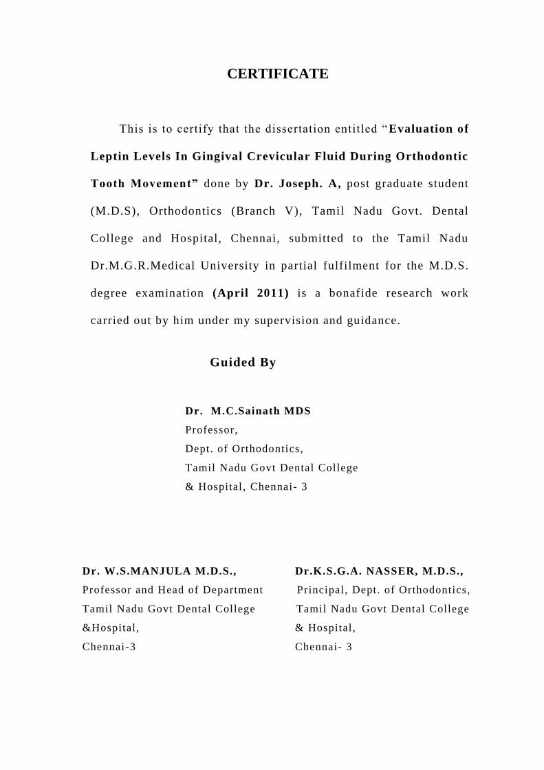
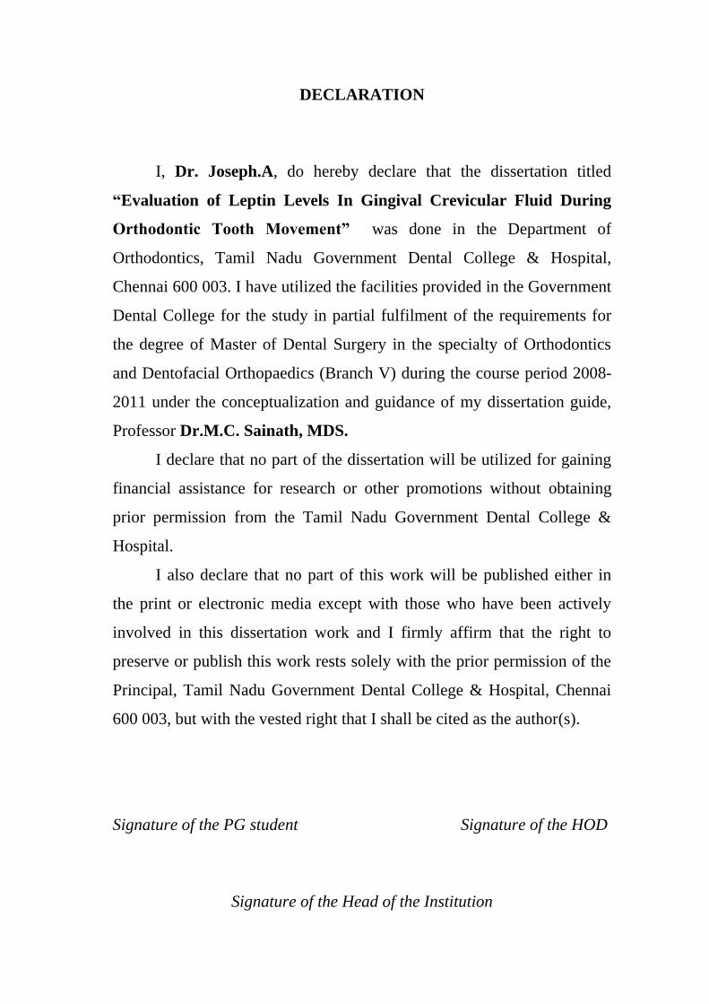
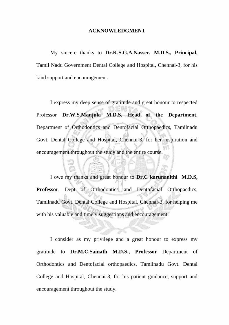


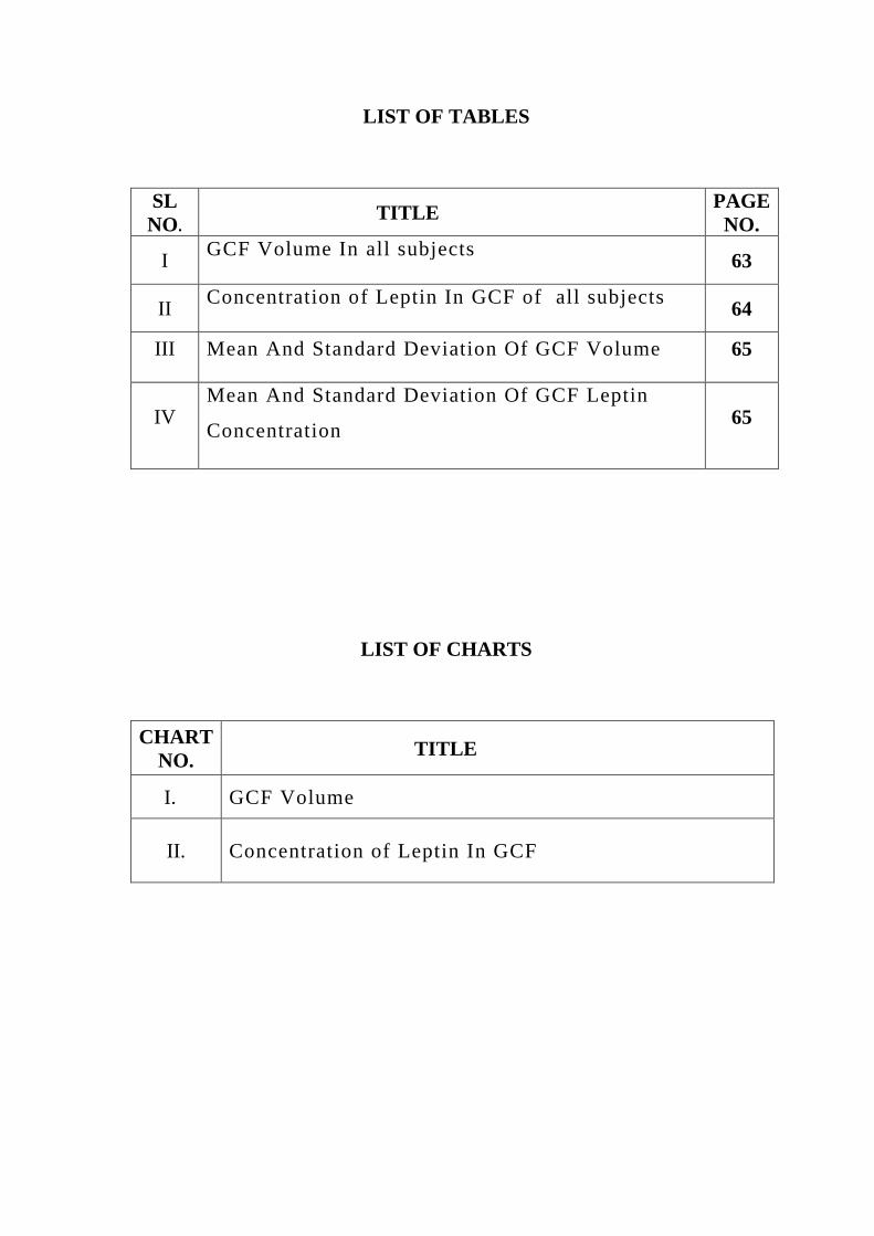
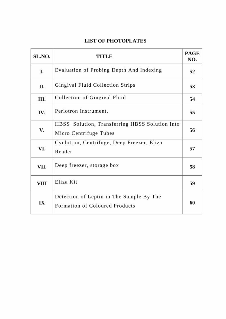


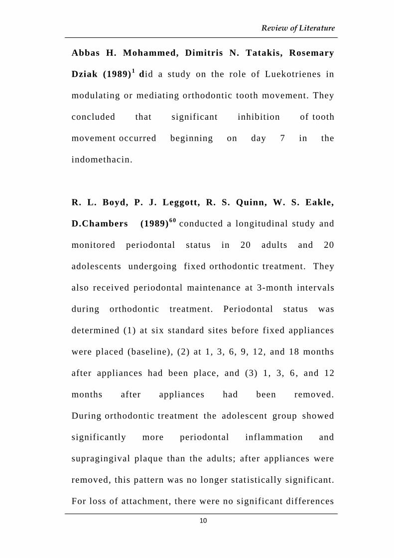
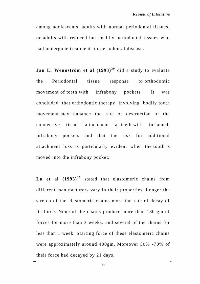

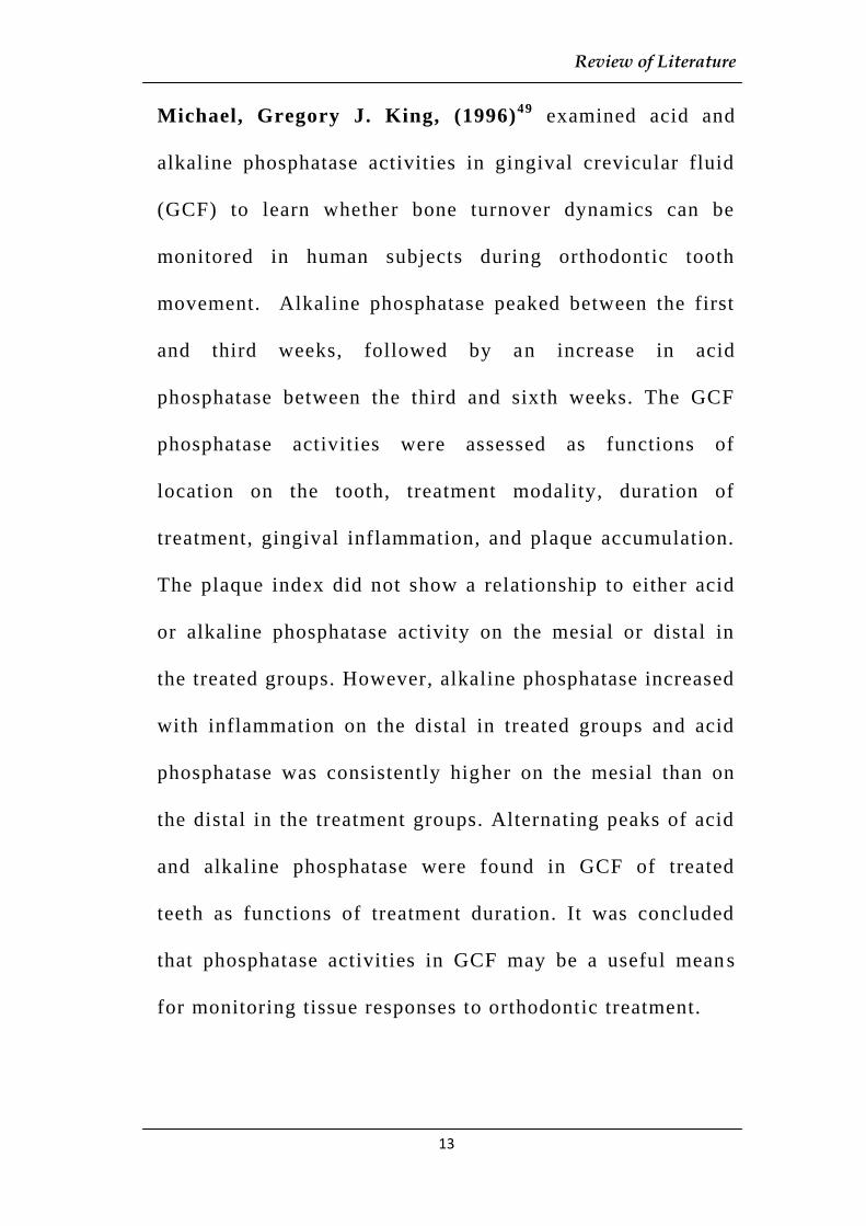


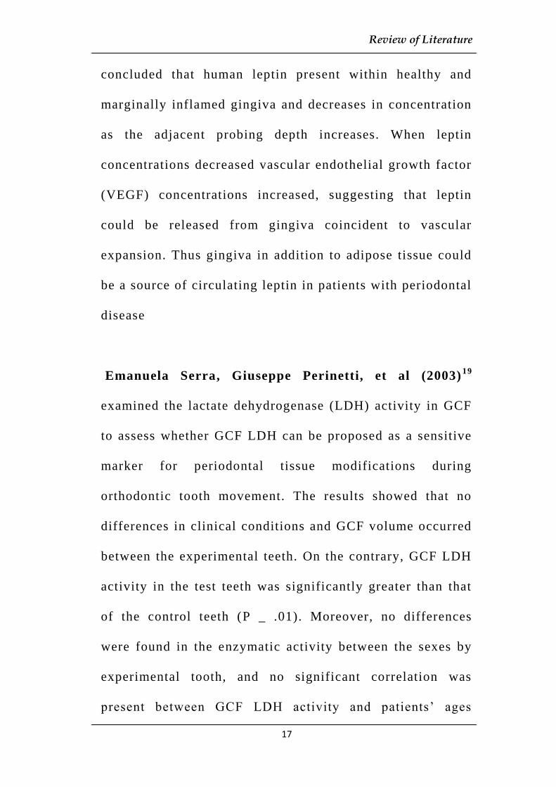


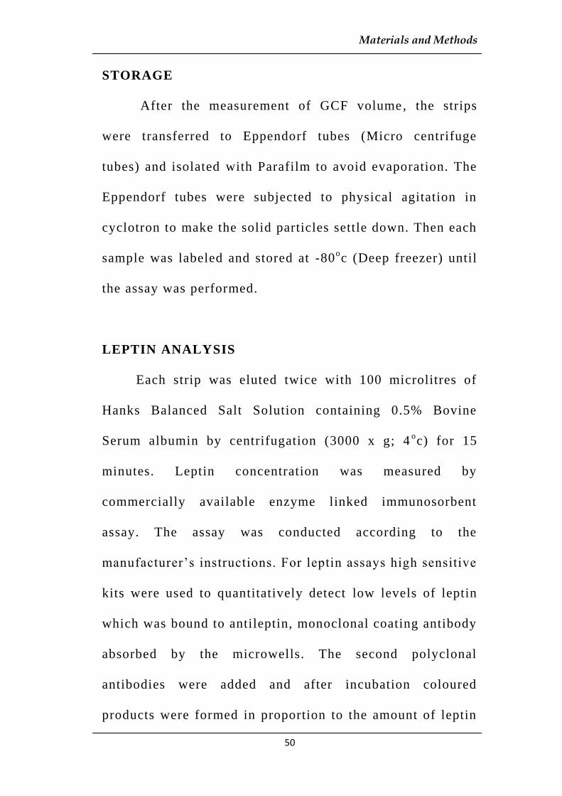
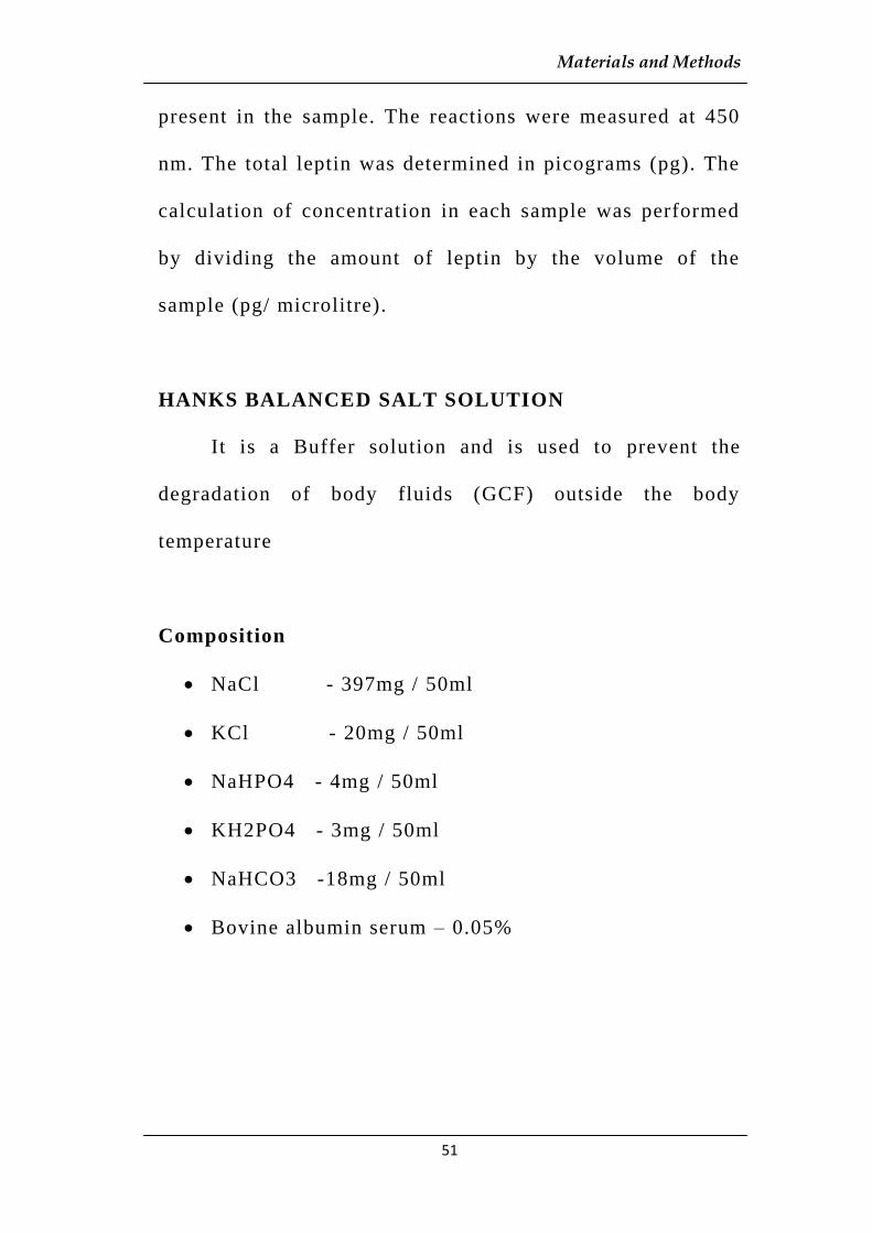

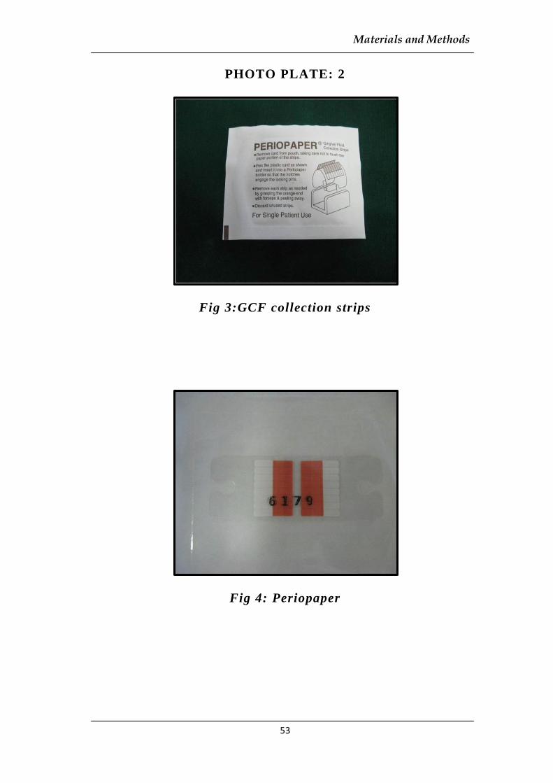
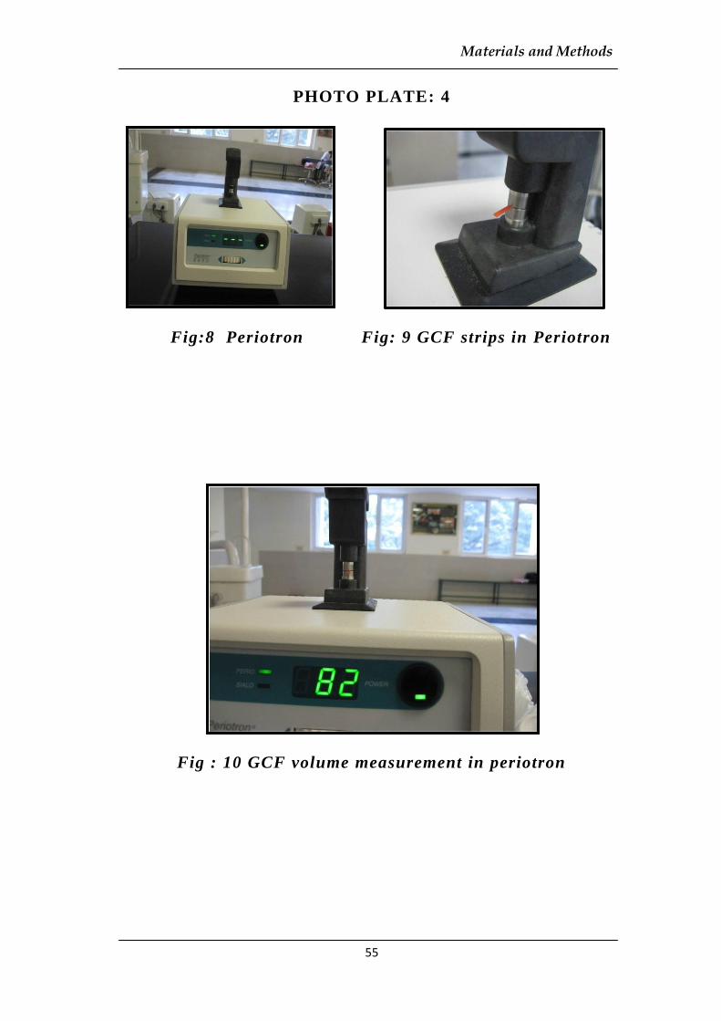


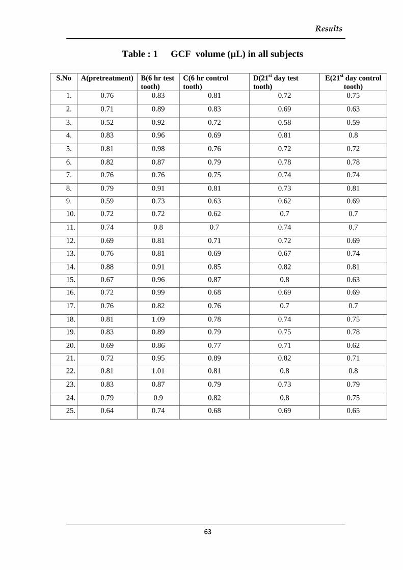
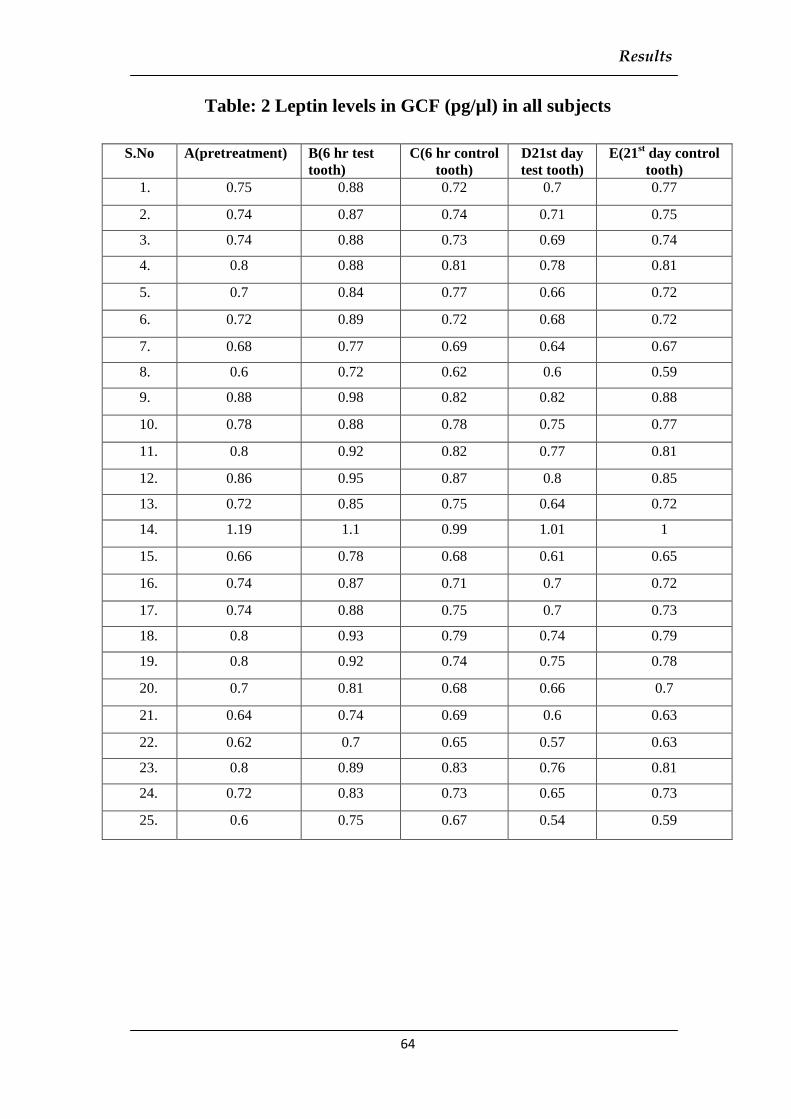


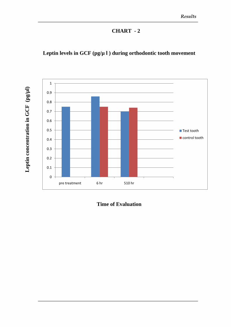

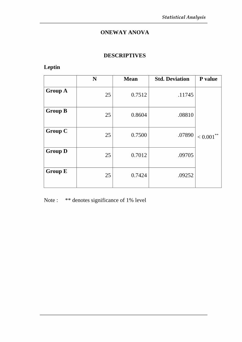

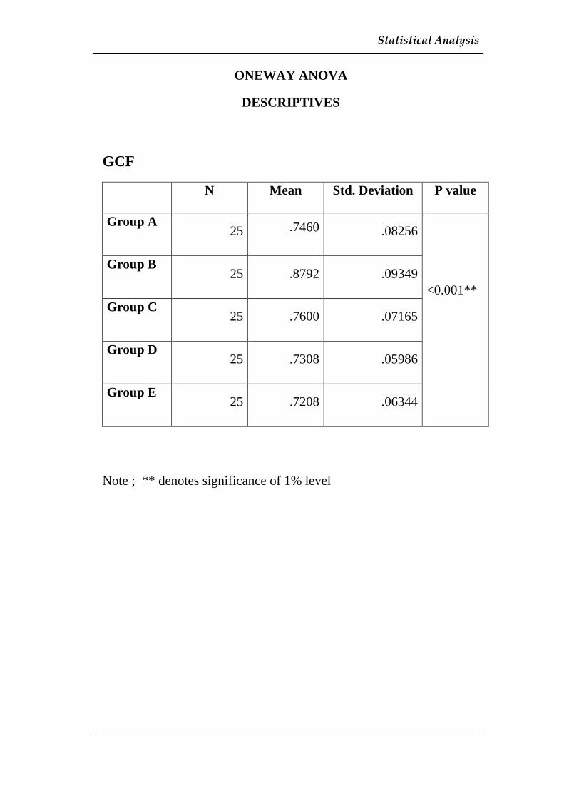


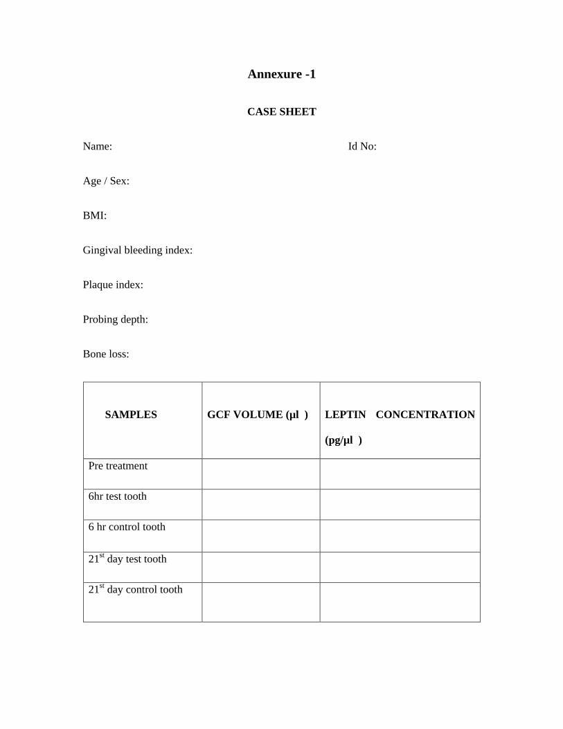


![Microbial and metabolomic analysis of gingival crevicular ... · 2]. Periodontitis affects the majority of adults worldwide and may cause various systemic diseases, including dia-betes](https://static.documents.pub/doc/80x56/5ffe1bd5f6ee7b7ad975583c/microbial-and-metabolomic-analysis-of-gingival-crevicular-2-periodontitis.jpg)


