Evaluation of PCR-based methods for the quantitation ofintegrated HIV-1 DNA
Raman Kumar a,1, Nick Vandegraaff a,1, Linda Mundy a,Christopher J. Burrell a,b, Peng Li a,*
a National Centre for HIV Virology Research, Infectious Diseases Laboratories, Institute of Medical and Veterinary Science,Frome Road, Adelaide 5000, Australia
b Department of Molecular Biosciences, Uni�ersity of Adelaide, North Terrace, Adelaide 5000, Australia
Received 14 March 2002; received in revised form 11 May 2002; accepted 14 May 2002
Human immunodeficiency virus (HIV), likeother retroviruses, reverse transcribes its genomicRNA to double stranded DNA after entering asusceptible cell. Newly synthesised viral DNA is
transported into the nucleus as a component ofthe preintegration complex where it is integratedinto the host cell chromosome to establish a stableprovirus (Bukrinsky et al., 1993; Fouchier andMalim, 1999; Karageorgos et al., 1993). In addi-tion to proviral DNA, large amounts of HIVDNA are found in three extrachromosomalforms; linear, single long-terminal repeat 1-LTRand double long-terminal repeat (2-LTR) circles.These unintegrated viral DNA forms have beenshown to be incapable of directing a productiveinfection and degrade over time (Barbosa et al.,
R. Kumar et al. / Journal of Virological Methods 105 (2002) 233–246234
1994; Vandegraaff et al., 2001a). However, it re-mains controversial whether these unintegratedviral DNA forms play some role in the HIVreplication cycle (Stevenson et al., 1990; Wu andMarsh, 2001).
Recent reports suggest that current antiretrovi-ral therapy cannot eliminate HIV-1 from the in-fected patients (Chun et al., 1997b; Finzi et al.,1997; Furtado et al., 1999; Zhang et al., 1998,1999). The efficacy of antiretroviral therapy isprimarily gauged by its effect on patients’ CD4+
T cell numbers and plasma HIV RNA levels,assayed using commercially available kits. How-ever, despite their undetectable plasma viral loadsusing highly sensitive techniques, patients invari-ably exhibit viral rebound on cessation of therapyindicating the persistence of virus within the body(Schrager and D’Souza, 1998). As additional toolsto monitor response to antiretroviral therapy andto identify sites of viral persistence, several labo-ratories have recently developed extremely sensi-tive polymerase chain reaction (PCR) -basedmethods to quantify cell-associated DNA andRNA in different populations of peripheral bloodmononuclear cells (PBMCs) (Burgard et al., 2000;Christopherson et al., 2000; Chun et al., 1997a;Lewin et al., 1999; Sharkey et al., 2000; Yerly etal., 2000). These approaches have allowed theidentification of a stable pool of long-lived restingCD4+ T cells carrying the integrated provirus inpatients receiving antiretroviral therapy with un-detectable plasma viral RNA. Furthermore, co-culture assays to quantify the infectious viruslevels from total PBMCs and/or sub-fractionatedcell populations have shown that a fraction thesecells were capable of producing infectious virus(Chun et al., 1997b; Finzi et al., 1999; Wong etal., 1997). However, such co-culture assays arecumbersome, time consuming and consequentlyill-suited for routine clinical applications. There-fore, the quantitation of proviral HIV DNA inthis pool of cells would be important for monitor-ing the effectiveness of antiretroviral therapy, andmay assist in predicting the long-term clinicaloutcome in patients (Lafeuillade et al., 2001).
A number of assays to quantitate specificallythe proviral HIV DNA levels have been reported(Chun et al., 1997a,b; Vandegraaff et al., 2001a).
The Alu-PCR method (Chun et al., 1997b; Sonzaet al., 1996) exploits the occurrence of many Alurepeat elements throughout the human genome(Nelson et al., 1989). Primers designed to annealwithin the conserved regions of Alu repeat ele-ments, in conjunction with HIV-specific primers,allow PCR amplification and subsequent quanti-tation of integrated HIV sequences. An alterna-tive and novel linker primer polymerase chainreaction (LP-PCR) method has been developed inour laboratory (Vandegraaff et al., 2001a). Thisprotocol involves digestion of chromosomal DNAwith the restriction enzyme NlaIII generatingfragments with cohesive termini to which anoligonucleotide linker is ligated. This linker thenserves as a template from which priming canoccur in a subsequent PCR step using both linker-and HIV-specific primers.
This article describes modifications to the previ-ously reported nested Alu- and LP-PCR protocolsand a systematic evaluation of the two methodswith respect to both the specificity and sensitivityfor the detection and quantitation of integratedHIV DNA. The results show that each of thesemodified methods can be successfully used forinvestigations requiring accurate and sensitivequantification of the integrated HIV-1 DNA. Fur-thermore, the method of DNA isolation and theinclusion of adequate controls in these protocolswere shown to be crucial for the precise quantita-tion of integrated HIV DNA.
2. Materials and methods
2.1. Cells and �irus infection
HuT-78 cells, a CD4+ lymphoblastoid cell lineand the persistently HIV-infected ACH-2 and 8E5T-cell lines (Clouse et al., 1989; Folks et al., 1986)were obtained from the NIH AIDS Research andReference Reagent Program. The H3B cell line isa clone of H9 cells derived in our laboratory thatare infected persistently with the HXB2 strain ofHIV (Li and Burrell, 1992). All cells were main-tained in RPMI 1640 medium supplemented with10% foetal bovine serum, L-glutamine and stan-dard antibiotics at 37 °C and in 5% CO2.
R. Kumar et al. / Journal of Virological Methods 105 (2002) 233–246 235
HuT-78 cells were infected with HIVHXB2 in-oculum (consisting of clarified H3B cell culturesupernatant) at 0.5 TCID50 units per cell (using acentrifugal enhancement protocol) in the absenceor presence of 10 �M L-731,988 as described(Vandegraaff et al., 2001a,b). L-731,988, a diketoacid, is shown to inhibit HIV integration in cellculture (Hazuda et al., 2000; Vandegraaff et al.,2001b). Infected cells were harvested 26 h postinfection (p.i.). P24 release was measured in theculture supernatants using a commercially avail-able kit (NEN).
2.2. DNA extraction and preparation of �iralDNA copy number standards and controlconstructs
Extrachromosomal and chromosomal DNAfractions were separated by the Hirt method asthe Hirt supernatant and Hirt pellet, respectively(Hirt, 1967; Vandegraaff et al., 2001a). In thisprocedure, chromosomal DNA is precipitatedpreferentially in the presence of SDS and NaClleaving extrachromosomal DNA, including unin-tegrated viral and mitochondrial DNA, in thesupernatant fraction. DNA prepared from thetwo fractions from each infection was resus-pended in water at approximately 5000 cell-equiv-alents of DNA/�l and stored at −20 °C untiluse.
The HIV DNA standard (designated HA8) wasprepared by mixing 5×10⁵, 1×10⁶ and 1×10⁶
cells of the H3B, ACH-2 and 8E5 cell lines,respectively, and isolating chromosomal DNA bythe Hirt procedure (Hirt, 1967). These cell linescontain 2, 1 and 1 copies of the integrated HIVproviral DNA, respectively, with little or no ex-trachromosomal HIV DNA detectable by South-ern blot (Clouse et al., 1989; Folks et al., 1986; Liand Burrell, 1992). H3B, ACH-2 and 8E5 cellswere counted in quadruplicate, mixed and chro-mosomal DNA (HA8) extracted. HA8 was thenused as a copy number standard for measuringtotal HIV-1 (gag) DNA (1.2 HIV-1 copies/cell),integrated HIV DNA (1.2 HIV copies/cell) and�-globin DNA (2 copies/cell) in PCR assays.Where required, HuT-78 chromosomal DNA wasused as background DNA.
The full-length HIV linear control constructwas generated in a multi-step procedure. Firstly,the first 5350 bp of HIVHXB2 from a plasmidcontaining full-length HIVHXB2 was amplifiedwith primers AC+1–21 and Primer B (Table 1)using rTth DNA polymerase XL (Perkin–Elmer).The AC dinucelotide present at the 5�-end of theAC+1–21 primer ensured that the termini of thelinear construct mimicked precisely that of theunprocessed 5� end of linear HIV DNA present inthe infected cells (Freed and Martin, 2001). Theremaining HIV-1 sequence was obtained by am-plifying an 8413 bp fragment using the sameplasmid template, primers INT-2 and M13–20(Table 1) and rTth DNA polymerase XL. BothPCR products (5350 and 8413 bp) were gelpurified and then digested with PstI to generatefour fragments that were then subjected to elec-trophoresis through a 0.5% agarose gel. The twofragments (1415 and 8304 bp), which on ligationwould produce a full-length linear HIV-1 DNA(9719 bp), were then eluted and ligated. Followingelectrophoresis of the ligation mix, the full-lengthlinear HIV DNA fragment (9719 bp) was gel-purified. Full length linear HIV-1 DNA was as-sessed for copy number by comparative gag-PCRamplification against the HA8 standard mix usingprimers GAG-P1 and GAG-III(− ) (Table 1) inthe presence of appropriate cell-equivalents ofHuT-78 background chromosomal DNA. Allcontrol DNA preparations used were stored inaliquots at −70 °C in siliconised tubes until use.
2.3. Standard PCR procedures
All PCRs were carried out in a Perkin–ElmerGeneAmp PCR system 9700. The cell-equivalentcontent of all chromosomal DNA preparationswas estimated following simultaneous PCR am-plification (in duplicate) of the human �-globingene (2 copies/diploid cell) within both the samplepreparations and known cell-equivalents of theHA8 chromosomal DNA standard. PCRs werecarried out using primers �-glo 1 and �-glo 2(Table 1) as described (Vandegraaff et al., 2001b).
To measure all forms of HIV-1 DNA, quantita-tive gag-PCRs were performed on 50 cell-equiva-lents of Hirt supernatant and Hirt chromosomal
R. Kumar et al. / Journal of Virological Methods 105 (2002) 233–246236
DNA isolated from experimental samples or HA8standards in the presence of 2×10⁵ cell-equiva-lents of HuT-78 chromosomal DNA (1.2 �g).gag-PCRs were also performed on 100 cell-equiv-alents of ACH-2, H3B and 8E5 chromosomalDNA. Duplicate PCRs were performed usingprimers GAG-P1(+ ) and GAG-III(− ) as de-scribed (Vandegraaff et al., 2001b).
2.4. Modified nested Alu-PCR
The integrated HIV-1 DNA levels were assayedusing known cell-equivalents of chromosomalDNA (estimated by -globin PCR) and a modifica-tion of the nested Alu-PCR method (Chun et al.,
1997b). In the modified protocol, the LTR-516(Alu-LTR 3�) primer (Chun et al., 1997b) wasreplaced with the PBS-659(− ) primer (Table 1) tominimise asymmetric PCR amplification from the3�-LTR of integrated DNA and from both virallong-terminal repeats of the unintegrated DNAforms.
2.5. Modified LP-PCR
Chromosomal DNA was digested initially with10 U of NlaIII and 20 U of BglII in 1×Thermo-Pol buffer (New England Biolabs) for 3 h at37 °C in a final volume of 30 �l. Two nucleotides(C and T) of the BglII overhang were ‘filled-in’
LTR-516 540–516b5�-AGGCAAGCTTTATTGAGGCTTAAGC-3�5�-CTGCTAGTTCAGGGTCTACTTGTGTGC-3�Primer B 5350–5324b
5�-TCCCAGCTACTCGGGAGGCTGAGG-3�Alu-164 164–187c
5�-TCATGATCAATGGGACGATCACATG-3�LPNV same as B101d
5�-GTAAAACGACGGCCAGT-3�M13–20 600–616 pKS(+)e
Sequence position Coordinates (nt)Probe
Flanked by primers �-glo 1 and �-glo 2 671–938aGloFlanked by primers GAG-P1(+) and GAG-III(−)GAG 1408–1722b
B-K BamHI-KpnI fragment with 2-LTR junction 1-376+9648-8718b
a Human �-globin gene sequence GenBank accession number L26462.b HIV Type 1 (HXB2) GenBank accession number K03455.c Jurka and Smith (1988).d Wattel et al. (1995).e Stratagene.
R. Kumar et al. / Journal of Virological Methods 105 (2002) 233–246 237
with 5 U of Bst DNA polymerase by raising thevolume to 50 �l with 1×ThermoPol buffer,adding 0.25 mM dGTP and 0.25 mM dATP(Promega) and then incubating at 65 °C for 1h. The samples were then extracted with phenol/chloroform/isoamylalcohol (25:24:1) and ethanolprecipitated in the presence of 2 mg/ml glycogen(Boehringer Mannheim). DNA pellets werewashed with 70% ethanol and resuspended inwater. Linker (LPNV; Table 1) ligation, 1st-round PCR amplification using LPNV andU3NV primers (Table 1) and nested PCR am-plification using U3.1(+ ) and U3-106(− ) wereperformed as described previously (Vandegraaffet al., 2001a).
2.6. Analysis of PCR products
PCR products were electrophoresed, electro-blotted and Southern hybridised to �-32P-la-belled probes as described previously(Vandegraaff et al., 2001b). The GAG probe(Table 1) was used to detect fragments resultingfrom gag-PCR. The B-K probe (Table 1) wasused to detect the products arising from 2nd-round Alu- and LP-PCR. The Glo probe (Table1) was used to detect the products arising from�-globin PCR. Following Southern hybridisa-tion, the signals obtained in each case werequantified using PhosphorImager ImageQuantanalysis. A standard curve was generated fromthe signals arising from PCRs performed onknown copies of HA8 standards and used toquantify the DNA copy numbers in experimen-tal samples.
2.7. Cloning of the chromosomal-integrated HIVDNA junction sequence
The chromosomal sequence upstream of the5�-end of integrated HIV DNA in the ACH-2,8E5 and H3B was isolated by a modified in-verse-PCR method (Chun et al., 1997a). Briefly,0.5 �g of ACH-2, 8E5 and H3B chromosomalDNA was digested with PstI, subjected to chlo-roform–phenol extraction, precipitated with eth-anol and ligated under dilute conditions topromote intramolecular ligation. First-round in-
verse PCR on the ligated DNA was performedusing outward-directed primers INT-1 and PBS-659(− ) (Table 1). Initially, two primers (25pmol each), dNTPs (0.2 mM) and ampliwaxadded to PCR tubes (Perkin–Elmer) wereheated to 75 °C for 1 min and cooled to 4 °Cto form a solid wax barrier. PCRs were thencarried out in a final volume of 100 �l using theentire ligated template DNA in 1×PCR bufferII (Perkin–Elmer), 2.0 mM MgCl2 and 2.5 UAmpliTaq Gold DNA Polymerase. PCRs werecycled as follows; 94 °C 12 min; 35 cycles of94 °C 1 min, 58 °C 30 s, 72 °C 3 min; and afinal extension of 72 °C 10 min. Nested PCRswere carried out on 1/10th of the 1st-roundPCRs in a final volume of 50 �l using 25 pmolof two primers INT-2 and NI-2 (Table 1) in1×PCR buffer II (Perkin–Elmer), 2.5 mMMgCl2, 0.2 mM dNTPs and 2.5 U AmpliTaqGold DNA Polymerase. PCRs were cycled asfollows; 94 °C 12 min; 30 cycles of 94 °C 15 s,58 °C 30 s, 72 °C 2 min; and a final extensionof 72 °C 10 min. Nested PCR products weregel-purified and sequenced directly (dye-termina-tor, Applied Biosystems) using the U3PNVprimer (Table 1).
3. Results
In order to evaluate the two modified PCRmethods, both extrachromosomal and chromo-somal DNA fractions were prepared fromacutely infected HuT-78 cells. DNA fractionsisolated from the cells infected in the presenceof integrase inhibitor L-731,988 were used asnegative control to assess the specificity of theintegration assays. By 26 h p.i., drug-free cul-tures showed extensive syncytia formation andvirus release into the culture supernatant (30 ngP24/ml) indicating that significant HIV DNA in-tegration and subsequent steps in the viral repli-cation cycle had been completed by this time(Vandegraaff et al., 2001b). In contrast, syncytiaformation and P24 release was not observed by26 h p.i. from cells infected in the presence ofL-731,988 (data not shown).
R. Kumar et al. / Journal of Virological Methods 105 (2002) 233–246238
Fig. 1. Comparison of the sensitivity and specificity of modified Alu- and LP-PCR assays. HuT-78 cells were infected with HIV(HXB2) at 0.5 TCID50 units per cell in the absence (− ) or presence (+ ) of the HIV integrase inhibitor L-731,988, and theextrachromosomal (s/n) and chromosomal (chr) DNA isolated at 26 h p.i. As the two DNA fractions were resuspended in the samefinal volume of water, the amounts of extrachromosomal and chromosomal DNA present should represent equivalent cell numbers.PCR amplified �-globin fragment from Hirt pellets of test samples and known copy numbers of HA8 standards were Southernhybridised to radio-labelled probes, and the bands quantified by PhosphorImager analysis using a standard curve generated fromHA8 standards (D). Duplicate PCRs were performed throughout and duplicate infections are marked as 1 and 2. (A) HIV gag-DNAwas quantified from 100 cell-equivalents of�L-731,988 Hirt supernatant (s/n) and Hirt pellet (chr) samples using the gag-PCRprotocol (see Section 2). Integrated HIV DNA in 50 cell-equivalents of Hirt pellets was analysed by the modified Alu- (B) andLP-PCR (C) protocols (see Section 2). Alu-PCR reactions performed on 250 copies of linear HIV DNA (250 lin) or without theAlu-164 primer (250-Alu) are shown (B). Reactions in which LP-PCR was performed in the absence of linker ligation (250-ligase)are also shown (C). These controls were included to show the levels of signal due to 2nd-round amplification of the input DNAalone.
3.1. Total HIV DNA le�els
Using known cell-equivalents of either extra-chromosomal or chromosomal DNA (see Fig.1D), we quantified the levels of total HIV gag-DNA within each fraction at 26 h p.i. using agag-PCR protocol (Fig. 1A). The averageamounts of total HIV DNA within cells (extra-
chromosomal+chromosomal) infected in the ab-sence or presence of L-731,988 were comparable(�1400 and �1300 copies per 50 cells, respec-tively; Fig. 1A and Fig. 2, Total gag-PCR). Fur-thermore, the levels of gag-DNA in chromosomalDNA fractions from cells infected in the absenceof drug (�580 copies/50 cells) were higher thanthose infected in the presence of drug (�220
R. Kumar et al. / Journal of Virological Methods 105 (2002) 233–246 239
copies/50 cells)(Fig. 1A, compare lanes +1 chr,+2 chr with −1 chr, −2 chr and Fig. 2, Chrgag-PCR). This increase in HIV DNA withinchromosomal fractions isolated from cells infectedin absence of the integrase inhibitor was at-tributed to de no�o integration of part of theextrachromosomal HIV DNA by 26 h p.i. Sup-porting this, the increases in HIV DNA withinchromosomal fractions in these cells correspondedto a decrease in HIV DNA in the extrachromoso-mal fraction (Fig. 1A, compare +s/n, +chrlanes with −s/n, −chr lanes, analysis notshown). Although signals were obtained in thechromosomal fractions from L-731,988 treatedcells (Fig. 1A, lanes +1 chr, +2 chr), this likelyresulted from the incomplete separation of extra-chromosomal HIV DNA from the chromosomalfraction and not the incomplete suppression ofintegration by the drug (see below). The extent towhich extrachromosomal HIV DNA was presentin chromosomal fractions was therefore estimatedto be approximately 17% of the total signal recov-ered from drug-treated cells.
3.2. Integrated HIV DNA le�els as determined byLP-PCR method
An alternative and novel PCR-based assay ca-pable of the specific and highly sensitive detectionof integrated HIV DNA has been described (Van-degraaff et al., 2001a). This assay was based on a
principle described originally by Wattel et al.(1995). Briefly, LP-PCR involves digestion ofchromosomal DNA with the restriction enzymeNlaIII generating fragments with four base cohe-sive ends to which a single stranded oligonucle-otide linker is then ligated. The HIV 5�-U3 regionand upstream chromosomal sequence are thenamplified using the same linker and an LTR-spe-cific primer. Since integration occurs at randomthroughout cellular DNA, a nested PCR is carriedout using LTR-specific primers to generate a dis-crete band that can be quantitated. To preventamplification of extrachromosomal HIV DNA,samples are also digested with the restriction en-zyme BglII and treated with Klenow DNA poly-merase (lacking 3�-5� exonuclease; 3�-5� exo−). Wehave now shown that a BglII/NlaIII double-di-gest can be performed in 1×ThermoPol buffer(New England Biolabs) without compromising theefficiency of digestion. Furthermore, Bst DNApolymerase, also used in 1×ThermoPol buffer,can be used in place of Klenow DNA polymerase(3�-5� exo−). Together, these modifications havesimplified greatly sample preparation prior to per-forming the linker ligation reaction.
Using this protocol, we were able to detect 4copies of the HA8 integrated DNA standard inthe presence of 2×10⁵ cell-equivalents of HuT-78chromosomal DNA (Fig. 1C). Furthermore, verylow signals in control reactions amplifying 250gag-copies of the HA8 standard without linkerligation (Fig. 1C, 250 -ligase) confirmed that thefinal signal observed was due to the 1st-roundLP-PCR amplification, and not purely from thenested PCR. To confirm further the selectivity ofthe LP-PCR protocol for integrated HIV DNA,we also analysed the levels of HIV DNA integra-tion in 50 cell-equivalents of chromosomal DNAisolated from cells 26 h after infection with HIVin the absence or presence of L-731,988 (Fig. 1C).While integration was very low in chromosomalDNA preparations isolated from cells infected inthe presence of drug (Fig. 1C, lanes +1 chr, +2chr and Fig. 2, Chr LP-PCR), those cells infectedin the absence of drug accumulated approximately380 copies of integrated HIV DNA/50 cells by 26h p.i. (Fig. 1C, lanes −1 chr, −2 chr; Fig. 2, ChrLP-PCR). Taken together, these results confirmed
Fig. 2. Comparison of the levels of total cellular (gag-PCR onextrachromosomal+chromosomal fractions), total chromoso-mal (gag-PCR on chromosomal fractions) and integratedHIV-1 DNA (on chromosomal fractions using Alu- and LP-PCR) on cells infected with HIV for 26 h in the absence orpresence of the anti-integration drug L-731,988. Data is basedon PhosphorImager quantification of bands in Fig. 1A–C.
R. Kumar et al. / Journal of Virological Methods 105 (2002) 233–246240
that LP-PCR amplifies integrated HIV DNAwith high specificity.
3.3. Modified Alu-PCR
The quantitative Alu-PCR method exploits theoccurrence of Alu repeat elements throughout thehuman genomic DNA. In this protocol, inte-grated HIV sequences are first amplified using aprimer that is designed to anneal within the con-served region of the Alu repeat elements togetherwith an HIV-specific primer. As Alu-binding se-quences are expected to be present at varyingdistances from the sites of proviral integration,the 1st-round PCR generates fragments of vary-ing length. Nested-PCRs with a pair of HIV-spe-cific primers using the 1st-round amplifiedproduct as a template results in DNA fragmentswith a defined length, which can be quantified bycomparison with the copy number standards.Under our nested Alu-PCR conditions (see Sec-tion 2), we were able to detect 4 copies of theintegrated HIV standard in the presence of 2×10⁵ cell-equivalents of HuT-78 chromosomalDNA without significantly amplifying a con-struct mimicking the linear HIV DNA form (Fig.1B, 250 lin). In addition, levels of integrated HIVDNA within cells infected in the absence andpresence of L-731,988 were determined to be �290 and �25 copies/50 cell-equivalents, respec-tively (Fig. 1B and Fig. 2, Chr Alu-PCR).Therefore, although the levels of integrated HIVDNA determined by Alu-PCR were lower thanthat determined by LP-PCR following infection(Fig. 2, compare Chr Alu-PCR with Chr LP-PCR), both methods were able to amplify specifi-cally integrated HIV DNA.
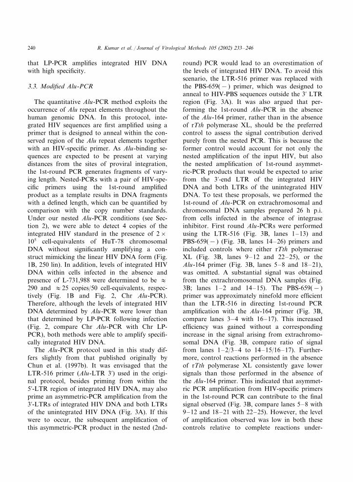
The Alu-PCR protocol used in this study dif-fers slightly from that published originally byChun et al. (1997b). It was envisaged that theLTR-516 primer (Alu-LTR 3�) used in the origi-nal protocol, besides priming from within the5�-LTR region of integrated HIV DNA, may alsoprime an asymmetric-PCR amplification from the3�-LTRs of integrated HIV DNA and both LTRsof the unintegrated HIV DNA (Fig. 3A). If thiswere to occur, the subsequent amplification ofthis asymmetric-PCR product in the nested (2nd-
round) PCR would lead to an overestimation ofthe levels of integrated HIV DNA. To avoid thisscenario, the LTR-516 primer was replaced withthe PBS-659(− ) primer, which was designed toanneal to HIV-PBS sequences outside the 3� LTRregion (Fig. 3A). It was also argued that per-forming the 1st-round Alu-PCR in the absenceof the Alu-164 primer, rather than in the absenceof rTth polymerase XL, should be the preferredcontrol to assess the signal contribution derivedpurely from the nested PCR. This is because theformer control would account for not only thenested amplification of the input HIV, but alsothe nested amplification of 1st-round asymmet-ric-PCR products that would be expected to arisefrom the 3�-end LTR of the integrated HIVDNA and both LTRs of the unintegrated HIVDNA. To test these proposals, we performed the1st-round of Alu-PCR on extrachromosomal andchromosomal DNA samples prepared 26 h p.i.from cells infected in the absence of integraseinhibitor. First round Alu-PCRs were performedusing the LTR-516 (Fig. 3B, lanes 1–13) andPBS-659(− ) (Fig. 3B, lanes 14–26) primers andincluded controls where either rTth polymeraseXL (Fig. 3B, lanes 9–12 and 22–25), or theAlu-164 primer (Fig. 3B, lanes 5–8 and 18–21),was omitted. A substantial signal was obtainedfrom the extrachromosomal DNA samples (Fig.3B; lanes 1–2 and 14–15). The PBS-659(− )primer was approximately ninefold more efficientthan the LTR-516 in directing 1st-round PCRamplification with the Alu-164 primer (Fig. 3B,compare lanes 3–4 with 16–17). This increasedefficiency was gained without a correspondingincrease in the signal arising from extrachromo-somal DNA (Fig. 3B, compare ratio of signalfrom lanes 1–2/3–4 to 14–15/16–17). Further-more, control reactions performed in the absenceof rTth polymerase XL consistently gave lowersignals than those performed in the absence ofthe Alu-164 primer. This indicated that asymmet-ric PCR amplification from HIV-specific primersin the 1st-round PCR can contribute to the finalsignal observed (Fig. 3B, compare lanes 5–8 with9–12 and 18–21 with 22–25). However, the levelof amplification observed was low in both thesecontrols relative to complete reactions under-
R. Kumar et al. / Journal of Virological Methods 105 (2002) 233–246 241
Fig. 3. Comparison of the LTR-516 (lanes 1–13) and PBS-659(− ) (lanes 14–26) primers in the Alu-PCR protocol. (A) Diagramof an integrated HIV-1 DNA showing Alu-164, PBS-659(− ) and LTR-516 binding sites. (B) Alu-PCRs were performed in duplicateon 50 cell-equivalents of extrachromosomal and chromosomal DNA fractions of cells infected for 26 h in the absence of L-731,988.The two DNA fractions were analysed using either the LTR-516 (1–13) or the PBS-659(− ) (14–26) primers in the presence (lanes1–4, 9–12, 13, 14–17, 22–25, 26) or absence (lanes 5–8, 18–21) of the Alu-164 primer, and in the presence (lanes 1–8, 13, 14–21,26) or absence (9–12, 22–25) of rTth DNA polymerase XL. The graph is drawn based on the values obtained from PhosphorImageanalysis of the Southern blot shown.
taken on chromosomal DNA when the PBS-659(− ) primer was used (Fig. 3B, compare lanes16–17 with 20–21). This confirmed that the ma-jority of signals obtained from samples amplifiedin complete reactions (see Fig. 3B, lanes 3–4 and16–17) were derived primarily from the 1st-roundamplification of integrated HIV sequences, andnot from the nested PCR amplification of eitherinitial input template DNA sequences or asym-metric PCR products.
3.4. Modified Alu- �s. LP-PCR
There are approximately 9×10⁵ Alu repeat ele-ments present throughout the haploid humangenome (Nelson et al., 1989). This equates to theoccurrence of an Alu repeat element approxi-mately every 4 kb of genomic DNA sequence.However, since these elements can exist in eitherorientation, the average distance between inte-grated HIV DNA and an Alu repeat element in
R. Kumar et al. / Journal of Virological Methods 105 (2002) 233–246242
the correct orientation for successful Alu-PCRamplification would be expected to be approxi-mately 8 kb. Many integration events may there-fore be outside the range of efficient PCRamplification, even if a long-range thermostablepolymerase is used. To determine the location ofAlu repeat elements immediately upstream of inte-grated HIV DNA in the ACH-2, 8E5 cell linesand at one of the two integration sites in H3B cellline, we used a modified inverse PCR approach toamplify the cellular DNA adjacent to the sites ofproviral integration. GenBank nucleotide data-base searches were undertaken on sequenced PCRproducts and showed that the chromosomal se-quences upstream of the HIV integration sites inthe ACH-2, 8E5 and H3B cell lines mapped to
regions within human chromosomes 7, 13 and 4,respectively (Fig. 4A). A search for Alu-164 bind-ing sequences (Jurka and Smith, 1988) upstreamof the integrated HIV DNA in the ACH-2 cellline showed that two primer-binding sites (in thecorrect orientation) were approximately 3.6 and6.7 kb from the site of proviral integration, re-spectively. However, in the 8E5 and H3B celllines, the nearest Alu-164 binding sites in thecorrect orientation for Alu-PCR amplificationwere approximately 12 and �20 kb away, respec-tively (Fig. 4A). To establish whether the distanceof the nearest Alu-164 binding-sequence corre-lated with ability of the Alu-PCR procedure todirect successful proviral DNA amplification,chromosomal DNA preparations from each cell
Fig. 4. Comparison of the modified Alu- and LP-PCR methods. (A) Diagram showing the position of conserved humanchromosomal Alu-164 binding sites upstream of the integrated HIV DNA in the ACH-2, 8E5 and H3B cell lines. The positions ofthe PBS-659(− ) primer, and the distance (in kb) and orientation of Alu repeat elements upstream of the sites of integration, isshown. The chromosomal-HIV junction sequences from the ACH-2, 8E5 and H3B cell lines matched to sequenced clones withinchromosomes 7 (GenBank accession number AC083863), 13 (GenBank accession number AL391374; see also published sequenceRodriguez-Alfageme et al., 1998) and 4 (GenBank accession number AC021120), respectively. (B) ACH-2, 8E5 and H3Bchromosomal DNA was prepared and equalised for cell-equivalents by �-globin PCR (�-Glo) and gag-DNA-equivalents bygag-PCR (gag). The modified Alu-PCR protocol was performed on 250 gag-DNA-equivalents (Alu-PCR) while the modifiedLP-PCR was performed on 100 gag-DNA-equivalents (LP-PCR) of chromosomal DNA.
R. Kumar et al. / Journal of Virological Methods 105 (2002) 233–246 243
line were equalised for both cell-equivalents (-globin PCR) and HIV copy number (gag-PCR)by comparison with HA8 (see Fig. 4B, -Glo andgag). The modified Alu-PCR procedure per-formed on ACH-2 and 8E5 chromosomal DNAgave a strong signal and no signal, respectively(Fig. 4B, Alu-PCR). Signal (albeit lower) was alsoobtained when the modified Alu-PCR procedurewas carried out on H3B chromosomal DNA (Fig.4B, Alu-PCR). This observation indicated that theAlu repeat element immediately upstream of thesecond unsequenced integrant in this cell line waslikely to be within the amplifiable range. In con-trast, when the same ACH-2, 8E5 and H3B chro-mosomal DNA preparations were analysed usingthe modified LP-PCR protocol, similar levels ofnested PCR product were detected from all three-cell lines (Fig. 4B, LP-PCR).
4. Discussion
A number of investigators have developed re-cently a range of assays to monitor cell-associatedviral DNA and RNA (Burgard et al., 2000; Butleret al., 2001; Christopherson et al., 2000; Lewin etal., 1999; Sharkey et al., 2000; Yerly et al., 2000).Longitudinal studies have suggested that theseadditional cell-associated virological markers mayhelp predict long-term clinical outcome (Yerly etal., 2000). The quantitation of the integratedproviral DNA load within cells may also be usedto identify and monitor sites of viral persistencewithin the body. However, a concerted effort hasnot been made to design sensitive and accuratemethods that can be used routinely to quantitatethe proviral DNA load within patients.
We have demonstrated that both the modifiednested Alu-PCR and LP-PCR protocols presentedin this study can be used to quantify levels ofintegrated HIV DNA with a high degree of sensi-tivity (4 copies of integrated HIV DNA in pres-ence of 2×10⁵ cell-equivalents of backgroundchromosomal DNA). In addition, it was demon-strated that a more appropriate control for Alu-PCR is a reaction lacking only the Alu-164 primerthat, unlike a reaction lacking only rTth poly-merase, would control for the asymmetric 1st-
round PCR amplification of input HIV DNAsequences. Furthermore, significant signals wereobserved when both the Alu- (Fig. 3B) and theLP-PCR (data not shown) procedures were per-formed on Hirt supernatant fractions (extrachro-mosomal HIV DNA) alone. The signals obtainedfrom these samples were unlikely to have resultedfrom chromosomal DNA contamination of theextrachromosomal DNA fraction, as the �-globinlevels within these Hirt supernatant fractions werenegligible (data not shown). Therefore, the specifi-city, and thus accuracy of each of these assaysappears to be compromised in the presence oflarge amounts of extrachromosomal HIV DNA.Considering earlier reports that levels of uninte-grated DNA within populations of resting CD4+
T cells were up to 28-fold higher than levels ofintegrated HIV DNA (Chun et al., 1997b), ourresults suggest that the quantification of inte-grated HIV DNA should not be performed ontotal cellular DNA, but on preferentially precipi-tated Hirt chromosomal DNA. Consistent withthe levels of polyoma virus DNA contaminationof chromosomal DNA observed when the Hirtprocedure was performed on infected mouse kid-ney cells (Hirt, 1967), it was demonstrated thatonly about 17% of extrachromosomal DNA re-mains associated with chromosomal DNA in theHirt method of chromosomal DNA extraction(Fig. 1A). This level of contamination did notaffect significantly the levels of integrated HIVDNA quantified by the Alu- and LP-PCRprocedures.
The levels of integrated HIV DNA copiesquantified by the modified Alu-PCR using theHA8 standards were consistently 20–30% lessthan those estimated by the modified LP-PCRprocedure. This discrepancy, also observed in anearlier study (Vandegraaff et al., 2001a), has beenattributed previously to inefficiencies of the Alu-PCR procedure arising from both the distanceand the orientation of the Alu repeat elementimmediately upstream of the site of proviral inte-gration. It was found that the Alu repeat elements(in the correct orientation for successful Alu-PCR) nearest to integrated HIV DNA in the 8E5and H3B cell lines are approximately 12 and �20kb, respectively. These distances are far greater
R. Kumar et al. / Journal of Virological Methods 105 (2002) 233–246244
than that predicted if Alu repeat elements werespaced evenly throughout the cellular chromo-some (�8 kb) and support earlier reports thatAlu repeat elements may be clustered within par-ticular chromosomal regions (Hattori et al., 2000;Korenberg and Rykowski, 1988). A signal wasnot obtained following Alu-PCR amplification of8E5 chromosomal DNA, and a reduced signalwas obtained from H3B chromosomal DNA. Thisindicated that these distances might be too largeto allow efficient Alu-PCR amplification, eventhough a DNA polymerase capable of amplifyinglong target sequences was used.
It is important to note, that rTth DNA poly-merase, like most other thermostable DNA poly-merases, has a 5��3� exonuclease activity thatwould direct the 5��3� hydrolysis of any DNAimmediately ahead of a nascent DNA molecule(Myers and Gelfand, 1991). In the context ofAlu-PCR, this activity might be expected to directthe hydrolysis of annealed (and extended) Alu-164primers ahead of nascent DNA chains throughoutthe genome. Therefore, the highly complex combi-nations of priming and degradation events that islikely to be occurring in Alu-PCR when rTthDNA polymerase is used would further limit thenumber of integration events that can be success-fully amplified. It is worth noting that if a long-range thermostable polymerase lacking stranddisplacement and 5��3� exonuclease activities isused (or when reaction conditions are uncondu-cive for 5��3� exonuclease activity), a maximumof 50% of all random integrants will be amplified.This is because such enzymes will be unable toamplify HIV DNA integrated immediately adja-cent to an Alu repeat element in the incorrectorientation (see for example, Fig. 4A, 8E5).
Although limitations associated with both thelocation of Alu repeat elements and the 5��3�exonuclease activity may reduce ultimately thesensitivity of the Alu-PCR assay, the accuracy ofthe Alu-PCR procedure should be maintained if arandom pool of integrants is used as the copynumber standard. Such a standard was recentlyreported by Butler et al. who used chromosomalDNA preparations from HIV-infected cells (cul-tured for 30 days to ensure that all the extrachro-mosomal forms of viral DNA were lost) as an
Alu-PCR copy number standard (Butler et al.,2001). In contrast, the use of large populations ofrandom integrants as copy number standards isless critical in the LP-PCR procedure (but never-theless recommended), due to the relative frequen-cies with which NlaIII and BglII cleave randomDNA sequence (Vandegraaff et al., 2001a). Addi-tionally, the LP-PCR procedure would not beaffected by the 5��3� exonuclease activity of ther-mostable polymerases as chromosomal DNA isinitially digested with restriction enzymes to gen-erate discrete templates for amplification. How-ever, despite being extensively optimised, theaccumulated error associated with the multi-stepnature of the LP-PCR procedure can also affectthe sensitivity of this assay.
In addition to monitoring the HIV proviralload within patients, the LP-PCR and Alu-PCRassays can be used to screen compounds for theirability to inhibit integration in cell culture and toassess the kinetics of HIV integration followinginfection of cultured cells. The results presentedabove suggest that either of the two assays can beused to assess proviral DNA in each of thesescenarios, provided the correct method of DNAextraction and the appropriate controls are used.However, the relative ease with which the Alu-PCR protocol can be performed compared to theLP-PCR procedure may make it a more suitableassay for use in situations where a high degree ofassay-sensitivity is not required.
Acknowledgements
We thank Helen Hocking for technical assis-tance, Melissa Egberton and Steven Young(Merck and Co.) for the sample of L-731,988 usedin this study, and Adrian Purins for his informedsuggestions throughout this project. The Aus-tralian Commonwealth AIDS Research GrantProgram supported this work.
References
Barbosa, P., Charneau, P., Dumey, N., Clavel, F., 1994.Kinetic analysis of HIV-1 early replicative steps in a cocul-ture system. AIDS Res. Hum. Retroviruses 10, 53–59.
R. Kumar et al. / Journal of Virological Methods 105 (2002) 233–246 245
Bukrinsky, M.I., Sharova, N., McDonald, T.L.,Pushkarskaya, T., Tarpley, W.G., Stevenson, M., 1993.Association of integrase, matrix, and reverse transcriptaseantigens of human immunodeficiency virus type 1 withviral nucleic acids following acute infection. Proc. Natl.Acad. Sci. USA 90, 6125–6129.
Burgard, M., Izopet, J., Dumon, B., Tamalet, C., Descamps,D., Ruffault, A., Vabret, A., Bargues, G., Mouroux, M.,Pellegrin, I., Ivanoff, S., Guisthau, O., Calvez, V., Seigneu-rin, J.M., Rouzioux, C., 2000. HIV RNA and HIV DNAin peripheral blood mononuclear cells are consistent mark-ers for estimating viral load in patients undergoing long-term potent treatment. AIDS Res. Hum. Retroviruses 16,1939–1947.
Butler, S.L., Hansen, M.S., Bushman, F.D., 2001. A quantita-tive assay for HIV DNA integration in vivo. Nat. Med. 7,631–634.
Christopherson, C., Kidane, Y., Conway, B., Krowka, J.,Sheppard, H., Kwok, S., 2000. PCR-based assay to quan-tify human immunodeficiency virus type 1 DNA in periph-eral blood mononuclear cells. J. Clin. Microbiol. 38,630–634.
Chun, T.W., Carruth, L., Finzi, D., Shen, X., DiGiuseppe,J.A., Taylor, H., Hermankova, M., Chadwick, K., Mar-golick, J., Quinn, T.C., Kuo, Y.H., Brookmeyer, R.,Zeiger, M.A., Barditch-Crovo, P., Siliciano, R.F., 1997a.Quantification of latent tissue reservoirs and total bodyviral load in HIV-1 infection. Nature 387, 183–188.
Chun, T.W., Stuyver, L., Mizell, S.B., Ehler, L.A., Mican,J.A., Baseler, M., Lloyd, A.L., Nowak, M.A., Fauci, A.S.,1997b. Presence of an inducible HIV-1 latent reservoirduring highly active antiretroviral therapy. Proc. Natl.Acad. Sci. USA 94, 13193–13197.
Clouse, K.A., Powell, D., Washington, I., Poli, G., Strebel, K.,Farrar, W., Barstad, P., Kovacs, J., Fauci, A.S., Folks,T.M., 1989. Monokine regulation of human immunodefi-ciency virus-1 expression in a chronically infected human Tcell clone. J. Immunol. 142, 431–438.
Finzi, D., Blankson, J., Siliciano, J.D., Margolick, J.B., Chad-wick, K., Pierson, T., Smith, K., Lisziewicz, J., Lori, F.,Flexner, C., Quinn, T.C., Chaisson, R.E., Rosenberg, E.,Walker, B., Gange, S., Gallant, J., Siliciano, R.F., 1999.Latent infection of CD4+ T cells provides a mechanism forlifelong persistence of HIV-1, even in patients on effectivecombination therapy. Nat. Med. 5, 512–517.
Finzi, D., Hermankova, M., Pierson, T., Carruth, L.M., Buck,C., Chaisson, R.E., Quinn, T.C., Chadwick, K., Mar-golick, J., Brookmeyer, R., Gallant, J., Markowitz, M.,Ho, D.D., Richman, D.D., Siliciano, R.F., 1997. Identifi-cation of a reservoir for HIV-1 in patients on highly activeantiretroviral therapy. Science 278, 1295–1300.
Folks, T.M., Powell, D., Lightfoote, M., Koenig, S., Fauci,A.S., Benn, S., Rabson, A., Daugherty, D., Gendelman,H.E., Hoggan, M.D., et al., 1986. Biological and biochem-ical characterization of a cloned Leu-3-cell surviving infec-tion with the acquired immune deficiency syndromeretrovirus. J. Exp. Med. 164, 280–290.
Freed, O.G., Martin, M.A., 2001. HIVs and their replication.In: Knipe, D.M., Howley, P.M., Griffin, D.E., Lamb,R.A., Martin, M.A., Roizman, B., Straus, S.E. (Eds.),Fields Virology, vol. 2, fourth ed. Lippincott Williams andWilkins, Philadelphia, USA, pp. 1971–2041.
Furtado, M.R., Callaway, D.S., Phair, J.P., Kunstman, K.J.,Stanton, J.L., Macken, C.A., Perelson, A.S., Wolinsky,S.M., 1999. Persistence of HIV-1 transcription in periph-eral-blood mononuclear cells in patients receiving potentantiretroviral therapy. N. Engl. J. Med. 340, 1614–1622.
Hattori, M., Fujiyama, A., Taylor, et al., 2000. The DNAsequence of human chromosome 21. Nature 405, 311–319.
Hazuda, D.J., Felock, P., Witmer, M., Wolfe, A., Stillmock,K., Grobler, J.A., Espeseth, A., Gabryelski, L., Schleif,W., Blau, C., Miller, M.D., 2000. Inhibitors of strandtransfer that prevent integration and inhibit HIV-1 replica-tion in cells. Science 287, 646–650.
Hirt, B., 1967. Selective extraction of polyoma DNA frominfected mouse cell cultures. J. Mol. Biol. 26, 365–369.
Jurka, J., Smith, T., 1988. A fundamental division in the Alufamily of repeated sequences. Proc. Natl. Acad. Sci. USA85, 4775–4778.
Karageorgos, L., Li, P., Burrell, C., 1993. Characterization ofHIV replication complexes early after cell-to-cell infection.AIDS Res. Hum. Retroviruses 9, 817–823.
Korenberg, J.R., Rykowski, M.C., 1988. Human genome or-ganization: Alu, lines, and the molecular structure ofmetaphase chromosome bands. Cell 53, 391–400.
Lafeuillade, A., Poggi, C., Chadapaud, S., Hittinger, G.,Khiri, H., Halfon, P., 2001. Impact of immune interven-tions on proviral HIV-1 DNA decay in patients receivinghighly active antiretroviral therapy. HIV Med. 2, 189–194.
Lewin, S.R., Vesanen, M., Kostrikis, L., Hurley, A., Duran,M., Zhang, L., Ho, D.D., Markowitz, M., 1999. Use ofreal-time PCR and molecular beacons to detect virus repli-cation in human immunodeficiency virus type 1-infectedindividuals on prolonged effective antiretroviral therapy. J.Virol. 73, 6099–6103.
Li, P., Burrell, C.J., 1992. Synthesis of human immunodefi-ciency virus DNA in a cell-to-cell transmission model.AIDS Res. Hum. Retroviruses 8, 253–259.
Myers, T.W., Gelfand, D.H., 1991. Reverse transcription andDNA amplification by a thermus thermophilus DNA poly-merase. Biochemistry 30, 7661–7666.
Nelson, D.L., Ledbetter, S.A., Corbo, L., Victoria, M.F.,Ramirez-Solis, R., Webster, T.D., Ledbetter, D.H.,Caskey, C.T., 1989. Alu polymerase chain reaction: amethod for rapid isolation of human-specific sequencesfrom complex DNA sources. Proc. Natl. Acad. Sci. USA86, 6686–6690.
Rodriguez-Alfageme, C., Chen, Z., Sonoda, G., Testa, J.R.,Damiani, R.D., Astrin, S.M., 1998. B cells malignantlytransformed by human immunodeficiency virus are poly-clonal. Virology 252, 34–38.
R. Kumar et al. / Journal of Virological Methods 105 (2002) 233–246246
Schrager, L.K., D’Souza, M.P., 1998. Cellular and anatomicalreservoirs of HIV-1 in patients receiving potent antiretrovi-ral combination therapy. JAMA 280, 67–71.
Sharkey, M.E., Teo, I., Greenough, T., Sharova, N.,Luzuriaga, K., Sullivan, J.L., Bucy, R.P., Kostrikis, L.G.,Haase, A., Veryard, C., Davaro, R.E., Cheeseman, S.H.,Daly, J.S., Bova, C., Ellison, R.T. III, Mady, B., Lai,K.K., Moyle, G., Nelson, M., Gazzard, B., Shaunak, S.,Stevenson, M., 2000. Persistence of episomal HIV-1 infec-tion intermediates in patients on highly active anti-retrovi-ral therapy. Nat. Med. 6, 76–81.
Sonza, S., Maerz, A., Deacon, N., Meanger, J., Mills, J.,Crowe, S., 1996. Human immunodeficiency virus type 1replication is blocked prior to reverse transcription andintegration in freshly isolated peripheral blood monocytes.J. Virol. 70, 3863–3869.
Stevenson, M., Stanwick, T.L., Dempsey, M.P., Lamonica,C.A., 1990. HIV-1 replication is controlled at the level of Tcell activation and proviral integration. EMBO J. 9, 1551–1560.
Vandegraaff, N., Kumar, R., Burrell, C.J., Li, P., 2001a.Kinetics of human immunodeficiency virus type 1 (HIV)DNA integration in acutely infected cells as determinedusing a novel assay for detection of integrated HIV DNA.J. Virol. 75, 11253–11260.
Vandegraaff, N., Kumar, R., Hocking, H., Burke, T.R. Jr.,Mills, J., Rhodes, D., Burrell, C.J., Li, P., 2001b. Specificinhibition of human immunodeficiency virus type 1 (HIV-
1) integration in cell culture: putative inhibitors of HIV-1integrase. Antimicrob. Agents Chemother. 45, 2510–2516.
Wattel, E., Vartanian, J.P., Pannetier, C., Wain-Hobson, S.,1995. Clonal expansion of human T-cell leukemia virustype I-infected cells in asymptomatic and symptomaticcarriers without malignancy. J. Virol. 69, 2863–2868.
Wong, J.K., Hezareh, M., Gunthard, H.F., Havlir, D.V.,Ignacio, C.C., Spina, C.A., Richman, D.D., 1997. Recov-ery of replication-competent HIV despite prolonged sup-pression of plasma viremia. Science 278, 1291–1295.
Wu, Y., Marsh, J.W., 2001. Selective transcription and modu-lation of resting T cell activity by preintegrated HIV DNA.Science 293, 1503–1506.
Yerly, S., Perneger, T.V., Vora, S., Hirschel, B., Perrin, L.,2000. Decay of cell-associated HIV-1 DNA correlates withresidual replication in patients treated during acute HIV-1infection. AIDS 14, 2805–2812.
Zhang, H., Dornadula, G., Beumont, M., Livornese, L. Jr.,Van Uitert, B., Henning, K., Pomerantz, R.J., 1998. Hu-man immunodeficiency virus type 1 in the semen of menreceiving highly active antiretroviral therapy. N. Engl. J.Med. 339, 1803–1809.
Zhang, L., Ramratnam, B., Tenner-Racz, K., He, Y.,Vesanen, M., Lewin, S., Talal, A., Racz, P., Perelson, A.S.,Korber, B.T., Markowitz, M., Ho, D.D., 1999. Quantify-ing residual HIV-1 replication in patients receiving combi-nation antiretroviral therapy. N. Engl. J. Med. 340,1605–1613.