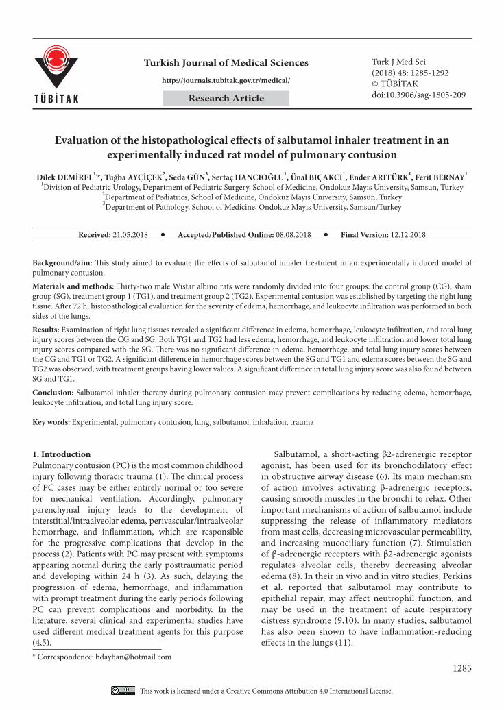
1Division of Pediatric Urology, Department of Pediatric Surgery, School of Medicine, Ondokuz Mayıs University, Samsun, Turkey2Department of Pediatrics, School of Medicine, Ondokuz Mayıs University, Samsun, Turkey3Department of Pathology, School of Medicine, Ondokuz Mayıs University, Samsun/Turkey
1. IntroductionPulmonary contusion (PC) is the most common childhood injury following thoracic trauma (1). The clinical process of PC cases may be either entirely normal or too severe for mechanical ventilation. Accordingly, pulmonary parenchymal injury leads to the development of interstitial/intraalveolar edema, perivascular/intraalveolar hemorrhage, and inflammation, which are responsible for the progressive complications that develop in the process (2). Patients with PC may present with symptoms appearing normal during the early posttraumatic period and developing within 24 h (3). As such, delaying the progression of edema, hemorrhage, and inflammation with prompt treatment during the early periods following PC can prevent complications and morbidity. In the literature, several clinical and experimental studies have used different medical treatment agents for this purpose (4,5).
Salbutamol, a short-acting β2-adrenergic receptor agonist, has been used for its bronchodilatory effect in obstructive airway disease (6). Its main mechanism of action involves activating β-adrenergic receptors, causing smooth muscles in the bronchi to relax. Other important mechanisms of action of salbutamol include suppressing the release of inflammatory mediators from mast cells, decreasing microvascular permeability, and increasing mucociliary function (7). Stimulation of β-adrenergic receptors with β2-adrenergic agonists regulates alveolar cells, thereby decreasing alveolar edema (8). In their in vivo and in vitro studies, Perkins et al. reported that salbutamol may contribute to epithelial repair, may affect neutrophil function, and may be used in the treatment of acute respiratory distress syndrome (9,10). In many studies, salbutamol has also been shown to have inflammation-reducing effects in the lungs (11).
Background/aim: This study aimed to evaluate the effects of salbutamol inhaler treatment in an experimentally induced model of pulmonary contusion.
Materials and methods: Thirty-two male Wistar albino rats were randomly divided into four groups: the control group (CG), sham group (SG), treatment group 1 (TG1), and treatment group 2 (TG2). Experimental contusion was established by targeting the right lung tissue. After 72 h, histopathological evaluation for the severity of edema, hemorrhage, and leukocyte infiltration was performed in both sides of the lungs.
Results: Examination of right lung tissues revealed a significant difference in edema, hemorrhage, leukocyte infiltration, and total lung injury scores between the CG and SG. Both TG1 and TG2 had less edema, hemorrhage, and leukocyte infiltration and lower total lung injury scores compared with the SG. There was no significant difference in edema, hemorrhage, and total lung injury scores between the CG and TG1 or TG2. A significant difference in hemorrhage scores between the SG and TG1 and edema scores between the SG and TG2 was observed, with treatment groups having lower values. A significant difference in total lung injury score was also found between SG and TG1.
Conclusion: Salbutamol inhaler therapy during pulmonary contusion may prevent complications by reducing edema, hemorrhage, leukocyte infiltration, and total lung injury score.
Received: 21.05.2018 Accepted/Published Online: 08.08.2018 Final Version: 12.12.2018
Research Article
This work is licensed under a Creative Commons Attribution 4.0 International License.
1286
DEMİREL et al. / Turk J Med Sci
The present experimental study evaluates the histopathological effects of salbutamol, a β2-agonist, on pulmonary parenchyma in an experimental model of PC in rats.
2. Materials and methodsThe current study was approved by the Animal Experimentation Ethics Committee of Ondokuz Mayıs University (Approval No. 2017/50). All surgical procedures were performed at the Research Center for Animal Experiments at Ondokuz Mayıs University. All rats were individually caged in a room under standard environmental conditions and fed with standard rat diet.2.1. Experimental groupsA total of 32 Wistar albino rats weighing 280–300 g were randomly divided into four groups (Table 1): the control group (CG) (n = 8) (no intervention performed), sham group (SG) (n = 8) (no treatment provided following experimental contusion), treatment group 1 (TG1) (n = 8) (100 μg per dose of salbutamol inhaler treatment administered once every 4 h for 24 h), and treatment group 2 (TG2) (100 μg per dose of salbutamol inhaler treatment administered once every 4 h for 72 h) (12).2.2. Experimental protocolsRats in the SG, TG1, and TG2 were intraperitoneally injected with a mixture of 80 mg/kg ketamine (Ketalar, Pfizer, Turkey) and 12 mg/kg xylazine (Rompun, Bayer, Turkey) after 8 h of fasting to achieve anesthesia. In the establishment of PC, the weight reduction method defined by Raghavendran et al. (13) was applied. The right lung anatomy of each rat was marked, targeting the right anterior axillary line–sternum–clavicle. Using a prepared device, a 500-g metal cylinder was lowered from a height of 40 cm to a protective silicone shield placed over the marked area of the rat’s chest. According to the formula E = mgh, an energy of 1.96 J was applied to the chest area [E: energy (J), m: mass of the cylinder in kg, g: gravitational constant (9.8 m/s2), h: height (m)].
2.3. Histopathological examinationRats were sacrificed 72 h after the experimental procedure and lung tissues were examined histopathologically. Tissue samples from both lungs were fixed with 10% neutral buffered formalin and embedded in paraffin. Interstitial/intraalveolar edema, perivascular/intraalveolar hemorrhage, and perivascular/interstitial leukocyte infiltration were evaluated under a light microscope (Olympus BX51, USA) with hematoxylin & eosin stain by the same pathologist who was blinded to the study. The scoring system was such that the absence of such features was awarded 0 points, whereas mild grade with 25% lung parenchyma involvement, moderate grade with 50% lung parenchyma involvement, and severe grade with 75% lung parenchyma involvement were awarded 1, 2, and 3 points, respectively. The total lung injury score was calculated by the obtained findings (14).2.4. Statistical analysisData were analyzed using IBM SPSS 23 (IBM Corp., Armonk, NY, USA). The Kruskal–Wallis test was used to compare intergroup measurements, whereas the Wilcoxon test was used for intragroup comparisons. Analysis results were presented as frequency (%). P ≤ 0.05 indicated statistical significance
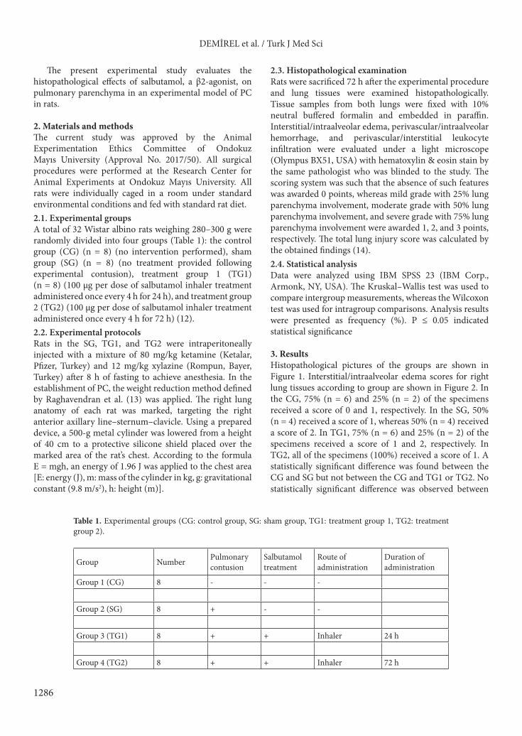
3. ResultsHistopathological pictures of the groups are shown in Figure 1. Interstitial/intraalveolar edema scores for right lung tissues according to group are shown in Figure 2. In the CG, 75% (n = 6) and 25% (n = 2) of the specimens received a score of 0 and 1, respectively. In the SG, 50% (n = 4) received a score of 1, whereas 50% (n = 4) received a score of 2. In TG1, 75% (n = 6) and 25% (n = 2) of the specimens received a score of 1 and 2, respectively. In TG2, all of the specimens (100%) received a score of 1. A statistically significant difference was found between the CG and SG but not between the CG and TG1 or TG2. No statistically significant difference was observed between
Table 1. Experimental groups (CG: control group, SG: sham group, TG1: treatment group 1, TG2: treatment group 2).
Group Number Pulmonary contusion
Salbutamol treatment
Route of administration
Duration of administration
Group 1 (CG) 8 - - -
Group 2 (SG) 8 + - -
Group 3 (TG1) 8 + + Inhaler 24 h
Group 4 (TG2) 8 + + Inhaler 72 h
1287
DEMİREL et al. / Turk J Med Sci
6
0 0 0
2
4
6
8
0
4
2
00 0 0 00
1
2
3
4
5
6
7
8
CG SG TG1 TG2
num
ber
of ra
ts
Groups
0
1
2
3
Figure 1. Histopathological evaluation of right lung tissues. Top left: Control group, mild interstitial congestion and slight edema in the lung (H&E, 100×). Top right: Sham group, severe leukocyte infiltration, alveolar and interstitial congestion, and edema (H&E, 400×). Bottom left: Treatment group 1, moderate leukocyte infiltration, alveolar and interstitial congestion, and edema (H&E, 400×). Bottom right: Treatment group 2, mild leukocyte infiltration, alveolar and interstitial congestion, and edema (H&E, 200×).
Figure 2. Interstitial/intraalveolar edema distribution according to group (CG: control group, SG: sham group, TG1: treatment group 1, TG2: treatment group 2).
1288
DEMİREL et al. / Turk J Med Sci
SG and TG1, though a statistically significant difference was found between SG and TG2 (Table 2).
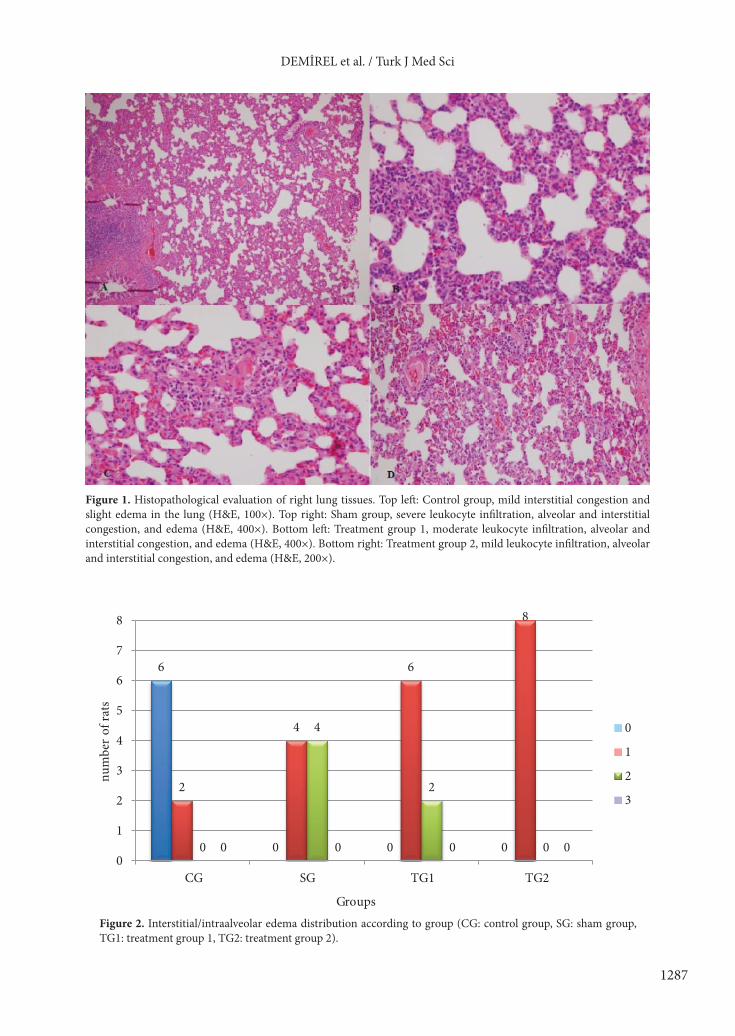
Interstitial/intraalveolar hemorrhage scores for right lung tissues according to group are shown in Figure 3. In the CG, 75% (n = 6) and 25% (n = 2) of the specimens received a score of 0 and 1, respectively. In the SG, 12.5% (n = 1), 75% (n = 6), and 12.5% (n = 1) of the specimens received a score of 1, 2, and 3, respectively. In TG1, all of the specimens received a score of 1. In TG2, 87.5% (n = 7) and 12.5% (n = 1) of the specimens received scores of 1 and 2, respectively. Statistical analysis revealed a statistically significant difference between the CG and SG but not between the CG and TG1 or TG2. Although no statistically significant difference was found between the SG and TG2, a significant difference was observed between the SG and TG1 (Table 3).
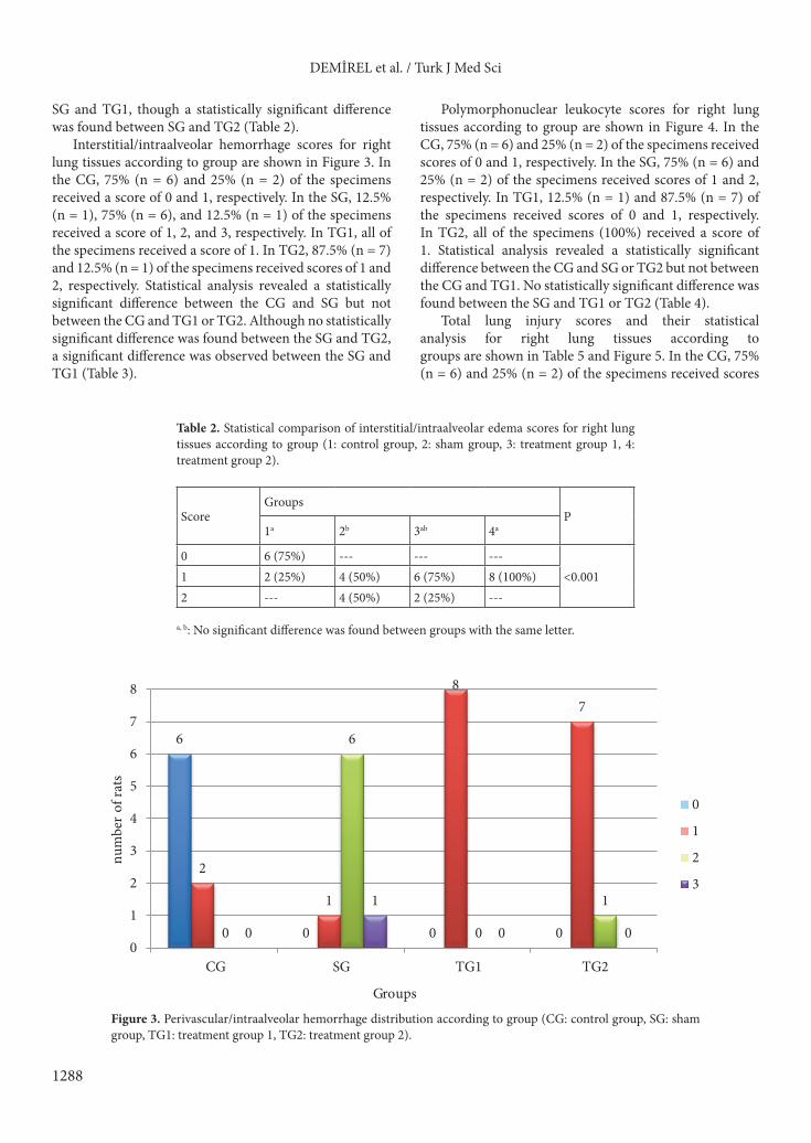
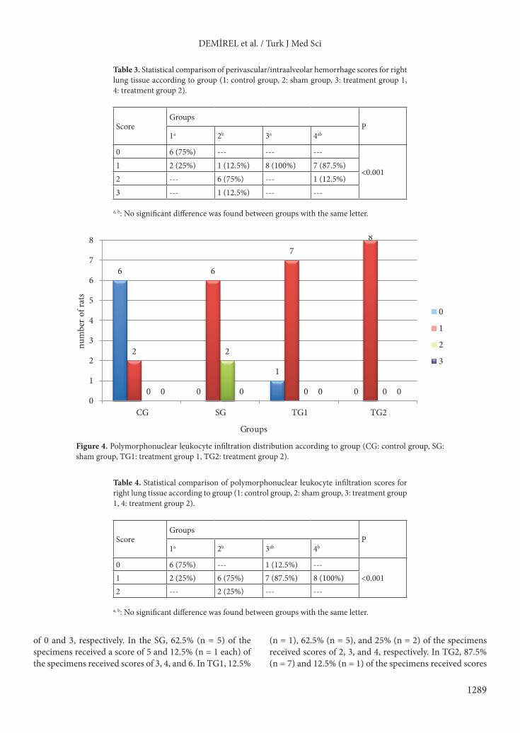
Polymorphonuclear leukocyte scores for right lung tissues according to group are shown in Figure 4. In the CG, 75% (n = 6) and 25% (n = 2) of the specimens received scores of 0 and 1, respectively. In the SG, 75% (n = 6) and 25% (n = 2) of the specimens received scores of 1 and 2, respectively. In TG1, 12.5% (n = 1) and 87.5% (n = 7) of the specimens received scores of 0 and 1, respectively. In TG2, all of the specimens (100%) received a score of 1. Statistical analysis revealed a statistically significant difference between the CG and SG or TG2 but not between the CG and TG1. No statistically significant difference was found between the SG and TG1 or TG2 (Table 4).
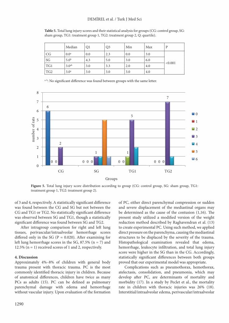
Total lung injury scores and their statistical analysis for right lung tissues according to groups are shown in Table 5 and Figure 5. In the CG, 75% (n = 6) and 25% (n = 2) of the specimens received scores
Table 2. Statistical comparison of interstitial/intraalveolar edema scores for right lung tissues according to group (1: control group, 2: sham group, 3: treatment group 1, 4: treatment group 2).
a, b: No significant difference was found between groups with the same letter.
6
0 0 0
2
1
87
0
6
0
1
0
1
0 00
1
2
3
4
5
6
7
8
CG SG TG1 TG2
num
ber
of ra
ts
Groups
0
1
2
3
Figure 3. Perivascular/intraalveolar hemorrhage distribution according to group (CG: control group, SG: sham group, TG1: treatment group 1, TG2: treatment group 2).
1289
DEMİREL et al. / Turk J Med Sci
Table 3. Statistical comparison of perivascular/intraalveolar hemorrhage scores for right lung tissue according to group (1: control group, 2: sham group, 3: treatment group 1, 4: treatment group 2).
a, b: No significant difference was found between groups with the same letter.
Table 4. Statistical comparison of polymorphonuclear leukocyte infiltration scores for right lung tissue according to group (1: control group, 2: sham group, 3: treatment group 1, 4: treatment group 2).
a, b: No significant difference was found between groups with the same letter.
6
0
1
0
2
6
78
0
2
0 00 0 0 00
1
2
3
4
5
6
7
8
CG SG TG1 TG2
num
ber
of ra
ts
Groups
0
1
2
3
Figure 4. Polymorphonuclear leukocyte infiltration distribution according to group (CG: control group, SG: sham group, TG1: treatment group 1, TG2: treatment group 2).
of 0 and 3, respectively. In the SG, 62.5% (n = 5) of the specimens received a score of 5 and 12.5% (n = 1 each) of the specimens received scores of 3, 4, and 6. In TG1, 12.5%
(n = 1), 62.5% (n = 5), and 25% (n = 2) of the specimens received scores of 2, 3, and 4, respectively. In TG2, 87.5% (n = 7) and 12.5% (n = 1) of the specimens received scores
1290
DEMİREL et al. / Turk J Med Sci
of 3 and 4, respectively. A statistically significant difference was found between the CG and SG but not between the CG and TG1 or TG2. No statistically significant difference was observed between SG and TG1, though a statistically significant difference was found between SG and TG2.
After intragroup comparison for right and left lung tissues, perivascular/intraalveolar hemorrhage scores differed only in the SG (P = 0.020). After examining for left lung hemorrhage scores in the SG, 87.5% (n = 7) and 12.5% (n = 1) received scores of 1 and 2, respectively.
4. DiscussionApproximately 4%–8% of children with general body trauma present with thoracic trauma. PC is the most commonly identified thoracic injury in children. Because of anatomical differences, children have twice as many PCs as adults (15). PC can be defined as pulmonary parenchymal damage with edema and hemorrhage without vascular injury. Upon evaluation of the formation
of PC, either direct parenchymal compression or sudden and severe displacement of the mediastinal organs may be determined as the cause of the contusion (1,16). The present study utilized a modified version of the weight reduction method described by Raghavendran et al. (13) to create experimental PC. Using such method, we applied direct pressure on the parenchyma, causing the mediastinal structures to be displaced by the severity of the trauma. Histopathological examination revealed that edema, hemorrhage, leukocyte infiltration, and total lung injury score were higher in the SG than in the CG. Accordingly, statistically significant differences between both groups proved that our experimental model was appropriate.
Complications such as pneumothorax, hemothorax, atelectasis, consolidation, and pneumonia, which may develop after PC, are determinants of mortality and morbidity (17). In a study by Peclet et al., the mortality rate in children with thoracic injuries was 26% (18). Interstitial/intraalveolar edema, perivascular/intraalveolar
Table 5. Total lung injury scores and their statistical analysis for groups (CG: control group, SG: sham group, TG1: treatment group 1, TG2: treatment group 2, Q: quartile).
a, b: No significant difference was found between groups with the same letter.
6
0 0 00 0 0 00 0
1
0
2
1
5
7
0
1
2
3
4
5
6
7
8
CG SG TG1 TG2
num
ber
of ra
ts
Groups
0
1
2
3
4
5
6
Figure 5. Total lung injury score distribution according to group (CG: control group, SG: sham group, TG1: treatment group 1, TG2: treatment group 2).
1291
DEMİREL et al. / Turk J Med Sci
References
1. Peclet MH, Newman KD, Eichelberger MR, Gotschall CS, Garcia VF, Bowman LM. Thoracic trauma in children: an indicator of increased mortality. J Pediatr Surg 1990; 25: 961-966.
2. Wu XJ, Xia ZY, Wang LL, Luo T, Zhan LY, Meng QT, Song XM. Effects of phencyclidine hydrochloride on pulmonary contusion from blunt chest trauma in rats. Injury 2012; 43: 232-236.
3. Bliss D, Silen M. Pediatric thoracic trauma. Crit Care Med 2002; 30: 409-415.
4. Akdemir HU, Güzel A, Katı C, Duran L, Alaçam H, Gacar A, Güvenç T, Murat N, Şişman B. The evaluation of different treatment protocols for trauma-induced lung injury in rats. J Thorac Dis 2014; 6: 66-73.
5. Ozdinc S, Oz G, Ozdemir C, Kilic I, Karakaya Z, Bal A, Koken T, Solak O. Melatonin: is it an effective antioxidant for pulmonary contusion? J Surg Res 2016; 204: 445-451.
6. Klassen TP, Rowe PC, Sutcliffe T, Ropp LJ, McDowell IW, Li MM. Randomized trial of salbutamol in acute bronchiolitis. J Pediatr 1991; 118: 807-811.
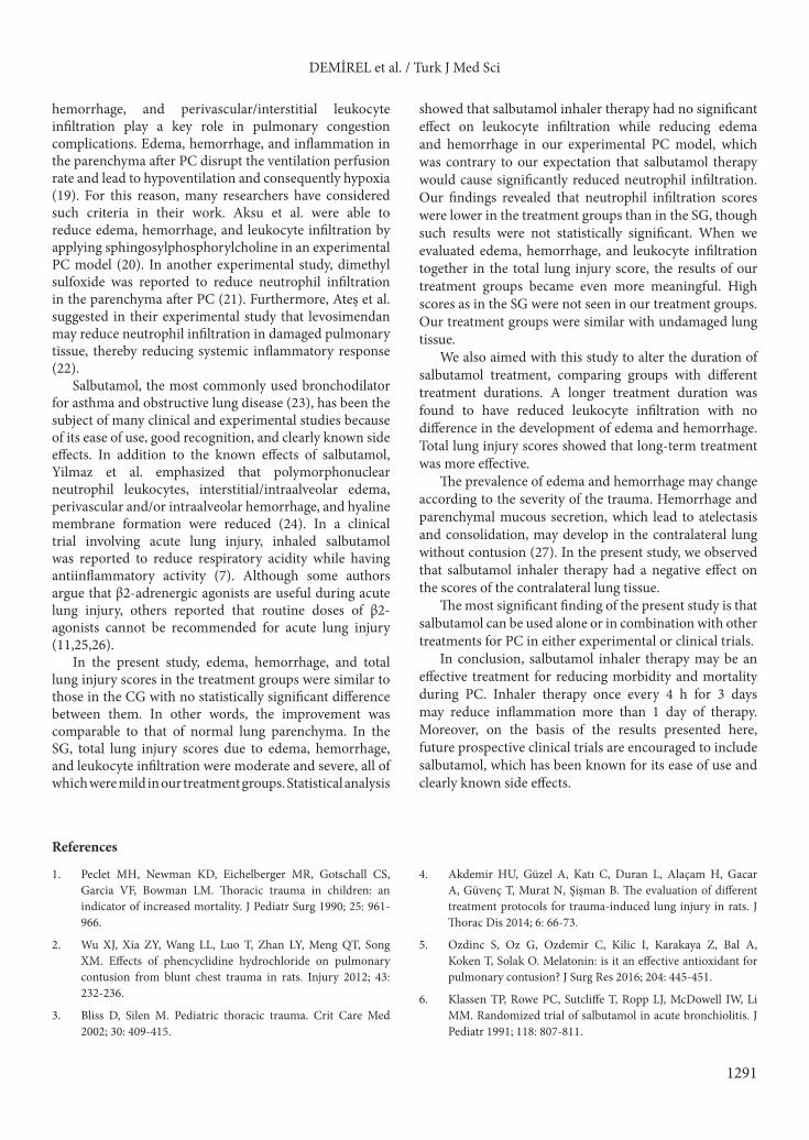
hemorrhage, and perivascular/interstitial leukocyte infiltration play a key role in pulmonary congestion complications. Edema, hemorrhage, and inflammation in the parenchyma after PC disrupt the ventilation perfusion rate and lead to hypoventilation and consequently hypoxia (19). For this reason, many researchers have considered such criteria in their work. Aksu et al. were able to reduce edema, hemorrhage, and leukocyte infiltration by applying sphingosylphosphorylcholine in an experimental PC model (20). In another experimental study, dimethyl sulfoxide was reported to reduce neutrophil infiltration in the parenchyma after PC (21). Furthermore, Ateş et al. suggested in their experimental study that levosimendan may reduce neutrophil infiltration in damaged pulmonary tissue, thereby reducing systemic inflammatory response (22).
Salbutamol, the most commonly used bronchodilator for asthma and obstructive lung disease (23), has been the subject of many clinical and experimental studies because of its ease of use, good recognition, and clearly known side effects. In addition to the known effects of salbutamol, Yilmaz et al. emphasized that polymorphonuclear neutrophil leukocytes, interstitial/intraalveolar edema, perivascular and/or intraalveolar hemorrhage, and hyaline membrane formation were reduced (24). In a clinical trial involving acute lung injury, inhaled salbutamol was reported to reduce respiratory acidity while having antiinflammatory activity (7). Although some authors argue that β2-adrenergic agonists are useful during acute lung injury, others reported that routine doses of β2-agonists cannot be recommended for acute lung injury (11,25,26).
In the present study, edema, hemorrhage, and total lung injury scores in the treatment groups were similar to those in the CG with no statistically significant difference between them. In other words, the improvement was comparable to that of normal lung parenchyma. In the SG, total lung injury scores due to edema, hemorrhage, and leukocyte infiltration were moderate and severe, all of which were mild in our treatment groups. Statistical analysis
showed that salbutamol inhaler therapy had no significant effect on leukocyte infiltration while reducing edema and hemorrhage in our experimental PC model, which was contrary to our expectation that salbutamol therapy would cause significantly reduced neutrophil infiltration. Our findings revealed that neutrophil infiltration scores were lower in the treatment groups than in the SG, though such results were not statistically significant. When we evaluated edema, hemorrhage, and leukocyte infiltration together in the total lung injury score, the results of our treatment groups became even more meaningful. High scores as in the SG were not seen in our treatment groups. Our treatment groups were similar with undamaged lung tissue.
We also aimed with this study to alter the duration of salbutamol treatment, comparing groups with different treatment durations. A longer treatment duration was found to have reduced leukocyte infiltration with no difference in the development of edema and hemorrhage. Total lung injury scores showed that long-term treatment was more effective.
The prevalence of edema and hemorrhage may change according to the severity of the trauma. Hemorrhage and parenchymal mucous secretion, which lead to atelectasis and consolidation, may develop in the contralateral lung without contusion (27). In the present study, we observed that salbutamol inhaler therapy had a negative effect on the scores of the contralateral lung tissue.
The most significant finding of the present study is that salbutamol can be used alone or in combination with other treatments for PC in either experimental or clinical trials.
In conclusion, salbutamol inhaler therapy may be an effective treatment for reducing morbidity and mortality during PC. Inhaler therapy once every 4 h for 3 days may reduce inflammation more than 1 day of therapy. Moreover, on the basis of the results presented here, future prospective clinical trials are encouraged to include salbutamol, which has been known for its ease of use and clearly known side effects.
1292
DEMİREL et al. / Turk J Med Sci
7. Eteraf-Oskouei T, Akbarzadeh-Atashkhosrow A, Maghsudi M, Najafi M. Effects of salbutamol on the inflammatory parameters and anjiogenesisi in the rat air pouch model of inflammation. Res Pharm Sci 2017; 12: 364-372.
8. McAuley DF, Frank JA, Fang X, Matthay MA. Clinically relevant concentrations of beta2-adrenergic agonists stimulate maximal cyclic adenosine monophosphate-dependent airspace fluid clearance and decrease pulmonary edema in experimental acid-induced lung injury. Crit Care Med 2004; 32: 1470-1476.
9. Perkins GD, Gates S, Park D, Gao F, Knox C, Holloway B, McAuley DF, Ryan J, Marzouk J, Cooke MW et al. The beta agonist lung injury trial prevention. A randomized controlled trial. Am J Respir Crit Care Med 2014; 189: 674-683.
10. Perkins GD, Nathani N, McAuley DF, Gao F, Thickett DR. In vitro and in vivo effects of salbutamol on neutrophil function in acute lung injury. Thorax 2007; 62: 36-42.
11. Roca O, Gomez-Olles S, Cruz MJ, Munoz X, Griffiths MJ, Masclans JR. Effects of salbutamol on exhaled breath condensate biomarkers in acute lung injury: prospective analysis. Crit Care 2008; 12: R72.
12. Akpinar ME, Tekke NS, Yigit O, Ercan F, Durna Y, Kiran D. Histological effects of inhaled corticosteroids and β2-agonists on laryngeal mucosa in allergic rat model. Otolaryngol Head Neck Surg 2013; 149: 457-465.
13. Raghavendran K, Davidson BA, Helinski JD, Marschke CJ, Manderscheid P, Woytash JA, Notter RH, Knight PR. A rat model for isolated bilateral lung contusion from blunt chest trauma. Anesth Analg 2005; 101: 1482-1489.
14. Yang Z, Deng Y, Su D, Tian J, Gao Y, He Z, Wang X. TLR4 as receptor for HMGB1-mediated acute lung injury after liver ischemia/reperfusion injury. Lab Invest 2013; 93: 792-800.
15. Holmes JF, Sokolove PE, Brant WE, Kuppermann N. A clinical decision rule for identifying children with thoracic injuries after blunt torso trauma. Ann Emerg Med 2002; 39: 492.
16. Cooper A, Barlow B, DiScala C, String D. Mortality and truncal injury: the pediatric perspective. J Pediatr Surg 1994; 29: 33-38.
17. Ganie FA, Lone H, Lone GN, Wani ML, Singh S, Dar AM, Wani N, Wani SN, Nazeer N. Lung contusion: a clinico-pathological entity with unpredictable clinical course. Bull Emerg Trauma 2013; 1: 7-16.
18. Peclet MH, Newman KD, Eichelberger MR, Gotschall CS, Garcia VF, Bowman LM. Thoracic trauma in children: an indicator of increased mortality. J Pediatr Surg 1990; 25: 961-966.
19. Cohn SM, Dubose JJ. Pulmonary contusion: an update on recent advances in clinical management. World J Surg 2010; 34: 1959-1970.
20. Aksu B, Ayvaz S, Aksu F, Karaca T, Cemek M, Ayaz A, Demirtas S. Effects of sphingosylphosphorylcholine against oxidative stress and acute lung injury induced by pulmonary contusion in rats. J Pediatr Surg 2015; 50: 591-597.
21. Boybeyi O, Bakar B, Aslan MK, Atasoy P, Kisa U, Soyer T. Evaluation of dimethyl sulfoxide and dexamethasone on pulmonary contusion in experimental blunt thoracic trauma. Thorac Cardiovasc Surg 2014; 62: 710-715.
22. Ateş G, Yaman F, Bakar B, Kısa U, Atasoy P, Büyükkocak U. Evaluation of the systemic antiinflammatory effects of levosimendan in an experimental blunt thoracic trauma model. Ulus Travma Acil Cer 2017; 23: 368-376.
23. Cazzola M, Page CP, Rogliani P, Matera MG. β2-Agonist therapy in lung disease. Am J Respir Crit Care Med 2013; 187: 690-696.
24. Yilmaz S, Yildizdas D, Daglioglu K, Acikalin A, Acipayam C, Bayram I, Gumurdulu D, Tanyeli A. The effects of salbutamol in an experimental model with acute respiratory distress syndrome. J Acute Dis 2012; 1: 94-99.
25. O’Kane CM, McKeown SW, Perkins GHD, Bassford CR, Gao F, Thickett DR, McAuley DF. Salbutamol up-regulates matrix metalloproteinase-9 in the alveolar space in the acute respiratory distress syndrome. Crit Care Med 2009; 37: 2242-2249.
26. Matthay MA, Brower RG, Carson S, Douglas IS, Eisner M, Hite D, Holets S, Kallet RH, Liu KD, MacIntyre N et al. Randomized, placebo-controlled clinical trial of an aerosolized β2-agonist for treatment of acute lung injury. Am J Respir Crit Care Med 2011; 184: 561-568.
27. Hellinger A, Konerding MA. Does lung contusion affect both the traumatized and the noninjured lung parenchyma? A morphological and morphometric study in the pig. J Trauma 1995; 39: 712-719.