Page 1
Evaluation of the remineralization potential of two non-fluoridated
remineralizing pastes using scanning electron microscope with
energy dispersive X-ray analysis: A randomized controlled
in-vitro trial
Dissertation submitted to
THE TAMIL NADU Dr M.G.R. MEDICAL UNIVERSITY
In partial fulfilment for the degree of
MASTER OF DENTAL SURGERY
BRANCH – VIII
PEDODONTICS AND PREVENTIVE DENTISTRY
MAY 2018
Page 2
KSR INSTITUTE OF DENTAL SCIENCE AND RESEARCH
DEPARTMENT OF PEDODONTICS AND PREVENTIVE DENTISTRY
CERTIFICATE
This is to certify that the dissertation titled “Evaluation of the remineralization potential of
two non-fluoridated remineralizing pastes using scanning electron microscope with energy
dispersive X-ray analysis: A randomized controlled in-vitro trial”
is a bonafide workdone by Dr. VIJAYASANKARI.V, Postgraduate student, during the
course of the study for the degree of “Master of Dental Surgery” in Department of
Pedodontics and Preventive Dentistry, KSR Institute of Dental Science and Research,
Tiruchengode during the period of 2015-2018.
Date: Dr. G.S. Kumar, M.D.S.,
Place: Tiruchengode Principal
Page 3
KSR INSTITUTE OF DENTAL SCIENCE AND RESEARCH
DEPARTMENT OF PEDODONTICS AND PREVENTIVE DENTISTRY
CERTIFICATE
This is to certify that the dissertation titled “Evaluation of the remineralization potential of
two non-fluoridated remineralizing pastes using scanning electron microscope with energy
dispersive X-ray analysis: A randomized controlled in-vitro trial”
is a bonafide workdone by Dr. VIJAYASANKARI.V, Postgraduate student, during the
course of the study for the degree of “Master of Dental Surgery” in Department of
Pedodontics and Preventive Dentistry, KSR Institute of Dental Science and Research,
Tiruchengode during the period of 2015-2018.
Date: Dr. Sharath Asokan, M.D.S., Ph.D
Place: Tiruchengode Professor and Head
Page 4
DECLARATION BY THE CANDIDATE
TITLE OF DISSERTATION
Evaluation of the remineralization potential of
two non-fluoridated remineralizing pastes
using scanning electron microscope with
energy dispersive X-ray analysis: A
randomized controlled in-vitro trial
PLACE OF STUDY
K.S.R Institute of Dental Science and Research
DURATION OF COURSE
3 Years (2015-2018)
NAME OF THE GUIDE
Dr. Sharath Asokan, M.D.S., Ph.D
HEAD OF THE DEPARTMENT
Dr. Sharath Asokan, M.D.S., Ph.D
I hereby declare that no part of the dissertation will be utilized for gaining financial
assistance for research or other promotions without obtaining prior permission from the
principal, K.S.R Institute of Dental Science and Research, Tiruchengode. In addition, I declare
that no part of this work will be published either in print or electronic without the guide who has
been actively involved in this dissertation. The author has the rights reserved for publishing the
work solely with prior permission of the principal, K.S.R Institute of Dental Science and
Research, Tiruchengode.
Head of the Department Signature of candidate
Page 5
ACKNOWLEDGEMENT
“Gratitude opens the door to the power, the wisdom and the creativity of the universe”
Foremost, I thank my grandmother Mrs.N.K.Subbulakshmi, my parents
Mr.S.Venkatesamurthy and Mrs.A.K.Maheshwari for their love, care, prayers and moral
support. Without them I couldn’t have made this in my life. I would also like to thank my sister
Mrs.V.Angayarkanni, My brother in law Mr.R.Manikandan and our little joy
Master.M.Aaryan for supporting me and making me happier all the time.
I express my sincere thanks to Dr.G.S.Kumar, M.D.S., Principal for providing the
opportunity of doing post-graduation in this college.
I would like to express my sincere gratitude to my guide, professor and head Dr. Sharath
Asokan, M.D.S, Ph.D, for the continuous support, his valuable guidance, patience, motivation,
enthusiasm and immense knowledge throughout the study. Also I thank him for providing me
facilities to conduct this study.
I owe my thanks and great honor to my postgraduate incharge professor Dr. P.R.Geetha
Priya, M.D.S for her motivation supervision, constructive critics and words of encouragement
which have gone a long way in successful completion of my study.
I would like to express my sincere thanks to Dr. V.Rajendran, Director R&D, Centre for
Nano Science and Technology, for offering me permission to conduct the study. I extend my
thanks to Mr.K.S.Balu, Research scholar, Centre for Nanoscience and technology, for his
support, for sharing his knowledge and spending his valuable time during the study.
I would like to thank my senior lectures Dr.Yogesh Kumar M.D.S,
Dr.G.Thiruvenkadam M.D.S and Dr.Lokeshwari M.D.S for their advice, motivation and
Page 6
helping me in clinical cases. I would also like to thank Dr.Lakshmi Prabha B.D.S for her
support. I would like to thank my super seniors Dr.Seby Thomas and Dr.Saravana Kumar for
helping me giving clinical tips and guidance.
I would like to express my gratitude to my seniors Dr.Allwyn Samuel, Dr.Kameshwaran
for their constant support and encouragement throughout the course.
I feel fortunate to have Dr.Janani RG as my colleague during the postgraduate course.
Her support and encouragement drove me through difficult situation. A good friend for life.
I would like to thank my juniors Dr.V.Chitravadhana, Dr.Jijo Mon, Dr.V.Sudhandra,
Dr.B.Kesavaraj for their never ending enthusiasm and meticulous help during the course. I
would like to express my thanks to the interns Dr.Selvalaksmi, Dr.Sreedhana and
Dr.Naleena for helping me in the study.
I am thankful to Dr.Carl Lewis, B.S.M.S., M.Sc., (Epidemiology), Chennai and
Dr.K.Sadhana M.D.S, Chennai for the Biostatistics work. I thank the non teaching faculty from
the department of Pedodontics and Preventive Dentistry for their prompt and patient help
throughout the course.
I came across many people who have been so kind to me. Thank you so much everyone
who has helped me along my student journey.
Page 7
CONTENTS
S.NO TITLE PAGE NO
1. INTRODUCTION 1
2. AIM AND OBJECTIVES 5
3. REVIEW OF LITERATURE 6
4. METHODOLOGY 20
5. RESULTS 39
6. DISCUSSION 62
7. SUMMARY AND CONCLUSION 69
8. REFERENCES 72
9. APPENDIX 81
Page 9
Dental caries is a localized chemical dissolution of the tooth surface caused by metabolic
events taking place in the biofilm covering the affected area. Biofilm is a prerequisite for the
carious lesion to occur. Any shift in the ecology and metabolic activity of the biofilm can cause
an imbalance in the equilibrium between the tooth minerals and the biofilm in the form of pH
fluctuations. The shift in the pH can influence the chemical composition of the tooth structure.
The tooth surface apatites are liable to such chemical modifications on countless events from the
very moment of eruption.45 Hydroxyapatite is the main component of enamel (95%) and dentine
(75%). The crystals of hydroxyapatite are hexogonal in cross section which are arranged to form
enamel rods. The solubility of hydroxyapatite is the primary determinant of dissolution of
enamel and it is related to pH.52
The enamel surface is in a state of dynamic equanimity with its surrounding
environment.45 When the pH in the surrounding medium drops down, the solubility of the apatite
crystals increases45 resulting in demineralization phase. During this phase, environment of the
oral cavity becomes undersaturated with mineral ions compared to the minerals content of the
tooth.50 The reverse takes place when the pH increases and remineralization takes place in the
interface between biofilm and the tooth surface.45 When the demineralization phase prolongs,
excessive loss of mineral ions takes place making the enamel surface sufficiently porous to be
seen clinically called as white spot lesion (WSL). This appearance has also been described as an
early, initial or incipient lesion.45 This process may progress further that eventually leads to
cavitation and total destruction of the tooth. Thus formation of caries is an ubiquitous, natural
process. It is impossible to prevent the formation of the biofilm and its metabolic activity, but the
progression of the disease can be controlled.18 As dental caries is a slow process, during early
stages non-invasive interventions can convert the initial lesion from an active to inactive state.26
Page 10
Traditionally, the management of dental caries is through surgical-restorative approach. It
involves diagnosis of carious lesions followed by surgical intervention to remove and restore the
affected part of the tooth. It is known that restoring the carious tooth alone might not stop the
disease process. This results in replacement of larger and larger subsequent restorations and
shorter associated survival times, resulting in a more invasive procedures over time. It is
estimated that 71% of all restorative treatments are performed on previously restored teeth, with
recurrent carious lesions as a predominant cause.31
The preventive approach of identification, conservative, non-restorative treatment of
incipient caries saves both dental manpower for profession as well as expense and suffering for
the patient.33 Thus the concept of minimal intervention dentistry has changed the perspective of
caries management, from “extension for prevention,” as proposed by GV Black. Today’s cavity
preparation is designed to preserve the health of the tooth over lifetime. The goal of modern
dentistry is to manage non-cavitated carious lesions non-invasively through remineralization in
an attempt to prevent disease progression, and to improve strength, esthetics and function of
teeth.22 Diagnosis of carious lesions at earlier stages and its remineralization has led to new era
in the modern preventive dentistry.33
Remineralization is defined as the process whereby calcium and phosphate ions are
supplied from a source external to the tooth to promote ion deposition into crystal voids in
demineralized enamel, to produce net mineral gain.12 For the past 60 years fluoride has been
used as a gold standard treatment for remineralizing teeth.61 The topical application of fluoride
agents, including dentifrices, mouthwash solutions, gels, and varnishes can help in
remineralization of initial enamel carious lesion.16 It results in the formation of calcium fluoride
like layer on the enamel surface thus preventing the subsequent acid attack and partially reduces
Page 11
the enamel mineral loss.16 Intake of fluoride during tooth formation systemically has been found
to be effective in prevention of caries.52
According to recommended daily allowances, a dose of 0.1mg fluoride/kg body weight
/day in children up to 8 years of age are considered to be safe causing no significant form of
fluorosis in permanent teeth. However it is crucial to consider the amount of fluoride in water,
tooth paste, dietary supplements and topical applications which have been identified as source of
enamel fluorosis. Moreover, the probable toxic dose of fluoride is 5 mg/kg of bodyweight which
triggers therapeutic intervention and hospitalization.52 Since most of the dental products contain
sufficient amount of fluoride, there is a high chance of chronic consumption of these products by
young children which could result in exceeding the toxic dose.42 Research has been looking for
an alternative caries remineralizing agents which should be as effective as fluoride.
Newer tooth remineralizing agents such as calcium phosphate based technologies and
biomimetic dental products containing nano sized hydroxyapatite particles are emerging in
modern preventive dentistry. Synthetic nano hydroxyapatite (nHAP) has the physical and
chemical properties similar to apatite structure in enamel42 making it most biocompatible and
bioactive material. nHAP has strong affinity to tooth and adsorbs strongly on the enamel
surfaces. Nano sized crystals smaller than 100nm improves bioactivity of the agent due to the
increase in the superficial surface area and wettability of nHAP. It promotes the remineralization
by increasing the saturation level of calcium and phosphate in saliva.16 With advancements in
nanotechnology, incorporation of nano sized biomimetic apatite particles in dentrifices and
mouthrinses have been increased. Since 1980, nHAP has been used in tooth paste in Japan and it
was accepted as anti caries agent in 1993.42 Various in-vitro and in-situ studies in the literature
have proved that nHAP has the potential to remineralize the artificial carious lesion. A study
Page 12
done by Roveri et al in 200952 demonstrated that biomimetic nanosized hydroxyapatite particles
produced an apatitic coating deposition on the demineralized enamel surface. Huang et al in
200930 proved that nHAP had the potential to remineralize the initial enamel carious lesions in
which 10% nHAP has been proved to be most effective. In an in situ study by Najibfard et al in
201142, nHAP dentrifice caused remineralization comparable to fluoride dentrifice. They
suggested that nHAP dentrifice can be used as an effective alternative to fluoride dentrifice.
Aclaim® (Group pharmaceuticals ltd) is one of the commercially available nHAP
toothpaste in India. According to the studies done by Verma P et al 201365 and Singh A et al
201757 Aclaim® has both densensitizing and remineralizing effect. So, the present study attempts
to synthesize nHAP toothpaste at 1% and 10% concentrations and compare its remineralization
efficacy with commercially available non-fluoridated remineralizing paste.
Page 14
AIMS
The present study was conducted with following aims:
1. To formulate experimental nHAP tooth pastes with 1% and 10% concentrations.
2. To evaluate the remineralization potential of experimental 1% and 10% nHAP tooth
paste.
3. To compare the remineralization potential of experimental 1% nHAP with commercially
available nHAP toothpaste using energy dispersive X-ray analysis (EDX).
4. To compare the remineralization potential of experimental 10% nHAP with commercially
available casein phosphopeptide amorphous calcium phosphate (CPP ACP) using
scanning electron microscope with energy dispersive X-ray analysis (SEM EDX).
HYPOTHESIS
The study hypothesis was that experimental nHAP pastes were equally effective in
comparison with commercially available pastes and control.
Page 15
REVIEW OF LITERATURE
Page 16
Shen P, Cai F, Nowicki A, Vincent J, Reynolds EC (2001)54 conducted a randomized
controlled cross over in-situ double blind study to evaluate the ability of sugar free CPP ACP
chewing gum in remineralizing enamel subsurface lesions. A total of 30 human subjects wore
removable palatal appliances with six human-enamel half-slabs inset containing sub-surface
demineralized lesions. The appliances were inserted immediately before gum chewing for 20
minutes and then retained for another 20 minutes. This was performed 4 times a day for 14 days.
At the completion of each treatment, the enamel half-slabs were paired with their respective
demineralized control half-slabs, embedded, sectioned, and subjected to microradiography and
densitometric image analysis, for measurement of the level of remineralization. They found that
addition of CPP-ACP to either sorbitol or xylitol based gum resulted in an increase in enamel
remineralization.
Andersson A, Skold-Larsson K, Hallgren A, Petersson LG, Twetman S (2007)2
compared the effects of a dental cream containing complexes of CPP ACP and fluoride
mouthwashes on the regression of WSLs. A total of 26 healthy adolescents exhibiting 60 teeth
with 152 visible WSL sites on incisors and canines were included. Baseline visual scoring and
laser fluorescence measurements were carried out. The patients were randomly assigned to two
different remineralization protocols A) daily topical applications of a dental cream containing
CPP ACP (Topacal) for 3 months followed by a 3-month period of daily tooth brushing with
fluoridated dentifrice, or B) daily 0.05% sodium fluoride mouthwash combined with fluoridated
dentifrice for 6 months. The laser fluorescence measurements and visual scoring were repeated at
1, 3, 6 and 12 months. A significant improvement (regression of WSL) of the clinical WSL
scores was found over time in both groups, but CPP ACP showed a statistically significant
difference (p< 0.01) in the decrease in number of lesions after 12 months. There was no
Page 17
statistically significant difference in laser fluorescence measurements. They concluded that both
the treatment could promote the regression of white spot lesions, but CPP ACP provided a more
favourable aesthetic outcome.
Kumar VL, Itthagarun A, King NM (2008)35 investigatedthe efficacy of CPP ACP
containing Tooth Mousse on the remineralization of enamel lesions and compared its efficacy to
that of a fluoride containing toothpaste. A total of 50 specimens were prepared from permanent
third molars and were randomly assigned in to 5 groups. Group A, fluoridated toothpaste
(positive control), Group B, non-fluoridated toothpaste (negative control), Tooth Mousse
containing CPP ACP was tested by three different means: Group C (as a toothpaste); Group D
(as a topical coating ); and Group E (as a topical coating after treating the sections with Group A
paste). All the specimens underwent a pH cycling for a period of 10 days with respective groups.
Polarizing light microscopy and microradiography were utilized to record the lesion depth and
the mineral content of each lesion before and after the 10 days of pH cycling. They found that
the lesion depth decreased by 13.1 per cent in Group E and it was statistically significant. They
concluded that CPP ACP containing Tooth Mousse showed a higher remineralizing potential
when applied as a topical coating after the use of fluoridated toothpaste.
Pai D, Bhat SS, Taranath A, Sargod S, Pai VM (2008)46 performed an in vitro study to
evaluate the remineralization of incipient enamel lesions by the topical application of CPP ACP
using laser fluorescence and scanning electron microscope (SEM). Sixty caries free teeth were
randomly divided in to 3 groups; 40 teeth were used as test sample (CPP ACP), 10 as positive
control (artificial saliva), 10 as negative control (normal saline). The samples were demineralized
and then remineralized by the topical application of CPP ACP for a period of 14 days. The
results showed that both laser fluorescence readings and SEM scores of test samples after
Page 18
remineralization were highly significant (p <0.001). They concluded that CPP ACP can prevent
demineralization and also bring about remineralization in enamel lesions.
Yengopal V, Mickenautsch S (2009)67 did systematic review with meta analysis to
assess the caries preventive effect of CPP ACP. Five in situ randomized control trials (RCT)
werepooled for meta-analyses. The results of the clinical in situ trials indicated a short-term
remineralization effect of CPP ACP. Additionally, in vivo RCT results suggested a caries
preventing effect for long-term clinical CPP ACP use. They concluded that further in vivo
randomized trials are needed to confirm these initial results.
Bailey DL, Adams GG, Tsao CE, Hyslop A, Escobar K, Manton DJ, Reynolds EC,
Morgan MV (2009)4 compared the effect of CPP ACP on post orthodontic WSL among
adolescents of 12-18 years of age for a period of 12 weeks. Four hundred and eight WSLs in 45
participantswere randomly divided in to 2 groups: CPP ACP group and placebo group. Clinical
assessments were performed according to International Caries Detection and Assessment System
(ICDAS) II criteria. They found that WSLs with severity codes 2 and 3 at baseline had a
significantly greater chance of regressing at 12 weeks in the CPP ACP group compared with
those in the placebo group.
Srinivasan N, Kavitha M, Loganathan SC (2009)59 conducted a in-situ study to
compare the remineralization potential of pastes containing CPP ACP and CPP ACP with 900
ppm fluoride on human enamel softened by a cola drink. Forty five enamel specimens obtained
from human third molar teeth were eroded in a cola drink for 8 minutes and then attached to
intra-oral devices worn by five volunteers. The specimens were subjected to three different in
situ remineralization protocols using: (1) CPP ACP (Group I), (2) CPP ACP with 900 ppm
Page 19
fluoride (Group II), and (3) saliva (Group III, control). Vickers microhardness measurements
were recorded at baseline, demineralization and remineralization stages. The results revealed
statistically significant differences in the mean microhardness values between pastes containing
CPP ACP and CPP ACP with 900 ppm fluoride. They concluded that both pastes substantially
remineralized the softened enamel, with the CPP ACP and fluoride combination showing higher
remineralization potential than CPP ACP.
Altenburger MJ, Gmeiner B, Hellwig E, Wrbas KT, Schirrmeister JF (2010)1
performed an in-vivo study to evaluate the effect of CPP ACP on initial enamel caries in pits and
fissures using DIAGNOdent and visual assessment. A total of 32 volunteers with premolars and
molars showing DIAGNOdent scores between 15 and 20 were randomly assigned in to 2
treatment groups. The intervention period consisted of 3 weeks, first 2 weeks of wash out period
and third week of treatment period. During a wash-out period of 2 weeks and during the 3-week
treatment period all subjects used only standard fluoride toothpaste without any oral hygiene
products. During the treatment period, one group additionally applied a CPP ACP containing
cream on the respective fissures for 3 minutes, once per day. At days 1, 8, 15, and 22,
DIAGNOdent measurements and a visual assessment of the fissures were done. They found that
CPP ACP group showed significantly lower fluorescence values at day 15 and day 21 compared
to the control group. There was no significant difference in the visual assessment scores in both
the groups.
Huang S, Gao S, Cheng L, Yu H (2010)28 investigated the combined effects of
nanohydroxyapatite and Gallachinensis (GCE) on remineralization of initial enamel lesion. In
vitro demineralized bovine enamel blocks were subjected to a pH-cycling regime for 12 days.
Each daily cycle included 3-4 minutes application with one of five treatments: NaF (positive
Page 20
control), deionised water (negative control), crude aqueous extract of GCE, nHAP and GCE with
nHAP. The samples were subsequently evaluated using a microhardness tester, polarised light
microscopy (PLM), X-ray diffraction (XRD) and SEM. GCE–nHAP combined treatment group
showed significant reduction in lesion depth and more mineral deposition occurred in the lesion
body. They concluded that there was a significant synergistic effect of combined GCE and nHAP
treatment on promoting the remineralization of initial enamel lesion.
Brochner A, Christensen C, Kristensen B, Tranæus S, Karlsson L, Sonnesen L,
Twetman S (2011)6 compared the effect of CPP ACP on post orthodontic WSLs among fifty
adolescents. Twenty two belonged to the intervention group (CPP ACP) and 28 in the control
group (standard fluoride toothpaste). The outcome was measured by quantitative light induced
fluorescence (QLF) and visual scores from digital photographs at baseline and 4 weeks. Both the
groups showed a statistically significant regression of WSL from the baseline, but there was no
difference between the groups. They concluded that topical application of CPP ACP resulted in
significantly reduced fluorescence and reduced area in the lesion size after 4 weeks.
Najibfard K, Ramalingam K, Chedjieu I, Amaechi BT (2011)42 evaluated the
efficacy of nHAP dentifrices on caries remineralization and demineralization inhibition by
conducting a double-blind randomized crossover in situ study. A total of 30 adults wore an intra
oral appliance containing 3 demineralized enamel blocks and one healthy enamel block cut from
each of 30 molars, were exposed respectively to dentifrices of A) 5% nHAP, B) 10% nHAP, C)
1100 ppm fluoride, and D) 10% nHAP in 4 phases lasting 28 days per phase. Baseline and post-
test mineral loss and lesion depth were quantified using microradiography. They concluded that
nHAP dentifrice caused remineralization comparable to a fluoride dentifrice, and inhibited caries
Page 21
development, thus suggesting that an HAP dentifrice can be an effective alternative to fluoride
toothpaste.
Ferrazzano GF, Amato I, Cantile T, Sangianantoni G, Ingenito A (2011)21
investigated the remineralizing effect of GC Tooth Mousse on early dental enamel lesions using
SEM analysis. 40 volunteers were randomly divided into two groups. In group (CPP ACP group)
two demineralized enamel specimens were placed on the buccal surfaces of the first molars and
were instructed to apply GC Tooth Mousse only on the right-sided specimen and a placebo
mousse on the left, for 1 month. In Group B (control group) two enamel specimens were placed
into the mouth without any intervention. The results of SEM analysis revealed a diffuse and
homogenous mineral coating reducing the surface alteration in CPP ACP group. They concluded
that CPP ACP was able to promote remineralization of early enamel lesions.
Tschoppe P, Zandim DL, Martus P, Kielbassa AM (2011)62 performed anin-vitro
study to evaluate the effects of nHAP toothpastes on the remineralization of bovine enamel and
dentine subsurface lesions. A total of 70 enamel specimens were randomly divided in 5 groups
and exposed to an aqueous remineralizing solution for two and fiveweeks (37 ̊
C).Brushingprocedures were performed with the respective toothpaste/storage solution slurry
twice daily:1) storage in remineralizing solution only; 2) additional brushing with 20 weight%
zinc carbonate nano hydroxyapatite, ZnCO3/nHAP; 3) 24 weight% ZnCO3/n-Hap; 4) 0.14
weight% amine fluoride; 5) 7 weight% pure nHAP. Differences in mineral loss (DDZ) before
and after storage/treatment were microradiographically evaluated. They concluded that
toothpastes containing nHAP revealed higher remineralizing effects compared to amine fluoride
toothpastes.
Page 22
Zhang Q, Zou J, Yang R, Zhou X (2011)68 conducted an in-vitro study to evaluate the
remineralizing effect of CPP ACP cream on the artificial enamel lesion of the primary teeth
andto assess its caries prevention efficiency. A total of 90 enamel specimens were randomly
divided in to 3 groups: group A: distilled and deionized water, group B: CPP ACP, group C:
sodium fluoride solution. The enamel surface microhardness was measured before, after
demineralization and 30 days after remineralization. The enamel specimens were also subjected
to SEM analysis. They found that there was a significant increase in microhardness in all the
groups. On comparison between the groups, CPP ACP group showed significant increase in
microhardness than sodium fluoride group. The results of SEM analysis revealed CPP ACP
group exhibited more homogenous arrangement of crystals than sodium fluoride group. They
concluded that CPP ACP cream effective in remineralizing enamel lesions in primary teeth.
Hegde MN, Moany A (2012)27 quantitatively evaluated the remineralization potential of
CPP ACP paste on enamel subsurface lesions using SEM EDX analysis. A total of 90 specimens
were randomly assigned into two groups: group 1 contained 15 specimens (control group) and
group 2 contained 75 specimens (study group). The study group was subdivided into five groups
of 15 specimens per group. Each subgroup was treated with remineralizing paste [10% CPP ACP
paste seven days (subgroup 2a), 14 days (subgroup 2b), 21 days (subgroup 2c), 28 days
(subgroup 2d), and 35 days (subgroup 2e), twice daily for three minutes. SEM EDX was done to
measure mineral content before, after demineralization and remineralization procedure. They
found that all the study groups showed very highly significant differences between Calcium
phosphorus ratios of the demineralized and remineralized samples. There was no significant
difference seen in the control group. They concluded that the remineralization achieved was
Page 23
dose-dependent as the remineralizing rate increased with the timeof exposure of enamel to CPP
ACP paste.
Comar LP, Souza BM, Gracindo LF, Buzalaf MA, Magalhães AC (2013)14 conducted
an in-vitro trial to evaluate the preventive potential of experimental pastes containing 10% and
20% nHAP, with or without fluoride, on dental demineralization. According to the surface
hardness, bovine enamel (n=15) and root dentin (n=15) specimens were divided into 9 groups :
control (without treatment), 20 Nanop paste (20% HAP), 20 Nanop paste plus (20% HAP +
0.2% NaF), 10 Nanop paste (10% HAP), 10 Nanop paste plus (10% HAP + 0.2% NaF), placebo
paste (without fluoride and HAP), fluoride paste (0.2% NaF), MI paste (CPP ACP), and MI paste
plus (CPP ACP + 0.2% NaF). All the specimens were subjected to pH cycling model for 7 days
and the dental subsurface demineralizat ion was analyzed using cross-sectional hardness.
They found that 0.2% NaF significantly reduced the loss of enamel and dentin sub surface
hardness. Experimental Nanop pastes, regardless of the addition of fluoride, were unable to
reduce demineralization in-vitro
Rallan M, Chaudhary S, Goswami M, Sinha A, Arora R, Kishor A (2013)49
determined the effect of three remineralizing agents on eroded enamel of human primary anterior
teeth. A total of 40 primary anterior teeth were randomly divided in to 4 groups. Group I: CPP
ACP, Group II: CPP ACP with fluoride (CPP ACPF) and Group III: fluoridated toothpaste and
Group IV: artificial saliva (control). A thin layer of respective paste were applied on the enamel
surface and left undisturbed for 3 minutes and then stored in artificial saliva for 8 hours. The
Knoop microhardness of the labial surface of enamel was measured at baseline, after erosion and
after the remineralization procedures. They found that CPP ACPF showed significant increase in
microhardness compared to the other groups.
Page 24
de Carvalho FG, Vieira BR, Santos RL, Carlo HL, Lopes PQ, de Lima BA (2014)16
analyzed the protective effect of remineralizing agents on enamel caries lesions. An in vitro
study conducted using 48 human enamel specimens were randomly divided in to 4 groups: (1)
control (without agent); (2) fluoride varnish (Duraphat); (3) nHAP paste (DesensibilizeNano P);
and (4) CPP ACPF paste (MI Paste Plus). Artificial carious lesion were developed in all the
specimens and subjected to cariogenic challenge (pH cycling) for 7 days. The surface
microhardness was evaluated at baseline, after artificial caries lesion formation and after pH
cycling. The percentage of surface microhardness recovery (%SMHR) was performed, and the
surface morphology was evaluated by atomic force microscopy (AFM). They found after the pH
cycling, the nHAP group showed significantly higher Knoop hardness number (KHN) and
%SMHR values than varnish whereas CPP ACP group showed no increase in KHN. On
evaluation by AFM nano HAP group showed protective layer formation with globular deposits
on the surface and concluded that nHAP paste has protective effect against in-vitro enamel caries
development.
Haghgoo R, Rezvani MB, Salehi Zeinabadi M (2014)25 compared sodium fluoride
mouthrinse and nHAP at different concentrations (0-2-5-10%) for remineralization of initial
enamel carious lesions. A total of 60 human premolars were randomly divided and subjected to
microhardness evaluation at baseline, post demineralization and remineralization. They
concluded that nHAP and NaF mouthrinses can greatly enhance remineralization and increase
tooth microhardness though the results were not statistically significant.
Vano M, Derchi G, Barone A, Covani U (2014)63 conducted a double blind randomized
controlled trial among 105 subjects to compare the efficacy in reducing dentin hypersensitivity.
The subjects were divided into 3 groups receiving treatment, 1) nHAP 15% tooth paste fluoride
Page 25
free; 2) fluoride toothpaste 3) placebo. By using air blast and tactile test dentin hypersensitivity
was evaluated at baseline and after 2 weeks and 4 weeks. In addition, subjective evaluation using
visual analog scale (VAS) was also used. The results showed statistically significant lower
values for sensitivity and VAS scores for group 1. They concluded that fluoride free nano
hydroxyapatite is considered as an effective desensitizing agent providing quick relief from
symptoms of sensitivity after 2 and 4 weeks.
Mielczarek A, Michalik J (2014)40 performed an in-vitro study comparing toothpaste
containing nHAP and 1,450 ppm F; toothpaste containing 1,450 ppm F; and Placebo group
(distilled water). Ninety human enamel specimens were randomly divided into 3 groups and
subjected to demineralization and remineralization phases. The surface microhardness of each
specimen were measured at each phase. They concluded surface microhardness increased
significantly in all the groups but none of the group reached the baseline level.
Memarpour M, Fakhraei E, Dadaein S, Vossoughi M (2015)39 conducted a 1 year
randomized controlled trial among 146 preschool children with WSLs on maxillary anterior
teeth. They compared the effect of CPP ACP, fluoride varnish and oral hygiene with dietary
counseling. They used dmft index and scored dental probe. There was a significant reduction in
the size of the lesion in CPP ACP group. They concluded that preventive interventional methods
play an important role in reducing white spot lesions in children. Oral hygiene instruction
together with the application of CPP ACP and fluoride varnish was an effective method to
reduce WSL size and dmft index values in primary teeth.
Llena C, Leyda AM, Forner L (2015)37 performed a double blind prospective study to
evaluate the effect of CPP ACP and CPP ACPF versus fluoride varnish on the remineralization
Page 26
of enamel white spot lesions. The participants were divided randomly into three groups. Groups
A: GC Tooth Mouse (CPP ACP), Group B: Mi Paste Plus (CPP ACPF) and Group C: Duraphat
fluoride varnish. WSLs were categorized according to the ICDAS criteria (ICDAS II; grades 0–
3) and assessed by laser fluorescence (DIAGNOdent) at baseline and at 4, 8 and 12 weeks.
DIAGNOdent values were significantly reduced in Group B (CPP ACPF) at 4 weeks, and in
Groups A (CPP ACP) and group C (Fluoride varnish) at 8 weeks. They concluded that CPP-
ACPF have a significant effect on smooth surface caries.
Vyavhare S, Sharma DS, Kulkarni VK (2015)66 evaluated the effect of nHAP (10%),
CPP-ACP (10%), NaF (1000 ppm) and Deionized water (negative control) on remineralization
of initial enamel lesion. A total of 26 human permanent incisors were subjected to
demineralization and pH cycling. Surface microhardness measurements were performed before,
after demineralization and after 3, 6, 9 and 12 days of pH cycling. The specimens were examined
by SEM. Percentage surface microhardness of nHAPand fluoride was significantly greater than
CPP ACP and negative control. When observed under SEM, nHAP particles were deposited on
the demineralized surface whereas CPP-ACP does not show any significant surface
remineralization. They concluded that nHAP and fluoride had the potential to remineralize initial
enamel lesions.
Souza BM, Comar LP, Vertuan M, Fernandes Neto C, Buzalaf MA, Magalhães AC
(2015)58 conducted a randomized cross over double blind in situ study. They evaluated the effect
of an experimental paste containing hydroxyapatite in nanoparticles/ fluoride on dental de-
remineralization. Thirteen subjects took part in this study which was performed in 4 phases (14
days each). Four sound and 4 predemineralised specimens were worn intraorally at each phase
corresponding to the following treatments: Nanop Plus (10% HA, 0.2% NaF, nano-HA/fluoride),
Page 27
MI Paste Plus (CPP-ACP, 0.2% NaF), F (0.2% NaF) and placebo. The demineralization and
remineralisation was quantified by transversal microradiography. They concluded that Nanop
Plus significantly reduced the dentin demineralization and improved enamel remineralization.
Haghgoo R, Ahmadvand M, Moshaverinia S (2016)24 evaluated the remineralizing
effect of topical NovaMin and nHAP on caries-like lesions. Thirty human primary incisors were
randomly subjected to demineralization / remineralization cycle and surface microhardness of
each tooth was measured at baseline, post demineralization and remineralization. They
concluded that no significant differences were detected in their efficacy and both nHAP and
NovaMin were effective for remineralization of caries like lesion in primary teeth.
Ebadifar A, Nomani M, Fatemi SA (2017)17 assessed the effect of nHAP on
microhardness of artificially created carious lesion. A total of 80 extracted teeth were
randomized in two groups, Group A contained nHAP and fluoride and Group B contained
fluoride alone. They concluded that toothpaste containing nHAP showed significant increase in
microhardness and was more effective than the toothpaste without NHA for the purpose of
remineralization.
Esteves-Oliveira M, Santos NM, Meyer-Lueckel H, Wierichs RJ, Rodrigues JA
(2017)20 performed a in vitro study to investigate the caries preventive effect of nHAP containing
toothpastes. Two hundred enamel specimens were prepared from bovine incisors and
randomized into 5 groups, which received different fluoride treatments: fluoride-free toothpaste
(0 ppm F-), as negative control; AmF (1400 ppm F-) anti-caries; AmF/NaF/ SnCl2/Chitosan (700
ppm F-/700 ppm F-/3500 ppm Sn2+), anti-erosion; NaF/KNO3 (1400 ppm F-), anti-erosion; and
nHAPcontaining (0 ppm F−) toothpastes. Changes in mineral loss and lesion depth were
determined using transversal microradiography. All toothpastes caused significantly less
Page 28
demineralization than negative control except for nHAP. They concluded that both anti-erosive
and anti-caries toothpastes reduced mineral loss to a similar extent, whereas the fluoride-free
nHAP containing toothpaste did not show inhibition of caries demineralization in vitro.
Vano M, Derchi G, Barone A, Pinna R, Usai P, Covani U (2017)64 conducted a
randomized double-blind clinical trial to compare the efficacy in reducing dentin hypersensitivity
of a dentifrice formulation containing nHAP with a fluoride dentifrice. A total of 115
participants were randomly divided into 3 groups: Group 1: Nano-hydroxyapatite 2%gel
toothpaste (fluoride free), Group 2: Fluoride gel toothpaste, Group 3: Placebo. The participant’s
dentin hypersensitivity was evaluated at baseline, after 2 and 4 weeks using airblast, tactile tests.
Subjective evaluation was done by visual analogue scale. They concluded that the application of
nHAP in gel toothpaste was an effective desensitizing agent providing relief from symptoms
after 2 and 4 weeks.
Shaik ZA, Rambabu T, Sajjan G, Varma M, Satish K, Raju VB, Ganguru S,
Ventrapati N (2017)53 quantitatively assessed the remineralization potential of CPP ACP,
Vantej and Icon by SEM EDX. A total of 78 maxillary premolars were randomly divided into 3
groups. Group 1:Vantej (Bioactive glass), Group 2: CPP ACP, Group 3: Icon (Resin infiltration).
All the samples were subjected to EDX analysis before, after demineralization and after
remineralization. Calcium and phosphate ratio increased significantly in CPP ACP group
compared to the other groups. They concluded that CPP ACP showed greater potential of
remineralization followed by Vantej and Icon.
Kamath P, Nayak R, Kamath SU, Pai D (2017)32 conducted a in-vitro double blind
randomized controlled trial to evaluate the remineralization potential of commercially available
Page 29
remineralization agents on WSLs in primary teeth. Forty extracted or exfoliated primary
teethwere selected and randomized into 4 groups: Group I: FTCP (Tricalcium phosphate with
fluoride), Group II: fluoridated dentifrice, Group III: CPP ACPF, and Group IV: nHAP. They
evaluated at baseline, post demineralization and post remineralization values by DIAGNOdent
and SEM EDX. Evaluation by SEM showed favorable surface changes in all the four study
groups. Intragroup comparison of DIAGNOdent and EDX readings showed a highly significant
difference between baseline, post demineralization, and post remineralization values. However,
the intergroup comparison was statistically non significant. They concluded that all the test
agents were comparable in their remineralization potential.
Page 31
The present randomized controlled invitro study was conducted from the Department of
Pediatric and Preventive Dentistry, KSR Institute of Dental Science and Research (KSRIDSR).
The study design and protocol was analyzed and approved by the Institutional Review Board and
Institutional Ethics Committee of KSRIDSR, Tiruchengode, Tamil Nadu.
Armamentarium
1. 35 human primary molars
2. Contra-angle hand piece
3. Self-cure resin
4. Nail polish
5. Gloves
6. Mask
7. Eye wear
8. Plastic containers
9. Double faced diamond disc
10. 10% formaldehyde
11. 0.1% thymol solution
12. Demineralization solution
13. pH meter (Slope Labtronics, Model LTD 11, Panchkula, Haryana, India)
14. Incubator (Coslab, ISO 9001:2000, Ambala Cant, Haryana, India)
15. Pipette
16. Burette
17. Hot air oven
18. Magnetic stirrer
Page 32
19. Stirring rod
20. Dry ball mill (PM 100; Retsch Corporation, Germany)
21. Aclaim® Tooth paste (Sorbitol, Glycerine, Silica, Purified water, Hydroxyapatite,
Cocomidopropylbetaine, Hydroxyethyl cellulose, Titanium dioxide, Flavour, Sodium
saccharin; Group pharmecuticals Ltd, Bangalore, India )
22. CPP ACP paste (GC tooth mousse, Recaldent™, Belgium, Germany)
Inclusion criteria
1. Extracted first and second human primary molars
Exclusion criteria
1. Teeth with developmental defects
2. Teeth with caries and white spot lesion
3. Teeth subjected to previous treatment
Specimen preparation
Thirty five extracted human primary molars were selected for this study. The selected teeth
were cleaned using an ultrasonic scalerto remove soft tissue debris. They were disinfected for 2
days using 10% formaldehyde. The teeth were decoronated at the cemento enamel junction and
sectioned into buccal and lingual halves using a double faced diamond disc mounted on a
contra-angle hand piece. Each specimen was embedded in self-cure acrylic with the enamel
surface exposed thus involving 65 enamel specimens from 35 teeth. All the specimens were
stored in 0.1% thymol solution till the study was carried out.
Page 33
Elemental analysis by EDX
The analysis was done in South Indian textile research association (SITRA), Coimbatore.
The specimens were kept in moisture free environment and care was taken to avoid contact with
air or moisture. The specimens were placed on a metal mounting block and quantification of
calcium and phosphorus content was done using EDX analysis.
Baseline EDX analysis
A 3 × 3 mm square window was prepared on enamel surface of each specimen by
applying nail polish varnish in rest of the tooth surface. All the specimens were subjected to
EDX analysis for assessing the mineral content at baseline.
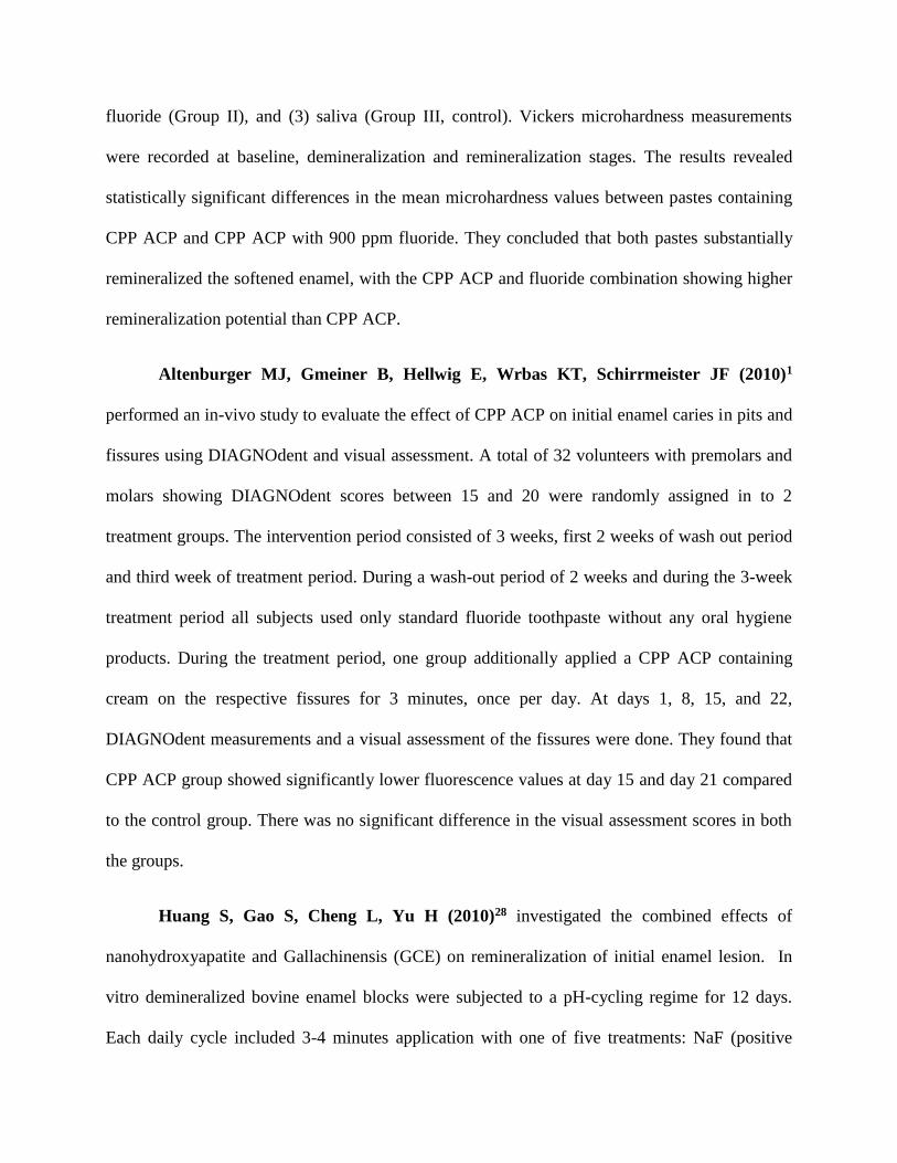
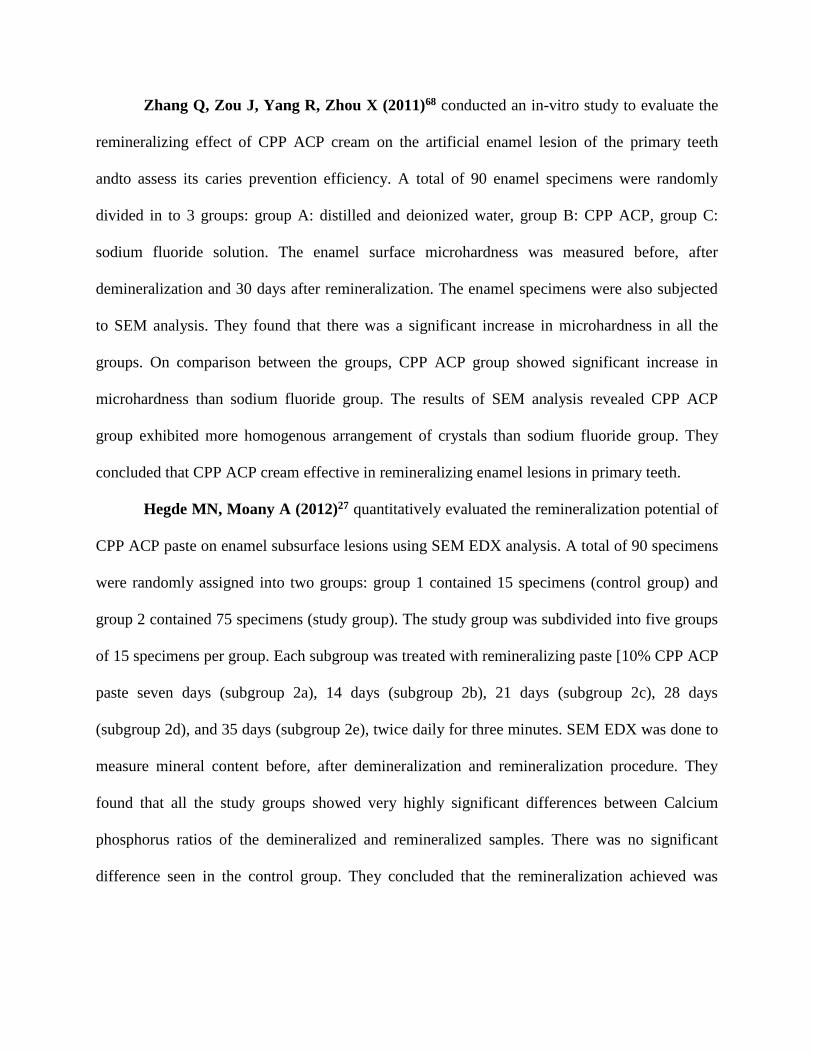
Preparation of the demineralization solution
Demineralizing solution was prepared in the Centre for Nano Science and Technology, KSR
College of Technology, Tiruchengode. The composition of demineralizing solution used was as
follows:
• 2.2 mM calcium chloride, CaCl2.2H2O (LobaChemiePvt. Ltd., Mumbai, Maharashtra,
India)
• 2.2 mM monosodium phosphate, NaH2PO4.7H2O (LobaChemiePvt. Ltd., Mumbai,
Maharashtra, India)
• 0.05 M lactic acid, C3H6O3 (LobaChemiePvt.Ltd.,Mumbai, Maharashtra, India).
A digital pH meter was used to check pH during and after preparation of solution.The final
pH was adjusted to 4.5 with 50% sodium hydroxide (NaOH).
Page 34
Induction of artificial carious lesions
Each of the specimens were immersed separately into sterile plastic container containing
12ml of prepared demineralizing solution and incubated for a period of 96 hours at 37°C in
universal incubator. After incubation, the teeth were washed with deionized water and dried with
the help of an air syringe. All the specimens were assessed for the formation of white opaque
areas. The demineralised specimens were subjected to EDX analysis for measuring the loss in
mineral content.
Formulation of experimental nano hydroxyapatite tooth paste
Experimental nano hydroxyapatite tooth pastewas prepared in the Centre for Nano
Science and Technology, KSR College of Technology, Tiruchengode.
Synthesis of nano hydroxyapatite
nHAP was synthesised according to the method described by Chen et al 200210. HAP was
prepared from calcium nitratetetrahydrate (Ca(NO3)2.4H2O) and ammonium dihydrogen
phosphate (NH4H2PO4) by sol-gel procedure. The calcium/phosphorus stoichiometric ratio of
pure HAP was kept constant as 1.67. Calcium nitratetetrahydrate(3.93 grams) was weighed and
mixed with 100 ml distilled water and stirred using a magnetic stirrer. Ammonium dihydrogen
phosphate was weighed and mixed with 100 mL distilled water and stirred using a magnetic
stirrer. This solution was added drop wise to Calcium nitratetetrahydratesolution using a burette
till the colour of the solution changed to milky white. The solution was allowed to stir for 1 hour
and the pH of the solution was found to be 6.0. The stable HAP can be obtained when the pH
value is above 10. Hence to raise the pH of the solution, ammonia was added drop wise to the
solution using pipette, optimizing the pH to 10.4. The solution was allowed to stir for 1 hour and
Page 35
it was then kept in a hot air oven for 48 hours at 33K. The dried solution was collected,
powdered, and then calcined at 773 K to obtain HAP powder. The obtained HAP was ground in
a dry ball mill at 400 rpm for 1 hour to obtain fine nHAP.
Characterization of formulated HAP
The Fourier Transform Infrared (FTIR) spectra of theHAP were obtained using an FTIR
spectrometer (Spectrum 100; PerkinElmer, USA) in the frequency range from 4000 to 400 cm−1
using a potassium bromide (KBr) pellet. The pellet was obtained by mixing 200:1 ratio of KBr
and HAP. The mixture was grinded initially in an agate mortar and then the pellet was obtained
using a hydraulic pellet maker. The obtained pellet was used to ascertain the functional groups
through measurement of infrared spectra.
The FTIR spectrum shows peaks of HAP. The peak 3572 Cm-1 indicates OH- group41 and
3431Cm-1stretching peak shows presence of water molecule.19 2351 Cm-1 peak shows C-H group
present material. 1643 Cm-1and 1373 Cm-1 peaks indicate CO32- functional group present in
material.7 960 Cm-1 and 568Cm-1 peaks represent phosphate group present in HAP.19
Page 36
Preparation of experimental nHAP tooth paste
Two experimental nHAP tooth pastes with the concentration of 1% nHAP and 10%
nHAP were prepared by mixing with the ingredients of standard tooth paste. The ingredients
added to prepare nHAp tooth paste were as follows:
S.no Ingredients Role Quantity/100g
1. Sorbitol Humectant 10000mg
2. Propylene glycol Humectant 30000mg
3. Silica Inorganic thickening agent 1000mg
4. Sodium lauryl sulphate
Organic thickening agent and
emulsifier
1000mg
5. Nano hydroxyapatite Active ingredient
1000mg for 1% nHAP paste
10000mg for 10% nHAP paste
6.
Sodium carboxy methyl
cellulose
Gum 1000mg
7. Titanium dioxide Whitening agent 500mg
8. Methyl paraben Antibacterial agent 100mg
9. Propyl paraben Antibacterial agent 20mg
Page 37
10. Clove oil Flavor Quantity required
11. Sodium saccharin Sweetening agent 30mg
12. Distilled water Bulking agent Quantity required for 100gm
Remineralization procedure
The demineralised specimens were randomly divided into 5 groups for remineralization
procedure.
• GROUP I –Aclaim® (n = 15)
• GROUP II – 1% experimental nHAP tooth paste (n = 15)
• GROUP III – 10% experimental nHAP tooth paste ( n = 15)
• GROUP IV – CPP ACP paste (n = 15)
• GROUP V – control (n = 5)
Page 38
Preparation of artificial saliva
The artificial saliva was prepared according to McKnight Hane and Whitfort formula
(1992).38 The composition used was as follows (grams/litre):
Methyl p- hydroxybenzoate(Merck) 2.00
Sodium carboxy methyl cellulose(Merck ) 10.00
Potassium chloride ( KCl ) (Merck ) 0.025
Magnesium chloride dihydrate (MgCl2.2H2O)
(LobaChemiePvt. Ltd.,)
0.059
Calcium chloride dehydrate (CaCl2.2H2O)
(Merck)
0.166
Dipotassium hydrogen phosphate (K2HPO4)
(Rankem)
0.804
Monopotassium hydrogen phosphate
(KH2PO4)(Merck)
0.326
• The pH of artificial saliva was adjusted to 6.75 with potassium hydroxide (KOH)
Page 39
Remineralization regimen
The specimens in groups I, II, III and IV were treated with respective tooth paste twice
daily for 14 days. Specimens were rubbed with respective tooth paste with the help of cotton
applicator for 3 minutes, washed with deionized water, and then placed in artificial saliva
maintained at ambient temperature. In the control group, specimens were only washed with
deionized water and placed in artificial saliva. Artificial saliva was renewed every 24 hours just
before immersion of freshly treated samples.
The specimens in group I and group II were subjected to EDX analysis to measure the
change in mineral content after remineralization procedure. Similarly the specimens in group III,
IV and V were subjected to SEM with EDX analysis to analyse the surface topographical
changes after remineralization.
Structural analysis
The surface characteristics of the demineralized and remineralized enamel specimens
were analysed by SEM. The specimens wereplacedonametalmountingblockandthenkept inside
the gold sputter coater (Q150R, Quorum technologies, UK). After sputtering the specimens were
observed under SEM (GeminiSEM, Zeiss microscopy, Germany) at ×5,000 and ×10,000
magnifications at 15kv.
Statistical analysis
The statistical analysis was done using IBM SPSS Statistics for Windows, Version 22.0.
Armonk, NY: IBM Corp. The statistical significance was set at p≤0.05. The distribution of
normality for the data were done using Kolmogorov-Smirnov and Shapiro-Wilks test. The
intragroup comparison was done using Friedman test and Wilcoxan Signed Rank Test.
Page 40
KruskallWallis and Mann Whitney U test were employed to analyse the change in the values for
intergroup comparison.
Page 41
Flow Chart of Methodology
Primary human molars
(n=35)
Enamel specimens prepared
(n=65)
Baseline
EDX analysis
Specimens subjected to
demineralization
EDX analysis after
demineralization
Group I
Aclaim
(n=15)
Group II
1% nHAP
(n=15)
Group III
10% nHAP
(n=15)
Group IV
CPP ACP
(n=15)
Group V
Control
(n=5)
EDX
analysis
SEM with EDX
analysis
Analysis of results
Page 42
Figure 1. Prepared enamel specimens
Figure 2. Armamentarium for the preparation of demineralizing solution
Page 43
Figure 3. Components for demineralizing solution
Figure 4. Preparation of demineralizing solution
Page 44
Figure 5. Preparation of nano hydroxyapatite powder
Page 45
Figure 6. Components for toothpaste preparation
Figure 7. Armamentarium for toothpaste preparation
Page 46
Figure 8. Preparation of tooth paste Figure 9. 1% and 10% nHAP paste
Figure 10.Test groups
Figure 11. Colour coded sample distribution
Page 47
Figure 12. Components for artificial saliva
Figure 13. Preparation of artificial saliva
Page 48
Figure 15. Ball Milling machine
Figure 16. Fourier Transform Infrared Spectroscopy
Page 49
Figure 17. Scanning electron microscope with energy dispersive X-ray analysis attachment
Page 51
Table 1.Comparison of mean distribution of calcium in weight percentage in different
stages of demineralization and remineralization cycle within each group
*Friedman test
Table 1 shows the comparison of mean values of calcium wt% at baseline, after demineralization
and after remineralization in each group. All the groups except Group V showed significant change
(p<0.05) in calcium wt% after each stage of demineralization and remineralization cycle.
Group N
Baseline
Mean±S.D(wt%)
After
demineralization
Mean±S.D (wt%)
After
remineralization
Mean±S.D (wt%)
p value*
Group I
(Aclaim®)
15 22.27±7.216 16.53±5.817 23.67±3.792 0.001
Group II
(1% nHAP)
15 22.87±3.204 17.40±5.591 21.87±4.086 0.001
Group III
(10% nHAP)
15 22.33±4.237 14.07±4.217 22.33±2.320 <0.001
Group IV
(CPP ACP)
15 24.00±2.952 18.13±3.357 20.13±3.543 0.001
Group V
(Control)
5 25.60±4.159 20.20±4.550 24.40±3.050 0.211
Page 52
Graph1. Comparison of mean values of calcium weight percentage
Graph 1 illustrates the comparison of mean values of change in calcium wt% at baseline,
after demineralization and after remineralization in each group.
0
5
10
15
20
25
30
Group I Group II Group III Group IV Group V
Mea
n c
on
cen
trat
ion
(w
t%)
Mean calcium weight percentage
Baseline After demineralization After remineralization
Page 53
Table 2. Post hoc analysis of change in calcium weight percentage within each group
**Wilcoxan Signed Rank test
Table 2 shows the comparison of mean values of calcium wt% within the groups. All the 4
groups except Group V showed a statistically significant (p<0.05) change in the calcium wt%
from baseline to demineralization. Similarly, there was a statistically significant change in all the 4
groups in calcium wt% from demineralization to remineralization, except in Group V. Only Group
IV showed a statistically significant change in calcium wt%, when the baseline values were
compared with the post remineralization values.
Group
Baseline to
Demineralization
p value**
Demineralization
to Remineralization
p value**
Baseline to
Remineralization
p value**
Group I
(Aclaim®) 0.002 0.001 0.271
Group II
(1% nHAP) 0.001 0.006 0.977
Group III
(10% nHAP) 0.001 0.001 0.723
Group IV
(CPP ACP) 0.001 0.043 0.009
Group V
(Control) 0.109 0.588 0.104
Page 54
Table 3. Intragroup and intergroup comparisons for the change in calcium wt% in
different stages of demineralization and remineralization cycle
* Friedman test;***Kruskal Wallis ANOVA
Group
Change in
baseline to
demineralization
Mean±S.D (wt%)
Change in
demineralization to
remineralization
Mean±S.D (wt%)
Change in
baseline to
remineralization
Mean±S.D (wt%)
p value*
Group I
(Aclaim®)
5.84±5.90 7.25±6.97 1.40±8.01 0.002
Group II
(1% nHAP)
5.30±5.47 4.37±4.94 -.92±5.81 0.001
Group III
(10% nHAP)
8.03±4.43 8.05±3.88 .02±4.11 0.001
Group IV
(CPP ACP)
5.88±3.02 2.02±3.98 -3.85±4.50 <0.001
Group V
(Control)
5.38±6.82 4.18±4.46 -1.20±6.42 0.549
p value*** 0.227 0.011 0.073
Page 55
Table 3 shows intergroup and intragroup comparison for change in calcium wt%. In
intergroup comparison, there was a statistically significant difference (p=0.011) between the
groups when demineralization to remineralization values of calcium wt% was compared. All the
groups, except Group V showed statistically significant difference in the change of calcium wt%
in the intragroup comparison.
Page 56
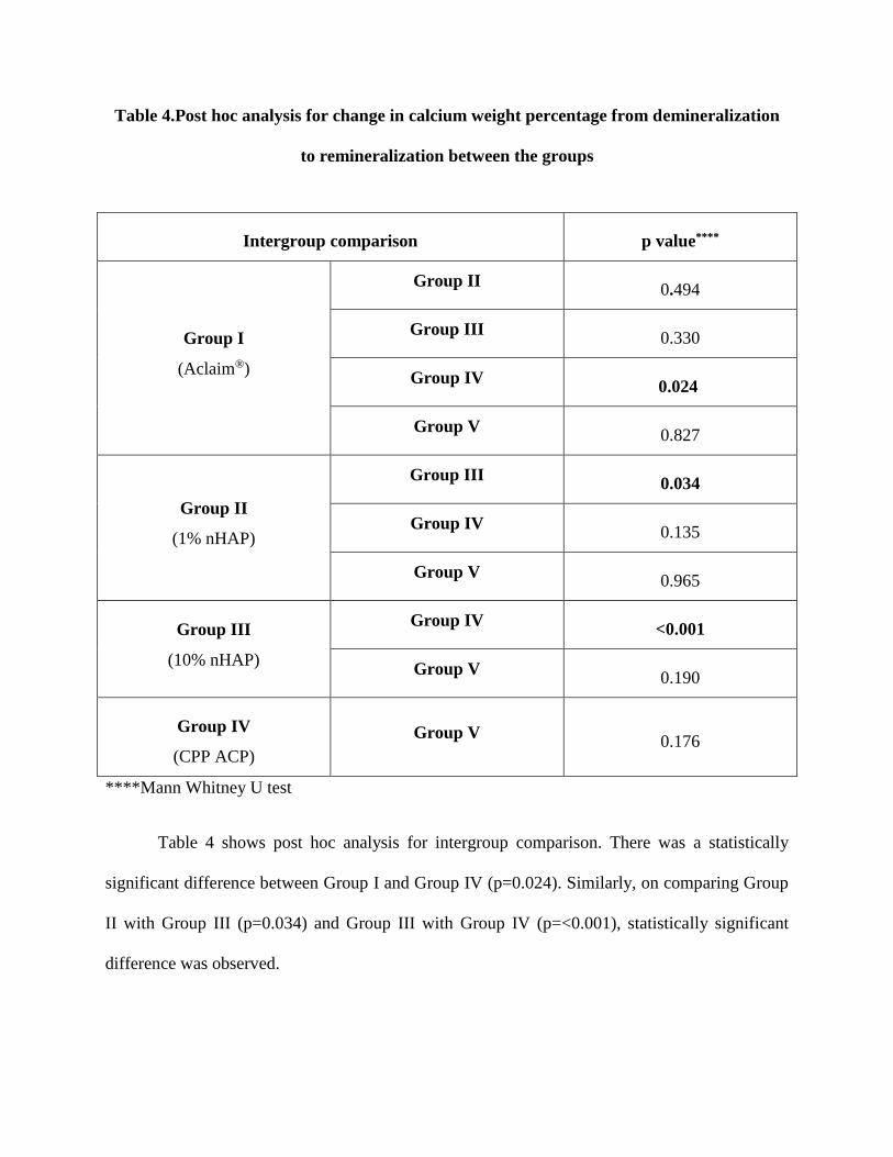
Table 4.Post hoc analysis for change in calcium weight percentage from demineralization
to remineralization between the groups
****Mann Whitney U test
Table 4 shows post hoc analysis for intergroup comparison. There was a statistically
significant difference between Group I and Group IV (p=0.024). Similarly, on comparing Group
II with Group III (p=0.034) and Group III with Group IV (p=<0.001), statistically significant
difference was observed.
Intergroup comparison p value****
Group I
(Aclaim®)
Group II 0.494
Group III 0.330
Group IV 0.024
Group V 0.827
Group II
(1% nHAP)
Group III 0.034
Group IV 0.135
Group V 0.965
Group III
(10% nHAP)
Group IV <0.001
Group V 0.190
Group IV
(CPP ACP)
Group V 0.176
Page 57
Table 5. Comparison of mean distribution of phosphorus in weight percentage in different
stages of demineralization and remineralization cycle within each group
*Friedman test
Table 5 shows the comparison of mean values of phosphorus wt% in each group after each
stage of demineralization and remineralization cycle. All the groups except Group V showed
significant changes in phosphorus wt% after each stage.
Group N
Baseline
Mean±S.D (wt%)
After
demineralization
Mean±S.D (wt%)
After
remineralization
Mean±S.D (wt%)
p value*
Group I
(Aclaim®)
15 12.40±2.501 9.80±2.883 13.67±1.877
<0.001
Group II
(1% nHAP)
15 13.73±1.792 11.00±3.381 12.73±2.187
0.034
Group III
(10% nHAP)
15 13.07±2.604 9.20±2.808 13.93±1.831
<0.001
Group IV
(CPP ACP)
15 14.60±1.957 11.33±1.718 12.73±1.792
<0.001
Group V
(Control)
5 14.80±2.775 12.60±2.702 13.80±1.924S
0.165
Page 58
Graph 2. Comparison of mean values of phosphorus weight percentage
Graph 2 illustrates the comparison of mean values of phosphorus wt% in each group after
each stage of demineralization and remineralization cycle.
0
2
4
6
8
10
12
14
16
Group I Group II Group III Group IV Group V
Mea
n c
on
cen
trat
ion
(w
t%)
Mean phosphorus weight percentage
Baseline After demineralization After remineralization
Page 59
Table 6. Post hoc analysis of change in phosphorus weight percentage within each group
**Wilcoxan Signed Rank test
Table 6 shows the comparison of mean values of phosphorus wt% within the groups. All
the groups except Group V showed statistically significant change in the phosphorus wt%
from baseline to demineralization. Group I, III and IV showed statistically significant
change in phosphorus wt% from demineralization to remineralization. Only Group IV
showed a significant change in phosphorus wt%, when the baseline values were compared
with the post remineralization values.
Group
Baseline to
Demineralization
p value**
Demineralization to
Remineralization
p value**
Baseline to
Remineralization
p value**
Group I
(Aclaim®) 0.001 0.001 0.213
Group II
(1% nHAP) 0.008 0.092 0.323
Group III
(10% nHAP) 0.001 0.001 0.177
Group IV
(CPP ACP) 0.001 0.031 0.024
Group V
(Control) 0.279 0.498 0.414
Page 60
Table 7. Intragroup and intergroup comparisons for the change in phosphorus weight
percentage in different stages of demineralization and remineralization cycle
* Friedman test;***Kruskal Wallis ANOVA
Group
Change in
baseline to
demineralization
Mean±S.D (wt%)
Change in
demineralization to
remineralization
Mean±S.D (wt%)
Change in
baseline to
remineralization
Mean±S.D (wt%)
p value*
Group I
(Aclaim®) 2.48±2.21 3.60±3.79 1.12±3.50 0.008
Group II
(1% nHAP) 2.66±4.18 1.69±3.38 -0.97±3.02 0.002
Group III
(10% nHAP) 3.85±3.30 4.74±2.62 0.89±2.93 0.011
Group IV
(CPP ACP) 3.18±1.54 1.42±2.30 -1.7±2.93 <0.001
Group V
(Control) 1.97±4.61 0.91±4.39 -1.06±2.22 0.449
p value*** 0.519 0.021 0.035
Page 61
Table 7 shows intragroup and intergroup comparison for change in phosphorus wt%. In
intergroup comparison there was a statistically significant difference (p=0.021) between the
groups, when change in demineralization to remineralization value of phosphorus wt% was
compared. Similarly, statistically significant difference (p=0.035) was observed when change in
baseline to remineralization values were compared. All the groups, except Group V showed
statistically significant difference in the change of phosphorus wt% in the intragroup
comparison.
Page 62
Table 8. Post hoc analysis for change in phosphorus weight percentage from
demineralization to remineralization and baseline to remineralization between the groups
****Mann Whitney U test
Intergroup comparison
Change in
demineralization to
remineralization
p value****
Change in
baseline to
remineralization
p value****
Group I
(Aclaim®)
Group II 0.198 0.130
Group III 0.110 0.852
Group IV 0.101 0.019
Group V 0.694 0.206
Group II
(1% nHAP)
Group III 0.014 0.059
Group IV 0.983 0.468
Group V 0.965 0.694
Group III
(10% nHAP)
Group IV 0.002 0.007
Group V 0.81 0.127
Group IV
(CPP ACP)
Group V 0.570 0.896
Page 63
Table 8 shows post hoc analysis for intergroup comparison. There was a statistically
significant difference, when change in demineralization to remineralization values were
compared between Group II and Group III (p=0.014), Group III and Group IV (p=0.002)
Similarly, on comparing Group I with Group IV (p=0.019) and Group III with Group IV
(p=0.007), statistically significant differences were observed between the baseline values
and the post remineralization values.
Page 64
Table 9.Comparison of mean distribution of calcium phosphorus ratio in weight
percentage in different phases of demineralization and remineralization cycle within each
group
*Friedman test
Table 9 shows the comparison of mean values of calcium phosphorus ratio in each groups
after each stage of demineralization and remineralization cycle. Group III showed significant
changes in calcium phosphorus ratio after each stage.
Group N
Baseline
Mean±S.D
After
demineralization
Mean±S.D
After
remineralization
Mean±S.D
p value*
Group I
(Aclaim®)
15 2.00±.655 1.67±.488 1.87±.352 0.156
Group II
(1% nHAP)
15 1.73±.458 1.80±.414 1.93±.258 0.311
Group III
(10% nHAP)
15 1.80±.414 1.47±.516 1.87±.352 0.045
Group IV
(CPP ACP)
15 1.87±.352 1.80±.414 1.87±.352 0.819
Group V
(Control)
5 1.80±.447 1.40±.548 2.00±.000 0.174
Page 65
Graph 3. Comparison of mean values of calcium phosphorus ratio weight percentage
Graph 3 illustrates the comparison of mean values of change in calcium phosphorus
ratio wt% in each group after each stage of demineralization and remineralization cycle.
0
0.5
1
1.5
2
2.5
Group I Group II Group III Group IV Group V
Mea
n c
on
cen
tra
tio
n (
wt%
)
Mean calcium phosphorus ratio weight
percentage
Baseline After demineralization After remineralization
Page 66
Table 10. Intragroup and intergroup comparisons for the change in calcium phosphorus
ratio weight percentage on different stage of demineralization and remineralization cycle
* Friedman test; **Kruskal Wallis ANOVA
Table 10 shows intragroup and intergroup comparison for change in calcium phosphorus
ratio wt%. There was no statistically significant difference between the groups and within the
groups after each stage of demineralization and remineralization cycle.
Group
Change in
baseline to
demineralization
Mean±S.D
Change in
demineralization to
remineralization
Mean±S.D
Change in
baseline to
remineralization
Mean±S.D
p value*
Group I
(Aclaim®)
0.16±0.60 0.13±0.22 -0.03±0.66 0.420
Group II
(1% nHAP)
0.09±0.28 0.16±0.25 0.06±0.40 0.247
Group III
(10% nHAP)
0.17±0.32 0.05±0.26 -0.12±0.25 0.057
Group IV
(CPP ACP)
0.72±0.20 -0.006±0.19 -0.07±0.27 0.886
Group V
(Control)
0.17±0.47 0.21±0.26 0.03±0.23 0.819
p value** 0.707 0.461 0.250
Page 67
Image 1. Elemental analysis of baseline enamel specimen by EDX analysis
Image 2. Elemental analysis of demineralized enamel specimen by EDX analysis
Page 68
Image 3. Elemental analysis of remineralized enamel specimen of Group I (Aclaim®) by
EDX analysis
Image 4. Elemental analysis of remineralized enamel specimen of Group II (1%nHAP) by
EDX analysis
Page 69
Image 5. Elemental analysis of remineralized enamel specimen of Group III (10% nHAP)
by EDX analysis
Image 6. Elemental analysis of remineralized enamel specimen of Group IV (CPP ACP) by
EDX analysis
Page 70
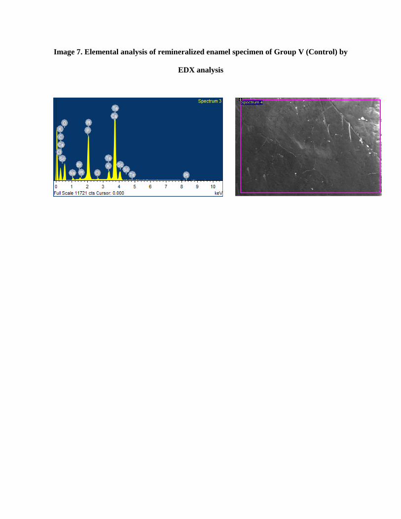
Image 7. Elemental analysis of remineralized enamel specimen of Group V (Control) by
EDX analysis
Page 71
Image 8. Structural analysis of demineralized enamel specimen by SEM analysis
Page 72
Image 9.Structural analysis of remineralized enamel specimen of Group III (10% nHAP)
by SEM analysis
Page 73
Image 10. Structural analysis of remineralized enamel specimen of Group IV (CPP ACP)
by SEM analysis
Page 75
Dental caries is the most common reason for tooth loss in children and young adults. It is
often described as a chronic disease which causes localized destruction of the tooth.45 Dental
caries is a multifactorial disease occurring as a result of complex interactions among tooth
structure, dental biofilm, dietary, salivary, and genetic influences.23 Any disturbance in the
physiological equilibrium of the biofilm (dental plaque) covering the affected site results in
demineralization.45 Demineralization of enamel leads to dissolution of hydroxyapatite and
diffusion of calcium and phosphate ions toward the enamel surface. Reprecipitation of
hydroxyapatite occurs, when the enamel surface is supersaturated with calcium and phosphate
ions forming an intact superficial layer.32
Caries process is said to be active when the demineralization period exceeds
remineralization.43 The disease will continue to progress, unless the dental plaque covering the
site is removed. The localized destruction of the hard tissues is often referred to as the lesion.
The amount of destruction of lesion can range from initial loss of mineral at the ultra structural
or nanoscale level to total tooth destruction.45
The concept of demineralization and remineralization cycle has led to the scope for
remineralizing incipient carious lesion.43 Thus focus on caries prevention shifted to the
development of methods for detecting the caries at early stages. The use of non-invasive
treatment for incipient carious lesions by remineralization has been considered as a promising
advancement in the clinical management of the disease.44 It bridges the traditional gap between
prevention and restorative procedures.32 Remineralization of initial carious lesions may be
possible with a variety of currently available agents containing fluoride, bioavailable calcium
and phosphate, CPP ACP, self-assembling peptide and nano hydroxyapatite.44
Page 76
Presently there is a lack of knowledge about the remineralizing efficacy of biomaterials
based on nanotechnology which may be an important preventive approach to be applied in high
caries risk patients.14 The Japanese company Sangi Co. Ltd was the first one to take an interest in
hydroxyapatite, after purchasing the rights from NASA (U.S. National Aeronautics and Space
Authority) in 1970. NASA introduced a synthetic hydroxyapatite as a repairing material for the
astronauts who lost minerals from the teeth and bones in the space due to the absence of gravity.
Later in 1978, Sangi Co. Ltd had the idea to create a toothpaste containing nHAP (Apadent) that
could repair the tooth enamel.48 The function of biomimetic hydroxyapatite is to protect the teeth
with the creation of a new layer of synthetic enamel around the tooth, rather than hardening the
existing layer with fluoride.
Toothpaste with nHAP had the strong ability to bond with the proteins present in plaque
and bacteria. The size of the nano particles markedly increases the surface area to which proteins
can bind.48 Accordingly, materials containing nHAP are able to provide calcium and phosphate
ions for reducing tooth demineralization and/or improving tooth remineralization.
Hydroxyapatite nano particles may penetrate tooth porosities (Mechanical imbrications) and
produce a protective layer on the tooth surface.12,29 nHAP helps in providing more mineral
deposits on the outer layer than the body of the lesion.14 The efficacy of nHAP increases in the
presence of biofilm.58
Toothpastes containing nHAP are now commercially produced worldwide. This indicates
the need for more precise evaluation of their efficacy. Considering the lack of adequate studies,
the present study was aimed at formulation of nHAP toothpaste at 2 different concentrations (1%
and 10%) and evaluating their remineralization potential with comparison to commercially
available nHAP paste (Aclaim®) and CPP ACP (GC Tooth mousse).
Page 77
Products containing CPP ACP have also proved to have some potential to prevent enamel
demineralization and increase remineralization in-vitro.35,9,69 Based on in-situ studies8,13 and
randomized clinical trials.50,5 CPP ACP paste was able to increase the remineralization of initial
enamel caries lesions. However it does not seem to have a significant difference in its
remineralization effect from fluorides.4,67,36 According to the systematic reviews by Chen et al
201311 and Li et al 201436, there were a lack of reliable scientific evidence to support the
remineralization effect of CPP ACP.
Evaluation method
In-vitro demineralization and remineralization can be assessed by using various methods.
The commonly used methods are SEM / SEM-EDX, DIAGNOdent, surface micro hardness, and
polarized light microscopy. It is wise to measure the changes in the mineral content of the
carious lesions quantitatively in order to provide more promising results in remineralization
process. One of the most common techniques was SEM with EDX attachment. It is a micro
analytical technique that is employed to estimate quantitatively the amounts of minerals in a
given tooth sample. SEM gives the topographical pictures and is used to assess the surface
changes seen on enamel. EDX gives quantification of various elements like calcium, phosphorus,
fluoride, magnesium and sodium in both atomic and weight percentage.43 In the present study,
quantitative elemental analysis for each specimen was measured at three levels; baseline, after
induction of carious lesions (demineralization) and after remineralization.
Demineralization regimen
Artificial caries like lesions can be induced in enamel in in-vitro conditions. These
carious lesions were considered to be more homogenously reproducible than natural carious
lesions and thus provide more reliable experimental model. The in-vitro model provides an area
Page 78
of enamel having a defined lesion of constant depth beneath the surface. They facilitate the
testing of multiple areas in any enamel lesion at different time intervals, in order to assess the
remineralizing phenomena.3,15,66 In the present study, artificial carious lesions were induced
using the demineralization protocol described by Patil N et al47 which is in accordance to Ten
Cate and Duijsters60. The results of the present study revealed that calcium and phosphorus wt%
of all the enamel specimens were significantly reduced from baseline after demineralization
regimen.
Remineralization regimen
The present study used paste type formulation of the test agents, applied with disposable
cotton tip applicators. This was done in view of replication of patient convenience in using the
tooth cream like a tooth paste with toothbrushes or along with cotton tip applicators. The
remineralization regimen comprised 3 minutes twice daily application of 14 days similar to
Shriahatti et al.56 In contrast, study done by Pai et al46 treatment pastes were applied 3 minutes
once daily for 14 days. Hegde and Moany27 in their study performed twice daily application of
CPP ACP for 3 minutes for 7, 14, 21, 28, and 35 days. They mentioned that remineralization
efficacy of paste is dose dependant, i.e. remineralization rate increases with the duration of time
the paste is in contact with the enamel surface.
In the present study, artificial saliva was changed every 24 hours during the
remineralization regimen to ensure ionic balance and maintenance of pH. This is in accordance
with Patil N et al47 and Pujan Kamath et al32. In contrast, Shirahatti et al56 changed the artificial
saliva solution every 72 hours in their study.
In the present study quantitative elemental analysis for each specimen was done at three
stages: baseline, after demineralization and after remineralization. The results of 4 test groups
Page 79
and a control group were summarized as calcium wt%, phosphorus wt% and calcium phosphorus
ratio wt%.
Experimental nHAP paste
In the present study experimental 1% nHAP paste showed significant increase in calcium
wt% after remineralization whereas phosphorus wt% did not increase significantly. Experimental
10% nHAP paste showed a significant higher mean change in both calcium and phosphorus wt%
after remineralization compared to 1% nHAP. This was in accordance with the results of Huang
et al30 who compared the remineralization effect of different concentrations (1%, 5%, 10% and
15%) of nHAP on artificially demineralized bovine enamel. They found that 1% nHAP did not
show significant effect on remineralization of initial enamel caries whereas 10% nHAP was most
effective. They also concluded that 10% nHAP was considered as the optimum concentration for
the remineralization of early enamel lesions. Similarly Kim et al34 reported that higher
concentration (10%) of nHAP with longer treatment time showed greater remineralization effect.
As the concentration of nHAP increased, the rate and amount of its precipitation also increased
along with the deposition of excessive amount of calcium and phosphate ions. Contrary to our
results, Haghoo et al25 concluded that there was no significant difference between the effect of
various concentration of nHAP (2%, 5% and 10%). The difference in the result of their study
might be because the formulated nHAP was used as mouth rinse. Another reason could be the
shorter treatment time of 12 hours, in which the specimens were immersed in the mouth rinse.
The results of the present study revealed that, there was a significant change in calcium
phosphorus ratio wt% after remineralization in 10% nHAP paste. This is in contrast with the
results obtained by Pujan Kamath et al32 where there was no significant difference between the
Page 80
groups. The variation in the result might be due to the concentration of the nHAP used in the
present study.
Commercially available nHAP pate (Aclaim®)
The present study revealed that Aclaim® showed a significant increase in calcium and
phosphorus wt% after remineralization. This suggests a positive effect on remineralization of
early enamel caries. Similar results were obtained in a study done by Singh A et al57 where
Aclaim® demonstrated a significant effect in preventing demineralization under polarized light
microscopy. Shetty P et al55 mentioned that the particle size of nHAP in Aclaim® was <100nm.
The nano sized hydroxyapatite remained relatively stable under acidic conditions and thus nHAP
layer formed on the enamel surface became resistant to dissolution.
CPP ACP
In the present study CPP ACP demonstrated a significant change in calcium and
phosphorus wt% after remineralization, however the mean value was lesser than the values at
baseline. When compared with CPP ACP, both Aclaim® and 10% nHAP showed higher mean
change of calcium and phosphorus wt% after remineralization. But there was no significant
difference between 1% nHAP and CPP ACP. The results were in accordance to the study done
by deCarvalho et al.16 This might be because CPP ACP needs an acid challenge for activation
and for the separation of ACP from casein. This activation increases the degree of saturation of
calcium and phosphate ions in saliva with respect to hydroxyapatite. In a study done by Comar et
al14 both CPP ACP and nHAP were unable to reduce demineralization which is contrary to our
results. The difference in results might be due to the pH cycling method used which focused on
the prevention of demineralization rather than the remineralization of preformed lesions.
Page 81
Structural Analysis
SEM analysis of the demineralized samples at 5,000 magnification revealed rough and
irregular surface with marked irregular porosity. Minor honey comb pattern of demineralized
enamel was evident on the surface. Distinct surface coating was evident in remineralized
specimens of both 10% nHAP and CPP ACP. SEM image of 10% nHAP revealed numerous
small deposits covering the irregularities of the enamel surface. CPP ACP also showed similar
deposits on the enamel surface but in fewer numbers. Roveri et al52 demonstrated an apatitic
coating deposition on the enamel surface when biomimetic nHAP was used. Pujan Kamath et
al32 noticed superior texture of enamel treated by nHAP paste under SEM. This could be
attributed to the nHAP particles that have similar morphology, crystal structure, and crystallinity
like the apatite crystals of tooth enamel.
In the present study, null hypothesis was rejected because 1% and 10% nHAP paste
showed significant increase in calcium wt% and phosphorus wt% compared to CPP ACP and
control group.
Limitations
1) Selection of the enamel specimen was done based on visual inspection. This might be
the reason for varied baseline values of calcium and phosphorus wt%.
2) The present study did not make any attempt to compare the remineralization of nHAP
with fluoride.
Page 82
SUMMARY AND CONCLUSION
Page 83
The present randomized controlled trial was conducted in Department of Pediatric and
Preventive dentistry, KSR Institute of Dental Science and Research, Tiruchengode, Tamil Nadu.
The in-vitro trial study was conducted with following aims: 1. To formulate two experimental
nHAP tooth pastes with two concentrations 1% and 10% concentrations; 2. To evaluate the
remineralization potential of experimental 1% and 10% nHAP tooth paste; 3. To compare the
remineralization potential of experimental 1% nHAP with commercially available nHAP
toothpaste using EDX analysis and 4. To compare the remineralization potential of 10% nHAP
with commercially available CPP ACP using SEM with EDX analysis. A total of 65 enamel
specimens were randomly divided into 4 test groups and one control group; Group I: Aclaim®,
Group II: Custom made 1% nHAP, Group III: Custom made 10% nHAP, Group IV: CPP ACP,
Group V: Control. Baseline quantitative measurement of mineral contents (calcium and
phosphorus wt%) in enamel specimens were done by EDX analysis. Artificial carious lesion was
induced in all the enamel specimens, according to the demineralization regimen by Tencate and
Duijsters. All the enamel specimens were evaluated for loss in mineral content using EDX
analysis. The specimens in Groups I, II, III and IV were treated with respective paste twice daily
for 14 days. Specimens were rubbed with respective paste with the help of a cotton applicator for
3 minutes, washed with deionized water and then placed in artificial saliva and maintained at
ambient temperature. In the control group, specimens were only washed once with deionized
water and placed in artificial saliva. After the remineralization cycle, all the enamel specimens
were again subjected to EDX analysis to evaluate the change in mineral content. The data were
collected and subjected to statistical analysis. SEM analysis was done for Group III, IV and
control group to analyze the surface topographical changes after remineralization.
Page 84
The following conclusions can be inferred from this study
1) All the test groups showed significant change in calcium and phosphorus wt% after
remineralization.
2) Among the 5 groups, post reminerlization calcium wt% was greatest in the 10% nHAP
followed by Aclaim®, 1% nHAP, control and CPP ACP
3) Among the 5 groups, post reminerlization phosphorus wt% was greatest in the 10%
nHAP followed by Aclaim®, 1% nHAP, CPP ACP and control
4) Among the test groups, post reminerlization calcium phosphorus ratio wt% was greatest
in the 10% nHAP followed by Aclaim®, 1% nHAP and CPP ACP
5) Significant change in calcium phosphorus ratio wt% after remineralization was seen only
in 10% nHAP group.
6) The mean change in calcium and phosphorus wt% of 10% nHAP was significantly higher
than 1% nHAP and CPP ACP after remineralization.
7) Aclaim® showed significantly higher mean change in calcium wt%, than CPP ACP after
remineralization.
8) Both 10% nHAP and CPP ACP showed favorable surface changes in enamel after
remineralization in SEM analysis.
9) SEM image of 10% nHAP showed more deposits on the enamel surface than CPP ACP
Page 85
Conclusion
Within the limitations of this in-vitro trial, nHAP had the potential to remineralize
artificially induced carious lesions. A concentration of 10% nHAP followed by Aclaim® was
found to be most effective in increasing calcium and phosphorus wt%. CPP ACP and 1% nHAP
were found to have less remineralizing potential.
Page 87
1. Altenburger MJ, Gmeiner B, Hellwig E, Wrbas KT, Schirrmeister JF. The evaluation of
fluorescence changes after application of casein phosphopeptides (CPP) and amorphous
calcium phosphate (ACP) on early carious lesions. Am J Dent 2010;23(4):188-92.
2. Andersson A, Larsson KS, Hallgren A, Petersson LG, Twetman S. Effect of a Dental
Cream Containing Amorphous Calcium Phosphate Complexes on White Spot Lesion
Regression Assessed by Laser Fluorescence. Oral Health Prev Dent 2007;5:229-233.
3. Arends J, Christoffersen J. The nature of early caries lesions in enamel. J Dent Res
1986;65:2-11.
4. Bailey DL, Adams GG, Tsao CE, Hyslop A, Escobar K, Manton DJ, et al. Regression of
post-orthodontic lesions by a remineralizing cream. J Dent Res 2009;88(12):1148-1153.
5. Beerens MW, Vanderveen MH, Vanbeek H, Tencate JM. Effects of casein
phosphopeptide amorphous calcium fluoride phosphate paste on white spot lesions and
dental plaque after orthodontic treatment: a 3-month follow-up. Eur J Oral Sci
2010;118:610-17.
6. Brochner A, Christensen C, Kristensen B, Tranaeus S, Karlsson L, Sonnensen L, et al.
Treatment of post-orthodontic white spot lesions with casein phosphopeptide-stabilised
amorphous calcium phosphate. Clin Oral Invest 2011;15:369-373.
7. Brundavanam S, Poinern GEJ, Fawcett D. Kinetic and Adsorption Behaviour of Aqueous
Fe2+, Cu2+ and Zn2+ Using a 30 nm Hydroxyapatite Base Powder Synthesized via a
Combined Ultrasound and Microwave Based Technique. American Journal of Materials
Science 2015;5(2):31-40.
Page 88
8. Cai F, Manton DJ, Shen P, Walker GD, Cross KJ, Yuan Y,et al. Effect of addition of
citric acid and casein phosphopeptide-amorphous calcium phosphate to a sugar-free
chewing gum on enamel remineralization in situ. Caries Res 2007;41(5):377-83.
9. Cao Y, Mei ML, Xu J, Lo EC, Li Q, Chu CH. Biomimetic mineralisation of
phosphorylated dentine by CPP-ACP. J Dent 2013;41:818–25.
10. Chen F, Wang ZC, Lin CJ. Preparation and characterization of nano-sized hydroxyapatite
particles and hydroxyapatite/chitosan nano-composite for use in biomedical materials.
Mater. Lett 2002;57(4):858–61.
11. Chen H, Liu X, Dai J, Jiang Z, Guo T, Ding Y. Effect of remineralizing agents on white
spot lesions after orthodontic treatment: a systematic review. Am J Orthod Dentofacial
Orthop 2013;143:376–382.
12. Cochrane NJ, Cai F, Huq NL, Burrow MF, Reynolds EC. New approaches to enhanced
remineralization of tooth enamel. J Dent Res 2010;89:1187-97.
13. Cochrane NJ, Reynolds EC. Calcium phosphopeptides—mechanisms of action and
evidence for clinical efficacy. Adv Dent Res 2012;24(2):41-7.
14. Comar LP, Souza BM, Gracindo LF, Buzalaf MA, Magalhaes AC. Impact of
experimental nano-HAP pastes on bovine enamel and dentin submitted to a pH cycling
model. Braz Dent J 2013;24(3):273-8.
15. Damle SG, Bengude V, Saini S. Evaluation of ability of dentifrices to remineralize
artificial caries like lesions. Dent Res J 2010;7:12-7.
16. deCarvalho FG, Vieira BR, Santos RL, Carlo HL, Lopes PQ, de Lima BA. In vitro
effects of nano-hydroxyapatite paste on initial enamel carious lesions. Pediatr Dent
2014;36(3):85-9.
Page 89
17. Ebadifar A, Nomani M, Fatemi SA. Effect of nano-hydroxyapatite toothpaste on
microhardness of artificial carious lesions created on extracted teeth. J Dent Res Dent
Clin Dent Prospects 2017;11(1):14-17.
18. Edwina AM Kidd. Essentials of dental caries: the disease and its management. 3rd ed.
USA: Oxford medical publication; 2005.
19. Ereiba KM, Mostafa AG, Gamal GA, Said AH. In vitro study of iron doped
hydroxyapatite. J Phys Chem B 2013;4(4):122.
20. Esteves-Oliveira M, Santos NM, Meyer-Lueckel H, Wierichs RJ, Rodrigues JA. Caries-
preventive effect of anti-erosive and nano-hydroxyapatite-containing toothpastes in vitro.
Clin Oral Investig 2017;21(1):291-300.
21. Ferrazzano GF, Amato I, Cantile T, Sangianantoni G, Ingenito A. In vivo remineralising
effect of GC tooth mousse on early dental enamel lesions: SEM analysis. Int Dent J
2011;61(4):210-6.
22. Goswami M, Saha S, Chaitra TR. Latest developments in non fluoridated remineralizing
technologies. J Indian Soc Pedod Prev Dent 2012;30(1):2-6.
23. Goswami S. Remineralize early demineralized enamel structure. SRM J Res Dent Sci
2016;7:231-4.
24. Haghgoo R, Ahmadvand M, Moshaverinia S. Remineralizing Effect of Topical NovaMin
and Nano-hydroxyapatite on caries-like Lesions in Primary teeth. J Contemp Dent Pract
2016;17(8):645-9.
25. Haghgoo R, Rezvani MB, SalehiZeinabadi M. Comparison of nano-hydroxyapatite and
sodium fluoride mouthrinse for remineralization of incipient carious lesions. J Dent
2014;11(4):406-10.
Page 90
26. Hedge S, Roma M, Shetty D. Non fluoridated remineralizing agents in dentistry. J.
Pharm. Sci. & Res 2016;8(8):884-887.
27. Hegde MN, Moany A. Remineralization of enamel subsurface lesions with casein
phosphopeptide-amorphous calcium phosphate: A quantitative energy dispersive X-ray
analysis using scanning electron microscopy: An in vitro study. J Conserv Dent
2012;15(1):61-7.
28. Huang S, Gao S, Cheng L, Yu H. Combined effects of nano-hydroxyapatite and
Gallachinensis on remineralisation of initial enamel lesion in vitro. J Dent
2010;38(10):811-9.
29. Huang S, Gao S, Cheng L, Yu H. Remineralization potential of nano-hydroxyapatite on
initial enamel lesions: an in vitro study. Caries Res 2011;45:460–468.
30. Huang SB, Gao SS, Yu HY. Effect of nano-hydroxyapatite concentration on
remineralization of initial enamel lesion in vitro. Biomed Mater 2009;4(3):034104.
31. Hurlbutt M. CAMBRA: Best practices in dental caries management. A Peer-Reviewed
Publication 2011:96-108.
32. Kamath P, Nayak R, Kamath SU, Pai D. A comparative evaluation of the
remineralization potential of three commercially available remineralizing agents on white
spot lesions in primary teeth: An in vitro study. J Indian Soc Pedod Prev Dent
2017;35:229-37.
33. Keerthi V, Manish R. Remineralizing Agents in Dentistry: A Review. Journal of Dental
and Medical Sciences (IOSR-JDMS) 2014;13(4):57-60.
34. Kim MY, Kwon HK, Choi CH, Kim BI. Combined effects of nano-hydroxyapatite and
NaF on remineralization of early caries lesion. Key Eng Mater 2007;1347:330-2.
Page 91
35. Kumar VL, Itthagarun A, King NM. The effect of casein phosphopeptide-amorphous
calcium phosphate on remineralization of artificial caries-like lesions: an in vitro study.
Aust Dent J 2008;53(1):34-40.
36. Li J, Xie X, Wang Y, Yin W, Antoun JS, Farella M, Mei L. Long-term remineralizing
effect of casein phosphopeptide-amorphous calcium phosphate (CPP-ACP) on early
caries lesions in vivo: a systematic review. J Dent 2014;42:769-77.
37. Llena C, Leyda A M, Forner L. CPP-ACP and CPP-ACFP versus fluoride varnish in
remineralisation of early caries lesions. A prospective study. Eur J Paediatr Dent
2015;16(3):181-6.
38. Mc Knight-Hanes C, Whitford GM. Fluoride release from three glass ionomer materials
and the effects of varnishing with or without finishing. Caries Res 1992;26:345-50.
39. Memarpour A, Fakhraei E, Dadaein S, Vossoughi M. Efficacy of Fluoride Varnish and
Casein Phosphopeptide-Amorphous Calcium Phosphate for Remineralization of Primary
Teeth: A Randomized Clinical Trial. Med Princ Pract 2015;24:231–37.
40. Mielczarek A, Michalik J. The effect of nano-hydroxyapatite toothpaste on enamel
surface remineralization.An in vitro study. Am J Dent 2014;27(6):287-90.
41. Mondal S, Mondal B, Dey A, Mukhopadhyay SS. Studies on processing and
characterization of hydroxyapatite biomaterials from different bio wastes. J Miner Mater
Charact Eng 2012;11(1):55-67.
42. Najibfard K, Ramalingam K, Chedjieu I, Amaechi BT. Remineralization of early caries
by a nano-hydroxyapatite dentifrice. J Clin Dent 2011;22(5):139-43.
Page 92
43. Narayana SS, Deepa VK, Ahamed S, Sathish ES, Meyappan R, Satheesh Kumar KS.
Remineralization efficiency of bioactive glass on artificially induced carious lesion an in-
vitro study. J Indian Soc Pedod Prev Dent 2014;32:19-25.
44. Naveena P, Nagarathana C, Sakunthala BK. Remineralizing Agent -Then and Now -An
Update. Dentistry 2014;4(9):1-5.
45. Ole Fejerskov, Edwina AM Kidd. Dental caries: the disease and its clinical management.
2nd ed. UK: Blackwell Munsgaard publication; 2008.
46. Pai D, Bhat SS, Taranath A, Sargod S, Pai VM. Use of laser fluorescence and scanning
electron microscope to evaluate remineralization of incipient enamel lesions
remineralized by topical application of casein phospho peptide amorphous calcium
phosphate (CPP-ACP) containing cream. J Clin Pediatr Dent 2008;32(3):201-6.
47. Patil N, Choudhari S, Kulkarni S, Joshi SR. Comparative evaluation of remineralizing
potential of three agents on artificially demineralized human enamel: An in vitro study. J
Conserv Dent 2013;16:116-20.
48. Pepla E, Besharat LK, Palaia G, Tenore G, Migliau G. Nano-hydroxyapatite and its
applications in preventive, restorative and regenerative dentistry: a review of literature.
Ann Stomatol 2014;5:108-114.
49. Rallan M, Chaudhary S, Goswami M, Sinha A, Arora R, Kishor A. Effect of various
remineralising agents on human eroded enamel of primary teeth. Eur Arch Paediatr Dent
2013;14(5):313-8.
50. Rao A, Malhotra N. The role of remineralizing agents in dentistry: a review. Compend
Contin Educ Dent 2011;32(6):26-33.
Page 93
51. Rao SK, Bhat GS, Aradhya S, Devi A, Bhat M. Study of the efficacy of toothpaste
containing casein phosphopeptide in the prevention of dental caries: a randomized
controlled trial in 12- to 15-year-old high caries risk children in Bangalore, India. Caries
Res 2009;43:430-435.
52. Roveri N, Battistella E, Bianchi CL, Foltran I, Foresti E, Iafisco M, et al. Surface Enamel
Remineralization: Biomimetic Apatite Nanocrystals and Fluoride Ions Different Effects. J
Nanomaterials 2009;Article ID 746383:1-9.
53. Shaik ZA, Rambabu T, Sajjan G, Varma M, Satish K, Raju VB, et al. Quantitative
analysis of remineralization of artificial carious lesions with commercially available
newer remineralizing agents using SEM-EDAX –In vitro study. J Clin Diagn Res
2017;11(4):20-23.
54. Shen P, Cai F, Nowicki A, Vincent J, Reynolds EC. Remineralization of enamel
subsurface lesions by sugar-free chewing gum containing casein phosphopeptide-
amorphous calcium phosphate. J Dent Res 2001;80(12):2066-70.
55. Shetty PA, Shetty D, Shetty S. A comparison of clinical efficacy of dentifrices containing
calcium sodium phosphosilicate, nanoparticle hydroxyapatite and a dentifrice containing
casein phosphopeptide amorphous calcium phosphate on dentinal hypersensitivity - A
comparative triple blind randomized study. Adv Hum Biol 2014;4:57-64.
56. Shirahatti RV, Ankola AV, Nagesh L, Hallikerimath S. The effects of three different
pastes on enamel caries formation and lesion depth progression – An in vitro study. J
Oral Health Comm Dent 2007;1:1-6.
57. Singh A, Shetty B, Mahesh CM, Reddy VP, Chandrashekar BS, Mahendra S. Evaluation
of efficiency of two nano hydroxyl apatite remineralizing agents with a hydroxyapatite
Page 94
and a conventional dentifrice: A comparative In vitro study. J Indian Orthod Soc
2017;51:92-102.
58. Souza BM, Comar LP, Vertuan M, FernandesNeto C, Buzalaf MA, Magalhaes AC.
Effect of an Experimental Paste with Hydroxyapatite Nanoparticles and Fluoride on
Dental Demineralisation and Remineralisation in situ. Caries Res 2015;49(5):499-507.
59. Srinivasan N, Kavitha M, Loganathan SC. Comparison of the remineralization potential
of CPP-ACP and CPP-ACP with 900 ppm fluoride on eroded human enamel: An in situ
study. Arch Oral Biol 2010;55(7):541-4.
60. TenCate JM and Duijsters PP. Influence of fluoride in solution on tooth demineralization.
Caries Res 1983;17:193–9.
61. TenCate JM. Review on fluoride, with special emphasis on calcium fluoride mechanisms
in caries prevention. Eur J Oral Sci 1997;105(5):461-5.
62. Tschoppe P, Zandim DL, Martus P, Kielbassa AM. Enamel and dentine remineralization
by nano-hydroxyapatite toothpastes. J Dent 2011;39(6):430-7.
63. Vano M, Derchi G, Barone A, Covani U. Effectiveness of nano-hydroxyapatite
toothpaste in reducing dentin hypersensitivity: a double-blind randomized controlled trial.
Quintessence Int 2014;45(8):703-11.
64. Vano M, Derchi G, Barone A, Pinna R, Usai P, Covani U. Reducing dentine
hypersensitivity with nano-hydroxyapatite toothpaste: a double-blind randomized
controlled trial Clin Oral Investig 2017 Mar 30doi: 10.1007/s00784-017-2113-3.
65. Verma P , Gupta U , Dodwad V , Kukreja BJ , Arora K. Evaluation of the Clinical
Efficacy of a new Desensitizing Tooth Paste Containing Nano-crystalline Hydorxyapatite
Page 95
in Dentine Hypersensitivity Patients : A Double Blind Randomized Controlled Clinical
Trial. J Dent Specialities 2013;1(2):47-54.
66. Vyavhare S, Sharma DS, Kulkarni VK. Effect of three different pastes on
remineralization of initial enamel lesion: an in vitro study. J Clin Pediatr Dent
2015;39(2):149-60.
67. Yengopal V, Mickenautsch S. Caries preventive effect of casein phosphopeptide-
amorphous calcium phosphate (CPP ACP): a meta-analysis. Acta Odonto Scand
2009;67(6):321-32.
68. Zhang Q, Zou J, Yang R, Zhou X. Remineralization effects of casein phosphopeptide-
amorphous calcium phosphate crème on artificial early enamel lesions of primary teeth.
Int J Paediatr Dent 2011;21(5):374-81.
69. Zhou C, Zhang D, Bai Y, Li S. Casein phosphopeptide-amorphous calcium phosphate
remineralization of primary teeth early enamel lesions. J Dent 2014;42:21–29.
Page 99
APPENDIX III
CERTIFICATE - II
This is to certify that this dissertation work titled “Evaluation of the
remineralization potential of two non-fluoridated remineralizing
pastes using scanning electron microscope with energy dispersive X-
ray analysis: A randomized controlled in-vitro trial” of the candidate
Dr.Vijayasankari V with registration number for the
award of “Master of Dental Surgery” in the branch of Pedodontics and
Preventive Dentistry. I personally verified the urkund.com website for
the purpose of plagiarism Check. I found that the uploaded thesis file
contains from introduction to conclusion pages and result shows 7%
percentage of plagiarism in the dissertation.
Guide & Supervisor sign with seal
Page 100
APPENDIX IV
ABBREVIATIONS
S no Abbreviation Expansion
1. WSL White spot lesion
2. Nhap Nano hydroxyapatite
3. CPP ACP Casein phophopeptide amorphous calcium phosphate
4. CaCl2.2H2O Calcium chloride
5. NaH2PO4.7H2O Monosodium phosphate
6. C3H6O3 Lactic acid
7. NaOH Sodium hydroxide
8. SEM Scanning Electron Microscope
9. EDX Energy Dispersive X-Ray
10. Ca(NO3)2.4H2O Calcium nitratetetrahydrate
11. KCl Potassium chloride
12. MgCl2.2H2O Magnesium chloride dehydrate
13. K2HPO4 Potassium dihydrogen phosphate
Page 101
14. KH2PO4 Monopotassium hydrogen phosphate
15. KOH Potassium hydroxide
16. NaOH Sodium hydroxide
17. kg Kilogram
18. mg milligram