1 Thyroid Cytology: The Bethesda System and Molecular Testing William C. Faquin, M.D., Ph.D. Director, Head and Neck Pathology Massachusetts General Hospital & Massachusetts Eye and Ear Infirmary Harvard Medical School Boston, MA Speaker Disclosure No Disclosures to make. WC Faquin, M.D., Ph.D. Even if you do not signout cytopathology, a working knowledge of basic thyroid cytology is valuable (e.g. frozen section lab intraop smears, interpreting cytology reports) “FNA is the most accurate and cost effective method for evaluating thyroid nodules.” The American Thyroid Association Guidelines Taskforce, Thyroid 2006: 16: 1-33. Thyroid FNA
Transcript
1
Thyroid Cytology:The Bethesda System and Molecular Testing
William C. Faquin, M.D., Ph.D.Director, Head and Neck PathologyMassachusetts General Hospital &
Massachusetts Eye and Ear InfirmaryHarvard Medical School
Boston, MA
Speaker DisclosureNo Disclosures to make.
WC Faquin, M.D., Ph.D.
Even if you do not signout cytopathology, a working knowledge of basic thyroid cytology is valuable (e.g. frozen section lab intraop smears,
interpreting cytology reports)
“FNA is the most accurate and cost effective method for evaluating
thyroid nodules.”
The American Thyroid Association Guidelines Taskforce, Thyroid 2006: 16: 1-33.
Thyroid FNA
2
Each year over 450,000 thyroid FNAs are performed in the U.S. !!!
THYROID FNA: THE GOOD NEWS…
�Reduced the number of surgeries by 50% [benign result in 60-70% of FNAs]� Increased the yield of malignancies by 2-3X�Decreased the costs of management by over
25%�But with Bethesda and advances in
molecular testing, we can do better!
THYROID FNA RATIONALE
RATIONALE:
�High prevalence of thyroid nodules (4-7%)�Low incidence of
malignancy (5%)�Surgery for all nodules is
not practical
Some examples of challenges in thyroid FNA
3
Thyroid FNA is often a critical test for the diagnosis of
undifferentiated thyroid carcinoma
Undifferentiated Thyroid Carcinoma:Patterns that are easily recognized
Bizarre tumor giant cells
Multinucleated tumor cells
Undifferentiated Thyroid Carcinoma:Malignant atypia and frequent mitoses
Undifferentiated Thyroid Carcinoma:Pitfall: Predominance of spindled cells –
a subset of these are keratin negative!
4
How to distinguish from other thyroid and non-thyroid lesions:
– Immunocytochemistry –often not helpful:» LMW keratin +» P53 +» Thyroglobulin – often NEGATIVE» TTF-1 – often NEGATIVE» Pax 8 +» B-catenin +» Calcitonin & CEA -
– EM:» Demonstrates epithelial features
– Clinical:» Radiologic evidence of thyroid origin» Clinical history of prior well differentiated
thyroid carcinoma
Undifferentiated Thyroid Carcinoma
Medullary carcinoma presents challenges for FNA:Important to recognize due to impact on management
Focal Amyloid
Salt & Pepper Chromatin
Medullary Carcinoma:Key to diagnosis is single cell pattern MTC – Oncocytic Variant
Can be mistaken for a Hurthle cell tumor
5
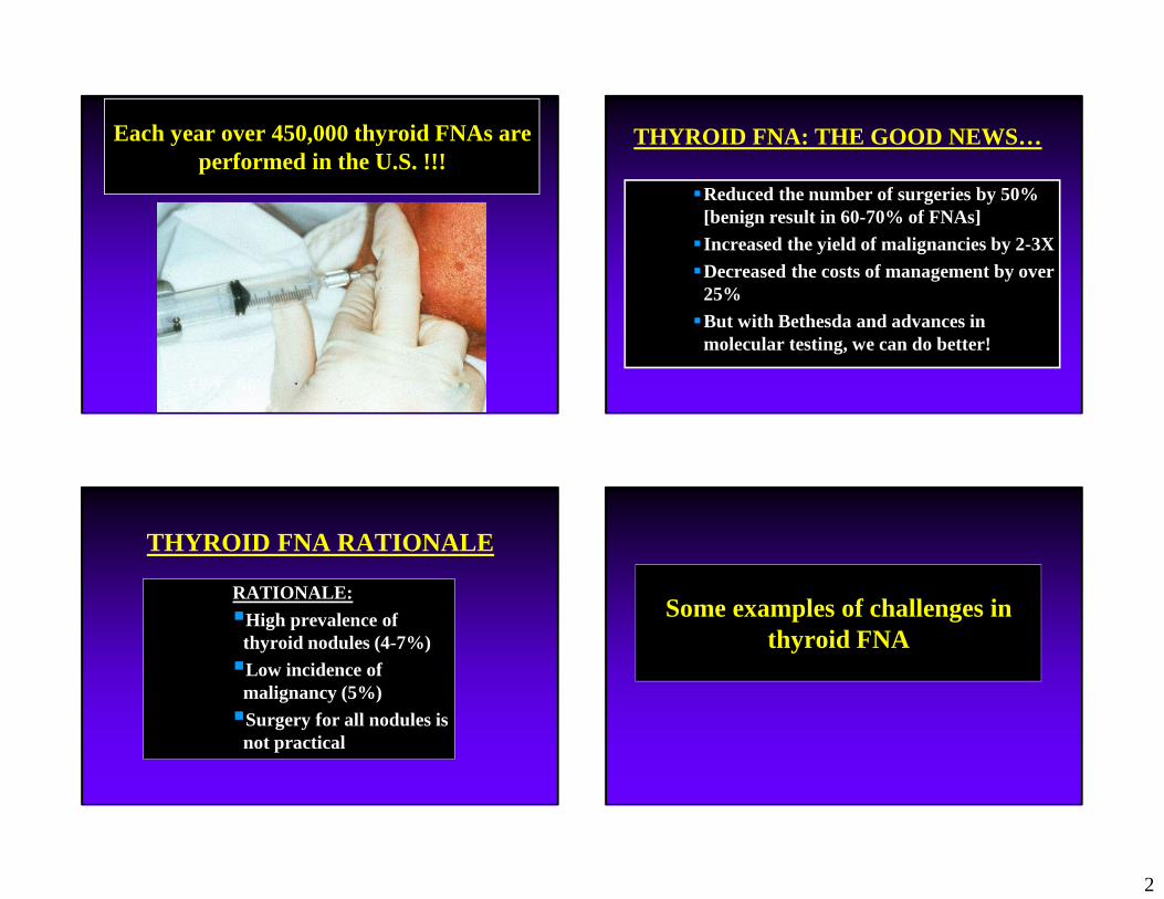
Suspicious for a Hurthle cell neoplasm?Lobectomy vs Total Thyroidectomy & LN Dissection
Medullary Thyroid Carcinoma
• Immunocytochemistry for calcitonin is recommended before making a definitive FNA diagnosis.
Anytime that the FNA diagnosis describes single cells or an unusual pattern, consider medullary thyroid carcinoma…and consider getting a serum calcitonin.
Thyroid FNA and Follicular-Patterned Lesions
6
Reporting of Thyroid FNAs
A major problem in the application of thyroid FNA has been the widespread inconsistency in reporting terminology.
The Bethesda System for Reporting Thyroid Cytopathology
The Bethesda System for Reporting Thyroid Cytopathology: 6 Diagnostic Categories
I. NONDIAGNOSTIC or UNSATISFACTORY
II. BENIGN
III. ATYPIA OF UNDETERMINED SIGNIFICANCE or FOLLICULAR LESION OF UNDETERMINED SIGNIFICANCE
IV. FOLLICULAR NEOPLASM or SUSPICIOUS FOR A FOLLICULAR NEOPLASM- specify if Hürthle cell (oncocytic) type
V. SUSPICIOUS FOR MALIGNANCY
VI. MALIGNANT
Bethesda Terminology: Relationship to Clinical Algorithms
Category Management Implied Risk of Malignancy (%)
Non-Diagnostic Repeat FNA 1-4%
Benign Follow <1-3%
AUS/FLUS Repeat FNA ~5-15*%
Susp for Follicular Neoplasm Lobectomy 20-30%
Susp for Hurthle Cell Neoplasm Lobectomy 20-30%
Suspicious for Malignancy Lobectomy/ Total Thyroid
60-75%
Malignant Total Thyroidectomy
97-99%
7
Summary of the BSRTC:Our experience at MGH and BWH
Study Category TOTAL CASESND Benign AUS SusF SUS Malignant
AUS/FLUS: The Problem• The classic “indeterminate” category• Cases that don’t fulfill criteria of other categories:
– “The findings are not convincingly benign, yet the degree of cellular or architectural atypia is not sufficient for an interpretation of ‘’follicular neoplasm’ or ‘suspicious for malignancy.“
• 8 scenarios outlined in the Bethesda Atlas• Heterogeneous category – WASTEBASKET• Often a compromised specimen (obscuring blood,
etc.)– Note: low cellularity, poor fixation, obscuring elements
by themselves not sufficient for AUS/FLUS
AUS/FLUS- Scenario: Hypocellular but Microfollicular
Rare microfollicles
AUS/FLUS- Scenario: Mixed Architectural Pattern
15
AUS/FLUS- Scenario: Scant Hurthle Cells Only AUS/FLUS – Scenario: Artifact
Obscuring blood and mild atypia
AUS/FLUS Scenario:“Benign” …But Focal Features of
Papillary Carcinoma
Figs. 4.5 A and B, The Bethesda atlas Air-drying of Pap-stained smear (Fig. 4.2, The Bethesda Atlas)
AUS/FLUS – Scenario: Preparation Artifact and Mild Atypia
16
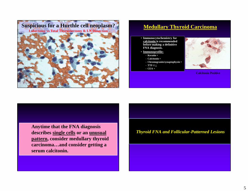
AUS/FLUS:• Less than 7% of thyroid FNAs (range: 3-20% in lit.) –
needs adjusting! …probably 10-12%• Potential for overuse/abuse –
– Role for intralab monitoring (QA metric)
• Recommended management: Repeat FNA or molecular– >50% of cases are reclassified as BENIGN on repeat FNA
• Surgery for “repeat atypicals”– 27% malignant with repeat AUS/FLUS FNA[Faquin and Baloch, 2009]
• Should AUS/FLUS be further subdivided? -Maybe!
• Nuclear atypia = increased risk for PTC• Architectural atypia only = lower risk for PTC
Follicular Variant of PTC:Common Cause of Difficult Thyroid FNA
A subset of these lesions will fall into the “Suspicious for follicular neoplasm’” “Suspicious
for malignancy’” “AUS/FLUS” categories.
Non-Invasive Follicular Thyroid (NIFT) Neoplasm with Papillary-Like Nuclear Features
�Reclassify non-invasive FVPTC as NIFT
�The prospects of NIFT will create some issues for thyroid cytopathology:�The ROM for certain diagnostic categories of the
Bethesda System will decrease�The PPV/NPV of molecular testing panels will
change
�Future modifications in our approach to the indeterminate thyroid FNA may be warranted
What is the role for ancillary testing of thyroid FNAs?
IHC, Afirma, MiRInform, others???
INDETERMINATE THYROID FNAS
17
INDETERMINATE THYROID FNAS (15-30%)
• Clinical management for the “benign” and “malignant” categories is clear
• Most patients with “suspicious for malignancy” will have surgery (often total thyroidectomy)
• Management options for the AUS/FLUS and FN/SFN categories are more complex – molecular testing offers a solution
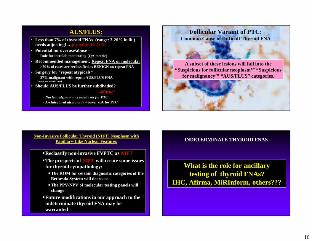
Immunocytochemistry�Used primarily in Europe (Fadda et al., EJE 2011)
�Stratify indeterminate thyroid FNAs into low and high risk groups�Liquid-based and smears�Inexpensive and fast TAT
�HBME-1 and Galectin-3 are most popular�Difficulties in reproducibility, specificity, and interp retation
– Convenient– Objective result– Avoids waiting for repeat FNA– Defines management and save dollars
• CONS:– Expensive if inappropriately applied– Reflex testing
• Takes clinician out of picture• Can add to overall expense (unnecessary testing)
– Loss of cyto-histo correlation
Molecular Testing and Thyroid FNA
18
The Afirma Test
Molecular Testing and Thyroid FNACASE
A 47-year-old euthyroid woman presented to the endocrinology clinic with a 2.0 cm right thyroid nodule. A previous FNA on this patient’s thyroid nodule at an outside hospital was reported to have been diagnosed as AUS/FLUS. An FNA was performed.
Mixed Macro- and Microfollicles Increased Proportion of Microfollicles
19
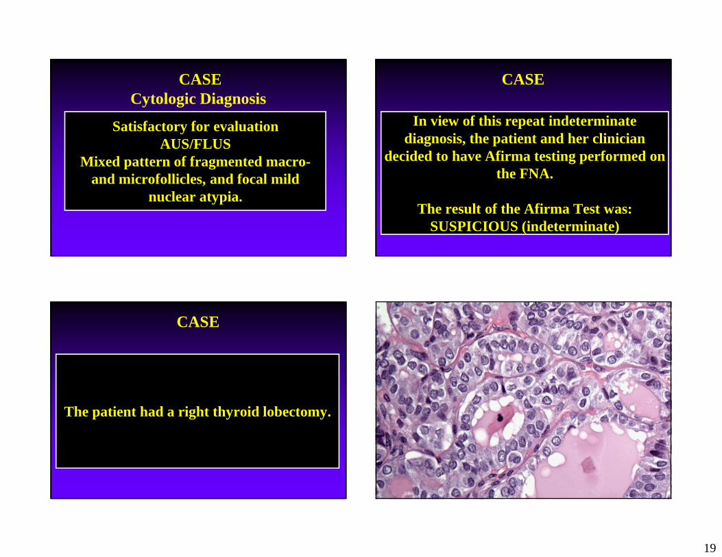
Satisfactory for evaluationAUS/FLUS
Mixed pattern of fragmented macro-and microfollicles, and focal mild
nuclear atypia.
CASECytologic Diagnosis
In view of this repeat indeterminate diagnosis, the patient and her clinician
decided to have Afirma testing performed on the FNA.
The result of the Afirma Test was:SUSPICIOUS (indeterminate)
CASE
The patient had a right thyroid lobectomy.
CASE
20
Histologic Diagnosis
Encapsulated follicular variant of papillary thyroid carcinoma (2.0 cm).
{aka NIFT}
The Afirma Test
2012
• Benign fingerprint {high NPV} – “rule out” test– Microarray data from 167 genes– “Benign” vs “Suspicious” Classification– $3350 cost– Requires 2 additional FNA passes– Also includes BRAF and RET mutation tests
• Nikiforov et al. J Clin Endocrinol Metab 2011– 479 indeterminate FNAs; 18% had mutations– 75% of PTC; 70% of FC– Overall specificity = 98%; sensitivity = 60%– $ 2250 cost
MiRInform Test
• Probably most useful for “susp. for malignancy” group• Does not decrease need for surgery, but can allow for
TT in 56% of patients with a malignancy• PPV:
– AUS/FLUS 88%– FN/SFN 87%– Susp Mal. 95%
• All FPs due to RAS+ follicular adenomas
MiRInform Test:Nikiforov et al. J Clin Endocrinol Metab 2011
The ThyroSeq Test v.2
Molecular Testing and Thyroid FNAThyroSeq v.2
– Next generation sequencing gene mutation panels– Mutations in 14 genes
– 42 gene fusions
• Indications: – Thyroid FNA diagnosed as indeterminate by cytology
– Thyroid FNA diagnosed as malignant, when molecular testing is expected to affect the decision to perform surgery or extent of surgery
– Thyroid FNA diagnosed as benign by cytology, when strong clinical suspicion for cancer exists based on imaging and clinical studies.
– Diagnosis of cancer is established in FNA or surgically excised thyroid tissue, when molecular profiling of cancer will affect administration of radioactive iodine, intensity of follow up, or targeted therapies for advanced cancer.
23
ThyroSeq v.2
• Gene List for Mutations: • AKT1, BRAF, CTNNB1, GNAS, HRAS, KRAS, NRAS, PIK3CA, PTEN, RET,
TP53, TSHR, TERT, EIF1AX
• Gene List for Gene Fusions and Gene Expression: • RET, PPARG, NTRK1, NTRK3, ALK, IGF2BP3, BRAF, MET, CALCA, PTH,
• TERT promoter activating mutations– Seen in any type of follicular-derived carcinoma
• PTC-7.5%• Follicular -17.1%
• Poorly differentiated -29.0%
• Anaplastic-33.3%
– Associated with aggressive behavior
Liu et al Endocr Relat Cancer (2013), Landa et al J Clin Endocrinol Metab (2013), Melo et al J Clin Endocrinol Metab (2014)
TERT and BRAF:Act synergystically to predict aggressive behavior
Xing et al J Clin Oncol (2014)
Case Example Using ThyroSeq v.2•55 yo male with 3.0 cm right thyroid nodule•FNA diagnosis: Susp for FN•ThyroSeq v.2 testing
Case Example Using ThyroSeq v.2
25
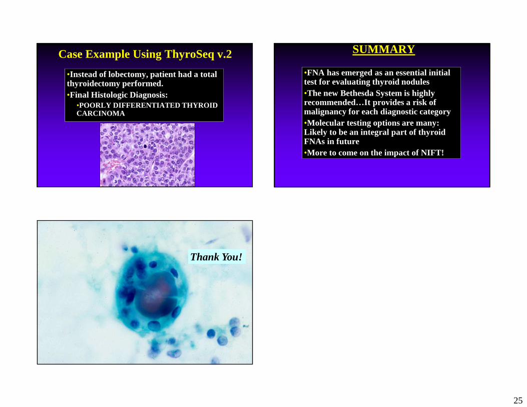
Case Example Using ThyroSeq v.2
•Instead of lobectomy, patient had a total thyroidectomy performed.•Final Histologic Diagnosis:
•POORLY DIFFERENTIATED THYROID CARCINOMA
SUMMARY
•FNA has emerged as an essential initial test for evaluating thyroid nodules•The new Bethesda System is highly recommended…It provides a risk of malignancy for each diagnostic category•Molecular testing options are many: Likely to be an integral part of thyroid FNAs in future•More to come on the impact of NIFT!