Evolutionarily conserved primary TNF sequences relate to itsprimitive functions in cell death inductionWenshu Lu1,*, Qiongyu Chen1,*, Songmin Ying1, Xiaobing Xia1, Zhanru Yu2, Yuan Lui2, George Tranter3,Boquan Jin4, Chaojun Song4, Leonard W. Seymour1,‡ and Shisong Jiang1,‡
ABSTRACTTNF is a primitive protein that has emerged from more than 550million years of evolution. Our bioinformatics study of TNF from ninedifferent taxa in vertebrates revealed several conserved regions in theTNF sequence. By screening overlapping peptides derived fromhuman TNF to determine their role in three different TNF-inducedprocesses – apoptosis, necrosis and NF-κB stimulation – we foundthat TNF conserved regions are mostly related to cell death ratherthanNF-κB stimulation. Among themost conserved regions, peptides(P)12, P13 and P1213 (comprising P12 and P13) induced apoptosis,whereas P14, P15, P16 and P1516 (comprising P15 and P16)induced necrosis. Cell death induced by these peptides was notthrough binding to the TNF receptor. P16-induced necrosis wasmainly through disruption of the cell membrane, whereas P1213-induced apoptosis involved activation of TRADD followed byformation of complex II. Finally, using a monoclonal antibody and amutant TNF protein, we show that TNF-induced apoptosis isdetermined by a conserved linear sequence that corresponds tothat within P1213. Our results reveal the determinant sequence that iskey to the TNF primitive function of inducing apoptosis.
INTRODUCTIONTumor necrosis factor (TNF) is a multifunctional cytokine that issecreted by various cells, but mainly macrophages and T cells. TNFis thought to have emerged during the Precambrian era (more than550 million years ago) before the animal phyla started to evolve(Quistad et al., 2014). The fact that certain functions of TNF – e.g.induction of cell death – have remained unchanged during theprocess of evolution indicates that some parts of the TNF structure,either primary or conformational, are probably conserved betweendifferent species.TNF has two forms, a 26-kDa membrane form and a 17-kDa
soluble form. The latter is formed through enzymatic cleavage of themembrane form by a TNF-converting enzyme (TCE). TNFperforms functions as a trimer, which then binds to one of its two
receptors, the 55-kDa TNF receptor 1 (TNFR1; also known asTNFRSF1A) or the 75-kDa TNF receptor 2 (TNFR2; also known asTNFRSF1B). TNFR1 exists constitutively on most cell surfaces andfunctions when activated by trimerized TNF. TNFR2 is expressed,in a highly regulated way, on the surface of several cell types, suchas immune cells, endothelial cells, epithelial cells and neuronal cells(Martin et al., 2014). Activation of TNFR2 leads to signal pathwaysthat are different to that of TNFR1 (Hehlgans and Pfeffer, 2005).TNF induces multiple biological functions, and many of thesefollow from three major intracellular events: (1) stimulation of thetranscription factor nuclear factor kappa B (NF-κB), leading to cellactivation and cytokine production; (2) induction of the extrinsicpathway of cell apoptosis (Declercq et al., 2009; Galluzzi andKroemer, 2008; Hitomi et al., 2008); and (3) induction of necrosis.In some situations, it has been reported that inhibition of caspasesduring apoptosis can lead to a form of programmed cell death thatcan occur with features of necrosis, termed ‘necroptosis’ (Bergheet al., 2010; Chan et al., 2003; Declercq et al., 2009; Galluzzi andKroemer, 2008; Hitomi et al., 2008; Vandenabeele et al., 2010).
Many reports have explored the mechanism of TNFmultifunctionality by studying its signal transduction pathways(Declercq et al., 2009). In most cases, stimulating cells with TNFprimarily activates NF-κB to promote cell survival (Fig. S1).Apoptosis and necrosis are only triggered when the NF-κB pathwayis inhibited (Varfolomeev and Ashkenazi, 2004). It has beenproposed that TNF stimulates a membrane-bound complex(complex I), which includes the following components: TNFR1,TNF-receptor-associated death domain (TRADD), the receptor-interacting protein kinase 1 (RIP1), TNF-receptor-associated factor2 (TRAF2) and cellular inhibitor of apoptosis 1 and 2 (c-IAP1 andc-IAP2, respectively) (Micheau and Tschopp, 2003). Complex Iinitiates NF-κB activation but not apoptosis and/or necrosis.However, if NF-κB activation is absent, TNF stimulates its targetcells to form a second complex (complex II) in the cytoplasm, whichcompared to complex I, lacks TNFR1 and c-IAP1 but includes Fas-associated death domain (FADD), caspase-8 and caspase-10.Complex II then triggers apoptosis and/or necrosis (Fig. S1).RIP1 has been thought to be important in the activation of NF-κB,but its role has been recently challenged by the observation that NF-κB activation can occur independently of RIP1 (Wong et al., 2010).Moreover, another RIP-family member in complex II, RIP3, hasbeen shown to be crucial in the cell death process. RIP3 induces adeath signal that leads to necrosis (necroptosis) when caspase-8 isinhibited (He et al., 2009; Zhang et al., 2009) (Fig. S1).
Although the mechanism of how intracellular signals aretransduced after TNF–TNFR1 binding is now well characterized,the study of the target cells alone has not so far created any clinicallyvalued opportunity for intervention of TNF related pathogenesis. Bycontrast, blocking TNF with monoclonal antibodies or solubleTNFR2 (Etanercept) has generated great benefit for patientsReceived 11 June 2015; Accepted 10 November 2015
1Department of Oncology, University of Oxford, Old Road Campus ResearchBuilding, Roosevelt Drive, Oxford OX3 7DQ, UK. 2MRC Immunology Unit,Weatherall Institute of Molecular Medicine, University of Oxford, John RadcliffeHospital, Headington OX3 9DS, UK. 3Chiralabs Limited, Begbroke Science Park,Woodstock Road, Begbroke, Oxfordshire OX5 1PF, UK. 4Department ofImmunology, Fourth Military Medical University, Xi’an City 710032, ShaanxiProvince, China.*These authors contributed equally to this work
(Feldmann, 2009), indicating the importance of investigating theligand itself. Nevertheless, the role that the TNF primary sequenceplays in its various functions has not been widely studied. Forexample, do distinct parts of the TNF molecule play a role instimulating different functions? It is widely believed thatevolutionarily conserved sequences in a protein are often offunctional importance. Some amino acid sequences of TNF mightremain unchanged in different species, but any correlation betweenthe conserved sequences and the functions has yet to be established.Identification of these functional ‘footprints’ does not only help usto understand how TNF evolved but also provides potentialbiomarkers for intervention of TNF-related diseases.As a result, this study sought to compare TNF sequences from
different species to identify conserved sequences. Next, in orderto identify whether the conserved sequences correlate withparticular functions of TNF, we made a series of overlapping 20-mer peptides comprising the full-length of the membrane form ofTNF. The ability of these peptides to mediate three distinctpharmacological outcomes (NF-κB stimulation, apoptosis andnecrosis) was measured using a series of cell lines, but mainlythe Jurkat A3 T-cell line and the mouse fibrosarcoma L929 cells.Some of the peptides mediated strong pro-apoptotic and pro-necrotic mechanisms, and these were studied in detail. Inducingthe inflammation-inhibitory process of apoptosis is one of themost important functions of TNF, and is a gateway leading toother forms of cell death – e.g. inflammatory necroptosis.Therefore, we investigated whether the apoptotic function ofspecific TNF peptides correlates with the function of the wholeTNF molecule.
RESULTSMost regions of TNF that are conserved across differentvertebrates correlate with cell deathConservation of protein sequence often serves as evidence forcellular functionality (Bejerano et al., 2004; Ghosh et al., 1998).TNF exists in different species within the vertebrate subphylum(Baena et al., 2007; Wiens and Glenney, 2011).We therefore compared the TNF protein sequences in nine
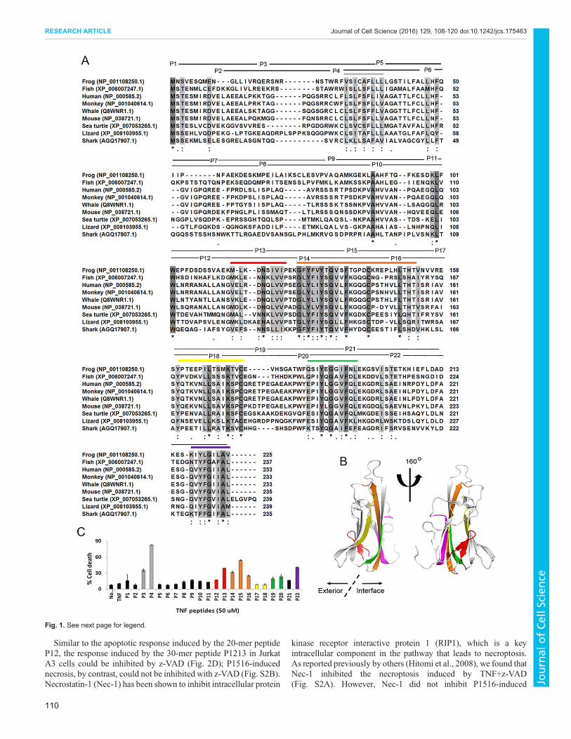
genetically diverse species (monkey, human, whale, mouse, seaturtle, lizard, fish, frog and shark) in the vertebrate subphylum.Fig. 1Ashows a multiple sequence alignment of whole TNF sequences fromthe nine species (Fig. 1A). There are several conserved regions, whichare marked by horizontal colored bars above the TNF sequences inFig. 1A. These conserved regions are also shown in the TNF crystalstructure (Eck and Sprang, 1989) in Fig. 1B (using the same colors asin Fig. 1A). The black lines and designation P1–P22 above thesequences in Fig. 1A indicate the overlapping peptides of the humanTNF sequence that were made in order to screen for TNF-likeactivities. Fig. 1C shows a cytotoxicity assay using the overlappingTNF-derived peptides. The colors used in Fig. 1C represent the sameconserved regions shown in Fig. 1A,B, and it is interesting that thepeptides inducing the greatest cytotoxicity are those containingevolutionarily conserved sequences.The conserved region shown in gray is located in overlapping
peptides P3 and P4 (Fig. 1A). Because this region represents thetrans-membrane domain of TNF, it is not shown in the crystalstructure of soluble TNF. Both P3 and P4 were found to be cytotoxic(Fig. 1C). The conserved region in red partly comprises P12 and P13(Fig. 1A). This region includes two β-strands that are connected by aloop and is located at the exterior surface of the trimeric TNF crystalstructure (Fig. 1B). Overlapping peptides comprising this region arealso cytotoxic (Fig. 1C). The conserved regions in orange, green and
purple relate to P13–P16, P19–P20 and P22, respectively. All theseconserved sequences are hidden at the trimer-interface side of theTNF crystal structure (Fig. 1B); however, the peptides derived fromthese regions are all cytotoxic (Fig. 1C). The conserved region inyellow is comprised by the sequences of P16–P18 (mainly P17–P18) (Fig. 1A), and the region is mainly located at the externalsurface of the trimeric TNF crystal structure (Fig. 1B). Interestingly,although P16 induces cytotoxicity, P17 and P18 contain conservedregions without obvious cytotoxic effects (Fig. 1C).
Peptides containing conserved sequences induce apoptosisas well as non-programmed necrosis in Jurkat A3 cells andL929 cellsTo investigate systematically whether the TNF conserved sequences(vs non-conserved sequences) mediate special cellular functions,overlapping synthetic peptides corresponding to the human TNFsequence were screened in an NF-κB assay, and in a high-throughput flow-cytometry-based screen for induction of apoptosisand necrosis. The overlapping synthetic peptides were designed as aseries of linear 20-amino-acid peptides, P1–P22, the sequences ofwhich comprised the complete length of the membrane form (or‘pro-form’) of TNF (amino acids 1–233) (http://www.uniprot.org/uniprot/NP_000585.2). Each peptide sequence overlapped theadjacent peptides by 10 amino acids, and the C-terminal peptidewas 23 amino acids in length (Fig. 1A). We used the assays to screenwhether the peptides could mediate the major functions of TNF,including activation of NF-κB and induction of cell death (apoptosisand/or necrosis).
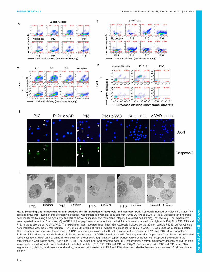
Several peptides showed activities of inducing cell death. Asmentioned above, cell-death-inducing peptides largely coincidedwith conserved sequences (Fig. 1). In this article, we focused on theseries of peptides P12–P16, as these sequences comprised the mostconserved sequences of TNF (Fig. 1A). Moreover, these peptidesare consecutively linked in tandem but can be divided functionallyinto two groups; in both Jurkat A3 and L929 cells, P12 and P13induced mainly apoptosis, and P14–P16 induced mainly necrosis(Fig. 2A,B). Peptide P18 never induced cell death and was thereforeused as a negative control.
Within the region covered by P12–P13, there was a slightsequence variation for induction of apoptosis between Jurkat A3 andL929 cells; both P12 and P13 induced strong apoptosis in Jurkat A3cells (Fig. 2A–C), whereas only P13 induced apoptosis in L929 cells(Fig. 2B). For the induction of necrosis, P14, P15 and P16 inducedstrong necrosis in both Jurkat A3 and L929 cells (Fig. 2A,B).
z-VAD-fluoromethylketone (z-VAD-FMK or z-VAD) is aspecific caspase inhibitor (Van Noorden, 2001). A combination ofTNF and z-VAD has been reported to induce regulated necrosis ornecroptosis in L929 cells (Hitomi et al., 2008; Vercammen et al.,1998; Wu et al., 2011). We found a similar phenomenon in JurkatA3 T cells when apoptosis was mediated by the TNF peptides. Theapoptosis in Jurkat A3 cells that was induced by these peptidescould be inhibited with z-VAD (Fig. 2C). P12- and P13- inducedapoptosis in Jurkat A3 cells was converted into partially live cellsand partially regulated necrosis after addition of z-VAD (Fig. 2C).
Because the peptide sequence varied slightly between cell lines,we made 30-mer-long peptides P1213 (comprising P12 andP13, P1213: ALLANGVELRDNQLVVPSEGLYLIYSQVLF;P12: ALLANGVELRDNQLVVPS) and P1516 (comprising P15and P16, P1516: KGQGCPSTHVLLTHTISRIAVSYQTKVNLL;P15: KGQGCPSTHVLLTHTISRIA; P16: LLTHTISRIAVSYQT-KVNLL) – P1213 induced predominantly apoptosis, whereasP1516 induced necrosis in Jurkat A3 cells (Fig. 2D; Fig. S2B).
109
RESEARCH ARTICLE Journal of Cell Science (2016) 129, 108-120 doi:10.1242/jcs.175463
Similar to the apoptotic response induced by the 20-mer peptideP12, the response induced by the 30-mer peptide P1213 in JurkatA3 cells could be inhibited by z-VAD (Fig. 2D); P1516-inducednecrosis, by contrast, could not be inhibited with z-VAD (Fig. S2B).Necrostatin-1 (Nec-1) has been shown to inhibit intracellular protein
kinase receptor interactive protein 1 (RIP1), which is a keyintracellular component in the pathway that leads to necroptosis.As reported previously by others (Hitomi et al., 2008), we found thatNec-1 inhibited the necroptosis induced by TNF+z-VAD(Fig. S2A). However, Nec-1 did not inhibit P1516-induced
Fig. 1. See next page for legend.
110
RESEARCH ARTICLE Journal of Cell Science (2016) 129, 108-120 doi:10.1242/jcs.175463
necrosis (Fig. S2B), suggesting that P1516-induced necrosis mightnot be necroptosis.Cell death was further confirmed with fluorescence microscopy
analyses. DNA fragmentation during peptide-induced apoptosis, asvisualized with DAPI staining, coincided with expression of activecaspase-3 (Fig. 2E). These experiments suggest that P12-inducedapoptosis in Jurkat A3 cells might be converted into programmednecrosis in the presence of caspase inhibitors, and this feature hasbeen defined as necroptosis (Galluzzi and Kroemer, 2008).We also investigated the cell death by using transmission electron
microscopy. P12- and P13-treated Jurkat A3 cells showedcondensation and fragmentation of nuclei and blebbing; P15- andP16-treated cells showed disintegration of the cell membrane,swelling of organelles and release of intracellular contents (Fig. 2F).Soluble TNF activated NF-κB in Jurkat A3 T cells at
concentrations as low as 0.5–1 nM (Fig. S3A). However, weobserved that, in Jurkat A3 T cells, none of the peptides were able toactivate NF-κB, even at concentrations of 100 µM (Fig. S3B). At1 min after treatment, there was no significant NF-κB activation ineither TNF- or peptide-treated cells. At 5 min, only TNF stimulatedNF-κB. This result indicates that the peptides have different NF-κB-stimulation kinetics compared with TNF. The peptides did not showNF-κB stimulation activity at any of the multiple time points ordoses tested (Fig. S3B,C). Peptides P11–P18 were also tested with aNF-κB reporter cell line (THP-1 human monocyte) and showed noactivation of NF-κB across a broad concentration range (Fig. S3C).These results suggest that either the NF-κB activation processrequires involvement of more than one TNF peptide chain, forexample the ability to trimerize, or that it requires conformationalproperties of the TNFmolecule that are lost in the peptide fragments.
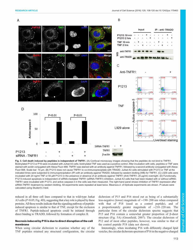
Cell death induced by peptides is independent of TNFR1We firstly tested whether P1213 and P16 bound to TNFR1 byusing immunofluorescence staining, which can be observed with aconfocal microscope. Staining the peptides and TNFR1 in Saos-2(human Osteosarcoma) cells showed no colocalization between thepeptides and TNFR1. Only TNF (positive control) was found tocolocalize with TNFR1 (Fig. 3A). This result indicates that, unlikethe whole molecule of TNF, the functioning peptides do not bind toTNFR1. This result was further confirmed by using a TRADDimmunoprecipitation assay in Jurkat A3 cells. TRADD is a key
intracellular protein that serves as a scaffold to attract variousproteins to form either complex I or complex II in order to initiatedifferent TNF functions. We therefore tried to immunoprecipitateTRADD after peptide and/or TNF stimulation. TNFR1 could beco-precipitated with TRADD after TNF stimulation, whereas itcould not be co-immunoprecipitated with TRADD afterstimulation with P1213 (Fig. 3B). The receptor-binding ability ofthe peptides was also assessed with an ELISA assay in whichELISA plates were coated with avidin, followed by the addition ofbiotinylated peptides (biotinylation did not affect the pro-apoptoticfunction of P1213; see Fig. S4A). Next, the plates were incubatedwith TNFR1 before detection with an antibody against TNFR1 anda second antibody conjugated with horseradish peroxidase (HRP).As Fig. S4B shows, none of the peptides tested was able to bind toTNFR1. Binding of TNFR1 and TNFR2 to TNF, P1213 or P16was also tested using Biacore surface plasmon resonance analysis.As shown in Fig. S4C, only TNF bound to both TNFR1 andTNFR2. Neither P1213 nor P16 bound to TNF receptors(Fig. S4C). We also tested whether blocking TNFR1 with aneutralizing monoclonal antibody against TNFR1 would inhibitP1213- or TNF-induced apoptosis. As Fig. 3C shows, blockingTNFR1 only inhibited TNF-induced apoptosis in L929 cells andnot P1213-induced apoptosis (Fig. 3C). Lastly, we tested whetherP1213 induces apoptosis in Jurkat A3 cells in which TNFR1 hadbeen knocked down with small interfering (si)RNAs. As shown inFig. 3D, after knockdown of TNFR1, the TNFR1 expression levelwas highly reduced (right panel), but apoptosis was induced nomatter whether TNFR1 was inhibited or not (Fig. 3D, left panel).These data suggest that TNFR1 is not required for peptide-relatedapoptosis or necrosis.
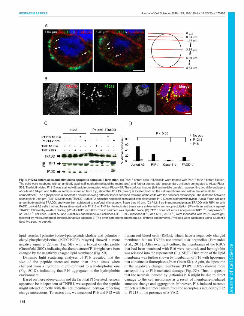
Apoptosis induced by P1213 involves formation of complexII, comprising TRADD, RIP1, caspase-8 and FADDTo study the signal pathway of peptide-induced apoptosis, wefocused on P1213-induced apoptosis. Although P1213-inducedapoptosis was independent of TNFR1 (Fig. 3), the fact that itstimulated activation of caspase-3 (apoptosis was assayed bymeasuring active caspase-3; Fig. 3D) hints that the intracellularsignals must be involved in the process. To examine this, we firstchecked whether P1213 enters cells. We performed an experimentin which we incubated P1213-biotin with HT29 cells (chosenbecause their membrane can be clearly stained with an antibodyagainst E-cadherin) for 2 h at 37°C, and stained P1213 and the cellmembrane. We used a confocal microscope to observe the positionof P1213 in relation to the cell membrane. As shown in Fig. 4A,P1213 (green) was found in different confocal-section layers on thecell membrane (red) and within the cell.
We then investigated whether P1213 colocalized with TRADD inSaos-2 cells. As shown in Fig. 4B, colocalization was seen as earlyas incubating P1213 and Saos-2 cells for 10 min, suggesting thatbinding of the peptide to TRADD is an early event.
Next, by immunoprecipitating TRADD from Jurkat A3 cells, wefound that after 2 h of stimulation with TNF, both RIP1 and FADDcould be co-immunoprecipitated upon exposure of cells to P1213(Fig. 4C), implying that a complex (most likely complex II) wasformed, comprising at least TRADD, RIP1 and FADD. Thenonspecific bands seen either side of the RIP1 signal relate to the50-kDa and 100-kDamonomer and dimer heavy chains, respectively,of the antibody against TRADD, which are recognized by the anti-mouse secondary antibody. Finally, we employed Jurkat-A3-basedknockout cell lines – Jurkat RIP (RIP1−/−), I9.2 (caspase-8−/−) andI2.1 (FADD−/−). Apoptosis induced by P1213 was significantly
Fig. 1. TNF conserved sequences across nine vertebrate taxa, theirposition in the crystal structure and their relationship to induction of celldeath. (A) Multiple TNF protein sequence alignment of frog(NP_001108250.1), fish (XP_006007247.1), human (NP_000585.2), monkey(NP_001040614.1), whale (Q8WNR1.1), mouse (NP_038721.1), sea turtle(XP_007053265.1), lizard (XP_008103955.1) and shark (AGQ17907.1). Theamino acids that are 100% conserved are shaded in black and marked with anasterisk (*) underneath the shading. The gray shading and a colon (:)underneath indicate conservation between groups of strongly similar properties– scoring >0.5 in the Gonnet PAM 250 matrix (http://www.ebi.ac.uk/Tools/msa/clustalo/help/faq.html#23). The lighter gray shading and a period (.)underneath indicate conservation between groups of weakly similar properties– scoring ≤0.5 in the Gonnet PAM 250 matrix. Above the alignment, a series ofoverlapping peptides derived from the human TNF sequence (P1–P22) andthe lengths of these peptides (lines after the names) is shown. The colored barsrepresent different regions of conserved sequences. (B) The position of theconserved sequences are shown in the corresponding colors in the crystalstructure of TNF. (C) Jurkat A3 cell death induced by the human TNFoverlapping peptides. The cell death shown represents the sum of apoptosis(measured using caspase-3 activation) and necrosis (live–dead staining). Theconcentration of the peptides is 50 µM. The colors correspond to the regions ofconserved sequence shown in A,B. C shows a typical result from three replicateexperiments. Mean±s.d. of three experiments are shown. No, no peptide.
111
RESEARCH ARTICLE Journal of Cell Science (2016) 129, 108-120 doi:10.1242/jcs.175463
Fig. 2. Screening and characterizing TNF peptides for the induction of apoptosis and necrosis. (A,B) Cell death induced by selected 20-mer TNFpeptides (P12–P18). Each of the overlapping peptides was incubated overnight at 50 µM with Jurkat A3 (A) or L929 (B) cells. Apoptosis and necrosiswere measured by using flow cytometry analysis of active caspase-3 and membrane integrity (live–dead cell staining), respectively. The experimentswere repeated more than five times. (C) z-VAD inhibited peptide-induced apoptosis. Jurkat A3 cells were incubated overnight with 100 µM of P12, P13 andP18, in the presence of 10 µM z-VAD. The experiment was repeated three times. (D) Apoptosis induced by the 30-mer peptide P1213. Jurkat A3 cellswere incubated with the 30-mer peptide P1213 at 30 µM overnight, with or without the presence of 10 µM z-VAD, P18 was used as a control peptide.The experiment was repeated three times. (E) DNA fragmentation coincided with active caspase-3 expression in P12- and P13-induced apoptosis.P12- and P13-induced apoptosis is shown in fluorescence images of DAPI-stained nuclei with DNA fragmentation (upper panel) and fluorescence-labeledactive caspase-3 (lower panel). White arrows point to nuclear DNA fragmentation (upper panel), which coincides with caspase-3 activation in thecells without z-VAD (lower panel). Scale bar: 20 µm. The experiment was repeated twice. (F) Transmission electron microscopy analysis of TNF-peptide-treated cells. Jurkat A3 cells were treated with selected peptides (P12, P13, P15 and P16) at 100 µM. Cells cultured with P12 and P13 show DNAfragmentation, blebbing and membrane shedding, whereas cells treated with P15 and P16 show necrosis-like features, such as loss of cell membraneintegrity.
112
RESEARCH ARTICLE Journal of Cell Science (2016) 129, 108-120 doi:10.1242/jcs.175463
Journal
ofCe
llScience
reduced in all three cell lines compared to that in wild-type JurkatA3 cells (P<0.05; Fig. 4D), suggesting that a key role is played by theseproteins.All these results indicate that the signaling pathwayof peptide-induced apoptosis is similar to that of TNF, except for the exclusionof TNFR1. Peptide-induced apoptosis could be initiated throughdirect binding to TRADD, followed by formation of complex II.
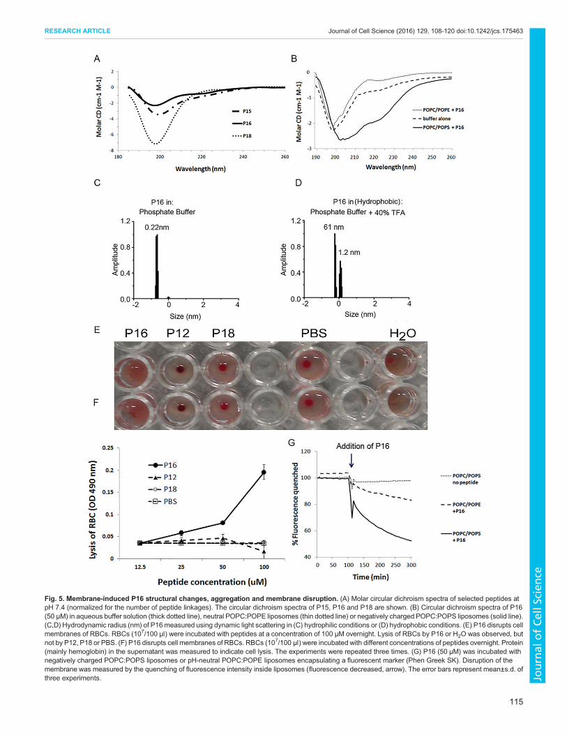
Necrosis induced by P16 is due to direct disruption of the cellmembraneWhen using circular dichroism to examine whether any of theTNF peptides retained any structural configuration, the circular
dichroism of P15 and P16 stood out as being of a substantiallyless-negative (lower) magnitude of ∼190–200 nm when comparedwith that of P18 (used as a control peptide), and ofa proportionately greater magnitude of ∼210–220 nm. Theparticular form of the circular dichroism spectra suggests thatP15 and P16 contain a somewhat greater proportion of β-sheetstructure (Fig. 5A) (Greenfield, 2007). The circular dichroism ofP12 and of most other peptides, however, was similar to that ofthe control peptide P18 (data not shown).
Interestingly, when incubating P16 with differently charged lipidvesicles, the circular dichroismspectrumofP16 in thenegative-charged
Fig. 3. Cell death induced by peptides is independent of TNFR1. (A) Confocal microscopy images showing that the peptides do not bind to TNFR1.Biotinylated P1213 or P16 were incubated with Jurkat A3 cells; biotinylated TNF was used as a positive control. After incubation with cells, peptides or TNF werestained with avidin conjugated with Alexa-Fluor-488. TNFR1 was stained with an antibody against TNFR1, followed by a second antibody conjugated with Alexa-Fluor-568. Scale bar: 10 μm. (B) P1213 does not cause TNFR1 to co-immunoprecipitate with TRADD. Jurkat A3 cells stimulated with P1213 or TNF at theindicated times were subjected to immunoprecipitation (IP) with an antibody against TRADD, followed by western blotting (WB) for TNFR1. (C) L929 cells wereincubated with 20 ng/ml TNF or 50 µM P1213 in the presence or absence of an antibody against TNFR1 (Anti-TNFR1; 20 µg/ml) overnight. (D) Functionally,P1213-induced apoptosis is independent of siRNA-mediated TNFR1 (siRNA-TNFR1) inhibition. Jurkat A3 cells that had been treated with or without siRNA-TNFR1 were incubated with P1213, and active caspase-3 in the cells was then measured. The right-hand panel shows inhibition of TNFR1 expression aftersiRNA-TNFR1 treatment by western blotting. All experiments were repeated at least twice. Means±s.d. of triplicate experiments are shown. P-values werecalculated using Student’s t-test.
113
RESEARCH ARTICLE Journal of Cell Science (2016) 129, 108-120 doi:10.1242/jcs.175463
Journal
ofCe
llScience
lipid vesicles [palmitoyl-oleoyl-phosphatidylcholine and palmitoyl-oleoyl-phosphatidylserine (POPC:POPS) bilayers] showed a morenegative signal at 220 nm (Fig. 5B), with a typical α-helix profile(Greenfield, 2007), indicating that the structure of P16might have beenchanged by the negatively charged lipid membrane (Fig. 5B).Dynamic light scattering analyses of P16 revealed that the
size of the peptide increased more than three times whenchanged from a hydrophilic environment to a hydrophobic one(Fig. 5C,D), indicating that P16 aggregates in the hydrophobicenvironment.Based on these observations and the fact that P16-related necrosis
appears to be independent of TNFR1, we suspected that the peptidemight interact directly with the cell membrane, perhaps reflectingcharge interactions. To assess this, we incubated P16 and P12 with
human red blood cells (RBCs), which have a negatively chargedmembrane but no TNFRs nor intracellular organelles (Fernandeset al., 2011). After overnight culture, the membranes of the RBCsthat had been incubated with P16 were ruptured, and hemoglobinwas released into the supernatant (Fig. 5E,F). Disruption of the lipidmembrane was further shown by incubation of P16 with liposomesthat contained a fluorophore (Phen Green SK). Again, the liposomeof the negatively charged membrane (POPC:POPS) showed moresusceptibility to P16-mediated damage (Fig. 5G). Thus, it appearsthat the necrosis induced by (cationic) P16 might be due to directdamage to the cell membrane as a result of membrane-mediatedstructure change and aggregation. Moreover, P16-induced necrosisreflects a different mechanism from the necroptosis induced by P12or P1213 in the presence of z-VAD.
Fig. 4. P1213 enters cells and stimulates apoptotic complex-II formation. (A) P1213 enters cells. HT29 cells were treated with P1213 for 2 h before fixation.The cells were incubated with an antibody against E-cadherin (to label the membrane) and further stained with a secondary antibody conjugated to Alexa-Fluor-568. The biotinylated P1213 was stained with avidin-conjugated Alexa-Fluor-488. The confocal images (left and middle panels), representing two different layersof cells at 3.84-µm and 6.40-µm sections scanning from top, show that P1213 (green) is located both on the cell membrane and within the intracellularcompartment. The right panel is a schematic picture showing different layers scanned from top of the cells with the confocal microscope. The distance betweeneach layer is 0.64 µm. (B) P1213 binds to TRADD. Jurkat A3 cells that had been stimulated with biotinylated P1213 were stained with avidin–Alexa-Fluor-488 andan antibody against TRADD, and were then subjected to confocal microscopy. Scale bar: 10 μm. (C) P1213 co-immunoprecipitates TRADD with RIP1 or withFADD. Jurkat A3 cells that had been stimulated with P1213 or TNF for the indicated times were subjected to immunoprecipitation (IP) with an antibody againstTRADD, followed by western blotting (WB) for RIP1 or FADD. The experiment was repeated twice. (D) P1213 does not induce apoptosis in RIP1−/−, caspase-8−/−
or FADD−/− cell lines. Jurkat A3 and Jurkat-A3-based knockout cell lines RIP−/−, I9.2 (caspase-8−/−) and I2.1 (FADD−/−) were incubated with P1213 overnight,followed by measurement of intracellular active caspase-3. The error bars represent mean±s.d. of three experiments. P-values were calculated using Student’sttest. No pep, no peptide.
114
RESEARCH ARTICLE Journal of Cell Science (2016) 129, 108-120 doi:10.1242/jcs.175463
Journal
ofCe
llScience
Fig. 5. Membrane-induced P16 structural changes, aggregation and membrane disruption. (A) Molar circular dichroism spectra of selected peptides atpH 7.4 (normalized for the number of peptide linkages). The circular dichroism spectra of P15, P16 and P18 are shown. (B) Circular dichroism spectra of P16(50 µM) in aqueous buffer solution (thick dotted line), neutral POPC:POPE liposomes (thin dotted line) or negatively charged POPC:POPS liposomes (solid line).(C,D) Hydrodynamic radius (nm) of P16 measured using dynamic light scattering in (C) hydrophilic conditions or (D) hydrophobic conditions. (E) P16 disrupts cellmembranes of RBCs. RBCs (107/100 µl) were incubated with peptides at a concentration of 100 µM overnight. Lysis of RBCs by P16 or H2O was observed, butnot by P12, P18 or PBS. (F) P16 disrupts cell membranes of RBCs. RBCs (107/100 µl) were incubated with different concentrations of peptides overnight. Protein(mainly hemoglobin) in the supernatant was measured to indicate cell lysis. The experiments were repeated three times. (G) P16 (50 µM) was incubated withnegatively charged POPC:POPS liposomes or pH-neutral POPC:POPE liposomes encapsulating a fluorescent marker (Phen Greek SK). Disruption of themembrane was measured by the quenching of fluorescence intensity inside liposomes (fluorescence decreased, arrow). The error bars represent mean±s.d. ofthree experiments.
115
RESEARCH ARTICLE Journal of Cell Science (2016) 129, 108-120 doi:10.1242/jcs.175463
Journal
ofCe
llScience
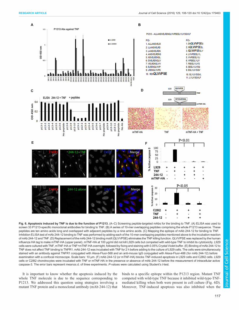
P1213 is key to TNF-induced apoptosisResults presented above characterize the function of the P1213peptide, which might be different from that of the whole TNFmolecule. To study whether the P1213 sequence in the whole TNFmolecule remains functional and is responsible for the apoptosis-inducing function of the whole TNF molecule, we produced 33monoclonal antibodies (mAbs) by immunizing mice with P1213,with a view to trying to block P1213-induced apoptosis. One pre-condition of this study was that the mAb should bind to TNF but notinterfere with the binding of TNF to its receptor. Five of these mAbswere able to bind to intact TNF (Fig. 6A), and we selected mAb 244-12, which exhibited the strongest binding to TNF, for further study.First, we investigated the binding epitope of mAb 244-12 by
screening a series of 10-mer overlapping peptides. These peptidesoverlapped the sequence of their adjacent peptides by nine aminoacids on each side (Fig. 6B), and they comprised the wholesequence of P1213. We employed an ELISA assay that measuredthe binding of mAb 244-12 to TNF in order to determine whetherthe binding could be inhibited by individual 10-mer peptides.Inhibition of mAb 244-12 binding to TNF in the presence of any 10-mer peptide meant that the epitope existed in the peptide. As shownin Fig. 6C, four consecutive peptides significantly inhibited thebinding of mAb 244-12 to TNF. The common sequence of thesefour peptides is QLVVPSE. Therefore, the binding epitope of mAb244-12 to TNF includes QLVVPSE (Fig. 6B,C).We engineered a mutant TNF protein (mTNF-HA) in which the
sequence QLVVPSE was replaced with the human influenzahemagglutinin (HA) tag (YPYDVPDYA) (Fig. 6D, upper panel).Functionally, unlike native TNF, expression of mTNF-HA did notkill L929 cells (Fig. 6D, lower panel) in the Crystal-Violet-basedTNF cytotoxicity assay (Hogan and Vogel, 2001). On the contrary,it inhibited the killing of L929 cells that was induced by wild-typeTNF (Fig. 6D, lower panel), suggesting a mechanism of receptor-competition and inhibition for mTNF-HA. This also suggests thatmTNF-HA might have a similar conformational structure to that ofwild-type TNF, which allows it to compete. To confirm that mTNF-HA binds to TNFR1, we performed an ELISA assay, which showedstrong binding activity (Fig. S4D).Next we examined whether binding of mAb 244-12 to TNF
interferes with the binding of TNF to TNFR1 on the surface of L929cells. As shown in Fig. 6E, mAb 244-12 colocalized with TNFR1only in the presence of TNF, suggesting that binding of mAb 244-12to TNF does not affect TNF binding to TNFR1 (Fig. 6E).Finally, we examined whether binding of mAb 244-12 to TNF
blocks TNF-induced apoptosis. Two cell lines, L929 and C28I2 (ahuman chondrocyte cell line) were employed in the study. AsFig. 6F shows, in both cell lines, adding mAb 244-12 to TNF-stimulated cells significantly inhibited activation of caspase-3compared to that without the mAb (P<0.01 and P<0.05 for L929and C28I2 cells, respectively). It was also noted that co-culture ofmTNF-HA with L929 cells instead of wild-type TNF alsoeliminated induction of apoptosis. These data suggest that theregion of TNF that corresponds to the sequence of P1213 is key tothe apoptosis-inducing function of TNF.
DISCUSSIONThe central scientific questions to be addressed in this study werewhether there are any genetically conserved sequences in theprimitive and multi-functional protein TNF, and whether any suchsequences are involved in important conserved TNF functions?Byaligning the TNF sequences of different species,we discovered
several genetically conserved regions (Fig. 1). Surprisingly, we
found that most of these regions appear to be linked with the TNFfunction of inducing cell death (Figs 1C and 2), but not stimulation ofcell proliferation (Fig. S3). Accordingly, it might be that induction ofcell death requires only a linear sequence, whereas stimulation ofproliferation could require a higher-level structure of TNF.
We sought to explain our discovery from the aspect of TNFevolution. TNF has evolved to be very important in both innate andadaptive immunity. As Quistat et al. have reported, the origin ofTNF can be dated back to 550 million years ago, well before thedivergence of vertebrates and invertebrates (Quistad et al., 2014).Our data suggest that the highly conserved regions of TNF structureare those that play a role in cell toxicity, and it is possible that thiswas originally the sole molecular function. However, as evolutionprogressed, the molecule appears to have adapted and divergedbetween species, with new functions (such as stimulation of NF-κB)developing and the sophisticated functions requiring TNF to have ahigher-order structure.
Here, we have explored the mechanisms of cell death inducedby these peptides. We chose P12 and P1213 for detailed study ofpeptide-induced apoptosis, and P16 and P1516 for necrosis. Noneof the peptides induce death through TNFR1 (Fig. 3). Althoughwe do not exclude the possibility of P1213 binding to cellmembrane receptors that directly activate TRADD to formcomplex II, it is likely that the apoptosis induced by P1213 inJurkat cells is mediated by binding of the intracellular peptide toTRADD directly, followed by formation of complex II (RIP,caspase-8, FADD and caspase-3; Fig. 4). This process isindependent of TNFR1 (Fig. 3).
Generally speaking, there are two mechanisms by which apeptide can enter cells: (1) in an energy-independent manner, suchas internalization and diffusion; (2) through energy-dependentpathways, such as receptor-mediated entrance. By analyzing thesequence of P1213 (with online software from Genescript), wefound it to be an acidic peptide (net charge of −2; isoelectric pointof pH 3.69). Because most cell membranes are either electronicallyneutral (e.g. normal cells) or negatively charged (e.g. tumor cells)(Hilchie et al., 2011; Riedl et al., 2011), it is unlikely that thepeptide crosses a cell membrane of the same electric charge usingdiffusion or an energy-independent pathway. Instead, energy willbe required for the peptide to enter the cells. For example, thepeptide might bind to a receptor (other than TNFR1), which helpsthe peptide to enter cells.
In summary, we have shown that P1213 probably enters the cellthrough an energy-dependent pathway (e.g. binding to a receptor)and that it then binds to TRADD to form complex II before initiatingapoptosis.
P12- and P1213-induced apoptosis showed similar features to thatinduced by TNF in L929 cells in that it can be inhibited by z-VAD,which changes the mode of cell death from apoptosis to necroptosis(Fig. 2). By contrast, P16 and P1516 induce necrosis in a differentmanner to TNF in L929 cells treated with z-VAD – P16-relatednecrosis is independent of caspase inhibition (Fig. S3) and P16 doesnot bind to TNFR1 (Fig. 3); instead, P16 probably works throughdirect disruption of the cell membrane because it permeabilizesRBCs (Fig. 5E,F) and liposome membranes (Fig. 5G). Moreover,these observations all suggest that P16-induced necrosis isfundamentally different from TNF-induced necroptosis. Thefeatures of P16 are very similar to certain characteristics ofcationic antimicrobial peptides (Peschel and Sahl, 2006), whichare regarded a primitive defense mechanism. Notably, the P16molecule, although very active in isolation, does not have anymembrane-permeabilizing activity in the intact TNF molecule.
116
RESEARCH ARTICLE Journal of Cell Science (2016) 129, 108-120 doi:10.1242/jcs.175463
It is important to know whether the apoptosis induced by thewhole TNF molecule is due to the sequence corresponding toP1213. We addressed this question using strategies involving amutant TNF protein and a monoclonal antibody (mAb 244-12) that
binds to a specific epitope within the P1213 region. Mutant TNFcompeted with wild-type TNF because it inhibited wild-type-TNF-mediated killing when both were present in cell culture (Fig. 6D).Moreover, TNF-induced apoptosis was also inhibited when the
Fig. 6. Apoptosis induced by TNF is due to the function of P1213. (A–C) Screening peptide-targeted mAbs for the binding to TNF. (A) ELISA was used toscreen 33 P1213-specific monoclonal antibodies for binding to TNF. (B) A series of 10-mer overlapping peptides comprising the whole P1213 sequence. Thesepeptides are ten amino acids long and overlapped with adjacent peptides by a nine amino acids. (C) Mapping the epitope of mAb 244-12 for binding to TNF.Inhibition ELISA test of mAb 244-12 binding to TNFwas performed by adding each of the 10-mer overlapping peptidesmentioned above to the incubation reactionof mAb 244-12 and TNF. (D) Replacement of themAb 244-12 bindingmotif (QLVVPSE) eliminates the TNF killing function. QLVVPSEwas replaced by the humaninfluenza HA tag to make mTNF-HA (upper panel). mTNF-HA at 100 µg/ml did not kill L929 cells but competed with wild-type TNF to inhibit its cytotoxicity. L929cells were culturedwith TNF,mTNF-HA or TNF+mTNF-HA overnight, followed by fixing and staining with 0.05%Crystal Violet buffer. (E) Binding of mAb 244-12 toTNF does not affect TNF binding to TNFR1. mAb 244-12 was incubated with TNF for 2 h before adding to the culture of L929 cells. The cells were simultaneouslystained with an antibody against TNFR1 conjugated with Alexa-Fluor-568 and an anti-mouse IgG conjugated with Alexa-Fluor-488 (for mAb 244-12) beforeexamination with a confocal microscope. Scale bars: 10 μm. (F) mAb 244-12 (or mTNF-HA) blocks TNF-induced apoptosis in L929 cells and C28I2 cells. L929cells or C28I2 chondrocytes were incubated with TNF or mTNF-HA in the presence or absence of mAb 244-12 before the measurement of intracellular activecaspase-3. The error bars represent mean±s.d. of three experiments. P-values were calculated using Student’s t-test.
117
RESEARCH ARTICLE Journal of Cell Science (2016) 129, 108-120 doi:10.1242/jcs.175463
Journal
ofCe
llScience
wild-type TNF was replaced with the mutant (Fig. 6F). By bindingto TNF, mAb 244-12 does not affect TNF binding to its receptor(Fig. 6E) but blocks the expression of caspase-3 (Fig. 6F). Thesedata strongly suggest that the P1213 region is key to TNF-inducedapoptosis. Previous reports have shown that after TNF binding to itsreceptor, it is degraded into fragments that might initiate the processof cell death within 2–6 h (Kull and Besterman, 1990; Tsujimotoet al., 1985). Our data support this by showing that a specificsequence in linear TNF peptides can directly bind to TRADD tostart the process of apoptosis.Our results suggest that the evolutionarily conserved primary
sequences of TNF relate to its original functions in cell death andthat secondary structure is important to the more recently evolvedfunction of NF-κB activation. The primitive molecule has evolvedto become sophisticated with diverse and sometimes apparentlyconflicting functions. The activities we have discerned here forsome of its component peptides could go someway to explaining itscomplex biology. The possibility of using TNF peptides to elicitspecific TNF effects raises new hope for potential applications indrug targeting and/or discovery.
MATERIALS AND METHODSCells lines, TNF and peptidesL929, Jurkat A3, RIP−/− and I9.2 (caspase-8−/−) cells were purchased fromAmerican Type Culture Collection (ATCC,Manassas, VA). I2.1 (FADD−/−)was a gift from L. Zheng (National Institutes of Health, Bethesda, MA).Human C28I2 chondrocytes were provided by Mary B. Goldring (Hospitalfor Special Surgery, New York City, NY). Human Saos-2 (osteosarcoma)cell line was purchased from Sigma-Aldrich.
TNF was purchased from Immunotools (Friesoythe, Germany). Peptides,whose sequences are shown in Fig. 1A, and their modification (e.g.biotinylation) were synthesized on an automatic APEX 396 using a standardsolid phase Fmoc strategy. In some of the validation experiments, peptides(e.g. P12 and P16) were purchased from Proimmune (Oxford, UK).
Mutant TNF-HA (mTNF-HA; where QLVVPSE was replaced with theHA tag YPYDVPDYA, starting at position 123) was synthesized byInvitrogen, and the mutant protein was expressed in Escherichia coli andpurified as described previously (Loetscher et al., 1993).
Apoptosis and necrosis assayCells were incubated overnight (or at the times indicated in the figures) withTNF (Immunotools) or TNF peptides with or without 10 µM z-VAD-FMK(R&D Systems) or TNFR1 (PeproTech, Rocky Hill, NJ). Cells were thenstained using a live–dead cell staining kit (Invitrogen, Paisley, UK),following the manufacturer’s instructions. The cells were then fixed with theCytofix/Cytoperm fixation and permeabilization solution kit (BDPharmingen), followed by intracellular staining with a FITC-conjugatedanti-caspase-3 antibody (BD Pharmingen). Cell data were acquired on aCyAn flow cytometer (Beckman Coulter, Fullerton, CA), and data wereanalyzed using Flowjo (Tree Star Inc., Ashland, Oregon).
TNF cytotoxicity assayAdherent L929 cells (100 µl, 4×10⁵/ml) were incubated with TNF orpeptides, with or without the presence of actinomycin D (8 µg/ml) (Sigma-Aldrich) overnight at 37°C under 5% CO2. After aspiration of all thesupernatant, 50 µl of 0.05% Crystal Violet was added to each well to stainthe live cells. After rinsing off Crystal Violet, the viability of the attachedcells could be determined.
NF-κB assayJurkat A3 cells, which were treated with TNF or their peptides at theindicated time points (Figs 1B and 2A), were harvested and lysed in RIPAlysis buffer (Cell Signaling Technology, Danvers, MA) with proteasecocktail inhibitor (Sigma-Aldrich). Cell extracts were then blotted ontonitrocellulose membranes (Amersham Life Science, Little Chalfont, UK),
followed by probing with an antibody against IκBα (Cell SignalingTechnology, Danvers, MA, USA) and detection using chemiluminescence.
Transmission electron microscopyIn some experiments, after the incubation of cells with peptides, the cellswere processed for transmission electron microscopy analysis (a serviceprovided by Oxford Brooks University).
Nuclear DNA fragmentationSome cells were stained with DAPI (Vector Laboratories), in addition toanalysis using the live–dead staining kit and anti-caspase-3 antibody (BDBiosciences). Cells were then observed under a fluorescent microscope.
siRNA knockdown of TNFR1Five million Jurkat A3 cells were mixed either with 240 pmol of validatedsiRNA against TNFR1 (Life Technologies; catalog number 4390824) orwith a negative siRNA control (Life Technologies; catalog number4390843) duplexes. The mixture was transferred to a 0.4-cm cuvette, andcells were electroporated under 260 V, 720 Ω and 1050 µF at roomtemperature using nucleofactor (Amaxa, Cologne, Germany). Afterelectroporation, the cells were resuspended in 6 ml of cell culture mediumand placed in a 6-well plate. The cells were cultured for 2 days before furthertreatment with or without P1213 peptides.
Immunoprecipitation and western blottingFor immunoprecipitation, 20 million Jurkat A3 cells were stimulated with100 ng/ml TNF or 100 µM P1213 for 10 min or 2 h at 37°C. The cellswere washed with cold PBS and lysed with 1 ml RIPA buffer (NEB,Hertfordshire, UK) containing protease inhibitor cocktail (Roche) on ice.Homogenized samples were microcentrifuged at full speed for 15 min at4°C. The supernatants, pre-cleared by incubation with 50 µl Protein-GSepharose beads, were subjected to immunoprecipitation overnight at 4°Cusing Protein-G beads coupled with 15 µg of antibody against TRADD(BD Biosciences). The immunoprecipitated beads were washed five timeswith cold lysate buffer and subjected to SDS-PAGE. The separatedproteins were transferred onto nitrocellulose membrane and probed withthe following antibodies: mouse anti-TRADD, rabbit anti-TNFR1(Abcam), mouse anti-RIP1 (BD Biosciences) and rabbit anti-FADD(Cell Signaling) antibodies.
Confocal microscopyCells that had been grown on cover slips were stimulated under one of thefollowing conditions: biotinylated TNF or peptides for 10 min at 37°C, orpre-mixed TNF+mAb 244-12 or mAb 244-12 alone for 30 min on ice. Thecells were fixed in 3% paraformaldehyde for 10 min at room temperature.After permeabilization (0.5% Triton X-100 in PBS), the cells were blockedwith phosphate-buffered gelatin and 1% FCS for 30 min, and incubated withappropriate primary antibodies for 1 h at room temperature. The cells werewashed and incubated for another hour with the appropriate secondaryantibodies conjugated with either Alexa-Fluor-488 or Alexa-Fluor-568(Invitrogen). Nuclei were counter stained with DAPI (Sigma-Aldrich). Thecells were imaged using a ZEISS 780 inverted confocal laser scanningmicroscope (ZEISS, Cambridge, UK) and analyzed by using Zen software.
Peptide structure – circular dichroismCircular dichroism spectra were acquired on a specially adapted Jasco J720spectrometer, with a bespoke thermostatic cell holder that maintained atemperature of 20.0±0.1°C in the spectroscopic cell and that was flushedwith copious evaporated N2 to improve performance in the far-UVwavelength region.
Circular dichroism spectroscopy analysis of P16 in liposomesCircular dichroism spectra were recorded between 190 and 260 nm on aJasco J-850 spectropolarimeter equipped with a temperature-controlledincubator at 25°C using 0.1-mm optical path length quartz cells. Circulardichroism spectra of P16 (50 µM) were measured in aqueous buffer solution(50 mM KH2PO4–K2HPO4, pH 7.2) or in the presence of 2.5 mM
118
RESEARCH ARTICLE Journal of Cell Science (2016) 129, 108-120 doi:10.1242/jcs.175463
Journal
ofCe
llScience
liposomes. The results are expressed as the mean residue ellipticity [θ] inunits of degree cm2 dmol−1.
Liposome preparationLiposomes comprising POPC and palmitoyl-oleoyl-phosphatidylethanolamine(POPE; POPC:POPE) (1:1) and POPC:POPS (1:1) were prepared bysonication. Briefly, POPC, POPE and POPS in chloroform were mixed atthe desired molar ratios. Chloroform was removed using a stream of argon atroom temperature to form a lipid film. The lipid film was then placed in avacuum desiccator overnight to remove residual chloroform. The lipid filmwas re-suspended in 50 mM KH2PO4–K2HPO4, pH 7.2 at a concentrationof 2.5 mg/ml with vigorous vortexing and then sonicated at 4°C using aprobe sonicator until a clear lipid suspension was obtained.
Dynamic light scatteringThe size of P16 was represented by its hydrodynamic radius (nm), measuredby dynamic light scattering under hydrophilic or hydrophobic conditions.
Briefly, the peptides at 1 mg/ml were measured using the Viscoteck 802DLS fromMalvern. Peptide samples in DMSO (50 mg/ml) were diluted into50 mM KH2PO4/K2HPO4, pH 7.2 or 40% TFAwith 60% 50 mM KH2PO4/K2HPO4 and spun at 12,000 g for 10 min before loading 10 µl into thecuvettes. Fifty scans of 5 s were acquired for each sample at 25°C. The datawas processed using default settings in the Wyatt software, corrected forsolvent signal.
Effect of peptides on cell membraneLysis of red blood cellsRBCswere separated and incubated with or without the presence of peptidesor sterile water at 37°C under 5% CO2 overnight. The hemoglobin in thesupernatant was measured at OD490 nm with a Wallac Victor2 1420multilabel counter (PerkinElmer, Massachusetts, MA, USA).
Lysis of lipid membranesFirst, the encapsulated membrane-impermeable fluorescent indicators wereprepared as follows – an aliquot of 10-mMmembrane-impermeable copper-ion-sensitive fluorescent dye Phen Green SK (Invitrogen) was added to theliposome re-suspension to a final concentration of 200 μM. The sampleswere sonicated for 10 s and subjected to one cycle of freeze–thaw (liquidnitrogen and room temperature), followed by an additional 10-s sonication.The untrapped indicator exterior to the liposomes was removed using anEcono-Pac 10 DG desalting column pre-equilibrated with 50 mM KH2PO4/K2HPO4, pH 7.2, 5 µM CuCl2. The indicator-loaded proteoliposomes wereeluted in the void fraction.
The liposome membrane disruption assay was performed on theFLUOstar Omega plate reader (BMG Labtech). The liposome leakagereactions were initiated by the injection of 10 µl of 500 µM of P16. Thequenching of fluorescence emission (the changes in Phen Green SKfluorescence monitored by exciting the samples at 490 nm and collectingemission data at 515 nm) was measured as a function of leakage.
Generation of peptide-P1213-specific monoclonal antibodiesFemale BALB/c mice (8 weeks old) were immunized with 30 μg of peptideP1213 conjugated with bovine serum albumin (P1213–BSA) (Generon,Maidenhead, UK) in a complete Freund’s adjuvant by subcutaneousinjection. The immunization was boosted 4 weeks later with 30 μg ofP1213–BSA mixed with an incomplete Freund’s adjuvant by subcutaneousinjection. The third immunization was performed 3 weeks later with 30 μgof P1213–BSA in PBS by intraperitoneal injection. The titer of anti-P1213antibodies in sera was monitored 10 days after the last immunization usingan indirect ELISA. P1213 was conjugated with keyhole limpet hemocyanin(P1213–KLH) (Generon) for the screening of mAbs. Subsequently, oneimmunized mouse with the highest sera titer of anti-P1213 antibodies wasfurther boosted with 30 μg of P1213–BSA in PBS by intraperitonealinjection. Three days later, splenocytes from the boosted mouse and SP2/0myeloma cells were fused in the presence of 50% polyethylene glycol (PEG;molecular mass 4000, Merck, Darmstadt, Germany). The positive hybridswere selected using an indirect ELISA with a coating of P1213–KLH and
then sub-cloned three times using the limiting dilution method. Monoclonalantibodies targeting the peptide P1516 were made in the same way asdescribed above using P1516 as the antigen.
ELISAPurified avidin (2 µg/ml, Sigma-Aldrich) or TNF (2 µg/ml) were coatedonto flat-bottomed 96-well microtiter plates (Nunc, Denmark) in PBSovernight at 4°C. The wells were blocked with 5% BSA for 1 h at roomtemperature before the addition of biotinylated peptides (2 µg/ml) or TNF(2 µg/ml). This followed by incubating with soluble TNFR1 (5 µg/ml) atroom temperature for 1 h. The binding was detected by using HRP-conjugated anti-TNFR1 antibodies.
In another experiment, TNF-coated- and BSA-blocked plates wereincubated at room temperature for 1 h with diluted anti-P1213 monoclonalantibodies (1:1000 or 1:10,000) and either further incubated with 10-meroverlapping peptides (Fig. 6C) or directly detected with HRP-conjugatedanti-mouse IgG (Fig. 6A). After washing, the plates were developed byadding 100 µl of tetramethylbenzidine substrate solution. The reaction wasstopped, and the absorbance at 450 nm was measured using a spectrometer.
Surface plasmon resonance experimentsBinding experiments were performed using surface plasmon resonancewith the Biacore® 3000 instrument (GE Healthcare). All analyses wereperformed at 37°C. Analyses of the interaction between TNFR1 and TNFR2and its ligands (TNF, P1213 and P16) were performed in HBS-EP buffer[0.01 M HEPES, pH 7.4, 0.15 M NaCl, 0.005% (v/v) surfactant P20]. Todetermine the binding responses of TNFR1 and TNFR2 for their ligands,TNF, P1213 and P16 were directly immobilized onto the dextran matrix ofresearch-grade CM5 sensor chips (GE Healthcare) by amine coupling usingthe manufacturer’s kit (GE Healthcare) with an activation time of 5 min,resulting in immobilization levels of 1000–10,000 response units, and aninactivation time of 5 min. The analytes, 5.3 µM TNFR1 and 5.4 µMTNFR2, were then subsequently injected over flow cells containing theimmobilized proteins simultaneously.
AcknowledgementsWe thank Dr L. Zheng for I2.1 cells and Dr Mary B. Goldring for human C28I2chondrocytes. We are grateful to Dr Robert Carlisle for his discussion of the project,Dr Suet Lin Chia (University of Oxford, UK) for providing the HT29 cell line andhelping with confocal microscope, and Dr Eugene Chang for his critical reading ofthis article.
Competing interestsS.J., W.L., S.Y. and Z.Y. have filed patents as a result of this project.
Author contributionsS.J. and L.W.S. designed and wrote the manuscript; W.L. and Q.C. performed mostof the experiments; S.Y. carried out some fluorescence microscopy experiments;X.X., Z.Y. and G.T. made peptides and performed peptide circular dichroismspectroscopy; Y.L. performed bioinformatic analyses; B.J. and C.S. mademonoclonal antibodies.
FundingThis work is supported by TheMary Kinross Charitable Trust. The funder had no rolein study design, data collection and analysis, decision to publish, or preparation ofthe manuscript.
Supplementary informationSupplementary information available online athttp://jcs.biologists.org/lookup/suppl/doi:10.1242/jcs.175463/-/DC1
ReferencesBaena, A., Mootnick, A. R., Falvo, J. V., Tsytsykova, A. V., Ligeiro, F., Diop,
O. M., Brieva, C., Gagneux, P., O’Brien, S. J., Ryder, O. A. et al. (2007). PrimateTNF promoters reveal markers of phylogeny and evolution of innate immunity.PLoS ONE 2, e621.
Bejerano, G., Pheasant, M., Makunin, I., Stephen, S., Kent, W. J., Mattick, J. S.and Haussler, D. (2004). Ultraconserved elements in the human genome.Science 304, 1321-1325.
Berghe, T. V., Vanlangenakker, N., Parthoens, E., Deckers, W., Devos, M.,Festjens, N., Guerin, C. J., Brunk, U. T., Declercq, W. and Vandenabeele, P.
119
RESEARCH ARTICLE Journal of Cell Science (2016) 129, 108-120 doi:10.1242/jcs.175463
(2010). Necroptosis, necrosis and secondary necrosis converge on similarcellular disintegration features. Cell Death Differ. 17, 922-930.
Chan, F. K.-M., Shisler, J., Bixby, J. G., Felices, M., Zheng, L., Appel, M.,Orenstein, J., Moss, B. and Lenardo, M. J. (2003). A role for tumor necrosisfactor receptor-2 and receptor-interacting protein in programmed necrosis andantiviral responses. J. Biol. Chem. 278, 51613-51621.
Declercq, W., Vanden Berghe, T. and Vandenabeele, P. (2009). RIP kinases atthe crossroads of cell death and survival. Cell 138, 229-232.
Eck, M. J. and Sprang, S. R. (1989). The structure of tumor necrosis factor-alpha at2.6 A resolution. Implications for receptor binding. J. Biol. Chem. 264,17595-17605.
Feldmann, M. (2009). Translating molecular insights in autoimmunity into effectivetherapy. Annu. Rev. Immunol. 27, 1-27.
Fernandes, H. P., Cesar, C. L. and Barjas-Castro, M. d. L. (2011). Electricalproperties of the red blood cell membrane and immunohematologicalinvestigation. Rev. Bras. Hematol. Hemoter. 33, 297-301.
Galluzzi, L. and Kroemer, G. (2008). Necroptosis: a specialized pathway ofprogrammed necrosis. Cell 135, 1161-1163.
Ghosh, S., May, M. J. and Kopp, E. B. (1998). NF-kappa B and Rel proteins:evolutionarily conserved mediators of immune responses. Annu. Rev. Immunol.16, 225-260.
Greenfield, N. J. (2007). Analysis of the kinetics of folding of proteins and peptidesusing circular dichroism. Nat. Protoc. 1, 2891-2899.
He, S., Wang, L., Miao, L., Wang, T., Du, F., Zhao, L. and Wang, X. (2009).Receptor interacting protein kinase-3 determines cellular necrotic response toTNF-alpha. Cell 137, 1100-1111.
Hehlgans, T. and Pfeffer, K. (2005). The intriguing biology of the tumour necrosisfactor/tumour necrosis factor receptor superfamily: players, rules and the games.Immunology 115, 1-20.
Hilchie, A. L., Doucette, C. D., Pinto, D. M., Patrzykat, A., Douglas, S. andHoskin, D. W. (2011). Pleurocidin-family cationic antimicrobial peptides arecytolytic for breast carcinoma cells and prevent growth of tumor xenografts. BreastCancer Res. 13, R102.
Hitomi, J., Christofferson, D. E., Ng, A., Yao, J., Degterev, A., Xavier, R. J. andYuan, J. (2008). Identification of a molecular signaling network that regulates acellular necrotic cell death pathway. Cell 135, 1311-1323.
Hogan, M. M. and Vogel, S. N. (2001). Measurement of tumor necrosis factor alphaand beta. Curr. Protoc. Immunol. Chapter 6, Unit 6 10.
Kull, F. C., Jr and Besterman, J. M. (1990). Drug-induced alterations of tumornecrosis factor-mediated cytotoxicity: discrimination of early versus late stageaction. J. Cell. Biochem. 42, 1-12.
Loetscher, H., Stueber, D., Banner, D., Mackay, F. and Lesslauer, W. (1993).Human tumor necrosis factor alpha (TNF alpha) mutants with exclusive specificityfor the 55-kDa or 75-kDa TNF receptors. J. Biol. Chem. 268, 26350-26357.
Martin, E. M., Remke, A., Pfeifer, E., Polz, J., Pietryga-Krieger, A., Steffens-Weber, D., Freudenberg, M. A., Mostbock, S. andMannel, D. N. (2014). TNFR2maintains adequate IL-12 production by dendritic cells in inflammatory responsesby regulating endogenous TNF levels. Innate Immun. 20, 712-720.
Micheau, O. and Tschopp, J. (2003). Induction of TNF receptor I-mediatedapoptosis via two sequential signaling complexes. Cell 114, 181-190.
Peschel, A. and Sahl, H.-G. (2006). The co-evolution of host cationic antimicrobialpeptides and microbial resistance. Nat. Rev. Microbiol. 4, 529-536.
Quistad, S. D., Stotland, A., Barott, K. L., Smurthwaite, C. A., Hilton, B. J.,Grasis, J. A.,Wolkowicz, R. andRohwer, F. L. (2014). Evolution of TNF-inducedapoptosis reveals 550 My of functional conservation. Proc. Natl. Acad. Sci. USA111, 9567-9572.
Riedl, S., Zweytick, D. and Lohner, K. (2011). Membrane-active host defensepeptides–challenges and perspectives for the development of novel anticancerdrugs. Chem. Phys. Lipids 164, 766-781.
Tsujimoto, M., Yip, Y. K. and Vilcek, J. (1985). Tumor necrosis factor: specificbinding and internalization in sensitive and resistant cells. Proc. Natl. Acad. Sci.USA 82, 7626-7630.
Van Noorden, C. J. F. (2001). The history of Z-VAD-FMK, a tool for understandingthe significance of caspase inhibition. Acta Histochem. 103, 241-251.
Vandenabeele, P., Galluzzi, L., Vanden Berghe, T. and Kroemer, G. (2010).Molecular mechanisms of necroptosis: an ordered cellular explosion. Nat. Rev.Mol. Cell Biol. 11, 700-714.
Varfolomeev, E. E. and Ashkenazi, A. (2004). Tumor necrosis factor: an apoptosisJuNKie?. Cell 116, 491-497.
Vercammen, D., Beyaert, R., Denecker, G., Goossens, V., Van Loo, G.,Declercq, W., Grooten, J., Fiers, W. and Vandenabeele, P. (1998). Inhibitionof caspases increases the sensitivity of L929 cells to necrosis mediated by tumornecrosis factor. J. Exp. Med. 187, 1477-1485.
Wiens, G. D. and Glenney, G. W. (2011). Origin and evolution of TNF and TNFreceptor superfamilies. Dev. Comp. Immunol. 35, 1324-1335.
Wong,W.W.-L., Gentle, I. E., Nachbur, U., Anderton, H., Vaux, D. L. and Silke, J.(2010). RIPK1 is not essential for TNFR1-induced activation of NF-kappaB. CellDeath Differ. 17, 482-487.
Wu, Y.-T., Tan, H.-L., Huang, Q., Sun, X.-J., Zhu, X. and Shen, H.-M. (2011).zVAD-induced necroptosis in L929 cells depends on autocrine production ofTNFalpha mediated by the PKC-MAPKs-AP-1 pathway. Cell Death Differ. 18,26-37.
Zhang, D.-W., Shao, J., Lin, J., Zhang, N., Lu, B.-J., Lin, S.-C., Dong, M.-Q. andHan, J. (2009). RIP3, an energy metabolism regulator that switches TNF-inducedcell death from apoptosis to necrosis. Science 325, 332-336.
120
RESEARCH ARTICLE Journal of Cell Science (2016) 129, 108-120 doi:10.1242/jcs.175463