British Heart Journal, 1977, 39, 1038-1042 Echocardiographic features of cardiac compression by a thymoma simulating cardiac tamponade and obstruction of the superior vena cava MARIO I. CANEDO, LUTHER OTKEN, AND MILTIADIS A. STEFADOUROS From the Sections of Cardiology and Pathology, Medical College of Georgia, Augusta, Georgia 30902, U.S.A. A patient with a large tumour of the superior mediastinum, detected by routine chest x-ray examination, is presented. Compression of the heart and the superior mediastinum produced the clinical picture of cardiac tamponade and obstruction of the superior vena cava and trachea. The solid nature of the tumour was estab- lished by echocardiography which, in addition, showed: (1) posterior displacement of the heart; (2) dimin- ished dimensions of the left atrium and left ventricle; (3) prominent respiratory variation in the position of the interventricular septum, the dimensions of both ventricles, and the duration of systolic separation of the aortic valve cusps; and (4) 'pseudo-prolapse' of the mitral valve. All abnormal clinical signs and echo- cardiographic findings disappeared promptly after surgical resection of the tumour which was found to be a thymoma. Large 'tumours' of the anterior mediastinum oc- casionally produce clinical symptoms and physical signs suggestive of heart disease (Shaver et al., 1965; Allee et al., 1973; Peterson et al., 1975; Schloss et al., 1975). Though plain chest x-ray films can easily show their presence, more sophisti- cated radiological (Ferrane et al., 1970), angiocardio- graphic (Kincaid et al., 1960), or ultrasonic (Birn- holz, 1973; Friday, 1973; Goldberg, 1973; Cardello et al., 1974; Peterson et al., 1975; Schloss et al., 1975) techniques are usually required to establish their extracardiac origin and to define their gross morphology further. B-scan ultrasonography has been successfully used to separate cystic from solid mediastinal masses (Friday, 1973; Peterson et al., 1975) but the very nature of this technique pre- cludes assessment of the possible mechanical effects of the tumour on the function of the contracting heart. In two recent reports of the use of M-mode echocardiography in the diagnosis of a large peri- cardial cyst (Peterson et al., 1975) or a cystic thymoma (Schloss et al., 1975), no echocardio- graphic evidence of mechanical interference of these 'tumours' with the function of the heart was noted. This report describes a patient with a large superior mediastinal tumour producing clinical manifesta- tions of cardiac tamponade and obstruction of the superior vena cava, in whom both the gross nature of the tumour and its effect on the functional anatomy of the heart were studied by echocardio- graphy. Case report The patient, a 17-year-old white youth, was ad- mitted to the Eugene Talmadge Memorial Hospital on 20 May 1976. He had been in apparently good health until one month before admission when he began having progressive dyspnoea at rest, followed by dysphagia, dysphonia, and weight loss of 6-8 kg (15 lb). He also noticed distension of his neck and arm veins and swelling of the neck and face. On physical examination the patient was in moderate respiratory distress with inspiratory stridor and a respiratory rate of 28/min. There was distinct venous distension with moderate oedema and cyanosis involving the head, neck, upper chest, and the arms. The jugular veins were distended up to the angle of the mandible in the sitting or standing position without any visible pulsations. The pulse rate was 88/min and showed sinus arrhythmia. The carotid and other peripheral arterial pulses were normal on expiration but very weak on in- spiration; the blood pressure on expiration was 110/80 mmHg, with a 40 mmHg pulsus paradoxus. The apex beat was not palpable. On auscultation the 1038 on March 11, 2022 by guest. Protected by copyright. http://heart.bmj.com/ Br Heart J: first published as 10.1136/hrt.39.9.1038 on 1 September 1977. Downloaded from
Transcript
British Heart Journal, 1977, 39, 1038-1042
Echocardiographic features of cardiac compressionby a thymoma simulating cardiac tamponadeand obstruction of the superior vena cava
MARIO I. CANEDO, LUTHER OTKEN, ANDMILTIADIS A. STEFADOUROS
From the Sections of Cardiology and Pathology, Medical College of Georgia, Augusta,Georgia 30902, U.S.A.
A patient with a large tumour of the superior mediastinum, detected by routine chest x-ray examination, ispresented. Compression of the heart and the superior mediastinum produced the clinical picture of cardiactamponade and obstruction of the superior vena cava and trachea. The solid nature of the tumour was estab-lished by echocardiography which, in addition, showed: (1) posterior displacement of the heart; (2) dimin-ished dimensions of the left atrium and left ventricle; (3) prominent respiratory variation in the position
of the interventricular septum, the dimensions of both ventricles, and the duration of systolic separation ofthe aortic valve cusps; and (4) 'pseudo-prolapse' of the mitral valve. All abnormal clinical signs and echo-cardiographic findings disappeared promptly after surgical resection of the tumour which wasfound to be a
thymoma.
Large 'tumours' of the anterior mediastinum oc-casionally produce clinical symptoms and physicalsigns suggestive of heart disease (Shaver et al.,1965; Allee et al., 1973; Peterson et al., 1975;Schloss et al., 1975). Though plain chest x-rayfilms can easily show their presence, more sophisti-cated radiological (Ferrane et al., 1970), angiocardio-graphic (Kincaid et al., 1960), or ultrasonic (Birn-holz, 1973; Friday, 1973; Goldberg, 1973; Cardelloet al., 1974; Peterson et al., 1975; Schloss et al.,1975) techniques are usually required to establishtheir extracardiac origin and to define their grossmorphology further. B-scan ultrasonography hasbeen successfully used to separate cystic from solidmediastinal masses (Friday, 1973; Peterson et al.,1975) but the very nature of this technique pre-cludes assessment of the possible mechanical effectsof the tumour on the function of the contractingheart. In two recent reports of the use of M-modeechocardiography in the diagnosis of a large peri-cardial cyst (Peterson et al., 1975) or a cysticthymoma (Schloss et al., 1975), no echocardio-graphic evidence of mechanical interference of these'tumours' with the function of the heart was noted.This report describes a patient with a large superiormediastinal tumour producing clinical manifesta-tions of cardiac tamponade and obstruction of thesuperior vena cava, in whom both the gross nature
of the tumour and its effect on the functionalanatomy of the heart were studied by echocardio-graphy.
Case report
The patient, a 17-year-old white youth, was ad-mitted to the Eugene Talmadge Memorial Hospitalon 20 May 1976. He had been in apparently goodhealth until one month before admission when hebegan having progressive dyspnoea at rest, followedby dysphagia, dysphonia, and weight loss of6-8 kg (15 lb). He also noticed distension of hisneck and arm veins and swelling of the neck andface. On physical examination the patient was inmoderate respiratory distress with inspiratorystridor and a respiratory rate of 28/min. There wasdistinct venous distension with moderate oedemaand cyanosis involving the head, neck, upper chest,and the arms. The jugular veins were distended upto the angle of the mandible in the sitting or standingposition without any visible pulsations. The pulserate was 88/min and showed sinus arrhythmia.The carotid and other peripheral arterial pulseswere normal on expiration but very weak on in-spiration; the blood pressure on expiration was110/80 mmHg, with a 40 mmHg pulsus paradoxus.The apex beat was not palpable. On auscultation the
1038
on March 11, 2022 by guest. P
rotected by copyright.http://heart.bm
j.com/
Br H
eart J: first published as 10.1136/hrt.39.9.1038 on 1 Septem
Echocardiographic features of heart compressed by thymoma
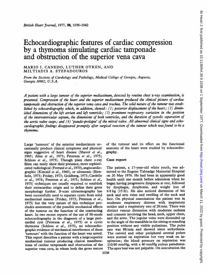
Fig. 1 Posteroanterior (left)and left lateral (right) chest filmtaken before (upper) and 11days after surgical resection ofthe thymoma (lower). Note theremaining part of the tumourin the superior mediastinum inthe right upper lung field. Alsonote posterior displacement of theheart by the tumour (upper right)with return of the heart in thenormal anterior position afterremoval of the tumour (lowerright).
heart sounds were normal; there were no murmursor added sounds. Examination of the remainder ofthe systems was noncontributory. The haemoglobinwas 15 g/dl and the haematocrit 43 per cent. Theelectrocardiogram and other laboratory findingswere normal. The chest x-ray film showed a largemass in the superior and anterior mediastinum; theheart was normal in size and configuration but dis-placed posteriorly (Fig. 1, upper).
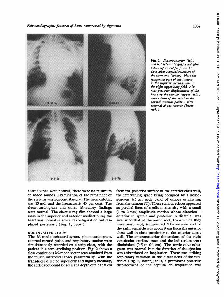
NONINVASIVE STUDYThe M-mode echocardiogram, phonocardiogram,external carotid pulse, and respiratory tracing weresimultaneously recorded on a strip chart, with thepatient in a semi-reclining position. Fig. 2 shows aslow continuous M-mode sector scan obtained fromthe fourth intercostal space parasternally. With thetransducer directed superiorly and slightly medially,the aortic root could be seen at a depth of 5-5 to 8 cm
from the posterior surface of the anterior chest wall,the intervening space being occupied by a homo-geneous 4 5 cm wide band of echoes originatingfrom the tumour (T). These tumour echoes appearedas parallel lines of medium intensity with a small(1 to 2 mm) amplitude motion whose direction-anterior in systole and posterior in diastole-wassimilar to that of the aortic root, from which theywere presumably transmitted. The anterior wall ofthe right ventricle was about 5 cm from the anteriorchest wall in close proximity to the anterior aorticwall. The anteroposterior dimensions of the rightventricular outflow tract and the left atrium werediminished (0 5 to 0*1 cm). The aortic valve echo-gram was normal but the duration of the ejectionwas abbreviated on inspiration. There was strikingrespiratory variation in the dimensions of the ven-tricles (Fig. 2, lower); thus, a prominent posteriordisplacement of the septum on inspiration was
1039
on March 11, 2022 by guest. P
rotected by copyright.http://heart.bm
j.com/
Br H
eart J: first published as 10.1136/hrt.39.9.1038 on 1 Septem
Fig. 2 Preoperativeechocardiographic scan(continuous recording). The bandof echoes from the tumour (T)are seen to occupy the spacebetween the anterior wall of theright ventricle (shown by thearrow on the lower tracing) andthe anterior chest wall(CW). Notethe progressive decrease in tumourdimension as the scan movesfromthe base of the heart towards theapex. See text for discussion.ECG, electrocardiogram, AO,aortic root; LA, left atrium;S, interventricular septum. Thetissue depth scale is constantthroughout and shown at the enSof the lower tracing. In this andall subsequent tracings therespiratory trace (R) movesupward on inspiration.
T
---------
1- Seo c4
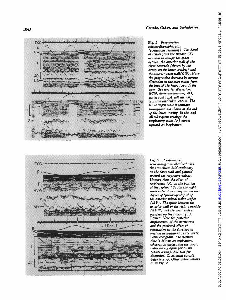
Fig. 3 Preoperativeechocardiograms obtained withthe transducer held stationaryon the chest wall and pointedtoward the respective valves.Upper: Note the effect ofrespiration (R) on the positionof the septum (S), on the rightventricular dimension, and on thedegree of 'pseudo-prolapse' ofthe anterior mitral valve leaflet(MV). The space between theanterior wall of the right ventricle(RVW) and the chest wall isoccupied by the tumour (T).Lower: Note the posteriordisplacement of the aortic rootand the profound effect ofrespiration on the duration ofejection as measured on the aortic-valve echogram. The ejectiontime is 240 ms on expiration,whereas on inspiration the aorticvalve barely opens for 80 ms(black arrow). See text fordiscussion. C, external carotidpulse tracing. Other abbreviationsas in Fig. 2.
I,-:!ll. 7.w :.: :;:,%. .::' .:,.:' :,,, .:: .:.
ECG ----
WOMPONNAMONSIOI_ * L _w-r
somma
1040
U.T -
-. .r"M=te
'IN
AO-1 NW, -
on March 11, 2022 by guest. P
rotected by copyright.http://heart.bm
j.com/
Br H
eart J: first published as 10.1136/hrt.39.9.1038 on 1 Septem
Echocardiographic features of heart compressed by thymoma
primarily responsible for an increase in right ven-tricular dimension at the expense of the left ventriclewhose end-diastolic dimension varied from about35 mm on expiration to 25 mm on inspiration. Themitral valve showed a prominent systolic posteriormovement that became maximal on expiration(Fig. 3, upper). In the lower strip of Fig. 3, theprofound effect of respiration on the volume andoutput of the left ventricle can be appreciated;thus, the ejection time, as measured on both thecarotid pulse tracing and the aortic valve echogram(Stefadouros and Witham, 1975), varied from amaximal value of 240 ms on expiration (5th beat,lower strip, Fig. 3) to a minimal value of 80 ms oninspiration, during which the aortic valve barelyopened and the carotid pulse tracing collapsed (3rdbeat, lower strip, Fig. 3). Intermediate values for theejection time were observed in other beats.
Because of increasing respiratory distress, anemergency thoractomy was carried out on the dayafter admission, and the greater part of the tumourwas excised. It was impossible to resect it entirelybecause of its extension into the superior portion ofthe anterior mediastinum laterally and posteriorlyaround the great vessels, trachea, and oesophagus.The excised mass, a poorly differentiated thymo-cytic thymoma, was a solid tumour of 988 g,containing a few areas of cystic degeneration. Afteroperation the patient was initially supported by amechanical respirator, and received a course ofirradiation to the mediastinum (total dose 4800rads), with considerable symptomatic improvement.The postoperative chest x-ray film (Fig. 1, lower)
showed the remaining part of the tumour in thesuperior mediastinum; the heart size, configuration,and position were normal. An echogram carried outon the 10th postoperative day showed the remainderof the tumour as a 1 cm band immediately behindthe anterior wall of the chest, normal dimensions ofthe cardiac cavities without respiratory variation,disappearance of the mitral valve 'pseudo-prolapse'seen before operation, and a small effusion behindthe posterior wall of the left atrium. The ejectiontime was normal and unaffected by respiration.
Discussion
The clinical syndrome of cardiac tamponade,superior vena caval obstruction, dysphagia, andrespiratory distress in this patient can be completelyaccounted for by the mechanical compression thetumour exerted on the heart, superior vena cava,oesophagus, and trachea, respectively. Though thetumour-being obvious on the chest film-was notactually 'discovered' by M-mode echocardiography,this technique was very useful in two respects:
it demonstrated the extracardiac situation and solidnature of the tumour, and it excluded possibly co-existing cardiac lesions whose presence might havebeen obscured by the dramatic clinical picture pro-duced by the tumour.Large anterior or superior mediastinal masses are
easily detectable on routine chest x-ray films buttheir differentiation from cardiomegaly or vasculartumours, and the determination of their grossanatomy usually necessitate additional radio-graphic, angiocardiographic, or ultrasonic studies.Ultrasound B-scan examination has been often,though not invariably (Cardello et al., 1974),successful in making the clinically important dis-crimination between solid and cystic mediastinalmasses (Friday, 1973; Peterson et al., 1975), andsimilar results have been obtained with A-modeultrasonography (Birnbolz, 1973; Goldberg, 1973).In at least two cases, M-mode echocardiographyhas been successful in defining the cystic nature ofa large pericardial cyst (Friday, 1973) and a largecystic thymoma (Schloss et al., 1975) and excludingany associated heart abnormality, other than aprolapsed mitral valve found in the former case(Friday, 1973).The echocardiographic appearance of the tumour
in our patient was indicative of a solid mass. Thus,at a gain level low enough to permit visualisationof the heart cavities as echo-free spaces, discretelinear echoes of medium intensity were present atany depth within the tumour (Fig. 3). Furthermore,these echoes showed a small but distinct systolicanterior motion which was concordant and syn-chronous with that of the aortic root from which itwas presumably transmitted. This echocardio-graphic pattern bears no resemblance to the echo-free space observed by M-mode echocardiographyin cases of cystic masses (Schloss et al., 1975;Peterson et al., 1975).The echogram of the heart itself was helpful in
excluding such conditions as constrictive pericarditisor pericardial effusion as a cause of the cardiactamponade. Mechanical external compression oftheheart by the tumour significantly affected theechocardiographic functional anatomy of the heart.Thus, the normal phenomenon of decreased leftventricular (and increased right ventricular) fillingon inspiration was much exaggerated in this patient,resulting in a drastic inspiratory curtailment of leftventricular stroke volume and ejection time, and aprominent pulsus paradoxus. Similar, pronouncedreciprocal respiratory variations in the echocardio-graphic dimension and hence volume of the twoventricles is a frequent finding in patients withsignificant pericardial effusion (unpublished obser-vations), in whom a posterior 'pseudo-prolapse' of
1041
on March 11, 2022 by guest. P
rotected by copyright.http://heart.bm
j.com/
Br H
eart J: first published as 10.1136/hrt.39.9.1038 on 1 Septem
the mitral valve is also occasionally seen (Levismanand Abbasi, 1976; Vignola et al., 1976). Thefunctional nature of this 'pseudo-prolapse' and itsindirect association with the compression of theheart in these cases is established by its promptdisappearance after aspiration of the pericardialeffusion (Levisman and Abbasi, 1976; Vignolaet al., 1976) or, in this patient, resection of thecompressing mediastinal tumour.Our experience from this patient together with
that of other investigators indicates that M-modeechocardiography is as useful as other noninvasiveradiographic or ultrasonic techniques in the evalua-tion of mediastinal tumours and their mechanicaleffect upon the function of the heart.
References
Allee G., Logue, B., and Mansour, K. (1973). Thymic cys Isimulating multiple cardiovascular abnormalities and pre-senting with pericarditis and pericardial tamponade.American Journal of Cardiology, 31, 377-380.
Birnholz, J. C. (1973). Sonic differentiation of cysts andhomogeneous solid masses. Radiology, 108, 699-702.
Cardello, F. P.,McQuown, D. S., and Dollinger, M. (1974).Ultrasound in diagnosis of parapericardial masses. (Letterto the Editor.) J'ournal of the American Medical Association,227, 1124.
Ferran6, J., Guermonprez, J. L., Vasile, N., and Maurice, P.(1970). Silhouette cardiaque anormale et tumeurs thy-miques. Journal de Radiologie et d'Electrologie et de MedecineNucleaire, 51, 207-216.
Friday, R. 0. (1973). Paracardiac cyst: diagnosis by ultra-sound and puncture. (Letter to the Editor.) Journal of theAmerican Medical Association, 226, 82.
Goldberg, B. B. (1973). Mediastinal ultrasonography. journalof Clinical Ultrasound, 1, 114-119.
Kincaid, 0. W., Brandenburg, R. O., and Bernatz, P. E.(1960). Experiences with angiography as a guide to medias-tinal exploration. J'ournal of the American Medical Associa-tion, 173, 613-624.
Levisman, J. A., and Abbasi, A. S. (1976). Abnormal motionof the mitral valve with pericardial effusion: pseudo-prolapse of the mitral valve. American Heart J7ournal, 91,18-20.
Peterson, D. T., Zatz, L. M., and Popp, R. L. (1975). Peri-cardial cyst ten years after acute pericarditis. Chest, 67,719-721.
Schloss, M., Kronzon, I., Gelber, P. M., Reed, G. E., andBerger, A. (1975). Cystic thymoma simulating constrictivepericarditis. The role of echocardiography in the differen-tial diagnosis. Journal of Thoracic and CardiovascularSurgery, 70, 143-146.
Shaver, V. C., Bailey, W. R., and Marrangoni, A. G. (1965).Acquired pulmonic stenosis due to external cardiac com-pression. American Journal of Cardiology, 16, 256-261.
Stefadouros, M. A., and Witham, A. C. (1975). Systolic timeintervals by echocardiography. Circulation, 51, 114-117.
Vignola, P. A., Pohost, G. M., Curfman, G. D., and Myers,G. S. (1976). Correlation of echocardiographic and clinicalfindings in patients with pericardial effusion. AmericanJournal of Cardiology, 37, 701-707.
Requests for reprints to Dr. Miltiadis A. Stefa-douros, Section of Cardiology, Department ofMedicine, Medical College of Georgia, Augusta,Georgia 30902, U.S.A.
Notice
Clinical application of intra-aortic balloonpumpA course will be held on 25-27 November 1977 atthe Konover Hotel, Miami Beach, Florida. It willcover indications for use of the intra-aortic balloonpump and other circulatory assist devices forpatients in cardiogenic shock.
Information can be obtained by writing toDivision of Continuing Medical Education, Uni-versity of Miami School of Medicine, PO Box520875, Miami, Florida 33152, U.S.A. (Tel. A/C305-547-6716).
1042
on March 11, 2022 by guest. P
rotected by copyright.http://heart.bm
j.com/
Br H
eart J: first published as 10.1136/hrt.39.9.1038 on 1 Septem