Date post: | 17-Dec-2015 |
Category: |
Documents |
Upload: | noreen-avis-shaw |
View: | 215 times |
Download: | 0 times |
Excitation-Contraction Coupling: At the heart of muscle function
Larry M. Frolich, Ph.D.March 17,2011
HOOK• Muscle is only biological cell/tissue that
can cause rapid, large-scale movement—THE evolutionary innovation that defines animals….and ourselves.
• Role of excitable membrane and filamentous muscle proteins understood as great and early breakthrough in cell/molecular biology and biochemistry
How does muscle work—outline • Motor Unit—motor neuron plus skeletal muscle
cells (review)• Action potential in neurons (reminder)• Muscle cell architecture• From arrival of an action potential to the
contraction of the muscle (excitation-contraction coupling)
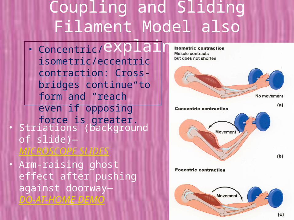
• Molecular basis of muscle movement—sliding filament model
• Whole muscles and their physiology as explained by the molecular/cellular basis of muscle function (lab activities)
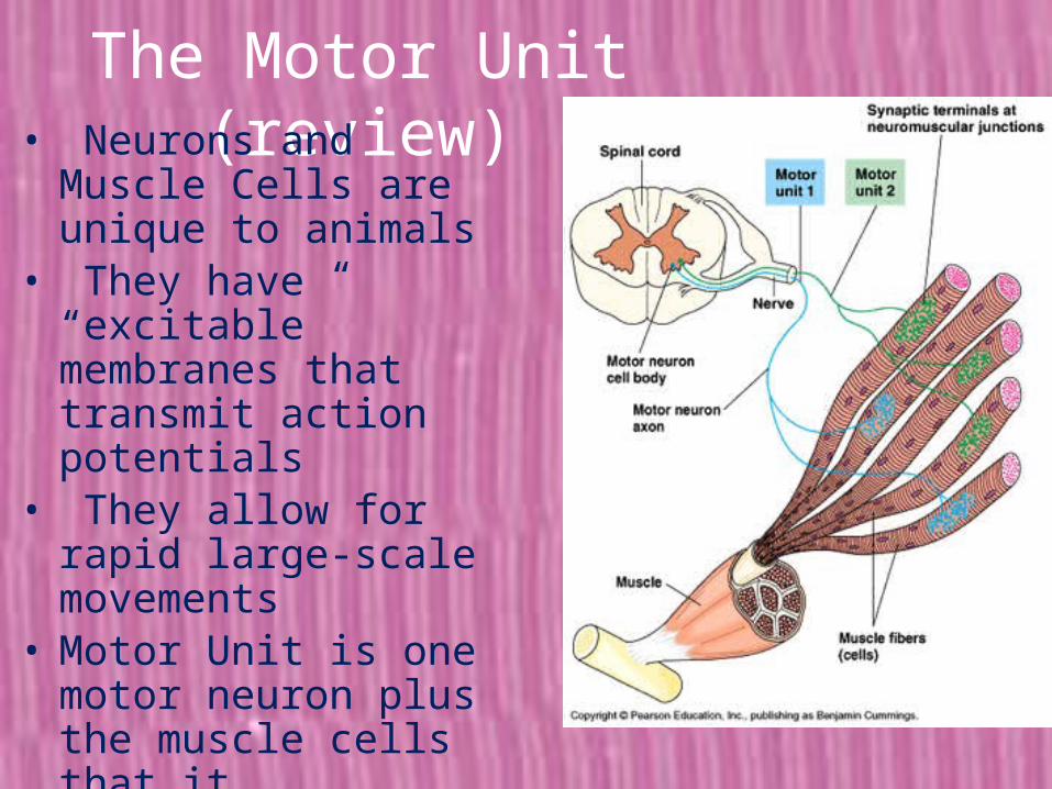
The Motor Unit (review)• Neurons and Muscle Cells
are unique to animals• They have “excitable”
membranes that transmit action potentials
• They allow for rapid large-scale movements
• Motor Unit is one motor neuron plus the muscle cells that it stimulates (or synapses with)--the minimal construct that allows for movement in our body
How do neurons carry a message—action potentials
REMINDER SLIDE
Single muscle cell or muscle “fiber” is composed of myofibrils which contain sarcomeres or contractile “units”
Myo (Latin for muscle)Sarco (Greek for flesh)
Muscle cell architecture
• Skeletal muscle fibers are BIG cells—visible to naked eye as fibers in meat, chicken, fish
• Sarcolemma is muscle cell membrane—”excitable” so has action potentials just like neurons
• Because cell is large, T-tubules carry action potential—ionic depolarization—into internal parts of cell
• Ionic depolarization in T-tubules causes sarcoplasmic reticulum to releases calcium
• Calcium triggers actin-myosin protein filaments to “slide” against each other.
Muscle cells
The Brain From Top to Bottom
From action potential to movement of muscle cell
Molecular Basis of Muscle Contraction• Actin-Myosin
“sliding filament” model
• Actin and myosin filamentous proteins are packed parallel and end-to-end in sarcomeres
• When muscle cell is “excited” (experiences action potential), Ca is released causing sarcomeres to contract
How does the actin-myosin complex (sarcomere) shorten and contract the muscle?
• Actin = thin filament “lattice-work”
• Myosin = thick filament “core”
• Ca release triggers the formation of molecular cross-bridges from myosin to actin
• Cross-bridges “row” or “reach” for more adjacent binding site on actin.
(would normally draw on board)
Details, details, details…
• Tropomyosin and troponin create binding site on actin filament
• Presence of Ca++ exposes binding site
• “Cocked” cross-bridge on myosin (uses ATP) then attaches to binding site and pulls or “rows” actin filament
• Cross-bridge linkage is broken and re-cocks to link with next binding site
Put th
e sli
ding
fila
men
ts b
ack
into
a w
hole
mus
cle…
And the result is muscle movement
Whole muscle
Excitation-Contraction Coupling and Sliding Filament Model explains:
• Why muscle has peak force at middle lengths: (ideal actin-myosin overlap for cross-bridge formation)—BUCKET DEMO
• More muscle cells active (“excited”) means more muscle force: (more cross-bridge formation)—• EMG’S• ISOLATED MUSCLE LAB
Excitation-Contraction Coupling and Sliding Filament Model also explains:• Concentric/isometric/
eccentric contraction: Cross-bridges continue to form and “reach” even if opposing force is greater.
• Striations (background of slide)—MICROSCOPE SLIDES
• Arm-raising ghost effect after pushing against doorway—DO-AT-HOME DEMO
Want more details(from 2008 Nature review)
“you'll thank me later” (for protecting from too much detail)
Evolutionary tinkering (where did this incredible system come from?:
• Actin is present in all eukaryotic cell as part of internal cell architecture
• Myosin is present as “motor protein” that hauls other structures along the actin highways
G
So, go get your actin and myosin sliding…Gracias por su atención.
More info on Frolich website: http://faculty.yc.edu/lfrolich/index.htm
Excitation-Contraction Coupling and Sliding Filament Model explains:
• Why muscle has peak force at middle lengths: (ideal actin-myosin overlap for cross-bridge formation)—BUCKET DEMO
• More muscle cells active (“excited”) means more muscle force: (more cross-bridge formation)—EMG’S, ISOLATED MUSCLE LAB
• Concentric/isometric/eccentric contraction: Cross-bridges continue to form and “reach” even if opposing force is greater.
• Striations (background of slide)—MICROSCOPE SLIDES
• Arm-raising ghost effect after pushing against doorway—DO-AT-HOME DEMO