Exosomal transfer of tumor-associatedmacrophage-derived miR-21 conferscisplatin resistance in gastric cancer cellsPeiming Zheng1†, Lei Chen2†, Xiangliang Yuan1*, Qin Luo1, Yi Liu1, Guohua Xie1, Yanhui Ma1 and Lisong Shen1*
Abstract
Background: Cisplatin-based chemotherapy is frequently used to treat advanced gastric cancer (GC). However, theresistance often occurs with the mechanisms being not well understood. Recently, emerging evidence indicatesthat tumor-associated macrophages (TAMs) play an important role in chemoresistance of cancer. As the importantmediators in intercellular communications, exosomes secreted by host cells mediate the exchange of genetic materialsand proteins to be involved in tumor aggressiveness. The aim of the study was to investigate whether exosomesderived from TAMs mediate cisplatin resistance in gastric cancer.
Methods: M2 polarized macrophages were obtained from mouse bone marrow or human PBMCs stimulated withIL-4 and IL-13. Exosomes isolated from M2 macrophages culture medium were characterized, and miRNA expressionprofiles of M2 derived exosomes (M2-exos) were analyzed using miRNA microarray. In vitro cell coculture was furtherconducted to investigate M2-exos mediated crosstalk between TAMs and tumor cells. Moreover, the in vivoexperiments were performed using a subcutaneous transplantation tumor model in athymic nude mice.
Results: In this study, we showed that M2 polarized macrophages promoted cisplatin (DDP) resistance in gastriccancer cells and exosomes derived from M2 macrophages (M2-exos) are involved in mediating the resistance to DDP.Using miRNA profiles assay, we identify significantly higher levels of microRNA-21 (miR21) isomiRNAs in exosomes andcell lysate isolated from M2 polarized macrophage. Functional studies revealed that exosomal miR-21 can be directlytransferred from macrophages to the gastric cancer cells, where it suppresses cell apoptosis and enhances activation ofPI3K/AKT signaling pathway by down-regulation of PTEN.
Conclusions: Our findings suggest that exosomal transfer of tumor-associated macrophages derived miR-21 conferDDP resistance in gastric cancer, and targeting exosome communication may be a promising new therapeutic strategyfor gastric cancer patients.
Keywords: Cisplatin resistance, Tumor-associated macrophages, Exosome, miR-21, Gastric cancer
BackgroundGastric cancer is currently the fourth most common ma-lignancy and the third leading cause of cancer-relateddeaths worldwide [1]. The incidence and mortality ofgastric cancer are the highest in East Asia (particularlyin Korea, Mongolia, Japan, and China), and it has becomethe second most lethal cancer in China [2]. The poorprognosis of this cancer resulted from late detection,
aggressive characteristics and poor response to availabletherapies. Although combined chemotherapy pre- andpost-operation has increased patient survival rates, the de-velopment of drug resistance is still the most significantobstacles to effective chemotherapy [3]. Cisplatin remainsto be a primary chemotherapeutic drug for gastric cancerpatients, especially for advanced stage ones. However, re-sistance often occurs with the mechanisms being not wellunderstood, which results in relapse of cancer and poorsurvival. The elucidation of molecular mechanisms tocisplatin resistance is important for improving gastriccancer survival.
* Correspondence: [email protected]; [email protected]†Equal contributors1Department of Clinical Laboratory, Xinhua Hospital, Shanghai Jiao TongUniversity School of Medicine, Shanghai 200092, ChinaFull list of author information is available at the end of the article
It is well known that the tumor microenvironmentcomprises a variety of nonmalignant stromal cells thatevolves with and provides support to tumor cells duringthe tumor progression [4, 5]. Among them, tumor-asso-ciated macrophages (TAMs) are the major componentsand play a pivotal role in tumor growth, angiogenesis, me-tastasis and therapy resistance [6–8]. Macrophages areheterogeneous cells that undergo different functional re-programming in response to various stimulating signals.M1- and M2-polarized macrophages, activated byIFNγ with LPS and IL-4 with IL-13 respectively, are ex-tremes of a broad range of functional states [9–11]. Inmost solid tumor, TAMs are typically a macrophage sub-population with M2 phenotype and a positive correlationbetween TAM density and poor prognosis has beenproved in several types of cancer [7, 12], including gas-tric cancer [13, 14]. Increasing evidence has shown thatTAM regulates therapeutic responses of cancer cell andimmunotherapy targeting TAM maybe an innovativecombination therapy designed to cure cancer [15, 16].Nevertheless, the detailed interaction between antican-cer therapies with TAM remains unclear.More recent studies have demonstrated that cells can
communicate with neighboring or distant cells throughthe secretion of exosomes. Exosomes are generated frommultivesicular bodies (MVBs) and are secreted into theextracellular space through fuse with the plasma mem-brane. These vesicles range in size from 50 to 100 nmcontaining proteins, lipids, mRNA, and are enrichedwith miRNA [17–19]. Several studies have shown thatmany types of cells can release exosomes and exoso-mal transmission among tumor microenvironment cellsmodulates therapeutic resistance of cancer cells [20–22]. MicroRNAs are small, noncoding RNAs that con-trol the expression of multiple target genes at the post-transcriptional level. Interestingly, exosomal miRNAsare more stable and the transfer of miRNA by exo-somes contributes to the development of chemoresis-tance in multiple tumourtypes [23, 24]. However, themiRNA signatures of TAM-derived exosomes have notbeen identified and whether these exosomal miRNAsare involved in chemoresistance in gastric cancer re-main unknown.In this study, we first construct the TAM-like M2
polarized macrophages activated by IL-4 with IL-13 andshow that macrophage-derived exosomes can be ingestedby gastric cancer cells, reducing the chemotherapy sen-sitivity to cisplatin. MicroRNA expression profiles usingmiRNA array reveals that miR-21a-5p is the most abun-dant in M2 macrophage-derived exosomes. Furtherinvestigation demonstrates that miR-21 can be directlytransferred, through exosomes, from TAM to gastriccancer cells, and regulates the chemotherapy resistanceof these cells. Our studies not only reveal a novel
communication mechanism between TAM and gastriccancer cell, but also may provide a promising newtherapeutic target for gastric cancer patients.
MethodsCell culture and treatmentThe gastric cancer cell line MFC,MGC-803 were pur-chased from the Chinese Academy of Sciences Cell Bankof Type Culture Collection. Murine bone marrow–de-rived macrophages (BMDM) were isolated and activatedas previously described [25]. Briefly, bone marrow cellsfrom femur of C57BL/6 male mice were isolated andcultured for 7 days in DMEM:F12 (Gibco, Life technolo-gies, USA) supplemented with 10% FBS (Gibco, USA)and 50 ng/ml M-CSF (R&D Systems, USA). Media waschanged every 3 days and contaminating nonadherentcells are eliminated and adherent cells are harvested forfurther stimulated. The cells were incubated for 48 hwith 20 ng/ml IL-4 plus 20 ng/ml IL-13 (PeproTech,USA) to achieve the M2 polarized macrophages. For invitro differentiation of human monocytes into macro-phages, monocytes were isolated by negative selectionfrom PBMCs using magnetic beads (MiltenyiBiotec),then isolated cells were subsequently cultured in inRPMI1640 supplemented with 10% FBS (Gibco) and100 IU/ml rhM-CSF for 7 days. The polarization of theresulting monocyte-derived macrophages was obtainedas above described. The other cells were cultured at 37 °Cwith 5% CO2 in DMEM containing 10% FBS supple-mented with 100 U/mL penicillin and 100 μg/mlstreptomycin (Gibco). For the co-culture experiment,Macrophage were grown on the 0.4um pore size trans-well insert (Corning) and the GC cells were grown inthe bottom well of the transwell chamber.
Flow cytometryThe M2 polarized macrophage were trypsinized andwashed twice in 1 × PBS, after resuspended in 100ul 1 ×PBS, fluorochrome-conjugated antibodies against F4/80,CD11b, CD206, CD86, CD163, CD68, CD80 or theirrespective isotype controls were added and stained for30 min at 4 °C. Following washed twice in 1 × PBS, la-beled cells were analyzed by flow cytometry on a FACSCanto II flow cytometer (BD Biosciences) and analyzedwith FlowJo software (Tree Star). All antibodies used forFACS are listed in (Additional file 1: Table S1).
Apoptosis assayApoptosis was measured using the FITC Annexin VApoptosis Detection Kit I (BD Pharmagen, USA) followingthe manufacturer’s protocol. In brief, cells were washedtwice with cold PBS and then resuspended in 100 μl of 1XBinding Buffer, then add 5 μl of FITC Annexin V and 5μlpropidium iodide (PI) for 15 min at room temperature
Zheng et al. Journal of Experimental & Clinical Cancer Research (2017) 36:53 Page 2 of 13
in the dark. After incubation 400 μl of 1X Binding Bufferwere added to each tube and analyzed by FACS Canto IIflow cytometry (BD Biosciences).
Exosome isolation and analysisMacrophages were incubated for 48 h in DMEM:F12medium with 10% exosome-free FBS. This conditionedmedium was collected and exosomes were isolated usingExosome Precipitation Solution (System Biosciences,USA). Identification of exosomes was processed accordingto the protocol described in Exosome Antibody Array(System Biosciences).For exosome uptake experiments, exosome prepara-
tions were labeled with PKH67 Fluorescent Cell LinkerKits (Sigma-Aldrich) according to the manufacturer’s in-structions, followed by washing through Exosome SpinColumns (MW3000) (Invitrogen, USA) to remove excessdye. Next, exosomes were incubated with gastric cancercells and examined under a SP5 confocal microscope(Leica, USA).
Transmission electron microscopy (TEM)For TEM, 10 μl of exosome suspension were absorbedonto carbon-coated cooper grids (200 mesh) for 1 min.Samples were washed with double-distilled water andnegatively stained with 2% uranyl acetate solution for1 min. After air dry, the samples were visualized at87000x in a Phillips Tecnai transmission electron micro-scope at 80 kV.
MicroRNA microarrayExosome pellets from 10 ml supernatant of M2 polarizedmacrophages were collected and homogenized in Trizol(Invitrogen). Total RNA was quantified with a NanoDrop2000c spectrophotometer (Thermo Scientific, USA) andits quality was assessed by capillary electrophoresis on anAgilent 2100 Bioanalyzer (Agilent Technologies, CA). ThemiRNA microarray analysis was performed by ShanghaiBiotechnology Corporation
Transfection of miRNA mimics and negative controlFor in vitro transfection of miRNA, Cy3-labeled miR-21mimics and negative control (GenePharma, China) weretransfected using Lipofectamine 3000 (Life Technolo-gies), according to the manufacturer’s instructions. After24 h of transfection cells were collected and used forfurther analysis
In vitro detection of miR-21 transferTo further observe the transfer of miRNA, Exosomesprepared from M2 macrophages transfected with Cy3-labelled miR-21 or without transfection (ctrl) were addedto MFC cell cultures. MFC cells were were fixed in 4%PFA, treated with 0.3% Triton X-100, blocked with 3%
BSA at 37 °C. After being washed with PBS, Cellular F-actin was visualized by staining with Alexa 488 phal-loidin (LifeTechnologies, USA) according to the manu-facturer’s guidelines. Cells were mounted with ProLong®Gold antifade Reagent with DAPI (LifeTechnologies,USA). Images were captured using Leica SP5 Laser scan-ning confocal microscope.
Cell viability and adhesion-dependent colony formationassayGastric cancer cells were seeded in 96-well plate at1500–3000 cells per well and incubated with DDP for24–72 h, cell viability was detected with the Cell count-ing Kit-8 (Dojindo Laboratories, Japan). The opticaldensity at 450 nm was measured on a multiwall platereader (FLX800, Bio-TEK). Transfected gastric cancercells were plated in 60-mm dishes at a density of 2 × 10³
cells per well for adhesion-dependent colony formationassay. DDP was added to the culture medium at a finalconcentration of 5uM. Culture medium that containedDDP was changed every 3–4 days. Then, 3–4 weekslater, the remaining colonies were fixed with 4% parafor-maldehyde and dyed with crystal violet. The colonieswere counted according to the defined colony size.
RNA extraction and quantitative real-time PCRTotal RNA was extracted using TRIzol reagent (Invitro-gen, USA) according to the manufacturer’s instructions.The concentration and quality of the total RNA wereassessed with Nanodrop Spectrophotometer (ThermoFisher Scientific, USA). For the mRNA expression ana-lysis, reverse transcription was performed using Prime-Script RT master mix (TaKaRa, Japan). For miRNAexpression analysis, total RNA was first reverse tran-scribed using Mir-X™ miRNA First-Strand Synthesis Kit(TaKaRa, Japan). Quantitative real-time PCR analysiswas performed in triplicate on 7900 HT Real-Time PCRSystem (Applied Biosystems, USA) using SYBR PremixEx Taq (TaKaRa, Japan) and the expression levels ofGAPDH or U6 was used as endogenous control. The5′primer used for miR-21 is TAGCTTATCAGACTGATGTTGA, the mRQ3′ primer and U6 primers aresupplied with the kit. Results were analyzed using the2–ΔΔct calculation method. Other primers sequences ofmentioned genes are described in (Additional file 2:Table S2).
Western blotThe cells were lysed in equal volumes of ice cold lysisbuffer and a protease inhibitor cocktail. Cell lysate wereseparated by SDS-PAGE and then transferred to a 0.2-μm PVDF membrane (Bio- Rad, USA). After blockingwith Odyssey Blocking Buffer (Li-COR Biosciences,USA), the membrane was incubated with primary
Zheng et al. Journal of Experimental & Clinical Cancer Research (2017) 36:53 Page 3 of 13
antibody (1:1000) at 4 °C overnight, followed by incuba-tion with IRDye 800CW or 680 secondary antibodies(1:5000, LI-COR Biosciences, USA). GAPDH was usedas endogenous control. The Odyssey Infrared ImagingSystem was used to visualize targeted protein bands. Allantibodies used for western blot are listed in (Additionalfile 1: Table S1).
In vivo xenograft and treatment experimentsFor in vivo studies, 4–6 week old male athymicC57 nudemice were purchased from Shanghai Laboratory AnimalCenter of China. MFC cells (3× 10⁵ cells in 200ul PBS)pretreated with or without M2-Exos were subcutane-ously injected into the nude mice to establish tumors.Another group received subcutaneous injections of thesame MFC cells transfected with miR-21 or miR-NC.After 10 days, 10 mg/kg DDP or PBSwas injected intra-peritoneally. M2-Exos or miR-21 were injected twiceintratumorally before the start of DDP treatment. Themice were examined every 2 days and sacrificed 6–7days after DDP treatment. The tumor sizes were mea-sured using digital caliper and tumor volume was cal-culated with the following formula: volume = 0.5 ×width2 × length. All animal procedures were carried outwith the approval of the Institutional Committee ofShanghai Jiao Tong University School of Medicine forAnimal Research.
Statistical analysisStatistical significance between groups was determinedby a two-tailed Student’s t-test and a one-way ANOVAtest. Differences were considered to be significant whenP < 0.05. All statistical data were displayed as means ±standard deviation (SD) and analyzed for statisticalsignificance with GraphPad Prism 5.0 for Windows(GraphPad Software, USA).
ResultsM2 polarized macrophages induces the resistance ofgastric cancer cells to cisplatinPrevious study and our unpublished data indicatedthat TAMs infiltrated in gastric cancer are primarily M2macrophages. To determine the functional biology of po-larized macrophages, we generated M2-polarized macro-phages in vitro from mouse bone marrow cells (MBMCs)cultured in the presence of M-CSF with IL-4/IL-13. TheM2 polarized macrophages were identified through flowcytometry analysis for expression of surface antigens, in-cluding CD11b, F4/80, CD206, and CD86 (Fig. 1a). Inaddition, specific gene expression was evaluated usingquantitative RT-PCR (Fig. 1b). The expression of proto-typical M2 markers (CD206, Arg1, IL-10, TGF-β) wasincreased, indicating that we successfully derived M2 mac-rophages from MBMC.A similar observation regarding
the markers was made in human M2-polarized macro-phages from human peripheral blood monocytes (PBMCs)(Additional file 3: Figure S1a).In order to determine the optimal dosage levels of
cisplatin (DDP), the gastric cancer cell lines MFC andMGC-803 were treated with various concentrations ofDDP for 24, 48, and 72 h. CCK8 assay showed that thecell viability was gradually decline along with prolongedtime and increasing concentration (Fig. 1c and Additionalfile 3: Figure S1b). Based on this observation, the dosesused in the drug resistance test were as follows: MFC(15uM, 48 h), MGC-803 (7uM, 48 h). To determinewhether M2 polarized macrophages confers chemoresis-tance in gastric cancer cells, we co-cultured gastric cancercells with M2 polarized macrophages in a transwell insert,which prevents direct cell-cell contact. Then the gastriccancer cells were treated with DDP for 48 h, and the re-sults showed a significant decrease of apoptosis rate incells co-cultured with M2 polarized macrophages com-pared with those co-cultured with unactivated macro-phages or normal control cells (Fig. 1d and Additional file3: Figure S1c). Taken together, these data indicated thatM2 polarized macrophages prompted the development ofresistance to DDP in gastric cancer cells.
Exosomes derived from M2 polarized macrophages canbe ingested by gastric cancer cellsPrevious studies have demonstrated that exosomes mayregulate therapy resistance through transfer of functionalsmall RNAs or proteins. To confirm whether M2 polar-ized macrophages-derived exosomes (M2-exos) play apivotal role in the development of resistance of gastriccancer, we first isolated exosomes from the conditionedmedium of M2 macrophages. The purified exosomesdisplayed as small round vesicles with a diameter ran-ging from 80 to120 nm, expressed the exosomalmarkersCD9, CD63 and CD81 (Fig. 2a, b, c).To examine whether M2-exos can be taken up by gas-
tric cancer cell, MFC cells were incubated with PKH67-labeled exosomes that were isolated from conditionedmedium of M2 macrophages. Examination using confocalmicroscopy confirmed the uptake of PKH67-labeled exo-somes by MFC cells (Fig. 2d), as evidenced by localizationof green fluorescence and DAPI.
M2-exos induce the resistance of gastric cancer cells toDDP in vitro and in vivoTo further investigate the functional roles of M2-exos inthe resistance of gastric cancer cells to DDP, MFC cellswere incubated with conditioned medium (CM) or exo-somes purified from M2 macrophages for 48 h. Thenthe cells were treated with 15uM DDP for another 48 h.CCK8 assay showed that co-cultured with M2-exos cansignificantly reduce the chemotherapy sensitivity of MFC
Zheng et al. Journal of Experimental & Clinical Cancer Research (2017) 36:53 Page 4 of 13
cells and improve their relative survival rate (Fig. 3a). Inaddition, MFC cells co-treatment with M2-exos had alower rate of apoptosis compared with normal treatedcells (Fig. 3b). A similar phenomenon of chemoresistancewas observed in human GC cell MGC-803 (Additionalfile 4: Figure S2a and S2b), confirming a common func-tion of M2-exos in human and mouse models.
To demonstrate the role of M2-exos in the develop-ment of chemoresistance in MFC cells in vivo, MFCcells pretreated with M2-exos were injected subcutane-ously into male nude mice, followed by an optimal doseof DDP treatment. MFC cells alone were injected as anegative control. We found that M2-exos significantlyinhibited the chemotherapeutic effect of DDP, while
Fig. 1 Co-cultivation with M2 polarized macrophages enhances the resistance of MFC cells to cisplatin. a FCM identification of M2 polarizedmacrophages derived from murine bone marrow stimulated with IL-4 and IL-13, CD206 was a specific marker for M2. (Un-Mac, unactivatedmacrophages; M2, macrophages activated by IL-4 and IL-13). b qRT-PCR detection of iNOS, Arg1, IL-10, TGF-β mRNA expression in unactivatedand M2 polarized macrophages, GAPDH was assayed as an internal control. (ns p > 0.05, *** p < 0.001). c Cell viability assay of MFC treated withvarious concentrations of DDP for 24 h, 48 h, and 72 h. d Flow cytometric analyses of apoptotic cells. MFC cells were cultured alone or co-culturedwith unactivated (Un-Mac) or M2 polarized macrophages, and then exposed to DDP for 48 h. The quantitative data are presented as the mean ± SDof triplicate experiments. (ns p > 0.05, *** p < 0.001)
Zheng et al. Journal of Experimental & Clinical Cancer Research (2017) 36:53 Page 5 of 13
M2-exos had minimal effect on the tumor growth(Fig. 3c). The final tumor size in M2-exos group was1104 ± 92.2 mm3 after DDP treatment, which was sig-nificantly larger than that in DDP alone group (687 ±78.3 mm3) (Fig. 3d). In addition, the mean tumorweight in M2-exos group was about 1.5 times heavierthan that in DDP alone group (Fig. 3e). Taken together,these results suggested that M2-exos could induce theresistance of gastric cancer cells to DDP both in vitroand in vivo.
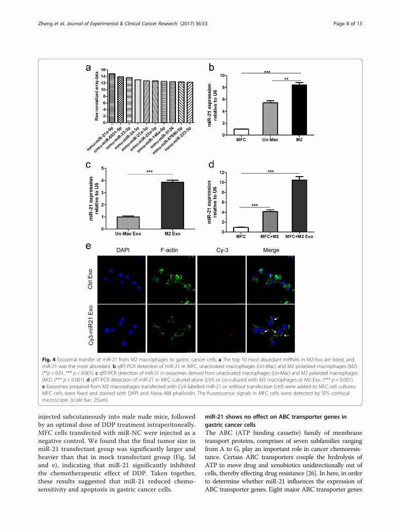
Functional miR-21 is transferred from macrophages togastric cancer cells through M2-exosEmerging evidence suggests that miRNAs which in-volved in cell-cell communication are frequently encap-sulated in exosomes, and implement their biologicalfunctions in the recipient cells. To explore the mecha-nisms by which M2-exos conferred chemoresistance ingastric cancer, we generated miRNA profiles of M2-exosby miRNA microarray analysis. Among the miRNAs thatwere identified in M2-exos, miR-21 was the most abundant(Fig. 4a). Additionally, previous studies have demonstratedthat miR-21 was involved in cancer chemoresistance;therefore, miR-21 was chose for further research. Using
qRT-PCR, we confirmed that the miR-21 expressionwas much higher in macrophages as compared to MFCcells, expecially in M2 polarized macrophages (Fig. 4b).In addition, M2-exos contained higher levels of miR-21than those from unactivated macrophages (Fig. 4c). Re-markably, the intracellular miR-21 levels in MFC cellswas obviously increased after co-cultured with M2 po-larized macrophages or M2-exos (Fig. 4d).To further determine whether the elevated miR-21
level in MFC cells was resulted from directly shuttledfrom M2 macrophages to gastric cancer cells via exo-somes, but not the tumor cells themselves. M2 polarizedmacrophages were transiently transfected with eitherCy3-labeled miR-21 or negative control. We thenisolated exosomes from conditioned media collectedfrom the above macrophages. The purified exosomeswere then added to gastric cancer cells culture medium.Using confocal microscopy, Cy3 labeled signals weredetected in the gastric cancer cells incubated with exo-somes isolated from Cy3-labeled miR-21 transfectedcells but not in those incubated with exosomes frommock-transfected cells (Fig. 4e). In summary, these datasuggest that M2 macrophage-derived exosomes mediatemiR-21 shuttling.
Fig. 2 Exosomes derived from M2 polarized macrophages can be internalized by gastric cancer cells. a Representative transmission electronmicroscopy image of M2-derived exosomes (scale bar, 100 nm). b Histogram showing the particle diameter (nm) of the purified exosomes.c Exosomal markers (CD63, CD9, CD81, HSP70) were analyzed in M2 cellular protein and corresponding exosomes using western blotting(GAPDH was used as an internal reference). d The uptake of the PKH67 labelled M2-Exos was evident in MFCcells after 12 h of incubation.No stain was observed in the negative control condition. (scale bar, 10 um)
Zheng et al. Journal of Experimental & Clinical Cancer Research (2017) 36:53 Page 6 of 13
miR-21 reduces chemosensitivity and apoptosis ingastric cancer cellsTo determine whether miR-21 can induce the che-moresistance in gastric cancer cells, we first examinedthe effects of miR-21 on gastric cancer cell chemosen-sitivity by directly transfecting miR-21 mimics ornegative control into MFC or MGC-803 cells, andthen treated them with DDP. The expression level ofmiR-21 in the transfected cells was increased mark-edly than did the mock transfectants (Additional file5: Figure S3a). Cell viability was determined by usingCCK-8 assay kit. Indeed, the cells transfected with themiR-21 mimics showed significantly lower chemosen-sitivity as compared to mock transfected cells (Fig. 5a
and Additional file 5: Figure S3b). We further deter-mined chemotherapy-induced apoptosis in gastriccancer cells transfected with miR-21 mimics or miR-NC. The apoptotic rate was analyzed by using FITCAnnexin V apoptosis detection kit. A lower rate ofapoptosis was observed in gastric cancer cells trans-fected with miR-21 mimics as compared to mocktransfected cells (Fig. 5c and Additional file 5: FigureS3c). In addition, cell colony formation assay alsodemonstrated that miR-21 conferred chemoresistancein MFC cells (Fig. 5b).To further investigate the functional roles of miR-21
in the chemoresistance of gastric cancer cells in vivo.MFC cells transfected with miR-21 mimics were
Fig. 3 M2-exos induce the resistance of MFC cells to DDP in vitro and in vivo. a Cell viability was assessed by CCK-8 assays. M2-derived conditionedmedium (CM) or exosomes (M2-Exo) attenuated DDP-induced cell suppression. MFC cells were pretreated with M2-derived CM or exosomes, and thenexposed to DDP for 48 h. b Flow cytometric analyses of apoptotic cells. MFC cells were exposed to DDP alone (ctrl) or DDP and M2-Exo for 48 h. Thequantitative data are presented as the mean ± SD of triplicate experiments. (*** p < 0.001). c Tumor growth curves in nude mice inoculated with MFCcells that were pretreated with or without M2-Exo.DDP or PBS (ctrl) treatment was initiated 10 days after subcutaneous injections of tumor cells.(n = 5, * p < 0.05). d The size of tumors at the end of the experiment from mice treated with PBS (ctrl), DDP, M2-Exo + DDP. (n = 5). e The meanweight of tumors from mice shown in (d). (n = 5, ** p < 0.01)
Zheng et al. Journal of Experimental & Clinical Cancer Research (2017) 36:53 Page 7 of 13
injected subcutaneously into male nude mice, followedby an optimal dose of DDP treatment intraperitoneally.MFC cells transfected with miR-NC were injected as anegative control. We found that the final tumor size inmiR-21 transfectant group was significantly larger andheavier than that in mock transfectant group (Fig. 5dand e), indicating that miR-21 significantly inhibitedthe chemotherapeutic effect of DDP. Taken together,these results suggested that miR-21 reduced chemo-sensitivity and apoptosis in gastric cancer cells.
miR-21 shows no effect on ABC transporter genes ingastric cancer cellsThe ABC (ATP binding cassette) family of membranetransport proteins, comprises of seven subfamilies rangingfrom A to G, play an important role in cancer chemoresis-tance. Certain ABC transporters couple the hydrolysis ofATP to move drug and xenobiotics unidirectionally out ofcells, thereby effecting drug resistance [26]. In here, in orderto determine whether miR-21 influences the expression ofABC transporter genes. Eight major ABC transporter genes
Fig. 4 Exosomal transfer of miR-21 from M2 macrophages to gastric cancer cells. a The top 10 most abundant miRNAs in M2-Exo are listed, andmiR-21 was the most abundant. b qRT-PCR detection of miR-21 in MFC, unactivated macrophages (Un-Mac) and M2 polarized macrophages (M2).(**p < 0.01, *** p < 0.001). c qRT-PCR detection of miR-21 in exosomes derived from unactivated macrophages (Un-Mac) and M2 polarized macrophages(M2). (*** p < 0.001). d qRT-PCR detection of miR-21 in MFC cultured alone (ctrl) or co-cultured with M2 macrophages or M2-Exo. (*** p < 0.001).e Exosomes prepared from M2 macrophages transfected with Cy3-labelled miR-21 or without transfection (ctrl) were added to MFC cell cultures.MFC cells were fixed and stained with DAPI and Alexa 488 phalloidin. The fluorescence signals in MFC cells were detected by SP5 confocalmicroscope. (scale bar, 25um)
Zheng et al. Journal of Experimental & Clinical Cancer Research (2017) 36:53 Page 8 of 13
were selected for real-time PCR detection, includingABCB1, ABCC1, ABCG2 and so on. However, the resultsshowed no significant changes between MFC cells trans-fected with miR-21 and the mock transfectants (Additionalfile 6: Figure S4), suggesting that miR-21 overexpression hasno effect on ABC transporter genes in gastric cancer cells.
Exosomal miR-21 regulates the PTEN/PI3K/AKT signalingpathway and enhances the anti-apoptotic ability ofgastric cancer cellsPrevious studies have showed that miR-21 could regulatetumor biological behavior through the PTEN/PI3K/AKT
signaling pathway in several types of cancer, includinggastric cancer. However, most of previous studies fo-cused on the miR-21 expression level of cancer cellsthemselves. In our model, whether the exosomal transferof functional miR-21 also has the same effect yet needfurther verification. So we examined the PI3K/AKTpathway related proteins in both M2-exos treated andmiR-21 overexpression MFC cells. We found that thepresence of M2-exos decreased the PTEN mRNA andprotein expression level and increased AKT phosphoryl-ation (Fig. 6a and b). Similar changes of MFC cells wereobserved after transfected with miR-21 (Fig. 6c and d).
Fig. 5 miR-21 induces the resistance of MFC cells to DDP in vitro and in vivo. a Cell viability assay of MFC cells treated with DDP after transfectedwith miR-21 mimics or miR-NC or without transfection (ctrl). b Colony formation assay of MFC cells transfected with miR-21 mimics or miR-NC orwithout transfection (up), and exposed to DDP (below). c Cell apoptosis assay of MFC cells treated with DDP after transfected with miR-21 mimicsor miR-NC or without transfection (ctrl). The quantitative data are presented as the mean ± SD of triplicate experiments. (ns p > 0.05,*** p < 0.001).d and e The size and mean weight of tumors at the end of the experiment from mice inoculated with MFC cells transfected with miR-21 mimicsor miR-NC. DDP or PBS (ctrl) treatment was initiated 10 days after subcutaneous injections of tumor cells. (n = 5, ns p > 0.05, ** p < 0.01)
Zheng et al. Journal of Experimental & Clinical Cancer Research (2017) 36:53 Page 9 of 13
These data suggested that exosomal transfer of miR-21enhances gastric cancer cells chemoresistance partiallythrough the regulation of PTEN/PI3K/AKT signalingpathway.In addition, there is accumulating evidence that re-
sistant to apoptosis is responsible for drug resistance.Previous reports have revealed that Bcl-2, which wasone of very important anti-apoptosis genes, was regu-lated by miR-21 directly [27]. So we next determinedwhether miR-21 affected Bcl-2 expression. Western blot-ting analyses indicated that M2-exos treatment andmiR-21 overexpression dramatically increased the Bcl-2protein level in MFC cells, which might be anothermechanism responsible for the enhanced anti-apoptosisability (Fig. 6b and d).
DiscussionCisplatin-based chemotherapy is now the most com-monly used chemotherapeutic criterion in gastric can-cer. Unfortunately, advanced GC patients who developresistance to cisplatin have limited therapeutic optionsin the clinic at present [3]. Besides genetic changes oftumor cells themselves causing increased drug efflux orenhanced anti-apoptosis, drug resistance can resultfrom the tumor microenvironment protecting tumorcells against treatment [28]. In the present study, we
showed that exosomes derived from M2 macrophagesexpress higher levels of miR-21 compared to unactivatedmacrophages, and that the exosomal transfer of miR-21from M2 macrophages to gastric cancer cells could con-fer DDP resistance in these cells. The exosome-shuttledmiR-21 promoted DDP resistance through the down-regulation of PTEN, leading to a more active signalingthrough the PI3K/AKT pathway. In addition, M2-exosprotected gastric cancer cells from chemotherapy in-duced apoptosis through the regulation of anti-apoptosisprotein Bcl-2.The role of exosomes in cancer as mediators of cell-
cell communication within the microenvironment hasgained increasing attention. Several studies have de-scribed the intriguing roles of exosomes in cancerprogression through transfer a variety of proteins,DNA, and RNA [29]. Importantly, exosomes have beenproved to be critically involved in the development ofchemoresistance. Sousa et al. have demonstrated thatexosomes from drug-resistant cancer cells can transferthe resistant phenotype to drug-sensitive cells, mainlythrough transferring of drug-efflux pumps and miR-NAs [30]. Qu L et al. have also suggested that lncRNAsembedded in exosomes derived from sunitinib-resistantcells could confer the resistant phenotype to recipientRCC cells [31].
Fig. 6 Exosomal transfer of miR-21 enhances gastric cancer cells chemoresistance via the PTEN/PI3K/AKT signaling pathway. a qRT-PCR detectionof PTEN in MFC cells treated with or without M2-Exo. (*** p < 0.001). b Western blot assays for the expression of PI3Kp85, PTEN, p-AKT, AKT, BCL-2in MFC cells treated with or without M2-Exo.GAPDH was used as the loading control. c qRT-PCR detection of PTEN in MFC cells transfected withmiR-21 mimics or miR-NC or without transfection (ctrl). d Western blot assays for the expression of PI3Kp85, PTEN, p-AKT, AKT, BCL-2 in MFC cellstransfected with miR-21 mimics or miR-NC or without transfection (ctrl). GAPDH was used as the loading control
Zheng et al. Journal of Experimental & Clinical Cancer Research (2017) 36:53 Page 10 of 13
Apart from tumor cells, exosomes from tumor stromalcells also contribute to the acquisition of a resistantphenotype in cancer cells. Boelens et al. demonstratedthat stromal cells orchestrate an intricate crosstalk withbreast cancer cells by utilizing exosomes to regulatetherapy resistance pathways [20]. Exosomes from cancer-associated fibroblast have been shown to promote prolif-eration and drug resistance of pancreatic cancer cellsthrough transfer of chemoresistance-inducing factorsnail [32]. A recent study reported that exosomal miR-21 can confer chemoresistance and an aggressive pheno-type in ovarian cancer cells through its transfer fromcancer-associated adipocytes and fibroblasts [24]. Simi-lar to the above researches, here we demonstrated thatexosomes from M2 polarized macrophages was suffi-cient to confer DDP resistance in gastric cancer cellsboth in vivo and in vitro; suggesting that tumor associ-ated macrophages may support gastric cancer aggres-siveness by secreting exosomes besides physicalcontact in the tumor microenvironment.In recent years, it has become increasingly clear that
miRNAs play an important role in the resistance of gas-tric cancer cells to chemotherapeutics [33, 34]. Import-antly, exosome-mediated transfer of miRNAs withinthe tumor microenvironment has also been indicated asa significant mechanism for dissemination of drug re-sistance [23]. In this study, using miRNA microarray inM2 macrophage-derived exosomes, we found that miR-21 was the most abundant miRNA among the identifiedmiRNAs. Furthermore, significantly higher miR-21 ex-pression levels were detected in both M2 polarizedmacrophages and M2-exos than in unactivated macro-phages by a qRT-PCR analysis. Next, we incubatedgastric cancer cells with purified exosomes from M2macrophages transfected with Cy3-labelled miR-21, andthe transfer of miR-21 was confirmed by the detectionof fluorescent signal in the gastric cancer cells usingconfocal microscopy.Additionally, we explored the possible mechanism by
which miR-21 may promote the DDP resistance ingastric cancer cells. Our data showed that miR-21 over-expression had no effect on ABC transporter genes ingastric cancer cells, one of the main mechanisms forchemoresistance. Previous studies have revealed thatmiR-21 expression was associated with resistance to avariety of chemotherapeutic agents in both solid andhaematologic tumors. miR-21 promotes cell survival andconfers resistance to cancer cells by regulating a set oftumor suppressor genes and apoptosis-associated genes[24, 35, 36]. Among these genes, PTEN/PI3K/AKT sig-naling pathway, which involved in cancer pathology andchemoresistance, has been shown to be managed bymiR-21 in several types of cancer [37, 38]. Consistentwith these findings, we demonstrated here that exosomal
transfer of miR-21 led to down-regulation of PTEN andincreased activation of AKT, resulting in more survivaland less apoptosis in gastric cancer cells when treatedwith DDP. In addition, one important apoptosis-associated gene Bcl-2 was also elevated along with miR-21 overexpression, though the exact mechanism wasnot documented in our data. Moreover, the other po-tential miR-21 target genes may also be responsible forthe effects of M2-exos on gastric cancer chemoresis-tance, which need further in-depth study.
ConclusionsIn summary, we demonstrate that M2 polarized mac-rophages confer DDP resistance in gastric cancer cellsthrough exosomal transfer of functional miR-21. Ourfindings suggest that TAM derived exosomes are animportant mediator for the reciprocity between TAMand gastric cancer cells, Moreover, we provide evi-dence to show that targeting exosomal miR-21 fromTAM maybe a promising adjuvant therapeutic strategyfor gastric cancer patients, especially cisplatin-resistant GC patients.
Additional files
Additional file 1: Table S1. Antibody list for Western blot and Flowcytometry. (DOC 44 kb)
Additional file 2: Table S2. List of primers used for candidate genes.(DOC 37 kb)
Additional file 3: Figure S1. Co-cultivation with M2 polarizedmacrophages enhances the resistance of MGC-803 cells to cisplatin.aFACS analyses of CD68, CD80, CD163, and CD206 in Un-Mac or M2-polarized macrophages from human peripheral blood monocytes. bCell viability assay of MGC-803 treated with various concentrations ofDDP for 24 h, 48 h, and 72 h. c Flow cytometric analyses of apoptoticcells. MGC-803 cells were cultured alone or co-cultured with unactivated(Un-Mac) or M2 polarized macrophages, and then exposed to DDP for48 h. The quantitative data are presented as the mean ± SD of triplicateexperiments. (ns p > 0.05, ***p < 0.001). (TIF 2027 kb)
Additional file 4: Figure S2. M2-exos induce the resistance of MGC-803cells to DDP in vitro.a Cell viability was assessed by CCK-8 assays. M2-derivedconditioned medium (CM) or exosomes (M2-Exo) attenuated DDP-inducedcell suppression. MGC-803 cells were pretreated with M2-derived CM orexosomes, and then exposed to DDP for 48 h. b Flow cytometric analysesof apoptotic cells. MGC-803 cells were exposed to DDP alone or DDP andM2-Exo for 48 h. The quantitative data are presented as the mean ± SD oftriplicate experiments. (***p < 0.001). (TIF 739 kb)
Additional file 5: Figure S3. miR-21 enhances chemoresistance anddecreases apoptosis in MGC-803 cells.aqRT-PCR detection of miR-21 inMFC and MGC-803 transfected with miR-21 mimics or miR-NC or withouttransfection (ctrl). (*** p < 0.001).b Cell viability assay of MGC-803 cellstreated with DDP after transfected with miR-21 mimics or miR-NC orwithout transfection (ctrl). (nsp > 0.05,*** p < 0.001). c Cell apoptosisassay of MGC-803 cells treated with DDP after transfected with miR-21mimics or miR-NC or without transfection (ctrl). The quantitative dataare presented as the mean ± SD of triplicate experiments. (nsp > 0.05,***p < 0.001). (TIF 1386 kb)
Additional file 6: Figure S4. qRT-PCR detection of ABCB1, ABCC1,ABCC2, ABCC3, ABCC4, ABCC5, ABCC6, ABCG2 mRNA expression in MFCcells transfected with miR-21 mimics or miR-NC or without transfection.GAPDH was used as an internal control. (TIF 660 kb)
Zheng et al. Journal of Experimental & Clinical Cancer Research (2017) 36:53 Page 11 of 13
AcknowledgementsWe thank our colleagues in the department of laboratory medicine forhelpful discussions and valuable assistance.
FundingThis study was supported in part by the National Natural Science Foundationof China (No. 81672363, 81372641, and 81472244).
Availability of data and materialsAll data generated or analysed during this study are included in thispublished article [and its additional files].
Authors’ contributionsLS and XY supervised the whole project, designed the experiments, analyzeddata. PZ performed the experiments, analyzed data, wrote the manuscriptand prepared the figures. LC contributed experiments and analyzed data.QL, YL, GX, and YM contributed experiments and provided the technicalsupport. All authors read and approved the final manuscript.
Competing interestsThe authors declare that they have no competing interests.
Consent for publicationNot applicable.
Ethics approvalThe study was approved by the ethics committee of the Xinhua Hospital.All animal procedures were performed according to national guidelinesandapproved by the Institutional Committee of Shanghai Jiao TongUniversity School of Medicine for Animal Research.
Publisher’s NoteSpringer Nature remains neutral with regard to jurisdictional claims inpublished maps and institutional affiliations.
Author details1Department of Clinical Laboratory, Xinhua Hospital, Shanghai Jiao TongUniversity School of Medicine, Shanghai 200092, China. 2Department ofGeneral Surgery, Xinhua Hospital, Shanghai Jiao Tong University School ofMedicine, Shanghai 200092, China.
Received: 24 January 2017 Accepted: 5 April 2017
References1. Torre LA, Bray F, Siegel RL, Ferlay J, Lortet-Tieulent J, Jemal A. Global cancer
statistics, 2012. CA Cancer J Clin. 2015;65:87–108.2. Chen W, Zheng R, Baade PD, Zhang S, Zeng H, Bray F, Jemal A, Yu XQ, He J.
Cancer statistics in China, 2015. CA Cancer J Clin. 2016;66:115–32.3. Orditura M, Galizia G, Sforza V, Gambardella V, Fabozzi A, Laterza MM,
Andreozzi F, Ventriglia J, Savastano B, Mabilia A, et al. Treatment ofgastric cancer. World J Gastroenterol. 2014;20:1635–49.
4. Gajewski TF, Schreiber H, Fu YX. Innate and adaptive immune cells in thetumor microenvironment. Nat Immunol. 2013;14:1014–22.
9. Sica A, Mantovani A. Macrophage plasticity and polarization: in vivo veritas.J Clin Invest. 2012;122:787–95.
10. Mills CD. M1 and M2 macrophages: oracles of health and disease. Crit RevImmunol. 2012;32:463–88.
11. Murray Peter J, Allen Judith E, Biswas Subhra K, Fisher Edward A, GilroyDerek W, Goerdt S, Gordon S, Hamilton John A, Ivashkiv Lionel B,Lawrence T, et al. Macrophage activation and polarization: nomenclatureand experimental guidelines. Immunity. 2014;41:14–20.
12. Chen J, Yao Y, Gong C, Yu F, Su S, Chen J, Liu B, Deng H, Wang F, Lin L, etal. CCL18 from tumor-associated macrophages promotes breast cancermetastasis via PITPNM3. Cancer Cell. 2011;19:541–55.
13. Cardoso AP, Pinto ML, Pinto AT, Oliveira MI, Pinto MT, Goncalves R, Relvas JB,Figueiredo C, Seruca R, Mantovani A, et al. Macrophages stimulate gastricand colorectal cancer invasion through EGFR Y(1086), c-Src, Erk1/2 and Aktphosphorylation and smallGTPase activity. Oncogene. 2014;33:2123–33.
14. Wu MH, Lee WJ, Hua KT, Kuo ML, Lin MT. Macrophage infiltration inducesgastric cancer invasiveness by activating the beta-catenin pathway. PLoS One.2015;10:e0134122.
15. Ruffell B, Coussens LM. Macrophages and therapeutic resistance in cancer.Cancer Cell. 2015;27:462–72.
16. Mantovani A, Allavena P. The interaction of anticancer therapies withtumor-associated macrophages. J Exp Med. 2015;212:435–45.
17. Thery C. Exosomes: secreted vesicles and intercellular communications.F1000 Biol Rep. 2011;3:15.
18. Tkach M, Thery C. Communication by extracellular vesicles: where we areand where we need to go. Cell. 2016;164:1226–32.
19. Penfornis P, Vallabhaneni KC, Whitt J, Pochampally R. Extracellular vesiclesas carriers of microRNA, proteins and lipids in tumor microenvironment.Int J Cancer. 2016;138:14–21.
20. Boelens Mirjam C, Wu Tony J, Nabet Barzin Y, Xu B, Qiu Y, Yoon T, AzzamDiana J, Twyman-Saint Victor C, Wiemann Brianne Z, Ishwaran H, et al.Exosome transfer from stromal to breast cancer cells regulates therapyresistance pathways. Cell. 2014;159:499–513.
21. Qu Z, Wu J, Wu J, Luo D, Jiang C, Ding Y. Exosomes derived from HCCcells induce sorafenib resistance in hepatocellular carcinoma both invivo and in vitro. J Exp Clin Cancer Res. 2016;35:159.
22. Wang X, Xu C, Hua Y, Sun L, Cheng K, Jia Z, Han Y, Dong J, Cui Y, Yang Z.Exosomes play an important role in the process of psoralen reverse multidrugresistance of breast cancer. J Exp Clin Cancer Res. 2016;35:186.
23. Challagundla KB, Wise PM, Neviani P, Chava H, Murtadha M, Xu T, Kennedy R,Ivan C, Zhang X, Vannini I, et al. Exosome-mediated transfer of microRNAswithin the tumor microenvironment and neuroblastoma resistance tochemotherapy. J Natl Cancer Inst. 2015;107:djv135.
24. Au Yeung CL, Co N-N, Tsuruga T, Yeung T-L, Kwan S-Y, Leung CS, Li Y, Lu ES,Kwan K, Wong K-K, et al. Exosomal transfer of stroma-derived miR21 conferspaclitaxel resistance in ovarian cancer cells through targeting APAF1.Nat Commun. 2016;7:11150.
25. Gordon S. Alternative activation of macrophages. Nat Rev Immunol. 2003;3:23–35.26. Szakács G, Annereau J-P, Lababidi S, Shankavaram U, Arciello A, Bussey KJ,
Reinhold W, Guo Y, Kruh GD, Reimers M, et al. Predicting drug sensitivityand resistance. Cancer Cell. 2004;6:129–37.
27. Dong J, Zhao Y-P, Zhou L, Zhang T-P, Chen G. Bcl-2 Upregulation induced bymiR-21 via a direct interaction is associated with apoptosis and chemoresistancein MIA PaCa-2 pancreatic cancer cells. Arch Med Res. 2011;42:8–14.
29. ELA S, Mager I, Breakefield XO, Wood MJ. Extracellular vesicles: biology andemerging therapeutic opportunities. Nat Rev Drug Discov. 2013;12:347–57.
30. Sousa D, Lima RT, Vasconcelos MH. Intercellular transfer of cancer drugresistance traits by extracellular vesicles. Trends Mol Med. 2015;21:595–608.
31. Qu L, Ding J, Chen C, Wu Z-J, Liu B, Gao Y, Chen W, Liu F, Sun W, Li X-F, et al.Exosome-transmitted lncARSR promotes sunitinib resistance in renal cancerby acting as a competing endogenous RNA. Cancer Cell. 2016;29:653–68.
32. Richards KE, Zeleniak AE, Fishel ML, Wu J, Littlepage LE, Hill R. Cancer-associated fibroblast exosomes regulate survival and proliferation ofpancreatic cancer cells. Oncogene. 2017;36:1770–78.
33. Dehghanzadeh R, Jadidi-Niaragh F, Gharibi T, Yousefi M. MicroRNA-induceddrug resistance in gastric cancer. Biomed Pharmacother. 2015;74:191–9.
34. Riquelme I, Letelier P, Riffo-Campos A, Brebi P, Roa J. Emerging role ofmiRNAs in the drug resistance of gastric cancer. Int J Mol Sci. 2016;17:424.
35. Giovannetti E, Erozenci A, Smit J, Danesi R, Peters GJ. Molecular mechanismsunderlying the role of microRNAs (miRNAs) in anticancer drug resistance andimplications for clinical practice. Crit Rev Oncol Hematol. 2012;81:103–22.
Zheng et al. Journal of Experimental & Clinical Cancer Research (2017) 36:53 Page 12 of 13
36. Shi GH, Ye DW, Yao XD, Zhang SL, Dai B, Zhang HL, Shen YJ, Zhu Y, Zhu YP,Xiao WJ, Ma CG. Involvement of microRNA-21 in mediating chemo-resistanceto docetaxel in androgen-independent prostate cancer PC3 cells. ActaPharmacol Sin. 2010;31:867–73.
37. Xiong B. MiR-21 regulates biological behavior through the PTEN/PI-3 K/Aktsignaling pathway in human colorectal cancer cells. Int J Oncol. 2013;42:219–28.
38. Yang S-m, Huang C, Li X-f, Yu M-z, He Y, Li J. miR-21 confers cisplatin resistancein gastric cancer cells by regulating PTEN. Toxicology. 2013;306:162–8.
• We accept pre-submission inquiries
• Our selector tool helps you to find the most relevant journal
• We provide round the clock customer support
• Convenient online submission
• Thorough peer review
• Inclusion in PubMed and all major indexing services
• Maximum visibility for your research
Submit your manuscript atwww.biomedcentral.com/submit
Submit your next manuscript to BioMed Central and we will help you at every step:
Zheng et al. Journal of Experimental & Clinical Cancer Research (2017) 36:53 Page 13 of 13


![Differential Regulation of Neutrophil and Macrophage Influx ...diabetes.diabetesjournals.org/content/diabetes/52/11/2821.full.pdf · nt 541–814 (for tumor necrosis factor [TNF]-](https://static.documents.pub/doc/80x56/5c8dc6ce09d3f20b4a8b9df9/differential-regulation-of-neutrophil-and-macrophage-inux-nt-541814.jpg)


