+ All Categories
Home > Documents > Expression Analysis of Macrodactyly Identifies ...

Expression Analysis of Macrodactyly Identifies ...

Date post: 12-Nov-2021
Category:
Upload: others
View: 2 times
Download: 0 times
Share this document with a friend
11
Expression Analysis of Macrodactyly Identifies Pleiotrophin Upregulation The Harvard community has made this article openly available. Please share how this access benefits you. Your story matters Citation Lau, Frank H., Fang Xia, Adam Kaplan, Felecia Cerrato, Arin K. Greene, Amir Taghinia, Chad A. Cowan, and Brian I. Labow. 2012. Expression analysis of macrodactyly identifies pleiotrophin upregulation. PLoS ONE 7(7): e40423. Published Version doi:10.1371/journal.pone.0040423 Citable link http://nrs.harvard.edu/urn-3:HUL.InstRepos:10464948 Terms of Use This article was downloaded from Harvard University’s DASH repository, and is made available under the terms and conditions applicable to Other Posted Material, as set forth at http:// nrs.harvard.edu/urn-3:HUL.InstRepos:dash.current.terms-of- use#LAA
Transcript

Expression Analysis of MacrodactylyIdentifies Pleiotrophin Upregulation

The Harvard community has made thisarticle openly available. Please share howthis access benefits you. Your story matters

Citation Lau, Frank H., Fang Xia, Adam Kaplan, Felecia Cerrato, ArinK. Greene, Amir Taghinia, Chad A. Cowan, and Brian I. Labow.2012. Expression analysis of macrodactyly identifies pleiotrophinupregulation. PLoS ONE 7(7): e40423.

Published Version doi:10.1371/journal.pone.0040423

Citable link http://nrs.harvard.edu/urn-3:HUL.InstRepos:10464948

Terms of Use This article was downloaded from Harvard University’s DASHrepository, and is made available under the terms and conditionsapplicable to Other Posted Material, as set forth at http://nrs.harvard.edu/urn-3:HUL.InstRepos:dash.current.terms-of-use#LAA

Expression Analysis of Macrodactyly IdentifiesPleiotrophin UpregulationFrank H. Lau1,2, Fang Xia1, Adam Kaplan1, Felecia Cerrato2, Arin K. Greene2, Amir Taghinia2,

Chad A. Cowan1, Brian I. Labow2*

1 Center for Regenerative Medicine and Cardiovascular Research Center, Massachusetts General Hospital, Boston, Massachusetts, United States of America, 2 Department

of Plastic and Oral Surgery, Children’s Hospital Boston, Boston, Massachusetts, United States of America

Abstract

Macrodactyly is a rare family of congenital disorders characterized by the diffuse enlargement of 1 or more digits. Multipletissue types within the affected digits are involved, but skeletal patterning and gross morphological features are preserved.Not all tissues are equally involved and there is marked heterogeneity with respect to clinical phenotype. The molecularmechanisms responsible for these growth disturbances offer unique insight into normal limb growth and development, ingeneral. To date, no genes or loci have been implicated in the development of macrodactyly. In this study, we performedthe first transcriptional profiling of macrodactyly tissue. We found that pleiotrophin (PTN) was significantly overexpressedacross all our macrodactyly samples. The mitogenic functions of PTN correlate closely with the clinical characteristics ofmacrodactyly. PTN thus represents a promising target for further investigation into the etiology of overgrowth phenotypes.

Citation: Lau FH, Xia F, Kaplan A, Cerrato F, Greene AK, et al. (2012) Expression Analysis of Macrodactyly Identifies Pleiotrophin Upregulation. PLoS ONE 7(7):e40423. doi:10.1371/journal.pone.0040423

Editor: Samuel J. Lin, Harvard Medical School, United States of America

Received February 15, 2012; Accepted June 5, 2012; Published July 27, 2012

Copyright: � 2012 Lau et al. This is an open-access article distributed under the terms of the Creative Commons Attribution License, which permits unrestricteduse, distribution, and reproduction in any medium, provided the original author and source are credited.

Funding: These authors have no support or funding to report.

Competing Interests: The authors have declared that no competing interests exist.

* E-mail: [email protected]

Introduction

Macrodactyly is characterized by the diffuse enlargement of 1 or

more digits [1–3] (Fig. 1). It is a rare, congenital disease with no

familial inheritance pattern, and usually presents as an isolated,

non-syndromic condition. While all tissue elements are involved,

including fat, skin, nerve, and bone, histopathologic analysis of

macrodactyly tissue is usually remarkable for excess mature

adipose tissue [3].

We currently possess no modern molecular insight into

macrodactyly. No genes or loci have been implicated in the

development of this disease. Neither transcriptional nor protein-

level characterization of affected tissues is available. There are no

cellular or animal models of macrodactyly.

In this study, we performed the first transcriptional profiling of

macrodactyly tissue. We found that a developmental cytokine,

pleiotrophin (PTN), was significantly overexpressed across all our

macrodactyly samples. As a mitogen that has been implicated in

nerve, bone, vascular, and tumor growth, the functions of PTN

correlate closely with the clinical characteristics of macrodactyly.

PTN thus represents a promising target for further investigation.

Materials and Methods

This Project was reviewed and approved by the Children’s

Hospital Boston Institutional Review Board, Assurance #07-11-

0428 and by the Brigham & Women’s Hospital Institutional

Review Board, Assurance # FWA00000484. Pediatric patients

who underwent elective surgical debulkings of isolated, nonsyn-

dromic macrodactyly were eligible for this study. Additionally,

normal abdominal subcutaneous adipose tissue (SAT) samples

were obtained from patients undergoing elective surgeries at either

Massachusetts General Hospital or Children’s Hospital Boston.

During the review of this Project, the IRB specifically considered

(i) the risks and anticipated benefits, if any, to subjects; (ii) the

selection of subjects; (iii) the procedures for securing and

documenting informed consent; (iv) the safety of subjects; and (v)

the privacy of subjects and confidentiality of the data. Written

informed consent was obtained from guardians on the behalf of all

minors/children participants.

Approximately 5 mg of adipose tissue were harvested from each

patient and sectioned into smaller pieces. Samples were either

snap frozen in liquid nitrogen and stored at 280C or fixed in

either fresh 4% paraformaldehyde or 10% neutral buffered

formalin (Sigma Aldrich) for 4 hours, transferred to phosphate

buffered saline and stored in a 4 C refrigerator.

For each patient, RNA was extracted in biological triplicate via

submersion in 1 ml of Trizol, mechanical lysis using an RNase free

pestle (Kimble Chase Kontes), addition of 200 ml chloroform, and

centrifugation (10,000 g, 15 minutes, 4 C). The supernatant was

extracted and purified with RNeasy Mini Columns (Qiagen).

1.2 mg RNA was synthesized into complementary DNA (cDNA)

with the Superscript III First Strand Synthesis Kit (Invitrogen).

All RNA samples were analyzed for quality using an Agilent

2100 Bioanalyzer. Hybridization to Affymetrix Human Genome

U133 Plus 2.0 Arrays and subsequent array processing was done

by Asuragen. All data is MIAME compliant and the raw data has

been deposited in the Gene Expression Omnibus database

(accession number GSE35820). Adult SAT datasets were found

using the Expression Omnibus (GEO) [4] and ArrayExpress [5]

databases by searching for the terms ‘‘adipose’’ and ‘‘fat’’, and

PLoS ONE | www.plosone.org 1 July 2012 | Volume 7 | Issue 7 | e40423

restricting the platform to Affymetrix Human Genome U133 Plus

2.0 Arrays.

Raw expression values were analyzed with the Partek Genomics

Suite (Partek). Data were normalized using Robust Multiarray

Averaging (RMA) with median scaling, quintile normalization,

and background correction. The resulting datasets were prepro-

cessed to remove probesets whose minimum fold change

(maximum gene expression value divided by the minimum value)

was ,2, or whose difference between maximum and minimum

values was less than 100. Prinicipal component analysis was

performed, and linear models were used to identify statistically

significant, differentially expressed probe sets. Hierarchical clus-

tering was performed across all samples using the differentially

expressed probesets.

Figure 1. Clinical photos from patients undergoing surgical treatment of macrodactyly. (A) 15 month-old boy with macrodactylyinvolving the thumb, index and middle fingers. As is often seen, there is associated syndactyly between the index and middle fingers and deviation ofall affected digits. (B) The same patient during separation and first stage debulking of the digits. The yellow loops are around the digital nerves, whichare enlarged. A large volume of overgrown fat and soft-tissue is being removed. (C) 8 year-old girl with macrodactyly isolated to the middle finger.The excess fat and soft-tissue has been removed revealing enlarged digital nerves tagged with yellow loops.doi:10.1371/journal.pone.0040423.g001

Expression Analysis Macrodactyly

PLoS ONE | www.plosone.org 2 July 2012 | Volume 7 | Issue 7 | e40423

GEA was performed using FuncAssociate 2.0 (http://llama.

mshri.on.ca/funcassociate/), which uses a Fisher’s exact test to

assess enrichment and a resampling approach to correct for

multiple hypotheses. For each of the sample populations, a false

discovery rate (FDR) of 0.01 was set as the threshold. The

differentially expressed probesets were uploaded into FuncAssoci-

ate 2.0 as ordered lists. Analysis was performed using the

hgnc_symbol namespace, with 1000 permutations for p-value

estimation and a p-value cutoff of 0.05.

For qPCR, expression levels of PTN were normalized to the

housekeeping gene hypoxanthine guanine phosphoribosyl trans-

ferase (HPRT) and measured via Quantifast SYBR Green PCR

Kit (Qiagen). To minimize the potential impact of any genomic

DNA contamination, primers were designed and verified to span

multiple exons. Three technical replicates were performed for

each sample. Error bars were computed by adding and subtracting

1 unit standard deviation of the delta Ct values from calibrated

delta Ct values. P-values were calculated in Microsoft Excel 2007

using 2-tailed heteroscedastic Student’s t-tests.

For immunohistochemical staining, tissues samples fixed in

freshly prepared 4% paraformaldehyde were dehydrated using an

ethanol gradient, sectioned (5-micron thickness), and mounted on

glass slides (Fisher). The sections were rehydrated and antigen

retrieval was performed using citric acid buffer (pH 6.0). The

sections were blocked using 5% donkey serum (Jackson Labs) with

0.01% Triton, and incubated with goat polyclonal anti-pleiotro-

phin antibody (1:100 dilution, Abcam ab10849). Bound anti-PTN

antibody was visualized using Alexa FluorH 546 donkey anti-goat

IgG (H+L) secondary antibody 1:700 (Invitrogen A-11056).

Images were captured using a Nikon Eclipse Ti-s.

DNA was isolated from our samples using a DNeasy Blood &

Tissue Kit (Qiagen). The isolates were analyzed for quality using

an Agilent 2100 Bioanalyzer. Sequencing of the promoter and

coding regions of PTEN was performed by Polymorphic DNA

Technologies.

Results

Between June 2009 and April 2011, 4 pediatric patients

(Patients 1 through 4) who underwent elective surgical debulkings

of isolated, nonsyndromic macrodactyly participated in the study.

Normal abdominal SAT from 2 adult patients (Patients 5 & 6) who

underwent elective surgeries at Massachusetts General Hospital

between June 2009 and March 2010 was obtained. Normal

abdominal SAT was also obtained from 1 pediatric patient

(Patient 7) who underwent an elective abdominal procedure at

Children’s Hospital Boston.

RNA was isolated from the 4 macrodactyly adipose tissue

samples and hybridized to Affymetrix U133A Plus 2.0 micro-

arrays. We sought to compare our samples against publicly

available data sets; since macrodactyly tissues are usually identified

as ‘‘mature adipose tissue’’ on histopathologic examination, we

chose subcutaneous adipose tissue (SAT) datasets as reference data

sets. No suitable data was available in the Gene Expression

Omnibus (GEO) [4] and ArrayExpress [5] databases for pediatric

samples, thus we performed our analysis against 4 adult SAT

datasets comprising 345 patients. Datasets GSE135063 [6],

GSE171704 [7], GSE157735 [8] were pooled and referred to as

the ‘‘GSM’’ pool. Dataset E-TABM-325 [8] was referred to as the

‘‘MolPAGE’’ pool.

To analyze these data, we performed principal component

analysis (Fig. 2A). The top 2 vectors accounted for 23.8% and

8.5% of inter-sample variation. This analysis demonstrated clear

separation between macrodactyly and SAT samples. We next

developed transcriptional profiles of macrodactyly and SAT. We

restricted our analysis to probe sets with false-discovery rates

(FDR) ,0.01, fold-changes $1.5, and p-values ,0.05. With these

stringent restrictions, we identified 3093 overexpressed and 4202

underexpressed genes. The large number of differentially ex-

pressed genes highlights the stark differences between macro-

dactyly and normal adipose tissue. The 10 features with the

highest fold-changes are shown in Table 1. When hierarchical

clustering was performed using these 7295 differentially expressed

genes, the macrodactyly samples clustered distinctly and distantly

from all SAT samples (Fig. 2B).

To better characterize the differentially expressed transcripts,

we performed unbiased gene enrichment analysis (GEA) using

FuncAssociate 2.0. [9] The 2 tissue types were each enriched for

different gene ontology categories (Table 2). In macrodactyly,

there was an enrichment of growth factor response (Table S1),

extracellular matrix (Table S2), and patterning (Table S3) genes.

In contrast, SAT was enriched for classic adipose tissue gene

categories such as regulation of fatty acid oxidation and response

to insulin (Table 2).

PTN was the most highly overexpressed gene in macrodactyly

(34.4-fold overexpression, p-value = 0.00). The developmental

cytokine pleiotrophin was present in both the extracellular space

and pattern binding GO annotation categories (Tables S2 & S3).

To confirm these findings, we performed quantitative real time

polymerase chain reaction (qPCR) and found that in macro-

dactyly, PTN averaged 127.6-fold overexpression (p = 0.049,

Fig. 3A).

In vivo, PTN is secreted into the extracellular space where it

binds to heparin. To demonstrate that transcriptional upregulation

of PTN resulted into protein overproduction, we analyzed sections

from adipose tissue from a patient with macrodactyly (Patient 4)

and pediatric SAT (Patient 6) tissue for PTN immunoreactivity.

We found no PTN staining in pediatric SAT (Fig. 4A). In contrast,

macrodactyly sections were marked by widespread punctate

expression of PTN (Fig. 4B). This assay did not allow the

localization of PTN expression to a specific cell type. We

attempted to quantify PTN overexpression by Western blot

[10,11] but the small volume of our samples made this technically

infeasible. Li et al. reported that deletion of phosphatase and

tensin homologue (PTEN) was associated with PTN upregulation

[12]. We thus isolated DNA from our macrodactyly samples and

sequenced the PTEN promoter and coding regions. No mutations

were identified (data not shown).

Discussion

Macrodactyly remains a difficult clinical problem, one with an

unknown etiology. Modern treatment strategies are strictly surgical

and involve either growth-limiting or digit reducing procedures,

or, in severe cases, finger amputation [2,3,13]. Prior to this study,

no molecular information was available regarding this disease.

Given the absence of molecular data, we elected to first generate

transcriptional profiles of macrodactyly tissue. Analysis of this data

identified 7295 differentially expressed genes in macrodactyly

compared to adult SAT. The candidate genes overexpressed in

macrodactyly include well-characterized mitogens such as bone-

morphogenetic proteins 5 & 7, transforming growth factor beta 3,

and Wnt signaling pathway members (WNT2, WNT5A) (Tables

S1 & S2). However, the mitogen with the highest fold-change

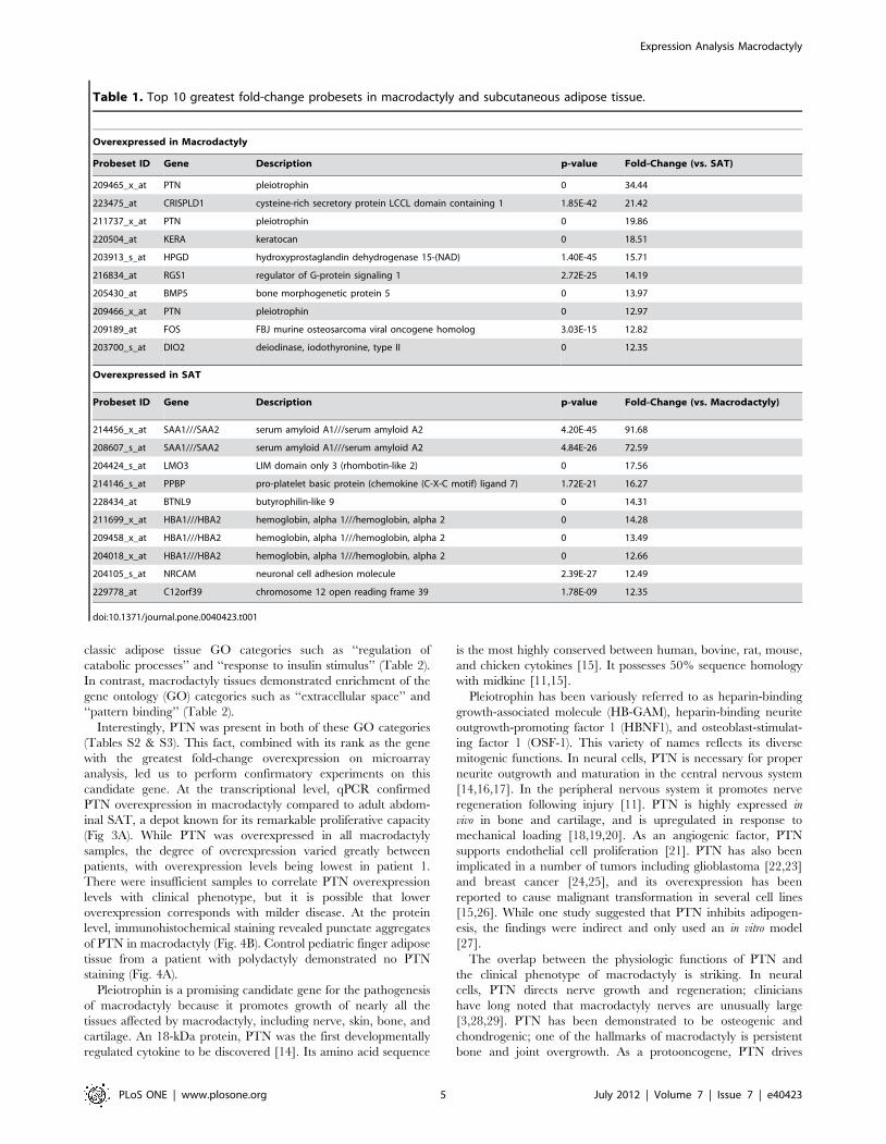
overexpression was pleiotrophin (Table 1).

To characterize our samples at the biological pathway level, we

performed gene enrichment analysis. This again underscored the

differences between SAT and macrodactyly: SAT was enriched for

Expression Analysis Macrodactyly

PLoS ONE | www.plosone.org 3 July 2012 | Volume 7 | Issue 7 | e40423

Figure 2. Results of principal component analysis (PCA) and hierarchical clustering of gene expression date from macrodactylysamples. (A) PCA of 4 macrodactyly samples (in triplicate) vs. 345 subcutaneous adipose tissue (SAT) samples. The top 2 vectors account 32.3% ofintersample variation. Macrodactyly samples cluster distinctly from SAT. (B) Hierarchical clustering of macrodactyly and SAT samples acrossdifferentially expressed genes with fold-change .1.5, p-value ,0.05, and false discovery rate ,0.05. Macrodactyly samples cluster distinctly anddistantly from all SAT samples.doi:10.1371/journal.pone.0040423.g002

Expression Analysis Macrodactyly

PLoS ONE | www.plosone.org 4 July 2012 | Volume 7 | Issue 7 | e40423

classic adipose tissue GO categories such as ‘‘regulation of

catabolic processes’’ and ‘‘response to insulin stimulus’’ (Table 2).

In contrast, macrodactyly tissues demonstrated enrichment of the

gene ontology (GO) categories such as ‘‘extracellular space’’ and

‘‘pattern binding’’ (Table 2).

Interestingly, PTN was present in both of these GO categories

(Tables S2 & S3). This fact, combined with its rank as the gene

with the greatest fold-change overexpression on microarray

analysis, led us to perform confirmatory experiments on this

candidate gene. At the transcriptional level, qPCR confirmed

PTN overexpression in macrodactyly compared to adult abdom-

inal SAT, a depot known for its remarkable proliferative capacity

(Fig 3A). While PTN was overexpressed in all macrodactyly

samples, the degree of overexpression varied greatly between

patients, with overexpression levels being lowest in patient 1.

There were insufficient samples to correlate PTN overexpression

levels with clinical phenotype, but it is possible that lower

overexpression corresponds with milder disease. At the protein

level, immunohistochemical staining revealed punctate aggregates

of PTN in macrodactyly (Fig. 4B). Control pediatric finger adipose

tissue from a patient with polydactyly demonstrated no PTN

staining (Fig. 4A).

Pleiotrophin is a promising candidate gene for the pathogenesis

of macrodactyly because it promotes growth of nearly all the

tissues affected by macrodactyly, including nerve, skin, bone, and

cartilage. An 18-kDa protein, PTN was the first developmentally

regulated cytokine to be discovered [14]. Its amino acid sequence

is the most highly conserved between human, bovine, rat, mouse,

and chicken cytokines [15]. It possesses 50% sequence homology

with midkine [11,15].

Pleiotrophin has been variously referred to as heparin-binding

growth-associated molecule (HB-GAM), heparin-binding neurite

outgrowth-promoting factor 1 (HBNF1), and osteoblast-stimulat-

ing factor 1 (OSF-1). This variety of names reflects its diverse

mitogenic functions. In neural cells, PTN is necessary for proper

neurite outgrowth and maturation in the central nervous system

[14,16,17]. In the peripheral nervous system it promotes nerve

regeneration following injury [11]. PTN is highly expressed in

vivo in bone and cartilage, and is upregulated in response to

mechanical loading [18,19,20]. As an angiogenic factor, PTN

supports endothelial cell proliferation [21]. PTN has also been

implicated in a number of tumors including glioblastoma [22,23]

and breast cancer [24,25], and its overexpression has been

reported to cause malignant transformation in several cell lines

[15,26]. While one study suggested that PTN inhibits adipogen-

esis, the findings were indirect and only used an in vitro model

[27].

The overlap between the physiologic functions of PTN and

the clinical phenotype of macrodactyly is striking. In neural

cells, PTN directs nerve growth and regeneration; clinicians

have long noted that macrodactyly nerves are unusually large

[3,28,29]. PTN has been demonstrated to be osteogenic and

chondrogenic; one of the hallmarks of macrodactyly is persistent

bone and joint overgrowth. As a protooncogene, PTN drives

Table 1. Top 10 greatest fold-change probesets in macrodactyly and subcutaneous adipose tissue.

Overexpressed in Macrodactyly

Probeset ID Gene Description p-value Fold-Change (vs. SAT)

209465_x_at PTN pleiotrophin 0 34.44

223475_at CRISPLD1 cysteine-rich secretory protein LCCL domain containing 1 1.85E-42 21.42

211737_x_at PTN pleiotrophin 0 19.86

220504_at KERA keratocan 0 18.51

203913_s_at HPGD hydroxyprostaglandin dehydrogenase 15-(NAD) 1.40E-45 15.71

216834_at RGS1 regulator of G-protein signaling 1 2.72E-25 14.19

205430_at BMP5 bone morphogenetic protein 5 0 13.97

209466_x_at PTN pleiotrophin 0 12.97

209189_at FOS FBJ murine osteosarcoma viral oncogene homolog 3.03E-15 12.82

203700_s_at DIO2 deiodinase, iodothyronine, type II 0 12.35

Overexpressed in SAT

Probeset ID Gene Description p-value Fold-Change (vs. Macrodactyly)

214456_x_at SAA1///SAA2 serum amyloid A1///serum amyloid A2 4.20E-45 91.68

208607_s_at SAA1///SAA2 serum amyloid A1///serum amyloid A2 4.84E-26 72.59

204424_s_at LMO3 LIM domain only 3 (rhombotin-like 2) 0 17.56

214146_s_at PPBP pro-platelet basic protein (chemokine (C-X-C motif) ligand 7) 1.72E-21 16.27

228434_at BTNL9 butyrophilin-like 9 0 14.31

211699_x_at HBA1///HBA2 hemoglobin, alpha 1///hemoglobin, alpha 2 0 14.28

209458_x_at HBA1///HBA2 hemoglobin, alpha 1///hemoglobin, alpha 2 0 13.49

204018_x_at HBA1///HBA2 hemoglobin, alpha 1///hemoglobin, alpha 2 0 12.66

204105_s_at NRCAM neuronal cell adhesion molecule 2.39E-27 12.49

229778_at C12orf39 chromosome 12 open reading frame 39 1.78E-09 12.35

doi:10.1371/journal.pone.0040423.t001

Expression Analysis Macrodactyly

PLoS ONE | www.plosone.org 5 July 2012 | Volume 7 | Issue 7 | e40423

fibroblast, endothelial cell, and epithelial cell growth; all of these

soft tissue elements are overgrown in macrodactyly. In many

patients, macrodactylous overgrowth is proportionally patterned

among all tissues of the affected region. The means by which

this specific, patterned overgrowth occurs is not known.

Table 2. Enriched gene ontology categories in macrodactyly and subcutaneous adipose tissue.

# of genes P-adjusted GO ID GO Category

Upregulated in Macrodactyly

6 0.001 GO:0071363 cellular response to growth factor stimulus

6 0.039 GO:0070848 response to growth factor stimulus

10 0.048 GO:0030199 collagen fibril organization

18 0.011 GO:0001501 skeletal system development

19 0 GO:0005578 proteinaceous extracellular matrix

22 0 GO:0031012 extracellular matrix

30 0 GO:0005539 glycosaminoglycan binding

34 0 GO:0001871 pattern binding

34 0 GO:0030247 polysaccharide binding

41 0 GO:0005615 extracellular space

41 0 GO:0009611 response to wounding

42 0.01 GO:0008284 positive regulation of cell proliferation

47 0 GO:0048731 system development

48 0.008 GO:2000026 regulation of multicellular organismal development

49 0.023 GO:0005102 receptor binding

51 0 GO:0044421 extracellular region part

52 0.023 GO:0009653 anatomical structure morphogenesis

59 0 GO:0007166 cell surface receptor linked signaling pathway

77 0 GO:0005576 extracellular region

Upregulated in Subcutaneous Adipose Tissue

3 0.002 GO:0015671 oxygen transport

3 0.008 GO:0005833 hemoglobin complex

3 0.012 GO:0015669 gas transport

3 0.012 GO:0005344 oxygen transporter activity

5 0 GO:0004556 alpha-amylase activity

5 0 GO:0016160 amylase activity

6 0.044 GO:0003823 antigen binding

12 0.013 GO:0003995 acyl-CoA dehydrogenase activity

17 0 GO:0046320 regulation of fatty acid oxidation

24 0.023 GO:0032869 cellular response to insulin stimulus

36 0.004 GO:0032868 response to insulin stimulus

43 0 GO:0001525 angiogenesis

55 0.013 GO:0051056 regulation of small GTPase mediated signal transduction

57 0.04 GO:0035467 negative regulation of signaling pathway

60 0.049 GO:0006732 coenzyme metabolic process

71 0.01 GO:0051270 regulation of cellular component movement

87 0.004 GO:0009894 regulation of catabolic process

96 0 GO:0005083 small GTPase regulator activity

123 0.004 GO:0030695 GTPase regulator activity

124 0.007 GO:0060589 nucleoside-triphosphatase regulator activity

197 0.015 GO:0046907 intracellular transport

204 0.012 GO:0019899 enzyme binding

222 0.04 GO:0035466 regulation of signaling pathway

234 0 GO:0044248 cellular catabolic process

doi:10.1371/journal.pone.0040423.t002

Expression Analysis Macrodactyly

PLoS ONE | www.plosone.org 6 July 2012 | Volume 7 | Issue 7 | e40423

Figure 3. Confirmation of pleiotrophin (PTN) expression in macrodactyly and the known PTN signaling cascade. (A) Relativeexpression of PTN in macrodactyly vs. adult subcutaneous adipose tissue as determined by quantitative real time polymerase chain reaction. Inmacrodactyly, PTN averaged 127.6-fold overexpression (p = 0.049). (B) The PTN signaling cascade and crosstalk with Wnt signalling (from Deuel etal.)14.doi:10.1371/journal.pone.0040423.g003

Expression Analysis Macrodactyly

PLoS ONE | www.plosone.org 7 July 2012 | Volume 7 | Issue 7 | e40423

Figure 4. Pleiotrophin (PTN) immunostaining. DAPI = nuclear stain, PTN = antibody staining for pleiotrophin, Merge = composite imagemerging Brightfield, DAPI, and PTN channels. A) No PTN is seen in pediatric subcutaneous adipose tissue from Patient 7. B) Large PTN aggregates areseen in macrodactyly patient 4.doi:10.1371/journal.pone.0040423.g004

Expression Analysis Macrodactyly

PLoS ONE | www.plosone.org 8 July 2012 | Volume 7 | Issue 7 | e40423

One mechanism whereby PTN overexpression might result in

macrodactyly was recently suggested by the discovery of an

activating AKT1 mutation in Proteus syndrome [30]. This

mutation leads to constitutive phosphorylation of residues

Ser473 and Thr308 in AKT1. PTN, on the other hand, has been

shown to rapidly phosphorylate Ser473 of AKT1 in a dose-

dependent manner [31]. This link is particularly intriguing

because partial gigantism of the hands and/or feet is a hallmark

of Proteus syndrome [32]. Unfortunately, phosphorylation of this

specific residue is also consistent with rapid proliferation and thus,

the use of this biomarker would not be conclusive as to mechanism

of overgrowth. For this reason, testing for phosphorylation of

residue Ser473 in AKT1 was not performed.

Little is currently known about the regulation of PTN. While it

has been reported that PTEN deletion is associated with PTN

upregulation, this is indirect and the direct regulation of PTN

stemming from PTEN deletion is unknown [12]. Our microarray

data did not demonstrate reduction of PTEN levels in macro-

dactyly (data not shown) and sequencing of the PTEN locus in

macrodactyly samples yielded no mutations.

This study was limited by the unavailability of normal pediatric

finger adipose tissue. Only under rare circumstances would it be

ethical to remove a significant amount of tissue from a child’s hand

for research purposes. Some pediatric hand conditions, such as

polydactyly, are managed by finger amputation and therefore are

potential sources of pediatric finger adipose tissue. However, these

conditions can be caused by germline genetic mutations and

therefore are not strictly normal [33]. Because of these limitations,

we believe future research will require in vitro and animal models of

macrodactyly. The identification of PTN as the first macrodactyly

candidate gene points the way towards the development of critical

research tools.

Supporting Information

Table S1 Genes present in the ‘‘Response to GrowthFactor Stimulus (GO:0070848)’’ gene ontology category.

(DOCX)

Table S2 Genes present in the ‘‘Extracellular Space(GO:0005615)’’ gene ontology category.

(DOCX)

Table S3 Genes present in the ‘‘Pattern Binding(GO:0001871)’’ gene ontology category.

(DOCX)

Author Contributions

Conceived and designed the experiments: FHL AK FC AKG AT CAC

BIL. Performed the experiments: FHL FX AK FC BIL. Analyzed the data:

FHL AK FC AKG AT CAC BIL. Contributed reagents/materials/

analysis tools: FHL AKG AT CAC BIL. Wrote the paper: FHL FC AKG

AT CAC BIL.

References

1. Ben-Bassat M, Casper J, Kaplan I, Laron Z (1966) Congenital macrodactyly. A

case report with a three-year follow-up. J Bone Joint Surg Br 48: 359–364.

2. McCombe D, Kay SP (n.d.) Macrodactyly. Green’s Operative Hand Surgery,

5th ed.

3. Upton J (2006) Failure of Differentiation and Overgrowth. Plastic Surgery.

Elsevier Inc., Vol. 8. 277–322.

4. Barrett T, Troup DB, Wilhite SE, Ledoux P, Evangelista C, et al. (2010) NCBI

GEO: archive for functional genomics data sets–10 years on. Nucleic Acids

Research 39: D1005–D1010. doi:10.1093/nar/gkq1184.

5. Parkinson H, Kapushesky M, Kolesnikov N, Rustici G, Shojatalab M, et al.

(2009) ArrayExpress update–from an archive of functional genomics experi-

ments to the atlas of gene expression. Nucleic Acids Res 37: D868–872.

doi:10.1093/nar/gkn889.

6. Plaisier CL, Kyttala M, Weissglas-Volkov D, Sinsheimer JS, Huertas-Vazquez

A, et al. (2009) Galanin preproprotein is associated with elevated plasma

triglycerides. Arterioscler Thromb Vasc Biol 29: 147–152. doi:10.1161/

ATVBAHA.108.178533.

7. Plaisier CL, Horvath S, Huertas-Vazquez A, Cruz-Bautista I, Herrera MF, et al.

(2009) A systems genetics approach implicates USF1, FADS3, and other causal

candidate genes for familial combined hyperlipidemia. PLoS Genet 5: e1000642.

doi:10.1371/journal.pgen.1000642.

8. Gene expression omnibus website. Expression data from human adipose tissue

(n.d. ) . Avai lable : ht tp://www.ncbi .nlm.nih.gov/geo/query/acc.

cgi?acc = GSE15773. Accessed 2011 April 1.

9. Berriz GF, Beaver JE, Cenik C, Tasan M, Roth FP (2009) Next generation

software for functional trend analysis. Bioinformatics 25: 3043–3044.

doi:10.1093/bioinformatics/btp498.

10. Orr B, Vanpoucke G, Grace OC, Smith L, Anderson RA, et al. (2011)

Expression of Pleiotrophin in the Prostate is Androgen Regulated and it

Functions as an Autocrine Regulator of Mesenchyme and Cancer Associated

Fibroblasts and as a Paracrine Regulator of Epithelia. Prostate 71: 305–317.

doi:10.1002/pros.21244.

11. Blondet B, Carpentier G, Lafdil F, Courty J (2005) Pleiotrophin Cellular

Localization in Nerve Regeneration after Peripheral Nerve Injury. Journal of

Histochemistry & Cytochemistry 53: 971–977. doi:10.1369/jhc.4A6574.2005.

12. Li G, Hu Y, Huo Y, Liu M, Freeman D, et al. (2006) PTEN Deletion Leads to

Up-regulation of a Secreted Growth Factor Pleiotrophin. Journal of Biological

Chemistry 281: 10663–10668. doi:10.1074/jbc.M512509200.

13. Akinci M, Ay S, Ercetin O (2004) Surgical treatment of macrodactyly in older

children and adults. J Hand Surg Am 29: 1010–1019. doi:10.1016/

j.jhsa.2004.07.004.

14. Rauvala H (1989) An 18-kd heparin-binding protein of developing brain that is

distinct from fibroblast growth factors. EMBO J 8: 2933–2941.

15. Deuel TF, Zhang N, Yeh H-J, Silos-Santiago I, Wang Z-Y (2002) Pleiotrophin: a

cytokine with diverse functions and a novel signaling pathway. Arch Biochem

Biophys 397: 162–171. doi:10.1006/abbi.2001.2705.

16. Kretschmer PJ, Fairhurst JL, Decker MM, Chan CP, Gluzman Y, et al. (1991)

Cloning, characterization and developmental regulation of two members of a

novel human gene family of neurite outgrowth-promoting proteins. Growth

Factors 5: 99–114.

17. Merenmies J, Rauvala H (1990) Molecular cloning of the 18-kDa growth-

associated protein of developing brain. J Biol Chem 265: 16721–16724.

18. Imai S, Heino TJ, Hienola A, Kurata K, Buki K, et al. (2009) Osteocyte-derived

HB-GAM (pleiotrophin) is associated with bone formation and mechanical

loading. Bone 44: 785–794. doi:10.1016/j.bone.2009.01.004.

19. Tare RS, Oreffo ROC, Clarke NMP, Roach HI (2002) Pleiotrophin/

Osteoblast-stimulating factor 1: dissecting its diverse functions in bone

format ion. J Bone Miner Res 17: 2009–2020. doi :10.1359/

jbmr.2002.17.11.2009.

20. Azizan A, Gaw JU, Govindraj P, Tapp H, Neame PJ (2000) Chondromodulin I

and pleiotrophin gene expression in bovine cartilage and epiphysis. Matrix

Biology 19: 521–531. doi:10.1016/S0945-053X(00)00110-4.

21. Zhang N, Zhong R, Perez-Pinera P, Herradon G, Ezquerra L, et al. (2006)

Identification of the angiogenesis signaling domain in pleiotrophin defines a

mechanism of the angiogenic switch. Biochem Biophys Res Commun 343: 653–

658. doi:10.1016/j.bbrc.2006.03.006.

22. Scrideli CA, Carlotti CG, Okamoto OK, Andrade VS, Cortez MAA, et al.

(2008) Gene expression profile analysis of primary glioblastomas and non-

neoplastic brain tissue: identification of potential target genes by oligonucleotide

microarray and real-time quantitative PCR. J Neurooncol 88: 281–291.

doi:10.1007/s11060-008-9579-4.

23. Lu KV, Jong KA, Kim GY, Singh J, Dia EQ, et al. (2005) Differential Induction

of Glioblastoma Migration and Growth by Two Forms of Pleiotrophin. Journal

of Biological Chemistry 280: 26953–26964. doi:10.1074/jbc.M502614200.

24. Wellstein A, Fang WJ, Khatri A, Lu Y, Swain SS, et al. (1992) A heparin-

binding growth factor secreted from breast cancer cells homologous to a

developmentally regulated cytokine. J Biol Chem 267: 2582–2587.

25. Relf M, LeJeune S, Scott PA, Fox S, Smith K, et al. (1997) Expression of the

angiogenic factors vascular endothelial cell growth factor, acidic and basic

fibroblast growth factor, tumor growth factor beta-1, platelet-derived endothelial

cell growth factor, placenta growth factor, and pleiotrophin in human primary

breast cancer and its relation to angiogenesis. Cancer Res 57: 963–969.

26. Fang W, Hartmann N, Chow DT, Riegel AT, Wellstein A (1992) Pleiotrophin

stimulates fibroblasts and endothelial and epithelial cells and is expressed in

human cancer. J Biol Chem 267: 25889–25897.

27. Gu D, Yu B, Zhao C, Ye W, Lv Q, et al. (2007) The effect of pleiotrophin

signaling on adipogenesis. FEBS Lett 581: 382–388. doi:10.1016/j.febs-

let.2006.12.043.

28. Moore BH (n.d.) Macrodactyly and associated peripheral nerve changes. J Bone

Joint Surg Am 24: 617–631.

29. Appenzeller O, Kornfeld M (1974) Macrodactyly and localized hypertrophic

neuropathy. Neurology 24: 767–771.

Expression Analysis Macrodactyly

PLoS ONE | www.plosone.org 9 July 2012 | Volume 7 | Issue 7 | e40423

30. Lindhurst MJ, Sapp JC, Teer JK, Johnston JJ, Finn EM, et al. (2011) A Mosaic

Activating Mutation in AKT1 Associated with the Proteus Syndrome.N Engl J Med 365: 611–619. doi:10.1056/NEJMoa1104017.

31. Powers C, Aigner A, Stoica GE, McDonnell K, Wellstein A (2002) Pleiotrophin

Signaling through Anaplastic Lymphoma Kinase Is Rate-limiting for Glioblas-toma Growth. Journal of Biological Chemistry 277: 14153–14158. doi:10.1074/

jbc.M112354200.

32. Wiedemann HR, Burgio GR, Aldenhoff P, Kunze J, Kaufmann HJ, et al. (1983)

The proteus syndrome. Partial gigantism of the hands and/or feet, nevi,

hemihypertrophy, subcutaneous tumors, macrocephaly or other skull anomalies

and possible accelerated growth and visceral affections. Eur J Pediatr 140: 5–12.

33. Upton J, Borud, Loren (n.d.) Molecular basis of human upper extremity

disorders. Plastic Surgery. Elsevier Inc., Vol. 8. 20–24.

Expression Analysis Macrodactyly

PLoS ONE | www.plosone.org 10 July 2012 | Volume 7 | Issue 7 | e40423


Recommended