JOURNAL OF VIROLOGY, Jan. 1996, p. 207–216 Vol. 70, No. 1 0022-538X/96/$04.0010 Copyright q 1996, American Society for Microbiology Expression in Animal Cells and Characterization of the Hepatitis E Virus Structural Proteins SHAHID JAMEEL, 1 * MOHAMMAD ZAFRULLAH, 1 MEHMET HAKAN OZDENER, 1 AND SUBRAT KUMAR PANDA 2 Virology Group, International Centre for Genetic Engineering and Biotechnology, 1 and Department of Pathology, All India Institute of Medical Sciences, 2 New Delhi, India Received 12 July 1995/Accepted 10 October 1995 Hepatitis E virus (HEV) is a major human pathogen in much of the developing world. It is a positive-strand RNA virus with a 7.5-kb polyadenylated genome consisting of three open reading frames (ORFs). In the absence of an in vitro culture system, the replication and expression strategy of HEV and the nature of its encoded polypeptides are not well understood. We have expressed the two ORFs constituting the structural portion of the HEV genome in COS-1 cells by using simian virus 40-based expression vectors and in vitro by using a coupled transcription-translation system. We show here that the major capsid protein, encoded by ORF2, is an 88-kDa glycoprotein which is expressed intracellularly as well as on the cell surface and has the potential to form noncovalent homodimers. It is synthesized as a precursor (ppORF2) which is processed through signal sequence cleavage into the mature protein (pORF2), which is then glycosylated (gpORF2). The minor protein, pORF3, encoded by ORF3 is a 13.5-kDa nonglycosylated protein expressed intracellularly and does not show any major processing. pORF3 interacts with a cellular protein of about 18 kDa which we call 3IP, the pORF3-interacting protein. The significance of these findings are discussed in light of an existing model of HEV genome replication and expression. Hepatitis E virus (HEV) is responsible for large epidemics and rampant sporadic cases of acute viral hepatitis in much of the developing world, where it is endemic (3, 11, 20, 28). In developed countries, this disease is seen primarily in travellers to areas where it is endemic. Though largely a self-limited infection, it results in significant morbidity and mortality, es- pecially among pregnant women (12) and in situations in which coinfection with other hepatic viruses may occur (17). The viral genome has been cloned and sequenced from a number of geographically distinct HEV strains and shows a high degree of nucleotide and amino acid sequence conserva- tion (1, 2, 6, 24, 26). The genome is a positive-stranded RNA of about 7.5 kb with short 59 and 39 noncoding regions span- ning a coding region that includes three open reading frames (ORFs) (24) (Fig. 1). Of these, the N-terminal ORF1 of about 5 kb is predicted to code for the putative nonstructural pro- teins, that include a methyltransferase, a papain-like cysteine protease, a replicase, and an RNA-dependent RNA poly- merase (13). The C-terminal region of about 2.4 kb codes for two putative structural proteins, pORF2 and pORF3, the prod- ucts of ORF2 and ORF3, respectively (Fig. 1). The fact that both of these structural-region ORFs are expressed during viral infection is demonstrated by the presence of antibodies in infected humans directed against epitopes present on pORF2 as well as on pORF3 (9, 10, 16, 18, 29). So far, HEV has not been classified conclusively into any virus family. Its provisional classification into the Caliciviridae family was based primarily on the presence of morphological features similar to those of other agents in this family (3, 14). However, the genome organization shows a major difference in that the small ORF, ORF3, is located mostly within ORF2 in HEV, whereas it is C terminal in calciviruses like the Norwalk agent (8). There has also been a suggestion that HEV is a nonenveloped ‘‘alpha-like’’ virus (13, 21). This is based on the presence of homologous regions across the genome (13), the detection of subgenomic HEV transcripts in the livers of ex- perimentally infected monkeys (24), and the presence of a nucleotide sequence stretch in the HEV genome that is ho- mologous to alphaviral junction sequences (21). A conclusive classification of HEV awaits further knowledge of its expres- sion and replication strategy and of the nature, processing, and properties of its component proteins. The inability to grow HEV in culture has so far precluded any such studies. No information on the nature and properties of the viral antigens is available. In this work, we expressed pORF2 and pORF3 in cultured animal cells and used this expression system to study the properties and interactions of these viral proteins. MATERIALS AND METHODS Construction of expression vectors. The cloning of ORF2 and ORF3 from an Indian strain of HEV has been described elsewhere (18). This sequence has been deposited in the GenBank database under accession number U22532. The ex- pression plasmid used in this study, pSGI, is a modification of plasmid pSG5 (Stratagene) in which a synthetic sequence was inserted between the EcoRI and BamHI sites. This resulted in a vector with a number of unique cloning sites, including EcoRI-SmaI-SacI-EcoRV-KpnI-HindIII-PstI-XhoI-NaeI-NotI-BamHI. Expression from this vector in animal cells is dependent on the simian virus 40 early promoter-enhancer region, and expression in vitro is dependent on the bacteriophage T7 promoter. For the construction of expression vector pSG- ORF2, a 2-kb NcoI-BamHI fragment encompassing the entire HEV ORF2 region was isolated, end filled with the Klenow fragment of DNA polymerase, and cloned into the EcoRV site within the polylinker region of plasmid pSGI. For the construction of expression vector pSG-ORF3, a 700-bp BamHI-EcoRI fragment encompassing the complete HEV ORF3 was isolated and similarly cloned into plasmid pSGI. The schematics and details of vector construction are presented in Fig. 1. Transfection and labeling of cultured cells. COS-1, HepG2, and Huh-7 cells were maintained in Dulbecco’s modified Eagle’s medium (DMEM) containing 10% fetal bovine serum and 20 mg of gentamicin per ml. Cells were transfected at about 50% confluency with plasmid DNA by using Lipofectin (GIBCO-BRL) according to the manufacturer’s guidelines. For each 60-mm-diameter culture dish, 2.5 mg of DNA and 10 ml of Lipofectin were used in 1.2 ml of DMEM * Corresponding author. Mailing address: Virology Group, ICGEB, NII Campus, Aruna Asaf Ali Marg, New Delhi 110067, India. Phone: 91-11-6865007. Fax: 91-11-6862316 or -6862317. 207 on April 4, 2019 by guest http://jvi.asm.org/ Downloaded from
Transcript
JOURNAL OF VIROLOGY, Jan. 1996, p. 207–216 Vol. 70, No. 10022-538X/96/$04.0010Copyright q 1996, American Society for Microbiology
Expression in Animal Cells and Characterization of theHepatitis E Virus Structural Proteins
SHAHID JAMEEL,1* MOHAMMAD ZAFRULLAH,1 MEHMET HAKAN OZDENER,1
AND SUBRAT KUMAR PANDA2
Virology Group, International Centre for Genetic Engineering and Biotechnology,1 and Department of Pathology,All India Institute of Medical Sciences,2 New Delhi, India
Received 12 July 1995/Accepted 10 October 1995
Hepatitis E virus (HEV) is a major human pathogen in much of the developing world. It is a positive-strandRNA virus with a 7.5-kb polyadenylated genome consisting of three open reading frames (ORFs). In theabsence of an in vitro culture system, the replication and expression strategy of HEV and the nature of itsencoded polypeptides are not well understood. We have expressed the two ORFs constituting the structuralportion of the HEV genome in COS-1 cells by using simian virus 40-based expression vectors and in vitro byusing a coupled transcription-translation system. We show here that the major capsid protein, encoded byORF2, is an 88-kDa glycoprotein which is expressed intracellularly as well as on the cell surface and has thepotential to form noncovalent homodimers. It is synthesized as a precursor (ppORF2) which is processedthrough signal sequence cleavage into the mature protein (pORF2), which is then glycosylated (gpORF2). Theminor protein, pORF3, encoded by ORF3 is a 13.5-kDa nonglycosylated protein expressed intracellularly anddoes not show any major processing. pORF3 interacts with a cellular protein of about 18 kDa which we call 3IP,the pORF3-interacting protein. The significance of these findings are discussed in light of an existing modelof HEV genome replication and expression.
Hepatitis E virus (HEV) is responsible for large epidemicsand rampant sporadic cases of acute viral hepatitis in much ofthe developing world, where it is endemic (3, 11, 20, 28). Indeveloped countries, this disease is seen primarily in travellersto areas where it is endemic. Though largely a self-limitedinfection, it results in significant morbidity and mortality, es-pecially among pregnant women (12) and in situations in whichcoinfection with other hepatic viruses may occur (17).The viral genome has been cloned and sequenced from a
number of geographically distinct HEV strains and shows ahigh degree of nucleotide and amino acid sequence conserva-tion (1, 2, 6, 24, 26). The genome is a positive-stranded RNAof about 7.5 kb with short 59 and 39 noncoding regions span-ning a coding region that includes three open reading frames(ORFs) (24) (Fig. 1). Of these, the N-terminal ORF1 of about5 kb is predicted to code for the putative nonstructural pro-teins, that include a methyltransferase, a papain-like cysteineprotease, a replicase, and an RNA-dependent RNA poly-merase (13). The C-terminal region of about 2.4 kb codes fortwo putative structural proteins, pORF2 and pORF3, the prod-ucts of ORF2 and ORF3, respectively (Fig. 1). The fact thatboth of these structural-region ORFs are expressed duringviral infection is demonstrated by the presence of antibodies ininfected humans directed against epitopes present on pORF2as well as on pORF3 (9, 10, 16, 18, 29).So far, HEV has not been classified conclusively into any
virus family. Its provisional classification into the Caliciviridaefamily was based primarily on the presence of morphologicalfeatures similar to those of other agents in this family (3, 14).However, the genome organization shows a major difference inthat the small ORF, ORF3, is located mostly within ORF2 inHEV, whereas it is C terminal in calciviruses like the Norwalk
agent (8). There has also been a suggestion that HEV is anonenveloped ‘‘alpha-like’’ virus (13, 21). This is based on thepresence of homologous regions across the genome (13), thedetection of subgenomic HEV transcripts in the livers of ex-perimentally infected monkeys (24), and the presence of anucleotide sequence stretch in the HEV genome that is ho-mologous to alphaviral junction sequences (21). A conclusiveclassification of HEV awaits further knowledge of its expres-sion and replication strategy and of the nature, processing, andproperties of its component proteins.The inability to grow HEV in culture has so far precluded
any such studies. No information on the nature and propertiesof the viral antigens is available. In this work, we expressedpORF2 and pORF3 in cultured animal cells and used thisexpression system to study the properties and interactions ofthese viral proteins.
MATERIALS AND METHODS
Construction of expression vectors. The cloning of ORF2 and ORF3 from anIndian strain of HEV has been described elsewhere (18). This sequence has beendeposited in the GenBank database under accession number U22532. The ex-pression plasmid used in this study, pSGI, is a modification of plasmid pSG5(Stratagene) in which a synthetic sequence was inserted between the EcoRI andBamHI sites. This resulted in a vector with a number of unique cloning sites,including EcoRI-SmaI-SacI-EcoRV-KpnI-HindIII-PstI-XhoI-NaeI-NotI-BamHI.Expression from this vector in animal cells is dependent on the simian virus 40early promoter-enhancer region, and expression in vitro is dependent on thebacteriophage T7 promoter. For the construction of expression vector pSG-ORF2, a 2-kb NcoI-BamHI fragment encompassing the entire HEV ORF2region was isolated, end filled with the Klenow fragment of DNA polymerase,and cloned into the EcoRV site within the polylinker region of plasmid pSGI.For the construction of expression vector pSG-ORF3, a 700-bp BamHI-EcoRIfragment encompassing the complete HEV ORF3 was isolated and similarlycloned into plasmid pSGI. The schematics and details of vector construction arepresented in Fig. 1.Transfection and labeling of cultured cells. COS-1, HepG2, and Huh-7 cells
were maintained in Dulbecco’s modified Eagle’s medium (DMEM) containing10% fetal bovine serum and 20 mg of gentamicin per ml. Cells were transfectedat about 50% confluency with plasmid DNA by using Lipofectin (GIBCO-BRL)according to the manufacturer’s guidelines. For each 60-mm-diameter culturedish, 2.5 mg of DNA and 10 ml of Lipofectin were used in 1.2 ml of DMEM
* Corresponding author. Mailing address: Virology Group, ICGEB,NII Campus, Aruna Asaf Ali Marg, New Delhi 110067, India. Phone:91-11-6865007. Fax: 91-11-6862316 or -6862317.
without serum or antibiotics, and DNA uptake allowed to proceed for 6 h at 378Cin a CO2 incubator. Forty hours posttransfection, cells were washed with 3 ml ofmethionine-deficient DMEM (GIBCO-BRL) and metabolically labeled with[35S]methionine (Amersham), with each 60-mm-diameter plate receiving 150mCi of label in 1 ml of methionine-deficient DMEM. After a 4-h labeling period,cells were washed with ice-cold phosphate-buffered saline (PBS) and harvestedfor further analysis. Besides HEV ORF-containing expression plasmids, eachexperiment also included a control (or mock) transfection in which the sameamount of the parent vector, pSGI, was used.Immunofluorescence. At about 44 h posttransfection, cells were fed with 1 ml
of fresh medium, kept on ice for 30 min, and scraped off with a disposablescraper. Staining was done with a 1:100 dilution of polyclonal antibodies raisedin rabbits against purified pORF2 and pORF3 polypeptides expressed in Esch-erichia coli (18). For surface immunofluorescence, cells in suspension were in-cubated at 48C for 1 h with diluted antibody in DMEM containing 10% fetalbovine serum. Cells were then washed three times with the above medium bycentrifugation in a cold centrifuge (Hermle GmbH) at 1,000 rpm. Washed cellswere incubated with a 1:100 dilution of anti-rabbit immunoglobulin G-fluores-cein isothiocyanate conjugate at 48C for 1 h and subsequently washed as de-scribed above. For intracellular localization, 105 cells in 0.5 ml were centrifugedonto glass slides at 2,000 rpm in a cytocentrifuge (Shandon). Cells were fixed inacetone and stained as described above, except that antibody dilutions andwashings were carried out in PBS (pH 7.2). Stained cells were observed with anepifluorescence microscope (Nikon).Immunoprecipitation. Transfected, PBS-washed COS-1 cells were harvested
directly in 0.5 ml of RIPA buffer (10 mM Tris-HCl [pH 8.0], 140 mM NaCl, 5mM iodoacetamide, 0.5% Triton X-100, 1% sodium deoxycholate, 0.1% sodiumdodecyl sulfate [SDS], 2 mM phenylmethylsulfonyl fluoride) after incubation onice for 15 to 30 min. Lysates were clarified at 10,000 3 g for 10 min, and thesupernatant was incubated on ice for 1 h with 5 ml of rabbit antiserum. Forimmunoprecipitation with HEV immune serum, 10 ml of pooled serum frompatients with hepatitis E was used. The mix was centrifuged again as describedabove, and the supernatant was removed to a fresh tube. To this was added 100ml of a 10% suspension of RIPA buffer-washed protein A-Sepharose beads(Pharmacia, Uppsala, Sweden), and the mixture was incubated with constantshaking at 48C for 1 h. The beads were washed five times, each time with 0.5 mlof RIPA buffer, after being centrifuged in a Costar microcentrifuge at 10,000 rpmfor 10 s. Washed beads were resuspended in 50 ml of SDS gel loading buffer (50mM Tris-HCl [pH 6.8], 5% 2-mercaptoethanol, 2% SDS, 0.1% bromophenolblue, 10% glycerol), heated at 1008C for 2 min, and centrifuged, and the super-natants were subjected to SDS-polyacrylamide gel electrophoresis (PAGE). Af-ter electrophoresis, the gels were soaked in 0.5 M sodium salicylate for 1 h, dried,and exposed to X-ray film.For the immunoprecipitation of antigens expressed in HepG2 and Huh-7 cells,
transfected and PBS-washed cells were harvested in 1 ml of RIPA buffer. Clar-ified lysates were incubated with 10 ml of preimmune rabbit serum on ice for 1
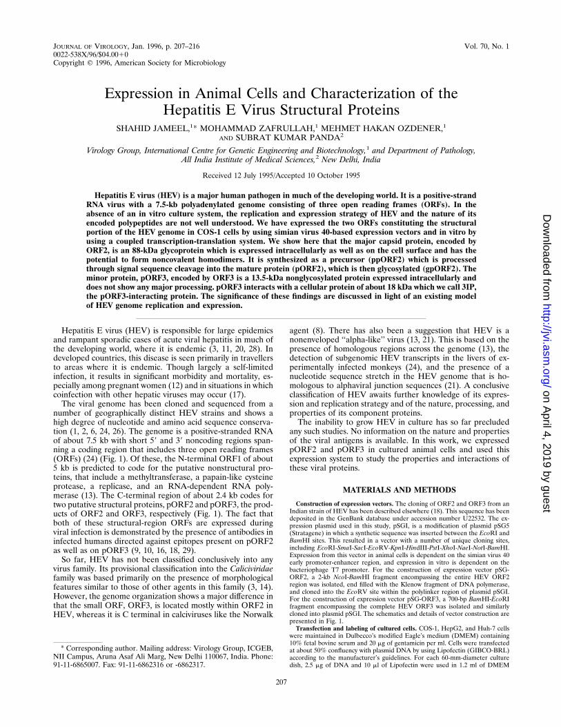
FIG. 1. HEV ORFs and cloning strategy. The three ORFs and their relativepositions along the HEV genomic polyadenylated RNA are indicated. The ex-pression vectors were constructed by subcloning the NcoI-BamHI (for ORF2) orBamHI-EcoRI (for ORF3) fragment at the EcoRV site within the polylinker ofplasmid pSGI. The vector backgrounds for ORF2 and ORF3 fragments (shadedregions) are shown. ATG initiator codons (open bars), part of the NcoI site, areindicated. Closed bars represent translation termination sequences. SV40 Pr,simian virus 40 promoter-enhancer and origin of replication sequences; T7 Pr,bacteriophage T7 promoter.
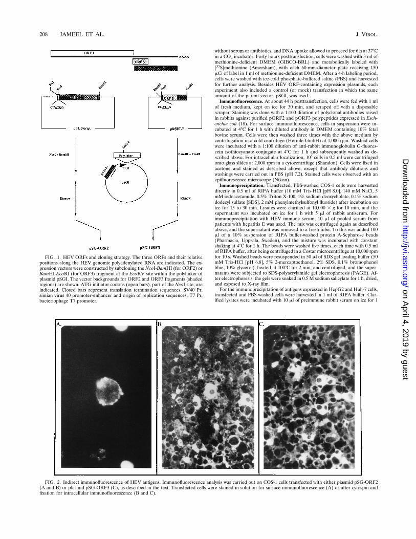
FIG. 2. Indirect immunofluorescence of HEV antigens. Immunofluorescence analysis was carried out on COS-1 cells transfected with either plasmid pSG-ORF2(A and B) or plasmid pSG-ORF3 (C), as described in the text. Transfected cells were stained in solution for surface immunofluorescence (A) or after cytospin andfixation for intracellular immunofluorescence (B and C).
h with the subsequent addition of 50 ml of a 50% protein A-Sepharose suspen-sion. The mixture was then incubated at 48C with shaking for 1 h. The beads werecentrifuged down, and precleared lysates were subjected to immunoprecipitationwith 10 ml of the specific antiserum as described above for COS-1 cells.Cell fractionation. COS-1 cells that had been transfected and labeled with
[35S]methionine as described above were washed once with PBS and thenscraped off the plate into 1 ml of PBS. After centrifugation, cell pellets equivalentto each 60-mm-diameter plate were resuspended in 0.5 ml of lysis buffer (10 mMTris-HCl [pH 8.0], 140 mM NaCl, 5 mM iodoacetamide, 0.5% Triton X-100, 2mM phenylmethylsulfonyl fluoride) and kept on ice for 1 h. Lysates were cen-trifuged in a Biofuge RS microcentrifuge (Heraeus Sepratech, GmbH) at 13,000rpm for 30 min. The supernatant (cytoplasmic fraction) was removed to a freshtube, and 50 ml of a 103 DOC-SDS solution (10% sodium deoxycholate, 1%SDS) was added to it. The pellets were washed once with 0.5 ml of lysis buffer asdescribed above. Washed pellets (nuclear fraction) were resuspended in 0.5 ml ofRIPA buffer. Both fractions were immunoprecipitated as described above.Tunicamycin treatment. At 40 h posttransfection, cells were shifted to 1 ml of
methionine-deficient DMEM without or with 10 mg of tunicamycin (BoehringerMannheim GmBH) for 1 h. Cells were then labeled with [35S]methionine for 4h, as described above, in the absence or presence of 10 mg of tunicamycin per ml.Total cell lysates in RIPA buffer were immunoprecipitated as described above.Pulse-chase analysis. At 40 h posttransfection, cells were shifted to 3 ml of
methionine-deficient DMEM for 1 h. Labeling was performed as describedabove, except that 300 mCi of [35S]methionine per 60-mm-diameter plate wasused and the labeling time was 20 min. After the removal of the labeling mix, 3ml of culture medium was added and cells were harvested either immediately orafter a chase of 30 min or 4 h. Total lysates in RIPA buffer were immunopre-cipitated as described above.In vitro translation. A coupled transcription-translation system (TNT; Pro-
mega) with bacteriophage T7 RNA polymerase was used for in vitro syntheses ofpolypeptides from pSG-ORF2 and pSG-ORF3 plasmid templates according tothe supplier’s guidelines. For cotranslational processing, 2 ml of canine pancre-atic membranes (Promega) was also included in the 25-ml in vitro transcription-translation reaction. [35S]methionine-labeled polypeptides synthesized in vitrowere separated by SDS-PAGE either directly or after immunoprecipitation andvisualized by fluorography.Endoglycosidase treatment. After the immunoprecipitation described above,
the protein A-Sepharose beads containing bound antigen were resuspended in 20ml of 0.5% SDS–1% 2-mercaptoethanol and heated at 1008C for 10 min. To thiswas added 2.5 ml of 0.5 M sodium citrate (pH 5.5) and 2 ml of endoglycosidaseH (500 U/ml; New England Biolabs, Beverly, Mass.). No enzyme was added tocontrol samples. After digestion at 378C for 2 h, 25 ml of 23 SDS gel loadingbuffer was added, and the samples were boiled for 5 min and analyzed byseparation on SDS–7.5% PAGE gels and fluorography.
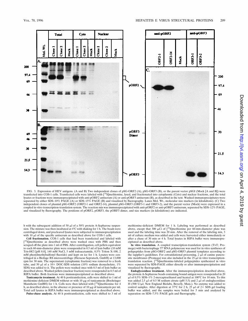
FIG. 3. Expression of HEV antigens. (A and B) Two independent clones of pSG-ORF2 (A), pSG-ORF3 (B), or the parent vector pSGI (Mock [A and B]) weretransfected into COS-1 cells. Transfected cells were labeled with [35S]methionine, lysed, and fractionated into cytoplasmic (Cyto) and nuclear fractions, and the totallysates or fractions were immunoprecipitated with anti-pORF2 antiserum (A) or anti-pORF3 antiserum (B), as described in the text. Washed immunoprecipitates wereseparated by either SDS–10% PAGE (A) or SDS–15% PAGE (B) and visualized by fluorography. Lanes Mol. Wt., molecular size markers (in kilodaltons). (C) Twoindependent clones of plasmid pSG-ORF2 (ORF2-1 and ORF2-14), plasmid pSG-ORF3 (ORF3-1 and ORF3-2), and the parent vector (Mock) were expressed in acoupled in vitro transcription-translation system. The reaction mix was immunoprecipitated with anti-pORF2 or anti-pORF3 antiserum, separated by SDS–12% PAGE,and visualized by fluorography. The positions of pORF2, pORF3, the pORF3 dimer, and size markers (in kilodaltons) are indicated.
VOL. 70, 1996 HEPATITIS E VIRUS STRUCTURAL PROTEINS 209
For an analysis of glycosylation in vitro, to 10 ml of the transcription-transla-tion mixture was added 2 ml of denaturing buffer (3% SDS, 6% 2-mercaptoetha-nol) and the mixture was heated at 1008C for 10 min. To this was added 20 ml ofreaction buffer (75 mM citrate-phosphate [pH 5.0], 100 mM EDTA, 5% TritonX-100, 1% 2-mercaptoethanol) and 0.4 U of endoglycosidase F (BoehringerMannheim GmbH), and the reaction was incubated at 378C. The control reactionincluded everything but the enzyme. Aliquots (10 ml) were removed at varioustimes, inactivated by being boiled in 40 ml of SDS gel loading buffer, and keptfrozen at2708C. Samples were analyzed by separation on SDS–7.5% PAGE gelsand fluorography.
RESULTS
Expression of HEV proteins. The expression vectors pSG-ORF2 and pSG-ORF3 contain the HEV ORFs driven by thesimian virus 40 control elements, including the ori sequences.These vectors are capable of replication to high copy numbersin T-antigen-producing monkey kidney COS-1 cells and shouldexpress high levels of the proteins encoded by HEV ORFs.Transfected cells were scored for antigen expression by immu-nofluorescence analysis with specific polyclonal antibodies.The results presented in Fig. 2 show that both pORF2 andpORF3 are expressed in transfected cells. pORF2 was foundon the cell surface (Fig. 2A) as well as in the cytoplasm (Fig.2B). On the other hand, pORF3 was found only in the cyto-plasm (Fig. 2C). Controls with preimmune sera did not showany staining on transfected COS-1 cells (data not shown). It isevident that antisera do not react with cellular componentsfrom the staining of only a fraction of the transfected cells in agiven field, in agreement with the expected transfection effi-ciency of about 40 to 50%.
Total lysates from COS-1 cells transfected with the appro-priate vectors and metabolically labeled with [35S]methioninewere also subjected to immunoprecipitation with polyclonalantisera. Cells transfected with two independent clones of plas-mid pSG-ORF2 showed the specific immunoprecipitation ofthe 74- to 88-kDa proteins absent in cells transfected with theparent vector pSGI (Fig. 3A). Similarly, a 13.5-kDa proteinwas found in cells transfected with two independent clones ofplasmid pSG-ORF3 but was not found in cells transfected withthe parent vector (Fig. 3B). Both pORF2 and pORF3 werealso found in Huh-7 hepatoma cells transfected with the ex-pression vectors, albeit at a level much reduced in comparisonwith that in COS-1 cells (data not shown).Subcellular fractionation of transfected COS-1 cells showed
that both pORF2 (Fig. 3A) and pORF3 (Fig. 3B) were presentin the cytoplasmic fraction, supporting the observations of im-munofluorescence studies (Fig. 2). A trace amount of pORF3was also found in the nuclear fraction. Neither protein wasfound to be secreted into the culture medium (data notshown).The expression of both proteins was also carried out in a
coupled in vitro transcription-translation system. Again, bothindependent clones of pSG-ORF2 as well as those of pSG-ORF3 expressed the respective proteins, as judged by immu-noprecipitation with specific antisera (Fig. 3C). In total trans-lation reactions without immunoprecipitation, pORF2 andpORF3 were the major protein bands, accounting for.80% ofthe synthesized protein (data not shown). The size of pORF2expressed in vitro was found to be 74 kDa, indicating that theprotein expressed in cultured cells may be subjected to post-translational modifications (Fig. 3A). The in vitro-expressedpORF3, like its cell-expressed counterpart (Fig. 3B), was 13.5kDa. In the in vitro system, however, a pORF3 species of about28 kDa was evident (Fig. 3C); it was reproducibly absent whenpORF3 was expressed in transfected cells. The fraction of this28-kDa form of pORF3 varied between different in vitro ex-pression experiments. Furthermore, it was observed that rabbitpolyclonal anti-pORF3 antiserum immunoprecipitated the 28-kDa form with a lower efficiency compared with that of the13.5-kDa form of pORF3.Both HEV antigens from lysates of pSG-ORF2- or pSG-
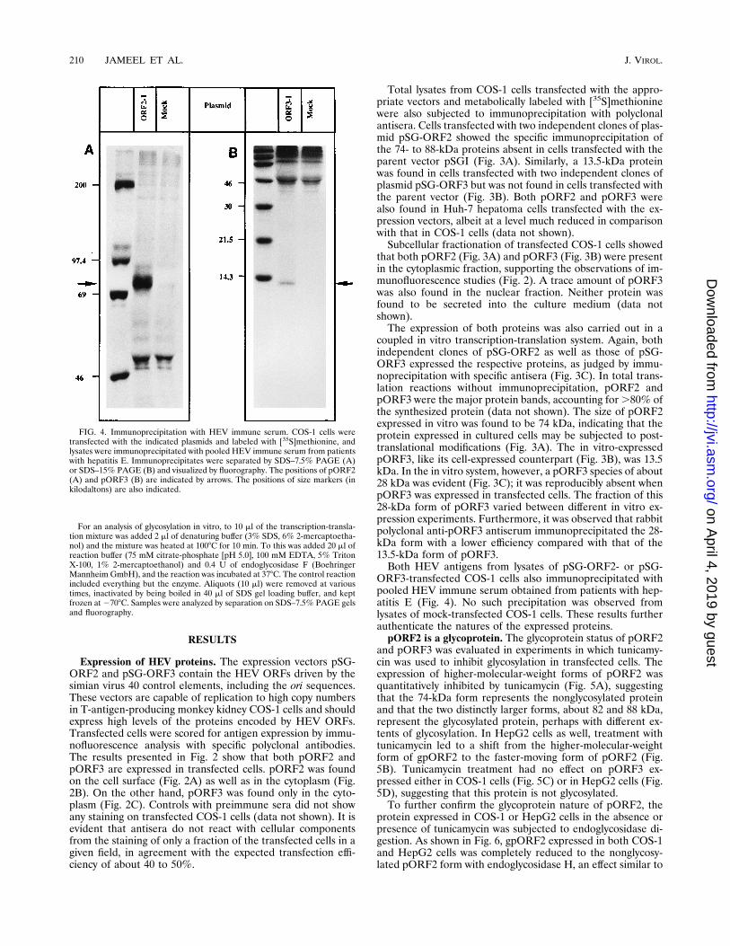
ORF3-transfected COS-1 cells also immunoprecipitated withpooled HEV immune serum obtained from patients with hep-atitis E (Fig. 4). No such precipitation was observed fromlysates of mock-transfected COS-1 cells. These results furtherauthenticate the natures of the expressed proteins.pORF2 is a glycoprotein. The glycoprotein status of pORF2
and pORF3 was evaluated in experiments in which tunicamy-cin was used to inhibit glycosylation in transfected cells. Theexpression of higher-molecular-weight forms of pORF2 wasquantitatively inhibited by tunicamycin (Fig. 5A), suggestingthat the 74-kDa form represents the nonglycosylated proteinand that the two distinctly larger forms, about 82 and 88 kDa,represent the glycosylated protein, perhaps with different ex-tents of glycosylation. In HepG2 cells as well, treatment withtunicamycin led to a shift from the higher-molecular-weightform of gpORF2 to the faster-moving form of pORF2 (Fig.5B). Tunicamycin treatment had no effect on pORF3 ex-pressed either in COS-1 cells (Fig. 5C) or in HepG2 cells (Fig.5D), suggesting that this protein is not glycosylated.To further confirm the glycoprotein nature of pORF2, the
protein expressed in COS-1 or HepG2 cells in the absence orpresence of tunicamycin was subjected to endoglycosidase di-gestion. As shown in Fig. 6, gpORF2 expressed in both COS-1and HepG2 cells was completely reduced to the nonglycosy-lated pORF2 form with endoglycosidase H, an effect similar to
FIG. 4. Immunoprecipitation with HEV immune serum. COS-1 cells weretransfected with the indicated plasmids and labeled with [35S]methionine, andlysates were immunoprecipitated with pooled HEV immune serum from patientswith hepatitis E. Immunoprecipitates were separated by SDS–7.5% PAGE (A)or SDS–15% PAGE (B) and visualized by fluorography. The positions of pORF2(A) and pORF3 (B) are indicated by arrows. The positions of size markers (inkilodaltons) are also indicated.
that observed with tunicamycin treatment. These results un-equivocally demonstrate that pORF2 is a glycoprotein and thatits glycosylation is not an artifact of overexpression in COS-1cells, which are not the natural target cells for HEV infection.Processing of HEV proteins. The processing of pORF2 and
pORF3 was studied in transfected cells in culture, as well as invitro. In a pulse-chase experiment after a 20-min pulse of[35S]methionine, a single 82-kDa form of pORF2 was predom-inant. After a 30-min chase with unlabeled methionine, threeforms of pORF2 were found, while after a 4-h chase, only the74- and 88-kDa forms were apparent. At this stage, the 88-kDaform of pORF2 was predominant. These results (Fig. 7A)suggest that the ORF2-encoded protein is made as a precursor(ppORF2), which is first processed into the mature form(pORF2) and then glycosylated (gpORF2). Similar experi-
ments with pORF3 showed only a single form of the proteinthroughout the pulse-chase period (Fig. 7B), suggesting thatthis protein does not undergo any major processing. Further,by comparing the behaviors of these two proteins during thepulse-chase, the turnover rate of pORF3 appeared to be higherthan that of pORF2.Cotranslational processing of these two proteins was studied
in vitro in the presence of canine pancreatic membranes (Fig.8). Apart from pORF2, a slightly larger form was also observedfor in vitro translations carried out in the presence of mem-branes (Fig. 8A). The fact that the larger form representedgpORF2 was established by endoglycosidase F digestion, whichresulted in the reduction of gpORF2 into pORF2 (Fig. 8B).The translation of pORF3 was inhibited by the addition ofmembranes. This was a nonspecific effect, as is apparent from
FIG. 5. Effects of tunicamycin on the expression of HEV proteins. (A and C) COS-1 cells were transfected with two independent clones of plasmid pSG-ORF2 (A)and plasmid pSG-ORF3 (C), with plasmid pSGI (Mock; A and C) as a control. (B and D) HepG2 cells were transfected with pSG-ORF2 (B) and pSG-ORF3 (D), withplasmid pSGI (Mock; B and D) as a control. The transfected cells were either treated (1) or not treated (2) with tunicamycin, as described in the text. Total cell lysateswere immunoprecipitated, separated by SDS–7.5% PAGE (A and B) or SDS–15% PAGE (C and D) and visualized by fluorography. The positions of size markers (inkilodaltons), gpORF2, pORF2, and pORF3 are indicated.
VOL. 70, 1996 HEPATITIS E VIRUS STRUCTURAL PROTEINS 211
the inhibition of pORF2 translation when translated eitheralone or along with pORF3 (Fig. 8A). In spite of the inhibitoryeffects of membranes, no cotranslational processing of pORF3was observed (Fig. 8A). These in vitro results support theobservations made for these two proteins expressed in trans-fected cells in culture.Interactions between HEV proteins. Homologous interac-
tions between pORF2 and pORF3 subunits were studied in thein vitro system. Proteins translated without the addition ofmembranes were subjected to SDS-PAGE in the absence orpresence of 2-mercaptoethanol (5%) and/or heat (1008C, 2min). As shown in Fig. 9A, pORF2 is unaffected by 2-mercap-toethanol treatment, but the result is a dimeric species if thesample is not heated prior to electrophoresis. This suggeststhat pORF2 forms a noncovalent homodimeric complex. Adistinct doublet observed in the monomeric pORF2 may be theresult of translation from alternate in-frame AUG codonspresent at amino acid positions 1, 12, and 16 in the primarysequence. As observed earlier, in vitro-translated pORF3showed a predominant 28-kDa form, which was unaffected byreduction and heating (Fig. 9B).Heteromeric interactions between subunits of pORF2 and
pORF3 were studied by transfection in COS-1 cells. Cells weretransfected with either pSG-ORF2, pSG-ORF3, or a combi-nation of two independent clones of each plasmid. [35S]methi-onine-labeled cell lysates were then immunoprecipitated withpolyclonal antibodies to one protein or the other. The results(Fig. 9C) show that while anti-pORF2 and anti-pORF3 wereable to precipitate the respective proteins from cotransfectedcells, there was no coimmunoprecipitation of the other protein.The specificities of the antisera were further substantiated bythe inability of anti-pORF2 to precipitate pORF3 and theinability of anti-pORF3 to precipitate pORF2. The lack ofcoimmunoprecipitation of pORF2 and pORF3 from cotrans-fected cells suggests that either these two proteins do not
interact with each other in vivo or that the interactions are notstrong enough to survive these immunoprecipitation condi-tions. Similar results were obtained when in vitro-translatedpORF2 and pORF3 were mixed and cross-immunoprecipi-tated with these two antisera (data not shown).pORF3 interacts with a cellular protein. Whenever pORF3
was immunoprecipitated from transfected COS-1 cells, an-other protein of about 18 kDa coprecipitated with it (Fig. 10A)(previously observed in Fig. 3B, 5C, and 9C). This protein wasalso observed in pORF3 immunoprecipitates from Huh-7 hep-atoma cells (data not shown), but not those from HepG2 cells(Fig. 5D). It was designated 3IP, the pORF3-interacting pro-tein. No such protein was precipitable from mock-transfectedcells (Fig. 10A), showing that it was not the result of antiserumcross-reactivity. Furthermore, a Western blot (immunoblot) oflysates from pSG-ORF3-transfected cells was positive forpORF3 but negative for 3IP (data not shown). By subcellularfractionation, like pORF3 3IP, was found in the cytoplasmicfraction (Fig. 3B). It was unaffected by tunicamycin treatmentof cells (Fig. 5C), suggesting that 3IP is not a glycoprotein.To further establish that 3IP is a cellular protein, in vitro-
translated pORF3 was mixed with [35S]methionine-labeled ly-sates of untransfected COS-1 cells and the mixture was pre-cipitated with anti-pORF3 antiserum. A very weak band wasseen in the presence of COS-1 lysates, but it was absent whenno lysates were added (Fig. 10B). As was the pORF3-3IPinteraction in transfected COS-1 cells, the in vitro interactionwas stable for RIPA buffer washing. In fact, identical resultswere obtained under washing conditions that were less strin-gent (10 mM Tris-HCl [pH 8.0], 140 mM Nacl, 0.1% TritonX-100 [TST buffer]). Quantitatively, however, far less pORF3-3IP interaction was observed in vitro than in transfected cells.
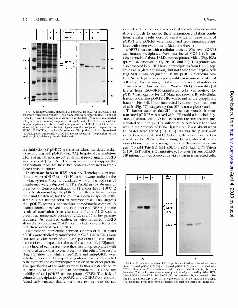
FIG. 6. Endoglycosidase digestion of gpORF2. HepG2 (A) and COS-1 (B)cells were transfected with pSG-ORF2, and cells were either treated (1) or nottreated (2) with tunicamycin, as described in the text. [35S]methionine-labeledcell lysates were immunoprecipitated with rabbit anti-pORF2, and washed im-munoprecipitates were treated with endoglycosidase H (Endo H) (1) or bufferalone (2), as described in the text. Digests were then subjected to separation onSDS–7.5% PAGE gels and to fluorography. The positions of the glycosylated(gpORF2) and nonglycosylated (pORF2) forms are shown. The positions of sizemarkers (in kilodaltons) are also indicated.
FIG. 7. Pulse-chase analysis of HEV proteins. COS-1 cells transfected witheither plasmid pSG-ORF2 (A) or plasmid pSG-ORF3 (B) were labeled with[35S]methionine for 20 min and chased with unlabeled methionine for the timesindicated. Total cell lysates were immunoprecipitated, separated by either SDS–7.5% PAGE (A) or SDS–15% PAGE (B), and visualized by fluorography. Thesize markers (from top to bottom) are 200, 97.4, 69, 46, 30, 21.5, and 14.3 kDa.The positions of multiple forms of pORF2 and that of pORF3 are indicated.
The genome of HEV has been cloned from multiple geo-graphically distinct isolates, but the inability to culture thisvirus has precluded studies into its molecular nature. Here wehave used a subgenomic fragment expression strategy to gainan insight into the putative structural proteins of HEV. Thestructural region of the HEV genome consists of two ORFs.These were expressed in COS-1 and HepG2 cells. The formercell line formed the basis of subsequent analyses as largeramounts of these proteins were expressed in this cell linebecause of replication of the expression vector to high copynumbers (5, 15). The latter cell line was used to ascertain thatHEV proteins are also expressed in liver cells, the only cellsknown to support viral replication.The major structural protein, pORF2, was found in multiple
forms as a 74- to 88-kDa protein by SDS-PAGE. In vitroexpression showed that the 74-kDa form corresponded to un-modified pORF2. This is in agreement with the predicted sizeof about 72 kDa, based on the 660-amino-acid ORF. On thebasis of tunicamycin inhibition experiments, cotranslationalprocessing with glycosylation-proficient membranes, and en-doglycosidase digestion experiments, we have shown that theapproximately 82- and 88-kDa forms are glycosylated versionsof pORF2. This is not an artifact of expression in COS-1 cells,as is borne out by the glycosylation of pORF2 expressed inHepG2 cells. Since this is a hepatoma line, it more closelymimics the natural target cell for HEV infection. In its primaryamino acid sequence, pORF2 contains three potentialN-linked glycosylation sites (Asn-X-Thr/Ser) at residues 137,310, and 561. These are conserved in all of the HEV strainssequenced so far (1, 6, 18, 24, 26).It has been suggested that pORF2 contains a signal se-
quence at its N-terminal end (21, 24). The putative signalsequence is a 22-amino-acid stretch consisting of positivelycharged residues (Arg) at the N-terminal end, a 14-residuehydrophobic core, and a turn-inducing stretch of proline resi-dues. However, direct proof of the presence of this signal
sequence is lacking. We have shown by means of pulse-chaseexperiments that pORF2 is indeed synthesized as a largerprecursor, ppORF2, of about 82 kDa. This is subsequentlyprocessed into the 74-kDa form and glycosylated to the matureform, gpORF2, of 88 kDa. The approximately 82-kDa speciesobserved in tunicamycin and pulse-chase experiments are mostlikely distinct from each other. While one may be an interme-diate in the glycosylation pathway leading to gpORF2 (88kDa), the other is the precursor which is processed to yieldpORF2. In Fig. 7A, both forms may be present but would notresolve on the gel because of very similar sizes.The various forms of pORF2 appear to run anomalously on
SDS-PAGE gels. On the basis of the primary sequence,ppORF2 should be about 72 kDa but runs as an 82-kDa pro-tein; its signal-cleaved form, which should be smaller, runs as a74-kDa protein. Such anomalous mobility due to conforma-tional and charge distribution effects has previously been ob-served for several classes of proteins, including glycoproteins,proline-rich proteins, maleylated proteins, calcium-bindingproteins, and histones (23). Apart from being a glycoprotein,pORF2 is also rich in proline residues in its N-terminal region.This is especially true for the signal sequence, which is aboutone-third proline (7 of 22 residues). In fact, there appears to bea far greater anomaly in the size of ppORF2 (82 versus 72kDa) than in the size of its signal-cleaved form, pORF2 (74versus 70 kDa).The glycosylation pattern of a glycoprotein reflects the cel-
lular compartment through which it has passed during its syn-thesis and processing. Our results with gpORF2 show that it iscompletely sensitive to endoglycosidase H and therefore con-tains only high-mannose residues. These structures are synthe-sized in the endoplasmic reticulum and the cis Golgi compart-ment (25). Thus, it appears that the processing of pORF2occurs at the endoplasmic reticulum and that the protein issubsequently transported either directly or through the cisGolgi compartment to the cell surface. Consistent with this,immunofluorescence localization showed pORF2 to be ex-
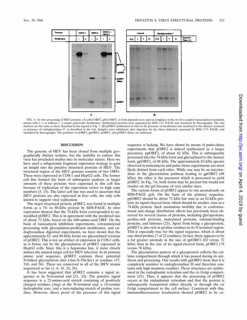
FIG. 8. In vitro processing of HEV proteins. (A) pSG-ORF2, pSG-ORF3, or both plasmids were used as templates in the in vitro coupled transcription-translationsystem with (1) or without (2) canine pancreatic membranes. Synthesized proteins were separated by SDS–12% PAGE and visualized by fluorography. The sizemarkers are the same as those described in the legend to Fig. 7. (B) pORF2 synthesized in vitro in the presence of membranes was incubated in the absence (control)or presence of endoglycosidase F, as described in the text. Samples were withdrawn after digestion for the times indicated, separated by SDS–7.5% PAGE, andvisualized by fluorography. The positions of pORF2, gpORF2, pORF3, and pORF3 dimer are indicated.
VOL. 70, 1996 HEPATITIS E VIRUS STRUCTURAL PROTEINS 213
pressed on the cell surface as well as in the cytoplasm. On thecell surface, the protein appeared to be concentrated in someregions, suggestive of an active process of association of pro-tein subunits, perhaps into some higher-order forms. It hasbeen shown here that pORF2 subunits can homodimerizethrough noncovalent interactions. Many positive-strand RNAviruses replicate and assemble on membrane surfaces and uti-lize a virally encoded RNA-binding capsid protein (4). pORF2,quite basic (pI, ;10.3) in its amino-terminal half, may well
serve this purpose (21, 22). Though the replication strategy ofHEV has not been worked out, such a proposal would fit wellinto the model proposed by Reyes et al. (22). It is not yet clearwhat bearing the glycosylation of pORF2 has on its cell surfacelocalization and on the assembly of the HEV nucleocapsid.This is currently being investigated with pORF2 mutants lack-ing individual or all N-linked glycosylation sites.The small ORF, ORF3, can code for a protein of 123 amino
acids (24). In accordance with this, we find a protein of 13.5
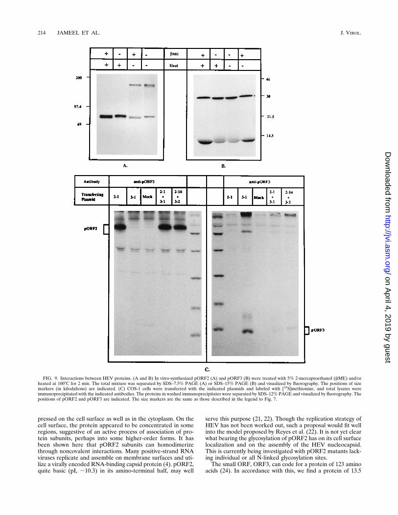
FIG. 9. Interactions between HEV proteins. (A and B) In vitro-synthesized pORF2 (A) and pORF3 (B) were treated with 5% 2-mercaptoethanol (bME) and/orheated at 1008C for 2 min. The total mixture was separated by SDS–7.5% PAGE (A) or SDS–15% PAGE (B) and visualized by fluorography. The positions of sizemarkers (in kilodaltons) are indicated. (C) COS-1 cells were transfected with the indicated plasmids and labeled with [35S]methionine, and total lysates wereimmunoprecipitated with the indicated antibodies. The proteins in washed immunoprecipitates were separated by SDS–12% PAGE and visualized by fluorography. Thepositions of pORF2 and pORF3 are indicated. The size markers are the same as those described in the legend to Fig. 7.
kDa expressed in cells transfected with ORF3 expression vec-tors. This protein is localized in the cytoplasm and nonglyco-sylated; it does not appear to undergo any modification thatwould significantly alter its size. In its N-terminal half, pORF3appears to contain two hydrophobic domains which have beenproposed to constitute either transmembrane segments or asignal sequence followed by a transmembrane region (22). Wehave provided clear evidence that pORF3 does not undergoany significant processing to remove the proposed signal se-quence.Two forms of pORF3, 13.5 and 28 kDa, were routinely
observed in vitro, but only the 13.5-kDa form was seen intransfected cells. One possibility is that the 28-kDa speciesrepresents a highly stable dimeric form of pORF3. In supportof this, a similar species is also observed when pORF3 isexpressed in E. coli (18). Alternatively, this could be an artifactof the in vitro system, whereby the ORF3 stop codon is skippedand translation terminates downstream in the vector se-quences. When a hexahistidine-pORF3 fusion protein is syn-thesized in vitro, two forms are evident. One is the expectedmonomeric (63His)pORF3 species of;17 kDa, and the otheris a species of ;28 kDa, significantly smaller than the expectedhomodimer (7). This supports the second alternative. In ani-mal cells, however, a cellular protein, which we have called 3IP,was found to interact with pORF3. The fact that the in vitro-expressed pORF3 monomer interacts very weakly with 3IPsuggests that this interaction takes place at the nascent chainlevel inside the cell. The role of 3IP, if any, in pORF3 expres-sion or function is not clear, considering that it coimmunopre-cipitates with pORF3 from COS-1 and Huh-7 hepatoma cells,not with pORF3 from HepG2 hepatoblastoma cells. However,the differences between the two liver cell lines may be ex-plained by their different stages of differentiation. We arepresently studying these homologous and heterologous inter-actions of pORF3 in greater detail.It is unknown whether pORF3 is a part of the virion. Here,
we tried to address this by looking for interactions betweenpORF2 (the major capsid protein) and pORF3. Expression intransfected cells and cross-immunoprecipitation with the re-
spective antibodies did not provide any evidence of an inter-action between these two proteins. It is possible that suchinteractions take place at the nascent chain level for these twoproteins or that the interactions are weak. Under both of thesecircumstances, our experimental conditions are likely to over-look such interactions. In the HEV genome, ORFs 2 and 3overlap the entire stretch of ORF3, except for a 9-amino-acidregion at the N terminus of ORF3. Though at least two sub-genomic RNAs coterminal with the 39 end of the HEV genomehave been reported (24), the subgenomic mRNA used for thetranslation of either or both of these ORFs has not beencharacterized. It is also possible that a single subgenomicmRNA is utilized for the synthesis of both proteins, perhaps byalternate initiator codon usage. In such a situation, the dynam-ics of translation and protein-protein interactions at the nas-cent chain level would be very distinct from those expected inour experimental system in which these two proteins are ex-pressed from different plasmids. Work currently in progress isaimed in this direction.How relevant are our findings to the infectious virus? Con-
tentious issues are the glycoprotein nature of pORF2 and thefact that most viral glycoproteins are components of the virionenvelope. So far, there is no evidence to suggest that HEVcontains a lipid membrane. We propose that pORF2 is themajor constituent of the HEV nucleocapsid, which is assem-bled at the cytoplasmic membrane. pORF2 is cotranslationallytranslocated via its N-terminal signal sequence into the endo-plasmic reticulum, where the signal is processed. The protein isglycosylated and transported to the cell surface by a bulk flowmechanism in the absence of any signals for retention in theendoplasmic reticulum (19), e.g., the C-terminal KDEL pep-tide found in endoplasmic reticulum lumenal proteins. Theglycosylation of pORF2 may be coincidental, considering itspassage through the endoplasmic reticulum (and perhaps thecis Golgi compartment as well) and its three highly conservedN-linked glycosylation sites. Some part of capsid assembly mayalso occur in association with the endoplasmic reticulum, asproposed by Reyes et al. (22), but final assembly and/or mat-uration has to be cytoplasmic to account for the encapsidation
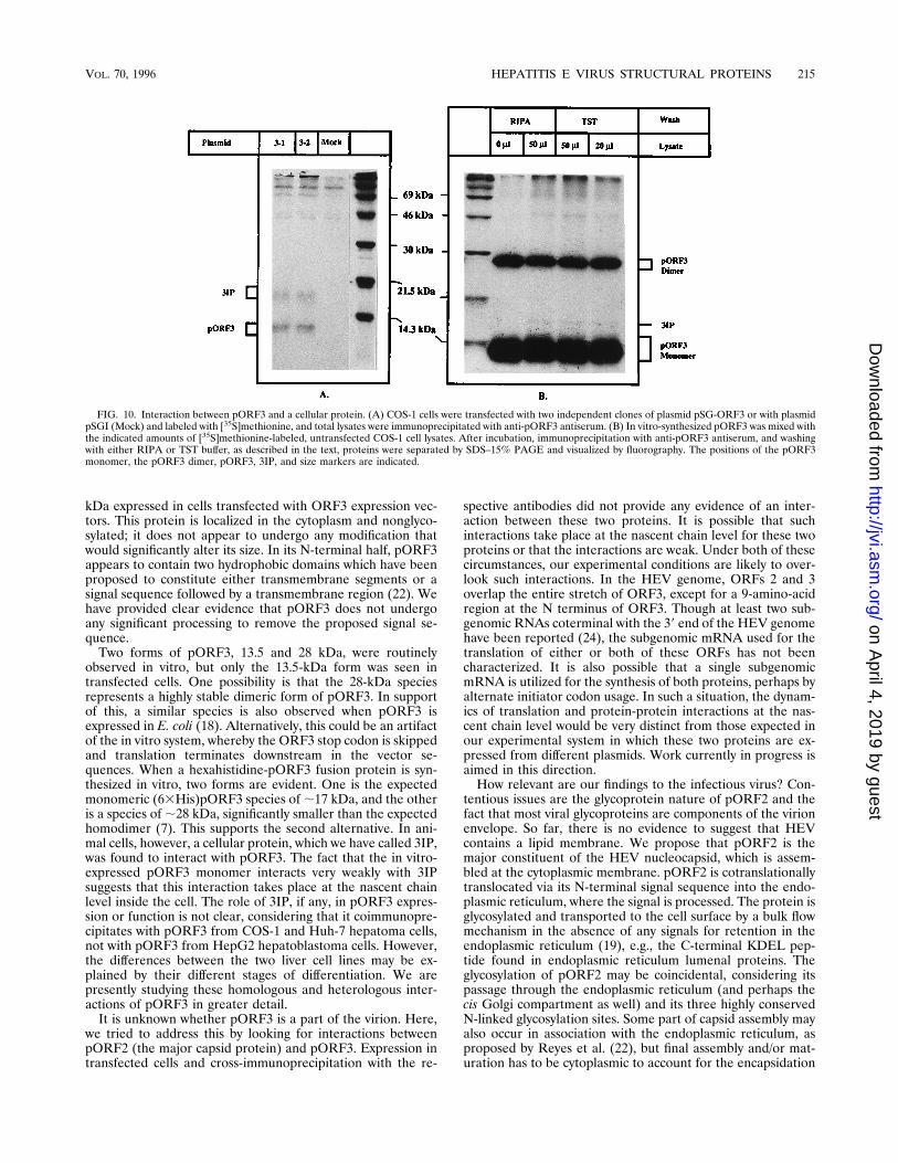
FIG. 10. Interaction between pORF3 and a cellular protein. (A) COS-1 cells were transfected with two independent clones of plasmid pSG-ORF3 or with plasmidpSGI (Mock) and labeled with [35S]methionine, and total lysates were immunoprecipitated with anti-pORF3 antiserum. (B) In vitro-synthesized pORF3 was mixed withthe indicated amounts of [35S]methionine-labeled, untransfected COS-1 cell lysates. After incubation, immunoprecipitation with anti-pORF3 antiserum, and washingwith either RIPA or TST buffer, as described in the text, proteins were separated by SDS–15% PAGE and visualized by fluorography. The positions of the pORF3monomer, the pORF3 dimer, pORF3, 3IP, and size markers are indicated.
VOL. 70, 1996 HEPATITIS E VIRUS STRUCTURAL PROTEINS 215
of HEV genomic RNA. At the cytoplasmic membrane, thenucleocapsid self-assembles along with HEV positive-strandedgenomic RNA. It is not clear whether viral RNA is an absoluterequirement for nucleocapsid assembly, since Tsarev et al. (27)have shown that pORF2 expressed in insect cells from a bacu-loviral vector is able to form virus-like particles. It is also notclear what role, if any, pORF3 plays in the assembly of the viralnucleocapsid. In the absence of either a tissue culture or agenomic RNA transfection system for HEV, we are trying toanswer some of these questions by using the COS-1 cell ex-pression system described above.
ACKNOWLEDGMENTS
We thank V. Kumar (ICGEB, New Delhi, India) for the expressionvector pSGI. We are grateful to Y. Vaishnav, D. Salunke, and S. E.Hasnain for fruitful discussions and critical readings of the manuscript.This work was supported by internal funds from the ICGEB.
REFERENCES
1. Aye, T. T., T. Uchida, X.-Z. Ma, F. Iida, T. Shikata, H. Zhuang, and K. M.Win. 1992. Complete nucleotide sequence of a hepatitis E virus isolated fromthe Xinjiang epidemic (1986–1988) of China. Nucleic Acids Res. 20:3512.
2. Bi, S. L., M. A. Purdy, K. A. McCaustland, H. S. Margolis, and D. W.Bradley. 1993. The sequence of hepatitis E virus isolated directly from asingle source during an outbreak in China. Virus Res. 28:233–247.
3. Bradley, D. W. 1990. Enterically-transmitted non-A, non-B hepatitis. Br.Med. Bull. 46:442–461.
4. Brinton, M. A., and F. X. Heinz (ed.). 1990. New aspects of positive-strandRNA viruses. American Society for Microbiology, Washington, D.C.
5. Gluzman, Y. 1981. SV40-transformed simian cells support the replication ofearly SV40 mutants. Cell 23:175–182.
6. Huang, C. C., D. Nguyen, J. Fernandez, K. Y. Yun, K. E. Fry, D. W. Bradley,A. W. Tam, and G. R. Reyes. 1992. Molecular cloning and sequencing of theMexico isolate of hepatitis E virus (HEV). Virology 191:550–558.
7. Jameel, S. Unpublished data.8. Jiang, X., D. Y. Graham, K. Wang, and M. K. Estes. 1990. Norwalk virusgenome cloning and characterization. Science 250:1580–1583.
9. Khudyakov, Y. E., M. O. Favorov, D. L. Jue, T. K. Hine, and H. A. Fields.1994. Immunodominant antigenic regions in a structural protein of thehepatitis E virus. Virology 198:390–393.
10. Khudyakov, Y. E., N. S. Khudyakova, H. A. Fields, D. Jue, C. Starling, M. O.Favorov, K. Krawczynski, L. Polish, E. Mast, and H. Margolis. 1993.Epitope mapping in proteins of hepatitis E virus. Virology 194:89–96.
11. Khuroo, M. S. 1980. Study of an epidemic of non-A, non-B hepatitis: pos-sibility of another human hepatitis virus distinct from post-transfusionnon-A, non-B type. Am. J. Med. 68:818–823.
12. Khuroo, M. S., M. R. Teli, S. Skidmore, M. A. Sofi, and M. Khuroo. 1981.Incidence and severity of viral hepatitis in pregnancy. Am. J. Med. 70:252–255.
13. Koonin, E. V., A. E. Gorbalenya, M. A. Purdy, M. N. Rozanov, G. R. Reyes,and D. W. Bradley. 1992. Computer-assisted assignment of functional do-mains in the nonstructural polyprotein of hepatitis E virus: delineation of anadditional group of positive-stranded RNA plant and animal viruses. Proc.Natl. Acad. Sci. USA 89:8259–8263.
14. Krawczynski, K. 1993. Hepatitis E. Hepatology 17:932–941.15. Learned, R. M., R. M. Myers, and R. Tjian. 1981. Replication in monkey
cells of plasmid DNA containing the minimal SV40 origin. ICN-UCLASymp. Mol. Cell. Biol. 22:555–566.
16. Li, F., H. Zhuang, S. Kolivas, S. A. Locarnini, and D. A. Anderson. 1994.Persistent and transient antibody responses to hepatitis E virus detected byWestern immunoblot using open reading frame 2 and 3 and glutathioneS-transferase fusion proteins. J. Clin. Microbiol. 32:2060–2066.
17. Panda, S. K. Unpublished results.18. Panda, S. K., S. K. Nanda, M. Zafrullah, I.-H. Ansari, M. H. Ozdener, and
S. Jameel. 1995. An Indian strain of hepatitis E virus (HEV): cloning,sequence, and expression of structural region and antibody responses in serafrom individuals from an area of high-level HEV endemicity. J. Clin. Mi-crobiol. 33:2653–2659.
19. Pelham, H. R. B., and S. Munro. 1993. Sorting of membrane proteins in thesecretory pathway. Cell 75:603–605.
20. Purcell, R. H., and J. R. Ticehurst. 1988. Enterically transmitted non-A,non-B hepatitis: epidemiology and clinical characteristics, p. 131–137. InA. J. Zuckerman (ed.), Viral hepatitis and liver disease. Alan R. Liss, Inc.,New York.
21. Purdy, M. A., A. W. Tam, C. C. Huang, P. O. Yarbough, and G. R. Reyes.1993. Hepatitis E virus: a non-enveloped member of the ‘alpha-like’ RNAvirus supergroup? Semin. Virol. 4:319–326.
22. Reyes, G. R., C. C. Huang, A. W. Tam, and M. A. Purdy. 1993. Molecularorganization and replication of hepatitis E virus (HEV). Arch. Virol. 7:15–25.
23. See, Y. P., and G. Jackowski. 1989. Estimating molecular weights of polypep-tides by SDS gel electrophoresis, p. 1–21. In T. E. Creighton (ed.), Proteinstructure: a practical approach. IRL Press, Oxford.
24. Tam, A. W., M. M. Smith, M. E. Guerra, C. C. Huang, D. W. Bradley, K. E.Fry, and G. R. Reyes. 1991. Hepatitis E virus (HEV): molecular cloning andsequencing of the full-length viral genome. Virology 185:120–131.
25. Tarentino, A. L., R. B. Trimble, and T. H. Plummer. 1989. Enzymatic ap-proaches for studying the structure, synthesis, and processing of glycopro-teins. Methods Cell Biol. 32:111–139.
26. Tsarev, S. A., S. U. Emerson, G. R. Reyes, T. S. Tsareva, L. J. Letgers, I. A.Malik, M. Iqbal, and R. H. Purcell. 1992. Characterization of a prototypestrain of hepatitis E virus. Proc. Natl. Acad. Sci. USA 89:559–563.
27. Tsarev, S. A., T. S. Tsareva, S. U. Emerson, A. Z. Kapikian, J. Ticehurst, W.London, and R. H. Purcell. 1993. ELISA for antibody to hepatitis E virus(HEV) based on complete open-reading frame-2 protein expressed in insectcells: identification of HEV infection in primates. J. Infect. Dis. 168:369–378.
28. Wong, D. C., R. H. Purcell, M. A. Sreenivasan, S. R. Prasad, and K. M.Pavri. 1980. Epidemic and endemic hepatitis in India: evidence for a non-A,non-B virus etiology. Lancet ii:876–879.
29. Yarbough, P. O., A. W. Tam, K. E. Fry, K. Krawczynski, K. A. McCaustland,D. W. Bradley, and G. R. Reyes. 1991. Hepatitis E virus: identification oftype-common epitopes. J. Virol. 65:5790–5797.