25
External Fixation Nursing Best Practic Guidelines Orthopedic Nursing, Part 2
| Date post: | 29-Dec-2015 |
| Category: |
Documents |
| Upload: | mervin-patterson |
| View: | 250 times |
| Download: | 2 times |
External Fixation
Nursing Best Practice
Guidelines
Orthopedic Nursing, Part 2


Reasons for External Fixation Device Placement….. a technique of fracture immobilization a series of transfixing pins are inserted
through the bone and attached to a rigid external metal frame
method is use mainly in the management of open fractures with severe soft-tissue damage


Types of External Fixation DevicesVary with the site of placement and type of fracture…
Unilateral Fixators Also called monolateral frame external fixators Attached to only one side of the limb to which they are fixated Unilateral frames allow the fixated limb to remain functional.
Taylor Spatial Frame Fixators One of the primary types of ring external fixators. Made up of rings, connecting rods, and struts. This type of external frames have been used primarily to correct
deformities Being used increasingly for managing fractures.

Advantages of External Fixation
Permits rigid support of: Commuted open fractures Infected nonunions Infected unstable joints
Facilitates wound care and soft tissue reconstruction Allows early function of muscle and joints Allows early patient comfort Reduces risk of compartment syndrome
https://youtu.be/21plYYmaxi8

Who is responsible for the care? Orthopedics Doctors, Physical Therapists and Nurses
all share responsibilities in providing care to the patient with an EFD
The Doctor orders the parameters of mobility and pin site care/dressing changes
The Physical Therapists trains the patient in initial mobility and use of assistive devices
Nurses assess for systemic and neurovascular changes, infection, safe mobility practices and insured skin integrity.
The next slide defines the distinct roles of the RN and Physical Therapist in the acute care setting related to the patient with an external fixation device (EFD).

Role of RN/ Nurse Role of Physical Therapist
Education of the patient pre- and post-operatively: multiple aspects including safety precautions, device purpose, diet, pain management,
follow-up.
Education of the patient pre- and post-operatively: multiple aspects including device type, safety precautions, allowed mobility, assistive devices, follow-up.
Initial assessment for post operative stability and complications following device placement (vs, bleeding…).
Assessment of fixation device precaution practices.
Positioning/moving/tilting patient to prevent skin breakdown.
Initial training for movement /gait with assistive devices.
Providing pin site care and education for self care post discharge.
Education of the patient : safety precautions, mobility, exercises.

Device Placement: Preoperative Best Nursing Practices Describe the device placement to the patient
so the patient is prepared for what to expect postoperatively
Carefully monitor for signs and symptoms of wound contamination that could lead to infection
Preoperative pain management/sedation Nutrition assessment/preoperative NPO
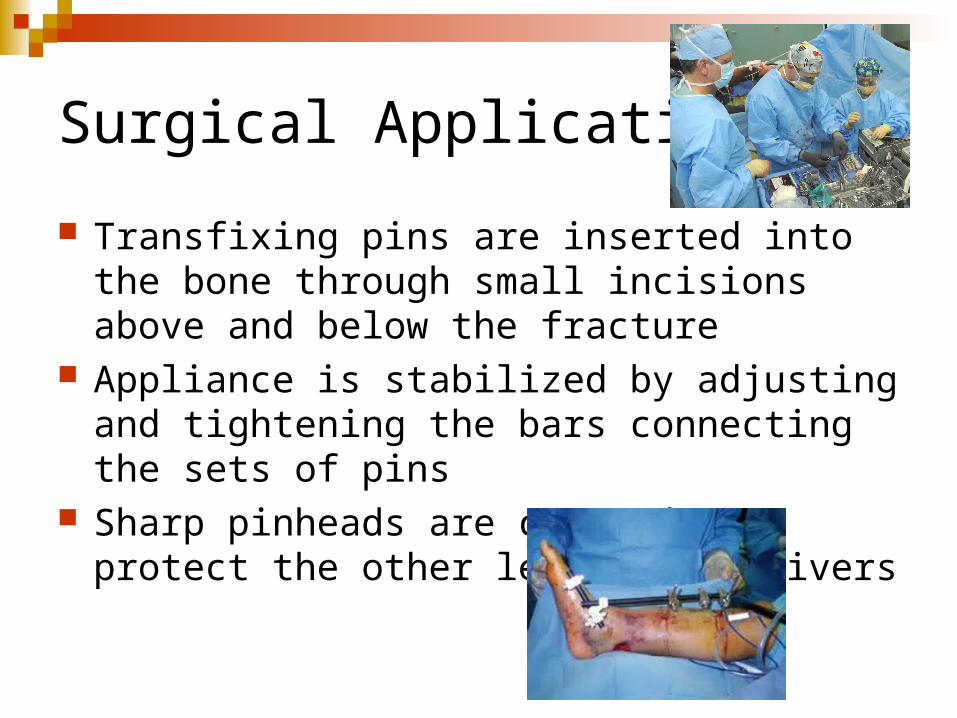
Surgical Application
Transfixing pins are inserted into the bone through small incisions above and below the fracture
Appliance is stabilized by adjusting and tightening the bars connecting the sets of pins
Sharp pinheads are covered to protect the other leg and caregivers

Assessment of the patient with an external fixation device
Determine patient’s understanding of procedure and fixation device
Evaluate neurovascular status of involved body part Inspect pin site
redness, drainage, tenderness, pain, loosening pins
Inspect open woundshealing, infection, devitalized tissue
Assess functioning of other body systems affected by injury or immobilization
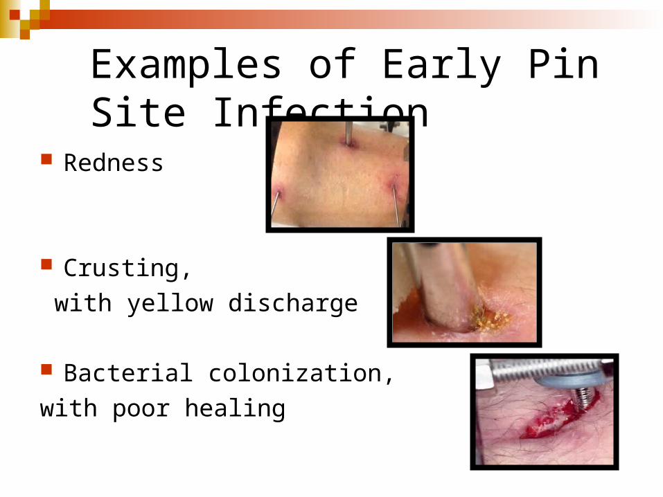
Examples of Early Pin Site Infection
Redness
Crusting,
with yellow discharge
Bacterial colonization,
with poor healing
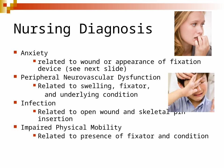
Nursing Diagnosis
Anxiety related to wound or appearance of fixation device
(see next slide) Peripheral Neurovascular Dysfunction
Related to swelling, fixator, and underlying condition
Infection Related to open wound and skeletal pin insertion
Impaired Physical Mobility Related to presence of fixator and condition

Interventions: Anxiety
Reassure the patient If possible do this before and after surgery. Explain that even
thought the device may look clumsy, cumbersome or intimidating, it should not hurt once in place.
Emphasize the positives Inform the patient that greater mobility can be achieved with an
external fixation device, thereby minimizing the development of other system problems.
Encourage patient to verbalize reaction to device Each patient will respond differently. By having the patient
verbalize their reactions to the device, nursing staff will better be able to reassure the patient specific to their particular concerns and issues.
Involve patient in the management and care of the device

Neurovascular Status
Assess status frequently Every 15 minutes to 1 hour while
swelling is significant After swelling has subsided, every 2
hours for 24 hours Then every 4 hours or as needed for
symptoms Establish baseline of functioning for
comparative monitoring Complex musculoskeletal injuries
frequently result in disruption of soft tissue functioning
Elevate extremity to reduce swelling Report any changes status or abnormal
findings
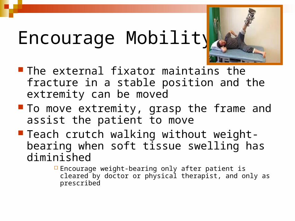
Encourage Mobility
The external fixator maintains the fracture in a stable position and the extremity can be moved
To move extremity, grasp the frame and assist the patient to move
Teach crutch walking without weight-bearing when soft tissue swelling has diminished
Encourage weight-bearing only after patient is cleared by doctor or physical therapist, and only as prescribed

Preventing Infection Provide wound care Monitor for local and systemic infection Provide site and fixator care
Clean fixator with clean cloth and water, as needed Clean pins and remove crusts with sterile
applicator Crusts formed by serous drainage can prevent
fluid from draining and can cause infection A small amount of drainage from the pin sites is
normal Report loosened pins Report inflammation, swelling, tenderness, and purulent drainage at pin site
Look for and report signs of infection

FYI….a note on pin placement and handling….
According to a local orthopedic specialist (Dr. Owen), a properly, securely placed fixation pin will not yield much fluid.
Loosened or poorly secured fixation pins may cause inflammation of the site and, therefore, discharge.
Proper aseptic handling during and after surgery will prevent site infection.

To clean or not to clean….that is the question…….
When is it appropriate to do clean the pin sites on an external fixation device?
When devices are placed for a short term (eg. a few days) often times orthopedic doctors will not order pin care or will do pin site care themselves when they visit the patient.
Gauze may be placed at the pin site by the doctor. If the gauze becomes damp, the prudent nurse will replace the damp gauze with dry gauze using aseptic technique.
Check pin site every day for signs of infection, such as: Skin redness Skin at the site is warmer Swelling or hardening of the skin Increased pain at the pin site Drainage that is yellow, green, thick, or smelly Fever Numbness or tingling at the pin site Movement or looseness of the pin >>>>If you think there is a site infection, call the surgeon right
away.

Cleaning pin sites: Clean pinsite only as MD orders
There are different types of pin-cleaning solutions. The two most common solutions are: Sterile water A mixture of half normal saline and half hydrogen peroxide
>>>Use the solution that your surgeon recommends.
Supplies you will need to clean your pin site include: Gloves Sterile cup Sterile cotton swabs (about 3 swabs for each pin) Sterile gauze Cleaning solution
Cleaning the pin site Clean the pin site twice a day only as ordered by the doctor. Do not put lotion or cream on the area unless your surgeon tells you
it is OK.

The basic steps for pin site cleaning are as follows:1. Check Physcian’s order.
2. Wash and dry your hands.
3. Put on gloves.
4. Pour the cleaning solution into a cup and put half of the swabs in the cup to moisten the cotton ends.
5. Use a clean swab for each pin site. Start at the pin site and clean your skin by moving the swab away from the pin. Move the swab in a circle around the pin, then make the circles around the pin larger as you move away from the pin site.
6. Remove any dried drainage and debris from around the pin site with the gentle movement of the swab.

The basic steps for pin site cleaning continue:6. Use a new swab or gauze to clean the pin. Start at the pin site
and move up the pin, away from the skin.7. When you are done cleaning, use a dry swab or gauze in the
same way to dry the area. 8. For a few days after the pin insertion surgery you may wrap
the pin site in dry sterile gauze while it heals. After this time, leave the pin site open to air.
9. If there is an external fixator (a steel bar that may be used for fractures of long bones), clean it with gauze and cotton swabs dipped in your cleaning solution every day.
10. Most patients who have pins can take a shower 10 days after surgery. Ask the surgeon how soon when the patient can shower
https://youtu.be/pP0BrZXxBGw

Discharge Planning for the Patient with and External Fixation Device Precautions related to mobility
Specific amount of weight-bearing or non-weight bearing ordered
Signs and symptoms to call the MD about Increased pain Fever Signs of infection at pin sites
Pain management plan medications ordered
Follow-up appointment plan

The EndOrthopedic Nursing Part 2: External Fixation DevicesNursing Best Practice


