Page 1/35 Characterization of the microRNA Transcriptomes and Proteomics of Cochlea Tissue-derived Small Extracellular Vesicles From Different Age of Mice After Birth Pei Jiang Southeast University Xiangyu Ma Southeast University Shanying Han Southeast University Leyao Ma Southeast University Jingru Ai Southeast University Leilei Wu Southeast University Yuan Zhang Southeast University Hairong Xiao Southeast University Mengyao Tian Southeast University W. Andy Tao Purdue University Shasha Zhang Southeast University Renjie Chai ( [email protected]) Southeast University https://orcid.org/0000-0002-3885-543X Research Article Keywords: small extracellular vesicles, cochlea, development, microRNAs, proteins, proteomics Posted Date: August 30th, 2021
Transcript
Page 1/35
Characterization of the microRNA Transcriptomesand Proteomics of Cochlea Tissue-derived SmallExtracellular Vesicles From Different Age of MiceAfter BirthPei Jiang
AbstractThe cochlea is an important sensory organ for both balance and sound perception, and the formation ofthe cochlea is a complex developmental process. The development of the mouse cochlea begins onembryonic day (E)9 and continues until postnatal day (P)21 when the hearing system is consideredmature. Small extracellular vesicles (sEVs), with a diameter ranging from 30 nm to 200 nm, have beenconsidered as a signi�cant medium for information communication in both the processing ofphysiological and pathological. However, there are no studies exploring the role of sEVs in thedevelopment of the cochlea. Here, we isolated tissue-derived sEVs from the cochleae of FVB mice at P3,P7, P14, and P21 by ultracentrifugation. These sEVs were �rst characterized by transmission electronmicroscopy, nanoparticle tracking analysis, and western blotting. Next, we used small RNA-seq and massspectrometry to characterize the microRNA transcriptomes and proteomics of cochlear sEVs from mice atdifferent ages. Many microRNAs and proteins were discovered to be related with inner ear development,anatomical structure development, and the auditory nervous system development. These results allsuggest that sEVs exist in the cochlea and are likely to be essential for the normal development of theauditory system. Our �ndings provide many sEV microRNA and protein targets for future studies of theroles of cochlear sEVs.
IntroductionThe cochlea in the inner ear is an important auditory signal transduction organ that develops fromembryonic day (E)9 through postnatal day (P)2 [1]. The detection of sound waves and transmission ofsound information to the brain are both dependent on cochlear hair cell (HCs) [2]. The �rst cochlear HCsdevelop at E11, and ultimately three rows of outer hair cells (OHCs), one row of inner hair cells (IHCs), andsupporting cells (SCs) beneath the HCs are formed [3]. By P3, the total number of HCs peaks and willremain basically unchanged, while the morphology of the HCs will change as the HCs mature from P3 toP21 [4, 5].
HC maturation involves many complex developmental processes, such as the formation of hair bundles,synapses, and mechanical transduction channels (METs) [6-8]. Hearing formation requires theestablishment of proper innervation, and the afferent nerves of the inner ear gradually form an outerspiral bundle of OHCs from P0 to P3 [9]. In the �rst seven days after birth, hair bundles and METs developgradually, and mature innervation patterns emerge gradually between P14 and P21 [9, 10]. At P7, HCshave mature mechanical transduction abilities, which is the most important aspect of formation of theauditory system [11]. HC synapses begin to mature at P14, which is when mice begin to gain hearingability [12]. At P21, the morphology and function of the cochlea are mature, and hearing function can bemeasured by auditory brainstem response.
During the process of HC maturation, the characteristics of SCs, especially inner ear progenitors, alsochange dramatically. SCs have been reported to act as inner ear stem cells and transdifferentiated into
Page 4/35
HCs by induction of Wnt signaling or inhibition of Notch signaling in newborn mice [13, 14]. However, thestemness of SCs deteriorates with age, and their capacity to divide is completely lost by P14 [15].
It has been reported that many important transcription factors and signaling pathways are associatedwith the development of the cochlea, such as Sox2, Atoh1 [16], and the Wnt, Notch, and FGF signalingpathways [17, 18]. In addition, many microRNAs (miRNAs), such as miR182, miR183, and miR124, arealso reported to regulate inner ear tissue differentiation and to maintain cell differentiation andproliferation [19, 20]. However, the cochlea’s development is a complicated process, and many regulatoryprocesses and the factors that are involved remain to be elucidated.
Small extracellular vesicles (sEVs) have become a research hotspot in latest years, and are reported to beinvolved in intercellular signal transmission during many important pathological and physiologicalprocesses [21-23]. sEVs have sizes between 30 nm to 200 nm and can be generated by various cells [24].The contents of sEVs include numerous proteins and nucleic acids that are protected by a phospholipidbilayer structure from being digested by extracellular substances, and these materials can be delivered torecipient cells and thus contribute to cellular communication and signal transmission [25, 26]. sEVsparticipate in cell proliferation and differentiation in both pathological and healthy situations throughsignaling pathways mediated by miRNAs [27-29], and sEVs are involved in intercellular signaltransmission during the development of brain neural circuits and in regulating growth patterns duringembryonic development [30, 31].
Although sEVs have been extensively studied in cancer and other diseases, limited studies have beenperformed on the role of sEVs in the cochlea. This may be because as the mice age the otic vesicleoutside the cochlea gradually becomes ossi�ed and becomes rigid, especially after P10, which makes itdi�cult to obtain the substances inside the cochlea. However, it is known that in the utricle SC-derivedexosomes can protect HCs against neomycin-induced ototoxicity [32] and that inner ear stem cell-derivedexosomes can reduce ototoxic drug damage by transferring miR-182-5p to HEI-OC1 cells [33, 34]. Atpresent, the research on inner ear-derived sEVs is based on in vitro models, and there is no research onsEVs in intact inner ear tissues.
In this study, we extracted cochlear tissue-derived sEVs from mice at different ages after birth andsystematically analyzed and characterized their protein and miRNA contents for the �rst time. We usedtransmission electron microscopy (TEM), western blotting, and nanoparticle tracking analysis (NTA) toquantify the characteristics of sEVs and then performed proteomics and small RNA-seq to analyze thedifferentially expressed proteins and miRNAs and to predict the functions of these proteins and miRNAs.These results are expected to provide important information for the subsequent functional analysis ofsEVs in the cochlea.
Materials And MethodsIsolation of cochlear tissue-derived sEVs
Page 5/35
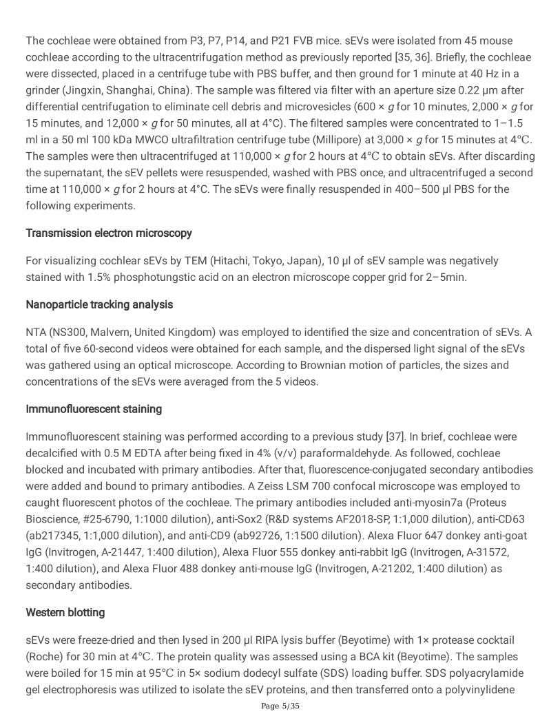
The cochleae were obtained from P3, P7, P14, and P21 FVB mice. sEVs were isolated from 45 mousecochleae according to the ultracentrifugation method as previously reported [35, 36]. Brie�y, the cochleaewere dissected, placed in a centrifuge tube with PBS buffer, and then ground for 1 minute at 40 Hz in agrinder (Jingxin, Shanghai, China). The sample was �ltered via �lter with an aperture size 0.22 µm afterdifferential centrifugation to eliminate cell debris and microvesicles (600 × g for 10 minutes, 2,000 × g for15 minutes, and 12,000 × g for 50 minutes, all at 4°C). The �ltered samples were concentrated to 1–1.5ml in a 50 ml 100 kDa MWCO ultra�ltration centrifuge tube (Millipore) at 3,000 × g for 15 minutes at 4℃.The samples were then ultracentrifuged at 110,000 × g for 2 hours at 4℃ to obtain sEVs. After discardingthe supernatant, the sEV pellets were resuspended, washed with PBS once, and ultracentrifuged a secondtime at 110,000 × g for 2 hours at 4°C. The sEVs were �nally resuspended in 400–500 μl PBS for thefollowing experiments.
Transmission electron microscopy
For visualizing cochlear sEVs by TEM (Hitachi, Tokyo, Japan), 10 µl of sEV sample was negativelystained with 1.5% phosphotungstic acid on an electron microscope copper grid for 2–5min.
Nanoparticle tracking analysis
NTA (NS300, Malvern, United Kingdom) was employed to identi�ed the size and concentration of sEVs. Atotal of �ve 60-second videos were obtained for each sample, and the dispersed light signal of the sEVswas gathered using an optical microscope. According to Brownian motion of particles, the sizes andconcentrations of the sEVs were averaged from the 5 videos.
Immuno�uorescent staining
Immuno�uorescent staining was performed according to a previous study [37]. In brief, cochleae weredecalci�ed with 0.5 M EDTA after being �xed in 4% (v/v) paraformaldehyde. As followed, cochleaeblocked and incubated with primary antibodies. After that, �uorescence-conjugated secondary antibodieswere added and bound to primary antibodies. A Zeiss LSM 700 confocal microscope was employed tocaught �uorescent photos of the cochleae. The primary antibodies included anti-myosin7a (ProteusBioscience, #25-6790, 1:1000 dilution), anti-Sox2 (R&D systems AF2018-SP, 1:1,000 dilution), anti-CD63(ab217345, 1:1,000 dilution), and anti-CD9 (ab92726, 1:1500 dilution). Alexa Fluor 647 donkey anti-goatIgG (Invitrogen, A-21447, 1:400 dilution), Alexa Fluor 555 donkey anti-rabbit IgG (Invitrogen, A-31572,1:400 dilution), and Alexa Fluor 488 donkey anti-mouse IgG (Invitrogen, A-21202, 1:400 dilution) assecondary antibodies.
Western blotting
sEVs were freeze-dried and then lysed in 200 μl RIPA lysis buffer (Beyotime) with 1× protease cocktail(Roche) for 30 min at 4℃. The protein quality was assessed using a BCA kit (Beyotime). The sampleswere boiled for 15 min at 95℃ in 5× sodium dodecyl sulfate (SDS) loading buffer. SDS polyacrylamidegel electrophoresis was utilized to isolate the sEV proteins, and then transferred onto a polyvinylidene
Page 6/35
di�uoride membrane at 275mA for 90 minutes. The membrane was blocked with 5% BSA [5% (v/v)bovine serum albumin in 0.1% (v/v) Tween-20 in PBS] for 1h at room temperature and then incubatedwith primary antibody overnight at 4°C. The second day, the membrane was incubated with HRP-conjugated secondary antibody (Abclonal, 1:2,000 dilution). SuperSignal West Pico Pluschemiluminescent substrate (Thermo Scienti�c) was employed for visualized target bands on a Tanon-5200 automatic chemical imaging system. The primary antibodies were anti-CD63 (ab217345, 1:1,000dilution), anti-CD9 (ab92726, 1:1500 dilution), anti-Tsg101 (ab125011, 1:2,000 dilution), anti-mouse EEA1(Santa Cruz Biotechnology, 1:100 dilution), and anti-rabbit Rab7 (Cell Signaling, 1:1,000 dilution), andanti-GAPDH (Kangchen, KC-5G4, 1:2,000 dilution).
RNA extraction and quantitative real-time PCR
sEV samples were mixed with 1 ml Trizol (Invitrogen,15596-026) on ice for 5 min, then centrifuged at17,970 × g for 5 min at 4℃. The sample was added with 200 μl chloroform, vortexed to mix well, andthen placed upon ice for 10 min. After centrifugation at 17,970 × g for 15 min at 4℃, the supernatant wasmixed with an equal amount of isopropanol, mixed well then hold on 10 min, and centrifuged at 17,970 ×g for 10 min at 4℃. The RNA pellet was washed with 70% ethanol after eliminating the supernatant, thendissolved in 25 μl RNase free water.
Total RNA from sEV was reverse-transcribed to cDNA using a miRNA 1st strand cDNA synthesis kit(Vazyme #MR101) following manufacturer’s directions. Real-time PCR was done using an AppliedBiosystems real-time RCR instrument by miRNA Universal SYBR qPCR Master Mix (Vazyme, #MR101-01)to quantify the miRNA expression levels. All primers sequences are listed in Table of supplement. Thelevels of miRNAs were compared utilizing two-tailed, unpaired Student's t-tests after being standardizedto small nuclear RNA U6.
Small RNA sequencing and analysis
For the small RNA-seq library, a minimum of 2 μg RNA single sample (n = 3) was used as buildingmaterial. Following the manufacturer's protocol, sequencing libraries were created employing NEBNext®Mltiplex Small RNA Library Prep Set for Illumina® (NEB, USA), and miRNA data was evaluated byFASTQC (v 0.11.5). Sequences were aligned to the reference genome derived from MirBase v22.1(http://www.mirbase.org/) using Bowtie2 (v 2.2.5). The miRNA expression level in each sample wasdetermined by featureCounts (v 2.0.0) and then normalized with the CPM (counts-per-million) algorithm,and differential expression analysis was performed in edgeR (v 3.30.3) using |log2FoldChange| > 2.0 andp < 0.05 as the threshold. Short Time-Series Expression Miner (STEM) (v 1.3.13) software was used forexpression trend analysis. In order to avoid too many false positives, only miRNA-targeted genes in theTarbase v7.0 database,[38] which were identi�ed experimentally, were selected.
DIANA-279 miRPath v.3 was used to assess miRNA enrichment pathways [39], and the Gene Ontology(GO) and Kyoto Encyclopedia of Genes and Genomes (KEGG) were employed to investigate functionalannotation and pathway enrichment. The cumulative effects of the speci�ed miRNAs were evaluated
Page 7/35
using the "genes-Union" algorithm. The Fisher accurate test with a microT threshold of 0.8, falsediscovery rate (FDR) correction, and a p-value threshold of 0.05 was used for enrichment analysis.
Protein digestion
The freeze-dried sEVs were dissolved in buffer consisting of phosphatase inhibitor cocktails, 10 mMTCEP, 40 mM 2-chloroacetamide, 12 mM sodium deoxycholate, 50 mM Tris-HCl, and 12 mM sodiumlauroyl sarcosinate (pH 8.5) (Sigma-Aldrich) by boiling for 10 minutes at 95°C. After that, the sampleswere diluted 5-fold with 50 mM triethylammonium bicarbonate and digested for 3 hours at 37°C with Lys-C (Wako) at 1:100 (w/w). To further degrade the peptides, the samples were treated overnight in a 37°Cwith trypsin at a ratio of 1:50 (w/w). To acidify the sample with a concentration of 1% TFA, ethyl acetatesolution and 10% tri�uoroacetic acid (TFA) were adjusted in a 1:1 ratio to the aforesaid combination. Thesample solution was vortexed before being centrifugation at 15,000 × g for 3 minutes. The organic phaseon the top was discarded, and the aqueous phase at the base was harvested and frozen dried byrefrigerated vacuum centrifuge (Laconco CentriVap). The desalting experiment was developed on an 8mm extraction disk as directly by manufacturer (3M Empore 2240-SDB-XC). All samples were stored at –80℃.
LC-MS/MS and quantitative data analysis
LC-MS/MS experiment method refer to previous study [40]. Brie�y, the peptides were solubilized in 10 µL0.1% formic acid (FA), then taken 2 µL into the nanoelute for proteomics analysis. All peptides can beseparated in a 25 cm internal packed column in the mobile phase with a �uid velocity of 300nl/min. ThetimsTOF Pro mass spectrometer (Bruker) is connected to Nanoeluate in real time, and the data settingsare adjusted to full scan (m/z 100 to 1,700) by the mass spectrometer.
Using the PEAKS Studio X+ program (Bioinformatics Solutions Inc), the raw �les were explicitly comparedwith the UniProt database to obtain clean data. There were no duplicate entries in the identi�cation ofproteins and peptides, but special peptides and proteins were found. To examine differential proteins,markers of exosome, and isolated inner ear proteins in various samples, the intensities of peptides werequanti�ed using a label-free approach. The Perseus software was utilized to investigate the differentialexpression of sEV proteins of the cochlea based on these data. DAVID (https://david.ncifcrf.gov/) wasconducted to identify biological process terms from GO and KEGG pathway analysis, and the protein-protein interaction network obtained by STRING database (http://string-db.org/).
Statistical analysis
All data in this study are shown as the mean ± SD, and all analyses were performed using GraphPadPrism 7 software. When analyzing the different groups, performed a two-tailed, unpaired Student’s t-teststo evaluate statistical signi�cance. Statistical signi�cance was de�ned as a value of p < 0.05.
Table1 Mass spectrometry analysis identi�ed typical sEV proteins
ResultsIsolation and characterization of cochlear tissue-derived sEVs
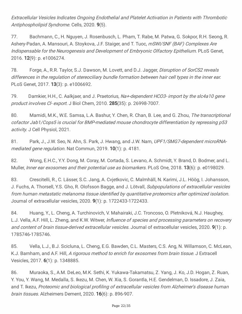
sEVs were isolated from the cochlear tissue of mice at P3, P7, P14, and P21 by ultracentrifugation asdescribed previously [35, 41] (Fig. 1a). Considering that the cochlea is surrounded by the rigid otic vesicle,we dissected the cochleae and ground them in a grinder at 40 Hz as gently as possible so as not to breakopen the cells. The samples were centrifuged at low speed (600 × g and 2,000 × g) to remove cell debrisand then at high speed (12,000 × g) to remove large extracellular vesicles. After passing through a 0.22µm �lter, the samples are concentrated by ultra�ltration with a 100 kDa MWCO ultra�lter. Finally, sEVswere isolated by ultracentrifugation at 110,000 × g. The RNAs and proteins extracted from sEVs wereused for miRNA sequencing and proteomics analysis, respectively. TEM by negative staining indicatedthe oval shape of sEVs (Fig. 1b), and we characterized the size and number of sEVs from mice ofdifferent ages by NTA and con�rmed that the diameter of the sEVs was 30–200 nm (Fig. 1d). Typical sEVmarker proteins – such as the tetraspanins CD63 and CD9 – and the composition of ESCRT-Ι complexTsg101 were detected in cochlear tissue-derived sEVs by western blotting (Fig. 1c). Marker proteins forother vesicles, including EEA1 (endosome marker), Rab7 (lysosome marker), and GAPDH, were used asnegative markers of sEVs and were not detected in the sEV samples (Fig. 1c). We also usedimmuno�uorescent staining to con�rm the presence of CD63 and CD9 in HCs and SCs (Fig. 1e). Together,these results suggest that this isolation method of cochlear tissue-derived sEVs is feasible and can yieldrelatively pure sEVs.
miRNA analysis of cochlear tissue-derived sEVs from mice of different ages
sEVs contain a variety of RNAs, especially miRNAs, that play important roles in gene regulation and thusmediate numerous biological processes [42, 43]. Because the role of sEV miRNA in the cochlea is poorly
Page 9/35
understood, we employed small RNA-seq to evaluated the cochlear tissue-derived exosomes from P3, P7,P14, and P21 mice to discover differentially expressed miRNA during the development of the cochlea.
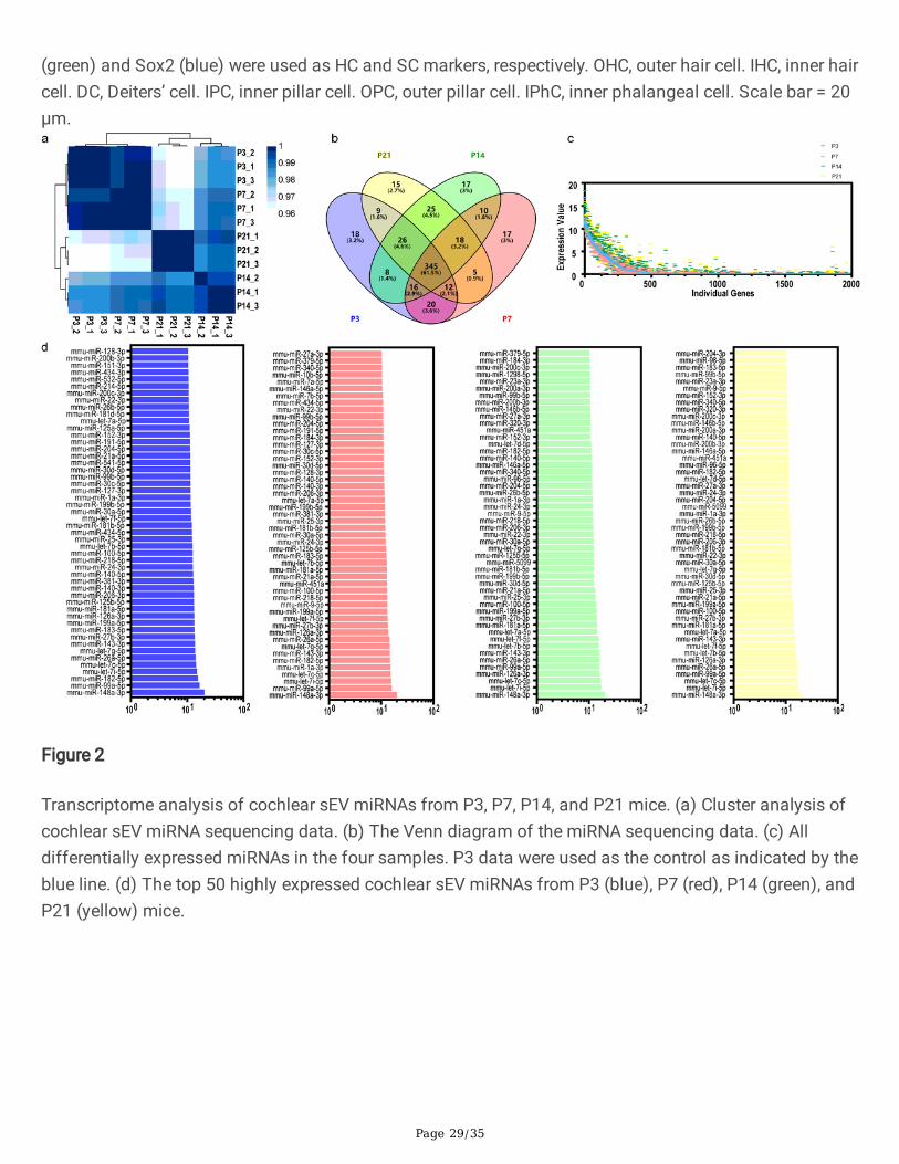
The correlations of the samples were tested by hierarchical clustering analysis, and the P3, P7, P14, andP21 groups were well-separated according to their Spearman correlation coe�cient (Fig. 2a). We detecteda total of 561 miRNAs, including 454, 453, 465, and 455 miRNAs from cochlear tissue-derived sEVs fromP3, P7, P14, and P21 mice, respectively (Fig. 2b). Furthermore, there were 18, 17, 17, and 15 miRNAs thatwere uniquely expressed at P3, P7, P14, and P21, respectively (Fig. 2b). The expression levels of allmiRNAs at P3, P7, P14, and P21 are shown in Fig. 2c. We compared the differentially expressed miRNAsbetween each of the age groups pairwise (Fig. S1), and the top 50 most abundant miRNAs in the four agegroups are shown in Fig. 2d.
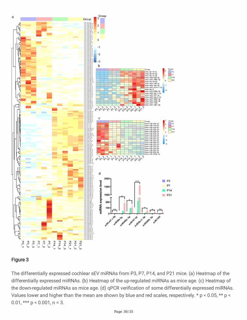
We found 179 miRNAs that were differentially expressed across the four age groups by pairwisecomparison, including 57, 33, 29, and 60 miRNAs that were highly expressed at P3, P7, P14, and P21,respectively (Fig. 3a, p < 0.05, fold change > 2). Further analysis of the 179 differentially expressedmiRNAs showed that 18 of these miRNAs became more prevalent in sEVs with age, while 17 miRNAsdecreased with age (Fig. 3b, c). Of the increased miRNAs, miRLet-7f-5p [44], miRLet-7e-5p [45], miRLet-7c-5p [46], miR29a-3p [47], miR146b-5p [48], miRLet-7d-5p [49], miR338-3p [50], miR144-3p [51], miRLet-7j [52], miR449a-5p [53], miR30c-1-3p [54], miR147-3p [55], miR30c-2-3p [56], and miR1195 [57] have beenattributed to a range of biological processes including cellular proliferation, cellular differentiation, andcellular signaling and communication. The miRNAs miR3074-1-3p, miR3095-3p, miR344b-3p, andmiR3057-5p have no reported biological functions. For the decreased miRNAs, miR495-3p [58], miR140-3p [59], miR434-5p [60], miR322-3p [61], miR409-3p [62], miR674-3p [63], miR335-3p [61], miR543-3p [64],miR341-3p [65], miR202-5p [66], miR369-3p [67], miR330-3p [68], miR370-3p [69], miR335-5p [70],miR503-3p [71], and miR503-5p [72] have been reported that related to biological processes, and onlymiR299a-3p has no reported function. We veri�ed the 18 increased miRNAs by qPCR, and Fig. 3d showsthat the expression of 7 miRNAs (miRLet7c-5p, miR29a-3p, miR449a-5p, miR147-3p, miR30c-2-3p,miR3095-3p, and miR1195) was matched to the results of bioinformatics analysis.
Functional analysis of differentially expressed miRNAs in cochlear sEVs
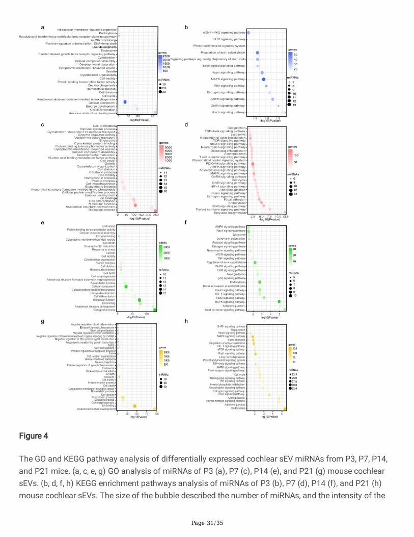
The GO and KEGG pathway analyses of the highly expressed miRNAs at P3, P7, and P14 were performedwith DIANA-mirPath v.3 (http://snf- 515788.vm.okeanos.grnet.gr/) using the target genes in the Tarbasev7.0 database (http://www.microrna.gr/tarbase). These miRNAs in cochlear sEVs at different ages havedifferent biological functions (Fig. 4). Notably, the GO analysis showed that these miRNAs are mainlyinvolved in anatomical structure development, cell differentiation, developmental maturation, growth, cellcycle, and vesicle-mediated transport (Fig. 4a, c, e, and g). Fig. 4(b, d, f, and h) show that the highlyexpressed miRNAs at P3, P7, P14, P21 are involved in the mTOR, PI3K-Akt, TGF-β, Wnt, Hippo, Notch, andcGMP-PKG signaling pathways. These �ndings suggest that these pathways likely actively involved inthe development of the cochlea and the formation of the auditory system. Among them Wnt, Notch, TGF-
β, and Hippo signaling have been implicated in progenitor cell proliferation and differentiation, as well ascell plane polarity during inner ear development [73, 74].
Label-free quantitative proteomics analysis of cochlear tissue-derived sEVs from mice of different ages
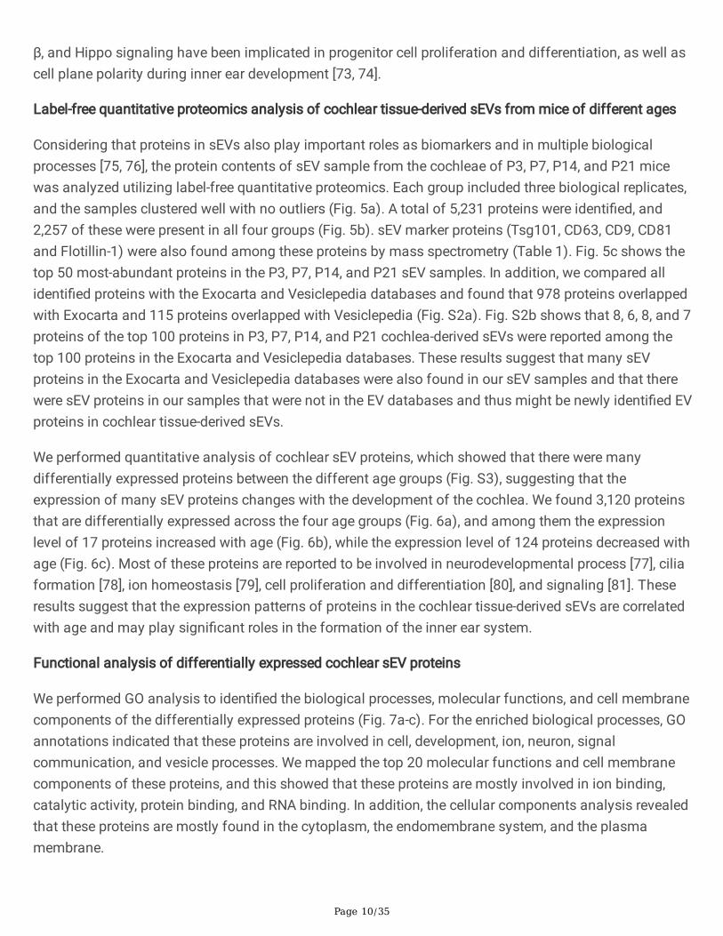
Considering that proteins in sEVs also play important roles as biomarkers and in multiple biologicalprocesses [75, 76], the protein contents of sEV sample from the cochleae of P3, P7, P14, and P21 micewas analyzed utilizing label-free quantitative proteomics. Each group included three biological replicates,and the samples clustered well with no outliers (Fig. 5a). A total of 5,231 proteins were identi�ed, and2,257 of these were present in all four groups (Fig. 5b). sEV marker proteins (Tsg101, CD63, CD9, CD81and Flotillin-1) were also found among these proteins by mass spectrometry (Table 1). Fig. 5c shows thetop 50 most-abundant proteins in the P3, P7, P14, and P21 sEV samples. In addition, we compared allidenti�ed proteins with the Exocarta and Vesiclepedia databases and found that 978 proteins overlappedwith Exocarta and 115 proteins overlapped with Vesiclepedia (Fig. S2a). Fig. S2b shows that 8, 6, 8, and 7proteins of the top 100 proteins in P3, P7, P14, and P21 cochlea-derived sEVs were reported among thetop 100 proteins in the Exocarta and Vesiclepedia databases. These results suggest that many sEVproteins in the Exocarta and Vesiclepedia databases were also found in our sEV samples and that therewere sEV proteins in our samples that were not in the EV databases and thus might be newly identi�ed EVproteins in cochlear tissue-derived sEVs.
We performed quantitative analysis of cochlear sEV proteins, which showed that there were manydifferentially expressed proteins between the different age groups (Fig. S3), suggesting that theexpression of many sEV proteins changes with the development of the cochlea. We found 3,120 proteinsthat are differentially expressed across the four age groups (Fig. 6a), and among them the expressionlevel of 17 proteins increased with age (Fig. 6b), while the expression level of 124 proteins decreased withage (Fig. 6c). Most of these proteins are reported to be involved in neurodevelopmental process [77], ciliaformation [78], ion homeostasis [79], cell proliferation and differentiation [80], and signaling [81]. Theseresults suggest that the expression patterns of proteins in the cochlear tissue-derived sEVs are correlatedwith age and may play signi�cant roles in the formation of the inner ear system.
Functional analysis of differentially expressed cochlear sEV proteins
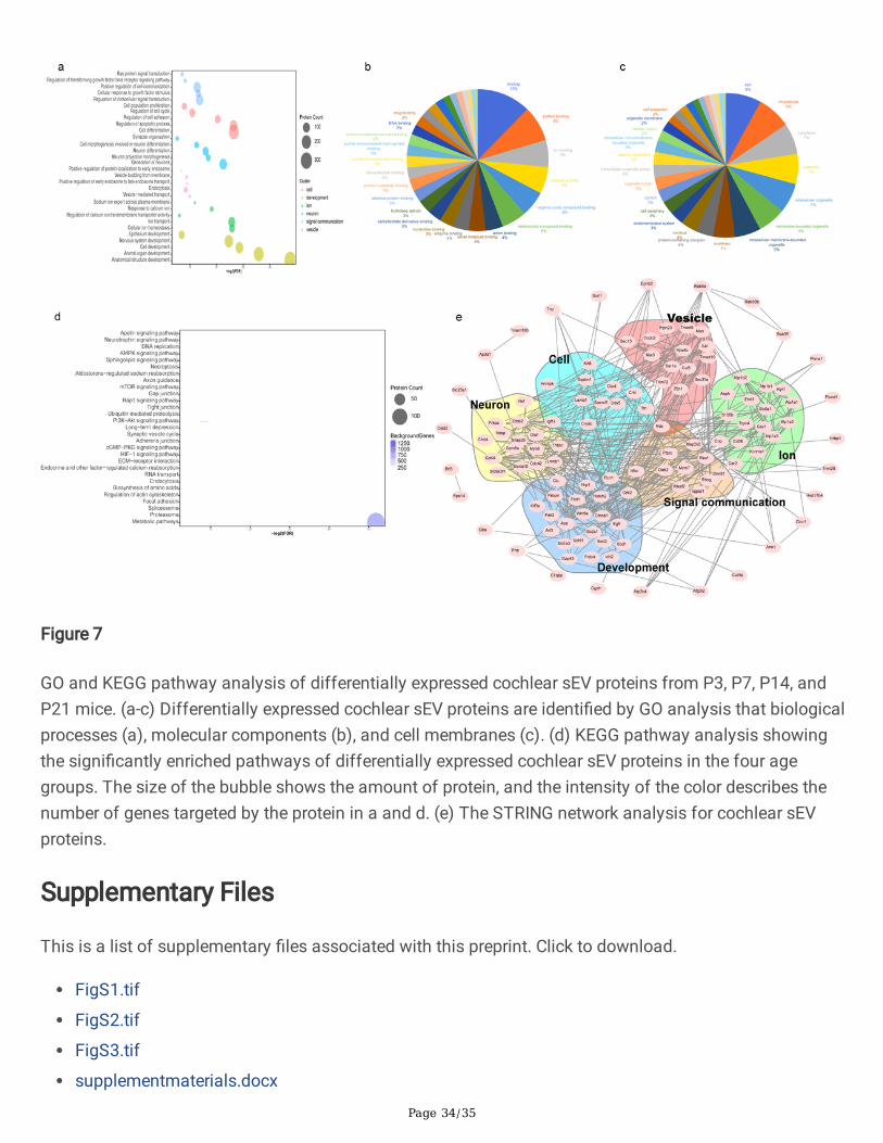
We performed GO analysis to identi�ed the biological processes, molecular functions, and cell membranecomponents of the differentially expressed proteins (Fig. 7a-c). For the enriched biological processes, GOannotations indicated that these proteins are involved in cell, development, ion, neuron, signalcommunication, and vesicle processes. We mapped the top 20 molecular functions and cell membranecomponents of these proteins, and this showed that these proteins are mostly involved in ion binding,catalytic activity, protein binding, and RNA binding. In addition, the cellular components analysis revealedthat these proteins are mostly found in the cytoplasm, the endomembrane system, and the plasmamembrane.
Page 11/35
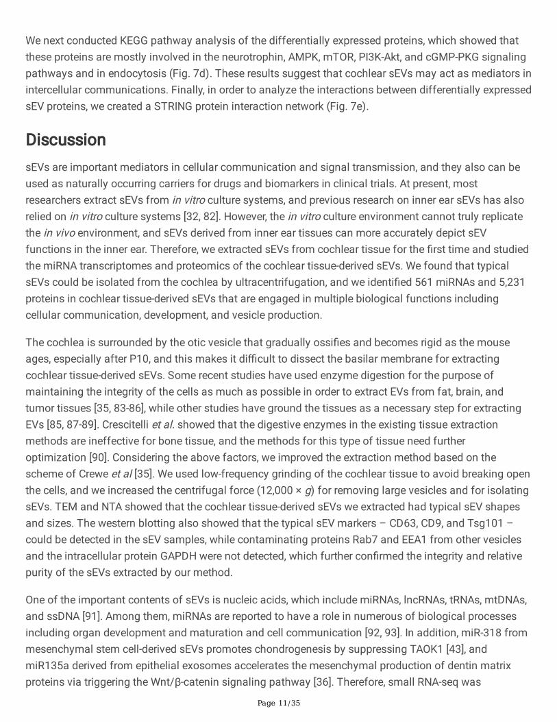
We next conducted KEGG pathway analysis of the differentially expressed proteins, which showed thatthese proteins are mostly involved in the neurotrophin, AMPK, mTOR, PI3K-Akt, and cGMP-PKG signalingpathways and in endocytosis (Fig. 7d). These results suggest that cochlear sEVs may act as mediators inintercellular communications. Finally, in order to analyze the interactions between differentially expressedsEV proteins, we created a STRING protein interaction network (Fig. 7e).
DiscussionsEVs are important mediators in cellular communication and signal transmission, and they also can beused as naturally occurring carriers for drugs and biomarkers in clinical trials. At present, mostresearchers extract sEVs from in vitro culture systems, and previous research on inner ear sEVs has alsorelied on in vitro culture systems [32, 82]. However, the in vitro culture environment cannot truly replicatethe in vivo environment, and sEVs derived from inner ear tissues can more accurately depict sEVfunctions in the inner ear. Therefore, we extracted sEVs from cochlear tissue for the �rst time and studiedthe miRNA transcriptomes and proteomics of the cochlear tissue-derived sEVs. We found that typicalsEVs could be isolated from the cochlea by ultracentrifugation, and we identi�ed 561 miRNAs and 5,231proteins in cochlear tissue-derived sEVs that are engaged in multiple biological functions includingcellular communication, development, and vesicle production.
The cochlea is surrounded by the otic vesicle that gradually ossi�es and becomes rigid as the mouseages, especially after P10, and this makes it di�cult to dissect the basilar membrane for extractingcochlear tissue-derived sEVs. Some recent studies have used enzyme digestion for the purpose ofmaintaining the integrity of the cells as much as possible in order to extract EVs from fat, brain, andtumor tissues [35, 83-86], while other studies have ground the tissues as a necessary step for extractingEVs [85, 87-89]. Crescitelli et al. showed that the digestive enzymes in the existing tissue extractionmethods are ineffective for bone tissue, and the methods for this type of tissue need furtheroptimization [90]. Considering the above factors, we improved the extraction method based on thescheme of Crewe et al [35]. We used low-frequency grinding of the cochlear tissue to avoid breaking openthe cells, and we increased the centrifugal force (12,000 × g) for removing large vesicles and for isolatingsEVs. TEM and NTA showed that the cochlear tissue-derived sEVs we extracted had typical sEV shapesand sizes. The western blotting also showed that the typical sEV markers – CD63, CD9, and Tsg101 –could be detected in the sEV samples, while contaminating proteins Rab7 and EEA1 from other vesiclesand the intracellular protein GAPDH were not detected, which further con�rmed the integrity and relativepurity of the sEVs extracted by our method.
One of the important contents of sEVs is nucleic acids, which include miRNAs, lncRNAs, tRNAs, mtDNAs,and ssDNA [91]. Among them, miRNAs are reported to have a role in numerous of biological processesincluding organ development and maturation and cell communication [92, 93]. In addition, miR-318 frommesenchymal stem cell-derived sEVs promotes chondrogenesis by suppressing TAOK1 [43], andmiR135a derived from epithelial exosomes accelerates the mesenchymal production of dentin matrixproteins via triggering the Wnt/β-catenin signaling pathway [36]. Therefore, small RNA-seq was
Page 12/35
performed to characterize the miRNAs in cochlear tissue-derived sEVs and to elucidate their possible rolesin the cochlea. We identi�ed 561 miRNAs in cochlear sEVs, including 179 differentially expressed miRNA,and we found that the expression of 18 miRNAs increased and 17 miRNAs decreased as the mice aged.We used qPCR to verify the expression of miRNAs and found that 7 miRNAs (miRLet7c-5p, miR29a-3p,miR449a-5p, miR147-3p, miR30c-2-3p, miR3095-3p, and miR1195) were consistent with the RNA-seqanalysis results. Overexpression of miRLet7c-5p can inhibit laryngeal squamous cell carcinoma cellproliferation and can regulate microglial activation during the repair of brain injury [46, 94], andupregulation of miR30c-2-3p suppresses gastric cancer and the proliferation of renal cell carcinomas [56,95]. miR29a-3p, miR449a-5p, and miR147-3p have been reported to be accumulated in exosomes derivedfrom oral squamous cells, macrophages, and bronchoalveolar lavage �uid [53, 55, 96, 97]. In addition,upregulation of miR29a-3p rescues bronchopulmonary dysplasia and has a negative regulatory effect onthe Smad, NFκB, and canonical Wnt signaling pathways [98-100]. Importantly, miR29a-3p directly targetsthe Wnt-related genes DVL3 (Dishevelled 3), CSNK2A2 (casein kinase 2 alpha 2 polypeptide), FZD3(Frizzled family receptor 3), and FZD5 (Frizzled family receptor 5) [100]. These 7 miRNAs, whoseexpression increases as mice age, may be involved in the development of the cochlea after birth and mayact as new targets to be further studied in the future to elucidate the detailed mechanisms behindcochlear development.
We also performed GO and KEGG analysis on the highly expressed miRNAs. GO analysis showed thatthese miRNAs are important for growth, development, maturation, anatomical structure development, ionbinding, cell differentiation, and cell proliferation, all of which are relevant to cochlear developmentevents. The enriched miRNAs in the sEVs are involved in the Hippo, MAPK, Wnt, Notch, TGF-β, and PI3K-Akt signaling pathways, most of which were essential to the development of the cochlea and inregulating the pluripotency of stem cells. These results showed that miRNAs enriched in cochlear tissue-derived sEVs may be essential for cell communication during inner ear development.
Proteins are another major component of sEVs and play signi�cant roles in cell communication,mediation of immune responses, and proliferation of cancer cells and as markers for diseasediagnosis [101, 102]. We performed proteomics analysis of the cochlear tissue-derived sEV proteins andidenti�ed 5,231 proteins, including 3,120 differentially expressed proteins, in the four age groups. We alsofound the sEV marker proteins CD63, CD9, CD81, and Tsg101 in the proteomics data, which again veri�edthe purity of our isolated cochlear sEVs. We identi�ed 1,051 proteins in the cochlear sEVs that overlappedwith proteins in the Vesiclepedia and Exocarta databases.
Among the differentially expressed sEV proteins, we found that the expression of 17 proteins increasedand 124 proteins decreased as the mice aged. For the 17 increased proteins, Slc4a10, Fbxo2, and S100bare related to the process of neurodevelopment and in regulating the differentiation and excitability ofneurons [103, 104], which suggests that these three proteins may be involved in the innervation of thecochlea that is required for hearing function. Fbxo2 is enriched in the inner ear and is a key regulator forage-related hearing loss [105]. Tlr3 is also presented in the inner ear and regulates immuneresponses [106, 107], and Tlr4 acts as a mediator in protecting HCs from damage by exosomes secreted
Page 13/35
by SCs [32]. This suggests that Tlr3 might also have a protective role on inner ear’s development. Inaddition, Slc4a10 is important for maintaining ion homeostasis of inner ear, and the absence of Slc4a10can lead to hearing loss [108, 109]. Among the proteins that decrease with age, Hnrnp [110], Ddx5 [111],Ilf3 [112], Lamtor5 [113], Psmd2 [114], Ddb1 [115], Psmd6 [116], and Chd4 [117] are reported to related tocell proliferation and differentiation. Ptbp1 [118], Chd4 [119], Ruvbl2 [120], Cul4a [121], andLama4 [122] are required for early developmental processes and neuronal differentiation, and someproteins also presented in the inner ear, such as P3h1 [123], Sorcs2 [78], Panx3 [124, 125], Idh1 [126],Lamb1 [127]. P3h1 knockout mice showed dysplasia of middle ear bones and hearing impairment [123].Sorcs2 regulates HC development by maintaining the shape of the cilia [78]. Idh1 is protein found in thecochlea and may play a role in age-related hearing loss act as an antioxidant [126, 128]. Panx3 is apannexin channel protein and is mainly presented in the cochlear bone structure and is essential for themaintenance of cochlear morphology [124, 125], and the expression of Panx3 is regulated duringdevelopment and reaches its peak at P8 [124]. According to previous reports, these cochlear sEV proteinsmay play important roles and may be used as new targets for the development of the cochlea in thefuture.
We conducted GO and KEGG analysis of the differentially expressed proteins. The GO analysis revealedthat cochlear tissue-derived sEV proteins play a signi�cant role in various biology processes such as Rasprotein signal transduction, cell proliferation, cell differentiation, neuron differentiation, endocytosis,cellular ion homeostasis, nervous system development, and organ development and that these proteinsare involved in many molecular functions, including ion binding, protein binding, and RNA binding. Thecellular components analysis showed that sEVs can be secreted from the cell, cytoplasm, andendomembrane system. These proteins are invested in axon guidance, the synaptic vesicle cycle, theAMPK signaling pathway, the mTOR signaling pathway, the PI3K-Akt signaling pathway, and endocytosis,according to the KEGG pathway analysis. Synapses on HCs are connected to spiral neurons fortransmitting signals to the brain, and this activity is essential for hearing function [8, 129, 130]. Inaddition, these pathways have also been reported to be critical for the biological functions of the innerear. Down-regulating the AMPK signaling pathway can reduce noise-induced damage to HCs and canprevent the age-related hearing loss [131, 132]. The mTOR signaling pathway is involved in reprogramingMyc/NICD to promote HC regeneration [15], and age-related hearing loss and HC damage can be relievedby inhibiting the mTOR signaling pathway [133, 134]. Balancing the AMPK and mTOR signalingpathways can further protect HCs from damage by ototoxic drugs [135]. We also created a STRINGprotein-interaction network investigating the interactions between differentially expressed sEV proteins,and this showed that sEV proteins involved in vesicles, development, neurons, signal communication,cellular processes, and ion homeostasis have close interactions with each other and with otherdifferentially expressed cochlear sEV proteins. These results indicate that sEV may be critical for thedevelopment of the cochlear nervous system, as well as for the protection and regeneration of HCs duringdevelopment.
In summary, we isolated cochlear tissue-derived sEVs from mice of different ages after birth byultracentrifugation and characterized the microRNA transcriptomes and proteomics of these sEVs to
Page 14/35
elucidate their possible roles. We found 561 miRNAs and 5,231 proteins in the cochlear sEVs, and amongthem 179 miRNAs and 3,120 proteins were differentially expressed at different ages. We further analyzedthese differentially expressed miRNA and proteins and found that the expression of many miRNAs andproteins may be relevant to the maturation of HCs, to changes in SCs characteristics, to neuraldevelopment, and to the protection of HCs from P3 to P21. These miRNAs and proteins might be used asnew targets for further studying the detailed mechanism of cochlear development after birth. Based onour results, we speculate that sEVs play a regulatory role in the maturation of HCs, HC regeneration frominner ear stem cells, and neural development during the development of the inner ear after birth, and thisshould be further con�rmed in future studies.
DeclarationsEthics approval and consent to participate
All studies of animal followed the authorized guidelines of Southeast University's Animal Care and UseCommittee and the National Institutes of Health Guide for the Care and Use of Laboratory Animals. Theamount of animals was kept to a minimum, and all efforts were made to reduce their suffering.
Consent for publication
Not applicable.
Availability of data and material
All the data analyzed by this research is included in this article and its supplementary �les.
Competing interests
The authors declare that they have no con�ict of interest.
Funding
This work was supported by grants from the National Key RD Program of China (No. 2017YFA0103903),the Strategic Priority Research Program of the Chinese Academy of Sciences (No. XDA16010303), theNational Natural Science Foundation of China (Nos. 81970892, 81970882, 81870721, 81900944, and81700913), the Natural Science Foundation of Jiangsu Province (Nos. BK20190062 and BE2019711), theShenzhen Fundamental Research Program (No. JCYJ20190814093401920), the Jiangsu ProvincialMedical Youth Talent of the Project of Invigorating Health Care through Science, Technology andEducation (No. QNRC2016002), and the Fundamental Research Funds for the Central Universities for theSupport Program of Zhishan Youth Scholars of Southeast University (No. 2242021R41136).
Author contributions
Page 15/35
RC, SZ, and WAT designed the experiments. PJ, JA, LW, YZ, HX, and MT isolated and characterized thecochlear sEVs. XM performed the miRNA analysis. SH and LM performed the proteomics analysis. PJ andSZ analyzed all data and wrote the manuscript. All authors read and approved the �nal manuscript.
Acknowledgments
Thanks to all colleagues who contributed to this research.
References1. Wright, T.J., E.P. Hatch, H. Karabagli, P. Karabagli, G.C. Schoenwolf, and S.L. Mansour,Expression of mouse �broblast growth factor and �broblast growth factor receptor genes during earlyinner ear development. Dev Dyn, 2003. 228(2): p. 267-72.
2. LeMasurier, M. and P.G. Gillespie, Hair-cell mechanotransduction and cochlear ampli�cation.Neuron, 2005. 48(3): p. 403-15.
3. Atkinson, P.J., E. Huarcaya Najarro, Z.N. Sayyid, and A.G. Cheng, Sensory hair cell developmentand regeneration: similarities and differences. Development, 2015. 142(9): p. 1561-71.
4. Burns, J.C., B.C. Cox, B.R. Thiede, J. Zuo, and J.T. Corwin, In vivo proliferative regeneration ofbalance hair cells in newborn mice. J Neurosci, 2012. 32(19): p. 6570-7.
5. Raft, S., E.J. Koundakjian, H. Quinones, C.S. Jayasena, L.V. Goodrich, J.E. Johnson, N. Segil, andA.K. Groves, Cross-regulation of Ngn1 and Math1 coordinates the production of neurons and sensory haircells during inner ear development. Development, 2007. 134(24): p. 4405-15.
6. Goutman, J.D., A.B. Elgoyhen, and M.E. Gómez-Casati, Cochlear hair cells: The sound-sensingmachines. FEBS Lett, 2015. 589(22): p. 3354-61.
7. Kim, K.X. and R. Fettiplace, Developmental changes in the cochlear hair cell mechanotransducerchannel and their regulation by transmembrane channel-like proteins. J Gen Physiol, 2013. 141(1): p. 141-8.
8. Fettiplace, R., Hair Cell Transduction, Tuning, and Synaptic Transmission in the MammalianCochlea. Compr Physiol, 2017. 7(4): p. 1197-1227.
9. Huang, L.C., P.R. Thorne, G.D. Housley, and J.M. Montgomery, Spatiotemporal de�nition ofneurite outgrowth, re�nement and retraction in the developing mouse cochlea. Development, 2007.134(16): p. 2925-33.
10. Fettiplace, R. and K.X. Kim, The physiology of mechanoelectrical transduction channels inhearing. Physiol Rev, 2014. 94(3): p. 951-86.
Page 16/35
11. D, Ó.M. and A.J. Ricci, A Bundle of Mechanisms: Inner-Ear Hair-Cell Mechanotransduction.Trends Neurosci, 2019. 42(3): p. 221-236.
12. Sun, S., T. Babola, G. Pregernig, K.S. So, M. Nguyen, S.M. Su, A.T. Palermo, D.E. Bergles, J.C.Burns, and U. Müller, Hair Cell Mechanotransduction Regulates Spontaneous Activity and Spiral GanglionSubtype Speci�cation in the Auditory System. Cell, 2018. 174(5): p. 1247-1263.e15.
13. Samarajeewa, A., D.R. Lenz, L. Xie, H. Chiang, R. Kirchner, J.F. Mulvaney, A.S.B. Edge, and A.Dabdoub, Transcriptional response to Wnt activation regulates the regenerative capacity of themammalian cochlea. Development, 2018. 145(23).
14. Wang, T., R. Chai, G.S. Kim, N. Pham, L. Jansson, D.H. Nguyen, B. Kuo, L.A. May, J. Zuo, L.L.Cunningham, and A.G. Cheng, Lgr5+ cells regenerate hair cells via proliferation and directtransdifferentiation in damaged neonatal mouse utricle. Nat Commun, 2015. 6: p. 6613.
15. Shu, Y., W. Li, M. Huang, Y.-Z. Quan, D. Scheffer, C. Tian, Y. Tao, X. Liu, K. Hochedlinger, A.A.Indzhykulian, Z. Wang, H. Li, and Z.-Y. Chen, Renewed proliferation in adult mouse cochlea andregeneration of hair cells. Nature communications, 2019. 10(1): p. 5530-5530.
16. Kelley, M.W., Regulation of cell fate in the sensory epithelia of the inner ear. Nat Rev Neurosci,2006. 7(11): p. 837-49.
17. Wu, J., W. Li, C. Lin, Y. Chen, C. Cheng, S. Sun, M. Tang, R. Chai, and H. Li, Co-regulation of theNotch and Wnt signaling pathways promotes supporting cell proliferation and hair cell regeneration inmouse utricles. Sci Rep, 2016. 6: p. 29418.
18. Kiernan, A.E., Notch signaling during cell fate determination in the inner ear. Semin Cell Dev Biol,2013. 24(5): p. 470-9.
19. Jiang, D., J. Du, X. Zhang, W. Zhou, L. Zong, C. Dong, K. Chen, Y. Chen, X. Chen, and H. Jiang,miR-124 promotes the neuronal differentiation of mouse inner ear neural stem cells. Int J Mol Med, 2016.38(5): p. 1367-1376.
20. Li, H., W. Kloosterman, and D.M. Fekete, MicroRNA-183 family members regulate sensorineuralfates in the inner ear. J Neurosci, 2010. 30(9): p. 3254-63.
21. Borghesan, M., J. Fa�án-Labora, O. Eleftheriadou, P. Carpintero-Fernández, M. Paez-Ribes, G.Vizcay-Barrena, A. Swisa, D. Kolodkin-Gal, P. Ximénez-Embún, R. Lowe, B. Martín-Martín, H. Peinado, J.Muñoz, R.A. Fleck, Y. Dor, I. Ben-Porath, A. Vossenkamper, D. Muñoz-Espin, and A. O'Loghlen, SmallExtracellular Vesicles Are Key Regulators of Non-cell Autonomous Intercellular Communication inSenescence via the Interferon Protein IFITM3. Cell Rep, 2019. 27(13): p. 3956-3971.e6.
22. Wang, C., V. Börger, M. Sardari, F. Murke, J. Skuljec, R. Pul, N. Hagemann, E. Dzyubenko, R.Dittrich, J. Gregorius, M. Hasenberg, C. Kleinschnitz, A. Popa-Wagner, T.R. Doeppner, M. Gunzer, B. Giebel,
Page 17/35
and D.M. Hermann, Mesenchymal Stromal Cell-Derived Small Extracellular Vesicles Induce IschemicNeuroprotection by Modulating Leukocytes and Speci�cally Neutrophils. Stroke, 2020. 51(6): p. 1825-1834.
23. Loyer, X., I. Zlatanova, C. Devue, M. Yin, K.Y. Howangyin, P. Klaihmon, C.L. Guerin, M. Khelou�, J.Vilar, K. Zannis, B.K. Fleischmann, D.W. Hwang, J. Park, H. Lee, P. Menasché, J.S. Silvestre, and C.M.Boulanger, Intra-Cardiac Release of Extracellular Vesicles Shapes In�ammation Following MyocardialInfarction. Circ Res, 2018. 123(1): p. 100-106.
24. Pegtel, D.M. and S.J. Gould, Exosomes. Annu Rev Biochem, 2019. 88: p. 487-514.
25. Cocucci, E. and J. Meldolesi, Ectosomes and exosomes: shedding the confusion betweenextracellular vesicles. Trends Cell Biol, 2015. 25(6): p. 364-72.
26. Chen, I.H., L. Xue, C.C. Hsu, J.S. Paez, L. Pan, H. Andaluz, M.K. Wendt, A.B. Iliuk, J.K. Zhu, andW.A. Tao, Phosphoproteins in extracellular vesicles as candidate markers for breast cancer. Proc NatlAcad Sci U S A, 2017. 114(12): p. 3175-3180.
27. Zhang, Z., T. Xing, Y. Chen, and J. Xiao, Exosome-mediated miR-200b promotes colorectal cancerproliferation upon TGF-β1 exposure. Biomed Pharmacother, 2018. 106: p. 1135-1143.
28. Liao, F.L., L. Tan, H. Liu, J.J. Wang, X.T. Ma, B. Zhao, Y. Chen, J. Bihl, Y. Yang, and R.L. Chen,Hematopoietic stem cell-derived exosomes promote hematopoietic differentiation of mouse embryonicstem cells in vitro via inhibiting the miR126/Notch1 pathway. Acta Pharmacol Sin, 2018. 39(4): p. 552-560.
29. Guo, L., Y. Zhu, L. Li, S. Zhou, G. Yin, G. Yu, and H. Cui, Breast cancer cell-derived exosomal miR-20a-5p promotes the proliferation and differentiation of osteoclasts by targeting SRCIN1. Cancer Med,2019. 8(12): p. 5687-5701.
30. Sharma, P., L. Schiapparelli, and H.T. Cline, Exosomes function in cell-cell communication duringbrain circuit development. Curr Opin Neurobiol, 2013. 23(6): p. 997-1004.
31. McGough, I.J. and J.P. Vincent, Exosomes in developmental signalling. Development, 2016.143(14): p. 2482-93.
32. Breglio, A.M., L.A. May, M. Barzik, N.C. Welsh, S.P. Francis, T.Q. Costain, L. Wang, D.E. Anderson,R.S. Petralia, Y.X. Wang, T.B. Friedman, M.J. Wood, and L.L. Cunningham, Exosomes mediate sensory haircell protection in the inner ear. J Clin Invest, 2020. 130(5): p. 2657-2672.
33. Men, Y., J. Yelick, S. Jin, Y. Tian, M.S.R. Chiang, H. Higashimori, E. Brown, R. Jarvis, and Y. Yang,Exosome reporter mice reveal the involvement of exosomes in mediating neuron to astrogliacommunication in the CNS. Nat Commun, 2019. 10(1): p. 4136.
Page 18/35
34. Lai, R., C. Cai, W. Wu, P. Hu, and Q. Wang, Exosomes derived from mouse inner ear stem cellsattenuate gentamicin-induced ototoxicity in vitro through the miR-182-5p/FOXO3 axis. J Tissue EngRegen Med, 2020. 14(8): p. 1149-1156.
35. Crewe, C., N. Jo�n, J.M. Rutkowski, M. Kim, F. Zhang, D.A. Towler, R. Gordillo, and P.E. Scherer, AnEndothelial-to-Adipocyte Extracellular Vesicle Axis Governed by Metabolic State. Cell, 2018. 175(3): p.695-708.e13.
36. Jiang, N., L. Xiang, L. He, G. Yang, J. Zheng, C. Wang, Y. Zhang, S. Wang, Y. Zhou, T.-J. Sheu, J.Wu, K. Chen, P.G. Coelho, N.M. Tovar, S.H. Kim, M. Chen, Y.-H. Zhou, and J.J. Mao, Exosomes MediateEpithelium-Mesenchyme Crosstalk in Organ Development. ACS nano, 2017. 11(8): p. 7736-7746.
37. Zhang, S., Y. Zhang, Y. Dong, L. Guo, Z. Zhang, B. Shao, J. Qi, H. Zhou, W. Zhu, X. Yan, G. Hong, L.Zhang, X. Zhang, M. Tang, C. Zhao, X. Gao, and R. Chai, Knockdown of Foxg1 in supporting cellsincreases the trans-differentiation of supporting cells into hair cells in the neonatal mouse cochlea. CellMol Life Sci, 2020. 77(7): p. 1401-1419.
38. Vlachos, I.S., M.D. Paraskevopoulou, D. Karagkouni, G. Georgakilas, T. Vergoulis, I. Kanellos, I.L.Anastasopoulos, S. Maniou, K. Karathanou, D. Kalfakakou, A. Fevgas, T. Dalamagas, and A.G.Hatzigeorgiou, DIANA-TarBase v7.0: indexing more than half a million experimentally supportedmiRNA:mRNA interactions. Nucleic Acids Res, 2015. 43(Database issue): p. D153-9.
39. Vlachos, I.S., K. Zagganas, M.D. Paraskevopoulou, G. Georgakilas, D. Karagkouni, T. Vergoulis, T.Dalamagas, and A.G. Hatzigeorgiou, DIANA-miRPath v3.0: deciphering microRNA function withexperimental support. Nucleic Acids Res, 2015. 43(W1): p. W460-6.
40. Sun, J., S. Han, L. Ma, H. Zhang, Z. Zhan, H.A. Aguilar, H. Zhang, K. Xiao, Y. Gu, Z. Gu, and W.A.Tao, Synergistically Bifunctional Paramagnetic Separation Enables E�cient Isolation of UrineExtracellular Vesicles and Downstream Phosphoproteomic Analysis. ACS Appl Mater Interfaces, 2021.13(3): p. 3622-3630.
41. Jiang, N., L. Xiang, L. He, G. Yang, J. Zheng, C. Wang, Y. Zhang, S. Wang, Y. Zhou, T.J. Sheu, J.Wu, K. Chen, P.G. Coelho, N.M. Tovar, S.H. Kim, M. Chen, Y.H. Zhou, and J.J. Mao, Exosomes MediateEpithelium-Mesenchyme Crosstalk in Organ Development. ACS Nano, 2017. 11(8): p. 7736-7746.
42. Zhang, Y., C. Li, Y. Qin, P. Cepparulo, M. Millman, M. Chopp, A. Kemper, A. Szalad, X. Lu, L. Wang,and Z.G. Zhang, Small extracellular vesicles ameliorate peripheral neuropathy and enhancechemotherapy of oxaliplatin on ovarian cancer. J Extracell Vesicles, 2021. 10(5): p. e12073.
43. Jing, H., X. Zhang, K. Luo, Q. Luo, M. Yin, W. Wang, Z. Zhu, J. Zheng, and X. He, miR-381-abundant small extracellular vesicles derived from kartogenin-preconditioned mesenchymal stem cellspromote chondrogenesis of MSCs by targeting TAOK1. Biomaterials, 2020. 231: p. 119682.
Page 19/35
44. Chen, G., H. Gu, T. Fang, K. Zhou, J. Xu, and X. Yin, Hypoxia-induced let-7f-5p/TARBP2 feedbackloop regulates osteosarcoma cell proliferation and invasion by inhibiting the Wnt signaling pathway.Aging (Albany NY), 2020. 12(8): p. 6891-6903.
45. Handgraaf, S., R. Dusaulcy, F. Visentin, J. Philippe, and Y. Gosmain, Let-7e-5p Regulates GLP-1Content and Basal Release From Enteroendocrine L Cells From DIO Male Mice. Endocrinology, 2020.161(2).
46. Wu, Y., Y. Zhang, X. Zheng, F. Dai, Y. Lu, L. Dai, M. Niu, H. Guo, W. Li, X. Xue, Y. Bo, Y. Guo, J. Qin, Y.Qin, H. Liu, Y. Zhang, T. Yang, L. Li, L. Zhang, R. Hou, S. Wen, C. An, H. Li, W. Xu, and W. Gao, Circular RNAcircCORO1C promotes laryngeal squamous cell carcinoma progression by modulating the let-7c-5p/PBX3axis. Mol Cancer, 2020. 19(1): p. 99.
47. Qu, F., B. Zhu, Y.L. Hu, Q.S. Mao, and Y. Feng, LncRNA HOXA-AS3 promotes gastric cancerprogression by regulating miR-29a-3p/LTβR and activating NF-κB signaling. Cancer Cell Int, 2021. 21(1):p. 118.
48. Tu, Z., J. Xiong, R. Xiao, L. Shao, X. Yang, L. Zhou, W. Yuan, M. Wang, Q. Yin, Y. Wu, S. Pan, J.Leng, D. Jiang, C. He, and Q. Zhang, Loss of miR-146b-5p promotes T cell acute lymphoblastic leukemiamigration and invasion via the IL-17A pathway. J Cell Biochem, 2019. 120(4): p. 5936-5948.
49. Suzuki, A., H. Yoshioka, D. Summakia, N.G. Desai, G. Jun, P. Jia, D.S. Loose, K. Ogata, M.V. Gajera,Z. Zhao, and J. Iwata, MicroRNA-124-3p suppresses mouse lip mesenchymal cell proliferation throughthe regulation of genes associated with cleft lip in the mouse. BMC Genomics, 2019. 20(1): p. 852.
50. Zhang, R., H. Shi, F. Ren, W. Feng, Y. Cao, G. Li, Z. Liu, P. Ji, and M. Zhang, MicroRNA-338-3psuppresses ovarian cancer cells growth and metastasis: implication of Wnt/catenin beta and MEK/ERKsignaling pathways. J Exp Clin Cancer Res, 2019. 38(1): p. 494.
51. Hou, G., J. Yang, J. Tang, and Y. He, LncRNA GAS6-AS2 promotes non-small-cell lung cancer cellproliferation via regulating miR-144-3p/ MAPK6 axis. Cell Cycle, 2021. 20(2): p. 179-193.
52. Zhang, J., N. Wang, and A. Xu, miR‐10b‐3p, miR‐8112 and let‐7j as potential biomarkers forautoimmune inner ear diseases. Mol Med Rep, 2019. 20(1): p. 171-181.
53. Ni, Z., L. Kuang, H. Chen, Y. Xie, B. Zhang, J. Ouyang, J. Wu, S. Zhou, L. Chen, N. Su, Q. Tan, X.Luo, B. Chen, S. Chen, L. Yin, H. Huang, X. Du, and L. Chen, The exosome-like vesicles from osteoarthriticchondrocyte enhanced mature IL-1β production of macrophages and aggravated synovitis inosteoarthritis. Cell Death Dis, 2019. 10(7): p. 522.
54. Zhang, L.S., Y.D. Zhou, Y.Q. Peng, H.L. Zeng, S. Yoshida, and T.T. Zhao, Identi�cation of alteredmicroRNAs in retinas of mice with oxygen-induced retinopathy. Int J Ophthalmol, 2019. 12(5): p. 739-745.
Page 20/35
55. Tang, B., Y. Wu, H. Fang, Y. Wu, and K. Shi, Small RNA Sequencing Reveals Exosomal miRNAsInvolved in the Treatment of Asthma by Scorpio and Centipede. Biomed Res Int, 2020. 2020: p. 1061407.
56. Tang, C.T., Q. Liang, L. Yang, X.L. Lin, S. Wu, Y. Chen, X.T. Zhang, Y.J. Gao, and Z.Z. Ge, RAB31Targeted by MiR-30c-2-3p Regulates the GLI1 Signaling Pathway, Affecting Gastric Cancer CellProliferation and Apoptosis. Front Oncol, 2018. 8: p. 554.
57. Tagne, J.B., O.R. Mohtar, J.D. Campbell, M. Lakshminarayanan, J. Huang, A.C. Hinds, J. Lu, andM.I. Ramirez, Transcription factor and microRNA interactions in lung cells: an inhibitory link between NK2homeobox 1, miR-200c and the developmental and oncogenic factors N�b and Myb. Respir Res, 2015.16(1): p. 22.
58. Xia, Y., Y. Zhou, H. Han, P. Li, W. Wei, and N. Lin, lncRNA NEAT1 facilitates melanoma cellproliferation, migration, and invasion via regulating miR-495-3p and E2F3. J Cell Physiol, 2019. 234(11):p. 19592-19601.
59. Dou, D., X. Ren, M. Han, X. Xu, X. Ge, Y. Gu, X. Wang, and S. Zhao, Circ_0008039 supports breastcancer cell proliferation, migration, invasion, and glycolysis by regulating the miR-140-3p/SKA2 axis. MolOncol, 2021. 15(2): p. 697-709.
60. Qiu, W.I., H.B. Chen, Z.Q. Jiang, and H.G. Zhou, [Effect of Xiaoai Jiedu Recipe on mIRNAExpression Pro�les in H Tumor-bearing Mice]. Zhongguo Zhong Xi Yi Jie He Za Zhi, 2016. 36(9): p.1112-1118.
61. Meyer, S.U., S. Sass, N.S. Mueller, S. Krebs, S. Bauersachs, S. Kaiser, H. Blum, C. Thirion, S.Krause, F.J. Theis, and M.W. Pfa�, Integrative Analysis of MicroRNA and mRNA Data Reveals anOrchestrated Function of MicroRNAs in Skeletal Myocyte Differentiation in Response to TNF-α or IGF1.PLoS One, 2015. 10(8): p. e0135284.
62. Liu, X., F. Zhou, Y. Yang, W. Wang, L. Niu, D. Zuo, X. Li, H. Hua, B. Zhang, Y. Kou, J. Guo, F. Kong,W. Pan, D. Gao, J.M. Meves, H. Sun, M. Xue, Q. Zhang, Y. Wang, and R. Tang, MiR-409-3p and MiR-1896co-operatively participate in IL-17-induced in�ammatory cytokine production in astrocytes andpathogenesis of EAE mice via targeting SOCS3/STAT3 signaling. Glia, 2019. 67(1): p. 101-112.
63. Eom, T.Y., S.B. Han, J. Kim, J.A. Blundon, Y.D. Wang, J. Yu, K. Anderson, D.B. Kaminski, S.M.Sakurada, S.M. Pruett-Miller, L. Horner, B. Wagner, C.G. Robinson, M. Eicholtz, D.C. Rose, and S.S.Zakharenko, Schizophrenia-related microdeletion causes defective ciliary motility and brain ventricleenlargement via microRNA-dependent mechanisms in mice. Nat Commun, 2020. 11(1): p. 912.
64. Wu, X., X. Meng, F. Tan, Z. Jiao, X. Zhang, H. Tong, X. He, X. Luo, P. Xu, and S. Qu, RegulatoryMechanism of miR-543-3p on GLT-1 in a Mouse Model of Parkinson's Disease. ACS Chem Neurosci, 2019.10(3): p. 1791-1800.
Page 21/35
65. Gässler, A., C. Quiclet, O. Kluth, P. Gottmann, K. Schwerbel, A. Helms, M. Stadion, I. Wilhelmi, W.Jonas, M. Ouni, F. Mayer, J. Spranger, A. Schürmann, and H. Vogel, Overexpression of Gjb4 impairs cellproliferation and insulin secretion in primary islet cells. Mol Metab, 2020. 41: p. 101042.
66. Liu, T., J. Guo, and X. Zhang, MiR-202-5p/PTEN mediates doxorubicin-resistance of breastcancer cells via PI3K/Akt signaling pathway. Cancer Biol Ther, 2019. 20(7): p. 989-998.
67. Galleggiante, V., S. De Santis, M. Liso, G. Verna, E. Sommella, M. Mastronardi, P. Campiglia, M.Chieppa, and G. Serino, Quercetin-Induced miR-369-3p Suppresses Chronic In�ammatory ResponseTargeting C/EBP-β. Mol Nutr Food Res, 2019. 63(19): p. e1801390.
68. Li, Q., W. Wang, M. Zhang, W. Sun, W. Shi, and F. Li, Circular RNA circ-0016068 Promotes theGrowth, Migration, and Invasion of Prostate Cancer Cells by Regulating the miR-330-3p/BMI-1 Axis as aCompeting Endogenous RNA. Front Cell Dev Biol, 2020. 8: p. 827.
69. Gao, X., D. Xu, S. Li, Z. Wei, S. Li, W. Cai, N. Mao, F. Jin, Y. Li, X. Yi, H. Liu, H. Xu, and F. Yang,Pulmonary Silicosis Alters MicroRNA Expression in Rat Lung and miR-411-3p Exerts Anti-�brotic Effectsby Inhibiting MRTF-A/SRF Signaling. Mol Ther Nucleic Acids, 2020. 20: p. 851-865.
70. Gu, X., X. Yao, and D. Liu, Up-regulation of microRNA-335-5p reduces in�ammation via negativeregulation of the TPX2-mediated AKT/GSK3β signaling pathway in a chronic rhinosinusitis mouse model.Cell Signal, 2020. 70: p. 109596.
71. Kim, K.S., J.I. Park, N. Oh, H.J. Cho, J.H. Park, and K.S. Park, ELK3 expressed in lymphaticendothelial cells promotes breast cancer progression and metastasis through exosomal miRNAs. Sci Rep,2019. 9(1): p. 8418.
72. Jee, Y.H., J. Wang, S. Yue, M. Jennings, S.J. Clokie, O. Nilsson, J.C. Lui, and J. Baron, mir-374-5p,mir-379-5p, and mir-503-5p Regulate Proliferation and Hypertrophic Differentiation of Growth PlateChondrocytes in Male Rats. Endocrinology, 2018. 159(3): p. 1469-1478.
73. Munnamalai, V. and D.M. Fekete, Wnt signaling during cochlear development. Seminars in cell &developmental biology, 2013. 24(5): p. 480-489.
74. Riccomagno, M.M., S. Takada, and D.J. Epstein, Wnt-dependent regulation of inner earmorphogenesis is balanced by the opposing and supporting roles of Shh. Genes Dev, 2005. 19(13): p.1612-23.
75. Eguchi, T., C. Sogawa, K. Ono, M. Matsumoto, M.T. Tran, Y. Okusha, B.J. Lang, K. Okamoto, andS.K. Calderwood, Cell Stress Induced Stressome Release Including Damaged Membrane Vesicles andExtracellular HSP90 by Prostate Cancer Cells. Cells, 2020. 9(3).
76. Štok, U., E. Blokar, M. Lenassi, M. Holcar, M. Frank-Bertoncelj, A. Erman, N. Resnik, S. Sodin-Šemrl,S. Čučnik, K.P. Pirkmajer, A. Ambrožič, and P. Žigon, Characterization of Plasma-Derived Small
Page 22/35
Extracellular Vesicles Indicates Ongoing Endothelial and Platelet Activation in Patients with ThromboticAntiphospholipid Syndrome. Cells, 2020. 9(5).
77. Bachmann, C., H. Nguyen, J. Rosenbusch, L. Pham, T. Rabe, M. Patwa, G. Sokpor, R.H. Seong, R.Ashery-Padan, A. Mansouri, A. Stoykova, J.F. Staiger, and T. Tuoc, mSWI/SNF (BAF) Complexes AreIndispensable for the Neurogenesis and Development of Embryonic Olfactory Epithelium. PLoS Genet,2016. 12(9): p. e1006274.
78. Forge, A., R.R. Taylor, S.J. Dawson, M. Lovett, and D.J. Jagger, Disruption of SorCS2 revealsdifferences in the regulation of stereociliary bundle formation between hair cell types in the inner ear.PLoS Genet, 2017. 13(3): p. e1006692.
79. Damkier, H.H., C. Aalkjaer, and J. Praetorius, Na+-dependent HCO3- import by the slc4a10 geneproduct involves Cl- export. J Biol Chem, 2010. 285(35): p. 26998-7007.
80. Mamidi, M.K., W.E. Samsa, L.A. Bashur, Y. Chen, R. Chan, B. Lee, and G. Zhou, The transcriptionalcofactor Jab1/Cops5 is crucial for BMP-mediated mouse chondrocyte differentiation by repressing p53activity. J Cell Physiol, 2021.
81. Park, J., J.W. Seo, N. Ahn, S. Park, J. Hwang, and J.W. Nam, UPF1/SMG7-dependent microRNA-mediated gene regulation. Nat Commun, 2019. 10(1): p. 4181.
82. Wong, E.H.C., Y.Y. Dong, M. Coray, M. Cortada, S. Levano, A. Schmidt, Y. Brand, D. Bodmer, and L.Muller, Inner ear exosomes and their potential use as biomarkers. PLoS One, 2018. 13(6): p. e0198029.
83. Crescitelli, R., C. Lässer, S.C. Jang, A. Cvjetkovic, C. Malmhäll, N. Karimi, J.L. Höög, I. Johansson,J. Fuchs, A. Thorsell, Y.S. Gho, R. Olofsson Bagge, and J. Lötvall, Subpopulations of extracellular vesiclesfrom human metastatic melanoma tissue identi�ed by quantitative proteomics after optimized isolation.Journal of extracellular vesicles, 2020. 9(1): p. 1722433-1722433.
84. Huang, Y., L. Cheng, A. Turchinovich, V. Mahairaki, J.C. Troncoso, O. Pletniková, N.J. Haughey,L.J. Vella, A.F. Hill, L. Zheng, and K.W. Witwer, In�uence of species and processing parameters on recoveryand content of brain tissue-derived extracellular vesicles. Journal of extracellular vesicles, 2020. 9(1): p.1785746-1785746.
85. Vella, L.J., B.J. Scicluna, L. Cheng, E.G. Bawden, C.L. Masters, C.S. Ang, N. Willamson, C. McLean,K.J. Barnham, and A.F. Hill, A rigorous method to enrich for exosomes from brain tissue. J ExtracellVesicles, 2017. 6(1): p. 1348885.
86. Muraoka, S., A.M. DeLeo, M.K. Sethi, K. Yukawa-Takamatsu, Z. Yang, J. Ko, J.D. Hogan, Z. Ruan,Y. You, Y. Wang, M. Medalla, S. Ikezu, M. Chen, W. Xia, S. Gorantla, H.E. Gendelman, D. Issadore, J. Zaia,and T. Ikezu, Proteomic and biological pro�ling of extracellular vesicles from Alzheimer's disease humanbrain tissues. Alzheimers Dement, 2020. 16(6): p. 896-907.
Page 23/35
87. Gallart-Palau, X., A. Serra, and S.K. Sze, Enrichment of extracellular vesicles from tissues of thecentral nervous system by PROSPR. Mol Neurodegener, 2016. 11(1): p. 41.
88. Perez-Gonzalez, R., S.A. Gauthier, A. Kumar, and E. Levy, The exosome secretory pathwaytransports amyloid precursor protein carboxyl-terminal fragments from the cell into the brain extracellularspace. J Biol Chem, 2012. 287(51): p. 43108-15.
89. Wan, S., S. Wang, L. Weng, G. Zhang, Z. Lin, X. Fei, F. Zhang, F. Yang, J. Wang, and Z. Cai,CD8α(+)CD11c(+) Extracellular Vesicles in the Lungs Control Immune Homeostasis of the RespiratoryTract via TGF-β1 and IL-10. J Immunol, 2018. 200(5): p. 1651-1660.
90. Crescitelli, R., C. Lässer, and J. Lötvall, Isolation and characterization of extracellular vesiclesubpopulations from tissues. Nat Protoc, 2021. 16(3): p. 1548-1580.
91. Kalluri, R. and V.S. LeBleu, The biology, function, and biomedical applications of exosomes.Science, 2020. 367(6478).
92. Mensà, E., M. Guescini, A. Giuliani, M.G. Bacalini, D. Ramini, G. Corleone, M. Ferracin, G. Fulgenzi,L. Graciotti, F. Prattichizzo, L. Sorci, M. Battistelli, V. Monsurrò, A.R. Bon�gli, M. Cardelli, R. Recchioni, F.Marcheselli, S. Latini, S. Maggio, M. Fanelli, S. Amatori, G. Storci, A. Ceriello, V. Stocchi, M. De Luca, L.Magnani, M.R. Rippo, A.D. Procopio, C. Sala, I. Budimir, C. Bassi, M. Negrini, P. Garagnani, C. Franceschi, J.Sabbatinelli, M. Bonafè, and F. Olivieri, Small extracellular vesicles deliver miR-21 and miR-217 as pro-senescence effectors to endothelial cells. J Extracell Vesicles, 2020. 9(1): p. 1725285.
93. Zhang, H., J. Huang, J. Liu, Y. Li, and Y. Gao, BMMSC-sEV-derived miR-328a-3p promotes ECMremodeling of damaged urethral sphincters via the Sirt7/TGFβ signaling pathway. Stem Cell Res Ther,2020. 11(1): p. 286.
94. Lv, J., Y. Zeng, Y. Qian, J. Dong, Z. Zhang, and J. Zhang, MicroRNA let-7c-5p improvesneurological outcomes in a murine model of traumatic brain injury by suppressing neuroin�ammationand regulating microglial activation. Brain Res, 2018. 1685: p. 91-104.
95. Mathew, L.K., S.S. Lee, N. Skuli, S. Rao, B. Keith, K.L. Nathanson, P. Lal, and M.C. Simon,Restricted expression of miR-30c-2-3p and miR-30a-3p in clear cell renal cell carcinomas enhances HIF2αactivity. Cancer Discov, 2014. 4(1): p. 53-60.
96. Cai, J., B. Qiao, N. Gao, N. Lin, and W. He, Oral squamous cell carcinoma-derived exosomespromote M2 subtype macrophage polarization mediated by exosome-enclosed miR-29a-3p. Am J PhysiolCell Physiol, 2019. 316(5): p. C731-c740.
97. Zhou, J., X. Li, X. Wu, T. Zhang, Q. Zhu, X. Wang, H. Wang, K. Wang, Y. Lin, and X. Wang,Exosomes Released from Tumor-Associated Macrophages Transfer miRNAs That Induce a Treg/Th17Cell Imbalance in Epithelial Ovarian Cancer. Cancer Immunol Res, 2018. 6(12): p. 1578-1592.
Page 24/35
98. Zhong, Q., L. Wang, Z. Qi, J. Cao, K. Liang, C. Zhang, and J. Duan, Long Non-coding RNA TUG1Modulates Expression of Elastin to Relieve Bronchopulmonary Dysplasia via Sponging miR-29a-3p. FrontPediatr, 2020. 8: p. 573099.
99. Song, Q., H. Zhang, J. He, H. Kong, R. Tao, Y. Huang, H. Yu, Z. Zhang, Z. Huang, L. Wei, C. Liu, L.Wang, Q. Ning, and J. Huang, Long non-coding RNA LINC00473 acts as a microRNA-29a-3p sponge topromote hepatocellular carcinoma development by activating Robo1-dependent PI3K/AKT/mTORsignaling pathway. Ther Adv Med Oncol, 2020. 12: p. 1758835920937890.
100. Le, L.T., T.E. Swingler, N. Crowe, T.L. Vincent, M.J. Barter, S.T. Donell, A.M. Delany, T. Dalmay, D.A.Young, and I.M. Clark, The microRNA-29 family in cartilage homeostasis and osteoarthritis. J Mol Med(Berl), 2016. 94(5): p. 583-96.
101. Servage, K.A., K. Stefanius, H.F. Gray, and K. Orth, Proteomic Pro�ling of Small ExtracellularVesicles Secreted by Human Pancreatic Cancer Cells Implicated in Cellular Transformation. Sci Rep,2020. 10(1): p. 7713.
102. Vinik, Y., F.G. Ortega, G.B. Mills, Y. Lu, M. Jurkowicz, S. Halperin, M. Aharoni, M. Gutman, and S.Lev, Proteomic analysis of circulating extracellular vesicles identi�es potential markers of breast cancerprogression, recurrence, and response. Sci Adv, 2020. 6(40).
103. Sinning, A., L. Liebmann, and C.A. Hübner, Disruption of Slc4a10 augments neuronal excitabilityand modulates synaptic short-term plasticity. Front Cell Neurosci, 2015. 9: p. 223.
104. Atkin, G., S. Moore, Y. Lu, R.F. Nelson, N. Tipper, G. Rajpal, J. Hunt, W. Tennant, J.W. Hell, G.G.Murphy, and H. Paulson, Loss of F-box only protein 2 (Fbxo2) disrupts levels and localization of selectNMDA receptor subunits, and promotes aberrant synaptic connectivity. J Neurosci, 2015. 35(15): p. 6165-78.
105. Nelson, R.F., K.A. Glenn, Y. Zhang, H. Wen, T. Knutson, C.M. Gouvion, B.K. Robinson, Z. Zhou, B.Yang, R.J. Smith, and H.L. Paulson, Selective cochlear degeneration in mice lacking the F-box protein,Fbx2, a glycoprotein-speci�c ubiquitin ligase subunit. J Neurosci, 2007. 27(19): p. 5163-71.
106. Yamada, T., K. Ogi, M. Sakashita, M. Kanno, S. Kubo, Y. Ito, Y. Imoto, T. Tokunaga, M. Okamoto, N.Narita, and S. Fujieda, Toll-like receptor ligands induce cytokine and chemokine production in humaninner ear endolymphatic sac �broblasts. Auris Nasus Larynx, 2017. 44(4): p. 398-403.
107. Gollmann-Tepeköylü, C., F. Nägele, M. Graber, L. Pölzl, D. Lobenwein, J. Hirsch, A. An, R. Irschick,B. Röhrs, C. Kremser, H. Hackl, R. Huber, S. Venezia, D. Hercher, H. Fritsch, N. Bonaros, N. Stefanova, I.Tancevski, D. Meyer, M. Grimm, and J. Holfeld, Shock waves promote spinal cord repair via TLR3. JCIInsight, 2020. 5(15).
Page 25/35
108. Huebner, A.K., H. Maier, A. Maul, S. Nietzsche, T. Herrmann, J. Praetorius, and C.A. Hübner, EarlyHearing Loss upon Disruption of Slc4a10 in C57BL/6 Mice. J Assoc Res Otolaryngol, 2019. 20(3): p. 233-245.
109. Sun, S., D. Zhang, G. Sun, Y. Song, J. Cai, Z. Fan, and H. Wang, Solute carrier family 4 member 1might participate in the pathogenesis of Meniere's disease in a murine endolymphatic hydrop model. ActaOtolaryngol, 2019. 139(11): p. 966-976.
110. Chen, E.B., X. Qin, K. Peng, Q. Li, C. Tang, Y.C. Wei, S. Yu, L. Gan, and T.S. Liu, HnRNPR-CCNB1/CENPF axis contributes to gastric cancer proliferation and metastasis. Aging (Albany NY), 2019.11(18): p. 7473-7491.
111. Du, C., D.Q. Li, N. Li, L. Chen, S.S. Li, Y. Yang, M.X. Hou, M.J. Xie, and Z.D. Zheng, DDX5 promotesgastric cancer cell proliferation in vitro and in vivo through mTOR signaling pathway. Sci Rep, 2017. 7: p.42876.
112. Jia, R., M. Ajiro, L. Yu, P. McCoy, Jr., and Z.M. Zheng, Oncogenic splicing factor SRSF3 regulatesILF3 alternative splicing to promote cancer cell proliferation and transformation. Rna, 2019. 25(5): p. 630-644.
113. Liu, B.W., T.J. Wang, L.L. Li, L. Zhang, Y.X. Liu, J.Y. Feng, Y. Wu, F.F. Xu, Q.S. Zhang, M.Z. Bao, W.Y.Zhang, and L.H. Ye, Oncoprotein HBXIP induces PKM2 via transcription factor E2F1 to promote cellproliferation in ER-positive breast cancer. Acta Pharmacol Sin, 2019. 40(4): p. 530-538.
114. Li, Y., J. Huang, B. Zeng, D. Yang, J. Sun, X. Yin, M. Lu, Z. Qiu, W. Peng, T. Xiang, H. Li, and G. Ren,PSMD2 regulates breast cancer cell proliferation and cell cycle progression by modulating p21 and p27proteasomal degradation. Cancer Lett, 2018. 430: p. 109-122.
115. Zheng, W., J. Nazish, F. Wahab, R. Khan, X. Jiang, and Q. Shi, DDB1 Regulates Sertoli CellProliferation and Testis Cord Remodeling by TGFβ Pathway. Genes (Basel), 2019. 10(12).
116. Imai, F., A. Yoshizawa, N. Fujimori-Tonou, K. Kawakami, and I. Masai, The ubiquitin proteasomesystem is required for cell proliferation of the lens epithelium and for differentiation of lens �ber cells inzebra�sh. Development, 2010. 137(19): p. 3257-68.
117. Xu, N., F. Liu, S. Wu, M. Ye, H. Ge, M. Zhang, Y. Song, L. Tong, J. Zhou, and C. Bai, CHD4 mediatesproliferation and migration of non-small cell lung cancer via the RhoA/ROCK pathway by regulatingPHF5A. BMC Cancer, 2020. 20(1): p. 262.
118. Mokabber, H., N. Najafzadeh, and M. Mohammadzadeh Vardin, miR-124 promotes neuraldifferentiation in mouse bulge stem cells by repressing Ptbp1 and Sox9. J Cell Physiol, 2019. 234(6): p.8941-8950.
Page 26/35
119. Hirota, A., M. Nakajima-Koyama, Y. Ashida, and E. Nishida, The nucleosome remodeling anddeacetylase complex protein CHD4 regulates neural differentiation of mouse embryonic stem cells bydown-regulating p53. J Biol Chem, 2019. 294(1): p. 195-209.
120. Hong, S., J. Jo, H.J. Kim, J.E. Lee, D.H. Shin, S.G. Lee, A. Baek, S.H. Shim, and D.R. Lee, RuvB-LikeProtein 2 (Ruvbl2) Has a Role in Directing the Neuroectodermal Differentiation of Mouse Embryonic StemCells. Stem Cells Dev, 2016. 25(18): p. 1376-85.
121. Liu, L., Y. Yin, Y. Li, L. Prevedel, E.H. Lacy, L. Ma, and P. Zhou, Essential role of the CUL4Bubiquitin ligase in extra-embryonic tissue development during mouse embryogenesis. Cell Res, 2012.22(8): p. 1258-69.
122. Kim, I.M., S. Ramakrishna, G.A. Gusarova, H.M. Yoder, R.H. Costa, and V.V. Kalinichenko, Theforkhead box m1 transcription factor is essential for embryonic development of pulmonary vasculature. JBiol Chem, 2005. 280(23): p. 22278-86.
123. Pokidysheva, E., S. Tufa, C. Bresee, J.V. Brigande, and H.P. Bächinger, Prolyl 3-hydroxylase-1 nullmice exhibit hearing impairment and abnormal morphology of the middle ear bone joints. Matrix Biol,2013. 32(1): p. 39-44.
124. Abitbol, J.M., J.J. Kelly, K. Barr, A.L. Schormans, D.W. Laird, and B.L. Allman, Differential effectsof pannexins on noise-induced hearing loss. Biochem J, 2016. 473(24): p. 4665-4680.
125. Abitbol, J.M., B.L. O'Donnell, C.B. Wake�eld, E. Jewlal, J.J. Kelly, K. Barr, K.E. Willmore, B.L.Allman, and S. Penuela, Double deletion of Panx1 and Panx3 affects skin and bone but not hearing. JMol Med (Berl), 2019. 97(5): p. 723-736.
126. Kim, Y.R., K.H. Kim, S. Lee, S.K. Oh, J.W. Park, K.Y. Lee, J.I. Baek, and U.K. Kim, Expressionpatterns of members of the isocitrate dehydrogenase gene family in murine inner ear. Biotech Histochem,2017. 92(7): p. 536-544.
127. Meyer zum Gottesberge, A.M. and H. Felix, Abnormal basement membrane in the inner ear andthe kidney of the Mpv17-/- mouse strain: ultrastructural and immunohistochemical investigations.Histochem Cell Biol, 2005. 124(6): p. 507-16.
128. Tadros, S.F., M. D'Souza, X. Zhu, and R.D. Frisina, Gene expression changes for antioxidantspathways in the mouse cochlea: relations to age-related hearing de�cits. PLoS One, 2014. 9(2): p.e90279.
129. Kearney, G., J. Zorrilla de San Martín, L.G. Vattino, A.B. Elgoyhen, C. Wedemeyer, and E. Katz,Developmental Synaptic Changes at the Transient Olivocochlear-Inner Hair Cell Synapse. J Neurosci,2019. 39(18): p. 3360-3375.
Page 27/35
130. Michanski, S., K. Smaluch, A.M. Steyer, R. Chakrabarti, C. Setz, D. Oestreicher, C. Fischer, W.Möbius, T. Moser, C. Vogl, and C. Wichmann, Mapping developmental maturation of inner hair cell ribbonsynapses in the apical mouse cochlea. Proc Natl Acad Sci U S A, 2019. 116(13): p. 6415-6424.
131. Zhao, J., G. Li, X. Zhao, X. Lin, Y. Gao, N. Raimundo, G.L. Li, W. Shang, H. Wu, and L. Song, Down-regulation of AMPK signaling pathway rescues hearing loss in TFB1 transgenic mice and delays age-related hearing loss. Aging (Albany NY), 2020. 12(7): p. 5590-5611.
132. Nagashima, R., T. Yamaguchi, N. Kuramoto, and K. Ogita, Acoustic overstimulation activates 5'-AMP-activated protein kinase through a temporary decrease in ATP level in the cochlear spiral ligamentprior to permanent hearing loss in mice. Neurochem Int, 2011. 59(6): p. 812-20.
133. Fu, X., X. Sun, L. Zhang, Y. Jin, R. Chai, L. Yang, A. Zhang, X. Liu, X. Bai, J. Li, H. Wang, and J.Gao, Tuberous sclerosis complex-mediated mTORC1 overactivation promotes age-related hearing loss. JClin Invest, 2018. 128(11): p. 4938-4955.
134. Leitmeyer, K., A. Glutz, V. Radojevic, C. Setz, N. Huerzeler, H. Bumann, D. Bodmer, and Y. Brand,Inhibition of mTOR by Rapamycin Results in Auditory Hair Cell Damage and Decreased Spiral GanglionNeuron Outgrowth and Neurite Formation In Vitro. Biomed Res Int, 2015. 2015: p. 925890.
135. Bodmer, D. and S. Levano-Huaman, Sesn2/AMPK/mTOR signaling mediates balance betweensurvival and apoptosis in sensory hair cells under stress. Cell death & disease, 2017. 8(10): p. e3068-e3068.
Figures
Page 28/35
Figure 1
Isolation and characterization of cochlear tissue-derived sEVs. (a) The work�ow for isolating cochleartissue-derived sEVs by ultracentrifugation. (b) TEM of cochlear sEVs. Scale bar = 100 nm. (c) Westernblotting of cochlear sEV samples. CD63, CD9, and TSG101 were used as sEV markers, and EEA1, Rab7,and GAPDH from other organelles were used as negative markers. (d) NTA of cochlear sEVs from P3, P7,P14, and P21 mice. (e) Immuno�uorescent staining of CD63 and CD9 (red) in the P3 cochlea. Myo7a
Page 29/35
(green) and Sox2 (blue) were used as HC and SC markers, respectively. OHC, outer hair cell. IHC, inner haircell. DC, Deiters’ cell. IPC, inner pillar cell. OPC, outer pillar cell. IPhC, inner phalangeal cell. Scale bar = 20μm.
Figure 2
Transcriptome analysis of cochlear sEV miRNAs from P3, P7, P14, and P21 mice. (a) Cluster analysis ofcochlear sEV miRNA sequencing data. (b) The Venn diagram of the miRNA sequencing data. (c) Alldifferentially expressed miRNAs in the four samples. P3 data were used as the control as indicated by theblue line. (d) The top 50 highly expressed cochlear sEV miRNAs from P3 (blue), P7 (red), P14 (green), andP21 (yellow) mice.
Page 30/35
Figure 3
The differentially expressed cochlear sEV miRNAs from P3, P7, P14, and P21 mice. (a) Heatmap of thedifferentially expressed miRNAs. (b) Heatmap of the up-regulated miRNAs as mice age. (c) Heatmap ofthe down-regulated miRNAs as mice age. (d) qPCR veri�cation of some differentially expressed miRNAs.Values lower and higher than the mean are shown by blue and red scales, respectively. * p < 0.05, ** p <0.01, *** p < 0.001, n = 3.
Page 31/35
Figure 4
The GO and KEGG pathway analysis of differentially expressed cochlear sEV miRNAs from P3, P7, P14,and P21 mice. (a, c, e, g) GO analysis of miRNAs of P3 (a), P7 (c), P14 (e), and P21 (g) mouse cochlearsEVs. (b, d, f, h) KEGG enrichment pathways analysis of miRNAs of P3 (b), P7 (d), P14 (f), and P21 (h)mouse cochlear sEVs. The size of the bubble described the number of miRNAs, and the intensity of the
Page 32/35
color shows the amount of genes targeted by the miRNA in all �gures. P3 (blue), P7 (red), P14 (green),P21 (yellow).
Figure 5
Proteomics analysis of cochlear sEV proteins from P3, P7, P14, and P21 mice. (a) Cluster analysis ofcochlear sEV proteomics data. (b) The Venn diagram of cochlear sEV proteomics data. (c) The top-50most highly expressed proteins of cochlear sEVs from P3 (blue), P7 (red), P14 (green), and P21 (yellow)mice.
Page 33/35
Figure 6
The differentially expressed cochlear sEV proteins from P3, P7, P14, and P21 mice. (a) Heatmap of thedifferentially expressed cochlear sEV proteins. (b) Heatmap of the up-regulated proteins as mice age. (c)Heatmap of the down-regulated proteins as mice age. Values lower and higher than the mean are shownby blue and red scales, respectively.
Page 34/35
Figure 7
GO and KEGG pathway analysis of differentially expressed cochlear sEV proteins from P3, P7, P14, andP21 mice. (a-c) Differentially expressed cochlear sEV proteins are identi�ed by GO analysis that biologicalprocesses (a), molecular components (b), and cell membranes (c). (d) KEGG pathway analysis showingthe signi�cantly enriched pathways of differentially expressed cochlear sEV proteins in the four agegroups. The size of the bubble shows the amount of protein, and the intensity of the color describes thenumber of genes targeted by the protein in a and d. (e) The STRING network analysis for cochlear sEVproteins.
Supplementary Files
This is a list of supplementary �les associated with this preprint. Click to download.