Fabrication of Bifunctional Gold/Gelatin Hybrid Nanocomposites andTheir ApplicationQianling Cui,†,‡,§ Alexey Yashchenok,‡ Lu Zhang,‡ Lidong Li,*,† Admir Masic,‡ Gabriele Wienskol,‡
Helmuth Mohwald,‡ and Matias Bargheer*,§
†School of Materials Science and Engineering, University of Science and Technology Beijing, Beijing 100083, China‡Max-Planck Institute of Colloids and Interfaces, Golm-Potsdam 14476, Germany§Institute of Physics and Astronomy, University of Potsdam, Potsdam 14476, Germany
*S Supporting Information
ABSTRACT: Herein, a facile method is presented to integrate large goldnanoflowers (∼80 nm) and small gold nanoparticles (2−4 nm) into a singleentity, exhibiting both surface-enhanced Raman scattering (SERS) and catalyticactivity. The as-prepared gold nanoflowers were coated by a gelatin layer, in whichthe gold precursor was adsorbed and in situ reduced into small gold nanoparticles.The thickness of the gelatin shell is controlled to less than 10 nm, ensuring that thesmall gold nanoparticles are still in a SERS-active range of the inner Au core.Therefore, the reaction catalyzed by these nanocomposites can be monitored in situusing label-free SERS spectroscopy. In addition, these bifunctional nanocompositesare also attractive candidates for application in SERS monitoring of bioreactionsbecause of their excellent biocompatibility.
Surface-enhanced Raman scattering (SERS) is a highly sensitivetechnique, providing fingerprint vibrational information ofmolecules on a plasmonic surface for qualitative andquantitative analysis.1 Using SERS to monitor chemical orbiochemical reactions may help to better understand reactionmechanisms and kinetics.2−4 Among them, heterogeneouscatalysis with noble metal nanoparticles draws considerableinterests, but the information on the reactive interfaces is still tobe explored. From the different noble metal catalysts, goldnanoparticles (AuNPs) attract special attention as promisingcatalysts with high selectivity under mild conditions.5
In recent years, pioneering works have explored thefabrication of bifunctional systems combining both catalyticand SERS activity, providing new insights into the mechanism,kinetic and structural evolution of catalytic reactions by Ramanspectroscopy.2,4,6−10 One common strategy to achieve this goalis coating a SERS active core with a catalytically active surface(such as Pt, Pd),11 and an alternative way is to assemble theminto one unit.12 In the work of Joseph et al.,12 isolated 40 nmAuNPs and 2 nm PtNPs were deposited simultaneously ontothe glass substrate, and the kinetics of Pt-catalyzed reactionswere studied in detail using SERS. However, since the twokinds of metal NPs were not chemically bonded, they are notsuitable for a reaction as colloidal systems under real catalyticconditions. Xie and coworkers designed and preparedraspberry-like Au/Pt/Au core−shell bimetallic nanoparticles,comprising a SERS active large Au core and a catalytically activePt shell, where the SERS monitoring was achieved.5 Recently,
they also developed a core−satellite superstructure integratingboth SERS and catalytic activity.8 Small AuNPs were attachedonto the silica-shell-isolated AuNP core through their strongaffinity to thiol groups, which were modified on the silicasurface. However, the present preparation methods of suchbifunctional systems are quite elaborate, and finding a facile andefficient way to organize SERS and catalytically activenanoparticles into well-defined nanostructures is still a bigchallenge.In this work, we design a bifunctional hybrid core-shell
nanocomposite, where large AuNPs are chosen as the SERSactive core, and small AuNPs with size of about 2−4 nmobtained from an in situ reduction in a 10 nm thick gelatin shellact as the catalyst (see Scheme 1). The morphology of theAuNF@Gelatin-AuNP nanocomposites obtained was charac-terized by transmission electron microscopy (TEM). The 4-nitrothiophenol molecule was used as a probe to examine theSERS performance of this hybrid nanocomposite. Furthermore,the whole process of the gold-catalyzed reduction of 4-nitrothiophenol by sodium borohydride was recorded by in situSERS spectroscopy.
(NaBH4), hydrochloric acid (HCl), gelatin from cold water fish skinand 2-[4-(2-hydroxyethyl)-1-piperazinyl]ethanesulfonic acid (HEPES)were purchased from Sigma-Aldrich. All chemical reagents were usedas received without further purification. Ultrapure Millipore water(18.6 MΩ) was used throughout the experiments.Synthesis of AuNF@Gelatin-AuNP Nanocomposites. Gold
nanoflowers (AuNFs) were prepared as described previously.13,14
Briefly, 200 μL of 100 mM HEPES (pH 7.4 ± 0.5) were mixed with1.8 mL of deionized water, followed by addition of 40 μL of 25 mMHAuCl4 solution. The resultant solution was mixed by gentle inversionand left undisturbed at room temperature for 2 h.Twenty-five milligrams of gelatin was dissolved in 8 mL of H2O at
50 °C, and 2 mL of AuNF solution were then added under vigorousstirring. This mixture was stirred at 50 °C for 30 min, after which itspH was adjusted to ∼3.0 using 1 M HCl. Then under constant stirringat 40 °C, 30 mL of acetone were added dropwise during 15 min.Seventy microliters of 25% glutaraldehyde was admixed to the stirringmixture, which was further stirred at 40 °C for 1 h, followed byovernight incubation at room temperature. The AuNF@Gelatin NPswere then collected by centrifugation, washed with water twice, andfinally dispersed in H2O.For the formation of small gold nanoparticles (AuNPs), 30 μL of 25
mM HAuCl4 solution was added into the dispersion of AuNF@Gelatin NPs and incubated at room temperature for 30 min. Aftercentrifugation to remove the excess gold precursor, 200 μL of 10 mMNaBH4 solution was added quickly to the mixture under vigorousstirring. The AuNF@Gelatin−AuNP nanocomposites were thencollected by centrifugation and dispersed in H2O.In Situ Monitoring of the Catalytic Reaction with SERS. Ten
microliters of 10 mM 4-NTP ethanol solution was added to 1 mL ofcolloidal suspension of AuNF@Gelatin-AuNP nanocomposites. Toremove the free 4-NTP molecules, we washed the nanocompositeswith water and resuspended in 500 μL of H2O. One-hundredmicroliters of 20 mM NaBH4 solution were added to this mixture tostart the catalytic reaction. SERS spectra were collected directly fromthe colloidal suspension at different reaction times.Characterization. TEM images were obtained with a Zeiss EM
912 Omega transmission electron microscope operated at 120 kV, forwhich samples were placed onto the carbon-coated copper grids. TheRaman spectra were obtained using a confocal Raman microscope(alpha 300, WITec, Ulm, Germany) equipped with He−Ne laserexcitation at a wavelength of 633 nm. The laser beam was focusedthrough a 60× water immersion (Nikon, NA = 1.0) microscopeobjective. The spectra were acquired with a thermoelectrically cooledCCD detector (DU401A-BV, Andor, UK) placed behind thespectrometer (UHTS 300; WITec, ULM, Germany) with a spectralresolution of 3 cm−1.
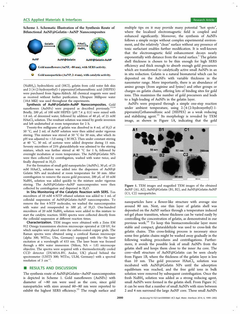
■ RESULTS AND DISCUSSIONThe synthesis route of AuNF@Gelatin−AuNP nanocompositesis depicted in Scheme 1. Gold nanoflowers (AuNFs) withdiameter of ∼80 nm were used as the core, since goldnanoparticles with sizes around 60−80 nm were reported toexhibit the highest SERS efficiency.15 The rough surface and
multiple tips on it may provide many potential “hot spots”,where the localized electromagnetic field is coupled andenhanced significantly. Moreover, the synthesis of AuNFsfollows a simple recipe without complex experimental environ-ment, and the relatively “clean” surface without any presence oftoxic surfactant enables further modification. It is well-knownthat the electromagnetic field enhancement decays nearlyexponentially with distance from the metal surface.1 The gelatinshell thickness is chosen to be thin enough for high SERSefficiency and thick enough to absorb enough gold precursorswhich are transformed to catalytically active small AuNPs in anin situ reduction. Gelatin is a natural biomaterial which can bedeposited on the AuNFs with variable thickness in thenanometer range. More importantly, there is an abundance ofamino groups (from arginine and lysine) and other groups orcharges on gelatin chains, offering lots of binding sites for goldions. This maximizes the number of gold precursors and leadsto a high-loading of AuNPs in the gelatin layer.AuNFs were prepared through a simple one-step reaction
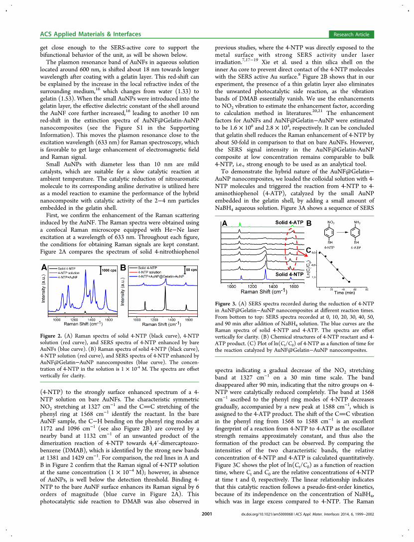
under ambient temperature, using 2-[4-(2-hydroxyethyl)-1-piperazinyl]ethanesulfonic acid (HEPES) as a weak reducingand stabilizing agent.13 Its morphology is revealed by TEMimage, as shown in Figure 1A, indicating that the gold
nanoparticles have a flower-like structure with average sizearound 80 nm. Next, one thin layer of gelatin shell wasdeposited on the AuNF surface through a temperature inducedsol-gel phase transition, whose thickness can be varied easily bycontrolling the concentration of gelatin, as demonstrated in ourprevious work.14 To keep this biomacromolecular layer morestable and compact, glutaraldehyde was used to cross-link thegelatin chains. This cross-linking process is necessary sincesome free gelatin chains might be washed away gradually in thefollowing washing procedures and centrifugations. Further-more, it avoids the possible leak of small AuNPs from thegelatin shell and keeps them close to the inner Au core. Thecore−shell structure of AuNF@Gelatin can be seen clearlyfrom Figure 1B, where the thickness of the gelatin layer is lessthan 10 nm. The gold precursor HAuCl4 solution wasincubated with AuNF@Gelatin NPs until the adsorptionequilibrium was reached, and the free gold ions in bulksolution were removed by subsequent centrifugation. Once thefresh NaBH4 solution was added as a strong reducing agent,small AuNPs were formed in the gelatin shell. From Figure 1Cit can be seen that a number of small AuNPs with sizes between2 and 4 nm surround the large AuNF core. These small AuNPs
Scheme 1. Schematic Illustration of the Synthesis Route ofBifunctional AuNF@Gelatin−AuNP Nanocomposites
Figure 1. TEM images and magnified TEM images of the obtainedAuNF (A1, A2), AuNF@Gelatin (B1, B2), and AuNF@Gelatin-AuNP(C1, C2) nanoparticles.
ACS Applied Materials & Interfaces Research Article
get close enough to the SERS-active core to support thebifunctional behavior of the unit, as will be shown below.The plasmon resonance band of AuNFs in aqueous solution
located around 600 nm, is shifted about 18 nm towards longerwavelength after coating with a gelatin layer. This red-shift canbe explained by the increase in the local refractive index of thesurrounding medium,16 which changes from water (1.33) togelatin (1.53). When the small AuNPs were introduced into thegelatin layer, the effective dielectric constant of the shell aroundthe AuNF core further increased,16 leading to another 10 nmred-shift in the extinction spectra of AuNF@Gelatin-AuNPnanocomposites (see the Figure S1 in the SupportingInformation). This moves the plasmon resonance close to theexcitation wavelength (633 nm) for Raman spectroscopy, whichis favorable to get large enhancement of electromagnetic fieldand Raman signal.Small AuNPs with diameter less than 10 nm are mild
catalysts, which are suitable for a slow catalytic reaction atambient temperature. The catalytic reduction of nitroaromaticmolecule to its corresponding aniline derivative is utilized hereas a model reaction to examine the performance of the hybridnanocomposite with catalytic activity of the 2−4 nm particlesembedded in the gelatin shell.First, we confirm the enhancement of the Raman scattering
induced by the AuNF. The Raman spectra were obtained usinga confocal Raman microscope equipped with He−Ne laserexcitation at a wavelength of 633 nm. Throughout each figure,the conditions for obtaining Raman signals are kept constant.Figure 2A compares the spectrum of solid 4-nitrothiophenol
(4-NTP) to the strongly surface enhanced spectrum of a 4-NTP solution on bare AuNFs. The characteristic symmetricNO2 stretching at 1327 cm−1 and the CC stretching of thephenyl ring at 1568 cm−1 identify the reactant. In the bareAuNF sample, the C−H bending on the phenyl ring modes at1172 and 1096 cm−1 (see also Figure 2B) are covered by anearby band at 1132 cm−1 of an unwanted product of thedimerization reaction of 4-NTP towards 4,4′-dimercaptoazo-benzene (DMAB), which is identified by the strong new bandsat 1381 and 1429 cm−1. For comparison, the red lines in A andB in Figure 2 confirm that the Raman signal of 4-NTP solutionat the same concentration (1 × 10⁻⁴ M); however, in absenceof AuNPs, is well below the detection threshold. Binding 4-NTP to the bare AuNF surface enhances its Raman signal by 6orders of magnitude (blue curve in Figure 2A). Thisphotocatalytic side reaction to DMAB was also observed in
previous studies, where the 4-NTP was directly exposed to themetal surface with strong SERS activity under laserirradiation.7,17−19 Xie et al. used a thin silica shell on theinner Au core to prevent direct contact of the 4-NTP moleculeswith the SERS active Au surface.8 Figure 2B shows that in ourexperiment, the presence of a thin gelatin layer also eliminatesthe unwanted photocatalytic side reaction, as the vibrationbands of DMAB essentially vanish. We use the enhancementsto NO2 vibration to estimate the enhancement factor, accordingto calculation method in literatures.20,21 The enhancementfactors for AuNFs and AuNF@Gelatin−AuNP were estimatedto be 1.6 × 10⁶ and 2.8 × 10⁴, respectively. It can be concludedthat gelatin shell reduces the Raman enhancement of 4-NTP byabout 50-fold in comparison to that on bare AuNFs. However,the SERS signal intensity in the AuNF@Gelatin-AuNPcomposite at low concentration remains comparable to bulk4-NTP, i.e., strong enough to be used as an analytical tool.To demonstrate the hybrid nature of the AuNF@Gelatin−
AuNP nanocomposites, we loaded the colloidal solution with 4-NTP molecules and triggered the reaction from 4-NTP to 4-aminothiophenol (4-ATP), catalyzed by the small AuNPembedded in the gelatin shell, by adding a small amount ofNaBH4 aqueous solution. Figure 3A shows a sequence of SERS
spectra indicating a gradual decrease of the NO2 stretchingband at 1327 cm−1 on a 30 min time scale. The banddisappeared after 90 min, indicating that the nitro groups on 4-NTP were catalytically reduced completely. The band at 1568cm−1 ascribed to the phenyl ring modes of 4-NTP decreasesgradually, accompanied by a new peak at 1588 cm−1, which isassigned to the 4-ATP product. The shift of the CC vibrationin the phenyl ring from 1568 to 1588 cm−1 is an excellentfingerprint of a reaction from 4-NTP to 4-ATP as the oscillatorstrength remains approximately constant, and thus also theformation of the product can be observed. By comparing theintensities of the two characteristic bands, the relativeconcentration of 4-NTP and 4-ATP is calculated quantitatively.Figure 3C shows the plot of ln(Ct/C0) as a function of reactiontime, where Ct and C0 are the relative concentrations of 4-NTPat time t and 0, respectively. The linear relationship indicatesthat this catalytic reaction follows a pseudo-first-order kinetics,because of its independence on the concentration of NaBH4,which was in large excess compared to 4-NTP. The Raman
Figure 2. (A) Raman spectra of solid 4-NTP (black curve), 4-NTPsolution (red curve), and SERS spectra of 4-NTP enhanced by bareAuNFs (blue curve). (B) Raman spectra of solid 4-NTP (black curve),4-NTP solution (red curve), and SERS spectra of 4-NTP enhanced byAuNF@Gelatin−AuNP nanocomposites (blue curve). The concen-tration of 4-NTP in the solution is 1 × 10‑4 M. The spectra are offsetvertically for clarity.
Figure 3. (A) SERS spectra recorded during the reduction of 4-NTPin AuNF@Gelatin−AuNP nanocomposites at different reaction times.From bottom to top: SERS spectra recorded at 0, 10, 20, 30, 40, 50,and 90 min after addition of NaBH4 solution. The blue curves are theRaman spectra of solid 4-NTP and 4-ATP. The spectra are offsetvertically for clarity. (B) Chemical structures of 4-NTP reactant and 4-ATP product. (C) Plot of ln(Ct/C0) of 4-NTP as a function of time forthe reaction catalyzed by AuNF@Gelatin−AuNP nanocomposites.
ACS Applied Materials & Interfaces Research Article
spectrum of the final product after the completion of thereduction is consistent with that of 4-ATP reported inliterature.17
To confirm the bifunctional nature of our AuNF@Gelatin−AuNP nanocomposites, we confirmed that the 4-NTPmolecules were preferentially catalyzed by the small AuNPs,which were embedded in the gelatin shell that prevents the sidereaction to DMAB: The reaction shown in Figure 3 wasmonitored once more using UV−vis absorption spectroscopy,as shown in Figure S2 in the Supporting Information. Theinitial 4-NTP solution with excess of NaBH4 showed anabsorption band around 410 nm, attributed to the formation of4-nitrothiophenolate ions in alkaline condition. After additionof AuNF@Gelatin−-AuNP colloids, the absorption banddecreased gradually, and the color of the mixture changedfrom yellow to colorless. When AuNF@Gelatin without smallAuNP were added no color change was observed. This controlexperiment confirms that the reaction is only catalytic, i.e., notphotoinduced. On the other hand, in the reaction process, itcan not be completely excluded that a small portion of 4-NTPmolecules perhaps reached the large core. However, thephotochemical reaction was not significant, because theRaman result did not show obvious Raman bands of the side-product, 4,4′-dimercaptoazobenzene (DMAB). We alsobelieved that the catalysis reaction indeed occurred on thesmall AuNP in gelatin layer, because the large AuNF itselfcannot catalyze efficiently this reduction reaction as demon-strated by UV-vis absorption spectra (see Figure S2 in theSupporting Information). Although UV−vis absorption spec-troscopy is widely used for monitoring this reduction reaction,it provides limited information about the structure, and it willnot work if the concentration of reactant is low. In addition,SERS allows simultaneously monitoring the emerging bands ofthe product even at low concentration, making it a betterchoice, in particular because of the additional structuralinformation encoded in multiple bands.
■ CONCLUSIONS
In conclusion, a facile method was presented to fabricate abifunctional AuNF@Gelatin-AuNP nanocomposite, consistingof a large AuNF core with SERS ability and small AuNPs in agelatin shell with catalytic activity. Since the thickness of thegelatin shell is less than 10 nm, the small AuNPs embedded init are still in the SERS active range of the AuNF core. Althoughthe Raman enhancement of the nanocomposite was reducedabout 50-fold compared to bare AuNFs due to the presence ofthe gelatin layer, the Raman intensity was still strong enough toderive information on a reaction, the reduction of 4-NTPcatalyzed by the nanocomposite. The photo-catalyzed dimeri-zation of 4-NTP to DMAB was avoided to a large amount,which had previously partly interfered in SERS monitoringusing bare plasmonic nanostructures. This reaction is well-studied, and we did not add new information, but the systemand proceeding described in this paper are well-suited forbroader application.
■ ASSOCIATED CONTENT
*S Supporting InformationExtinction spectra, absorption spectra during the catalyticreaction, estimation of Raman enhancement factor, comparisonof SERS ability of AuNFs and AuNPs. This material is availablefree of charge via the Internet at http://pubs.acs.org.
■ ACKNOWLEDGMENTSY.A. thanks the Alexander von Humboldt-Stiftung for thescholarship.
■ REFERENCES(1) Dieringer, J. A.; McFarland, A. D.; Shah, N. C.; Stuart, D. A.;Whitney, A. V.; Yonzon, C. R.; Young, M. A.; Zhang, X. Y.; VanDuyne, R. P. Faraday Discuss. 2006, 132, 9−26.(2) Heck, K. N.; Janesko, B. G.; Scuseria, G. E.; Halas, N. J.; Wong,M. S. J. Am. Chem. Soc. 2008, 130, 16592−16600.(3) Lantman, E. M. V.; Deckert-Gaudig, T.; Mank, A. J. G.; Deckert,V.; Weckhuysen, B. M. Nat. Nanotechnol. 2012, 7, 583−586.(4) Guerrini, L.; Lopez-Tobar, E.; Garcia-Ramos, J. V.; Domingo, C.;Sanchez-Cortes, S. Chem. Commun. 2011, 47, 3174−3176.(5) Min, B. K.; Friend, C. M. Chem. Rev. 2007, 107, 2709−2724.(6) Wang, A.; Huang, Y. F.; Sur, U. K.; Wu, D. Y.; Ren, B.; Rondinini,S.; Amatore, C.; Tian, Z. Q. J. Am. Chem. Soc. 2010, 132, 9534−9536.(7) Xie, W.; Herrmann, C.; Kompe, K.; Haase, M.; Schlucker, S. J.Am. Chem. Soc. 2011, 133, 19302−19305.(8) Xie, W.; Walkenfort, B.; Schlucker, S. J. Am. Chem. Soc. 2013,135, 1657−1660.(9) Ren, X. Q.; Tan, E. Z.; Lang, X. F.; You, T. T.; Jiang, L.; Zhang,H. Y.; Yin, P. G.; Guo, L. Phys. Chem. Chem. Phys. 2013, 15, 14196−14201.(10) Muralidharan, R.; McIntosh, M.; Li, X. Phys. Chem. Chem. Phys.2013, 15, 9716−9725.(11) Tian, Z. Q.; Ren, B.; Li, J. F.; Yang, Z. L. Chem. Commun. 2007,3514−3534.(12) Joseph, V.; Engelbrekt, C.; Zhang, J. D.; Gernert, U.; Ulstrup, J.;Kneipp, J. Angew. Chem., Int. Ed. 2012, 51, 7592−7596.(13) Xie, J. P.; Zhang, Q. B.; Lee, J. Y.; Wang, D. I. C. ACS Nano2008, 2, 2473−2480.(14) Cui, Q. L.; He, F.; Wang, X. Y.; Xia, B. H.; Li, L. D. ACS Appl.Mater. Interfaces 2013, 5, 213−219.(15) Krug, J. T.; Wang, G. D.; Sr, E.; Nie, S. M. J. Am. Chem. Soc.1999, 121, 9208−9214.(16) Kiel, M.; Klotzer, M.; Mitzscherling, S.; Bargheer, M. Langmuir2012, 28, 4800−4804.(17) Huang, Y. F.; Zhu, H. P.; Liu, G. K.; Wu, D. Y.; Ren, B.; Tian, Z.Q. J. Am. Chem. Soc. 2010, 132, 9244−9246.(18) Dong, B.; Fang, Y. R.; Xia, L. X.; Xu, H. X.; Sun, M. T. J. RamanSpectrosc. 2011, 42, 1205−1206.(19) Kang, L. L.; Xu, P.; Zhang, B.; Tsai, H. H.; Han, X. J.; Wang, H.L. Chem. Commun. 2013, 49, 3389−3391.(20) Zhang, L.; Dong, W. F.; Tang, Z. Y.; Song, J. F.; Xia, H.; Sun, H.B. Opt. Lett. 2010, 35, 3297−3299.(21) Orendorff, C. J.; Gole, A.; Sau, T. K.; Murphy, C. J. Anal. Chem.2005, 77, 3261−3266.
ACS Applied Materials & Interfaces Research Article