of paper-based analytical devicesfor rapid and highly selective colorimetricdetection of cesium in environmental samples†
Sung-Min Kang,‡a Sung-Chan Jang,‡ac Yuvaraj Haldorai,b A. T. Ezhil Vilian,b
Muruganantham Rethinasabapathy,a Changhyun Roh,*cd Young-Kyu Han *b
and Yun Suk Huh *ae
Cesium (Cs), a radioactive contaminant of the ecosystem, causes a major risk to human health and
environments. Till now, the cesium sensor fabrication methods have been reported with the usage of
expensive chemicals that are complex and time-consuming. In this work, we have fabricated a paper-
based colorimetric device impregnated with a chrysoidine G (CG) as chemo-indicator which is simple,
rapid, low-cost, and portable using a naked-eye quantitative technique for the detection and monitoring
of inactive cesium in environmental analysis. This chemo-indicator is designed to exhibit a powerful
detection capability featuring high selectivity and sensitivity to inactive Cs, by means of color
discrimination from light yellow to red orange. Interestingly, a portable smart phone camera, which
determined the relative red/green/blue (RGB) values within 3 s, provided us with further information on
environmental pollution. Using our new colorimetric reusable sensor (CRS) platform, the CRS shows
excellent detection linearity (R2 ¼ 0.99) of inactive Cs from the contaminated water. Our results will pave
the way for portable and versatile sensors and, in turn, for the detection and monitoring of toxic inactive
cesium in contaminated water samples.
1. Introduction
The nuclear accidents that befell the Chernobyl (Ukraine, 1986)and Fukushima (Japan, 2011) nuclear power plants are widelyregarded as being among the world's worst environmentaldisasters. The release of radionuclides as a result of thesenuclear accidents is an increasingly pressing global concern.1,2
In particular, most of the radionuclides (more than 80%)resulting from the Fukushima Daiichi nuclear accident wereallowed to contaminate the offshore area, causing their spreadto the world's sea and soil via the Pacic Ocean.3,4
hybrid Systems Research Center (BSRC),
12, Republic of Korea. E-mail: yunsuk.
: +82 32 860 9177
eering, Dongguk University-Seoul, Seoul,
dongguk.edu; Tel: +82 2 2260 4975
Radiation Technology Institute (ARTI),
ERI), 29 Geumgu-gil, Jeongeup, Jeonbuk,
aeri.re.kr; Tel: +82 63 570 3133
ioisotope Science, University of Science
, 34113, Republic of Korea
ip, Inha University, 100 Inha-ro, Incheon,
tion (ESI) available. See DOI:
is work.
5
Among these radioactive contaminants, cesium is known tobe a signicant byproduct of nuclear waste. Due to its highmobility and solubility in water, cesium (Cs) is responsible forcontaminating surface water and/or ground water which leadsto aquatic bioaccumulation of radioactive cesium ions and ispassed on to people through contaminated seafood anddrinking water.5–7 More seriously, the simple substitutionreaction of cesium ions with sodium and potassium ions in thered blood cells can induce adverse health problems in humanssuch as cardiovascular, gastrointestinal, fetal, and neurologicaldisease.8,9 Radioactive cesium and its radioisotopes are stronggamma emitters with a half-life of 2.06 years (134Cs) or 30.17years (137Cs), so their existence in the environment has long-term harmful effects.10,11 Currently, various analytical tech-niques, including laser-induced breakdown spectroscopy, cold-vapor inductively coupled plasma mass spectroscopy, atomicabsorption spectroscopy, and electrochemical devices havebeen accomplished to detect cesium.12 These analytical tech-niques have disadvantages such as high-cost, time-consumingcomplex operational procedures, and complicated non-portable equipment which are not suitable for environmentalmonitoring.
The paper which mainly consists of cellulose ber is highlyabundant and has become a growing concern amongresearchers as a potential substance in the fabrication ofsensors, chemical and analytical devices due to its low cost and
exibility in the fabrication of devices. Recently, a new paper-based analytical sensor is commonly used in environmentalmonitoring, food safety, and biomedical diagnostics applica-tion.13–16 As paper-based analytical devices exhibit a relativelynovel analytical sensing platform, several effective approacheshave been used to detect multiplex analytes, such as chemilu-minescent and FRET-based uorescence methods, electro-chemical and colorimetric methods, and surface-enhancedRaman spectroscopy.17–20 Among these analytical methods,colorimetric methods have established great attention due totheir excellent features such as simplicity, rapid execution, andlow-cost.21–24 In particular, the paper-based colorimetric deviceovercomes these existing high-cost time-consuming complexoperational procedures with ease of operation and detectscesium in the environmental samples very rapidly with highsensitivity and high selectivity.25,26 In addition, there are variousworks integrated on the use of smartphones as an on-sitedetection technique using the paper-based colorimetricdevice. Visual colorimetric sensors based on reusable substrates(e.g., cellulose, polyester, and non-woven fabric) are well-suitedfor the on-site detection and monitoring of environmentalcontaminants.27–30 The colorimetric data can be rapidly con-verted to a digital image by using smartphones. Their ability tosense harmful substances rapidly will have a signicant impacton a variety of danger warning and emergency responsesystems.
Currently, portable devices for detecting radioactive cesiumcan be fabricated using semiconductor materials such asgermanium (Ge), silicon (Si), diamond, and cadmium (zinc)telluride, which are capable of emitting gamma rays.31 However,even if these devices can provide a sensitive signal for thedetection of cesium, there are still a number of technicalproblems to be addressed before the general public will be ableto diagnose cesium easily using mobile sensors in the eld.Specically, in the case of the current portable cesium-measuring apparatus, there are limitations in terms of highproduction costs per unit, the size of the unit, the level of energyrequired, and the complex manufacturing processes involved.
To facilitate the development of an on-site portable sensordevice, we here present a colorimetric reusable sensor (CRS)technology for inactive cesium detection based on the colortransition of a chrysoidine G (CG) chemo-indicator with highlysensitive and selective signals through fabrication of a strip-typepaper-based sensor. The concept of our CRS for the rapid on-site diagnosis of Cs can be described by classifying theprocess into three major parts, namely, the fabrication ofa paper strip impregnated with CG, the detection of a change incolor through a reaction with the sample solution, and evalua-tion and conrmation via an image captured by a mobilephone. In order to implement a cheap, easy to make andoperate, and versatile sensor, we used exible substrates such aspaper, plastic, and fabric, which can be used easily in everydaylife. This method will detect and provide on-site diagnosticsignals about the presence of inactive Cs pollutants inhazardous environment within a short time that can be iden-tied by the human eye, as well as quantitative digital
information through analysis of images captured by a smartphone or portable camera.
2. Materials and method2.1. Materials
4-Phenylazo-m-phenylenediamine (chrysoidine G, CG) andWhatman cellulose chromatography paper were purchasedfrom Sigma-Aldrich Chemicals (MO, USA). Inactive cesium(133Cs) standard solutions were obtained from o2si smartsolutions (SC, USA). Lead, mercury, and zinc standard solutionwere purchased from CPI International (CA, USA). Iron(II)chloride tetrahydrate, iron(III) chloride hexahydrate, magne-sium chloride hexahydrate, manganese chloride tetrahydrate,and aluminum chloride hexahydrate were purchased fromSigma-Aldrich Chemicals (MO, USA). The real environmentalsamples from a stream and a lake were collected from KoreaAtomic Energy Research Institute (KAERI), Jeongeup, Republicof Korea. All reagents and chemicals were of analytical gradeand were used as purchased without further purication.
2.2. Fabrication of a text-reporting colorimetric reusablesensor
The text-reporting colorimetric reusable sensor was fabricatedusing a standard protocol of photolithography (Fig. S1†).28–30
Briey, various exible substrates (cellulose, polyester, and non-woven fabric) were coated with 1 mL of SU-8 photoresist (SU-82025, MicroChem Corp., MA, USA) by means of spin coating(1000 rpm, 10 s) (step 1). The SU-8 photoresist was immobilizedinto exible substrate networks and then so baked at 90 �C for60 min followed by stabilization at room temperature (step 2).Then, the glass photomask and paper were assembled in face toface stacking (step 3). Of note is the fact that we made a glassphotomask composed of two slide glasses and a piece of paperprinted with text. Also, to achieve high-resolution textpatterning, the conformal contact between the glass photomaskand the SU-8 impregnated paper was fastened by clamps. Theglass photomask was placed on SU-8 coated exible substratesand then exposed to UV irradiation (l ¼ 365 nm, UVItec, Lon-don, UK) for 1 min (step 4). Aer UV irradiation, the papersubstrate was developed in an alkaline solvent to remove theresidual photoresist (step 5). Then, the hydrophilic text regionbecame visible aer the paper was washed with distilled water.Finally, a CG aqueous chemo-indicator was introduced on to thehydrophilic text region (step 6).
2.3. Fabrication of a colorimetric reusable sensor
The colorimetric reusable sensor (CRS) was fabricated using thedrop-casting method.32 Briey, for the homogeneous distribu-tion of the CG chemo-indicator between the cellulose matrixes,cellulose papers were prepared through immersion in anaqueous CG chemo-indicator. This paper was then allowed todry in the oven at 50 �C for 30 min to induce slow evaporation(not by aggregated CG molecules, known as the coffee ringeffect).33 The fabrication of the CRS using various substrates isperformed in the same way.
Aqueous solutions of CG chemo-indicator (7 � 10�5 M) wereprepared in H2O. Inactive Cs standard solution (1000 mg L�1)was prepared in 20 mL of double-distilled water and a seriesconcentrations of the Cs solution was transferred to the solu-tion of CG (7 � 10�5 M). Working solutions were prepared bydiluting the stock solution to the desired concentration usingdouble-distilled water. Aer mixing them by hand shaking fora few seconds, UV-vis spectra were recorded on an Innite UVM200 spectrophotometer (TECAN, Austria).
2.5. Analysis of colorimetric reusable sensors
For the quantitative analysis, the reusable sensors wereanalyzed with a handy phone camera in high dynamic range(HDR) mode. The color changes of the reusable substrates(cellulose, polyester, and non-woven fabric) were analyzed usinga scanner and were saved in TIF format. The numerical single-color coordinate values of the colorimetric images were extrac-ted from nine-grid points in the resulting experimental imageusing ImageJ soware.34,35 The DR, DG, and DB values were thendetermined from the RGB values before and aer the sensingimages and were used for the histogram plot.
2.6. Detection and analysis of real samples
To demonstrate the utility of the CRS for a real sample in theenvironmental eld, we carried out a series of experiments inthe Korea Atomic Energy Research Institute (KAERI) aer takingeld real samples from two different locations near a small lake(latitude: 37� 380 0.5700 N and longitude: 127� 50 6.1800 E) andstream (latitude: 35� 300 44.9900 N and longitude: 126� 500 1.6400
E). In detail, the CRS was placed at the base and ve photo-graphs were taken within 5 s aer soaking it in a eld realsamples. The distance between the camera and CRS, lightingconditions, and camera setting were kept constant for allexperiments. No cesium was detected in the water samples, sosamples were spiked with a cesium standard solution. Subse-quently, 3 mL of a spiked real water sample was pipetted intoeach of plastic Petri dishes, before adding the standard cesiumsolution. The concentrations of cesium standard added were 0,0.2, and 0.5 mg L�1.
3. Results and discussion3.1. Fabrication of a text-reporting colorimetric reusablesensor (CRS)
Our proposed detection system is easy to make and operate andcan achieve the rapid diagnosis of inactive Cs with a high levelof sensitivity and accuracy. As described in Fig. 1a, the strategyfor our detection method can be guided through a straightfor-ward “design and fabrication, detection and analysis, andevaluation and conrmation” procedure, which does notrequire expensive materials, sophisticated devices, or analyticalspecialists.27,28 The major challenge of chemosensor's insolu-bility is resolved in our work by developing CRS through theimpregnation of a CG chemo-indicator with various types ofsubstrates. The CRS sensing strip was rst prepared using
48376 | RSC Adv., 2017, 7, 48374–48385
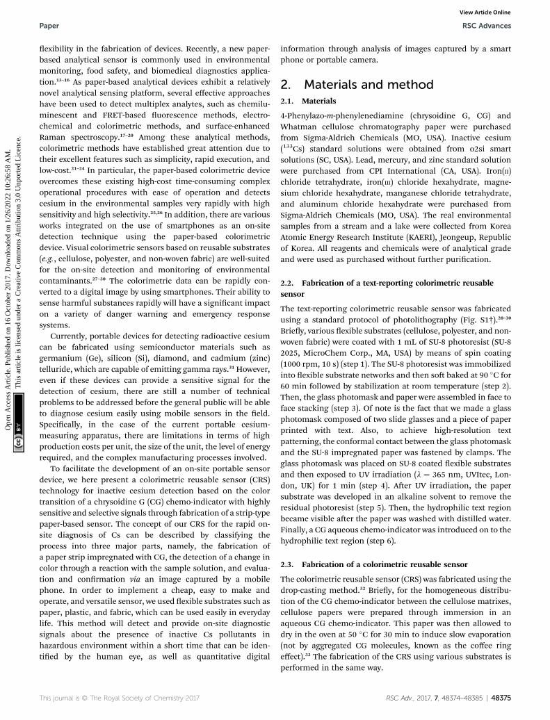
simple patterning techniques via a direct dipping process orphotolithography, so that the chemo-indicator was impreg-nated in a stable manner with regard to the position of theintended characters and patterns (Fig. 1a and S1†).29 Here, wepatterned the letter “CESIUM” on a substrate comprisinga variety of polymers and cotton materials through so lithog-raphy by utilizing surface properties. To be precise, SU-8photoresist penetrates the cellulose networks as a hydro-phobic barrier which contributes to selective wetting in thehydrophilic text region (see the Experimental section fordetails). As shown in Fig. 1b, the text-reporting CRS may allowthe potential use of cesium analysis in real environmentalsamples using proof-of-concept experiments. To demonstratethe usefulness of the CRS for a real sample in the environmentaleld, we next carried out a series of experiments in KAERI aertaking eld samples from two different locations near a smalllake and stream, (i) and (ii) (Fig. 1b). In the case of the twosamples (see the image (iii) of the CRS in Fig. 1b), there wasbarely any color change in the sensor, indicating that inactiveCs was not present. In order to conrm the capability of thecellulose-based CRS to distinguish the presence of inactive Cs,the environmental water was spiked with 0.5 mg L�1 of inactiveCs prior to testing. When a sample solution was introduced intothe cellulose CRS, the resulting color change remarkablyoccurred within 3 s ((iv) and (v) in Fig. 1b). To facilitatea quantitative analysis, the resulting CRS image was recordedusing a portable smart phone camera. Fig. S2† shows satisfac-tory analytical results for the articial contamination of cesiumspiked real aqueous samples. As shown in Fig. 1c, the recordeddata can be imported into generalized analytical freeware, andthe difference in color intensity can then be quantied usingthe histogram function. These results show the capability ofCRS to detect Cs ions in water with a more complex composi-tion. Also, the CRS can be designed and fabricated easily onexible substrates using patterning techniques, dipping, andphotolithography. In this study, the use of various exiblesubstrates including cellulose, polyester, and non-woven fabricprovided a proof-of-concept design for exible sensors (Fig. 2).Cellulose, which has a white background, is easier to observechanges in the color of the sensor owing to high-contrastenhancement. Unlike other conventional rigid sensing plat-forms including plastic, glass, and metal, these exiblesubstrates can offer a new type of custom tted sensing appli-cations for adjusting to various environmental conditions.
3.2. Cellulose-based CRS and its reusability
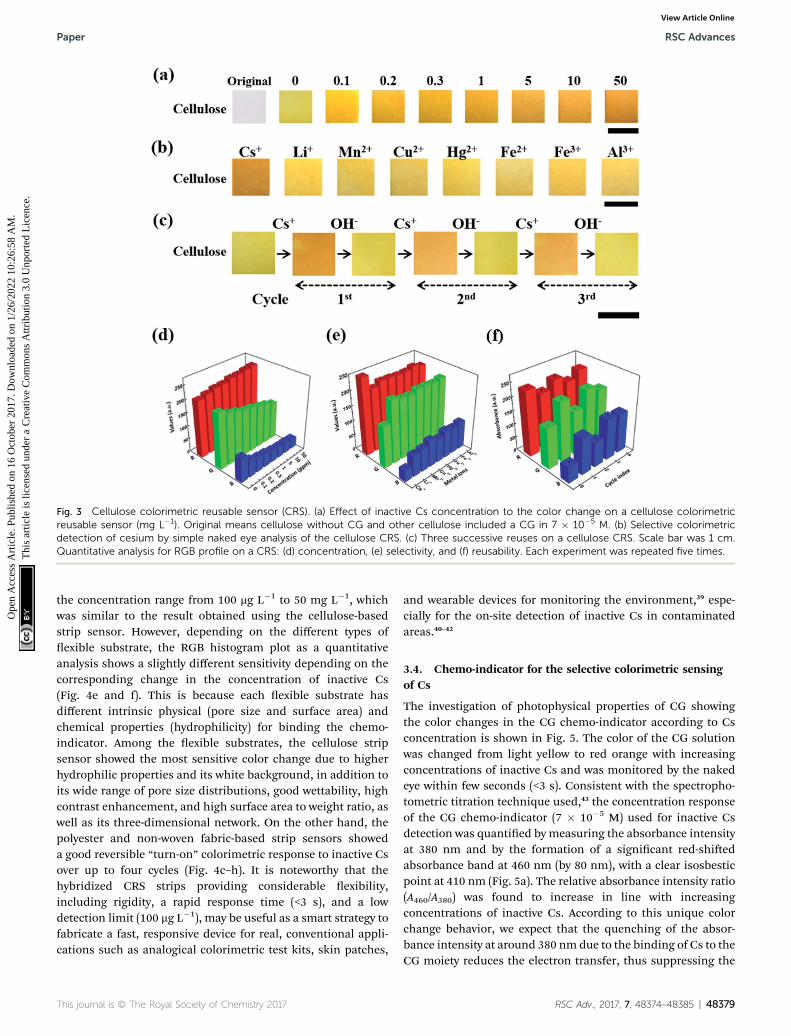
To attain the quantitative numerical values of the sensor, thecolor transitions of the CRS strips were recorded using portabledevices such as smart phone cameras and desktop scanners. Asshown in Fig. 3a, when the CRS strips were developed byimmersing them into solutions at different concentrations ofinactive Cs, the obvious color change was observed from lightyellow to red orange.36–38 The CRS strips showed excellentdetection sensitivity to cesium in the range from 100 mg L�1 to50 mg L�1 and provided good linearity up to a cesium concen-tration of 250 mg L�1 with a detection limit of 100 mg L�1
Fig. 1 Full spectrum showing a proof-of-concept for inactive Cs detection. (a) Design and fabrication process for text-reporting colorimetricreusable sensor (CRS). (b) Adaptation of CRS for real samples. Collecting the sample at (i) lake and (ii) stream around KAERI (Korea Atomic EnergyResearch Institute, Jeongeup, Jeonbuk, Korea). Real sample analysis on a text-reporting CRS compared (iii) before Cs treatment and (iv) after Cstreatment. (v) Color transition with cesium treatment in hydrophilic text region. (c) Quantitative RGB profile with cesium treatment. Theconcentration of Cs was 0.5 mg L�1. Scale bar was 2 cm.
Paper RSC Advances
Ope
n A
cces
s A
rtic
le. P
ublis
hed
on 1
6 O
ctob
er 2
017.
Dow
nloa
ded
on 1
/26/
2022
10:
26:5
8 A
M.
Thi
s ar
ticle
is li
cens
ed u
nder
a C
reat
ive
Com
mon
s A
ttrib
utio
n 3.
0 U
npor
ted
Lic
ence
.View Article Online
(Fig. S3†). In addition, we conducted a quantitative measure-ment of the degree of contamination using mathematicalconversion from the RGB color value (Fig. 3d). The red value (R)gradually increased in line with the concentration of cesium.Conversely, in terms of cesium concentration, the cellulosepaper showed decreased values for green (G) and blue (B)compared to the control. Therefore, we successfully demon-strated the ability to quantify inactive Cs concentration basedon the conversion of the RGB color values. The selectivedetection of target material among competitive ions by CRS isexamined by the changes in the color of the CG chemo-indicatorin the presence of other metal ions (Fig. 3b). In the selectivityexperiment using cesium and other metal ions with 0.5 mg L�1
concentration, cesium contaminated solution showeda maximum color transition, corresponding to the result of thequantitative RGB numerical analysis in Fig. 3e. These resultsclearly show that the CRS strips are not notably disturbed by thepresence of other metal ions. The most challenging aspects ofthe conventional sensors based on color changes are theirrecyclability and reusability. The reusability of our CRS isexamined by using a reversible reaction between a CG chemoindicator and Cs ion. As shown in Fig. 3c, a strong externalcomplexing agent was applied aer the detection of inactive Cs;that is, a Cs ion can combine favorably with NaOH to transforminto a more stable NaOH–Cs+ complex. Also, the colorimetriccellulose paper showed excellent reusability with no color
Fig. 2 Various reusable sensors with patterned flexible substrates. Colorimetric detection of Cs on the various substrates: (a) cellulose, (b)polyester, and (c) non-woven fabric. The concentration of Cs was 0.5 mg L�1. Scale bar was 2 cm. Each experiment was repeated five times.
RSC Advances Paper
Ope
n A
cces
s A
rtic
le. P
ublis
hed
on 1
6 O
ctob
er 2
017.
Dow
nloa
ded
on 1
/26/
2022
10:
26:5
8 A
M.
Thi
s ar
ticle
is li
cens
ed u
nder
a C
reat
ive
Com
mon
s A
ttrib
utio
n 3.
0 U
npor
ted
Lic
ence
.View Article Online
intensity decay over several cycles (Fig. 3f). These results suggestthat CRS strips have invaluable practical application potentialfor on-site environmental detection.
3.3. Fabrication of CRS strips using various exiblesubstrates
CRS strips of different exible substrates, cut to a constant size(1 cm� 1 cm), were prepared through a simple dipping method
48378 | RSC Adv., 2017, 7, 48374–48385
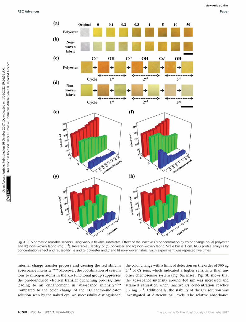
in a CG chemo-indicator solution of optimal concentration atvarious dipping intervals (10–60 min) followed by drying thestrips in order to impregnate a chemo-indicator to form exiblechips at 50 �C for 1 h. This method allows the stable immobi-lization of a chemo-indicator on all exible substrates by facil-itating electrostatic and hydrophilic interactions between them.Using the resulting strips with polyester and non-woven fabric(Fig. 4a and b),30 we were able to observe the color transition in
Fig. 3 Cellulose colorimetric reusable sensor (CRS). (a) Effect of inactive Cs concentration to the color change on a cellulose colorimetricreusable sensor (mg L�1). Original means cellulose without CG and other cellulose included a CG in 7 � 10�5 M. (b) Selective colorimetricdetection of cesium by simple naked eye analysis of the cellulose CRS. (c) Three successive reuses on a cellulose CRS. Scale bar was 1 cm.Quantitative analysis for RGB profile on a CRS: (d) concentration, (e) selectivity, and (f) reusability. Each experiment was repeated five times.
Paper RSC Advances
Ope
n A
cces
s A
rtic
le. P
ublis
hed
on 1
6 O
ctob
er 2
017.
Dow
nloa
ded
on 1
/26/
2022
10:
26:5
8 A
M.
Thi
s ar
ticle
is li
cens
ed u
nder
a C
reat
ive
Com
mon
s A
ttrib
utio
n 3.
0 U
npor
ted
Lic
ence
.View Article Online
the concentration range from 100 mg L�1 to 50 mg L�1, whichwas similar to the result obtained using the cellulose-basedstrip sensor. However, depending on the different types ofexible substrate, the RGB histogram plot as a quantitativeanalysis shows a slightly different sensitivity depending on thecorresponding change in the concentration of inactive Cs(Fig. 4e and f). This is because each exible substrate hasdifferent intrinsic physical (pore size and surface area) andchemical properties (hydrophilicity) for binding the chemo-indicator. Among the exible substrates, the cellulose stripsensor showed the most sensitive color change due to higherhydrophilic properties and its white background, in addition toits wide range of pore size distributions, good wettability, highcontrast enhancement, and high surface area to weight ratio, aswell as its three-dimensional network. On the other hand, thepolyester and non-woven fabric-based strip sensors showeda good reversible “turn-on” colorimetric response to inactive Csover up to four cycles (Fig. 4c–h). It is noteworthy that thehybridized CRS strips providing considerable exibility,including rigidity, a rapid response time (<3 s), and a lowdetection limit (100 mg L�1), may be useful as a smart strategy tofabricate a fast, responsive device for real, conventional appli-cations such as analogical colorimetric test kits, skin patches,
and wearable devices for monitoring the environment,39 espe-cially for the on-site detection of inactive Cs in contaminatedareas.40–42
3.4. Chemo-indicator for the selective colorimetric sensingof Cs
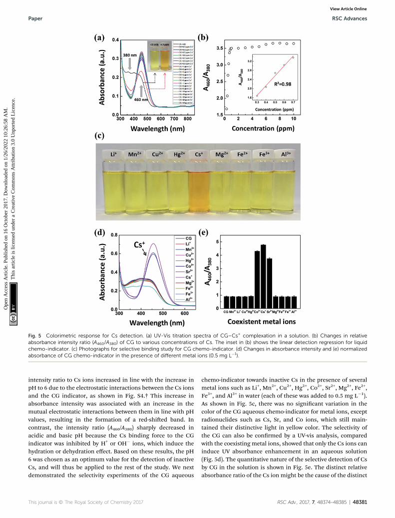
The investigation of photophysical properties of CG showingthe color changes in the CG chemo-indicator according to Csconcentration is shown in Fig. 5. The color of the CG solutionwas changed from light yellow to red orange with increasingconcentrations of inactive Cs and was monitored by the nakedeye within few seconds (<3 s). Consistent with the spectropho-tometric titration technique used,43 the concentration responseof the CG chemo-indicator (7 � 10�5 M) used for inactive Csdetection was quantied by measuring the absorbance intensityat 380 nm and by the formation of a signicant red-shiedabsorbance band at 460 nm (by 80 nm), with a clear isosbesticpoint at 410 nm (Fig. 5a). The relative absorbance intensity ratio(A460/A380) was found to increase in line with increasingconcentrations of inactive Cs. According to this unique colorchange behavior, we expect that the quenching of the absor-bance intensity at around 380 nm due to the binding of Cs to theCG moiety reduces the electron transfer, thus suppressing the
Fig. 4 Colorimetric reusable sensors using various flexible substrates. Effect of the inactive Cs concentration by color change on (a) polyesterand (b) non-woven fabric (mg L�1). Reversible usability of (c) polyester and (d) non-woven fabric. Scale bar is 1 cm. RGB profile analysis byconcentration effect and reusability: (e and g) polyester and (f and h) non-woven fabric. Each experiment was repeated five times.
RSC Advances Paper
Ope
n A
cces
s A
rtic
le. P
ublis
hed
on 1
6 O
ctob
er 2
017.
Dow
nloa
ded
on 1
/26/
2022
10:
26:5
8 A
M.
Thi
s ar
ticle
is li
cens
ed u
nder
a C
reat
ive
Com
mon
s A
ttrib
utio
n 3.
0 U
npor
ted
Lic
ence
.View Article Online
internal charge transfer process and causing the red shi inabsorbance intensity.44–46 Moreover, the coordination of cesiumions to nitrogen atoms in the azo functional group suppressesthe photo-induced electron transfer quenching process, thusleading to an enhancement in absorbance intensity.47,48
Compared to the color change of the CG chemo-indicatorsolution seen by the naked eye, we successfully distinguished
48380 | RSC Adv., 2017, 7, 48374–48385
the color change with a limit of detection on the order of 300 mgL�1 of Cs ions, which indicated a higher sensitivity than anyother chemosensor system (Fig. 5a, inset). Fig. 5b shows thatthe absorbance intensity around 460 nm was increased andattained saturation when inactive Cs concentration reaches0.7 mg L�1. Additionally, the stability of the CG solution wasinvestigated at different pH levels. The relative absorbance
Fig. 5 Colorimetric response for Cs detection. (a) UV-Vis titration spectra of CG–Cs+ complexation in a solution. (b) Changes in relativeabsorbance intensity ratio (A460/A380) of CG to various concentrations of Cs. The inset in (b) shows the linear detection regression for liquidchemo-indicator. (c) Photographs for selective binding study for CG chemo-indicator. (d) Changes in absorbance intensity and (e) normalizedabsorbance of CG chemo-indicator in the presence of different metal ions (0.5 mg L�1).
Paper RSC Advances
Ope
n A
cces
s A
rtic
le. P
ublis
hed
on 1
6 O
ctob
er 2
017.
Dow
nloa
ded
on 1
/26/
2022
10:
26:5
8 A
M.
Thi
s ar
ticle
is li
cens
ed u
nder
a C
reat
ive
Com
mon
s A
ttrib
utio
n 3.
0 U
npor
ted
Lic
ence
.View Article Online
intensity ratio to Cs ions increased in line with the increase inpH to 6 due to the electrostatic interactions between the Cs ionsand the CG indicator, as shown in Fig. S4.† This increase inabsorbance intensity was associated with an increase in themutual electrostatic interactions between them in line with pHvalues, resulting in the formation of a red-shied band. Incontrast, the intensity ratio (A460/A380) sharply decreased inacidic and basic pH because the Cs binding force to the CGindicator was inhibited by H+ or OH� ions, which induce thehydration or dehydration effect. Based on these results, the pH6 was chosen as an optimum value for the detection of inactiveCs, and will thus be applied to the rest of the study. We nextdemonstrated the selectivity experiments of the CG aqueous
chemo-indicator towards inactive Cs in the presence of severalmetal ions such as Li+, Mn2+, Cu2+, Hg2+, Co2+, Sr2+, Mg2+, Fe2+,Fe3+, and Al3+ in water (each of these was added to 0.5 mg L�1).As shown in Fig. 5c, there was no signicant variation in thecolor of the CG aqueous chemo-indicator for metal ions, exceptradionuclides such as Cs, Sr, and Co ions, which still main-tained their distinctive light in yellow color. The selectivity ofthe CG can also be conrmed by a UV-vis analysis, comparedwith the coexisting metal ions, showed that only the Cs ions caninduce UV absorbance enhancement in an aqueous solution(Fig. 5d). The quantitative nature of the selective detection of Csby CG in the solution is shown in Fig. 5e. The distinct relativeabsorbance ratio of the Cs ion might be the cause of the distinct
red orange color of the CG chemo-indicator containing cesium.Chrysoidine G is highly absorbing, monovalent and possesseslow molecular weight with simple chemical structure amongother azo dyes. When Cs is added to chrysoidine G, the Cs reactwith the –N]N– double bond of CG and binds with it. Thiscomplexation is selective for Cs among other cations such asMn2+, Cu2+, Hg2+, Co2+, Sr2+, Mg2+, Fe2+, Fe3+, and Al3+ witha remarkable color discrimination rapidly. The selectivecomplexation of Cs may be attributed to (i) the size of thehydrated Cs ions, and (ii) the entropic free volume and spatialarrangement of azobenzene and the amino groups present inthe CG.49,50 Also, the mobility of monovalent Cs+ cations arehigher than that of the divalent (Mn2+, Cu2+, Hg2+, Co2+, Sr2+,Mg2+, and Fe2+) and trivalent (Fe3+ and Al3+) cations and offersan acceptable mobility match for the low molecular weight CG.Thus Cs provides the highest possibility for binding with CGand exhibited maximum detection sensitivity. This studysuggests that other metal ions showed weak to negligiblecompetition in terms of the performance of the CG chemo-indicator for colorimetric detection.
3.5. Job's plot
To further understand the binding phenomenon and the stoi-chiometry of the complex formation, the Job's plot for absor-bance intensity was determined by changing the molar ratio ofinactive Cs (Xm ¼ [Cs+]/([Cs+] + [CG])).51 The total concentrationsof CG and Cs were xed at 10 mM, by continuously varying themolar fraction of Cs. The maximum absorbance intensity wasobserved in complexes of Cs, where the molar fraction of Cs was0.3. The plot of the relative absorbance intensity ratio versus Xm,as shown in Fig. S5† indicates that the complex formed betweenCG and Cs follows a 2 : 1 stoichiometry binding process.
3.6. Reversible CG chemo-indicator reaction
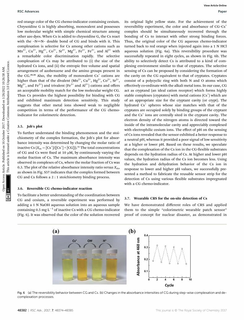
To facilitate a better understanding of the coordination betweenCG and cesium, a reversible experiment was performed byadding a 1 N NaOH aqueous solution into an aqueous samplecontaining 0.5 mg L�1 of inactive Cs with a CG chemo-indicator(Fig. 6). It was observed that the color of the solution recovered
Fig. 6 (a) The reversibility behavior between CG and Cs. (b) Changes in thcomplexation processes.
48382 | RSC Adv., 2017, 7, 48374–48385
its original light yellow state. For the achievement of thereversibility experiment, the color and absorbance of CG–Cs+
complex should be simultaneously recovered through thebonding of Cs to interact with other strong binding forces.Thus, the original color of the CG aqueous chemo-indicatorturned back to red orange when injected again into a 1 N HClaqueous solution (Fig. 6a). This reversibility procedure wassuccessfully repeated in eight cycles, as shown in Fig. 6b. Theability to selectively detect Cs is attributed to a kind of com-plexing environment similar to that of cryptates. The selectivesensing of Cs can be proposed by considering the formation ofthe cavity on the CG equivalent to that of cryptates. Cryptatesconsist of a polycyclic ring with both N and O atoms whicheffectively co-ordinate with the alkali metal ions. In our case, CGact as cryptand (an ideal cation receptor) which forms highlystable complexes (cryptates) with metal cations (Cs+) which areof an appropriate size for the cryptant cavity (or crypt). Thehydrated Cs+ spheres whose size matches with that of thecryptates are occupied solely by binding groups of the cryptantand the Cs+ ions are centrally sited in the cryptant cavity. Theelectron density of the nitrogen atoms is directed toward theinside of the intramolecular cavity and appreciably complexedwith electrophilic cesium ions. The effect of pH on the sensingof Cs ions revealed that the sensor exhibited a better response toa neutral pH, whereas it provided a poor signal of low sensitivityat a higher or lower pH. Based on these results, we speculatethat the complexation of the Cs ion in the CG-exible substratesdepends on the hydration radius of Cs. At higher and lower pHvalues, the hydration radius of the Cs ion becomes less. Usingthe hydration and dehydration behavior of the Cs ion inresponse to lower and higher pH values, we successfully pre-sented a method to fabricate the reusable sensor strip for thedetection of Cs using various exible substrates impregnatedwith a CG chemo-indicator.
3.7. Wearable CRS for the on-site detection of Cs
We have demonstrated different roles of CRS and appliedthem to the simple “colorimetric wearable patch sensor”proof of concept for nuclear disaster, as demonstrated in
e absorbance intensities of CG during step-wise complexation and de-
Fig. 7 (a) Simulation training for Cs contamination with applications of CRS. (b) A schematic illustration of the platform technology for inactive Csdetection on hybridized colorimetric reusable sensor. The identifiers of the cell-phone was removed by reconstructing the image to prevent anybiasing or advertising effects of specific goods.
Paper RSC Advances
Ope
n A
cces
s A
rtic
le. P
ublis
hed
on 1
6 O
ctob
er 2
017.
Dow
nloa
ded
on 1
/26/
2022
10:
26:5
8 A
M.
Thi
s ar
ticle
is li
cens
ed u
nder
a C
reat
ive
Com
mon
s A
ttrib
utio
n 3.
0 U
npor
ted
Lic
ence
.View Article Online
Fig. 7a. To prove the concept, we carried out simulationtraining for colorimetric inactive Cs detection. Basically, CRSsare used for detecting environmental contamination.However, in the case of rain, human contamination is inevi-table because contaminants such as Cs can be dissolved easilyin the atmospheric vapor. For this reason, the CRS is appliedto protective clothing (i.e., mask, glasses, and gloves) byattaching the exible CRS in the form of a wearable patchsensor. According to this simulation, it is anticipated thata newly developed CRS can be used for on-site detection in
real-life situations. Finally, the diagram of proposed processesfor detecting inactive Cs is schematically shown in Fig. 7b.The major advantages of CG-based detection of Cs usingappealing analytical features are illustrated in the followingpoints: (1) a facile and robust synthetic method using low-costmaterials; (2) high selectivity and good sensitivity; (3)comparable multiple regeneration cycles (i.e., reversibilityand reusability); (4) simplicity and portability; (5) rapidresponsive detection time; and (6) a water-compatible sensingsystem.
In this paper, we described the development of a CG-basednovel colorimetric sensor on hybridized exible substrates forthe on-site detection and monitoring of inactive Cs, a processthat facilitates the “naked eye” detection of Cs. By discrimi-nating the color changes of the CRS in accordance with theinteraction of the CG chemo-indicator with Cs ions, wesuccessfully detected inactive Cs with a detection limit of 100 mgL�1 and demonstrated the ability to quantify the sample solu-tion through the conversion of mathematical RGB color valuesfrom the image of the sensor strip. The colorimetric response ofthe RGB values and the corresponding Cs concentrationsshowed a well-matched relationship under dynamic environ-mental conditions. The recycle test with reversibility indicatedthat the detection process was stable even aer eight consecu-tive regeneration cycles. More importantly, this can be appliedto real samples as a photochromic paper sensor for environ-mental monitoring. Water sample detection can be achieved bygently dipping the hybridized reusable sensors in water, and theresults can be saved by using portable devices such as a smartphone camera. This will allow on-site analysis of unknownsamples since the method does not require skilled operators forthe colorimetric detection of Cs. Also, the detection time can beshortened to 3 s to change the color response, which offersfurther evidence of the potential for on-site detection and real-time monitoring. We believe that the newly developed colori-metric reusable sensors could be expanded to real situationsthat require sensing applications, particularly in the areas of theenvironmental and the analytical sciences.
Conflicts of interest
There are no conicts of interest to declare
Acknowledgements
This work was supported by the Radiation Fusion TechnologyProgram (2015M2A2A6A02045262(3)) from Nulear ResearchR&D Program through the National Research Foundation ofKorea (NRF) funded by the Ministry of Science, ICT & FuturePlanning (MSIP), Republic of Korea.
References
1 M. R. Awual, T. Yaita, T. Taguchi, H. Shiwaku, S. Suzuki andY. Okamoto, J. Hazard. Mater., 2014, 278, 227–235.
2 N. Kinoshita, K. Sueki, K. Sasa, J. Kitagawa, S. Ikarashi,T. Nishimura, Y. Wong, Y. Satou, K. Handa, T. Takahashi,M. Sato and T. Yamagata, Proc. Natl. Acad. Sci. U. S. A.,2011, 108, 19526–19529.
3 S.-C. Jang, Y. Haldorai, G.-W. Lee, S.-K. Hwang, Y.-K. Han,C. Roh and Y. S. Huh, Sci. Rep., 2015, 5, 17510.
4 Y. Morino, T. Ohara and M. Nishizawa, Geophys. Res. Lett.,2011, 38, L00G11.
5 J. Smith, O. Voitsekhovitch, L. Hakanson and J. Hilton, J.Environ. Radioact., 2001, 56, 11–32.
48384 | RSC Adv., 2017, 7, 48374–48385
6 J. Szabo and S. Minamyer, Environ. Int., 2014, 72, 129–132.7 T. Mizuno and H. Kubo, Sci. Rep., 2013, 3, 1742.8 B. Radaram, T. Mako and M. Levine, Dalton Trans., 2013, 42,16276–16278.
9 P. Melnikov and L. Z. Zanoni, Biol. Trace Elem. Res., 2010,135, 1–9.
10 X. Liu, G. R. Chen, D. J. Lee, T. Kawamoto, H. Tanaka,M. L. Chen and Y. K. Luo, Bioresour. Technol., 2014, 160,142–149.
11 H. Deng, Y. Li, Y. Huang, X. Ma, L. Wu and T. Cheng, Chem.Eng. J., 2016, 286, 25–35.
12 N. Kumar, I. Leray and A. Depauw, Coord. Chem. Rev., 2016,310, 1–15.
13 L.-J. Sun, Q.-M. Feng, Y.-F. Yan, Z.-Q. Pan, X.-H. Li,F.-M. Song, H. Yang, J.-J. Xu, N. Bao and H.-Y. Gu, Biosens.Bioelectron., 2014, 60, 154–160.
14 L. Feng, H. Li, L.-Y. Niu, Y.-S. Guan, C.-F. Duan, Y.-F. Guan,C.-H. Tung and Q.-Z. Yang, Talanta, 2013, 108, 103–108.
15 H. Tao, L. R. Chieffo, M. A. Brenckle, S. M. Siebert, M. Liu,A. C. Strikwerda, K. Fan, D. L. Kaplan, X. Zhang andR. D. Averitt, Adv. Mater., 2011, 23, 3197–3201.