224
FCUP Acknowledgements i

FCUP
Acknowledgements
i

FCUP
Acknowledgements
i
DEffects of interaction,
availability and
bioaccessibility of
microcystin-LR and
cylindrospermopsin in
terrestrial and aquatic
species
Marisa Alexandra Marques de FreitasEnvironmental Sciences and TechnologyDepartment of Geosciences, Environment and Spatial Planning
2014
SupervisorVítor Manuel de Oliveira Vasconcelos, Cathedratic Professor,
Faculty of Sciences of University of Porto
Co-supervisorAlexandre Campos, Auxiliary Researcher, Interdisciplinary Centre of Marine and
Environmental Research of University of Porto


FCUP
Acknowledgements
i
Acknowledgements
I would like to express my deepest gratitude to my supervisor, Professor Vitor
Vasconcelos, for his extraordinary support, pragmatic guidance and encouragement
throughout this work. His constant availability, patience and understanding made this
thesis possible.
I would like to thank my co-supervisor, Alexandre Campos, for his constant availability,
help and valuable suggestions, especially during the development of the experiments
with lettuce plants and proteomics analysis.
I am grateful for the assistance given by Professor António Paulo de Carvalho in the in
vitro enzymatic digestions and for his valuable critical suggestions during the writing of
the manuscripts.
I am thankful to Joana Azevedo for her constant cooperation, teachings and friendship.
A special thank to all LEGE team members for all support and essentially for keeping
an excellent working environment.
I would like to thank Edgar Pinto for his assistance in the analysis of minerals of the
lettuce plants.
I would like to thank Professor Piedade Barros for ceding the culture room, where the
lettuce experiments were developed.
My thanks to Joana Neves for her help in the lettuce experiments.
I would also like to extend my thanks to the Dr. Jenny Renaut and Dr. Sébastien
Planchon of the Centre de Recherche Public-Gabriel Lippmann, Department of
Environment and Agro-biotechnologies, for the analysis of lettuce proteins by MALDI-
TOF/TOF MS, and Dr. Vera Mendes and Dr. Bruno Manadas of the Center for
Neuroscience and Cell Biology, University of Coimbra, for the collaboration in the LC-
MS/MS analysis.
I would like to thank the Fundação para a Ciência e Tecnologia (FCT) for the Ph.D.
Grant SFRH/BD/85490/2012 and Escola Superior de Tecnologia da Saúde do Porto,

ii FCUP
Acknowledgements
Polythecnic Institute of Porto (ESTSP-IPP) for the financial support by the Programa de
Apoio à Formação Avançada de Docentes 2012.
I would like to thank my colleagues of department of Environmental Health of the
Escola Superior de Tecnologia da Saúde do Porto, Polytechnic Institute of Porto
(ESTSP-IPP), who have assumed many of my functions during the development of this
work. My special thanks to Professor Manuela Vieira da Silva, who transmitted me the
passion for research.
I am especially grateful to Miguel, my husband, for his remarkable patience and
unconditional support. I dedicate this thesis to him.
Finally, a special thank to my family, particularly to my parents and my sisters, who
always encouraged me to go further.
FCUP
Abstract
iii
Abstract
The occurrence and proliferation of toxic cyanobacterial blooms are an emergent
environmental concern worldwide. Microcystin-LR (MC-LR), mainly produced by
Microcystis aeruginosa, is the most documented and studied cyanotoxin.
Cylindrospermopsin (CYN) has been recognized of increased concern due to the
invasive nature of its main producer, Cylindrospermopsis raciborskii. Recent studies
support the hypothesis that MC-LR and CYN exert harmful effects on crop plants.
Lettuce is an important commercial leafy vegetable that supplies important nutrients for
human diet. Thus, it is of particular interest to know its sensitivity to environmentally
relevant concentrations of cyanotoxins, including mixtures. Proteomic technologies
seem to be suitable for the identification of early stress responses, which are not
perceptible by traditional endpoints. Proteomics may also provide the identification of
allergenic proteins, which may be of particular interest for human health risk
assessment. However, human health problems due to MC-LR and CYN are most likely
associated to the chronic exposure by contaminated drinking water and food. Previous
studies have shown that edible aquatic organisms, especially bivalves, can accumulate
high levels of these cyanotoxins without lethal effects. MC-LR and CYN are water-
soluble and stable at a wide range of temperatures and pHs, thus the knowledge of the
influence of storage and cooking practices as well as human digestion on MC-LR and
CYN concentration in food is required to achieve a more accurate risk assessment.
This thesis aimed to: (1) assess biochemical and physiological effects of MC-LR, CYN
and MC-LR/CYN mixture in lettuce, using conventional endpoints and a proteomic-
based approach; (2) assess the changes on MC-LR and CYN concentration in edible
bivalves after applying different storage and cooking practices, and (3) assess the MC-
LR and CYN bioaccessibility.
Lettuce plants (Lactuca sativa L.) were exposed to concentrations of 1, 10 and 100
µg/L of MC-LR, CYN and MC-LR/CYN mixture for five and ten days, and the effects
were assessed by the parameters fresh weight, activity of antioxidant enzymes and
mineral content in edible parts. The lettuce leaves were also studied by a comparative
proteomics approach. To assess the changes on MC-LR and CYN concentration in
bivalves after common food storage and processing practices, clams (C. fluminea) fed
MC-LR-producing M. aeruginosa and mussels (M. galloprovincialis) fed CYN-producing
C. raciborskii were refrigerated, frozen, boiled, steamed and subjected to microwave

iv FCUP
Abstract
radiation over different periods of time and then analyzed by LC-MS/MS.
Bioaccessibility of MC-LR and CYN were assessed in uncooked clams and in
uncooked and steamed mussels, respectively.
Overall, an increase in root growth was obtained, however, leaf-fresh weight was
significantly reduced in plants exposed to 100 µg/L. The GST activity was significantly
increased in roots, contrary to GPx activity, which decreased in roots and leaves. In
general, the mineral content in lettuce leaves decreased with MC-LR and increased
with CYN, and apparently these effects are time and concentration-dependent. The
effects of the MC-LR/CYN mixture were almost always similar to the single
cyanotoxins. Some of these physiological and biochemical effects were further
elucidated by the proteomics analysis, and at proteome level, the effects of the mixture
were clearly stronger than those of CYN alone. The biological functions of the proteins
that were most represented in both experiments were related to photosynthesis and
carbon metabolism and stress/defense response. Such variations could have altered
the rates of mineral uptake by lettuce plants and also conferred putative tolerance of
lettuce plants to CYN.
The recovery of free MC-LR in clams increased with freezing storage and with cooking
for short periods of time; specifically with the microwave radiation treatment for 0.5 and
1 min and boiling treatment for 5 and 15 min. The bioaccessibility of MC-LR after
proteolytic digestion was reduced to 83%, potentially because of MC-LR degradation
by pancreatic enzymes. In mussels stored frozen a significantly higher recovery of CYN
was obtained. The cooking treatments did not produce significant differences in CYN
concentration in mussel matrices (flesh), however, CYN was found in the cooking
water, suggesting that heat processing can be used to reduce the availability of CYN in
this food item. The in vitro digestion with salivary and gastrointestinal juices
considerably decreased the CYN availability in uncooked and steamed mussels,
highlighting the importance in integrating the bioaccessibility in the human health risk
assessment.
In conclusion, these findings provide new insights into the biochemical and
physiological mechanisms of the lettuce response to MC-LR and CYN (inclusive in
mixture) and may contribute to the understanding of potential mechanisms that may
confer tolerance to CYN. This study also provides an enhancement of knowledge on
the MC-LR and CYN concentration available in food after employing techniques
FCUP
Abstract
v
commonly used for their preservation and processing, which might be of particular
interest for the definition of critical control limits, considering the HACCP approach as a
promising tool for risk management. Our results also suggest that risk assessment
based on MC-LR and CYN concentration in raw products might not be representative
of true human exposure.
Keywords: Cylindrospermopsin, bioaccessibility, bivalves, Lactuca sativa, Microcystin-
LR, mixture.
vi FCUP
Resumo
Resumo
A ocorrência e a proliferação de florescências de cianobactérias tóxicas são um
problema ambiental emergente a nível mundial. Microcistina-LR (MC-LR), produzida
principalmente por Microcystis aeruginosa, é a cianotoxina mais estudada e
documentada. A cianotoxina cilindrospermopsina (CYN) tem sido alvo de crescente
preocupação devido à natureza invasiva da principal cianobatéria que a produz,
Cylindrospermopsis raciborskii. Estudos recentes suportam a hipótese de que a MC-
LR e a CYN podem exercer efeitos tóxicos em plantas agrícolas. A alface é um vegetal
de grande importancia a nível comercial, o qual fornece nutrientes essenciais para a
dieta humana. É portanto, de particular interesse conhecer a sua sensibilidade a
concentrações ambientalmente relevantes de cianotoxinas, inclusive quando em
mistura. A abordagem proteómica parece ser adequada para a identificação de
respostas ao stress que não são perceptíveis pelos endpoints tradicionais. O estudo
do proteoma pode também permitir a identificação de proteínas alergénicas, que
podem ter particular interesse em termos de avaliação de risco para a saúde humana.
No entanto, as implicações para a saúde humana devido às cianotoxinas, MC-LR e
CYN, são mais susceptíveis de ocorrer pela exposição crónica através da ingestão de
água e alimentos contaminados. Estudos prévios demonstraram que os organismos
aquáticos, especialmente bivalves, podem acumular níveis elevados de cianotoxinas
sem que ocorram efeitos letais. Por outro lado, a MC-LR e a CYN são solúveis em
água e estáveis a uma ampla gama de temperaturas e pHs. Portanto, a compreensão
da influência dos métodos de armazenamento e processamento de alimentos, assim
como da digestão humana sobre a concentração de MC-LR e CYN nos alimentos é de
grande relevância para alcançar uma avaliação de risco mais precisa. Os objetivos
desta tese foram: (1) avaliar os efeitos bioquímicos e fisiológicos provocados pela MC-
LR, CYN e pela sua mistura em alface, através de endpoints convencionais e de uma
abordagem proteómica; (2) avaliar o efeito de diferentes práticas de armazenamento e
de processamento de alimentos sobre a concentração de MC-LR e CYN em bivalves
edíveis, e (3) conhecer a biaccessibilidade de MC-LR e CYN.
As plantas de alface (Lactuca sativa L.) foram expostas às concentrações de 1, 10 e
100 µg/L de MC-LR, CYN e da mistura de MC-LR/CYN durante cinco e dez dias e os
efeitos foram avaliados pelos parâmetros peso fresco, atividade de enzimas
antioxidantes e conteúdo mineral na parte edível das plantas. As folhas de alface

FCUP
Resumo
vii
foram também estudadas utilizando uma abordagem proteómica comparativa. Para
avaliar os efeitos das práticas de armazenamento e procesamento dos alimentos na
concentração de MC-LR e CYN disponível em bivalves, ameijoas (C. fluminea) e
mexilhões (M. galloprovincilais) após serem alimentados com as cianobactérias
produtoras de MC-LR e CYN, M. aeruginosa e C. raciborskii, respetivamente, foram
refrigerados, congelados, cozidos, cozidos a vapor e submetidos a radiação
microondas durante diferentes períodos de tempo e de seguida analisados por LC-
MS/MS. A bioaccessibilidade da MC-LR e CYN foi estudada em ameijoas cruas e em
mexilhões crus e cozidos a vapor, respetivamente.
Na generalidade, obteve-se um aumento do crescimento das raízes, no entanto, o
peso fresco das folhas foi significativamente reduzido nas plantas expostas a 100
µg/L. A atividade da GST foi significativamente aumentada nas raízes, ao contrário da
atividade da GPx, que diminuiu nas raízes e nas folhas. A concentração de minerais
diminuiu nas folhas das plantas de alface expostas a MC-LR, contrariamente às
plantas expostas a CYN, onde o teor de minerais foi significativamente aumentado, e
aparentemente estes efeitos foram dependentes do tempo e da concentração de
exposição. Os efeitos da mistura de MC-LR/CYN foram semelhantes aos da exposição
às cianotoxinas individualmente. Alguns destes efeitos fisiológicos e bioquímicos foram
elucidados através da análise peoteómica, e a este nível, os efeitos da mistura foram
claramente mais potentes do que na exposição isolada a CYN. As funções biológicas
das proteínas que foram mais representadas em ambos os ensaios estavam
relacionadas com a fotossíntese e o metabolismo de carbono assim como com a
resposta ao stress/mecanismos de defesa. Estas variações poderiam ter alterado a
taxa de absorção de minerais pelas plantas de alface e também conferido uma
potencial tolerância destas plantas à CYN.
A recuperação da MC-LR livre nas ameijoas aumentou devido à armazenagem sob
congelação e ao cozimento por curtos períodos de tempo; especificamente, nos
tratamentos com radiação microondas durante 0,5 e 1 min e na cozedura durante 5 e
15 min. A biodisponibilidade da MC-LR após a digestão proteolítica foi reduzida para
83%, possivelmente devido à degradação da MC-LR pelas enzimas pancreáticas. O
armazenamento dos mexilhões sob congelação permitiu também uma maior
recuperação da CYN. Os tratamentos de cozedura não produziram diferenças
significativas na concentração da CYN na matriz do mexilhão (tecidos), no entanto, a
toxina foi encontrada na água de cozedura, o que sugere que o processamento pode
ser utilizado para reduzir a sua disponibilidade neste organismo edível. A digestão in
viii FCUP
Resumo
vitro com os sucos salivares e gastrointestinais diminuiu consideravelmente a
disponibilidade da CYN nos mexilhões crus e cozidos a vapor, o que destaca a
importância da integração da bioacessibilidade na avaliação de risco para a saúde
humana.
Em conclusão, estes resultados permitem uma compreensão mais abrangente dos
mecanismos bioquímicos e fisiológicos de resposta das plantas de alface à MC-LR e à
CYN (isoladas e em simultâneo), e podem contribuir para um maior entendimento do
mecanismo que parece conferir tolerância à CYN. Este estudo também potencia o
conhecimento sobre a concentração da MC-LR e CYN disponível em bivalves após o
uso de práticas comumente aplicadas na sua conservação e processamento, podendo
estes resultados ser utilizados para definição de limites críticos de controlo,
considerando a abordagem HACCP como uma ferramenta promissora para a gestão
de riscos para saúde humana. Estes resultados também sugerem que a avaliação de
riscos com base na concentração de MC-LR e CYN em produtos crus pode não ser
representativa da real exposição humana.
Palavras-chave: Cilindrospermopsina, bioacessibilidade, bivalves, Lactuca sativa,
Microcistina-LR, mistura.

FCUP
Table of contents
ix
Table of contents
Acknowledgements ..................................................................................................... I
Abstract ...................................................................................................................... III
Resumo ...................................................................................................................... VI
Table of contents ....................................................................................................... IX
List of tables.............................................................................................................. XII
List of figures .......................................................................................................... XIV
List of abbreviations ................................................................................................ XX
Chapter 1 ..................................................................................................................... 1
1. Introduction ............................................................................................................. 2
1.1. GENERAL INTRODUCTION ......................................................................................... 2
1.2. MICROCYSTIN-LR .................................................................................................... 4
1.3. CYLINDROSPERMOPSIN ............................................................................................ 5
1.4. Effects of MC-LR and CYN on plants .................................................................. 7
1.4.1. PROTEOMICS APPLIED TO AGRICULTURAL SECTOR TO ASSESS THE EFFECTS OF
CYANOTOXINS .............................................................................................................. 13
1.5. Human exposure to MC-LR and CYN ............................................................... 14
1.5.1. FACTORS AFFECTING HUMAN EXPOSURE TO MC-LR AND CYN BY THE
CONSUMPTION OF CONTAMINATED EDIBLE AQUATIC ORGANISMS ................................... 17
1.5.1.1. FOOD STORAGE AND PROCESSING ................................................................... 17
1.5.1.2. BIOACCESSIBILITY ............................................................................................ 18
1.6. References ......................................................................................................... 21
Chapter 2 ................................................................................................................... 31
2. Structure of the thesis and objectives..................................................................31
2.1. Structure of the thesis .......................................................................................... 32
2.2. Objectives...............................................................................................................34
x FCUP
Table of contents
Chapter 3 ................................................................................................................... 35
Effects of microcystin-LR, cylindrospermopsin and a microcystin-
LR/cylindrospermopsin mixture on growth, oxidative stress and mineral content
in lettuce plants (Lactuca sativa L.) ......................................................................... 36
Abstract ..................................................................................................................... 36
1. Introduction ........................................................................................................... 37
2. Materials and methods ......................................................................................... 40
3. Results and discussion ........................................................................................ 46
4. Conclusions .......................................................................................................... 54
5. References ............................................................................................................ 55
Lettuce (Lactuca sativa L.) leaf-proteome profiles after exposure to
cylindrospermopsin and a microcystin-LR/cylindrospermopsin mixture: a
concentration-dependent response ........................................................................ 61
Abstract ..................................................................................................................... 61
1. Introduction ........................................................................................................... 63
2. Materials and methods ......................................................................................... 66
3. Results and discussion ........................................................................................ 72
4. Conclusions .......................................................................................................... 90
5. References ............................................................................................................ 91
Chapter 4 ................................................................................................................... 98
Effects of storage, processing and proteolytic digestion on the microcystin-LR
concentration in edible clams .................................................................................. 99
Abstract ..................................................................................................................... 99
1. Introduction ......................................................................................................... 100
2. Material and methods ......................................................................................... 103
3. Results and discussion ...................................................................................... 107
4. Conclusions ........................................................................................................ 114
FCUP
Table of contents
xi
5. References .......................................................................................................... 115
Bioaccessibility and changes in the cylindrospermopsin concentration in edible
mussels with storage and processing time .......................................................... 119
Abstract ................................................................................................................... 119
1. Introduction ......................................................................................................... 120
2. Material and methods ......................................................................................... 123
3. Results and discussion ...................................................................................... 130
4. Conclusion .......................................................................................................... 138
5. References .......................................................................................................... 139
Chapter 5 ................................................................................................................. 145
5. General conclusions and future research ......................................................... 145
5.1. General discussion and conclusions .................................................................. 146
5.2. Future research.................................................................................................. 151
Chapter 6 ................................................................................................................. 152
6.1. Supporting information ....................................................................................... 152
6.2. References ........................................................................................................ 183
xii FCUP
List of tables
List of tables
Chapter 1
Table 1. General effects of the MC-LR on different species of aquatic and terrestrial
plants...............................................................................................................................8
Table 2. General effects of the CYN on different species of aquatic and terrestrial
plants.............................................................................................................................11
Table 3. General effects of the MC-LR/CYN on different species of aquatic and
terrestrial plants.............................................................................................................12
Table 4. The TDI of MC-LR and CYN derived from the respective no observed adverse
effect level (NOAEL). The LD50 of MC-LR and CYN by intraperitoneal and oral
routes…………………………………………………………………………………………..16
Table 5. Effects of food storage and processing practices on the MC-LR availability in
tissues of edible aquatic organisms………………………………………………………...18
Table 6. Digestion of MC-LR (in solution) by proteolytic enzymes of gastric and
intestinal juices…………………………………………………………………………….….20
Chapter 3
Effects of microcystin-LR, cylindrospermopsin and a microcystin-
LR/cylindrospermopsin mixture on growth, oxidative stress and mineral content
in lettuce plants (Lactuca sativa L.)
Table 1. Composition (constituents and concentration) of the culture medium used to
expose lettuce plants to MC-LR, CYN and a MC-LR/CYN mixture in a closed
hydroponic system.........................................................................................................43
Table 2. Mineral content in the lettuce leaves of the control groups from the three
experiments at the end of 5 and 10 days (5D and 10D, respectively). Values are
expressed as the mean ± SD (n=3)………………………………………………………...51
Table 3. Ratio of the mineral content in lettuce leaves exposed for 5 and 10 days (5D
and 10D, respectively) to MC-LR, CYN and a MC-LR/CYN mixture. Values express the
ratio between the mean concentrations obtained in each condition by the mean
concentration obtained in the respective control group. Values are expressed as the
mean ± SD (n=3). Values in bold represent the concentrations that exceeded the
screening value (control group, Table 2)…………………………………………………...52

FCUP
List of tables
xiii
Lettuce (Lactuca sativa L.) leaf-proteome profiles after exposure to
cylindrospermopsin and a microcystin-LR/cylindrospermopsin mixture: a
concentration-dependent response.
Table 1. Quantitative description of the differentially abundant protein spots of the
lettuce leaf-proteome profile obtained after treatment with ecologically relevant
concentrations of CYN and the MC-LR/CYN mixture....................................................76
Chapter 4
Effects of storage, processing and proteolytic digestion on the microcystin-LR
concentration in edible clams.
Table 1. Detailed information of the food storage and processing conditions applied in
experiment...................................................................................................................104
Bioaccessibility and changes in the cylindrospermopsin concentration in edible
mussels with storage and processing time
Table 1. Food storage and processing practices applied in the experiment………….125
Table 2. Composition (constituents and concentration) of digestive juices used in the in
vitro digestion, representing fed conditions……………………………………………....127
Chapter 5
Table 1. The general results obtained regarding to the effects of MC-LR, CYN and the
MC-LR/CYN mixture in lettuce plants………………………………………………….....146
Table 2. The general results obtained on the changes of MC-LR and CYN in bivalves
after applying different practices of food storage and processing as well as the effects
of digestive juices........................................................................................................148
Chapter 6
Supplemental table 1: Full data regarding to the proteins identification of the CYN
exposure experiment with lettuce plants.....................................................................162
Supplemental table 2: Full data regarding to the proteins identification of the MC-
LR/CYN exposure experiment with lettuce plants.......................................................166

xiv FCUP
List of figures
List of figures
Chapter 1
Fig. 1. The chemical structure of the heptapeptide MC-LR, where D-Ala is D-alanine
(1), L-Leu is L-leucine (2), D-Me-Asp is D-erythro-β-methylaspartic acid (3), L-Arg is
L-arginine (4), Adda is the unusual amino acid (2s,3s,8s,9s)-3-amino-9-methoxy-
2,6,8-trimethyl-10-phenyldeca-4(E),6(E)-dienoicacid (5), D-Glu is D-glutamic acid (6),
and Mdha is N-methyl-dehydroalanine (7)………………………………………………..4
Fig. 2. Global distribution of blooms of cyanobacteria known as CYN-producers.
Note: ‗non-CYN‘ denote a fluorescence from which toxin production was not
confirmed or studied....................................................................................................5
Fig. 3. The molecular structure of cylindrospermopsin (1) and its analogs 7-deoxy-
cylindrospermopsin (2) and 7-epicylindrospermopsin (3)............................................6
Fig. 4. The main factors that influence the expansion of toxic cyanobacterial blooms
and the routes of human exposure to cyanotoxins....................................................14
Fig. 5. Schematic representation of the difference between bioaccessibility and
bioavailability of the contaminants………………………………………………………..19
Chapter 2
Fig. 1. Scheme of the logical progression of the topics included in this thesis..........33
Chapter 3
Effects of microcystin-LR, cylindrospermopsin and a microcystin-
LR/cylindrospermopsin mixture on growth, oxidative stress and mineral
content in lettuce plants (Lactuca sativa L.)
Fig. 1. The fresh weight of lettuce plants (roots and leaves) after being exposed 5
and 10 days (5D and 10D, respectively) to MC-LR (A), CYN (B) and MC-LR/CYN
(C). Control: white bars; 1 µg/L: dark gray bars; 10 µg/L: black bars; 100 µg/L: light
gray bars. Values are expressed as the mean ± SD (n=10). Different letters (a, b and
c) indicate significant differences (p < 0.05)…………………………………………….46
Fig. 2. GST (A, B and C) and GPx (D, E and F) activities in lettuce plants (roots and
leaves) after being exposed 5 and 10 days to MC-LR, CYN and MC-LR/CYN,
respectively. Control: white bars; 1 µg/L: dark gray bars; 10 µg/L: black bars; 100

FCUP
List of figures
xv
µg/L: light gray bars. Values are expressed as the mean ± SD (n=3). Different letters
(a, b, c and d) means significant differences (p < 0.05)………………………………..49
Lettuce (Lactuca sativa L.) leaf-proteome profiles after exposure to
cylindrospermopsin and a microcystin-LR/cylindrospermopsin mixture: a
concentration-dependent response.
Fig. 1. Fr. wt (g) of leaves of lettuce plants exposed to CYN and MC-LR/CYN. The
values are expressed as the means ± SD (n=10). The asterisk (*) indicates
significant differences (P<0.05) between the control and exposed
groups........................................................................................................................73
Fig. 2. Venn diagram of the protein spots that exhibit differential abundance on leaf-
lettuce plants exposed to CYN (A) and MC-LR/CYN (B) compared with the control
group. The intersections also show the number of common proteins spots between
different groups. C1: 1 µg/l; C10: 10 µg/l; and C100: 100 µg/l...................................74
Fig. 3. PCA diagrams representing the first and second components and the second
and third components of the differential protein abundance (spot intensity) on the 2-
DE gel of leaf-lettuce plants exposed to CYN. C1: 1 µg/l; C10: 10 µg/l; and C100:
100 µg/l......................................................................................................................74
Fig. 4. PCA diagrams representing the first and second components and the second
and third components of the differential protein abundance (spot intensity) on the 2-
DE gel of leaf-lettuce plants exposed to MC-LR/CYN. C1: 1 µg/l; C10: 10 µg/l; and
C100: 100 µg/l............................................................................................................75
Fig. 5. Heat map of the proteins identified from the differentially abundant protein
spots of leaf-lettuce plants exposed to CYN. The values shown were normalized and
standardized using the control group as the reference. Two hierarchical clusterings
were made to display the similarities of the tested concentrations and the distribution
pattern of the differentially abundant proteins. The functional categorization of the
identified proteins is shown on the left side of the heat map. Photos. and C met.:
photosynthesis and carbon metabolism; Str. resp./Prot. fold.: stress response/protein
folding; ATP synth.: ATP synthesis; Def. resp./Allerg.: defense response/allergens;
Cytosk. Form.: cytoskeleton formation; Proteol.: proteolysis; Nucleos. assemb.:
nucleosome assembly; Transp. Act.: transport activity; Unk./Miscell.:
unknown/miscellaneous.............................................................................................77

xvi FCUP
List of figures
Fig. 6. Heat map of the proteins identified from the differentially abundant protein
spots of leaf-lettuce plants exposed to MC-LR/CYN. The values shown were
normalized and standardized using the control group as the reference. Two
hierarchical clusterings were made to display the similarities of the tested
concentrations and the distribution pattern of the differentially abundant proteins. The
functional categorization of identified proteins is shown on the left side of heat map.
Photos. and C met.: photosynthesis and carbon metabolism; Str. resp./Prot. Fold.:
stress response/protein folding; ATP synth.: ATP synthesis; Def. resp./Allerg.:
defense response/Allergens; Cytosk. Form.: cytoskeleton formation; Proteol.:
proteolysis; Cell wall biog./degrad.: Cell wall biogenesis/degradation; Transp. Act.:
transport activity; Prot. Synth. & Sig. transd.: protein synthesis and signal
transduction; Vit. B1 Bios.: vitamin B1 biosynthesis; Lip. metab.: lipid metabolism;
Inos. Bios.: inositol biosynthesis; Pigm. metab.: pigment metabolism; AA. metab.:
amino acid metabolism; Asc. bios.: ascorbic acid biosynthesis; Glut metab.:
glutathione metabolism; Unk./Miscell.: unknown/miscellaneous...............................78
Fig. 7. Functional categorization of the proteins identified from the differentially
abundant protein spots of leaf-lettuce plants exposed to CYN. C1: 1 µg/l; C10: 10
µg/l; and C100: 100 µg/l.............................................................................................80
Fig. 8. Functional categorization of the proteins identified from the differentially
abundant protein spots of leaf-lettuce plants exposed to MC-LR/CYN. C1: 1 µg/l;
C10: 10 µg/l; and C100: 100 µg/l...............................................................................80
Chapter 4
Effects of storage, processing and proteolytic digestion on the microcystin-LR
concentration in edible clams.
Fig.1. The chemical structure of the heptapeptide MC-LR, where D-Ala is D-alanine
(1), L-Leu is L-leucine (2), D-Me-Asp is D-erythro-β-methylaspartic acid (3), L-Arg is
L-arginine (4), Adda is the unusual amino acid (2s,3s,8s,9s)-3-amino-9-methoxy-
2,6,8-trimethyl-10-phenyldeca-4(E),6(E)-dienoicacid (5), D-Glu is D-glutamic acid (6),
and Mdha is N-methyl-dehydroalanine (7)...............................................................100
Fig. 2. Schematic representation of in vitro proteolytic digestion experiment..........105
Fig. 3. The MC-LR concentration (ng/g) in intoxicated C. fluminea after different
thermal storage conditions. Values are expressed as the mean ± SD (n=3). Different
letters (a, b, c, d, e, and f) indicate significant differences (P<0.05). Columns that
FCUP
List of figures
xvii
share the same letter are not significantly different.................................................108
Fig. 4. The MC-LR concentration (ng/g) in intoxicated C. fluminea after different
cooking conditions. Values are expressed as the mean ± SD (n=3). Different letters
(a, b, c, d, and e) indicate significant differences (P< 0.05). Columns that share the
same letter are not significantly different.................................................................110
Fig. 5. The MC-LR concentration (ng/g) after in vitro proteolytic gastric (G) and
gastrointestinal (GI) digestion. The column filled with black points represents the
bioaccessibility. Values are expressed as the mean ± SD (n=3). Different letters (a,
b, and c) indicate significant differences (P<0.05). Columns that share the same
letter are not significantly different...........................................................................113
Bioaccessibility and changes in the cylindrospermopsin concentration in
edible mussels with storage and processing time
Fig. 1. The molecular structure of cylindrospermopsin (1) and its analogs 7-deoxy-
cylindrospermopsin (2) and 7-epicylindrospermopsin (3)…………………………….121
Fig. 2. Schematic representation of the in vitro digestion model used in the
experiment…………………………………………………………………………..…….126
Fig. 3. The concentration of CYN (ng/g) in the mussel matrix submitted to different
storage treatments at different periods of time. Values are expressed as the mean ±
SD (n = 3). Different letters (a, b and c) indicate significant differences (p < 0.05).
Columns that share the same letter are not significantly different…………………..131
Fig. 4. The concentration of CYN (ng/g) in the mussel matrix submitted to different
processing conditions at different periods of time. Values are expressed as the mean
± SD (n = 3)………………………………………………………………………………..132
Fig. 5. The concentration of CYN (ng/mL) in the water in which the mussels were
cooked. Values are expressed as the mean ± SD (n = 3)……………………………133
Fig. 6. The concentration of CYN (ng/g) in uncooked (control), uncooked depurated
(1 week) and steamed (10 min) depurated mussels. Values are expressed as the
mean ± SD (n = 3)………………………………………………………………………..134
Fig. 7. Chromatographic peak area of CYN detection from uncooked and steamed
mussels before (CYN C) and after digestion (for liquid and solid fractions). Letters
represent each digestive step: mouth (M), gastric (G) and gastrointestinal (GI) with
proteolytic enzymes (PE). Values are expressed as the mean ± SD (n = 3). A.U.,
Arbitrary Units……………………………………………………………………………..136

xviii FCUP
List of figures
Fig. 8. Chromatographic peak area of CYN detection from uncooked and steamed
mussels before (CYN C) and after digestion (for liquid and solid fractions). Letters
represent each digestive step: mouth (M), gastric (G) and gastrointestinal (GI) with
pancreatic juice (P). Values are expressed as the mean ± SD (n = 3). A.U., Arbitrary
Units………………………………………………………………………………………..136
Fig. 9. The concentration of CYN (ng/mL) before (CYN C) and after digestion of the
free form in solution (purified CYN). Letters represent each digestive step: mouth
(M), gastric (G) and gastrointestinal (GI) with proteolytic enzymes (PE). Values are
expressed as the mean ± SD (n = 3). Different letters (a, b, c and d) indicate
significant differences (p<0.05). Columns that share the same letter are not
significantly different……………………………………………………………………...137
Chapter 6
Supplementary Figure 1: Variable representation of PCA along the first and second
dimensions...............................................................................................................153
Supplementary Figure 2: Variable representation of PCA along the second and
third dimensions.......................................................................................................154
Supplementary Figure 3: Percentage of dissimilarity rank obtained with the
ANOSIM test (ANOSIM statistic R = 0.3519; Significance = 0.027)........................155
Supplementary Figure 4: 2-DE gel of lettuce leaf-protein spots that changed in
abundance after CYN exposure. A protein mass of 400 µg was loaded on each
Immobiline IEF gel strip (17 cm, pH 4-7) for isoelectric focusing. The SDS-PAGE was
performed in 12% (w/v) polyacrylamide gels, which were stained with Colloidal
Coomassie G-250. The differentially abundant proteins were identified by MALDI-
TOF/TOF MS. The spots surrounded by a green color correspond to group C1, the
spots surrounded by a blue color correspond to group C10, and the spots
surrounded by a purple color correspond to group C100. The spot numbers labeled
red indicate that the same protein exhibited changes in abundance in multiple
groups......................................................................................................................156
Supplementary Figure 5: Variable representation of PCA along the first and second
dimensions...............................................................................................................157
Supplementary Figure 6: Variable representation of PCA along the second and
third dimensions.......................................................................................................158
Supplementary Figure 7: Percentage of dissimilarity rank obtained with the
ANOSIM test (ANOSIM statistic R = 0.6636; Significance = 0.002)........................159
FCUP
List of figures
xix
Supplementary Figure 8: 2-DE gel of lettuce leaf-protein spots that changed in
abundance after MC-LR/CYN exposure. A protein mass of 400 µg was loaded on
each Immobiline IEF gel strip (17 cm, pH 4-7) for isoelectric focusing. The SDS-
PAGE was performed in 12% (w/v) polyacrylamide gels, which were stained with
Colloidal Coomassie G-250. The differentially abundant proteins were identified by
MALDI-TOF/TOF MS. The spots surrounded by a green color correspond to group
C1, the spots surrounded by a blue color correspond to group C10, and the spots
surrounded by a purple color correspond to group C100. The spot numbers labeled
in red indicate that the same protein exhibited changes in abundance in multiple
groups......................................................................................................................160

xx FCUP
List of abbreviations
List of Abbreviations
2-DE Two-dimensional electrophoresis
AAS Atomic Absorption Spectroscopy
ACN Acetonitrile
APX Ascorbate peroxidase
BSA Bovine serum albumin
BW Body Weight
CAT Catalase
CHAPS 3-[(3-cholamidopropyl)dimethylamonio]-1-propanesulfonate
CYP450 Cytochrome P450
CYN Cylindrosdpermopsin
ESI Electrospray
EST Expressed sequence tag
FA Formic acid
FAS Fatty acid synthesis
FAAS Atomic absorption spectrometer
FAO Food and Agriculture Organization of the United Nations
GPx Glutathione peroxidase
GR Glutathione reductase
GSH Glutathione
GST Glutathione-S-transferase
HACCP Hazard Analysis Critical Control Points
HCl Hydrochloric acid
HPLC High-performance liquid chromatography
HSP Heat shock protein
IARC International Agency for Research on Cancer
IEF Isoelectric focusing
ICP–MS Inductively coupled plasma – mass spectrometry
I.P. Intraperitoneal
IPG Immobilized pH gradient
LC-MS/MS Liquid Chromatography Coupled to Tandem Mass Spectrometry
LD50 Median Lethal Dose
LEA Embryogenesis abundant protein
LOD Limit of detection
LOQ Limit of quantification

FCUP
List of abbreviations
xxi
MALDI-TOF/TOF-
MS
Matrix-assisted laser desorption/ionization time of flight-mass
spectrometry
Mdha N-methyl-dehydroalanine
MeOH Methanol
MC-LR Microcystin-LR
MCs Microcystins
MRM Multiple reaction monitoring mode
MW Microwave
NaHCO3 Sodium Bicarbonate
NOAEL No Observed Adverse Effect Levels
OATPs Organic Anion Transporting Polypeptides
PAL Phenylalanine ammonia lyase
PCA Principal component analysis
PDA Photoelectric diode array
PP Protein phosphatases
PPIase Peptidyl-prolyl cis-trans isomerase
PPO Polyphenoloxidase
POD Peroxidase
PR Pathogenesis-related
PRK Phosphoribulokinase
PS Photosystem
ROS Reactive oxygen species
RuBisCO Ribulose bisphosphate carboxylase/oxygenase
RuBP Ribulose-1,5-bisphosphate carboxylase/oxygenase
SB Solubilization buffer
SBPase Sedoheptulose-1,7-bisphosphatase
SD Standard deviation
SOD Superoxide dismutase
SPE Solid-Phase Extraction
TCA Tricarboxylic acid
TDI Tolerable daily intake
TFA Trifluoroacetic acid
WHO World Health Organization
WW Wet weight


FCUP
Chapter 1: Introduction
1
Chapter 1
Introduction
2 FCUP
Chapter 1: Introduction
1. Introduction
1.1. General introduction
Cyanobacteria, commonly designated as ‗blue-green algae‘, are a group of
unicellular and multicellular photosynthetic prokaryotes that occur worldwide in
freshwater, brackish and coastal marine ecosystems (Sivonen and Jones, 1999).
Blooms of cyanobacteria can be potentiated by a combination of several
environmental factors, such as nutrient availability, water temperature, light intensity,
salinity and water stagnation (Merel et al., 2013). The frequency and intensity of
cyanobacteria blooms, including toxin-producing taxa, have become increasingly in
the last decades because of eutrophication of surface waters mainly due to
anthropogenic sources (Paerl and Paul, 2012). In addition, warmer temperatures and
low river flows associated to global climate change seem to profit the occurrence and
distribution of highly toxic cyanobacteria (Elliott, 2012; O‘Neil et al., 2012; Paerl and
Paul, 2012). Warming can promote cyanobacterial expansion because as
prokaryotes, their growth rates are optimized at relatively high temperatures,
supporting a competitive advantage under nutrient-enriched conditions in comparison
to eukaryotic phytoplankton (Paerl and Paul, 2012). The blooms of cyanobacteria
represent an emerging human and environmental concern because of some species
produce toxins (cyanotoxins) that can affect a large number of organisms, such as
zooplankton, mollusks, crustaceans, fish, birds, mammals and plants.
Microcystins (MCs) are the highest widespread group of cyanotoxins, being the
microcystin-LR (MC-LR) the most common variant. Nevertheless, the increasing
occurrence of blooms of cylindrospermopsin-producing cyanobacteria inclusive in
temperate areas, suggests that cylindrospermopsin (CYN) may be regarded as an
emergent human and ecological threat worldwide.
The effects of MC-LR and CYN in agriculture have been a field of increasing interest,
since recent studies have suggested phytotoxic effects of these cyanotoxins on
terrestrial plants (Corbel et al., 2014). The use of water for irrigation from sources
containing toxic cyanobacterial blooms can present harmful effects on growth and
development of plants, and potential risks to human health due to the hypothetical
accumulation of cyanotoxins in edible parts (Corbel et al., 2014). Furthermore, in
aquatic ecosystems, it is common to find several cyanobacteria species; thus, the
FCUP
Chapter 1: Introduction
3
existence of mixtures of cyanotoxins is expected and it was already reported for MC-
LR and CYN (Brient et al., 2008). The exposure of crop plants to a mixture of MC-LR
and CYN may lead to potential additive, synergistic or antagonistic effects.
Nevertheless, only few studies have proven adverse effects of single cyanotoxins on
plants at environmentally relevant concentrations (Gehringer et al., 2003;
Pflugmacher et al., 2007; Pichardo and Pflugmacher et al., 2011), leading to the
hypothesis that plants have appropriate protective mechanisms to tolerate
cyanotoxins.
On the other hand, it can be questioned if the traditional endpoints used to assess
toxic effects (e.g., growth, photosynthetic rate and the activity of antioxidant enzymes
and nonenzymatic substances) exhibit enough sensitivity to evaluate understated
biochemical alterations. Proteomics is a field of growing interest in the agricultural
sector because it has contributed to a better understanding of the specific biological
functions of the proteins involved in plant responses to environmental stresses, and
may enable the discovery of proteins underlying stress tolerance (Afroz et al., 2011;
Kosová et al., 2011; Abreu et al., 2013). Nevertheless, some secreted proteins with
defensive or protective functions on stress factors are recognized to also have
allergenic potential (Abreu et al., 2013). From the health risk point of view,
proteomics data associated with allergen identification may provide promising
insights into the protein composition, quality, and safety of edible plants exposed to
environmentally relevant concentrations of cyanotoxins. Moreover, tolerant plants
can accumulate high levels of cyanotoxins, which can be considered a great risk to
public health.
Organisms in direct contact with toxic cyanobacterial blooms, such as aquatic
species, are more prone to accumulate cyanotoxins, and several studies have
reported the bioaccumulation of MC-LR and CYN in common aquatic vertebrates and
invertebrates, including zooplankton, mollusks, crustaceans and fish (Ibelings and
Chorus, 2007). Among them, bivalves (clams and mussels), as sessile species and
filter-feeding organisms, can be important vehicles of MC-LR and CYN to both
animals and humans. The oral route is by far the most representative of human
exposure to cyanotoxins. Thus, the pattern and frequency of exposed populations
through contaminated-food consumption is required to a more accurate health risk
assessment. Human exposure assessment has been based on the total
concentration of MC-LR and CYN in raw edible organisms (Ibelings and Chorus,
4 FCUP
Chapter 1: Introduction
2007). Nevertheless, the risks associated to the consumption of contaminated food
may change if the consumers use storage and processing practices that alter the
concentration of cyanotoxins. Furthermore, to exert toxic effects, the cyanotoxins
have to be released from food matrix to be absorbed by intestinal epithelium. Thus,
the study of the bioaccessibility of cyanotoxins is of major interest for a more
accurate human health risk assessment due to the consumption of contaminated
food, once bioaccessibility represents the maximum bioavailability of any
contaminant (Versantvoort et al., 2005).
1.2. Microcystin-LR
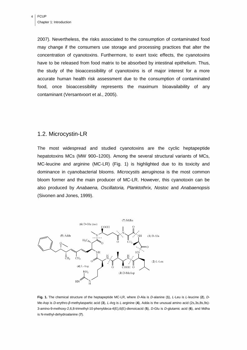
The most widespread and studied cyanotoxins are the cyclic heptapeptide
hepatotoxins MCs (MW 900–1200). Among the several structural variants of MCs,
MC-leucine and arginine (MC-LR) (Fig. 1) is highlighted due to its toxicity and
dominance in cyanobacterial blooms. Microcystis aeruginosa is the most common
bloom former and the main producer of MC-LR. However, this cyanotoxin can be
also produced by Anabaena, Oscillatoria, Planktothrix, Nostoc and Anabaenopsis
(Sivonen and Jones, 1999).
Fig. 1. The chemical structure of the heptapeptide MC-LR, where D-Ala is D-alanine (1), L-Leu is L-leucine (2), D-
Me-Asp is D-erythro-β-methylaspartic acid (3), L-Arg is L-arginine (4), Adda is the unusual amino acid (2s,3s,8s,9s)-
3-amino-9-methoxy-2,6,8-trimethyl-10-phenyldeca-4(E),6(E)-dienoicacid (5), D-Glu is D-glutamic acid (6), and Mdha
is N-methyl-dehydroalanine (7).
FCUP
Chapter 1: Introduction
5
The main mechanism of MC-LR toxicity in both animals and higher plants is the
irreversible inhibition of serine/threonine protein phosphatases (PP) (PP1 and PP2A)
(MacKintosh et al., 1990). The mechanism involved in PP inhibition by MC-LR
consists in a rapid and reversible binding, followed by a slower covalent binding
(occurs over several hours) between the N-methyl-dehydroalanine (Mdha) residue of
toxin and cysteine-273 of the catalytic subunit of PP1 (cysteine-266 of PP2A) (Craig
et al., 1996; MacKintosh et al., 1995).
1.3. Cylindrospermopsin
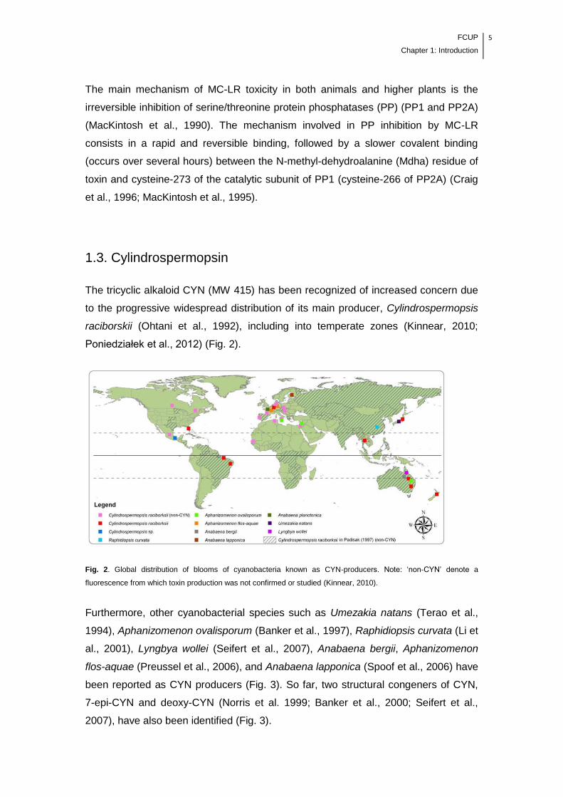
The tricyclic alkaloid CYN (MW 415) has been recognized of increased concern due
to the progressive widespread distribution of its main producer, Cylindrospermopsis
raciborskii (Ohtani et al., 1992), including into temperate zones (Kinnear, 2010;
Poniedziałek et al., 2012) (Fig. 2).
Fig. 2. Global distribution of blooms of cyanobacteria known as CYN-producers. Note: ‗non-CYN‘ denote a
fluorescence from which toxin production was not confirmed or studied (Kinnear, 2010).
Furthermore, other cyanobacterial species such as Umezakia natans (Terao et al.,
1994), Aphanizomenon ovalisporum (Banker et al., 1997), Raphidiopsis curvata (Li et
al., 2001), Lyngbya wollei (Seifert et al., 2007), Anabaena bergii, Aphanizomenon
flos-aquae (Preussel et al., 2006), and Anabaena lapponica (Spoof et al., 2006) have
been reported as CYN producers (Fig. 3). So far, two structural congeners of CYN,
7-epi-CYN and deoxy-CYN (Norris et al. 1999; Banker et al., 2000; Seifert et al.,
2007), have also been identified (Fig. 3).

6 FCUP
Chapter 1: Introduction
Fig. 3. The molecular structure of cylindrospermopsin (1) and its analogs 7-deoxy-cylindrospermopsin (2) and 7-
epicylindrospermopsin (3).
Although the molecular mechanism of CYN toxicity has not yet been established, it is
known that it inhibits eukaryotic protein synthesis with similar intensity in plant and
mammalian cell extracts (Terao et al., 1994; Runnegar et al., 2002; Froscio et al.,
2008). Moreover, CYN was reported to inhibits the glutathione (GSH) synthesis
(Runnegar et al., 1995) and the metabolic activation of CYN by cytochrome P450
(CYP450) seems to contribute to its high toxicity (Runnegar et al., 1995; Froscio et
al., 2003).
FCUP
Chapter 1: Introduction
7
1.4. Effects of MC-LR and CYN on plants
The majority of MC-LR- and CYN-related research have been focused on
mammalian toxicity; however, plants can also be affected by these cyanotoxins
through several molecular pathways (Babica et al., 2006; Corbel et al., 2014).
Cyanotoxins can be released from toxic cyanobacterial cells into water due to their
natural metabolism (e.g., CYN) (Chiswell et al., 1999; Rücker et al., 2007) or
following a cellular lyse during cell senescence or through water treatment processes
such as algaecide application (e.g., MC-LR) (Babica et al., 2006). The concentration
of MCs in surface waters vary from 4-50 µg/L up to 6500 µg/L, however, the higher
concentrations would be found in blooms and scums and correspond to intracellular
plus dissolved cyanotoxin (Corbel et al., 2014). Although the studies reporting the
concentrations of CYN in the environment are scarce, the concentration of total
extracellular CYN in water seem to vary from undetectable values up to 126 µg/L
(Corbel et al., 2014). The phytotoxic effects of MC-LR and CYN on higher plants
were firstly focused on aquatic macrophytes and floating plants that are naturally
exposed to cyanotoxins (Pflugmacher et al., 1999; Pflugmacher, 2002; Pietsch et al.,
2001; Mitrovic et al., 2005; Saqrane et al., 2007; Kinnear et al., 2008). MCs are very
stable and may persist in aquatic systems for weeks after being released from the
cells (Sivonen and Jones, 1999). Also, CYN can persist in the water because its
photodegradation is very low under natural conditions (Wörmer et al., 2010).
Therefore, the use of contaminated surface waters for agricultural irrigation may also
allows that these cyanotoxins enter into terrestrial ecosystems, leading to potential
risks for crop production and quality. MC-LR, by acting as PP1 and PP2A inhibitors
and inducers of ROS production, could be involved in several physiological and
molecular processes in higher terrestrial plants (Corbel et al., 2014). Indeed,
numerous studies have reported that MC-LR produces several perturbatory effects
on plant physiology and metabolism.
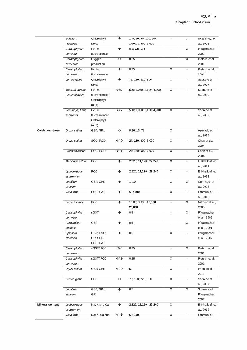
As is shown in Table 1, the growth, photosynthesis, antioxidant system and mineral
content of several aquatic and terrestrial plant species can be affected due to MC-LR
exposure.

8 FCUP
Chapter 1: Introduction
Table 1. General effects of the MC-LR on different species of aquatic and terrestrial plants.
Physiological
response
Plant species Endpoint Effect Concentration of
exposure (µg/L)
Crude
extract
Purified
toxin
Reference
Growth Oryza sativa Fresh weight 0.26; 13; 78 X - Azevedo et
al., 2014
Brassica napus;
Oryza sativa
Fresh and dry
weight; height
24; 120; 600; 3,000 X - Chen et al.,
2004
Medicago sativa Fresh weight;
stem length
2,220; 11,120; 22,240 X - El Khalloufi et
al., 2011
Lycopersicon
esculentum
Stem length 2,220; 11,120; 22,240 X - El Khalloufi et
al., 2012
Lepidium
sativum
Fresh weight;
length
1; 10 X X Gehringer et
al., 2003
Brassica
oleracea;
Sinapis alba
Stem length 1; 10 - X Järvenpää et
al., 2007
Vicia faba Length, dry
weight,
number of
nodes and
leaves
50; 100 X - Lahrouni et
al., 2013
Solanum
tuberosum
Fresh weight;
shoot length
1; 5; 10; 50; 100; 500;
1,000; 2,500; 5,000
- X McElhiney et
al., 2001
Lemma minor Fresh and dry
weight; frond
number
1,500; 3,000; 10,000;
20,000
- X Mitrovic et al.,
2005
Wolffia arrhiza Frond number 1,500; 3,000; 6,000;
10,000; 15,000
- X Mitrovic et al.,
2005
Ceratophyllum
demesum
Fresh weight 0.1; 0.5; 1; 5 - X Pflugmacher,
2002
Oryza sativa Fresh weight 50 X - Prieto et al.,
2011
Oryza sativa Fresh weight
of root
500; 1,000; 2,000; 4,000 - X Chen et al.,
2013
Lemna gibba Fronds
number
75; 150; 220; 300 X - Saqrane et
al., 2007
Triticum durum;
Zea mays;
Pisum sativum;
Lens esculenta
Fresh and dry
weight
500; 1,050; 2,100; 4,200 X - Saqrane et
al., 2009
Photosynthesis Oryza sativa Fv/Fm
fluorescence
0.26; 13; 78 X Azevedo et
al., 2014
Medicago sativa Fv/Fm
fluorescence
2,220; 11,120; 22,240 X - El Khalloufi et
al., 2011
Lycopersicon
esculentum
Fv/Fm
fluorescence
2,220; 11,120; 22,240 X - El Khalloufi et
al., 2012
Brassica
oleracea;
Sinapis alba
Fv/Fm
fluorescence;
Chlorophyll
(a+b)
1; 10 - X Järvenpää et
al., 2007
Vicia faba Fv/Fm
fluorescence;
Chlorophyll
(a+b)
50; 100 X - Lahrouni et
al., 2013
FCUP
Chapter 1: Introduction
9
Solanum
tuberosum
Chlorophyll
(a+b)
1; 5; 10; 50; 100; 500;
1,000; 2,500; 5,000
- X McElhiney, et
al., 2001
Ceratophyllum
demesum
Fv/Fm
fluorescence
0.1; 0.5; 1; 5 - X Pflugmacher,
2002
Ceratophyllum
demesum
Oxygen
production
0.25 - X Pietsch et al.,
2001
Ceratophyllum
demesum
Fv/Fm
fluorescence
0.25 X - Pietsch et al.,
2001
Lemna gibba Chlorophyll
(a+b)
75; 150; 220; 300 X - Saqrane et
al., 2007
Triticum durum;
Pisum sativum
Fv/Fm
fluorescence/
Chlorophyll
(a+b)
/ 500; 1,050; 2,100; 4,200 X - Saqrane et
al., 2009
Zea mays; Lens
esculenta
Fv/Fm
fluorescence/
Chlorophyll
(a+b)
/ 500; 1,050; 2,100; 4,200 X - Saqrane et
al., 2009
Oxidative stress Oryza sativa GST; GPx 0.26; 13; 78 X Azevedo et
al., 2014
Oryza sativa SOD; POD / 24; 120; 600; 3,000 X - Chen et al.,
2004
Brassica napus SOD/ POD / 24; 120; 600; 3,000 X - Chen et al.,
2004
Medicago sativa POD 2,220; 11,120; 22,240 X - El Khalloufi et
al., 2011
Lycopersicon
esculentum
POD 2,220; 11,120; 22,240 X - El Khalloufi et
al., 2012
Lepidium
sativum
GST; GPx 1; 10 X X Gehringer et
al., 2003
Vicia faba POD; CAT 50 ; 100 X - Lahrouni et
al., 2013
Lemma minor POD 1,500; 3,000; 10,000;
20,000
- X Mitrovic et al.,
2005
Ceratophyllum
demesum
sGST 0.5 - X Pflugmacher
et al., 1999
Phragmites
australis
GST 0.5 - X Pflugmacher
et al., 2001
Spinacia
oleracea
GST; GSH;
GR; SOD;
POD; CAT
0.5 X - Pflugmacher
et al., 2007
Ceratophyllum
demesum
sGST/ POD / 0.25 - X Pietsch et al.,
2001
Ceratophyllum
demesum
sGST/ POD / 0.25 X - Pietsch et al.,
2001
Oryza sativa GST/ GPx / 50 X - Prieto et al.,
2011
Lemna gibba POD 75; 150; 220; 300 X - Saqrane et
al., 2007
Lepidium
sativum
GST, GPx;
GR
0.5 X X Stüven and
Pflugmacher,
2007
Mineral content Lycopersicon
esculentum
Na; K and Ca 2,220; 11,120; 22,240 X El Khalloufi et
al., 2012
Vicia faba Na/ K; Ca and / 50; 100 X - Lahrouni et
10 FCUP
Chapter 1: Introduction
N al., 2013
Triticum durum;
Zea mays;
Pisum sativum;
Lens esculenta
Ca; Na; K; P;
N
500; 1,050; 2,100; 4,200 X - Saqrane et
al., 2009
Non-enzymatic
and enzymatic
antioxidant
content
Medicago sativa Phenols
content
2,220; 11,120; 22,240 X - El Khalloufi et
al., 2011
Lycopersicon
esculentum
Phenols
content
2,220; 11,120; 22,240 X - El Khalloufi et
al., 2012
Vicia faba PPO; PAL;
Phenols
content
50; 100 X - Lahrouni et
al., 2013
Medicago sativa α and β
Tocopherols
0.05; 0.5; 5 X X Peuthert and
Pflugmacher,
2010
Spinacia
oleracea
Ascorbate; α
Tocopherol
0.5 X - Pflugmacher
et al., 2007
Lemna gibba Phenols
content
75; 150; 220; 300 X - Saqrane et
al., 2007
Lepidium
sativum
α and β
Tocopherols
0.5 X X Stüven and
Pflugmacher,
2007
Increased in comparison to control group; Decreased in comparison to control group; There were no effects in comparison to
control group; - Not measured; Concentration values highlighted in bold indicate more pronounced effects.
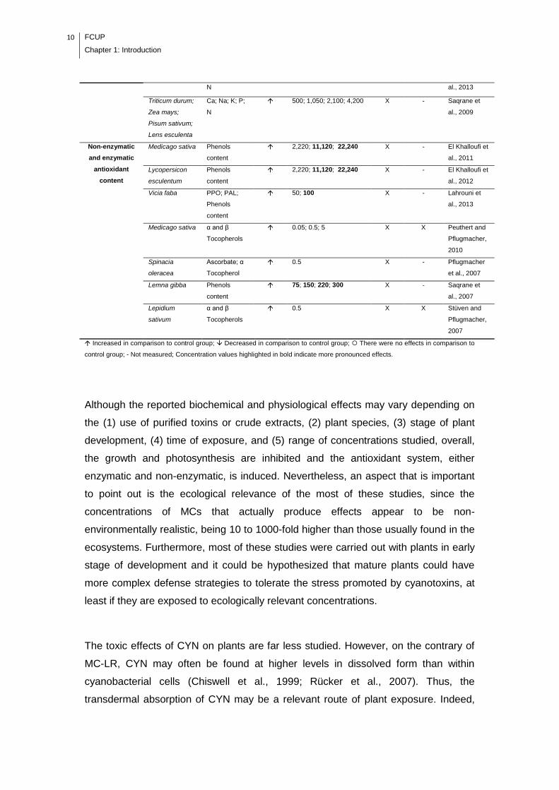
Although the reported biochemical and physiological effects may vary depending on
the (1) use of purified toxins or crude extracts, (2) plant species, (3) stage of plant
development, (4) time of exposure, and (5) range of concentrations studied, overall,
the growth and photosynthesis are inhibited and the antioxidant system, either
enzymatic and non-enzymatic, is induced. Nevertheless, an aspect that is important
to point out is the ecological relevance of the most of these studies, since the
concentrations of MCs that actually produce effects appear to be non-
environmentally realistic, being 10 to 1000-fold higher than those usually found in the
ecosystems. Furthermore, most of these studies were carried out with plants in early
stage of development and it could be hypothesized that mature plants could have
more complex defense strategies to tolerate the stress promoted by cyanotoxins, at
least if they are exposed to ecologically relevant concentrations.
The toxic effects of CYN on plants are far less studied. However, on the contrary of
MC-LR, CYN may often be found at higher levels in dissolved form than within
cyanobacterial cells (Chiswell et al., 1999; Rücker et al., 2007). Thus, the
transdermal absorption of CYN may be a relevant route of plant exposure. Indeed,
FCUP
Chapter 1: Introduction
11
implications of this cyanotoxin were already observed in vegetable cells and the few
studies that were performed indicate that, at low concentrations, the exposure to
CYN results in an induction of plant growth, inhibition of photosynthesis and increase
in oxidative stress (Table 2).
Table 2. General effects of the CYN on different species of aquatic and terrestrial plants.
Physiological
response
Plant
species
Endpoint Effect Concentration of
exposure (µg/L)
Crude
extract
Purified
toxin
Reference
Growth Hydrilla
verticillata
Biomass of
roots
25; 50; 100; 200; 400 X - Kinnear et
al., 2008
Brassica
juncea/
Brassica
oleracea
Fresh
weight of
leaves
/ 18.2; 35.5 X X Kittler et
al., 2012
Oryza
sativa
Roots/
leaves
fresh and
dry weight
/ 2.5 X - Prieto et
al., 2011
Sinapis
alba
Dry weight 2,500; 5,000; 10,000;
20,000; 40,000; 80,000;
160,000
- X Vasas et
al., 2002
Photosynthesis Hydrilla
verticillata
Chlorophyll
(a+b)
/ 25; 50; 100; 200; 400 X - Kinnear et
al., 2008
Oxidative stress Oryza
sativa
GST; GPx 2.5 X - Prieto et
al., 2011
Increased in comparison to control group; Decreased in comparison to control group; There were no effects in comparison to
control group; - Not measured; Concentration values highlighted in bold indicate more pronounced effects.
In aquatic ecosystems, the existence of mixtures of cyanotoxins is highly expected,
however, the toxicological experiments are predominantly carried out on individual
cyanotoxins. The impact of cyanotoxin mixtures, especially the more prevalent, is a
matter of high priority. Simultaneous exposure to MC-LR and CYN may lead to
changes in the response capability of crop plants, triggering potential synergistic,
additive or antagonistic effects. However, the studies regarding to effects of
interaction of the MC-LR/CYN mixture are very scarce. According to our knowledge,
only Prieto et al. (2011) have studied the interaction effects of MC-LR and CYN in
plants. The authors suggested a synergistic effect on the oxidative stress response
(GST activity) of rice plants when exposed to cyanobacterial extracts containing
ecological relevant concentrations of both CYN (0.13 µg/L) and MC-LR (50 µg/L)
(Table 3).
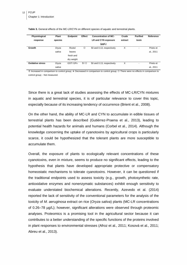
12 FCUP
Chapter 1: Introduction
Table 3. General effects of the MC-LR/CYN on different species of aquatic and terrestrial plants.
Physiological
response
Plant
species
Endpoint Effect Concentration of MC-
LR and CYN exposure
(µg/L)
Crude
extract
Purified
toxin
Reference
Growth Oryza
sativa
Roots/
leaves
fresh and
dry weight
50 and 0.13, respectively X - Prieto et
al., 2011
Oxidative stress Oryza
sativa
GST/ GPx / 50 and 0.13, respectively X - Prieto et
al., 2011
Increased in comparison to control group; Decreased in comparison to control group; There were no effects in comparison to
control group; - Not measured.
Since there is a great lack of studies assessing the effects of MC-LR/CYN mixtures
in aquatic and terrestrial species, it is of particular relevance to cover this topic,
especially because of its increasing tendency of occurrence (Brient et al., 2008).
On the other hand, the ability of MC-LR and CYN to accumulate in edible tissues of
terrestrial plants has been described (Gutiérrez-Praena et al., 2013), leading to
potential health hazards for animals and humans (Corbel et al., 2014). Although the
knowledge concerning the uptake of cyanotoxins by agricultural crops is particularly
scarce, it could be hypothesized that the tolerant plants are more susceptible to
accumulate them.
Overall, the exposure of plants to ecologically relevant concentrations of these
cyanotoxins, even in mixture, seems to produce no significant effects, leading to the
hypothesis that plants have developed appropriate protective or compensatory
homeostatic mechanisms to tolerate cyanotoxins. However, it can be questioned if
the traditional endpoints used to assess toxicity (e.g., growth, photosynthetic rate,
antioxidative enzymes and nonenzymatic substances) exhibit enough sensitivity to
evaluate understated biochemical alterations. Recently, Azevedo et al. (2014)
reported the lack of sensitivity of the conventional parameters for the analysis of the
toxicity of M. aeruginosa extract on rice (Oryza sativa) plants (MC-LR concentrations
of 0.26–78 µg/L); however, significant alterations were observed through proteomic
analyses. Proteomics is a promising tool in the agricultural sector because it can
contributes to a better understanding of the specific functions of the proteins involved
in plant responses to environmental stresses (Afroz et al., 2011; Kosová et al., 2011;
Abreu et al., 2013).
FCUP
Chapter 1: Introduction
13
1.4.1. Proteomics applied to agricultural sector to assess the
effects of cyanotoxins
Proteomics is an emergent research tool that offers several advantages over the
standard enzymatic and biochemical assays. By the investigation of protein
dynamics and variations in plant metabolism in response to an environmental
stimulus, proteomic technologies may enable the potential identification of protein
biomarkers of stress response and the discovery of proteins involved in stress
tolerance (Afroz et al., 2011; Gutiérrez-Praena et al., 2014; Kosová et al., 2011).
Recent advances in accomplishment of genome sequencing of crop plants (e.g.,
rice) and the development/improvement of analytical methods for protein
characterization makes the proteomics analysis appropriate for the agricultural
sector. Proteomic studies are extensively applied to genetically modified plants, in
which new proteins are incorporated into food crops, for instance to promote
resistance to pests, pesticides and other stressors. Proteomic studies investigating
the effects of CYN and MC-LR have been performed on bivalves, including mixtures
with other environmental pollutants (e.g., herbicides) (Martins et al., 2009; Puerto et
al., 2011; Malécot et al., 2013). However, so far only two studies were developed
using a proteomic approach to investigate the effects of cyanotoxins (MC-LR) in crop
plants. Concisely, Azevedo et al. (2014) studied the biochemical responses of rice
(Oryza sativa) seedlings exposed to low concentrations of MC-LR; and Gutiérrez-
Praena et al. (2014) studied the effects of MC-LR in the leaf proteome of tomato
(Lycopersicon esculentum). In these two studies, the combination of two-dimensional
electrophoresis (2DE) and matrix-assisted laser desorption/ionization time of flight-
tandem mass spectrometry (MALDI-TOF/TOF MS), allowed the identification of the
function of several proteins that complement the current understanding of the mode
of action of MC-LR in those plants. Thus, these techniques could be applied for other
commercially important species, such as lettuce (Lactuca sativa), a leafy vegetable
worldwide used for human consumption.
From the health risk point of view, proteomics may provide also new insights on
safety and quality of edible plants exposed to environmentally relevant
concentrations of cyanotoxins due to the potential identification of allergenic proteins
secreted as a defensive or protective mechanism, such as pathogenesis-related (PR)
proteins (Abreu et al., 2013).
14 FCUP
Chapter 1: Introduction
1.5. Human exposure to MC-LR and CYN
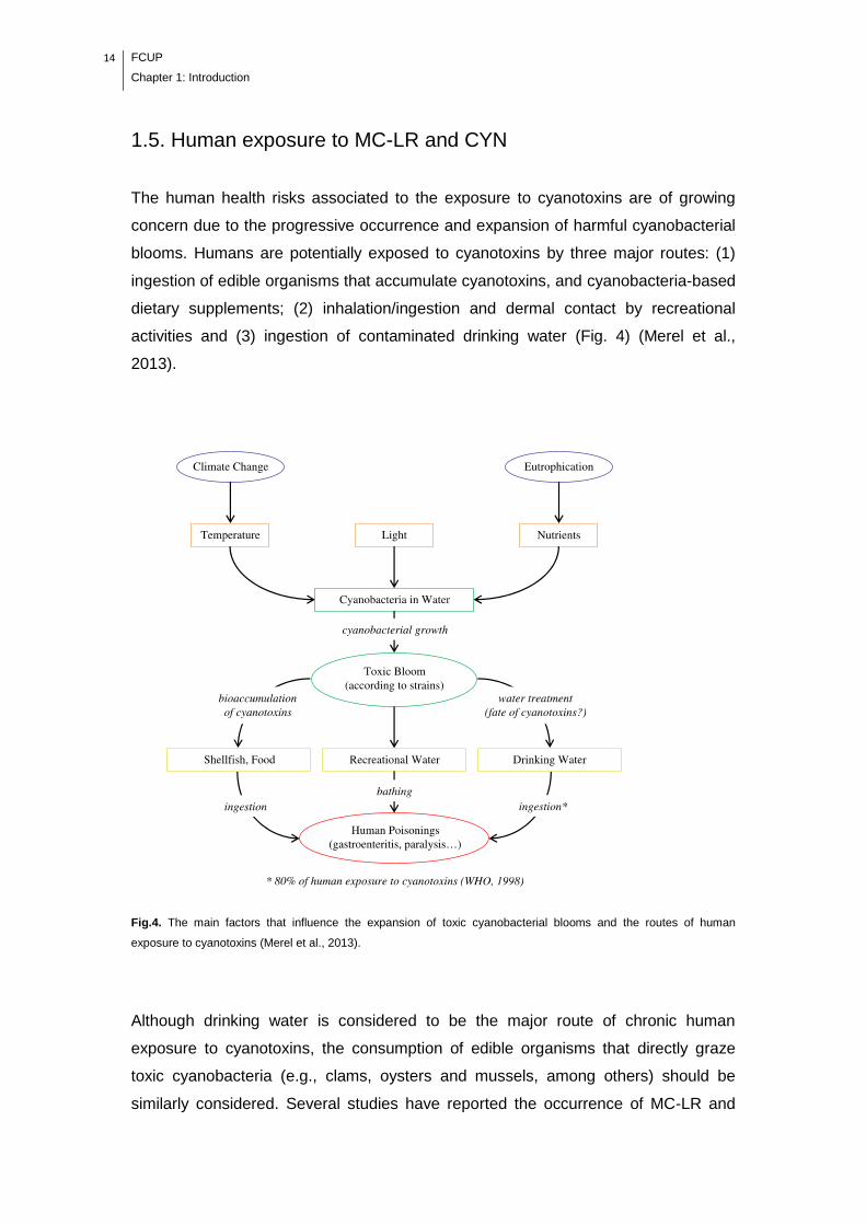
The human health risks associated to the exposure to cyanotoxins are of growing
concern due to the progressive occurrence and expansion of harmful cyanobacterial
blooms. Humans are potentially exposed to cyanotoxins by three major routes: (1)
ingestion of edible organisms that accumulate cyanotoxins, and cyanobacteria-based
dietary supplements; (2) inhalation/ingestion and dermal contact by recreational
activities and (3) ingestion of contaminated drinking water (Fig. 4) (Merel et al.,
2013).
Fig.4. The main factors that influence the expansion of toxic cyanobacterial blooms and the routes of human
exposure to cyanotoxins (Merel et al., 2013).
Although drinking water is considered to be the major route of chronic human
exposure to cyanotoxins, the consumption of edible organisms that directly graze
toxic cyanobacteria (e.g., clams, oysters and mussels, among others) should be
similarly considered. Several studies have reported the occurrence of MC-LR and

FCUP
Chapter 1: Introduction
15
CYN in the tissues of edible aquatic organisms (e.g., fish, bivalves) (Gutiérrez-
Praena et al., 2013; Kinnear, 2010; Ibelings and Chorus, 2007), reaching levels at
which human consumption should be avoided (Chen and Xie, 2005; Ibelings and
Chorus, 2007). Bivalves, as filter-feeding organisms, have shown to accumulate high
concentrations of MCs and CYN in both laboratory and field conditions (Gutiérrez-
Praena et al., 2013; Kinnear, 2010), making them potential vehicles of these
cyanotoxins to higher trophic levels (Saker et al., 2004; Vasconcelos et al., 2007).
Considering the human health effects, cyanotoxins can be classified according to
their target organs, as follows: (1) neurotoxins (nervous system), (2) hepatotoxins
(liver), (3) cytotoxins (several organs: liver, kidneys, adrenal glands, small intestine),
and (4) dermatotoxins (causing skin irritation). In mammals, the acute exposure to
the hepatotoxin MC-LR produces a cascade of events (cytoskeleton alterations, lipid
peroxidation, oxidative stress, apoptosis) leading to cell necrosis, intrahepatic
hemorrhage and death (Funari and Testai, 2008). Although human acute intoxication
is rare, unfortunately, it already occurred in 1996 at the Brazilian dialysis center of
Caruaru, which caused the death of 60 patients due to the use of contaminated water
for hemodialysis (Pouria et al., 1998). Nevertheless, human health problems due to
MC-LR are most likely associated with chronic exposure, where phosphatases
inhibition induces cellular proliferation and hepatic hypertrophy (Funari and Testai,
2008). Epidemiological studies in China support the association of chronic exposure
to MCs from contaminated drinking water with primary liver and colorectal cancer
(Ueno et al., 1996; Zhou et al., 2002). On the basis of data on tumor promoting
mechanisms, the International Agency for Research on Cancer (IARC) classified
MC-LR as ‗‗possibly carcinogenic to humans‘‘ (group 2B) (Grosse et al., 2006).
Regarding to the cytotoxin CYN, the most famous case of human intoxication
occurred in 1979 in Australia, where 149 people supplied with drinking water from a
reservoir with CYN-producing C. raciborskii suffered a hepatoenteritis-like illness
(Griffiths & Saker, 2003). Although a lack of epidemiological studies regarding to
chronic exposure to CYN, it has been suggested that tumors are generated in mice
by oral exposure to the toxin (Falconer & Humpage, 2001; Falconer and Humpage,
2006).
Based on the potential for human health risks, the World Health Organization (WHO)
proposed a provisional upper limit in drinking water of 1 µg/L for MC-LR and a
tolerable daily intake (TDI) of 0.04 µg/kg of body weight (WHO, 1998). Likewise, a
guideline for maximum concentration of 1 μg/L was proposed in drinking water for
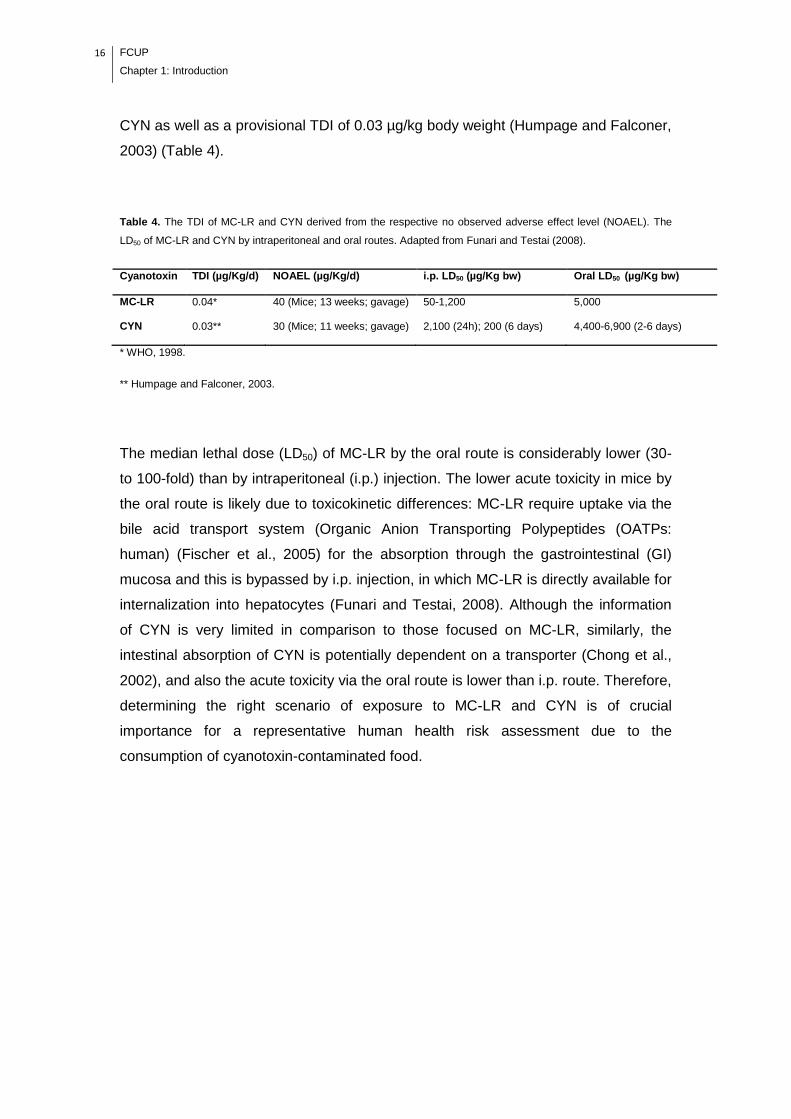
16 FCUP
Chapter 1: Introduction
CYN as well as a provisional TDI of 0.03 µg/kg body weight (Humpage and Falconer,
2003) (Table 4).
Table 4. The TDI of MC-LR and CYN derived from the respective no observed adverse effect level (NOAEL). The
LD50 of MC-LR and CYN by intraperitoneal and oral routes. Adapted from Funari and Testai (2008).
Cyanotoxin TDI (µg/Kg/d) NOAEL (µg/Kg/d) i.p. LD50 (µg/Kg bw) Oral LD50 (µg/Kg bw)
MC-LR 0.04* 40 (Mice; 13 weeks; gavage) 50-1,200 5,000
CYN 0.03** 30 (Mice; 11 weeks; gavage) 2,100 (24h); 200 (6 days) 4,400-6,900 (2-6 days)
* WHO, 1998.
** Humpage and Falconer, 2003.
The median lethal dose (LD50) of MC-LR by the oral route is considerably lower (30-
to 100-fold) than by intraperitoneal (i.p.) injection. The lower acute toxicity in mice by
the oral route is likely due to toxicokinetic differences: MC-LR require uptake via the
bile acid transport system (Organic Anion Transporting Polypeptides (OATPs:
human) (Fischer et al., 2005) for the absorption through the gastrointestinal (GI)
mucosa and this is bypassed by i.p. injection, in which MC-LR is directly available for
internalization into hepatocytes (Funari and Testai, 2008). Although the information
of CYN is very limited in comparison to those focused on MC-LR, similarly, the
intestinal absorption of CYN is potentially dependent on a transporter (Chong et al.,
2002), and also the acute toxicity via the oral route is lower than i.p. route. Therefore,
determining the right scenario of exposure to MC-LR and CYN is of crucial
importance for a representative human health risk assessment due to the
consumption of cyanotoxin-contaminated food.
FCUP
Chapter 1: Introduction
17
1.5.1. Factors affecting human exposure to MC-LR and CYN by
the consumption of contaminated edible aquatic organisms
The severity degree of MC-LR and CYN induced toxicity depends on the levels and
duration of exposure, determined by the balance between the absorption,
detoxification and excretion. In addition, another factor of uncertainty in assessing
human exposure derives from the fact that it is not clear whether the levels of MC-LR
and CYN measured in raw edible aquatic organisms correspond to the total
bioavailable amount. Some external factors such as food storage, preparation,
processing and the human digestion itself can change the MC-LR and CYN
availability and thus the risk of human exposure.
1.5.1.1. Food storage and processing
Food is generally consumed after being stored and processed. The processing of
food products generally implies the transformation of the perishable raw commodity
to value added product that has larger shelf life (Kaushik et al., 2009). The common
food storage (e.g., refrigeration, freezing) and processing practices (e.g., boiling,
frying, microwave cooking, steaming, canning) at domestic and industrial level could
lead to significant variations of MC-LR and CYN availability in edible organisms.
Domingo (2011) has studied the influence of cooking processes on the
concentrations of toxic metals and various organic environmental pollutants in food
and found that the main changes in contaminant levels depend basically on the food
item and the cooking conditions, as follows: (1) cooking method, (2) time, (3)
temperature, and (4) medium of cooking. Kaushik et al. (2009) reviewed the effects
of food processing in pesticide dissipation and found that in most cases processing
leads to large reductions in pesticide residue levels. MC-LR and CYN are water-
soluble and stable at high temperatures (Chiswell et al., 1999; Sivonen and Jones,
1999), thus studies are necessary to know whether storage and cooking practices
can be suitable of reducing the content of these cyanotoxins in food. Very few
studies have been developed in the investigation of the effects of food storage and
processing practices in the availability of cyanotoxins in edible organisms (Guzmán-
Guillén et al., 2011; Morais et al., 2008; Zhang et al., 2010), and these studies
concern only to MC-LR (Table 5).
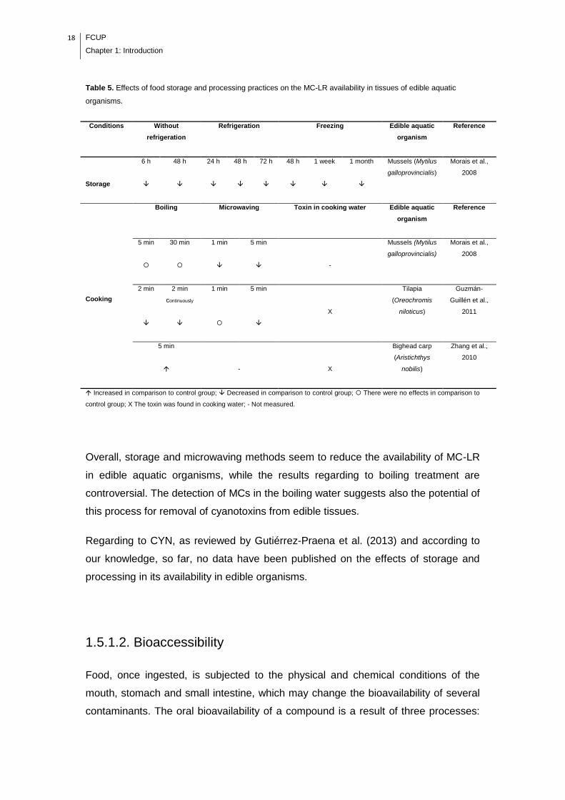
18 FCUP
Chapter 1: Introduction
Table 5. Effects of food storage and processing practices on the MC-LR availability in tissues of edible aquatic
organisms.
Conditions Without
refrigeration
Refrigeration Freezing Edible aquatic
organism
Reference
Storage
6 h 48 h 24 h 48 h 72 h 48 h 1 week 1 month Mussels (Mytilus
galloprovincialis)
Morais et al.,
2008
Cooking
Boiling Microwaving Toxin in cooking water Edible aquatic
organism
Reference
5 min 30 min 1 min 5 min
-
Mussels (Mytilus
galloprovincialis)
Morais et al.,
2008
2 min 2 min
continuously
1 min 5 min
X
Tilapia
(Oreochromis
niloticus)
Guzmán-
Guillén et al.,
2011
5 min
-
X
Bighead carp
(Aristichthys
nobilis)
Zhang et al.,
2010
Increased in comparison to control group; Decreased in comparison to control group; There were no effects in comparison to
control group; X The toxin was found in cooking water; - Not measured.
Overall, storage and microwaving methods seem to reduce the availability of MC-LR
in edible aquatic organisms, while the results regarding to boiling treatment are
controversial. The detection of MCs in the boiling water suggests also the potential of
this process for removal of cyanotoxins from edible tissues.
Regarding to CYN, as reviewed by Gutiérrez-Praena et al. (2013) and according to
our knowledge, so far, no data have been published on the effects of storage and
processing in its availability in edible organisms.
1.5.1.2. Bioaccessibility
Food, once ingested, is subjected to the physical and chemical conditions of the
mouth, stomach and small intestine, which may change the bioavailability of several
contaminants. The oral bioavailability of a compound is a result of three processes:

FCUP
Chapter 1: Introduction
19
(1) release of the compound from food matrix into digestive juices (i.e.,
bioaccessibility), (2) transport across the intestinal epithelium into the vena Portae
(intestinal transport) and (3) degradation of the compound in the liver (and intestine)
(metabolism) (Versantvoort et al., 2005). In more detail, bioaccessibility is defined as
the fraction of the contaminant that is released from the food matrix by the action of
digestive enzymes and is then available for absorption by the intestinal mucosa
(Versantvoort et al., 2005). The bioaccessibility indicates the maximal oral
bioavalability of any contaminant (Versantvoort et al., 2005) (Fig. 5).
Fig. 5. Schematic representation of the difference between bioaccessibility and bioavailability of food contaminants
(Adapted from Guerra et al., 2012).
Metabolism
Liver
20 FCUP
Chapter 1: Introduction
There are several in vitro methods that simulate the human digestion process, most
of them comprising three phases: mouth, stomach and small intestine. Thus, these
models enable the investigation of the bioaccessibility of a compound during the
transit in the gastrointestinal tract (Versantvoort et al., 2005; Maulvault et al., 2011;
Guerra et al., 2012). The bioaccessibility of a contaminant can be influenced by
several factors, as follows: (1) the type of food matrix, (2) cooking preparation, (3)
digestion model, (4) initial concentration of the contaminat, and (5) the chemical
properties of the contaminant (Hur et al., 2009; Maulvault et al., 2011; Xing et al.,
2008). The determination of the bioaccessibility of MC-LR and CYN is of great
relevance, once it will provide data for a more accurate human health risk
assessment due to consumption of contaminated food. The current health risk
assessment generally considers the concentration of these cyanotoxins in the raw
edible organisms.
There were only few reports on the digestibility of MCs with gastric and intestinal
juices, but these studies were carried out with toxin in its free form in solution
(without food matrix) (Table 6).
Table 6. Digestion of MC-LR (in solution) by proteolytic enzymes of gastric and intestinal juices.
Pepsin Trypsin Chymotrypsin Reference
+ - - Moreno et al., 2004
- - - Smith et al., 2010
NA + + Kankaanpää et al., 2005
+ Digested; - non-digested; NA, not analyzed.
Although the contradictory results of these studies, probably due to the differences in
the experimental designs and/or analysts and analysis equipment, they give an
indication of the effects occurring in MCs (heptapeptides) when they are alternatively
exposed to gastric or intestinal digestive conditions. However, it is important to notice
that if the toxin is ingested within food, this may be not straightforward because of the
matrix effects. According to our knowledge there are no studies on the
bioaccessibility of MC-LR and CYN. The study of bioaccessibility of these
cyanotoxins in edible bivalves can provide relevant information for risk assessment,
supporting potential improvements in the management measures.
FCUP
Chapter 1: Introduction
21
1.6. References
Abreu, I.A., Farinha, A.P., Negrão, S., Gonçalves, N., Fonseca, C., Rodrigues, M.,
Batista, R., Saibo, N.J.M., Oliveira, M.M., 2013. Coping with abiotic stress: Proteome
changes for crop improvement. J. Proteomics. 93, 145-168.
Afroz, A., Ali, G.M., Mir, A., Komatsu, S., 2011. Application of proteomics to
investigate stress-induced proteins for improvement in crop protection. Plant Cell
Rep. 30, 745–763.
Azevedo, C.C., Azevedo, J., Osório, H., Vasconcelos, V., Campos, A., 2014. Early
physiological and biochemical responses of rice seedlings to low concentration of
microcystin-LR. Ecotoxicology. 23, 107–121.
Babica P., Blaha L., Marsalek B., 2006. Exploring the natural role of microcystins -
review of effects on photoautotrophic organisms. J. Phycol. 42, 9-20.
Banker, R., Carmeli, S., Hadas, O., Teltsch, B., Porat, R., Sukenik, A., 1997.
Identification of cylindrospermopsin in Aphanizomenon ovalisporum (cyanophyceae)
isolated from Lake Kinneret, Israel. J. Phycol. 33, 613–616.
Banker, R., Teltsch, B., Sukenik, A., Carmeli, S., 2000. 7-Epicylindrospermopsin, a
toxic minor metabolite of the cyanobacterium Aphanizomenon ovalisporum from
Lake Kinneret, Israel. J. Nat. Prod. 63, 387–389.
Brient, L., Lengronne, M., Bormans, M., Fastner, J., 2008. First occurrence of
cylindrospermopsin in freshwater in France. Environ. Toxicol. 24, 415–420.
Chen, J., Song, L., Dai, J., Gan, N., Liu, Z., 2004. Effects of microcystins on the
growth and the activity of superoxide dismutase and peroxidase of rape (Brassica
napus L.) and rice (Oryza sativa L.). Toxicon. 43, 393–400.
Chen, J., Xie, P., 2005. Seasonal Dynamics of the Hepatotoxic Microcystins in
Various Organs of Four Freshwater Bivalves from the Large Eutrophic Lake Taihu of
Subtropical China and the Risk to Human Consumption. Environ Toxicol. 20, 572–
584.
Chen, J., Zhang, H-Q., Hu, L-B., Shi, Z-Q., 2013. Microcystin-LR-induced
phytotoxicity in rice crown root is associated with the cross-talk between auxin and
22 FCUP
Chapter 1: Introduction
nitric oxide. Chemosphere 93, 283–293.
Chiswell, R.K., Shaw, G.R., Eaglesham, G., Smith, M.J., Norris, R.L., Seawright,
A.A., Moore, M.R., 1999. Stability of cylindrospermopsin, the toxin from the
cyanobacterium. Cylindrospermopsis raciborskii: Effect of pH, temperature, and
sunlight on decomposition. Environ. Toxicol. 14, 155–161.
Chong, M.W.K., Wong, B.S.F., Lam, P.K.S., Shaw, G.R., and Seawright, A.A., 2002.
Toxicity and uptake mechanism of cylindrospermopsin and lophyrotomin in primary
rat hepatocytes. Toxicon 40, 205–211.
Corbel, S., Mougin, C., Bouaicha, N., 2014. Cyanobacterial toxins: Modes of actions,
fate in aquatic and soil ecosystems, phytotoxicity and bioaccumulation in agricultural
crops. Chemosphere 96, 1–15.
Craig, M., Luu, H.A., McCready, T.L., Williams, D., Andersen, R.J., Holmes, C.F.B.,
1996. Molecular mechanisms underlying the interaction of motuporin and
microcystins with type-1 and type-2A protein phosphatases. Biochem. Cell Biol. 74,
569–578.
Domingo, J.L., 2011. Influence of cooking processes on the concentrations of toxic
metals and various organic environmental pollutants in food: a review of the
published literature. Crit. Rev. Food Sci. Nutr. 51, 29–37.
El Khalloufi, F., El Ghazali, I., Saqrane, S., Oufdou, K., Vasconcelos, V., Oudra, B.,
2012. Phytotoxic effects of a natural bloom extract containing microcystins on
Lycopersicon esculentum. Ecotoxicol. Environ. Saf. 79, 199–205.
El Khalloufi, F., Oufdou, K., Lahrouni, M., El Ghazali, I., Saqrane, S., Vasconcelos,
V., Oudra, B., 2011. Allelopatic effects of cyanobacteria extracts containing
microcystins on Medicago sativa-Rhizobia symbiosis. Ecotoxicol. Environ. Saf. 74,
431-438.
Elliott, J.A., 2012. Is the future blue-green? A review of the current model predictions
of how climate change could affect pelagic freshwater cyanobacteria. Water Res. 46,
1364-1371.
Falconer, I., Humpage, A.R., 2006. Cyanobacterial (blue-green algal) toxins in water
supplies: cylindrospermopsins. Environ. Toxicol. 21, 299–304.
FCUP
Chapter 1: Introduction
23
Falconer, I.R., Humpage, A.R., 2001. Preliminary evidence for in vivo tumour
initiation by oral administration of extracts of the blue-green alga Cylindrospermopsis
raciborskii containing the toxin cylindrospermopsin. Environ. Toxicol. 16, 192–195.
Fischer, W.J., Altheimer, S., Cattori, V., Meier, P.J., Dietrich, D.R., Hagenbuch, B.,
2005. Organic anion transporting polypeptides expressed in liver and brain mediate
uptake of microcystin. Toxicol. Appl. Pharmacol. 203, 257–263.
Froscio, S.M., Humpage, A.R., Wickramasinghe, W., Shaw, G., Falconer, I.R., 2008.
Interaction of the cyanobacterial toxin cylindrospermopsin with the eukaryotic protein
synthesis system. Toxicon. 51, 191–198.
Froscio, S.M., Humpage, A.R., Burcham, P.C., Falconer, I.R., 2003.
Cylindrospermopsin-induced protein synthesis inhibition and its dissociation from
acute toxicity in mouse hepatocytes. Environ. Toxicol. 18, 243–251.
Funari, E., Testai, E., 2008. Human Health Risk Assessment Related to Cyanotoxins
Exposure. Crit. Rev. Toxicol. 38, 97-125.
Gehringer, M.M., Kewada, V., Coates, N., Downing, T.G., 2003. The use of Lepidium
sativum in a plant bioassay system for the detection of microcystin-LR. Toxicon. 41,
871–876.
Griffiths, D.J., Saker, M.L., 2003. The Palm Island mystery disease twenty years on:
a review of research on the cyanotoxin cylindrospermopsin. Environ. Toxicol. 18, 78–
93.
Grosse, Y., Baan, R., Straif, K., Secretan, B., Ghissassi, F.El., Cogliano, V., 2006.
Carcinogenicity of nitrate, nitrite, and cyanobacterial peptide toxins. Lancet Oncol. 7,
628–629.
Guerra, A., Etienne-Mesmin, L., Livrelli. V., Denis, S., Blanquet-Diot, S., Alric, M.,
2012. Relevance and challenges in modelinghuman gastric and small intestinal
digestion. Trends Biotechnol. 30, 591-600.
Gutiérrez-Praena, D., Campos, A., Azevedo, J., Neves, J., Freitas, M., Guzmán-
Guillén, R., Cameán, A.M., Renaut, J., Vasconcelos, V., 2014. Exposure of
Lycopersicon esculentum to microcystin-LR: Effects in the leaf proteome and toxin
translocation from water to leaves and fruits. Toxins 6, 1837-1854.
24 FCUP
Chapter 1: Introduction
Gutiérrez-Praena, D., Jos, Á., Pichardo, S., Moreno, I.M., Cameán, A.M., 2013.
Presence and bioaccumulation of microcystins and cylindrospermopsin in food and
the effectiveness of some cooking techniques at decreasing their concentrations: A
review. Food Chem. Toxicol. 53, 139–152.
Guzmán-Guillén, R., Prieto, A.I., Moreno, I., Soria, M.E., Cameán, A.M., 2011.
Effects of thermal treatments during cooking, microwave oven and boiling, on the
unconjugated microcystin concentration in muscle of fish (Oreochromis niloticus).
Food Chem. Toxicol. 49, 2060–2067.
Humpage, A.R., Falconer, I.R., 2003. Oral toxicity of the cyanobacterial toxin
cylindrospermopsin in male swiss albino mice: Determination of no observed adverse
effect level for deriving a drinking water guideline value. Environ. Toxicol. 18, 94–
103.
Hur, S.J., Decker, E.A., McClements, D.J., 2009. Influence of initial emulsifier type on
microstructural changes occurring in emulsified lipids during in vitro digestion. Food
Chem. 114, 253–262.
Ibelings, B.W., Chorus I., 2007. Accumulation of cyanobacterial toxins in freshwater
‗‗seafood‘‘ and its consequences for public health: A review. Environ. Pollut. 150,
177-192.
Järvenpää, S., Lundberg-Niinistö, C., Spoof, L., Sjövall, O., Tyystjärvi, E., Meriluoto,
J., 2007. Effects of microcystins on broccoli and mustard, and analysis of
accumulated toxin by liquid chromatography–mass spectrometry. Toxicon 49, 865–
874.
Kankaanpää, H.T., Holliday, J., Schröder, H., Goddard, T.J., von Fister, R.,
Carmichael, W.W., 2005. Cyanobacteria and prawn farming in northern New South
Wales, Australia—a case study on cyanobacteria diversity and hepatotoxin
bioaccumulation. Toxicol. Appl. Pharmacol. 203, 243–256.
Kaushik, G., Satya, S., Naik, S.N., 2009. Food processing a tool to pesticide residue
dissipation – a review. Food Res. Int. 42, 26–40.
Kinnear, S., 2010. Cylindrospermopsin: a decade of progress on bioaccumulation
research. Mar. Drugs 8, 542-564.

FCUP
Chapter 1: Introduction
25
Kinnear, S., Fabbro, L., Duivenvoorden, L., 2008. Variable growth responses of
water thyme (Hydrilla verticillata); to whole-cell extracts of Cylindrospermopsis
raciborskii. Arch. Environ. Contam. Toxicol. 54, 187–194.
Kittler, K., Schreiner, M., Krumbein, A., Manzei, S., Koch, M., Rohn, S., Maul, R.,
2012. Uptake of the cyanobacterial toxin cylindrospermopsin in Brassica vegetables.
Food Chem. 133, 875–879.
Kosová, K., Vítámvás, P., Prášil, I.T., Renaut, J., 2011. Plant proteome changes
under abiotic stress — Contribution of proteomics studies to understanding plant
stress response. J. Proteomics 74, 1301-1322.
Lahrouni, M., Oufdou, K., El Khalloufi, F., Baz, M., Lafuente, A., Dary, M., Pajuelo,
E., Oudra, B., 2013. Physiological and biochemical defense reactions of Vicia faba
L.-Rhizobium symbiosis face to chronic exposure to cyanobacterial bloom extract
containing microcystins. Environ. Sci. Pollut. Res. Int. 20, 5405-5415.
Li, R., Carmichael, W.W., Brittain, S., Eaglesham, G.K., Shaw, G.R., Liu, Y.,
Watanabe, M.M., 2001. First report of the cyanotoxins cylindrospermopsin and
deoxycylindrospermopsin from Raphidiopsis curvata (cyanobacteria.). J. Phycol. 3,
1121–1126.
MacKintosh, C., Beattie, K.A., Klumpp, S., Cohen, P., Codd, G.A., 1990.
Cyanobacterial microcystin-LR is a potent and specific inhibitor of protein
phosphatases 1 and 2A from both mammals and higher plants. FEBS Lett. 264, 187–
192.
MacKintosh, R.W., Dalby, K.N., Campbell, D.G., Cohen, P.T.W., Cohen, P.,
MacKintosh, C., 1995. The cyanobacterial toxin microcystin binds covalently to
cysteine-273 on protein phosphatase 1. FEBS Lett. 371, 236–240.
Malécot, M., Guével, B., Pineau, C., Holbech, B.F., Bormans, M., Wiegand, C., 2013.
Specific proteomic response of Unio pictorum mussel to a mixture of glyphosate and
microcystin-LR. J. Proteome Res. 12, 5281–5292.
Martins, J.C., Leão, P.N., Vasconcelos, V., 2009. Differential protein expression in
Corbicula fluminea upon exposure to a Microcystis aeruginosa toxic strain. Toxicon.
53, 409–416.

26 FCUP
Chapter 1: Introduction
Maulvault, A.L., Machado, R., Afonso C., Lourenço, H.M., Nunes, M.L., Coelho, I.,
Langerholc T., Marques, A., 2011 Bioaccessibility of Hg, Cd and As in cooked black
scabbard fish and edible crab. Food Chem. Toxicol. 49. 2808–2815
McElhiney, J., Lawton, L.A., Leifert, C., 2001. Investigations into the inhibitory effects
of microcystins on plant growth, and the toxicity of plant tissues following exposure.
Toxicon. 39, 1411-1420.
Merel, S., Walker, D., Chicana, R., Snyder, S., Baurès, E., Thomas, O., 2013. State
of knowledge and concerns on cyanobacterial blooms and cyanotoxins. Environ. Int.
59, 303–327.
Mitrovic, S.M., Allis, O., Furey, A., James, K.J., 2005. Bioaccumulation and harmful
effects of microcystin-LR in the aquatic plants Lemna minor and Wolffia arrhiza and
the filamentous alga Chladophora fracta. Ecotoxicol. Environ. Saf. 61, 345–352.
Morais, J., Augusto, M., Carvalho, A.P., Vale, M., Vasconcelos, V.M., 2008.
Cyanobacteria hepatotoxins, microcystins: bioavailability in contaminated mussels
exposed to different environmental conditions. Eur. Food Res. Technol. 227, 949–
952.
Moreno, I.M., Maraver, J., Aguete, E.C., Leao, M., Gago-Martínez, A., Cameán,
A.M., 2004. Decomposition of Microcystin-LR, Microcystin-RR, and Microcystin-YR in
water samples submitted to in Vitro dissolution tests. J. Agric. Food Chem. 52,
5933−5938.
Norris, R.L.G., Eaglesham, G.K., Pierens, G., Shaw, G.R., Smith, M.J., Chiswell,
R.K., Seawright, A.A., Moore, M.R., 1999. Deoxycylindrospermopsin, an analog of
cylindrospermopsin from Cylindrospermopsis raciborskii. Environ. Toxicol. 14, 163–
165.
O‘Neil, J.M., Davis, T.W., Burford, M.A., Gobler, C.J., 2012. The rise of harmful
cyanobacteria blooms: The potential roles of eutrophication and climate change.
Harmful Algae. 14, 313–334.
Paerl, H.W., Paul, V.J., 2012. Climate change: Links to global expansion of harmful
cyanobacteria. Water Res. 46, 1349-1363.
Peuthert, A., Pflugmacher, S., 2010 Influence of the cyanotoxin microcystin-LR on
FCUP
Chapter 1: Introduction
27
tocopherol in Alfalfa seedlings (Medicago sativa). Toxicon 56, 411–417.
Pflugmacher, S., 2002. Possible allelopathic effects of cyanotoxins, with reference to
microcystin-LR, in aquatic ecosystems. Environ. Toxicol. 17, 407–413.
Pflugmacher, S., Codd, G.A., Steinberg, C.E.W., 1999. Effects of the cyanobacterial
toxin microcystin-LR on detoxication enzymes in aquatic plants. Environ. Toxicol. 14,
111-115.
Pflugmacher, S., Hofmann, J., Hübner, B., 2007. Effects on growth and physiological
parameters in wheat (Triticum aestivum L.) grown in soil and irrigated with
cyanobacterial toxin contaminated water. Environ. Toxicol. Chem. 26, 2710-2716.
Pflugmacher, S., Wiegand, C., Beattie, K.A., Krause, E., Steinberg, C.E.W., Codd,
G.A., 2001. Uptake, effects, and metabolism of cyanobacterial toxins in the emergent
reed plant Phragmites australis (cav.) trin. ex steud. Environ. Toxicol. Chem. 20,
846–852.
Pichardo, S., Pflugmacher, S., 2011. Study of the antioxidant response of several
bean variants to irrigation with water containing MC-LR and cyanobacterial crude
extract. Environ. Toxicol. 26, 300-306.
Pietsch, C., Wiegand, C., Amé, M.V., Nicklisch, A., Wunderlin, D., Pflugmacher, S.,
2001. The effects of a cyanobacterial crude extract on different aquatic organisms:
evidence for cyanobacterial toxin modulating factors. Environ. Toxicol. 16, 535–542.
Poniedziałek, B., Rzymski, P., Kokociński, M., 2012. Cylindrospermopsin: Water-
linked potential threat to human health in Europe. Environ. Toxicol. Phar. 34, 651–
660.
Pouria, S., de Andrade, A., Barbosa, J., Cavalcanti, R.L., Barreto, V.T.S., Ward, C.J.,
Preiser, W., Poon, G.K., Neild, G.H., Codd, G.A., 1998. Fatal microcystin intoxication
in haemodialysis unit in Caruaru, Brazil. Lancet 352, 21–26.
Preussel, K., Stüken, A., Wiedner, C., Chorus, I., Fastner, J., 2006. First report on
cylindrospermopsin producing Aphanizomenon flos-aquae (Cyanobacteria) isolated
from two German lakes. Toxicon 47, 156–162.

28 FCUP
Chapter 1: Introduction
Prieto, A., Campos, A., Cameán, A., Vasconcelos, V., 2011. Effects on growth and
oxidative stress status of rice plants (Oryza sativa) exposed to two extracts of toxin-
producing cyanobacteria (Aphanizomenon ovalisporum and Microcystis aeruginosa).
Ecotoxicol. Environ. Saf. 74, 1973-1980.
Puerto, M., Campos, A., Prieto, A.I., Cameán, A.M., Almeida, A., Varela Coelho, A.,
Vasconcelos, V.M., 2011. Differential protein expression in two bivalve species;
Mytilus galloprovincialis and Corbicula fluminea; exposed to Cylindrospermopsis
raciborskii cells. Aquat. Toxicol. 101, 109–116.
Rücker, J., Stüken, A., Nixdorf, B., Fastner, J., Chorus, I., Wiedner, C., 2007.
Concentrations of particulate and dissolved cylindrospermopsin in 21
Aphanizomenon-dominated temperate lakes. Toxicon 50, 800–809.
Runnegar, M.T., Kong, S.M., Zhong, Y.Z., Lu, S.C., 1995. Inhibition of reduced
glutathione synthesis by cyanobacterial alkaloid cylindrospermopsin in cultured rat
hepatocytes. Biochem. Pharmacol. 49, 219–225.
Runnegar, M.T., Xie, C., Snider, B.B., Wallace, G.A., Weinreb, S.M., Kuhlenkamp, J.,
2002. In vitro hepatotoxicity of the cyanobacterial alkaloid cylindrospermopsin and
related synthetic analogues. Toxicol. Sci. 67, 81–87.
Saker, M.L., Metcalf, J.S., Codd, G.A., Vasconcelos, V.M., 2004. Accumulation and
depuration of the cyanobacterial toxin cylindrospermopsin in the freshwater mussel
Anodonta cygnea. Toxicon 43, 185–194.
Saqrane, S., Ouahid, Y., El Ghazali, I., Oudra, B., Bouarab, L., del Campo, F.F.,
2009. Physiological changes in Triticum durum, Zea mays, Pisum sativum and Lens
esculenta cultivars, caused by irrigation with water contaminated with microcystins: a
laboratory experimental approach. Toxicon. 53, 786-796.
Saqrane, S., El ghazali, I., Ouahid, Y., El Hassni, M., El Hadrami, I., Bouarab, L., del
Campo, F.F., Oudra, B., Vasconcelos, V., 2007. Phytotoxic effects of cyanobacteria
extract on the aquatic plant Lemna gibba: Microcystin accumulation,
detoxication and oxidative stress induction. Aquat. Toxicol. 83, 284–294.
Seifert, M., Mcgregor, G., Eaglesham, G., Wickramasinghe, W., Shaw, G., 2007.
First evidence for the production of cylindrospermopsin and deoxy-
cylindrospermopsin by the freshwater benthic cyanobacterium, Lyngbya wollei

FCUP
Chapter 1: Introduction
29
(Farlow ex Gomont) Speziale and Dyck. Harmful Algae 6, 73–80.
Sivonen, K.,Jones, G., 1999. Cyanobacterial toxins, in: Chorus, I., Bartram, J. (Eds.),
Toxic cyanobacteria in water: A guide to their public health consequences,
monitoring and management. World Health Organization, Taylor and Francis,
London and New York, pp. 41–111.
Smith, J.L., Schulz, K.L., Zimba, P.V., Boyer, G.L., 2010. Possible mechanism for the
foodweb transfer of covalently bound microcystins. Ecotoxicol. Environ. Saf. 73(5),
757-761.
Spoof, L., Berg, K.A., Rapala, J., Lahti, K., Lepisto, L., Metcalf, J.S., Codd, G.A.,
Meriluoto, J., 2006. First observation of cylindrospermopsin in Anabaena lapponica
isolated from the boreal environment (Finland). Environ. Toxicol. 21, 552–560.
Stüven, J., Pflugmacher, S., 2007. Antioxidative stress response of Lepidium sativum
due to exposure to cyanobacterial secondary metabolites. Toxicon. 50, 85–93.
Ohtani, I., Moore, R.E., Runnegar, M.T.C., 1992. Cylindrospermopsin: A potent
hepatotoxin from the blue-green alga Cylindrospermopsis raciborskii. J. Am. Chem.
Soc. 114, 7941-7942.
Terao, K., Ohmori, S., Igarashi, K., Ohtani, I., Watanabe, M.F., Harada, K.I., Ito, E.,
Watanabe, M., 1994. Electron microscopic studies on experimental poisoning in mice
induced by cylindrospermopsin isolated from blue-green alga Umezakia natans.
Toxicon 32: 833–843.
WHO. 1998. Guidelines for drinking-water quality. Addendum to volume 2, health
criteria and other supporting information. Second ed. Geneva: World Health
Organization.
Wörmer, L., Huerta-Fontela, M., Cirés, S., Carrasco, D., Quesada, A., 2010. Natural
photodegradation of the cyanobacterial toxins microcystin and cylindrospermopsin.
Environ. Sci. Technol. 44, 3002–3007.
Ueno, Y., Nagata, S., Tsutsumi, T., Hasegawa, A., Watanabe, M.F., Park, H.D.,
Chen, G-C., Chen, G., Yu S-Z., 1996. Detection of microcystins, a blue-green algal
hepatotoxin, in drinking water sampled in Haimen and Fusui, endemic areas of
primary liver cancer in China, by highly sensitive immunoassay. Carcinogenesis. 17,

30 FCUP
Chapter 1: Introduction
1317-1321.
Vasas, G., Gáspár, A., Surányi, G., Batta, G., Gyémánt, G., M-Hamvas, M., Máthé,
C., Grigorszky, I., Molnár, E., Borbély, G., 2002. Capillary electrophoretic assay and
purification of cylindrospermopsin, a cyanobacterial toxin from Aphanizomenon
ovalisporum, by plant test (blue-green Sinapis test). Anal. Biochem. 302, 95–103.
Vasconcelos, V.M., Wiegand, C., Pflugmacher, S., 2007. Dynamics of glutathione-S-
transferases in Mytilus galloprovincialis exposed to toxic Microcystis aeruginosa
cells, extracts and pure toxins. Toxicon 50, 740–745.
Versantvoort, C.H.M., Oomen, A.G., Van de Kamp, E., Rompelberg, C.J.M., Sips,
A.J.A.M., 2005. Applicability of an in vitro digestion model in assessing the
bioaccessibility of mycotoxins from food. Food Chem. Toxicol. 43, 31–40.
Xing, G.H., Yang, Y., Chan, J.K.Y., Tao, S., Wong, M.H., 2008. Bioaccessibility of
polychlorinated biphenyls in different foods using an in vitro digestion method.
Environ. Pollut. 156, 1218–1226.
Zhang, D., Xie, P., Chen, J., 2010. Effects of temperature on the stability of
microcystins in muscle of fish and its consequences for food safety. Bull. Environ.
Contam. Toxicol.84, 202–207.
Zhou, L., Yu, H., Chen, K., 2002. Relationship between microcystin in drinking water
and colorectal cancer. Biomed. Environ. Sci. 15, 166-171.

FCUP
Chapter 2: Structure of the thesis and objectives
31
Chapter 2
Structure of the thesis and objectives
32 FCUP
Chapter 2: Structure of the thesis and objectives
2.1. Structure of the thesis
This thesis comprises six chapters consisting in a general introduction (Chapter 1),
which describes the state-of-the-art of the field of this work, followed by the
description of the objectives (Chapter 2).
The Chapter 3 comprises two manuscripts already submitted to international peer-
reviewed journals (ISI) that explore the effects of interaction of MC-LR and CYN on
lettuce, an important edible vegetable. The first manuscript describe the effects these
cyanotoxins on the traditional endpoints, such as growth, activity of the antioxidative
enzymes and mineral content. The second manuscript describes the effects of CYN
and MC-LR/CYN mixture at the proteome level, in an attempt to identify early stress
responses and affected pathways, which are not perceptible by traditional endpoints,
as well as to identify effects which may have implications on lettuce nutritional quality
and safety, such as the presence of allergenic proteins.
The Chapter 4 comprises one scientific paper and one manuscript already submitted
to an international peer-reviewed journal (ISI) that explore the availability and
bioaccessibility of MC-LR and CYN in bivalves, which are an important human food
resource and also susceptible to accumulate high levels of these cyanotoxins. In this
chapter the effects of common practices of food storage and processing, as well as
the human digestion were considered to assess the availability and the
bioaccessibility of MC-LR and CYN in edible bivalves.
In Chapter 5 is presented the overall discussion of the results, the conclusions and
future perspectives for research.
Finally, the Chapter 6 includes supporting information of the second manuscript
presented in this thesis: Lettuce (Lactuca sativa L.) leaf-proteome profiles after
exposure to cylindrospermopsin and to microcystin-LR/cylindrospermopsin mixture: a
concentration-dependent response.
These four experimental works were published or submitted for publication in
international peer-reviewed journals (ISI), as is indicated in the respective chapters.
The structure of the manuscripts and the scientific paper was maintained according
to the journal guidelines in which they were published or submitted, including the
reference style.

FCUP
Chapter 2: Structure of the thesis and objectives
33
Overall, the scope of this thesis was to study the effects of these two emergent
cyanotoxins in an important crop plant, Lactuca sativa; and to explore the effects of
common practices of food storage and processing, as well as human digestion on
the concentration of MC-LR and CYN available in two edible bivalves, in order to
improve human health and environmental risk assessment associated to the
cyanobacterial toxins as a threat to food safety and security. The structure of this
thesis follows a logical progression of the topics addressed that are briefly shown in
Fig. 1.
Fig. 1. Scheme of the logical progression of the topics included in this thesis.

34 FCUP
Chapter 2: Structure of the thesis and objectives
2.2. Objectives
This thesis aimed to enhance the knowledge on the effects of MC-LR, CYN and the
mixture of the two cyanotoxins in lettuce plants; and the effects of the common food
storage and processing practices as well as human digestion on the availability and
bioaccessibility, respectively, of MC-LR and CYN in edible bivalves. Specific aims
were as follows:
1 - To assess the effects of environmentally relevant concentrations (1, 10 and 100
µg/L) of MC-LR, CYN and a mixture of MC-LR and CYN on growth (fresh weight),
antioxidant defense system (GST and GPx activities), and mineral content of lettuce
plants (Lactuca sativa L.).
2 - To use a 2-DE proteomic approach and MALDI-TOF/TOF MS to investigate the
leaf-proteome profiles of lettuce (Lactuca sativa L.) plants exposed to
environmentally relevant concentrations (1, 10, and 100 µg/l) of a CYN and MC-
LR/CYN mixture.
3 - To assess the changes on MC-LR concentration in edible clams after applying
common food storage and processing practices as well as to determine MC-LR
bioaccessibility after proteolytic digestion.
4 - To assess the changes on CYN concentration in edible mussels with storage and
processing as well as to assess the bioaccessibility of CYN in raw and processed
(steamed) mussels.
FCUP
Chapter 3: Effects of interaction of microcystin-LR and cylindrospermopsin in lettuce
35
Chapter 3
Effects of interaction of microcystin-LR and
cylindrospermopsin in lettuce
This chapter has been adapted from the following manuscripts:
Marisa Freitas, Joana Azevedo, Edgar Pinto, Joana Neves, Alexandre Campos, Vitor
Vasconcelos. Effects of microcystin-LR, cylindrospermopsin and a microcystin-LR/
cylindrospermopsin mixture on growth, oxidative stress and mineral content in lettuce
plants (Lactuca sativa L.). Submitted to the Journal Ecotoxicology and Environmental
Safety.
Marisa Freitas, Alexandre Campos, Joana Azevedo, Aldo Barreiro, Sébastien
Planchon, Jenny Renaut, Vitor Vasconcelos. Lettuce (Lactuca sativa L.) leaf-proteome
profiles after exposure to cylindrospermopsin and a microcystin-LR/cylindrospermopsin
mixture: a concentration-dependent response. Accepted for publication in the Journal
Phytochemistry.

36 FCUP
Chapter 3: Effects of interaction of microcystin-LR and cylindrospermopsin in lettuce
Effects of microcystin-LR, cylindrospermopsin and a
microcystin-LR/cylindrospermopsin mixture on growth,
oxidative stress and mineral content in lettuce plants (Lactuca
sativa L.)
Marisa Freitas1,2,3, Joana Azevedo1, Edgar Pinto4, Joana Neves1, Alexandre
Campos1,5, Vitor Vasconcelos1,2
1 CIIMAR/CIMAR - Interdisciplinary Centre of Marine and Environmental Research,
University of Porto, Rua dos Bragas 289, P 4050-123 Porto, Portugal.
2 Faculty of Sciences, Porto University, Rua do Campo Alegre, 4169-007 Porto,
Portugal.
3 Polytechnic Institute of Porto. Escola Superior de Tecnologia da Saúde do Porto.
Environmental Health Department and CISA/Research Center in Environment and
Health, Rua de Valente Perfeito, 322, 4400-330 Gaia, Portugal.
4 REQUIMTE/ Departamento de Ciências Químicas, Laboratório de Bromatologia e
Hidrologia da Faculdade de Farmácia da Universidade do Porto, Portugal.
5 Present address: Department of Clinical and Experimental Medicine, Cell Biology,
Faculty of Health Science Linköping University, SE-581 83, Linköping, Sweden.
Abstract
Toxic cyanobacterial blooms are documented worldwide as an emerging environmental
concern. Recent studies support the hypothesis that microcystin-LR (MC-LR) and
cylindrospermopsin (CYN) produce toxic effects in crop plants. Lettuce (Lactuca sativa
L.) is an important commercial leafy vegetable that supplies essential elements for
human nutrition; thus, the study of its sensitivity to MC-LR, CYN and a MC-LR/CYN
mixture is of major relevance. This study aimed to assess the effects of
environmentally relevant concentrations (1, 10 and 100 µg/L) of MC-LR, CYN and a
MC-LR/CYN mixture on growth, antioxidant defense system and mineral content in
lettuce plants.
FCUP
Chapter 3: Effects of interaction of microcystin-LR and cylindrospermopsin in lettuce
37
In almost all treatments, an increase in root fresh weight was obtained; however, the
fresh weight of leaves was significantly decreased in plants exposed to 100 µg/L
concentrations of each toxin and the toxin mixture. Overall, GST activity was
significantly increased in roots, contrary to GPx activity, which decreased in roots and
leaves. The mineral content in lettuce leaves changed due to its exposure to
cyanotoxins; in general, the mineral content decreased with MC-LR and increased with
CYN, and apparently these effects are time and concentration-dependent. The effects
of the MC-LR/CYN mixture were almost always similar to the single cyanotoxins,
although MC-LR seems to be more toxic than CYN. Our results suggest that lettuce
plants in non-early stages of development are able to cope with lower concentrations of
MC-LR, CYN and the MC-LR/CYN mixture; however, higher concentrations (100 µg/L)
can affect both lettuce yield and nutritional quality.
Keywords: microcystin-LR, cylindrospermopsin, GST, GPx, minerals, Lactuca sativa
Abbreviations: AAS, atomic absorption spectroscopy; APX, ascorbate peroxidase;
BSA, bovine serum albumin; CAT, catalase; CYN, cylindrospermopsin; GSH,
glutathione; GPx, glutathione peroxidase; GST, glutathione-S-transferase; ICP–MS,
inductively coupled plasma – mass spectrometry; MeOH, methanol; MC-LR,
microcystin-LR; MCs, microcystins; PDA, photoelectric diode array; PP, protein
phosphatases; PPO, polyphenoloxidase; POD, peroxidase; ROS, reactive oxygen
species; SPE, solid phase extraction; SOD, superoxide dismutase; TFA, trifluoroacetic
acid.
1. Introduction
Toxic cyanobacterial blooms have become increasingly widespread in aquatic
ecosystems, potentially as a consequence of eutrophication and climate change
(Elliott, 2012; O‘Neil et al., 2012). Among cyanobacteria, Microcystis is recognized as
the most common bloom forming genus, and microcystin-LR (MC-LR), primarily
produced by Microcystis aeruginosa, is the predominant variant of microcystins (MCs).
Nevertheless, the tricyclic alkaloid cylindrospermopsin (CYN) has been recognized to
38 FCUP
Chapter 3: Effects of interaction of microcystin-LR and cylindrospermopsin in lettuce
be of increased concern due to the invasive nature of its main producer,
Cylindrospermopsis raciborskii (Kinnear, 2010; Poniedziałek et al., 2012). The use of
irrigation water containing toxic cyanobacterial blooms can be hazardous to the
agricultural sector because several studies have reported that cyanotoxins negatively
impact the yield, quality and safety of crop plants. The primary mechanism of the
toxicity of MC-LR in both animals and higher plants is well recognized and consists of
the irreversible inhibition of serine/threonine protein phosphatases 1 and 2A (PP; PP1
and PP2A) by covalent binding (Mackintosh et al., 1990). Potentially associated with
this mechanism, several studies have shown that MCs, including MC-LR, inhibit
germination, decrease plant growth and crop yield and alter chlorophyll content and
photosynthesis (Chen et al., 2004; El Khalloufi et al., 2011; Gehringer et al., 2003;
McElhiney et al., 2001; Mitrovic et al., 2005; Pflugmacher, 2002; Pflugmacher et al.,
2006; Pflugmacher et al., 2007; Pietsch et al., 2001; Saqrane et al., 2009). The
induction of oxidative stress by the production of reactive oxygen species (ROS) seems
to be another important biochemical mechanism of MC-LR toxicity in plants. Several
studies have been performed on the oxidative stress generated in plants due to MC
exposure, and changes in the antioxidant mechanisms (enzymatic and non-enzymatic
components) have been reported (Gehringer et al., 2003; El Khalloufi et al., 2011;
2012; 2013; Lahrouni et al., 2013; Pereira et al., 2009; Pflugmacher et al., 1999;
Pflugmacher et al., 2001; Pflugmacher, 2004; Pflugmacher et al., 2006; Pflugmacher et
al., 2007; Saqrane et al., 2009; Stüven and Pflugmacher, 2007). Among the antioxidant
enzymes, glutathione-S-transferase (GST) has been successfully employed to assess
the oxidative stress promoted by MC-LR in plants. This strategy was developed
because the described pathway of MC-LR detoxification is by its conjugation with
tripeptide glutathione (GSH), catalyzed via GST (Pflugmacher et al., 1998; 2001).
Nevertheless, Gehringer et al. (2003) and Stüven and Pflugmacher (2007) have
obtained a significant increase in glutathione peroxidase (GPx) activity in seedlings of
Lepidium sativum exposed to MC-LR either purified or from extracts, suggesting that
GPx may play an important role to mitigate the negative effects of ROS generated by
MC-LR in plants. However, if the antioxidant mechanisms are not efficient to scavenge
the enhanced amount of ROS promoted by cyanotoxins, extensive cellular damage can
occur, which may lead to potential negative effects on plant nutrient uptake and
translocation. Minerals are essential to plant growth and development; they are intrinsic
components in their structure and normal metabolism and function (Taiz and Zeiger,
2002). Interestingly, Saqrane et al. (2009) have reported that the exposure of Triticum
durum, Zea mays, Pisum sativum and Lens esculenta plants to MC-containing extracts
resulted in changes in the mineral content in roots in a concentration-dependent
FCUP
Chapter 3: Effects of interaction of microcystin-LR and cylindrospermopsin in lettuce
39
manner. More recently, El Khalloufi et al. (2012) and Lahrouni et al. (2013) have also
demonstrated that cyanobacterial bloom extracts containing MCs induced changes in
mineral assimilation and content in tomato (Lycopersicon esculentum) and faba bean
(Vicia faba).
Although the effects of CYN in plants have been studied to a much lesser extent than
MC-LR, this toxin is expected to become increasingly recurrent and thus enhancing the
knowledge of its impact on crop plants is of critical importance. So far, the molecular
mechanism of toxicity of CYN has not yet been established; however, CYN is known to
inhibit protein synthesis with similar intensities in plant and mammalian cell extracts
(Froscio et al., 2008). The few studies that have arisen regarding the effects of CYN on
plants indicate that CYN results in the induction of oxidative stress (Prieto et al., 2011),
the reduction of pollen germination (Metcalf et al., 2004) and the inhibition of growth
(Beyer et al., 2009; Vasas et al., 2002).
In the majority of the studies performed on the effects of cyanotoxins in plants, the
concentrations of the cyanotoxins used did not take into account their ecological
relevance. Furthermore, in the aquatic environment, the simultaneous occurrence of
different cyanotoxins can be highly expectable; inclusively, the co-occurrence of MC-
LR and CYN has already been reported (Brient et al., 2008). In laboratory studies,
synergistic effects have been suggested on the oxidative stress response (GST
activity) of rice plants (Oryza sativa) exposed to cyanobacterial extracts containing
CYN (0.13 µg/L) and MC-LR (50 µg/L) (Prieto et al., 2011). Thus, a study of the effects
of the mixture of these two prevalent cyanotoxins (MC-LR and CYN) at environmentally
relevant concentrations is of major significance to predict the potential impact of their
interaction in crop plants.
Lettuce (Lactuca sativa L.) is a leafy vegetable widely used for human consumption
due to its extensive production, convenience and nutritional value. Among other
nutrients, lettuce provides an important source of minerals for the human diet (Pinto et
al., 2014). The inhibition of lettuce root growth has been demonstrated after exposure
to extracts of Microcystis aeruginosa containing MCs in a concentration ranging from
5.9 to 56.4 µg/L (Pereira et al., 2009). However, this study was performed with plants in
early stages of development, and mature plants could be hypothesized to have more
complex defense strategies to cope with stress promoted by cyanotoxins.
The aim of this study was to assess the effects of environmentally relevant
concentrations (1, 10 and 100 µg/L) of MC-LR, CYN and a mixture of MC-LR and CYN
on growth, antioxidant defense systems and mineral content in lettuce plants (Lactuca
40 FCUP
Chapter 3: Effects of interaction of microcystin-LR and cylindrospermopsin in lettuce
sativa L.) in non-early stages of development.
2. Materials and Methods
2.1. Cyanobacterial culture and toxin purification and quantification
2.1.1. Culture of Microcystis aeruginosa and Cylindrospermopsis
raciborskii
M. aeruginosa (LEGE 91094) and C. raciborskii (LEGE 97047) were grown to
exponential phase in Z8 medium (Kotai, 1972) (6-L flasks) under fluorescent light with
a light/dark cycle of 14/10 h and a temperature of 25 ± 1°C. The cultured cells were
gathered by centrifugation (20 min, 4°C, 4495 g), frozen at -80°C and then freeze-
dried. As CYN is highly hydrophilic, the culture medium of C. raciborskii was also
freeze-dried. The lyophilized material was stored at room temperature in the dark until
toxin extraction and purification. In this study, purified toxins were chosen for the
experiments to find the specific effects of the MC-LR/CYN mixture, avoiding
interferences of other potentially toxic metabolites (e.g., lipopolysaccharides) deriving
from cyanobacterial crude extracts.
2.1.2. MC-LR extraction, purification and quantification by HPLC-
PDA
MC-LR was extracted from M. aeruginosa cells according to Ramanan et al. (2000),
with some modifications. Briefly, the lyophilized M. aeruginosa biomass was extracted
with 75% (v/v) methanol (MeOH) (Fisher Scientific, UK) by continuous stirring for 20
min at room temperature. The sample was then ultrasonicated five times on ice at 60
Hz for 1 min (Vibra-Cell 50-sonics & Material Inc. Danbury, CT, USA). The homogenate
was centrifuged at 10,000 g for 15 min, and the resulting supernatant was collected
and stored at 4°C. The pellet was re-extracted with an equal volume of solvent, and the
pooled supernatants were subjected to solid phase extraction (SPE) with a Water Sep-
Pak® Vac 6-ml C18 cartridge preconditioned with 100% MeOH and distilled water at a
flow rate of 1 mL/min. The loaded column was washed with 20% MeOH, and the MC-
LR was then eluted using 80% MeOH. The MC-LR fraction was evaporated by rotary
evaporation at 35°C to remove the entire MeOH portion. The concentrated MC-LR was

FCUP
Chapter 3: Effects of interaction of microcystin-LR and cylindrospermopsin in lettuce
41
thereafter purified and quantified by a Waters Alliance e2695 HPLC system coupled
with a photoelectric diode array (PDA) 2998. The MC-LR semi-preparative assay was
performed using a reversed-phase column (Phenomenex Luna RP-18 (250 mm × 10
mm, 10 μm) maintained at 35°C. The gradient elution was performed with MeOH and
water, both acidified with 0.1% trifluoroacetic acid (TFA), with a flow rate of 2.5 mL/min.
The injection volume was 500 µL. The peak purity and percentage of purified MC-LR
were calculated at 214 and 238 nm. The fraction with purified MC-LR was then
evaporated with nitrogen air for one day until all of the solvent was removed. Then, the
residue was resuspended in distilled water. The chromatographic purity of MC-LR was
97%. The purified fractions of MC-LR were then quantified in the same HPLC system
on a Merck Lichrospher RP-18 endcapped column (250 mm x 4.6 mm, 5 µm) equipped
with a guard column (4 × 4 mm, 5 µm), both maintained at 45°C. The PDA range was
210-400 nm with a fixed wavelength of 238 nm. The linear gradient elution consisted of
(A) MeOH + 0.1% TFA and (B) H2O + 0.1% TFA (55% A at 0 min, 65% A at 5 min, 80%
A at 10 min, 100% A at 15 min, and 55% A at 15.1 and 20 min) with a flow rate of 0.9
mL/min. The injected volume was 20 µL. The MC-LR was identified by a comparison of
its spectra and retention time with that of the MC-LR standard (batch number
018K1209, 10.025 µg/mL in MeOH, 98% purity, Cyano Biotech GmbH, Berlin,
Germany). The system was calibrated using a set of seven dilutions of the MC-LR
standard (0.5 to 20 μg/mL) in 50% MeOH. Each vial was injected in duplicate, and
every HPLC runs series of 10 samples included a blank and two different standard
concentrations. The Empower 2 Chromatography Data Software was used for
calculating and reporting the peak information. The retention time of the MC-LR peak
was 10.44 min (data from method validation not published).
2.1.3. CYN extraction, purification and quantification by HPLC-PDA
CYN was extracted from C. raciborskii cells and culture medium following a modified
version of the method described by Welker et al. (2002). Briefly, the freeze-dried
material (0.7 g) was first sonicated in a bath for 15 min in 5 mL of 0.1% (v/v) TFA
(spectrophotometric grade) and then subjected to five cycles of ultrasonication with a
Vibra-Cell at 60 Hz for 1 min. The homogenate was stirred for 1 h at room temperature
and centrifuged (20,000 g, 4°C, 20 min). The supernatant was collected and the pellet
was subjected to a second extraction. The supernatants were then pooled and stored
at -20°C. CYN was thereafter purified in the same HPLC-PDA system on a semi-

42 FCUP
Chapter 3: Effects of interaction of microcystin-LR and cylindrospermopsin in lettuce
preparative Gemini C18 column (250 mm x 10 mm, 5 µm) from Phenomenex that was
maintained at 40°C. The isocratic elution utilized a 5% MeOH solution containing 2 mM
sodium 1-heptanesulfonate monohydrate (99%) with a flow rate of 2.5 mL/min and an
injection volume of 1000 µL. Working solutions of CYN (0.08-5.0 µg/mL) were prepared
in water. Standard CYN was supplied by Alexis (San Diego, CA, USA). The purified
CYN fractions were then quantified in an HPLC system on an Atlantis® HILIC phase
column (250 mm x 4.6 mm, 5 µm) from Waters maintained at 40°C. The PDA range
was 210-400 nm with a fixed wavelength of 262 nm. The isocratic elution was also a
5% MeOH solution containing 2 mM sodium 1-heptanesulfonate monohydrate (99%)
with a flow rate of 0.9 mL/min and an injection volume of 10 µL. The system was
calibrated using a set of seven dilutions of the CYN standard (25, 20, 10, 5, 2, 1 and
0.5 μg/mL) in ultrapure water. Each vial was injected in duplicate, and every HPLC run
consisting of a series of 10 samples included a blank and two different standard
concentrations. The chromatographic purity of CYN was 98%. The Empower 2
Chromatography Data Software was used for the calculation and reporting the peak
information. The retention time of the CYN peak was 7.35 min (data from method
validation not published).
2.2. Plant material and exposure to MC-LR, CYN and MC-LR/CYN
mixture
The lettuce plants (Lactuca sativa L. var. ‗Susybel‘) were purchased in a commercial
soil substrate at four to five weeks‘ maturity. The roots were carefully washed with tap
water until complete soil removal, and 20 lettuce plants were then transferred to the
holes of plastic boards (PVC), which were placed on black glass trays (35 x 25 x 5 cm
deep). The roots were completely immersed in 3 L culture medium (adapted from
Jensen and Malter (1995), (table 1)), pH 6.5, which was continuously aerated.
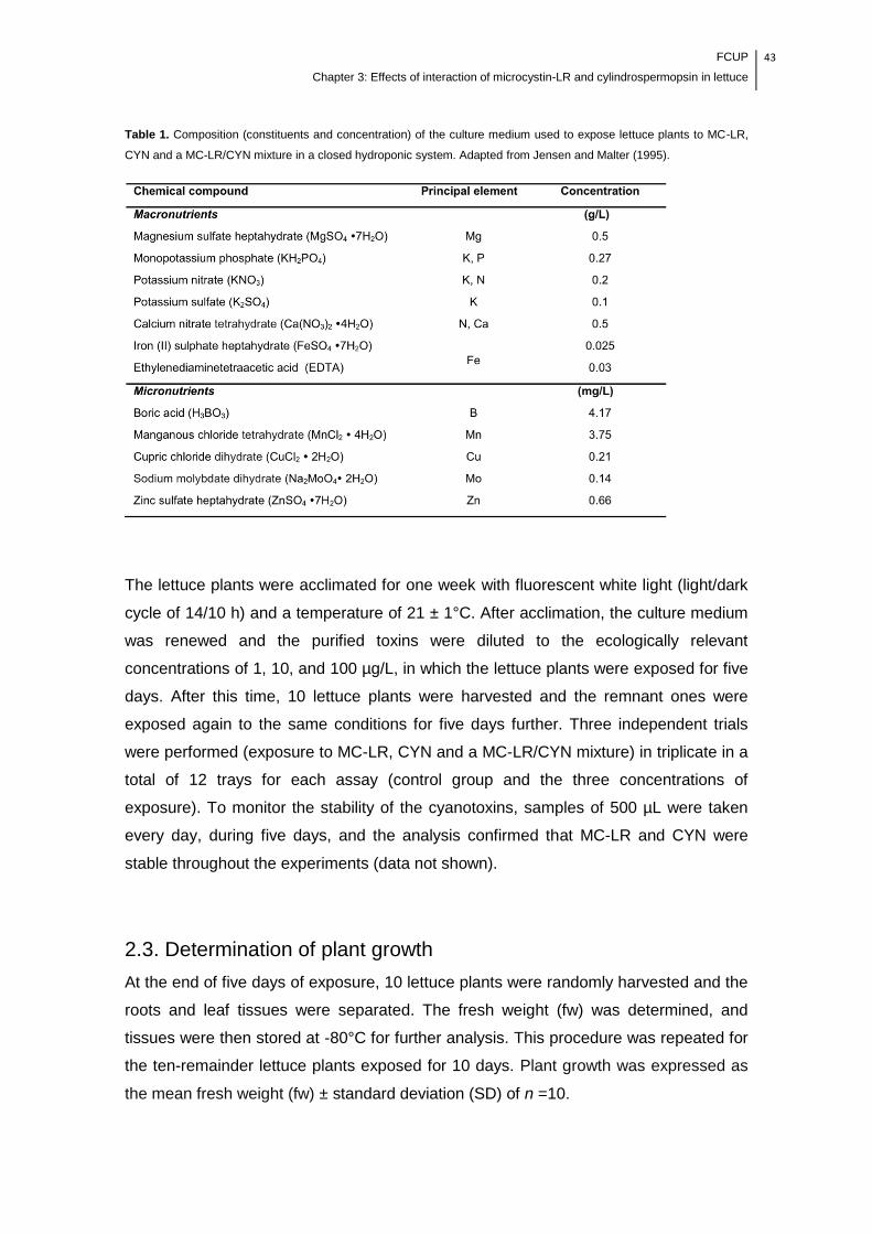
FCUP
Chapter 3: Effects of interaction of microcystin-LR and cylindrospermopsin in lettuce
43
Table 1. Composition (constituents and concentration) of the culture medium used to expose lettuce plants to MC-LR,
CYN and a MC-LR/CYN mixture in a closed hydroponic system. Adapted from Jensen and Malter (1995).
The lettuce plants were acclimated for one week with fluorescent white light (light/dark
cycle of 14/10 h) and a temperature of 21 ± 1°C. After acclimation, the culture medium
was renewed and the purified toxins were diluted to the ecologically relevant
concentrations of 1, 10, and 100 µg/L, in which the lettuce plants were exposed for five
days. After this time, 10 lettuce plants were harvested and the remnant ones were
exposed again to the same conditions for five days further. Three independent trials
were performed (exposure to MC-LR, CYN and a MC-LR/CYN mixture) in triplicate in a
total of 12 trays for each assay (control group and the three concentrations of
exposure). To monitor the stability of the cyanotoxins, samples of 500 µL were taken
every day, during five days, and the analysis confirmed that MC-LR and CYN were
stable throughout the experiments (data not shown).
2.3. Determination of plant growth
At the end of five days of exposure, 10 lettuce plants were randomly harvested and the
roots and leaf tissues were separated. The fresh weight (fw) was determined, and
tissues were then stored at -80°C for further analysis. This procedure was repeated for
the ten-remainder lettuce plants exposed for 10 days. Plant growth was expressed as
the mean fresh weight (fw) ± standard deviation (SD) of n =10.
44 FCUP
Chapter 3: Effects of interaction of microcystin-LR and cylindrospermopsin in lettuce
2.4. Enzyme activity measurement
The measurements of soluble (cytosolic) GST and GPx activities were performed in the
roots and leaves of two lettuce plants. Tissues were ground in liquid nitrogen to a
powder with a pestle and mortar and then homogenized in phosphate buffer, (100 mM)
pH 6.5, in a ratio of 1.5 g/1 mL (tissue/buffer). The homogenates were centrifuged at
4495 g for 20 min at 4°C, and the supernatants were recovered. Aliquots were stored
at –80°C until further analysis. Protein content was determined according to the
method of Bradford (1976), in which bovine serum albumin (BSA) was used as a
standard. The sGST activity was determined according to the method of Habig et al.
(1974), whereas GPx activity was determined according to the method of Lawrence
and Burk (1976). GST and GPx activities were performed with 0.1 mg protein/mL, and
the enzymatic activities were calculated according to Azevedo et al. (2014) and
expressed in nkat/mg protein.
2.5. Determination of mineral content in lettuce leaves
Leaf tissues of three lettuce plants from the MC-LR, CYN and MC-LR/CYN
experiments were analyzed regarding to their content of calcium (Ca), magnesium
(Mg), potassium (K), phosphorus (P), sodium (Na), manganese (Mn), iron (Fe), zinc
(Zn), copper (Cu) and molybdenum (Mo). The digestion and analysis of lettuce
samples were performed according to Pinto et al. (2014). Freeze-dried leaves of lettuce
plants were digested in an MLS 1200 Mega high performance microwave digestion
(Milestone, Sorisole, Italy) unit. Samples were weighed into the PTFE vessels and 5
mL of HNO3 (65% w/w, TraceSELECT® Ultra) and 2 mL of 30% H2O2 (30% v/v,
TraceSELECT® Fluka) were added to each vessel. Afterwards, the mixture was
submitted to the following microwave heating programs (power/time): 250 W/1 min, 0
W/2 min, 250 W/5 min, 400 W/5 min and 600 W/5 min. Sample solutions were then
analyzed by a 3100 flame atomic absorption spectroscopy (AAS) instrument (Perkin
Elmer, Überlingen, Germany) and by inductively coupled plasma – mass spectrometry
(ICP-MS) (VG Elemental PlasmaQuad 3, Winsford, UK) for total metal content. For
AAS analysis, multi-element calibration standards were prepared from 1000 mg/L
single-element standard solutions (Sigma, MO) of Ca, Mg, K and Na. For ICP-MS
analysis, calibration standards were prepared from AccuStandard® (New Haven, CT)
10 μg/mL multi-element ICP-MS standard solution (ICP-MS-200.8-CAL1-1). All
solutions were prepared using ultrapure water (>18.2 MΩ cm at 25°C) obtained with a

FCUP
Chapter 3: Effects of interaction of microcystin-LR and cylindrospermopsin in lettuce
45
Milli-Q (Millipore, Billerica, MA) water purification system.
The ICP–MS instrument was equipped with a glass concentric nebulizer (Meinhard®
Type A), a water-cooled glass spray chamber with impact beads, a standard quartz
torch and nickel skimmer and sampling cones. For sample introduction, a Minipuls 3
(Gilson, Villiers le Bel, France) peristaltic pump was used. Argon of 99.9% purity
(Alphagaz 2TM, supplied by Air Liquide, Maia, Portugal) was used as the plasma
source. The ICP-MS analysis was performed under the following conditions: argon flow
rate (13 L/min); auxiliary argon flow rate (0.7 L/min); nebulizer flow rate (0.8 L/min); RF
power (1350 W); scan regions dwell time (200 ms); and detection mode (pulse
counting). The elemental isotopes (m/z ratios) 55Mn, 56Fe, 65Cu, 66Zn and 95Mo were
monitored for analytical determination and 45Sc, 89Y and 115In were used as internal
standards. The instrument was tuned daily for maximum signal sensitivity and stability
using 115In as the target isotope. P was determined according to Murphy and Riley
(1962). Results were expressed on a dry weight (dw) basis.
2.6. Statistical analysis
The statistical analysis of lettuce growth and enzymatic activity was performed by one-
way analysis of variance (ANOVA) followed by Tukey‘s post hoc test. The homogeneity
of variance was checked with the Levene test, and when it was not observed, data
were transformed to achieve this assumption. Statistical analysis of mineral content
was conducted using the Mann-Whitney U test (IBM® SPSS® Statistics version 21.0
for Mac OS X). The significance level was set at P < 0.05.
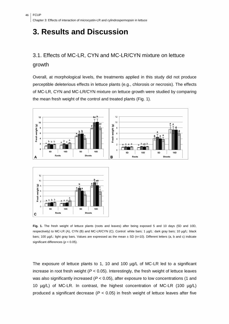
46 FCUP
Chapter 3: Effects of interaction of microcystin-LR and cylindrospermopsin in lettuce
3. Results and Discussion
3.1. Effects of MC-LR, CYN and MC-LR/CYN mixture on lettuce
growth
Overall, at morphological levels, the treatments applied in this study did not produce
perceptible deleterious effects in lettuce plants (e.g., chlorosis or necrosis). The effects
of MC-LR, CYN and MC-LR/CYN mixture on lettuce growth were studied by comparing
the mean fresh weight of the control and treated plants (Fig. 1).
Fig. 1. The fresh weight of lettuce plants (roots and leaves) after being exposed 5 and 10 days (5D and 10D,
respectively) to MC-LR (A), CYN (B) and MC-LR/CYN (C). Control: white bars; 1 µg/L: dark gray bars; 10 µg/L: black
bars; 100 µg/L: light gray bars. Values are expressed as the mean ± SD (n=10). Different letters (a, b and c) indicate
significant differences (p < 0.05).
The exposure of lettuce plants to 1, 10 and 100 µg/L of MC-LR led to a significant
increase in root fresh weight (P < 0.05). Interestingly, the fresh weight of lettuce leaves
was also significantly increased (P < 0.05), after exposure to low concentrations (1 and
10 µg/L) of MC-LR. In contrast, the highest concentration of MC-LR (100 µg/L)
produced a significant decrease (P < 0.05) in fresh weight of lettuce leaves after five
FCUP
Chapter 3: Effects of interaction of microcystin-LR and cylindrospermopsin in lettuce
47
days of exposure; however, this effect attenuated after 10 days of exposure. Unlike our
results, lettuce root growth has been inhibited by extracts of M. aeruginosa containing
MCs in concentrations ranging from 5.9 to 56.4 µg/L, following five days of exposure
(Pereira et al., 2009). Furthermore, several studies have reported the growth inhibition
of roots and leaves of plants as result of MC-LR exposure (Chen et al., 2004; El
Khalloufi et al., 2011; Gehringer et al., 2003; McElhiney et al., 2001; Mitrovic et al.,
2005; Pflugmacher, 2002; Pflugmacher et al., 2006; Pflugmacher et al., 2007; Saqrane
et al., 2009). However, as evidenced by these studies, various factors can influence the
toxicological effects. These include plant species, stage of plant development, the use
of purified toxins or crude extracts, time of exposure and the range of concentrations
studied. In most of these studies, the toxic effects of MC-LR were produced in plants in
early stages of development and/or by high concentrations, approximately 100- to
1,000-fold higher than those used in this study. The contradictory results obtained by
Pereira et al. (2009) could be explained by the differences in the stage of development
of lettuce plants, the parameter used to assess the growth (root length) and the use of
crude extracts instead of purified toxin. Interestingly, in the latter study, the strain of M.
aeruginosa that produced the most pronounced root growth inhibition contained the
lowest concentration of MCs. This suggests that at ecologically relevant
concentrations, other components of the extracts of M. aeruginosa may induce
inhibitory effects on lettuce root growth, but these findings may not be a result of MC-
LR by itself.
The effects of time of exposure were more evident in the CYN trial. The fresh weight of
lettuce plants was only affected after 10 days of exposure, whereas the growth of roots
was stimulated in all concentrations of exposure, and the leaf fresh weight decreased
at 100 µg/L (P < 0.05) of CYN. Similar results in leaf fresh weight of rice plants
exposed nine days to extracts of A. ovalisporum containing 2.5 µg/L of CYN have been
reported (Prieto et al., 2011). Hydrilla verticillata has been reported to exhibit in
increase in root growth following 14 days of exposure to extracts of C. raciborskii
containing 400 µg/L of CYN (Kinnear et al., 2008). According to Kinnear et al. (2008),
several benefits could result from the increased root production, such as the potential
production of exudates, allelochemicals or phytochelatins, which might scavenge CYN,
preventing its uptake by plant cells. In all of the experiments in our study, we observed
a gradual abundance of exudates in the culture medium that were proportional to the
toxin concentration (data not shown). Although whether these compounds are effective
to protect lettuce plants to cyanotoxins is not known, their potential contribution should
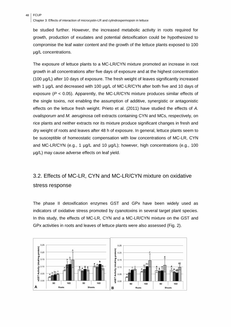
48 FCUP
Chapter 3: Effects of interaction of microcystin-LR and cylindrospermopsin in lettuce
be studied further. However, the increased metabolic activity in roots required for
growth, production of exudates and potential detoxification could be hypothesized to
compromise the leaf water content and the growth of the lettuce plants exposed to 100
µg/L concentrations.
The exposure of lettuce plants to a MC-LR/CYN mixture promoted an increase in root
growth in all concentrations after five days of exposure and at the highest concentration
(100 µg/L) after 10 days of exposure. The fresh weight of leaves significantly increased
with 1 µg/L and decreased with 100 µg/L of MC-LR/CYN after both five and 10 days of
exposure (P < 0.05). Apparently, the MC-LR/CYN mixture produces similar effects of
the single toxins, not enabling the assumption of additive, synergistic or antagonistic
effects on the lettuce fresh weight. Prieto et al. (2011) have studied the effects of A.
ovalisporum and M. aeruginosa cell extracts containing CYN and MCs, respectively, on
rice plants and neither extracts nor its mixture produce significant changes in fresh and
dry weight of roots and leaves after 48 h of exposure. In general, lettuce plants seem to
be susceptible of homeostatic compensation with low concentrations of MC-LR, CYN
and MC-LR/CYN (e.g., 1 µg/L and 10 µg/L); however, high concentrations (e.g., 100
µg/L) may cause adverse effects on leaf yield.
3.2. Effects of MC-LR, CYN and MC-LR/CYN mixture on oxidative
stress response
The phase II detoxification enzymes GST and GPx have been widely used as
indicators of oxidative stress promoted by cyanotoxins in several target plant species.
In this study, the effects of MC-LR, CYN and a MC-LR/CYN mixture on the GST and
GPx activities in roots and leaves of lettuce plants were also assessed (Fig. 2).
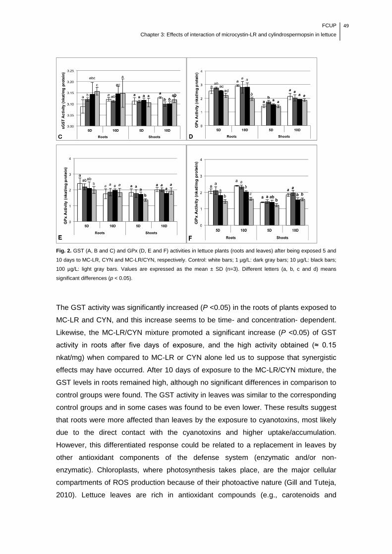
FCUP
Chapter 3: Effects of interaction of microcystin-LR and cylindrospermopsin in lettuce
49
Fig. 2. GST (A, B and C) and GPx (D, E and F) activities in lettuce plants (roots and leaves) after being exposed 5 and
10 days to MC-LR, CYN and MC-LR/CYN, respectively. Control: white bars; 1 µg/L: dark gray bars; 10 µg/L: black bars;
100 µg/L: light gray bars. Values are expressed as the mean ± SD (n=3). Different letters (a, b, c and d) means
significant differences (p < 0.05).
The GST activity was significantly increased (P <0.05) in the roots of plants exposed to
MC-LR and CYN, and this increase seems to be time- and concentration- dependent.
Likewise, the MC-LR/CYN mixture promoted a significant increase (P <0.05) of GST
activity in roots after five days of exposure, and the high activity obtained (≈ 0.15
nkat/mg) when compared to MC-LR or CYN alone led us to suppose that synergistic
effects may have occurred. After 10 days of exposure to the MC-LR/CYN mixture, the
GST levels in roots remained high, although no significant differences in comparison to
control groups were found. The GST activity in leaves was similar to the corresponding
control groups and in some cases was found to be even lower. These results suggest
that roots were more affected than leaves by the exposure to cyanotoxins, most likely
due to the direct contact with the cyanotoxins and higher uptake/accumulation.
However, this differentiated response could be related to a replacement in leaves by
other antioxidant components of the defense system (enzymatic and/or non-
enzymatic). Chloroplasts, where photosynthesis takes place, are the major cellular
compartments of ROS production because of their photoactive nature (Gill and Tuteja,
2010). Lettuce leaves are rich in antioxidant compounds (e.g., carotenoids and
50 FCUP
Chapter 3: Effects of interaction of microcystin-LR and cylindrospermopsin in lettuce
phenolics) that play an important role to protect the photosynthetic apparatus against
oxidative stress. Inclusively, the non-enzymatic antioxidants, such as phenolic
compounds and α- and β-tocopherol, have been reported to be enhanced in plants
after their exposure to MCs (El Khalloufi et al., 2012; Lahrouni et al., 2013; Stüven and
Pflugmacher, 2007). This raises attention to the impact of cyanotoxins in the nutritional
value of lettuce leaves because the content of antioxidants can be enhanced as a
physiological mechanism of response.
According to our results, GST activity in the roots of lettuce plants showed that it was a
good marker of stress induced by MC-LR, CYN and its mixture. Similarly, Prieto et al.
(2011) have reported that the exposure of rice plants to a mixture of A. ovalisporum
and M. aeruginosa cell extracts containing CYN and MC-LR, respectively, resulted in a
significant increase in GST activity in roots, also suggesting that a synergistic effect of
both extracts may exist; however, no changes in GPx activity have been observed in
rice plants after 48 h of exposure.
In this study, the GPx activity was significantly decreased (P <0.05) in the roots of
lettuce plants exposed for 10 days to 100 µg/L of MC-LR, whereas it was significantly
increased (P <0.05) in leaves of plants exposed to 1 µg/L for five days. GPx activity
decreased in both roots and leaves of lettuce plants exposed to 100 µg/L of CYN for
five days. A similar pattern was found for the MC-LR/CYN trial, and in this experiment,
the GPx activity significantly decreased (P <0.05) in roots and leaves exposed for 10
days to 10 and 100 µg/L of the mixture. Although the GPx activity has been
successfully used to assess the effects of MC-LR in Lepidium sativum (Gehringer et
al., 2003; Stüven and Pflugmacher 2007), GPx may not be the best biomarker of these
cyanotoxins in lettuce plants. The activity of superoxide dismutase (SOD), catalase
(CAT), and ascorbate peroxidase (APX) increased in M. sativa seedlings after
exposure to ecologically relevant concentrations of MCs (Pflugmacher et al., 2006).
Recently, El Khalloufi et al. (2013) also reported that peroxidase (POD),
polyphenoloxidase (PPO) and CAT activities were significantly increased in leaves,
roots and nodules of M. sativa exposed to cyanobacterial extract containing MCs. The
enzyme GPx scavenges peroxides, especially hydrogen peroxide. However, CAT and
APX can also convert hydrogen peroxide to water (Gill and Tuteja, 2010); thus, these
enzymes should be further studied as potential indicators of oxidative stress generated
in lettuce plants due to the exposure to cyanotoxins.
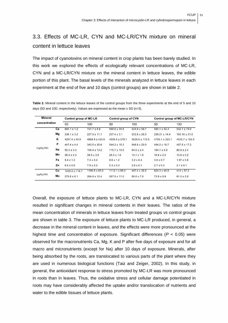
FCUP
Chapter 3: Effects of interaction of microcystin-LR and cylindrospermopsin in lettuce
51
3.3. Effects of MC-LR, CYN and MC-LR/CYN mixture on mineral
content in lettuce leaves
The impact of cyanotoxins on mineral content in crop plants has been barely studied. In
this work we explored the effects of ecologically relevant concentrations of MC-LR,
CYN and a MC-LR/CYN mixture on the mineral content in lettuce leaves, the edible
portion of this plant. The basal levels of the minerals analyzed in lettuce leaves in each
experiment at the end of five and 10 days (control groups) are shown in table 2.
Table 2. Mineral content in the lettuce leaves of the control groups from the three experiments at the end of 5 and 10
days (5D and 10D, respectively). Values are expressed as the mean ± SD (n=3).
Overall, the exposure of lettuce plants to MC-LR, CYN and a MC-LR/CYN mixture
resulted in significant changes in mineral contents in their leaves. The ratios of the
mean concentration of minerals in lettuce leaves from treated groups vs control groups
are shown in table 3. The exposure of lettuce plants to MC-LR produced, in general, a
decrease in the mineral content in leaves, and the effects were more pronounced at the
highest time and concentration of exposure. Significant differences (P < 0.05) were
observed for the macronutrients Ca, Mg, K and P after five days of exposure and for all
macro and micronutrients (except for Na) after 10 days of exposure. Minerals, after
being absorbed by the roots, are translocated to various parts of the plant where they
are used in numerous biological functions (Taiz and Zeiger, 2002). In this study, in
general, the antioxidant response to stress promoted by MC-LR was more pronounced
in roots than in leaves. Thus, the oxidative stress and cellular damage potentiated in
roots may have considerably affected the uptake and/or translocation of nutrients and
water to the edible tissues of lettuce plants.

52 FCUP
Chapter 3: Effects of interaction of microcystin-LR and cylindrospermopsin in lettuce
Table 3. Ratio of the mineral content in lettuce leaves exposed for 5 and 10 days (5D and 10D, respectively) to MC-LR,
CYN and a MC-LR/CYN mixture. Values express the ratio between the mean concentrations obtained in each condition
by the mean concentration obtained in the respective control group. Values are expressed as the mean ± SD (n=3).
Values in bold represent the concentrations that exceeded the screening value (control group, Table 2).
The mineral content in the roots of T. durum, Z. mays, P. sativum and L. esculenta (Ca,
Na, K, P and N), as well as L. esculentum (Ca, Na, K), increased in a concentration-
dependent manner after 30 days of exposure to M. aeruginosa extract containing MCs
(500-4,200 µg/L; 2,220-22,240 µg/L, respectively) (El Khalloufi et al., 2012; Saqrane et
al., 2009). However, similar to our results and the range of concentrations used in this
study, the K and Ca content in the shoots of V. faba have been recently reported to
FCUP
Chapter 3: Effects of interaction of microcystin-LR and cylindrospermopsin in lettuce
53
decrease, whereas Na increased after two months of exposure to M. aeruginosa
extract containing 50 and 100 µg/L of MCs (Lahrouni et al., 2013).
Contrary to the MC-LR, the exposure of lettuce plants to CYN produced an
enhancement in leaf mineral content at almost all concentrations after five days of
exposure. Moreover, after 10 days of exposure to CYN, the content of the minerals Mn,
Fe, Zn, Cu and Mo in the leaves was significantly increased at all concentrations of
exposure (P < 0.05). In the CYN experiment only P was significantly decreased at all
concentrations after 10 days of exposure (P < 0.05). However, generally, crop plants
need small amounts of micronutrients; thus the excessive increase of these elements
in the leaves of lettuce plants exposed to CYN for 10 days could be an indication of
some physiological disorders, the minerals can be hypothesized to have been used as
a defense mechanism. Exploring the mechanisms underlying the enhancement of the
uptake of mineral nutrients after five days of exposure to CYN is important. The lettuce
plants were able to retain almost all of the minerals (especially the macronutrients) in a
concentration higher than the corresponding control value, and this can be an
indication of the potential tolerance of lettuce plants to CYN. However, studies on the
effects of cyanotoxins in crop plants have been primarily focused on yield losses; yet,
the physiological stress promoted by cyanotoxins seems to alter the chemical
composition of plants and can therefore change its nutritional quality. The ability of crop
plants to cope with abiotic stress and also maximize its nutritional quality is of major
relevance for food security. The leaves of lettuce plants exposed to CYN demonstrate
the ability to retain a higher content of minerals; however other quality parameters
should be assessed, such as antioxidants, proteins, non-structural carbohydrates,
lipids and sensory quality traits, which are particularly prone to be changed due to
exposure to abiotic stress (Wang and Frei, 2011).
The mixture of MC-LR/CYN resulted in a general decrease of nutrient content after five
days of exposure. However, this tendency was reverted with the increase in time of
exposure, and after 10 days only the 100 µg/L concentration resulted in lower
concentrations of the minerals in comparison to the correspondent control group. Once
again, apparently, the MC-LR/CYN mixture did not produce additive, synergistic or
antagonistic effects on mineral content in lettuce leaves.
Plants require Ca, Mg, K and P in relatively high amounts, and these nutrients are
essential to ensure its life cycle. Although each mineral contributes to several metabolic
reactions, some general functions in plant metabolism could be affected due to the
54 FCUP
Chapter 3: Effects of interaction of microcystin-LR and cylindrospermopsin in lettuce
disturbance of mineral uptake and translocation promoted by MC-LR, CYN and its
mixture in lettuce plants. Mg supports other functions related to respiration and the
synthesis of DNA and RNA and is also a central atom of the chlorophyll molecule; thus,
it plays an important role in the light-dependent reactions of photosynthesis.
Additionally, both K and P are crucial for respiration and photosynthesis. Mn also plays
an important role in the structure of photosynthetic proteins and enzymes, as well as
ATP synthesis, as it activates several enzymes involved in tricarboxylic acid
metabolism. Likewise, Zn is required for many enzymes and also for chlorophyll
biosynthesis (Taiz and Zeiger, 2002). The decline of these minerals in lettuce leaves as
was found after MC-LR exposure, could compromise the chlorophyll content and
photosynthetic rates; thus, the chronic exposure of lettuce to MC-LR could result in loss
of productivity. Indeed, the photosynthesis impairment by MC-LR is well recognized
and has already been reported by several authors (El Khalloufi et al., 2011; McElhiney
et al., 2001; Pietsch et al., 2001; Pflugmacher, 2002; Saqrane et al., 2009). Although
other biological functions could be affected by mineral imbalances, including
antioxidant activity and cell growth (for example, Fe and Cu are associated with
enzymes involved in redox reactions; and Ca and Na are involved in the synthesis of
new cell walls, cell division and cell expansion (Taiz and Zeiger, 2002)),
photosynthesis, which plays an important role as energy source, and thus crop yield,
could be one of the most affected processes by mineral imbalance due to exposure to
MC-LR and CYN.
4. Conclusions
In the present study, the effects of ecologically relevant concentrations of MC-LR, CYN
and a MC-LR/CYN mixture were investigated regarding to growth, oxidative stress and
mineral content in lettuce plants. This is of particular relevance, facing a tendency
towards an increasing occurrence of toxic cyanobacterial blooms and the challenge of
the intensification of agricultural productivity. Our results suggest that lettuce plants are
able to cope with lower concentrations (1 and 10 µg/L) of MC-LR, CYN and MC-
LR/CYN mixture by ensuring the maintenance and even increasing the fresh weight
and mineral content and controlling oxidative stress, as was indicated by the significant
increase of the GST activity in roots. Furthermore, the enhancement of the mineral

FCUP
Chapter 3: Effects of interaction of microcystin-LR and cylindrospermopsin in lettuce
55
content in the leaves of plants exposed to CYN for five days may provide an indication
of the potential tolerance of lettuce plants to this cyanotoxin. However, the
concentration and the time of exposure are preponderant factors for the toxic effects of
these cyanotoxins in lettuce plants. The exposure of lettuce plants to 100 µg/L of
cyanotoxins resulted in a significant decrease in fresh weight of leaves and its mineral
content, especially for MC-LR and the MC-LR/CYN mixture, highlighting the potential
implications of these toxins for lettuce yield and nutritional quality. Furthermore, the
potential tolerance of lettuce plants to CYN raises the possibility of its accumulation in
edible tissues, and this issue should be further studied to avoid risks in human health.
Conflict of Interest
The authors declare that there are no conflicts of interest.
Acknowledgments
Marisa Freitas thanks the Fundação para a Ciência e Tecnologia (FCT) for the Ph.D.
Grant SFRH/BD/85490/2012. This work was partially funded by the FCT project PesT-
C/MAR/LA0015/2011.
5. References
Azevedo, C.C., Azevedo, J., Osório, H., Vasconcelos, V., Campos, A., 2014. Early
physiological and biochemical responses of rice seedlings to low concentration of
microcystin-LR. Ecotoxicology 23, 107–121.
Beyer, D., Surányi, G., Vasas, G., Roszik, J., Erdődi, F., M-Hamvas, H., Bácsi, I.,
Bátori, R., Serfőző, Z., Szigeti, Z.M., Vereb, G., Demeter, Z., Gonda, S., Máthé, C.,
2009. Cylindrospermopsin induces alterations of root histology and microtubule
organization in common reed (Phragmites australis) plantlets cultured in vitro. Toxicon
54, 440–449.
56 FCUP
Chapter 3: Effects of interaction of microcystin-LR and cylindrospermopsin in lettuce
Bradford, M.M., 1976. A rapid and sensitive method for the quantitation of microgram
quantities of protein utilizing the principle of protein-dye binding. Anal. Biochem. 72,
248-254.
Brient, L., Lengronne, M., Bormans, M., Fastner, J., 2008. First occurrence of
cylindrospermopsin in freshwater in France. Environ. Toxicol. 24, 415–420.
Chen, J., Song, L., Dai, J., Gan, N., Liu, Z., 2004. Effects of microcystins on the growth
and the activity of superoxide dismutase and peroxidase of rape (Brassica napus
L.) and rice (Oryza sativa L.). Toxicon 43, 393–400.
El Khalloufi, F., El Ghazali, I., Saqrane, S., Oufdou, K., Vasconcelos, V., Oudra, B.,
2012. Phytotoxic effects of a natural bloom extract containing microcystins on
Lycopersicon esculentum. Ecotoxicol. Environ. Saf. 79, 199–205.
El Khalloufi, F., Oufdou, K., Lahrouni, M., El Ghazali, I., Saqrane, S., Vasconcelos, V.,
Oudra, B., 2011. Allelopatic effects of cyanobacteria extracts containing microcystins
on Medicago sativa-Rhizobia symbiosis. Ecotoxicol. Environ. Saf. 74, 431-438.
El Khalloufi, F., Oufdou, K., Lahrouni, M., Faghire, M., Peix, A., Ramírez-Bahena, M.H.,
Vasconcelos, V., Oudra, B., 2013. Physiological and antioxidant responses of
Medicago sativa- rhizobia symbiosis to cyanobacterial toxins (Microcystins) exposure.
Toxicon 76, 167–177.
Elliott, J.A., 2012. Is the future blue-green? A review of the current model predictions of
how climate change could affect pelagic freshwater cyanobacteria. Water Res. 46,
1364-1371.
Froscio, S.M., Humpage, A.R., Wickramasinghe, W., Shaw, G., Falconer, I.R., 2008.
Interaction of the cyanobacterial toxin cylindrospermopsin with the eukaryotic protein
synthesis system. Toxicon 51, 191–198.
Gehringer, M.M., Kewada, V., Coates, N., Downing, T.G., 2003. The use of Lepidium
sativum in a plant bioassay system for the detection of microcystin-LR. Toxicon 41,
871–876.
Gill, S.S., Tuteja, N., 2010. Reactive oxygen species and antioxidant machinery in
abiotic stress tolerance in crop plants. Plant Physiol Biochem. 48, 909-930.
Habig, W.H., Pabst, M.J., Jakoby, W.B., 1974. Glutathione S-transferases. The first
FCUP
Chapter 3: Effects of interaction of microcystin-LR and cylindrospermopsin in lettuce
57
enzymatic step in mercapturic acid formation. J. Biol. Chem. 249, 7130–7139.
Jensen, M.H., Malter, A.J., 1995. Protected Agriculture: A Global Review. Chapter 7,
Water supply, water quality and mineral nutrition. World Bank Publications.
Kinnear, S., 2010. Cylindrospermopsin: a decade of progress on bioaccumulation
research. Mar. Drugs 8, 542-564.
Kinnear, S., Fabbro, L., Duivenvoorden, L., 2008. Variable growth responses of water
thyme (Hydrilla verticillata); to whole-cell extracts of Cylindrospermopsis raciborskii.
Arch. Environ. Contam. Toxicol. 54, 187–194.
Kotai, J., 1972. Instruction for preparation of modified nutrient solution Z8 for algae.
Norwegian Institute for Water Research, Oslo, Norway, Report nr B-11/69, 1-5.
Lahrouni, M., Oufdou, K., El Khalloufi, F., Baz, M., Lafuente, A., Dary, M., Pajuelo, E.,
Oudra, B., 2013. Physiological and biochemical defense reactions of Vicia faba L.-
Rhizobium symbiosis face to chronic exposure to cyanobacterial bloom extract
containing microcystins. Environ. Sci. Pollut. Res. Int. 20, 5405-5415.
Lawrence, R.A., Burk, R.F., 1976. Glutathione peroxidase activity in selenium- deficient
rat liver. Biochem. Biophys. Res. Commun. 71, 952–958.
MacKintosh, C., Beattie, K.A., Klumpp, S., Cohen, P., Codd, G.A., 1990.
Cyanobacterial microcystin-LR is a potent and specific inhibitor of protein
phosphatases 1 and 2A from both mammals and higher plants. FEBS Lett. 264, 187–
192.
McElhiney, J., Lawton, L.A., Leifert, C., 2001. Investigations into the inhibitory effects of
microcystins on plant growth, and the toxicity of plant tissues following exposure.
Toxicon 39, 1411-1420.
Metcalf, J.S., Barakate, A., Codd, G.A., 2004. Inhibition of plant protein synthesis by
the cyanobacterial hepatotoxin, cylindrospermopsin. FEMS Microbiol. Lett. 235, 125–
129.
Mitrovic, S.M., Allis, O., Furey, A., James, K.J., 2005. Bioaccumulation and harmful
effects of microcystin-LR in the aquatic plants Lemna minor and Wolffia arrhiza and the
filamentous alga Chladophora fracta. Ecotoxicol. Environ. Saf. 61, 345–352.
58 FCUP
Chapter 3: Effects of interaction of microcystin-LR and cylindrospermopsin in lettuce
Murphy J., Riley, J., 1962. A modified single solution method for the determination of
phosphate in natural waters. Anal. Chim. Acta. 27: 31–36.
O‘Neil, J.M.,
Davis, T.W., Burford, M.A., Gobler, C.J., 2012. The rise of harmful
cyanobacteria blooms: The potential roles of eutrophication and climate change.
Harmful Algae 14, 313–334.
Pereira, S., Saker, M.L., Vale, M., Vasconcelos, V.M., 2009. Comparison of sensitivity
of grasses (Lolium perenne L. and Festuca rubra L.) and lettuce (Lactuca sativa L.)
exposed to water contaminated with microcystins. Bull Environ. Contam. Toxicol. 83,
81-84.
Pflugmacher, S., 2002. Possible allelopathic effects of cyanotoxins, with reference to
microcystin-LR, in aquatic ecosystems. Environ. Toxicol. 17, 407–413.
Pflugmacher, S., 2004. Promotion of oxidative stress in the aquatic macrophyte
Ceratophyllum demersum during biotransformation of the cyanobacterial toxin
microcystin-LR. Aquat. Toxicol. 70, 169–178.
Pflugmacher, S., Codd, G.A., Steinberg, C.E.W., 1999. Effects of the cyanobacterial
toxin microcystin-LR on detoxication enzymes in aquatic plants. Environ. Toxicol. 14,
111-115.
Pflugmacher, S., Hofmann, J., Hübner, B., 2007. Effects on growth and physiological
parameters in wheat (Triticum aestivum L.) grown in soil and irrigated with
cyanobacterial toxin contaminated water. Environ. Toxicol. Chem. 26, 2710-2716.
Pflugmacher, S., Jung, K., Lundvall, L., Neumann, S., Peuthert, A., 2006. Effects of
cyanobacterial toxins and cyanobacterial cell-free crude extract on germination of
alfalfa (Medicago sativa) and induction of oxidative stress. Environ. Toxicol. Chem. 25,
2381–2387.
Pflugmacher, S., Wiegand, C., Beattie, K.A., Krause, E., Steinberg, C.E.W., Codd,
G.A., 2001. Uptake, effects, and metabolism of cyanobacterial toxins in the emergent
reed plant Phragmites australis (cav.) trin. ex steud. Environ. Toxicol. Chem. 20, 846–
852.
Pflugmacher, S., Wiegand, C., Oberemm, A., Beattie, K.A., Krause, E., Codd, G.A.,
Steinberg C.E.W., 1998. Identification of an enzymatically formed glutathione conjugate
of the cyanobacterial hepatotoxin microcystin-LR: the first step of detoxication.
FCUP
Chapter 3: Effects of interaction of microcystin-LR and cylindrospermopsin in lettuce
59
Biochim. Biophys. Acta 1425, 527-533.
Pietsch, C., Wiegand, C., Amé, M.V., Nicklisch, A., Wunderlin, D., Pflugmacher, S.,
2001. The effects of a cyanobacterial crude extract on different aquatic organisms:
evidence for cyanobacterial toxin modulating factors. Environ. Toxicol. 16, 535–542.
Pinto, E., Almeida, A. A., Aguiar, A.A.R.M., Ferreira, I.M.P.L.V.O., 2014. Changes in
macrominerals, trace elements and pigments content during lettuce (Lactuca sativa L.)
growth: Influence of soil composition. Food Chem. 152, 603–611.
Poniedziałek, B., Rzymski, P., Kokociński, M., 2012. Cylindrospermopsin: Water-linked
potential threat to human health in Europe. Environ. Toxicol. Phar. 34, 651–660.
Prieto, A., Campos, A., Cameán, A., Vasconcelos, V., 2011. Effects on growth and
oxidative stress status of rice plants (Oryza sativa) exposed to two extracts of toxin-
producing cyanobacteria (Aphanizomenon ovalisporum and Microcystis aeruginosa).
Ecotoxicol. Environ. Saf. 74, 1973-1980.
Ramanan, S., Tang, J., Velayudhan, A., 2000. Isolation and preparative purification of
microcystin variants. J. Chromatogr A. 883,103-112.
Saqrane, S., Ouahid, Y., El Ghazali, I., Oudra, B., Bouarab, L., del Campo, F.F., 2009.
Physiological changes in Triticum durum, Zea mays, Pisum sativum and Lens
esculenta cultivars, caused by irrigation with water contaminated with microcystins: a
laboratory experimental approach. Toxicon 53, 786-796.
Stüven, J., Pflugmacher, S., 2007. Antioxidative stress response of Lepidium sativum
due to exposure to cyanobacterial secondary metabolites. Toxicon 50, 85–93.
Taiz, L., Zeiger, E., 2002. Plant Physiology, Third Edition. Sinauer Associates,
Sunderland, MA. 690 pps.
Vasas, G., Gáspár, A., Surányi, G., Batta, G., Gyémánt, G., M-Hamvas, M., Máthé, C.,
Grigorszky, I., Molnár, E., Borbély, G., 2002. Capillary electrophoretic assay and
purification of cylindrospermopsin, a cyanobacterial toxin from Aphanizomenon
ovalisporum, by plant test (blue-green Sinapis test). Anal. Biochem. 302, 95–103.
Wang, Y., Frei, M., 2011. Stressed food – The impact of abiotic environmental stresses
on crop quality. Agr. Ecosyst. Environ. 141, 271–286.

60 FCUP
Chapter 3: Effects of interaction of microcystin-LR and cylindrospermopsin in lettuce
Welker, M., Bickel, H., Fastner, J., 2002. HPLC-PDA detection of cylindrospermopsin -
opportunities and limits. Water Res. 36, 4659-4663.

FCUP
Chapter 3: Effects of interaction of microcystin-LR and cylindrospermopsin in lettuce
61
Lettuce (Lactuca sativa L.) leaf-proteome profiles after
exposure to cylindrospermopsin and a microcystin-
LR/cylindrospermopsin mixture: a concentration-dependent
response
Marisa Freitas1,2,3, Alexandre Campos1, Joana Azevedo1, Aldo Barreiro1, Sébastien
Planchon4, Jenny Renaut4, Vitor Vasconcelos1,2
1 CIIMAR/CIMAR - Interdisciplinary Centre of Marine and Environmental Research,
University of Porto, Rua dos Bragas 289, P 4050-123 Porto, Portugal.
2 Faculty of Sciences, Porto University, Rua do Campo Alegre, 4169-007 Porto,
Portugal.
3 Polytechnic Institute of Porto. Escola Superior de Tecnologia da Saúde do Porto.
CISA/Research Center in Environment and Health, Rua de Valente Perfeito, 322,
4400-330 Gaia, Portugal.
4 Department of Environment and Agro-biotechnologies (EVA), Centre de Recherche
Public-Gabriel Lippmann, 41, rue du Brill, 4422 Belvaux, Luxembourg.
Abstract
The intensification of agricultural productivity is an important challenge worldwide.
However, environmental stressors can provide challenges to this intensification. The
progressive occurrence of the cyanotoxins CYN and MC-LR as a potential
consequence of eutrophication and climate change is of increasing concern in the
agricultural sector because it has been reported that these cyanotoxins exert harmful
effects in crop plants. A proteomic-based approach has been shown to be a suitable
and powerful tool for the detection and identification of the early responses of
organisms exposed to cyanotoxins. The aim of this study was to compare the leaf-
proteome profiles of lettuce plants exposed to environmentally relevant concentrations
of CYN and a MC-LR/CYN mixture. Lettuce plants were exposed to 1, 10, and 100 µg/l

62 FCUP
Chapter 3: Effects of interaction of microcystin-LR and cylindrospermopsin in lettuce
CYN and a MC-LR/CYN mixture for five days. The proteins of lettuce leaves were
separated by two-dimensional electrophoresis (2-DE), and those that were differentially
abundant were then identified by matrix-assisted laser desorption/ionization time of
flight-mass spectrometry (MALDI-TOF/TOF MS). Although the lettuce genome has not
been sequenced, a high rate of differentially abundant proteins was identified (>73%).
The biological functions of the proteins that were most represented in both experiments
were photosynthesis and carbon metabolism and stress/defense response. Proteins
involved in protein synthesis and signal transduction were also highly observed in the
MC-LR/CYN experiment. Although distinct protein abundance patterns were observed
in both experiments, the effects appear to be concentration-dependent, and the effects
of the mixture were clearly stronger than those of CYN alone. The obtained results
highlight the putative tolerance of lettuce to CYN at concentrations up to 100 µg/l.
Furthermore, the combination of CYN with MC-LR at low concentrations (1 µg/l)
stimulated a significant increase in the fresh weight (fr. wt) of lettuce leaves and at the
proteomic level resulted in the increase in abundance of a high number of proteins. In
contrast, many proteins exhibited a decrease in abundance or were absent in the gels
of the simultaneous exposure to 10 and 100 µg/l MC-LR/CYN. In the latter, also a
significant decrease in the fr. wt of lettuce leaves was obtained. These findings provide
new insights into the molecular mechanisms of the lettuce response to CYN and MC-
LR/CYN and may contribute to the identification of potential protein markers of
exposure and novel proteins that may confer tolerance to CYN and MC-LR/CYN,
although these need to be functionally characterized and validated. Furthermore,
because lettuce is an important crop worldwide, this study may improve our
understanding of the potential impact of environmental stress conditions on its quality
traits (e.g., presence of allergenic proteins).
Keywords: Lactuca sativa, Proteomics, Cylindrospermopsin, Microcystin-LR, Mixture
Abbreviations: 2-DE, two-dimensional electrophoresis; APX, ascorbate peroxidase;
CHAPS, 3-[(3-cholamidopropyl)dimethylamonio]-1-propanesulfonate; CYN,
cylindrospermopsin; EST, expressed sequence tag; FAS, fatty acid synthesis; GSH,
glutathione; GST, glutathione-S-transferase; HPLC, High-performance liquid
chromatography; HSP, heat shock protein; IPG, immobilized pH gradient; IEF,
isoelectric focusing; LEA, embryogenesis abundant protein; MALDI-TOF/TOF MS,
matrix-assisted laser desorption/ionization time of flight-mass spectrometry; MC-LR,
microcystin-LR; MeOH, methanol; PCA, principal component analysis; PDA,

FCUP
Chapter 3: Effects of interaction of microcystin-LR and cylindrospermopsin in lettuce
63
photoelectric diode array; PP, protein phosphatases; PPIase, peptidyl-prolyl cis-trans
isomerase; PR, pathogenesis-related; PRK, phosphoribulokinase; PS, photosystem;
ROS, reactive oxygen species; RuBisCO, ribulose bisphosphate
carboxylase/oxygenase; RuBP, ribulose-1,5-bisphosphate carboxylase/oxygenase; SB,
solubilization buffer; SBPase, sedoheptulose-1,7-bisphosphatase; SD, standard
deviation; SOD, superoxide dismutase; TCA, tricarboxylic acid; TFA, trifluoroacetic
acid.
1. Introduction
The progressive occurrence and global expansion of harmful cyanobacteria blooms
have been forecasted as consequences of eutrophication and climate change (Elliott,
2012; O‘Neil et al., 2012). Among freshwater cyanobacteria, Microcystis aeruginosa is
the most common bloom former (O‘Neil et al., 2012). However, the invasive species
Cylindrospermopsis raciborskii has shown a substantial widespread distribution,
including into temperate zones (Kinnear, 2010; Poniedziałek et al., 2012). The use of
irrigation water from sources that contain toxic cyanobacterial blooms of C. raciborskii
and M. aeruginosa may pose a threat on the agricultural sector because their
cyanotoxins (the cytotoxic cylindrospermopsin (CYN) and the hepatotoxic microcystin-
LR (MC-LR), respectively) appear to generate phytotoxic effects on crop plants. CYN is
a tricyclic alkaloid, and although the molecular mechanism of its toxicity has not yet
been established, it is known that CYN inhibits eukaryotic protein synthesis with similar
intensity in plant and mammalian cell extracts (Froscio et al., 2008). Although studies
on the toxic effects of CYN on plants are scarce, the few studies that have analyzed
CYN indicate that it results in the reduction of pollen germination (Metcalf et al., 2004),
inhibition of plant growth (Vasas et al., 2002), induction of abnormal mitosis, alteration
of microtubule organization, inhibition of root and shoot elongation (Beyer et al., 2009),
and increase in oxidative stress (Prieto et al., 2011). MC-LR, the most studied
structural variant of microcystins, is a cyclic heptapeptide that irreversibly inhibits, by
covalent binding, serine/threonine protein phosphatases (PP; PP1 and PP2A), and this
is the main mechanism of its toxicity in both animals and higher plants (Mackintosh et
al., 1990). The induction of oxidative stress by the production of reactive oxygen
species (ROS) appears to be another important biochemical mechanism of MC-LR
64 FCUP
Chapter 3: Effects of interaction of microcystin-LR and cylindrospermopsin in lettuce
toxicity that may cause serious oxidative damage to DNA (Pflugmacher, 2004; Stüven
and Pflugmacher, 2007). The toxic effects of MC-LR on plants have also been
characterized. It has been reported that MC-LR results in the inhibition of germination
(Pflugmacher et al., 2006; Pflugmacher et al., 2007; El Khalloufi et al., 2011), inhibition
of growth and development (McElhiney et al., 2001; Pflugmacher, 2002; Gehringer et
al., 2003; Chen et al., 2004; Mitrovic et al., 2005; Pflugmacher et al., 2006;
Pflugmacher et al., 2007; El Khalloufi et al., 2011), alteration of microtubule
organization (Máthé et al., 2009), induction of changes in photosynthesis (Pietsch et
al., 2001; Pflugmacher, 2002; El Khalloufi et al., 2011), induction of changes in
chlorophyll content (McElhiney et al., 2001; Pflugmacher, 2002), and induction of
changes in antioxidative response parameters (Pflugmacher et al., 1999; Pflugmacher
et al., 2001; Gehringer et al., 2003; Pflugmacher, 2004; Pflugmacher et al., 2006;
Stüven and Pflugmacher, 2007; Pflugmacher et al., 2007; Saqrane et al., 2009; El
Khalloufi et al., 2011). Nevertheless, the reported biochemical and physiological effects
vary depending on the (1) use of purified toxins or crude extracts, (2) the plant species,
(3) the stage of plant development, (4) the time of exposure, and (5) the range of
concentrations studied. Therefore, it is important to note the ecological relevancy of
these studies because few have confirmed the effects at environmentally relevant
concentrations (Gehringer et al., 2003; Pichardo and Pflugmacher et al., 2011). The
concentrations required to exhibit effects in a wider range of species appear to be non-
environmentally realistic because these are 10–1000-fold higher than those usually
found in ecosystems (McElhiney et al., 2001; Mitrovic et al., 2005; Crush et al., 2008;
Beyer et al., 2009; Saqrane et al., 2009). It has been reported that exposure
concentrations of pure CYN below 100 µg/l appear to have no significant harmful
effects on a wide range of species (e.g., a floating macrophytes and green algae)
(Kinnear, 2010), leading to the hypothesis that plants have developed appropriate
protective mechanisms to tolerate CYN. Otherwise, it can be questioned whether the
traditional endpoints used to assess toxicity exhibit sufficient sensitivity to evaluate
understated biochemical alterations. Recently, Azevedo et al. (2014) reported the lack
of sensitivity of the conventional parameters for the analysis of the toxicity of M.
aeruginosa extract on rice (Oryza sativa) plants (MC-LR concentrations of 0.26–78
µg/l); however, significant alterations were observed through proteomic analyses. The
inhibition of protein synthesis by CYN and the inhibition of PP1/PP2A activities by MC-
LR appear to interfere with a wide range of molecular processes in plants (Máthé et al.,
2013). Although the conventional biochemical biomarkers of stress induced by CYN
and MC-LR (antioxidative enzymes and nonenzymatic substances) appear to be
suitable, because proteins are the main targets of these cyanotoxins, it is particularly
FCUP
Chapter 3: Effects of interaction of microcystin-LR and cylindrospermopsin in lettuce
65
important to investigate how these operate in plant systems at the protein level.
Proteomics is a field of growing interest in the agricultural sector because it has
contributed to a better understanding of the specific functions of the proteins involved in
plant responses to environmental stresses (Afroz et al., 2011; Kosová et al., 2011;
Abreu et al., 2013). A proteomic approach may enable the identification of protein
biomarkers of the plant stress response and the discovery of the biological processes
underlying stress tolerance, which may be used to enhance agricultural productivity
(Kosová et al., 2011; Abreu et al., 2013). Moreover, some secreted proteins with
defensive or protective functions on stress factors are recognized to also have
allergenic potential (Abreu et al., 2013). From the health risk point of view, proteomics
data associated with allergen identification may provide new insights into the protein
composition, quality, and safety of edible plants exposed to environmentally relevant
concentrations of cyanotoxins. Nevertheless, in aquatic ecosystems, single species of
cyanobacteria are almost never found; hence, the existence of mixtures of cyanotoxins
in the water column is likely and it was already reported for MC-LR and CYN (Brient et
al., 2008). Simultaneous exposure to a mixture of CYN and MC-LR may lead to
changes in the response capability of crop plants, triggering potential synergistic or
antagonistic effects. Recently, Prieto et al. (2011) suggested a synergistic effect on the
oxidative stress response of rice plants due to its exposure to cyanobacterial extracts
containing low concentrations of both CYN (0.13 µg/l) and MC-LR (50 µg/l). Proteomics
studies investigating the effects of CYN and MC-LR have been performed on bivalves,
including mixtures with other environmental pollutants (e.g., herbicides) (Martins et al.,
2009; Puerto et al., 2011; Malécot et al., 2013). Proteomics studies on the plant
response to abiotic stress factors are mainly related to cold, heat, drought,
waterlogging, salinity, ozone treatment, hypoxia/anoxia, herbicide treatments,
inadequate or excessive light conditions, deficient or excessive mineral nutrition,
enhanced concentrations of heavy metals, radioactivity, and mechanical wounding, as
was reviewed by Kosová et al. (2011). As above mentioned, more recently, Azevedo et
al. (2014) successfully applied a proteomic approach to assess the early physiological
and biochemical responses of rice seedlings to environmentally relevant concentrations
of MC-LR.
This work aimed to use a 2-DE proteomic approach and MALDI-TOF/TOF MS to
investigate the leaf-proteome profiles of lettuce (Lactuca sativa L.) plants exposed to
environmentally relevant concentrations (1, 10, and 100 µg/l) of a CYN and MC-
LR/CYN mixture.

66 FCUP
Chapter 3: Effects of interaction of microcystin-LR and cylindrospermopsin in lettuce
2. Materials and Methods
2.1. General Experimental Procedures
The lettuce plants (Lactuca sativa L. var. ‗Susybel‘) were exposed to environmentally
relevant concentrations (1, 10, and 100 µg/l) of a CYN and MC-LR/CYN mixture in a
hydroponic system. The CYN and MC-LR were purified and quantified by High-
performance liquid chromatography (HPLC) and then diluted in the culture medium to
the desired concentrations of exposure. All of the HPLC solvents were of high-purity
chromatography grade and were filtered (Pall GH Polypro 47 mm, 0.2 μm) and
degassed with an ultrasound bath. After exposure, to investigate the leaf-proteome
profiles, the proteins of lettuce leaves were separated by 2-DE, and those that were
differentially abundant were then identified by Matrix-assisted laser
desorption/ionization time of flight-mass spectrometry (MALDI-TOF/TOF MS).
2.2. Cyanobacterial culture and toxin purification and quantification
2.2.1. Culture of Microcystis aeruginosa and Cylindrospermopsis
raciborskii
M. aeruginosa (LEGE 91094) and C. raciborskii (LEGE 97047) were grown to
exponential phase in Z8 medium (Kotai, 1972) (6-l flasks) under fluorescent light with a
light/dark cycle of 14/10 h and a temperature of 25 ± 1ºC. The cultured cells were
gathered by centrifugation (20 min, 4ºC, 4495 g), frozen at -80ºC, and then freeze-
dried. Due to the high hydrophilicity of CYN, the culture medium from C. raciborskii was
also frozen at -80ºC and freeze-dried. The lyophilized material was stored at room
temperature in the dark until toxin extraction and purification. In this study, purified
toxins were chosen for the experiments to address the specific effects of CYN and MC-
LR/CYN on the lettuce leaf-proteome and to avoid interference from other potentially
toxic metabolites (e.g., lipopolysaccharides) because cyanobacterial crude extracts
appear to induce more pronounced effects than purified toxins at equivalent
concentrations (Pietsch et al., 2001).
FCUP
Chapter 3: Effects of interaction of microcystin-LR and cylindrospermopsin in lettuce
67
2.2.2. CYN extraction, purification, and quantification by HPLC-PDA
CYN was extracted from C. raciborskii cells and culture medium following a modified
version of the method described by Welker et al. (2002). Briefly, the freeze-dried cells
and medium (0.7 g) were first sonicated in a bath for 15 min in 5 ml of 0.1%, v/v
trifluoroacetic acid (TFA) of spectrophotometric grade and then subjected to five cycles
ultrasonication with Vibra-Cell at 60 Hz for 1 min. The homogenate was stirred for 1 h
at room temperature and centrifuged (20,000 g, 4ºC, 20 min), and the supernatant was
collected. The cell pellet was subjected to a second extraction. The supernatants were
pooled and stored at -20ºC. CYN was thereafter purified using a Waters Alliance e2695
HPLC system coupled with a photoelectric diode array (PDA) 2998 on a semi-
preparative Gemini C18 column (250 mm x 10 mm i.d., 5 µm) from Phenomenex that
was maintained at 40ºC. The isocratic elution utilized a 5% methanol (MeOH) solution
containing 2 mM sodium 1-heptanesulfonate monohydrate (99%) with a flow rate of 2.5
ml/min and an injection volume of 1000 µl. Working solutions of CYN (0.08-5.0 µg/ml)
were prepared in water. Standard CYN was supplied by Alexis (San Diego, CA, USA).
The purified CYN fractions were then quantified in an HPLC system on an Atlantis®
HILIC phase column (250 mm x 4.6 mm i.d., 5 µm) from Waters maintained at 40ºC.
The PDA range was 210-400 nm with a fixed wavelength of 262 nm. The isocratic
elution was also a 5% MeOH solution containing 2 mM sodium 1-heptanesulfonate
monohydrate (99%) with a flow rate of 0.9 ml/min and an injection volume of 10 µl. The
system was calibrated using a set of seven dilutions of the CYN standard (25, 20, 10,
5, 2, 1, and 0.5 μg/ml) in ultrapure water. Each vial was injected in duplicate, and every
HPLC run series of ten samples included a blank and two different standard
concentrations. The chromatographic purity of CYN was of 98%. The Empower 2
Chromatography Data Software was used for the calculation and reporting the peak
information. The retention time of the CYN peak was 7.35 min (data from method
validation not published).
2.2.3. MC-LR extraction, purification, and quantification by HPLC-
PDA
MC-LR was extracted from M. aeruginosa cells according to Ramanan et al. (2000)
with some modifications. Briefly, the lyophilized M. aeruginosa biomass was extracted
with 75% (v/v) MeOH (Fisher Scientific, UK) by continuous stirring for 20 min at room

68 FCUP
Chapter 3: Effects of interaction of microcystin-LR and cylindrospermopsin in lettuce
temperature. The sample was then ultrasonicated five times on ice at 60 Hz for 1 min
(Vibra-Cell 50-sonics & Material Inc. Danbury, CT, USA). The homogenate was
centrifuged at 10,000 g for 15 min to remove the cell debris. The resulting supernatant
was then collected and stored at 4ºC. The pellet was re-extracted with an equal volume
of solvent, and the pooled supernatants were subjected at a flow rate of at 1 ml/min to
SPE with a Water Sep-Pak® Vac 6-ml C18 cartridge preconditioned with 100% MeOH
and distilled water. The loaded column was washed with 20% (v/v) MeOH, and the MC-
LR was then eluted using 80% (v/v) MeOH. The MC-LR fraction was evaporated by
rotary evaporation at 35ºC to remove the entire MeOH portion. The concentrated MC-
LR extract was thereafter purified and quantified by HPLC-PDA. The MC-LR semi-
preparative assay was performed using a reversed-phase column (Phenomenex Luna
RP-18 (250 mm × 10 mm, 10 μm) maintained at 35ºC. The gradient elution was
performed with MeOH and water, both of which were acidified with 0.1% TFA, with a
flow rate of 2.5 ml/min. The injection volume was 500 µl. The peak purity and
percentage of purified MC-LR were calculated at 214 nm and 238 nm. The fraction with
purified MC-LR was then evaporated with nitrogen air for 1 day until all of the solvent
was removed. The residue was resuspended in distilled water to the desired
concentration. The chromatographic purity of MC-LR was of 97%. The purified fractions
of MC-LR were then quantified in the same HPLC system on a Merck Lichrospher RP-
18 endcapped column (250 mm x 4.6 mm i.d., 5 µm) equipped with a guard column (4
× 4 mm, 5 µm), both of which were maintained at 45ºC. The PDA range was 210-400
nm with a fixed wavelength of 238 nm. The linear gradient elution consisted of (A)
MeOH + 0.1% TFA and (B) H2O + 0.1% TFA (55% A and 45% B at 0 min, 65% A and
35% B at 5 min, 80% A and 20% B at 10 min, 100% A at 15 min, and 55% A and 45%
B at 15.1 and 20 min) with a flow rate of 0.9 ml/min. The injected volume was 20 µl.
The MC-LR was identified by a comparison of its spectra and retention time with that of
the MC-LR standard (batch nº 018K1209, 10.025 µg/ml in MeOH, 98% purity, Cyano
Biotech GmbH, Berlin, Germany). The system was calibrated using a set of seven
dilutions of the MC-LR standard (0.5 to 20 μg/ml) in 50% MeOH. Each vial was injected
in duplicate, and every HPLC run series of ten samples included a blank and two
different standard concentrations. The Empower 2 Chromatography Data Software was
used for calculating and reporting the peak information. The retention time of the MC-
LR peak was 10.44 min (data from method validation not published).

FCUP
Chapter 3: Effects of interaction of microcystin-LR and cylindrospermopsin in lettuce
69
2.3. Plant exposure to CYN and MC-LR/CYN mixture
Lettuce is an important leafy vegetable worldwide that contains substantial amounts of
health-promoting phytochemicals (including polyphenols, carotenoids, and vitamin C).
Lettuce has been shown to be an excellent experimental system for the assessment of
the effects of MC-LR (Crush et al., 2008; Pereira et al., 2009), and although its genome
has not been sequenced, lettuce has been the object of genomics and proteomics
studies (Choi et al., 2008; Cho et al., 2009). The lettuce plants used in the experiments
were purchased at four to five weeks‘ maturity after sowing in a commercial soil
substrate. The roots were carefully washed with tap water until complete soil removal
was achieved, and twenty lettuce plants were then transferred to the holes of plastic
boards (PVC), which were placed on black glass trays (35 x 25 x 5 cm deep) in a
hydroponic system that was continuously aerated. The roots were completely
immersed in 3 l of culture medium (Jensen and Malter, 1995), pH 6.5. The lettuce
plants were acclimated for 1 week with fluorescent white light (light/dark cycle of 14/10
h) and a temperature of 21 ± 1ºC. After the acclimation period, the culture medium was
renewed, and the plants were exposed to CYN and the MC-LR/CYN mixture at
ecologically relevant concentrations of 1, 10, and 100 µg/l for five days. After the
exposure time, the plants were harvested. The roots and leaf tissues were separated,
weighed (fr. wt) and stored at -80ºC for proteomic analysis. Three biological replicates
for each experimental group (control (C), C1, C10, and C100) were prepared in a total
of 12 trays.
2.4. Proteomics analysis
A proteomics analysis of the lettuce roots was attempted; however, likely due to salt
interference, a slow progression of the first-dimensional isoelectric focusing (IEF) and
the heat produced on the IPG gel strips may the analysis impracticable.
2.4.1. Protein extraction from lettuce leaves
The leaf tissues from one lettuce plant exposed for five days to the above-mentioned
concentrations of CYN and MC-LR/CYN were ground in liquid nitrogen to a powder
with a pestle and mortar. The protein extraction was performed immediately with
70 FCUP
Chapter 3: Effects of interaction of microcystin-LR and cylindrospermopsin in lettuce
acetone containing 10% trichloroacetic acid and 0.07% β-mercaptoethanol for 1 h at -
20ºC. After centrifugation at 4495 g and 0ºC for 45 min, the pellet was washed with
acetone containing 0.07% β-mercaptoethanol for 1 h at -20ºC and centrifuged at 4495
g and 0ºC for 50 min. The pellet was dried with nitrogen gas, and the proteins were
then solubilized in solubilization buffer (SB) composed of urea (7 M), thiourea (2 M), 3-
[(3-cholamidopropyl)dimethylamonio]-1-propanesulfonate (CHAPS) (4%, w/v),
dithiothreitol (65 mM), ampholytes, pH 4–7 (0.8%, v/v), and polyvinylpolypyrrolidone
(0.025 g/mL) for 1 h. The homogenate was centrifuged at 16,000 g and 4ºC for 20 min.
The supernatant was collected, and the proteins were quantified according to the
method described by Bradford (1976) using bovine serum albumin as the standard.
The protein samples were stored at -80ºC until further analysis.
2.4.2 IEF and 2-DE
IEF and 2-DE were performed according to Puerto et al. (2011). Briefly, 300 µl of SB
with 400 µg of proteins were loaded in 17-cm pH 4–7 IPG gel strips (Bio-Rad,
Hercules, CA, USA). The proteins were separated by IEF in a Protean IEF Cell (Bio-
Rad, Hercules, CA, USA) using the following program: 16 h at 50 V (strip rehydration);
step 1, 15 min at 250 V; step 2, linear voltage increase to 10,000 V over 3 h; step 3,
linear increase from 10,000 V to 90,000 V/h; and step 4: 500 V/h. After rehydration, wet
paper wicks were placed between the immobilized pH gradient (IPG) gel strip and
electrode to remove the excess salts from the samples. The IPG gel strips were frozen
at -20ºC prior to 2-DE. The IPG gel strips were equilibrated with 10 mg/ml dithiothreitol
in buffer containing urea (6 M), SDS (2%, w/v), glycerol (30%, v/v), and Tris (50 mM),
pH 8.8, for 15 min and then with 25 mg/ml iodoacetamide in the same buffer for 15 min.
The equilibrated IPG gel strips were placed on top of 12% (w/v) acrylamide SDS-PAGE
slab gels (20 cm×20 cm) and sealed with 0.5% agarose. The proteins were separated
by SDS-PAGE in a Protean Xi Cell (Bio-Rad, Hercules, CA, USA) at 18 mA per gel for
30 min and then at 24 mA per gel until the dye reached the bottom of the gel. One 2-
DE gel was run for each experimental replicate for a total of 24 gels. The gels were
stained overnight at room temperature with Colloidal Coomassie Blue as described by
Neuhoff et al. (1988).
FCUP
Chapter 3: Effects of interaction of microcystin-LR and cylindrospermopsin in lettuce
71
2.4.3. Gel image acquisition and protein abundance variation
analysis
The image acquisition and analysis of the protein abundance from the 2-DE gels were
performed as described previously by Puerto et al. (2011). Briefly, the gel images were
acquired using a calibrated scanner (GS-800, Bio-Rad, Hercules, CA, USA), and the
protein spots were detected automatically with the PDQuest 2-D Analysis Software
(Bio-Rad, Hercules, CA, USA). The spot intensities were normalized based on the total
density in the gel image, and manual spot corrections, including re-matching, were also
made using the software. The protein spots were considered differentially abundant
when the intensity levels exhibited at least a two-fold difference that was statistically
significant at a level of p<0.05 (univariate approach, t-test).
2.4.4. Protein identification by MALDI-TOF-TOF MS and peptide
mass fingerprinting
A selection list of the spots of interest was generated based on the differentially
abundant protein spots (p<0.05) between the C group and the C1, C10, and C100
groups of the CYN and MC-LR/CYN exposure experiments. The spots were excised
from the gels and automatically digested with trypsin and spotted on MALDI targets as
previously described by Printz et al. (2013). The identification of proteins was
performed through mass spectrometry (MALDI-TOF/TOF). For each spot 1 MS
spectrum was acquired and the 10 highest peaks selected for MS/MS analysis,
excluding known contaminants and trypsin autocleavage products. All of the searches
were conducted allowing for a peptide mass tolerance of 100 ppm and a fragment
mass tolerance of 0.5 Da within NCBInr Viridiplantae (Green Plants) (downloaded
2013.01.18; 1,162,105 sequences) and EST_Lettuce databases (downloaded on
2013.04.19; 1,763,496 sequences; 357736570 residues) using MASCOT (Matrix
Science, www.matrixscience.com, London, UK). Variable modifications [Dioxidation
(W), Oxidation (HW), Oxidation (M), Trp -> Kynurenine (W)] and fixed modifications
[Carbamidomethyl (C)] have been allowed for the database search. In general, a
protein was considered identified when the protein scores were greater than 73 and
were significant (p<0.05). When a protein or peptide was identified based on an EST
sequence or a protein with a trivial name, the protein sequence was used in a Blast

72 FCUP
Chapter 3: Effects of interaction of microcystin-LR and cylindrospermopsin in lettuce
alignment. Proteins were considered as being significantly identified when two
individual non-identical peptides surpassed the threshold for identification or when one
peptide resulted in a protein e-value of < 0.005. All identifications were manually
validated and extra data was acquired when insignificant identifications were obtained.
The manual validations were done as previously described by Printz et al. (2013). For
some spots, manual sequence determinations were performed and the sequences
found used for cross-species identification with FASTS and MSBlast.
2.5. Statistical analysis
The statistical analysis of the fr. wt of the lettuce plants (n=10) was conducted using
the Mann-Whitney U test (p<0.05) (IBM® SPSS® Statistics version 21.0 for Mac OS
X). The remaining statistical analyses and plots were performed using R software
2.15.1 (R Core Team, 2012). A multivariate analysis using PCA was performed after
the data were pre-processed through missing value imputation (sequential KNN
method, R Environment) and logarithm transformation. Significant differences between
the groups in each PCA dataset were tested by ANOSIM test (p<0.05). The heatmap.2
function in the gplots package was used to generate the heat map. The clustering
method that was used with this function was the default method, which consists of
hierarchical clustering using the complete linkage method operated with a matrix of
dissimilarities calculated as Euclidean distances.
3. Results and Discussion
In the present study, well-defined differences were observed in the leaf-proteome
profiles of lettuce plants exposed for five days to ecologically relevant concentrations
(1, 10 and 100 µg/l) of CYN and MC-LR/CYN. The proteomics approach was found to
be a suitable tool that was sufficiently sensitive to recognize changes in the plant
physiological responses that were not perceptible at the morphological level. Overall,
the treatments applied in this study did not affect lettuce plants at the morphological
level, with the exception of the leaf fr. wt of plants exposed to 1 µg/l and 100 µg/l MC-
LR/CYN, which was significantly higher and lower than that of the control group
(p<0.05), respectively (Fig. 1).

FCUP
Chapter 3: Effects of interaction of microcystin-LR and cylindrospermopsin in lettuce
73
Fig. 1. Fr. wt (g) of leaves of lettuce plants exposed to CYN and MC-LR/CYN. The values are expressed as the means
± SD (n=10). The asterisk (*) indicates significant differences (P<0.05) between the control and exposed groups.
Although there is scarce information on the lettuce genome, a high rate of proteins was
successfully identified by the combination of 2-DE, MALDI-TOF/TOF MS, and lettuce
expressed sequence tag (EST) databases (NCBI). A functional characterization of the
differentially abundant leaf-lettuce proteins was performed to better understand the
physiological response of this important crop plant to CYN and MC-LR/CYN exposure.
3.1. Differential leaf-proteome profiles related to CYN and MC-
LR/CYN exposure
The analysis of the leaf-proteome profiles of lettuce plants exposed to 1 (C1), 10 (C10),
and 100 (C100) µg/l CYN and MC-LR/CYN revealed a total of 68 and 286 protein spots
with significant abundance variations, respectively (p<0.05, spot intensity variation of at
least two-fold). Specifically, the individual comparisons were made between the control
group (C) and the groups with different exposure concentrations (C1, C10, and C100),
as follows: C/C1, C/C10, and C/C100 (Figs. 2A and 2B).
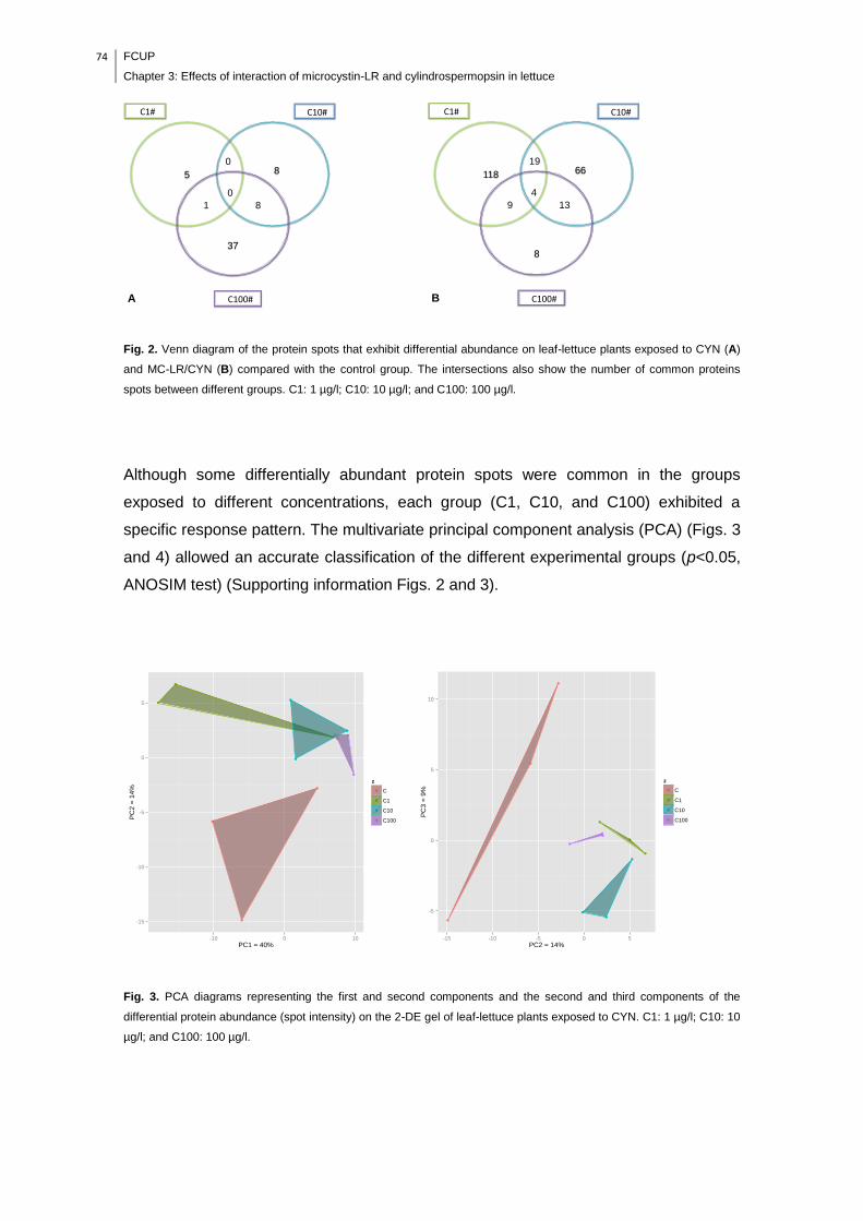
74 FCUP
Chapter 3: Effects of interaction of microcystin-LR and cylindrospermopsin in lettuce
Fig. 2. Venn diagram of the protein spots that exhibit differential abundance on leaf-lettuce plants exposed to CYN (A)
and MC-LR/CYN (B) compared with the control group. The intersections also show the number of common proteins
spots between different groups. C1: 1 µg/l; C10: 10 µg/l; and C100: 100 µg/l.
Although some differentially abundant protein spots were common in the groups
exposed to different concentrations, each group (C1, C10, and C100) exhibited a
specific response pattern. The multivariate principal component analysis (PCA) (Figs. 3
and 4) allowed an accurate classification of the different experimental groups (p<0.05,
ANOSIM test) (Supporting information Figs. 2 and 3).
Fig. 3. PCA diagrams representing the first and second components and the second and third components of the
differential protein abundance (spot intensity) on the 2-DE gel of leaf-lettuce plants exposed to CYN. C1: 1 µg/l; C10: 10
µg/l; and C100: 100 µg/l.
0
5 8
37
C1# C10#
C100#
8 1
A
0
4
118 66
8
C1# C10#
C100#
13 9
B
19
-15
-10
-5
0
5
-10 0 10
PC1 = 40%
PC
2 =
14
%
z
C
C1
C10
C100
-5
0
5
10
-15 -10 -5 0 5
PC2 = 14%
PC
3 =
9%
z
C
C1
C10
C100

FCUP
Chapter 3: Effects of interaction of microcystin-LR and cylindrospermopsin in lettuce
75
Fig. 4. PCA diagrams representing the first and second components and the second and third components of the
differential protein abundance (spot intensity) on the 2-DE gel of leaf-lettuce plants exposed to MC-LR/CYN. C1: 1 µg/l;
C10: 10 µg/l; and C100: 100 µg/l.
In the CYN exposure experiment, the first component explains 40% of the variation and
separates groups C and C1 from groups C10 and C100. The second and third
components explain 14% and 9% of the proteome variation, respectively, and these
separate group C from groups C1, C10, and C100. In the MC-LR/CYN exposure
experiment, the experimental groups were also well separated by the first three
components. PC1 explains 33% of the proteome variation and separates groups C,
C10, and C100 from group C1. Groups C10 and C100 are in the same component.
PC2 explains 25% of the variation and separates groups C and C100 from groups C1
and C10, whereas the third component, which explains 11% of the variation, clearly
separates group C from group C100.
The number of protein spots with significant abundance variation appears to be
concentration-dependent in the CYN exposure experiment (Table 1).
-10
0
10
20
-20 -10 0 10 20 30
PC1 = 33%
PC
2 =
25
%
z
C
C1
C10
C100
-10
0
10
-10 0 10 20
PC2 = 25%
PC
3 =
11
%
z
C
C1
C10
C100
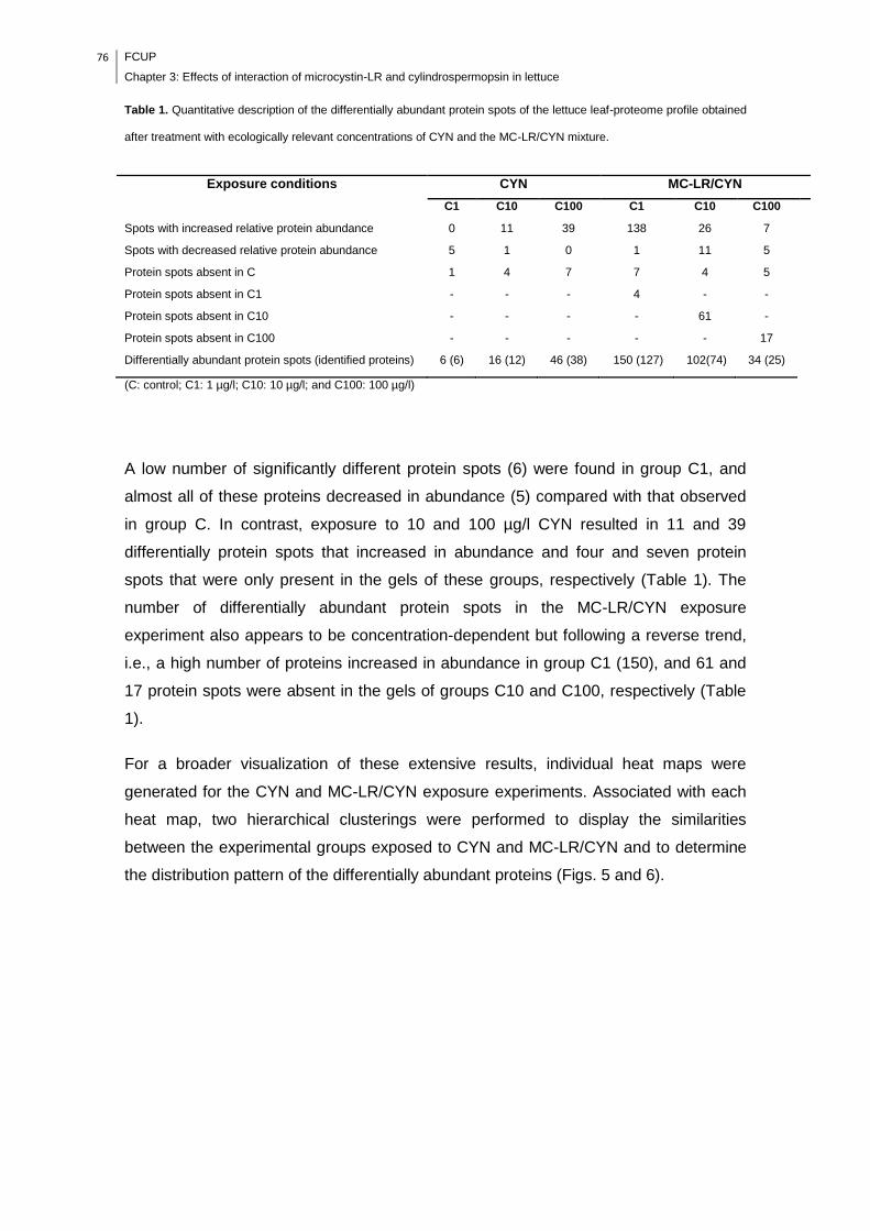
76 FCUP
Chapter 3: Effects of interaction of microcystin-LR and cylindrospermopsin in lettuce
Table 1. Quantitative description of the differentially abundant protein spots of the lettuce leaf-proteome profile obtained
after treatment with ecologically relevant concentrations of CYN and the MC-LR/CYN mixture.
Exposure conditions CYN MC-LR/CYN
C1 C10 C100 C1 C10 C100
Spots with increased relative protein abundance 0 11 39 138 26 7
Spots with decreased relative protein abundance 5 1 0 1 11 5
Protein spots absent in C 1 4 7 7 4 5
Protein spots absent in C1 - - - 4 - -
Protein spots absent in C10 - - - - 61 -
Protein spots absent in C100 - - - - - 17
Differentially abundant protein spots (identified proteins) 6 (6) 16 (12) 46 (38) 150 (127) 102(74) 34 (25)
(C: control; C1: 1 µg/l; C10: 10 µg/l; and C100: 100 µg/l)
A low number of significantly different protein spots (6) were found in group C1, and
almost all of these proteins decreased in abundance (5) compared with that observed
in group C. In contrast, exposure to 10 and 100 µg/l CYN resulted in 11 and 39
differentially protein spots that increased in abundance and four and seven protein
spots that were only present in the gels of these groups, respectively (Table 1). The
number of differentially abundant protein spots in the MC-LR/CYN exposure
experiment also appears to be concentration-dependent but following a reverse trend,
i.e., a high number of proteins increased in abundance in group C1 (150), and 61 and
17 protein spots were absent in the gels of groups C10 and C100, respectively (Table
1).
For a broader visualization of these extensive results, individual heat maps were
generated for the CYN and MC-LR/CYN exposure experiments. Associated with each
heat map, two hierarchical clusterings were performed to display the similarities
between the experimental groups exposed to CYN and MC-LR/CYN and to determine
the distribution pattern of the differentially abundant proteins (Figs. 5 and 6).
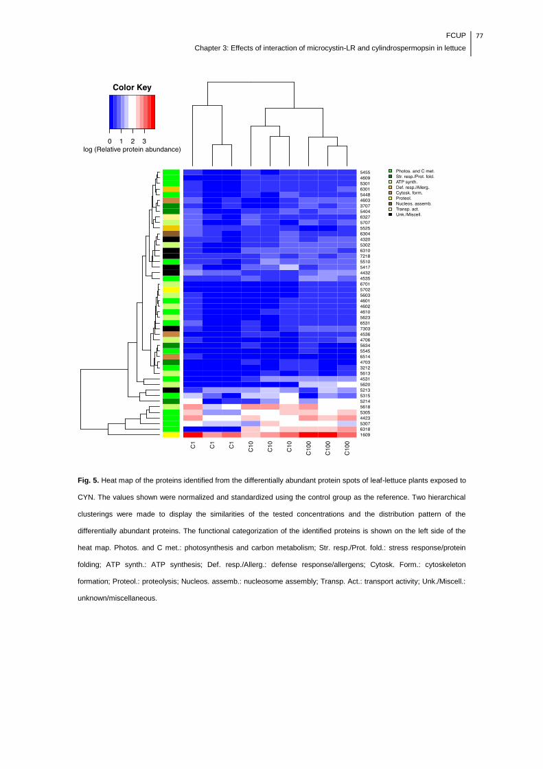
FCUP
Chapter 3: Effects of interaction of microcystin-LR and cylindrospermopsin in lettuce
77
Fig. 5. Heat map of the proteins identified from the differentially abundant protein spots of leaf-lettuce plants exposed to
CYN. The values shown were normalized and standardized using the control group as the reference. Two hierarchical
clusterings were made to display the similarities of the tested concentrations and the distribution pattern of the
differentially abundant proteins. The functional categorization of the identified proteins is shown on the left side of the
heat map. Photos. and C met.: photosynthesis and carbon metabolism; Str. resp./Prot. fold.: stress response/protein
folding; ATP synth.: ATP synthesis; Def. resp./Allerg.: defense response/allergens; Cytosk. Form.: cytoskeleton
formation; Proteol.: proteolysis; Nucleos. assemb.: nucleosome assembly; Transp. Act.: transport activity; Unk./Miscell.:
unknown/miscellaneous.
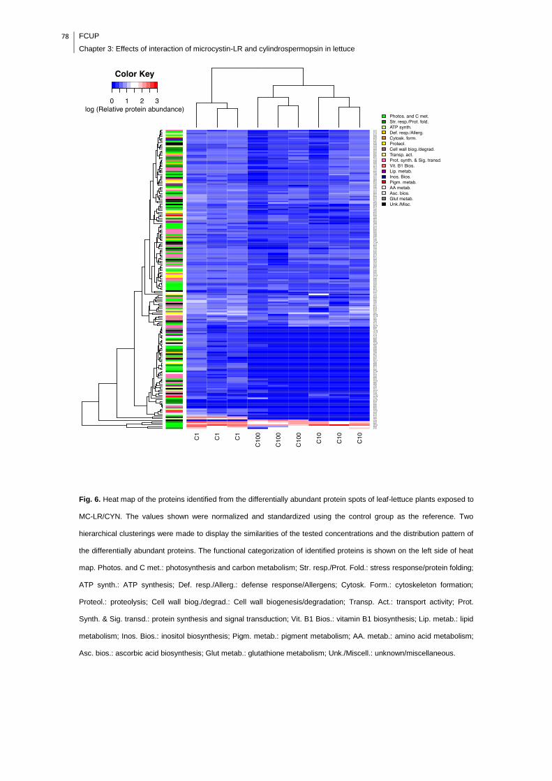
78 FCUP
Chapter 3: Effects of interaction of microcystin-LR and cylindrospermopsin in lettuce
Fig. 6. Heat map of the proteins identified from the differentially abundant protein spots of leaf-lettuce plants exposed to
MC-LR/CYN. The values shown were normalized and standardized using the control group as the reference. Two
hierarchical clusterings were made to display the similarities of the tested concentrations and the distribution pattern of
the differentially abundant proteins. The functional categorization of identified proteins is shown on the left side of heat
map. Photos. and C met.: photosynthesis and carbon metabolism; Str. resp./Prot. Fold.: stress response/protein folding;
ATP synth.: ATP synthesis; Def. resp./Allerg.: defense response/Allergens; Cytosk. Form.: cytoskeleton formation;
Proteol.: proteolysis; Cell wall biog./degrad.: Cell wall biogenesis/degradation; Transp. Act.: transport activity; Prot.
Synth. & Sig. transd.: protein synthesis and signal transduction; Vit. B1 Bios.: vitamin B1 biosynthesis; Lip. metab.: lipid
metabolism; Inos. Bios.: inositol biosynthesis; Pigm. metab.: pigment metabolism; AA. metab.: amino acid metabolism;
Asc. bios.: ascorbic acid biosynthesis; Glut metab.: glutathione metabolism; Unk./Miscell.: unknown/miscellaneous.

FCUP
Chapter 3: Effects of interaction of microcystin-LR and cylindrospermopsin in lettuce
79
In both the CYN and MC-LR/CYN exposure experiments, the clusters above the plots
show two major groups (C1 and C10/C100). These clusters are corroborated by PCA
(first component in the CYN experiment and first and second components in the MC-
LR/CYN experiment) and suggest that the concentration of 1 µg/l in both experiments
produced distinctive effects relative to the concentrations of 10 and 100 µg/l. The
clustering of proteins by their abundance variation also generated two major groups.
The first small group encloses the proteins that displayed major differences in
abundance comparatively to group C (absence of spots in the gels or high-fold
variation). In the CYN experiment, the color gradient shows a common pattern of the
proteins that decreased in abundance in group C1 (dark blue/grey pattern). Group
C100 is adjacent to group C10 and is clustered away from group C1 because all of the
protein spots increased in abundance (light blue/grey pattern). With respect to the MC-
LR/CYN experiment, the trend is reversed, and the majority of proteins identified in
group C1 exhibited an increase in abundance (light blue/grey pattern), whereas the
proteins in groups C10 and C100 decreased in abundance or were absent in the gels
(dark blue/grey pattern).
3.2. Identification and functional classification of differentially
abundant leaf-proteins
Six (100%), 12 (75%), and 38 (83%) proteins from the C1, C10, and C100 groups in
the CYN exposure experiment, respectively, and 127 (85%), 74 (73%), and 25 (74%)
proteins from the C1, C10, and C100 groups of the MC-LR/CYN exposure experiment,
respectively, were successfully identified (Table 1). In both experiments, there were
multiple spots corresponding to the same protein, such as ATP synthase CF1 β-subunit
(spots 4706, 5603, and 5707) in the CYN experiment and chloroplastic-like thiamine
thiazole synthase chloroplastic-like (spots 3309 and 4308) in the MC-LR/CYN
experiment. Furthermore, some of the identified proteins exhibited an unexpected
molecular mass relative to its position in the 2-DE gels (Supporting information Figs. 4
and 8). Similar results have been reported in other proteomics studies, and this
variability may be related to the identification of proteins from other plant species, the
identification of multiple isoforms of the same protein, and post-translational
modifications (Sheoran et al., 2007; Pinheiro et al., 2013).
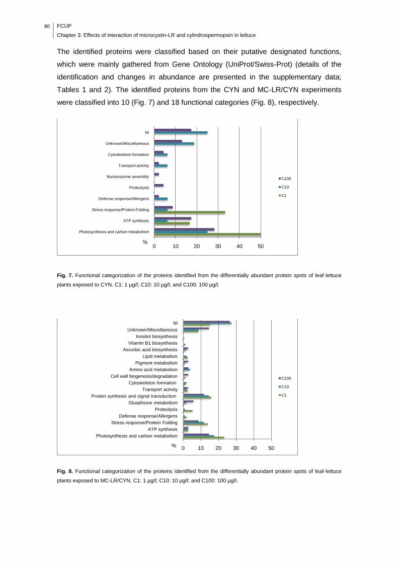
80 FCUP
Chapter 3: Effects of interaction of microcystin-LR and cylindrospermopsin in lettuce
The identified proteins were classified based on their putative designated functions,
which were mainly gathered from Gene Ontology (UniProt/Swiss-Prot) (details of the
identification and changes in abundance are presented in the supplementary data;
Tables 1 and 2). The identified proteins from the CYN and MC-LR/CYN experiments
were classified into 10 (Fig. 7) and 18 functional categories (Fig. 8), respectively.
Fig. 7. Functional categorization of the proteins identified from the differentially abundant protein spots of leaf-lettuce
plants exposed to CYN. C1: 1 µg/l; C10: 10 µg/l; and C100: 100 µg/l.
Fig. 8. Functional categorization of the proteins identified from the differentially abundant protein spots of leaf-lettuce
plants exposed to MC-LR/CYN. C1: 1 µg/l; C10: 10 µg/l; and C100: 100 µg/l.
0 10 20 30 40 50
Photosynthesis and carbon metabolism
ATP synthesis
Stress response/Protein Folding
Defense response/Allergens
Proteolysis
Nucleossome assembly
Transport activity
Cytoskeleton formation
Unknown/Miscellaneous
NI
%
C100
C10
C1
0 10 20 30 40 50
Photosynthesis and carbon metabolism
ATP synthesis
Stress response/Protein Folding
Defense response/Allergens
Proteolysis
Glutathione metabolism
Protein synthesis and signal transduction
Transport activity
Cytoskeleton formation
Cell wall biogenesis/degradation
Amino acid metabolism
Pigment metabolism
Lipid metabolism
Ascorbic acid biosynthesis
Vitamin B1 biosynthesis
Inositol biosynthesis
Unknown/Miscellaneous
NI
C100
C10
C1
%
FCUP
Chapter 3: Effects of interaction of microcystin-LR and cylindrospermopsin in lettuce
81
A large number of identified proteins in both experiments with all exposure
concentrations are mostly involved in photosynthesis and carbon metabolism, ATP
synthesis, and stress/defense response and protein folding (Figs. 7 and 8). Proteins
related to protein synthesis and signal transduction were also well represented in the
MC-LR/CYN experiment. To summarize, groups of more comprehensive functions
were created to discuss the potential effects of environmentally relevant concentrations
of CYN and MC-LR/CYN on leaf-lettuce plants.
3.2.1. Energy-related metabolism (photosynthesis/carbon
metabolism; ATP synthesis)
The impact of the cyanotoxins on photosynthesis is of major interest because plant
productivity and hence crop yield depend strongly of the efficiency of this process.
In the C1 group of the CYN experiment, although only six proteins showed differential
abundance, three (50%) were related to energy production, and these decreased in
abundance (plastocyanin, ATP synthase CF1 β subunit, and NADP-dependent malate
dehydrogenase (pyruvate metabolism)). In contrast, energy production appears to be
enhanced in the leaves of the plants in groups C10 and C100, the latter of which
presented the highest number of proteins involved in this process. The abundance of
chlorophyll a-b-binding proteins, which gather and transfer light energy to
photosynthetic reaction centers, was increased; in particular, chlorophyll a-b-binding
protein 8 exhibited a 50.7-fold increase in abundance in the C10 group. Oxygen-
evolving enhancer proteins, which are responsible for water oxidation in photosystem II
(PSII), are also increased in abundance in groups C10 and C100; and the same results
were obtained with quinone oxidoreductase-like protein At1g23740, ATP synthase CF1
α (C10 and C100) and β subunits (C100), and ATPase epsilon chain (C100). In
addition to proteins associated with primary photosynthesis reactions (light reactions),
proteins involved in the Calvin cycle (carbon fixation reactions) exhibit an increase in
abundance in group C100: ribulose-1,5-bisphosphate carboxylase/oxygenase activase
(RuBP activase), ribulose bisphosphate carboxylase/oxygenase activase 1 (RuBisCO
activase 1), phosphoribulokinase (PRK), and sedoheptulose-1,7-bisphosphatase
(SBPase).
The effects of MC-LR/CYN were much more complex and appear to have been

82 FCUP
Chapter 3: Effects of interaction of microcystin-LR and cylindrospermopsin in lettuce
stronger in groups C10 and C100, the gels of which showed several absent proteins.
The combined MC-LR/CYN exposure affected not only photosynthesis but also cellular
respiration. Interestingly, in group C1, several proteins involved in energy production
are increased in abundance. In detail, these proteins are associated with the light
reactions of photosynthesis (chlorophyll a-b-binding proteins, oxygen-evolving
enhancer proteins, chloroplast PsbO4 precursor, cytochromeb6/f heme-binding protein
2-like, ATP synthase α subunit, PSI reaction center subunit II, PSII stability/assembly
factor HCF136, quinone oxidoreductase-like protein At1g23740 chloroplastic-like, and
ferredoxin-NADP reductase), photorespiration (gamma carbonic anhydrase-like 2), the
Calvin cycle (RuBP activase and large subunit, RuBisCO activase 1, and PRK),
glycolysis and pentose phosphate pathway (β-xylosidase/α-L-arabinofuranosidase 2-
like, fructan 1-exohydrolase IIa, triosephosphate isomerase, 2,3-bisphosphoglycerate-
independent phosphoglycerate mutase, phosphoglycerate kinase 3, ribose-5-
phosphate transaldolase), tricarboxylic acid (TCA) cycle (mitochondrial
dihydrolipoyllysine-residue succinyltransferase component of 2-oxoglutarate
dehydrogenase complex 2), and oxidative and photo phosphorylation (mitochondrial
ATP synthase subunit delta' and chloroplastic soluble inorganic pyrophosphatase 1). In
contrast, few proteins related to photosynthesis and cellular respiration were increased
in abundance in groups C10 and C100 (chloroplast light-harvesting chlorophyll a/b-
binding protein, RuBP activase (C100), pyruvate dehydrogenase E1 component
subunit β-like, transaldolase-like, and chloroplastic soluble inorganic pyrophosphatase
1). Furthermore, there were proteins that were absent in the gels or exhibited
decreases in abundance in groups C10 and C100, and these proteins are associated
with the light reactions of photosynthesis (oxygen-evolving enhancer protein 1, ATP
synthase α subunit, ATP synthase gamma chain, and chloroplastic-like isoform 1) and
the Calvin cycle (RuBP activase, transketolase, putative), which is dependent on the
ATP and NADPH generated by the light reactions to assimilate CO2. Additionally,
proteins involved in glycolysis (pyrophosphate--fructose 6-phosphate 1-
phosphotransferase β subunit, and glyceraldehyde-3-phosphate dehydrogenase
(C100)) and the TCA cycle (putative cytosolic NADP-malic enzyme, α isopropylmalate
synthase, succinate dehydrogenase, isocitrate dehydrogenase (NAD+) (C100), and
malate dehydrogenase (C100)) were absent in the gels or presented decreases in
abundance in groups C10 and C100. Several studies have proposed that
photosynthesis-related proteins are differentially regulated under abiotic stress. In
general, photosynthesis is impaired in sensitive plants and enhanced in tolerant plants
(Kosová et al., 2011; Abreu et al., 2013). Nothing definite is known regarding how CYN
interferes in plant photosynthesis. However, negative effects of MC-LR in the
FCUP
Chapter 3: Effects of interaction of microcystin-LR and cylindrospermopsin in lettuce
83
photosynthesis activity of plants have been reported (Abe et al., 1996; Pflugmacher,
2002; El Khalloufi et al., 2011). Our results show that MC-LR/CYN concentrations of 10
and 100 µg/l may produce harmful effects on the energy production of lettuce leaves.
Nevertheless, the highest number of proteins that decreased in abundance was found
in group C10, which may suggest that the effects are produced through different
mechanisms. The roots exposed to 100 µg/l MC-LR/CYN had a high amount of
exudates (data not shown). It could be hypothesized that this accumulation could
prevent the uptake of the toxins, thereby mitigating their negative effects, but can also
impair the uptake of water and nutrients, which can be related to the significant
reduction in the fr. wt of these lettuce leaves (p<0.05) (Fig. 1). In contrast, the fr. wt of
the lettuce leaves of group C1 was significantly increased (p<0.05), likely due to the
enhancement of photosynthesis and carbon metabolism. As mentioned above, no
differences were found in the fr. wt of lettuce leaves in the CYN experiment (Fig. 1);
thus, it is likely that the increase in abundance of proteins related to photosynthesis
and carbon metabolism in group C100 may contribute to the maintenance of cellular
homeostasis and an active adaptation to the stress promoted by CYN. Plant
productivity and hence crop yield depend strongly on the prevailing photosynthetic
rates. The Calvin cycle is autocatalytic and can thus be enhanced by increases in the
concentrations of its biochemical intermediates (Taiz and Zeiger, 2002). Some of the
identified proteins belonging to the Calvin cycle, such as SBPase (which exhibits a
11.8-fold increase in abundance in group C100 of the CYN exposure experiment), have
been reported to be effective for the improvement of abiotic stress tolerance (Abreu et
al., 2013) and have also been studied to determine whether their use may increase
crop production (Lefebvre et al., 2005). Lefebvre et al. (2005) reported that an increase
in SBPase activity in tobacco plants (Nicotiana tabacum) leads to improvements in the
photosynthesis rate, levels of sucrose and starch, leaf area, and biomass, which may
be associated with an increase in the RuBP regenerative capacity. Because
photosynthesis and carbon metabolism seem to have been the main biological
processes affected by CYN and MC-LR/CYN, it is hypothesized that some of these
identified proteins can be considered potential biomarkers of these cyanotoxins.
3.2.2. Stress and defense response (stress response/protein folding;
glutathione metabolism; proteolysis; defense response/allergens)
In the stress/defense response, plants need to regulate a variety of processes that

84 FCUP
Chapter 3: Effects of interaction of microcystin-LR and cylindrospermopsin in lettuce
require energy. In fact, the profile of abundance of proteins involved in the
stress/defense response followed the same pattern observed in the analysis of energy-
related proteins: group C100 of the CYN exposure experiment and group C1 of the
MC-LR/CYN exposure experiment stand out due to their high number of proteins that
exhibited an increase in abundance. Although it is well recognized that ROS are
generated during normal plant metabolism (e.g., in chloroplasts), it has also been
stated that the cyanotoxins CYN and MC-LR induce oxidative stress in plants
(Pflugmacher et al., 2006; Pflugmacher et al., 2007; Stüven and Pflugmacher, 2007;
Saqrane et al., 2009; Prieto et al., 2011). A protein involved in glutathione (GSH)
metabolism (S-formylglutathione hydrolase), specifically in GSH synthesis, was only
present in the C group of the MC-LR/CYN experiment. However, protein IN2-1
homolog B-like (glutathione-S-transferase (GST) superfamily) increased in abundance
in group C100 (3.3-fold). Runnegar et al. (1995) showed that CYN inhibits GSH
synthesis in cultured rat hepatocytes. It is interesting to note that in this study this effect
was not observed in lettuce leaves exposed to CYN even at a concentration of 100
µg/l. Nevertheless, it is well recognized that MC-LR is detoxified by conjugation with
GSH via GST (Pflugmacher et al., 1998). It can be hypothesized that the simultaneous
exposure to MC-LR/CYN may lead to a reduction of GSH pool in cells due to MC-LR
detoxification (via GST), which may have resulted in a higher requirement of GSH
biosynthesis. Because CYN is an inhibitor of GSH synthesis, it can impair the capacity
of plants to detoxify MC-LR. On the other hand, the weakened response of lettuce
plants to MC-LR may contribute to oxidative stress and may enhance its toxic effects in
a concentration-dependent manner, leading to an inefficient response of plants to CYN
and consequently the inhibition of a higher number of proteins in the groups exposed to
higher concentrations of the toxin. In contrast, at low concentrations, lettuce plants
appear to be able to cope with oxidative stress by inducing the production of other
antioxidant enzymes. Chloroplastic Cu/Zn superoxide dismutase (SOD), ascorbate
peroxidase (APX), other peroxidases, and ferritin, which is reported to be responsive to
stress, were identified in leaf-lettuce plants exposed to 1 µg/l MC-LR/CYN
(chloroplastic 2-Cys peroxiredoxin BAS1, peroxiredoxin 2, thioredoxin-dependent
peroxidase, oxidoreductase, chloroplastic SOD [Cu-Zn], aldo-keto reductase 2-like, and
chloroplastic peroxiredoxin-2E (also in the group C10)). The coordinated action of
these enzymes prevents the oxidative damage of cells generated by ROS. SOD
catalyzes the dismutation of superoxide radical to hydrogen peroxide and oxygen. The
resulting hydrogen peroxide is reduced by APX and other peroxidases to yield water
(Pflugmacher et al., 2006). However, if the detoxification enzymes produced are not
sufficient, proteins damaged by ROS may accumulate in plant cells under stress
FCUP
Chapter 3: Effects of interaction of microcystin-LR and cylindrospermopsin in lettuce
85
conditions. Some of the strategies developed by plants to overcome this accumulation
are to refold misfolded proteins using helper proteins, such as chaperones, and to
remove these by protease activity. Heat shock protein (HSP) 70, which is one of the
most important HSPs involved in the plant response to abiotic stresses (Abreu et al.,
2013), was decreased in abundance in the C1 group of the CYN exposure experiment.
However, in group C100 of the CYN exposure experiment, putative thioredoxin-
dependent peroxidase and RuBisCO large subunit-binding protein subunit α, which has
chaperone and refolding activity, exhibited increases in abundance. In addition, the
chloroplastic protein peptidyl-prolyl cis-trans isomerase (PPIase) FKBP16-3, which is a
folding catalyst, exhibited a 107-fold increase in abundance in group C100 (CYN
exposure). With respect to the MC-LR/CYN experiment, several proteins involved in
redox homeostasis and some proteins with chaperone functions (chloroplastic PPIase
CYP38, RuBisCO subunit binding-protein α subunit precursor, 60-kDa chaperonin α
subunit, chloroplastic 20-kDa chaperonin, protein disulfide isomerase-like 2-3-like,
calreticulin (C10), protein disulfide isomerase (C10), and HSP 90 (C10)) were
increased in abundance in groups C1 and C10 (exhibited less fold-variation in group
C10). The late embryogenesis abundant protein (LEA), Lea14-A, is well recognized for
its role in stress tolerance (Abreu et al., 2013); however, this protein was absent in the
gels of groups C1 and C10 of the MC-LR/CYN exposure experiment. Additionally,
some proteins involved in redox homeostasis and some proteins with chaperone
activity were either decreased in abundance or absent in the gels of groups C10 and
C100 of the MC-LR/CYN exposure experiment (chloroplastic chaperone protein ClpC
(C10), glutaredoxin S16 (C10), thylakoid-bound ascorbate peroxidase (C10),
thioredoxin reductase 2-like (C10), PITH domain-containing protein At3g04780 (C100),
and chloroplastic chaperone protein ClpB3). The latter protein was also decreased in
abundance in the C1 group. Almost all of the chaperones and peroxidases are
chloroplastic, which suggests that photosynthetic complexes are the most affected by
CYN and MC-LR/CYN exposure. In addition to detoxification and chaperone proteins,
an increase in the abundance of proteases and proteasomes was observed in group
C1 of the MC-LR/CYN exposure experiment. Probable 26S proteasome non-ATPase
regulatory subunit 7, which belongs to the proteolytic complex that regulates cytosolic
protein turnover by the ubiquitin pathway (Taiz and Zeiger, 2002) and plays an
essential role in removing regulatory proteins and abnormal polypeptides in plants
during stress, exhibited a 4-fold increase in abundance. The putative zinc-dependent
protease was increased in abundance in group C100 of the CYN exposure experiment.
86 FCUP
Chapter 3: Effects of interaction of microcystin-LR and cylindrospermopsin in lettuce
Proteins related to the bacterium defense response also presented changes in
abundance in the CYN and MC-LR/CYN experiments. Eugenol synthase 1, which
catalyzes the synthesis of the phenylpropene eugenol, a defense compound with
antimicrobial properties, exhibited an increase in abundance in group C10 of the CYN
exposure experiment and in group C1 of the MC-LR/CYN exposure experiment. In the
latter experiment, harpin binding protein 1 also presented increases in abundance. The
increase in abundance of pathogenesis-related (PR) proteins may be promising for the
improvement of the response to abiotic stress; however, it may also constitute a threat
to food safety because most of these proteins have allergenic potential. Thaumatin-like
protein-like, which appears to have allergenic properties (Palacín et al., 2010), was
increased in abundance in group C100 of the CYN exposure experiment (2.7-fold) and
in group C1 of the MC-LR/CYN exposure experiment (2.2-fold).
3.2.3. Protein synthesis and signal transduction (transcription, RNA
processing and translocation, and translation)
Several proteins involved in protein synthesis and signal transduction were found in
almost all of the groups of the MC-LR/CYN experiment. The C1 group presented a high
number of these proteins that were increased in abundance. Although to a lower
extent, groups C10 and C100 also showed proteins associated with: mRNA processing
(chloroplast putative ribonucleoprotein (C1, C10, and C100), chloroplast 31-kDa
ribonucleoprotein (C1 and C100)), transcription factors containing DNA-binding motifs
and regulators of transcription (transcription factor Pur-alpha 1-like, U2 small nuclear
ribonucleoprotein A, zinc finger protein, proliferating cell nuclear antigen, minor allergen
Alt a, nascent polypeptide-associated complex subunit alpha-like), and mRNA transport
and translation (eukaryotic translation initiation factor 3 subunit D-like, elongation factor
1-beta, eukaryotic translation initiation factor 3 subunit F-like, eukaryotic translation
initiation factor 3 subunit J-like (C10), chloroplast 30S ribosomal protein S1 (C1 and
C10), 60S acidic ribosomal protein P0, and chloroplast 50S ribosomal protein L12
(C10)) that presented increases in abundance. The mRNA processing and translational
apparatus may indicate a rapid protein synthesis, which could be related to the
increase and maintenance of the fr. wt of lettuce leaves in groups C1 and C10,
respectively. Nevertheless, some proteins associated with RNA recognition motifs and
regulators of ribonuclease activity (poly(A)-binding protein (C10), and regulator of
ribonuclease activity A (C10)) and translation (eukaryotic translation initiation factor 5A
FCUP
Chapter 3: Effects of interaction of microcystin-LR and cylindrospermopsin in lettuce
87
(C10), 40S ribosomal protein (C10), eukaryotic translation initiation factor 3 subunit K-
like (C10), and elongation factor 2 (C100)) decreased in abundance or were absent in
the gels of groups C10 and C100. In this study, the changes in abundance of proteins
related to protein synthesis suggest that this activity may be of particular importance in
the lettuce response to MC-LR/CYN. The little that is known regarding how CYN
inhibits protein synthesis in plants is that the soluble proteins associated with the
eukaryotic translation system appear to be the target of the toxin (Froscio et al., 2008).
Additionally, this toxin appears to interfere with the elongation step, which indicates that
elongation factors may also be a target (Froscio et al., 2008). In fact, in group C100 of
the MC-LR/CYN experiment, elongation factor 2 was the unique protein that exhibited a
decrease in abundance. The potential decrease of energy-related enzymes (e.g.,
Calvin cycle enzymes), likely as a result of protein synthesis inhibition promoted by
CYN, may lead to reduced synthesis of carbohydrates, which are essential for the
support of cell division and elongation (Taiz and Zeiger, 2002). This decrease may also
have contributed to the reduction in the fr. wt of lettuce leaves; however, this
hypothesis should be further studied. Exposure to CYN results only in an increased
abundance of histone H4 in the C100 group. This protein is a core component of the
nucleosome and plays a central role in transcription regulation, DNA repair, DNA
replication, and chromosomal stability. It could be hypothesized that CYN at a
concentration up to 100 µg/l is not sufficient to cause cessation of protein synthesis;
however, in combination with MC-LR, the inhibition of PP1 and PP2A and the induction
of oxidative stress promoted by MC-LR may have enhanced the protein synthesis
inhibition produced by CYN. Furthermore, phosphorylation and dephosphorylation play
important roles in signal transduction, and in the MC-LR/CYN exposure experiment,
proteins involved in signal transduction pathways (14-3-3-like protein D-like (C1), 14-3-
3 protein 1-like, and 14-3-3 protein (C10)) present increases in abundance in groups
C1 and C10. The plant 14-3-3 isoforms regulate a diverse range of proteins, including
kinases, transcription factors, structural proteins, ion channels, and pathogen defense-
related proteins (Denison et al., 2011). The differential accumulation of these proteins
appears to be implicated in the response to abiotic stress (Abreu et al., 2013).
3.2.4. Transport activity
Proteins and lipoproteins involved in the transport of macromolecules, small molecules,
and ions, mainly in the chloroplast, exhibited changes in abundance in both

88 FCUP
Chapter 3: Effects of interaction of microcystin-LR and cylindrospermopsin in lettuce
experiments. In groups C10 and C100 of the CYN exposure experiment, temperature-
induced lipocalin exhibited 4.4-and 2.5-fold increases in abundance, respectively. This
protein as well as chloroplast processing peptidase-like, protein TIC 62 chloroplastic-
like, and apolipoprotein d, also increased in abundance in group C1 of the MC-LR/CYN
exposure experiment. However, in groups C10 and C100, chloroplastic protein TIC 62
was decreased in abundance, and GTP-binding nuclear protein Ran1A was absent in
the gels of the C10 group.
3.2.5. Structural activity (cytoskeleton formation; cell wall
biogenesis/degradation)
It is well recognized that MC-LR alters the cytoskeletal structure of animal cells (Toivola
and Eriksson, 1999). Phosphorylation/dephosphorylation and the synthesis of certain
regulatory proteins are key mechanisms in cytoskeletal regulation (Máthé et al., 2013).
Similarly, CYN induces the reorganization of the cytoskeleton in animal cells (CHO K1
cells) (Fessard and Bernard, 2003). In this study, exposure to CYN promoted a
decrease in the abundance of actin in the C10 group. In contrast, plastid-dividing ring
protein, which is related to microtubule-based process and protein polymerization,
increased in abundance in group C100 of the CYN exposure experiment and in group
C1 of the MC-LR/CYN exposure experiment. Additionally, fibrillin, a glycoprotein that
contributes to the structural integrity and assembly within or outside cells, increased in
abundance in groups C1 and C10 of the MC-LR/CYN exposure experiment. UDP-
arabinopyranose mutase 1, which is involved in the biosynthesis of the cell wall,
exhibited an increase in abundance in group C1 of the MC-LR/CYN exposure
experiment. However, xyloglucan endotransglucosylase/hydrolase, an enzyme
involved in wall assembly, decreased in abundance in groups C1 and C100 of the MC-
LR/CYN exposure experiment. Exposure to these cyanotoxins appears to lead to the
reorganization of cytoskeletal components in lettuce leaves. Furthermore, it could be
hypothesized that proteins involved in cytoskeleton assembly and cell wall biosynthesis
played a role in the increase, maintenance, and decrease of the observed fr. wt of
lettuce leaves.
FCUP
Chapter 3: Effects of interaction of microcystin-LR and cylindrospermopsin in lettuce
89
2.2.6. Other metabolisms (amino acid metabolism; pigment
metabolism; lipid metabolism; ascorbic acid biosynthesis; vitamin B1
biosynthesis; inositol biosynthesis; hormone regulator)
Overall, proteins involved in the metabolism of amino acids, pigments, lipids, ascorbic
acid, vitamin B1, inositol, and hormone regulation exhibited increases in abundance in
group C1 and decreases in abundance in groups C10 and C100 of the MC-LR/CYN
exposure experiment. Ketol-acid reductoisomerase, which promotes the formation of
amino acids containing a branched carbon skeleton, increased in abundance in the C1
group. In contrast, other proteins that have been identified to be involved in amino acid
metabolism, such as acetohydroxyacid synthase 1, diaminopimelate decarboxylase 2
chloroplastic isoform 1, vitamin-b12 independent methionine synthase 5-
methyltetrahydropteroyltriglutamate-homocysteine, and putative thiosulfate
sulfurtransferase (C100) decreased in abundance or were absent in groups C10 and
C100. Chloroplastic coproporphyrinogen-III oxidase, which is involved in porphyrin and
chlorophyll metabolism and biosynthesis, increased in abundance in the C1 group, and
chloroplastic uroporphyrinogen decarboxylase was absent in the C10 group. The
polyphenol oxidase precursor, which is involved in the pigment biosynthetic process,
was absent in group C100. A diverse family of lipases and esterases (GDSL
esterase/lipase At5g45670, GDSL esterase/lipase LTL1-like, and bifunctional epoxide
hydrolase 2-like) and a key enzyme of the type II fatty acid synthesis (FAS) system
(enoyl-ACP reductase 1) presented increases in abundance in the C1 group. However,
the protein 2-hydroxyacyl-CoA lyase-like, which plays a key role in redox signaling and
lipid homeostasis, and the protein acetyl-CoA C-acetyltransferase, which is involved in
the beta oxidation pathway of fatty acid degradation, were absent in the C10 group.
Phosphomannomutase, which is involved in ascorbic acid biosynthesis, was absent in
all of the groups of the MC-LR/CYN experiment. Chloroplastic-like thiamine thiazole
synthase and L-myo-inositol-1-phosphate synthase, which are involved in vitamin B1
and inositol biosynthesis, respectively, increased in abundance in the C1 group. Auxin-
binding protein ABP20-like, a probable receptor for the plant growth-promoting
hormone auxin, exhibited an increase in abundance in groups C1 and C10 (to a higher
extent in group C1), whereas abscisic acid receptor PYR1-like, a plant hormone
associated with signal transduction, was absent in group C10. The simultaneous
exposure to low concentrations of MC-LR and CYN (1 µg/l) appears to stimulate the
synthesis of different constituents in lettuce leaves that are concomitantly involved in

90 FCUP
Chapter 3: Effects of interaction of microcystin-LR and cylindrospermopsin in lettuce
plant growth and development and also in the response to stress, such as hormones,
amino acids, lipids, important membrane components, and vitamins. Exposure to
environmental stress induces several physiological changes in plants that can alter the
chemical composition and thus the quality of crops (Wang and Frei, 2011). Dependent
on numerous factors, such as the time of stress and the crop species, the plant stress
response appears to result in increasing the concentrations of some constituents, such
as proteins and antioxidants (Wang and Frei, 2011). Depending on the concentration of
MC-LR/CYN exposure, positive changes in the nutritional quality of lettuce leaves may
be attained. However, this issue should be further studied through the quantification of
the respective constituents in plants exposed to these cyanotoxins.
3.2.7. Unknown/miscellaneous
Proteins with no well-defined function (several functions or unknown function) were
identified as unknown/miscellaneous.
4. Conclusions
In this study, we applied a differential-expression proteomics approach to understand
the mechanisms underlying the response of leaf-lettuce plants to environmentally
relevant concentrations of CYN and MC-LR/CYN. The abundance of proteins was
affected in a concentration-dependent manner by the simultaneous activation of
several metabolic pathways, which are mainly related to photosynthesis,
response/defense to stress, and protein synthesis and transduction. The activation of
these pathways appears to confer tolerance to lettuce plants against CYN at
concentrations up to 100 µg/l. The simultaneous exposure to MC-LR and CYN resulted
in a ‗dualistic response‘, and exposure to a concentration of 1 µg/l promoted an
increase in the abundance of proteins associated with general biological processes and
a significant increase in the fr. wt of leaves. In contrast, the concentrations of 10 and
100 µg/l appeared to be deleterious to lettuce because a high number of proteins
associated with general biological processes exhibited decreases in abundance or
were absent in the gels. The latter concentration also promoted a reduction in the fr. wt
of the leaves. This effect is of major concern because the occurrence of mixtures of
cyanotoxins is expected to become increasingly recurrent. This study also provides
FCUP
Chapter 3: Effects of interaction of microcystin-LR and cylindrospermopsin in lettuce
91
new insights into potential protein markers of exposure to cyanotoxins and of novel
proteins that may confer tolerance to CYN and MC-LR/CYN, although these need to be
functionally characterized and validated. Furthermore, the proteomics analysis was
found to be suitable for the discovery of some traits associated with the quality and
safety of edible tissues of lettuce exposed to environmentally relevant concentrations of
CYN and MC-LR/CYN, such as the presence of allergenic proteins.
Conflict of Interest
The authors declare that there are no conflicts of interest.
Acknowledgments
Marisa Freitas thanks the Fundação para a Ciência e Tecnologia (FCT) for the Ph.D.
Grant SFRH/BD/85490/2012. This work was partially funded by the FCT project PesT-
C/MAR/LA0015/2011.
5. References
Abe, T., Lawson, T., Weyers, J.D.B., Codd, G.A., 1996. Microcystin-LR inhibits
photosynthesis of Phaseolus vulgaris primary leaves: imphcations for current spray
irrigation practice. New Phytol. 133, 651-658.
Abreu, I.A., Farinha, A.P., Negrão, S., Gonçalves, N., Fonseca, C., Rodrigues, M.,
Batista, R., Saibo, N.J.M., Oliveira, M.M., 2013. Coping with abiotic stress: Proteome
changes for crop improvement. J. Proteomics 93, 145-168.
Afroz, A., Ali, G.M., Mir, A., Komatsu, S., 2011. Application of proteomics to investigate
stress-induced proteins for improvement in crop protection. Plant Cell Rep. 30, 745–
763.
Azevedo, C.C., Azevedo, J., Osório, H., Vasconcelos, V., Campos, A., 2014. Early
physiological and biochemical responses of rice seedlings to low concentration of
92 FCUP
Chapter 3: Effects of interaction of microcystin-LR and cylindrospermopsin in lettuce
microcystin-LR. Ecotoxicology 23, 107–121.
Beyer, D., Surányi, G., Vasas, G., Roszik, J., Erdődi, F., M-Hamvas, H., Bácsi, I.,
Bátori, R., Serfőző, Z., Szigeti, Z.M., Vereb, G., Demeter, Z., Gonda, S., Máthé, C.,
2009. Cylindrospermopsin induces alterations of root histology and microtubule
organization in common reed (Phragmites australis) plantlets cultured in vitro. Toxicon
54, 440–449.
Bradford, M.M., 1976. A rapid and sensitive method for the quantitation of microgram
quantities of protein utilizing the principle of protein-dye binding. Anal. Biochem. 72,
248-254.
Brient, L., Lengronne, M., Bormans, M., Fastner, J., 2008. First occurrence of
cylindrospermopsin in freshwater in France. Environ. Toxicol. 24, 415–420.
Chen, J., Song, L., Dai, J., Gan, N., Liu, Z., 2004. Effects of microcystins on the growth
and the activity of superoxide dismutase and peroxidase of rape (Brassica napus
L.) and rice (Oryza sativa L.). Toxicon 43, 393–400.
Cho, W.K., Chen, X-Y., Uddin, N.M., Rim, Y., Moon, J., Jung, J-H., Shi, C., Chu, H.,
Kim, S., Kim, S-W., Park, Z-Y., Kim, J-Y., 2009. Comprehensive proteome analysis of
lettuce latex using multidimensional protein-identification technology. Phytochemistry
70, 570–578.
Choi, J-S., Cho, S-W., Kim, T-S., Cho, K., Han, S-S., Kim, H-K., Woo, S-H., Chung, K-
Y., 2008. Proteome analysis of greenhouse-cultured lettuce with the natural soil
mineral conditioner illite. Soil Biol. Biochem. 40, 1370–1378.
Crush, J.R., Briggs, L.R., Sprosen, J.M., Nichols, S.N., 2008. Effect of irrigation with
lake water containing microcystins on microcystin content and growth of ryegrass,
clover, rape, and lettuce. Environ. Toxicol. 23, 246-252.
Denison, F.C., Paul, A-L., Zupanska, A.K., Ferl, R.J., 2011. 14-3-3 proteins in plant
physiology. Semin. Cell Dev. Biol. 22, 720–727.
El Khalloufi, F., Oufdou, K., Lahrouni, M., El Ghazali, I., Saqrane, S., Vasconcelos, V.,
Oudra, B., 2011. Allelopatic effects of cyanobacteria extracts containing microcystins
on Medicago sativa-Rhizobia symbiosis. Ecotoxicol. Environ. Saf. 74, 431-438.
Elliott, J.A., 2012. Is the future blue-green? A review of the current model predictions of
FCUP
Chapter 3: Effects of interaction of microcystin-LR and cylindrospermopsin in lettuce
93
how climate change could affect pelagic freshwater cyanobacteria. Water Res. 46,
1364-1371.
Fessard, V., Bernard, C., 2003. Cell alterations but no DNA strand breaks induced In
Vitro by cylindrospermopsin in CHO K1 cells. Environ. Toxicol. 18, 353–359.
Froscio, S.M., Humpage, A.R., Wickramasinghe, W., Shaw, G., Falconer, I.R., 2008.
Interaction of the cyanobacterial toxin cylindrospermopsin with the eukaryotic protein
synthesis system. Toxicon 51, 191–198.
Gehringer, M.M., Kewada, V., Coates, N., Downing, T.G., 2003. The use of Lepidium
sativum in a plant bioassay system for the detection of microcystin-LR. Toxicon 41,
871–876.
Jensen, M.H., Malter, A.J., 1995. Protected Agriculture: A Global Review. Chapter 7,
Water supply, water quality and mineral nutrition. World Bank Publications.
Kinnear, S., 2010. Cylindrospermopsin: a decade of progress on bioaccumulation
research. Mar. Drugs 8, 542-564.
Kosová, K., Vítámvás, P., Prášil, I.T., Renaut, J., 2011. Plant proteome changes under
abiotic stress — Contribution of proteomics studies to understanding plant stress
response. J. Proteomics 74, 1301-1322
Kotai, J., 1972. Instruction for preparation of modified nutrient solution Z8 for algae.
Norwegian Institute for Water Research, Oslo, Norway, Report nr B-11/69, 1-5.
Lefebvre, S., Lawson, T., Fryer, M., Zakhleniuk, O.V., Lloyd, J.C., Raines, C.A., 2005.
Increased sedoheptulose-1,7-bisphosphatase activity in transgenic tobacco plants
stimulates photosynthesis and growth from an early stage in development. Plant
Physiol. 138, 451-460.
MacKintosh, C., Beattie, K.A., Klumpp, S., Cohen, P., Codd, G.A., 1990.
Cyanobacterial microcystin-LR is a potent and specific inhibitor of protein
phosphatases 1 and 2A from both mammals and higher plants. FEBS Lett. 264, 187–
192.
Malécot, M., Guével, B., Pineau, C., Holbech, B.F., Bormans, M., Wiegand, C., 2013.
Specific proteomic response of Unio pictorum mussel to a mixture of glyphosate and
94 FCUP
Chapter 3: Effects of interaction of microcystin-LR and cylindrospermopsin in lettuce
microcystin-LR. J. Proteome Res. 12, 5281–5292.
Martins, J.C., Leão, P.N., Vasconcelos, V., 2009. Differential protein expression in
Corbicula fluminea upon exposure to a Microcystis aeruginosa toxic strain. Toxicon 53,
409–416.
Máthé, C., Beyer, D., Erdodi, F., Serfozo, Z., Székvölgyi, L., Vasas, G., M-Hamvas, M.,
Jámbrik, K., Gonda, S., Kiss, A., Szigeti, Z.M., Surányi, G., 2009. Microcystin-LR
induces abnormal root development by altering microtubule organization in tissue-
cultured common reed (Phragmites australis) plantlets. Aquat. Toxicol. 92, 122-130.
Máthé, C., M-Hamvas, M., Vasas, G., 2013. Microcystin-LR and cylindrospermopsin
induced alterations in chromatin organization of plant cells. Mar. Drugs11, 3689-3717.
McElhiney, J., Lawton, L.A., Leifert, C., 2001. Investigations into the inhibitory effects of
microcystins on plant growth, and the toxicity of plant tissues following exposure.
Toxicon 39, 1411-1420.
Metcalf, J.S., Barakate, A., Codd, G.A., 2004. Inhibition of plant protein synthesis by
the cyanobacterial hepatotoxin, cylindrospermopsin. FEMS Microbiol. Lett. 235, 125–
129.
Mitrovic, S.M., Allis, O., Furey, A., James, K.J., 2005. Bioaccumulation and harmful
effects of microcystin-LR in the aquatic plants Lemna minor and Wolffia arrhiza and the
filamentous alga Chladophora fracta. Ecotoxicol. Environ. Saf. 61, 345–352.
Neuhoff, V., Arold, N., Taube, D., Ehrhardt, W., 1988. Improved staining of proteins in
polyacrylamide gels including isoelectric focusing gels with clear background at
nanogram sensitivity using Coomassie Brilliant Blue G-250 and R-250. Electrophoresis
9, 255-262.
O‘Neil, J.M.,
Davis, T.W., Burford, M.A., Gobler, C.J., 2012. The rise of harmful
cyanobacteria blooms: The potential roles of eutrophication and climate change.
Harmful Algae. 14, 313–334.
Palacín, A., Tordesillas, L., Gamboa, P., Sanchez-Monge, R., Cuesta-Herranz, J.,
Sanz, M.L., Barber, D., Salcedo, G., Díaz-Perales, A., 2010. Characterization of peach
thaumatin-like proteins and their identification as major peach allergens. Clin. Exp.
Allergy 40, 1422–1430.
FCUP
Chapter 3: Effects of interaction of microcystin-LR and cylindrospermopsin in lettuce
95
Pereira, S., Saker, M.L., Vale, M., Vasconcelos, V.M., 2009., Comparison of sensitivity
of grasses (Lolium perenne L. and Festuca rubra L.) and lettuce (Lactuca sativa L.)
exposed to water contaminated with microcystins. Bull Environ. Contam. Toxicol. 83,
81-84.
Pflugmacher, S., 2002. Possible allelopathic effects of cyanotoxins, with reference to
microcystin-LR, in aquatic ecosystems. Environ. Toxicol. 17, 407–413.
Pflugmacher, S., 2004. Promotion of oxidative stress in the aquatic macrophyte
Ceratophyllum demersum during biotransformation of the cyanobacterial toxin
microcystin-LR. Aquat. Toxicol. 70, 169–178.
Pflugmacher, S., Codd, G.A., Steinberg, C.E.W., 1999. Effects of the cyanobacterial
toxin microcystin-LR on detoxication enzymes in aquatic plants. Environ. Toxicol. 14,
111-115.
Pflugmacher, S., Hofmann, J., Hübner, B., 2007. Effects on growth and physiological
parameters in wheat (Triticum aestivum L.) grown in soil and irrigated with
cyanobacterial toxin contaminated water. Environ. Toxicol. Chem. 26, 2710-2716.
Pflugmacher, S., Jung, K., Lundvall, L., Neumann, S., Peuthert, A., 2006. Effects of
cyanobacterial toxins and cyanobacterial cell-free crude extract on germination of
alfalfa (Medicago sativa) and induction of oxidative stress. Environ. Toxicol. Chem. 25,
2381–2387.
Pflugmacher, S., Wiegand, C., Beattie, K.A., Krause, E., Steinberg, C.E.W., Codd,
G.A., 2001. Uptake, effects, and metabolism of cyanobacterial toxins in the emergent
reed plant Phragmites australis (cav.) trin. ex steud. Environ. Toxicol. Chem. 20, 846–
852.
Pflugmacher, S., Wiegand, C., Oberemm, A., Beattie, K.A., Krause, E., Codd, G.A.,
Steinberg C.E.W., 1998. Identification of an enzymatically formed glutathione conjugate
of the cyanobacterial hepatotoxin microcystin-LR: the first step of detoxication.
Biochim. Biophys. Acta1425, 527-533.
Pichardo, S., Pflugmacher, S., 2011. Study of the antioxidant response of several bean
variants to irrigation with water containing MC-LR and cyanobacterial crude extract.
Environ. Toxicol. 26, 300-306.
96 FCUP
Chapter 3: Effects of interaction of microcystin-LR and cylindrospermopsin in lettuce
Pietsch, C., Wiegand, C., Amé, M.V., Nicklisch, A., Wunderlin, D., Pflugmacher, S.,
2001. The effects of a cyanobacterial crude extract on different aquatic organisms:
evidence for cyanobacterial toxin modulating factors. Environ. Toxicol. 16, 535–542.
Pinheiro, C., Sergeant, K., Machado, C.M., Renaut, J., Ricardo, C.P., 2013. Two
traditional maize inbred lines of contrasting technological abilities are discriminated by
the seed flour proteome. J. Proteome Res. 12, 3152-3165.
Poniedziałek, B., Rzymski, P., Kokociński, M., 2012. Cylindrospermopsin: Water-linked
potential threat to human health in Europe. Environ. Toxicol. Phar. 34, 651–660.
Prieto, A., Campos, A., Cameán, A., Vasconcelos, V., 2011. Effects on growth and
oxidative stress status of rice plants (Oryza sativa) exposed to two extracts of toxin-
producing cyanobacteria (Aphanizomenon ovalisporum and Microcystis aeruginosa).
Ecotoxicol. Environ. Saf. 74, 1973-1980.
Printz, B., Sergeant, K., Lutts, S., Guignard, C., Renaut, J., Hausman, J.F., 2013. From
tolerance to acute metabolic deregulation: contribution of proteomics to dig into the
molecular response of alder species under a polymetallic exposure. J. Proteome Res.
12, 5160-5179.
Puerto, M., Campos, A., Prieto, A.I., Cameán, A.M., Almeida, A., Varela Coelho, A.,
Vasconcelos, V.M., 2011. Differential protein expression in two bivalve species; Mytilus
galloprovincialis and Corbicula fluminea; exposed to Cylindrospermopsis raciborskii
cells. Aquat. Toxicol. 101, 109–116.
Ramanan, S., Tang, J., Velayudhan, A., 2000. Isolation and preparative purification of
microcystin variants. J. Chromatogr A. 883,103-112.
Runnegar, M.T., Kong, S.M., Zhong, Y.Z., Lu S.C., 1995. Inhibition of reduced
glutathione synthesis by cyanobacterial alkaloid cylindrospermopsin in cultured rat
hepatocytes. Biochem. Pharmacol. 49, 219–225.
Saqrane, S., Ouahid, Y., El Ghazali, I., Oudra, B., Bouarab, L., del Campo, F.F., 2009.
Physiological changes in Triticum durum, Zea mays, Pisum sativum and Lens
esculenta cultivars, caused by irrigation with water contaminated with microcystins: a
laboratory experimental approach. Toxicon 53, 786-796.
Sheoran, I.S., Ross, A.R.S., Olson, D.J.H., Sawhney, V.K., 2007. Proteomic analysis of
tomato (Lycopersicon esculentum) pollen. J. Exp. Bot. 58, 3525–3535.

FCUP
Chapter 3: Effects of interaction of microcystin-LR and cylindrospermopsin in lettuce
97
Stüven, J., Pflugmacher, S., 2007. Antioxidative stress response of Lepidium sativum
due to exposure to cyanobacterial secondary metabolites. Toxicon 50, 85–93.
Taiz, L., Zeiger, E., 2002. Plant Physiology, Third Edition. Sinauer Associates,
Sunderland, MA. 690 pps.
Toivola, D.M., Eriksson J.E., 1999. Toxins affecting cell signalling and alteration of
cytoskeletal structure. Toxicol. In Vitro 13, 521-530.
Vasas, G., Gáspár, A., Surányi, G., Batta, G., Gyémánt, G., M-Hamvas, M., Máthé, C.,
Grigorszky, I., Molnár, E., Borbély, G., 2002. Capillary electrophoretic assay and
purification of cylindrospermopsin, a cyanobacterial toxin from Aphanizomenon
ovalisporum, by plant test (blue-green Sinapis test). Anal. Biochem. 302, 95–103.
Wang, Y., Frei, M., 2011. Stressed food – The impact of abiotic environmental stresses
on crop quality. Agr. Ecosyst. Environ. 141, 271–286.
Welker, M., Bickel, H., Fastner, J., 2002. HPLC-PDA detection of cylindrospermopsin -
opportunities and limits. Water Res. 36, 4659-4663.
98 FCUP
Chapter 4: Availability and bioaccessibility of microcystin-LR and cylindrospermopsin in bivalves
Chapter 4
Availability and bioaccessibility of microcystin-
LR and cylindrospermopsin in bivalves
This chapter has been adapted from the following scientific paper and manuscript:
Marisa Freitas, Joana Azevedo, António Paulo Carvalho, Alexandre Campos, Vitor
Vasconcelos. 2014. Effects of storage, processing and proteolytic digestion on the
microcystin-LR concentration in edible clams. Food and Chemical Toxicology 66 (2014)
217–223.
Marisa Freitas, Joana Azevedo, António Paulo Carvalho, Vera Mendes, Bruno
Manadas, Alexandre Campos, Vitor Vasconcelos. Bioaccessibility and changes on
cylindrospermopsin concentration in edible mussels over storage and processing time.
Submitted to the Journal Food and Chemical Toxicology.
FCUP
Chapter 4: Availability and bioaccessibility of microcystin-LR and cylindrospermopsin in bivalves
99
Effects of storage, processing and proteolytic digestion on the
microcystin-LR concentration in edible clams
Marisa Freitas1,2,3, Joana Azevedo1,3, António Paulo Carvalho1,2, Alexandre Campos1,
Vitor Vasconcelos1,2
1CIIMAR/CIMAR - Interdisciplinary Centre of Marine and Environmental Research,
University of Porto, Rua dos Bragas 289, P 4050-123 Porto, Portugal.
2Faculty of Sciences, University of Porto, Rua do Campo Alegre, 4169-007 Porto,
Portugal.
3 Polytechnic Institute of Porto. Escola Superior de Tecnologia da Saúde do Porto.
CISA/Research Center in Environment and Health, Rua de Valente Perfeito, 322,
4400-330 Gaia, Portugal.
Abstract
Accumulation of microcystin-LR (MC-LR) in edible aquatic organisms, particularly in
bivalves, is widely documented. In this study, the effects of food storage and
processing conditions on the free MC-LR concentration in clams (Corbicula fluminea)
fed MC-LR-producing Microcystis aeruginosa (1×10⁵ cell/mL) for four days, and the
bioaccessibility of MC-LR after in vitro proteolytic digestion were investigated. The
concentration of free MC-LR in clams decreased sequentially over the time with
unrefrigerated and refrigerated storage and increased with freezing storage. Overall,
cooking for short periods of time resulted in a significantly higher concentration (P <
0.05) of free MC-LR in clams, specifically microwave (MW) radiation treatment for 0.5
(57.5%) and 1 min (59%) and boiling treatment for 5 (163.4%) and 15 min (213.4%).
The bioaccessibility of MC-LR after proteolytic digestion was reduced to 83%,
potentially because of MC-LR degradation by pancreatic enzymes. Our results suggest
that risk assessment based on direct comparison between MC-LR concentrations
determined in raw food products and the tolerable daily intake (TDI) value set for the
MC-LR might not be representative of true human exposure.

100 FCUP
Chapter 4: Availability and bioaccessibility of microcystin-LR and cylindrospermopsin in bivalves
Keywords: Bioaccessibility; Clams; Food processing; Food storage; Microcystin-LR;
Proteolytic digestion
Abbreviations: ESI, Electrospray; GST, glutathione-S-transferase; HACCP, Hazard
Analysis Critical Control Points; HCl, Hydrochloric Acid; IARC, International Agency for
Research on Cancer; LC-MS, Liquid Chromatography-Mass Spectrometry; MCs,
Microcystins; MC-LR, Microcystin-LR; Mdha, N-methyl-dehydroalanine; MeOH,
Methanol; MRM, Multiple Reaction Monitoring mode; MW, Microwave; NaHCO3,
Sodium Bicarbonate; OATPs, Organic Anion Transporting Polypeptides; PDA,
Photoelectric Diode Array; PP, Protein Phosphatases; SD, Standard Deviation; SPE,
Solid-Phase Extraction; TDI, Tolerable Daily Intake; WHO, World Health Organization;
WW, Wet Weight.
1. Introduction
The occurrence of cyanobacterial blooms in freshwater is well recognized and
documented. The main concerns for the environment and human health due to
cyanobacterial blooms are the potential presence of high amounts of cyanotoxins in the
water. Globally, the most studied cyanotoxins are microcystins (MCs), which are mainly
produced by Microcystis but also by Anabaena, Oscillatoria, Planktothrix, Nostoc, and
Anabaenopsis (Sivonen and Jones, 1999). Several structural variants of MCs have
been identified. MC-LR (Fig. 1) is highlighted due to its toxicity and dominance in
cyanobacterial blooms.
Fig.1. The chemical structure of the heptapeptide MC-LR, where D-Ala is D-alanine (1), L-Leu is L-leucine (2), D-Me-
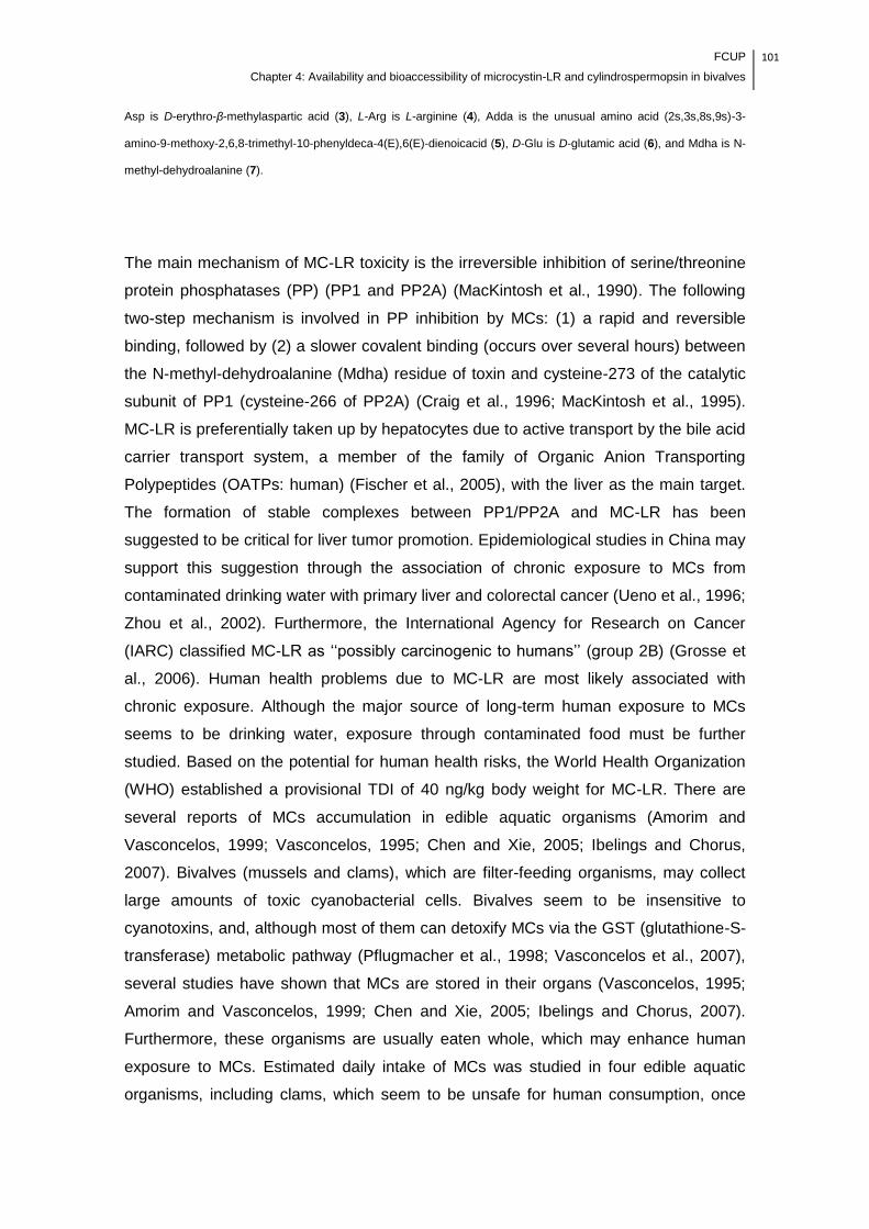
FCUP
Chapter 4: Availability and bioaccessibility of microcystin-LR and cylindrospermopsin in bivalves
101
Asp is D-erythro-β-methylaspartic acid (3), L-Arg is L-arginine (4), Adda is the unusual amino acid (2s,3s,8s,9s)-3-
amino-9-methoxy-2,6,8-trimethyl-10-phenyldeca-4(E),6(E)-dienoicacid (5), D-Glu is D-glutamic acid (6), and Mdha is N-
methyl-dehydroalanine (7).
The main mechanism of MC-LR toxicity is the irreversible inhibition of serine/threonine
protein phosphatases (PP) (PP1 and PP2A) (MacKintosh et al., 1990). The following
two-step mechanism is involved in PP inhibition by MCs: (1) a rapid and reversible
binding, followed by (2) a slower covalent binding (occurs over several hours) between
the N-methyl-dehydroalanine (Mdha) residue of toxin and cysteine-273 of the catalytic
subunit of PP1 (cysteine-266 of PP2A) (Craig et al., 1996; MacKintosh et al., 1995).
MC-LR is preferentially taken up by hepatocytes due to active transport by the bile acid
carrier transport system, a member of the family of Organic Anion Transporting
Polypeptides (OATPs: human) (Fischer et al., 2005), with the liver as the main target.
The formation of stable complexes between PP1/PP2A and MC-LR has been
suggested to be critical for liver tumor promotion. Epidemiological studies in China may
support this suggestion through the association of chronic exposure to MCs from
contaminated drinking water with primary liver and colorectal cancer (Ueno et al., 1996;
Zhou et al., 2002). Furthermore, the International Agency for Research on Cancer
(IARC) classified MC-LR as ‗‗possibly carcinogenic to humans‘‘ (group 2B) (Grosse et
al., 2006). Human health problems due to MC-LR are most likely associated with
chronic exposure. Although the major source of long-term human exposure to MCs
seems to be drinking water, exposure through contaminated food must be further
studied. Based on the potential for human health risks, the World Health Organization
(WHO) established a provisional TDI of 40 ng/kg body weight for MC-LR. There are
several reports of MCs accumulation in edible aquatic organisms (Amorim and
Vasconcelos, 1999; Vasconcelos, 1995; Chen and Xie, 2005; Ibelings and Chorus,
2007). Bivalves (mussels and clams), which are filter-feeding organisms, may collect
large amounts of toxic cyanobacterial cells. Bivalves seem to be insensitive to
cyanotoxins, and, although most of them can detoxify MCs via the GST (glutathione-S-
transferase) metabolic pathway (Pflugmacher et al., 1998; Vasconcelos et al., 2007),
several studies have shown that MCs are stored in their organs (Vasconcelos, 1995;
Amorim and Vasconcelos, 1999; Chen and Xie, 2005; Ibelings and Chorus, 2007).
Furthermore, these organisms are usually eaten whole, which may enhance human
exposure to MCs. Estimated daily intake of MCs was studied in four edible aquatic
organisms, including clams, which seem to be unsafe for human consumption, once

102 FCUP
Chapter 4: Availability and bioaccessibility of microcystin-LR and cylindrospermopsin in bivalves
TDI proposed by the WHO was exceeded several times (Chen and Xie, 2005).
Currently, most of the analytical methods used for MC-LR extraction from food matrices
use organic solvents such as methanol (MeOH), which do not enable complete MC-LR
(dissolved, non- and covalently bound) extraction (Williams et al., 1997). MeOH only
retrieves dissolved MCs or those in non-covalent bonds (free), while covalently bound
MCs are supposedly not available when food is consumed (Ibelings and Chorus, 2007;
Smith et al., 2010). Therefore, it is accepted that only free MCs are relevant to human
exposure estimation and risk assessment by contaminated food consumption.
Nevertheless, the estimation of exposure to MCs as food contaminants has been
based on the direct comparison of the concentration determined from studied
organisms (raw food) with the TDI value (Chen and Xie, 2005; Ibelings and Chorus,
2007), assuming that the available concentration of MCs in raw and in ready-to-eat
food products is similar. The risk assessment of human exposure to MCs through food
must include detailed knowledge of the variation of the free MC content in the different
steps of food storage (e.g., refrigeration, freezing) and processing (e.g., boiling, frying,
microwaving), because food is generally consumed after such processing. Data on the
effects of storage and processing practices on MC availability in food have been
reported (Morais et al., 2008; Zhang et al., 2010; Guzmán-Guillén et al., 2011). For
instance, Zhang et al. (2010) found that the mean concentration of MCs in bighead
carp muscle was significantly increased after boiling. These findings suggest that the
MC concentration in some contaminated food-stuffs may have been underestimated.
Furthermore, once ingested, the food is subjected to the physical and chemical
conditions of the stomach and small intestine, which may change the MC bioavail-
ability. Bioaccessibility is one of the main factors limiting bioavailability. Bioaccessibility
is defined as the fraction of the contaminant that is released from the food matrix by the
action of digestive enzymes and is then available for absorption by the intestinal
mucosa (Cabañero et al., 2004; Versantvoort et al., 2005). Bioaccessibility has been
studied for several food chemical contaminants, such as mycotoxins (Versantvoort et
al., 2005), mercury (Cabañero et al., 2004) and polychlorinated biphenyls (Xing et al.,
2008). To our knowledge, there are no studies that estimate MC-LR bioaccessibility
after in vitro proteolytic digestion. This study aims (1) to assess changes in the MC-LR
concentration after common practices of food storage and processing as well as (2) the
determination of MC-LR bioaccessibility after proteolytic digestion to create a more
suitable estimation of human exposure to MC-LR through consumption of
contaminated food.
FCUP
Chapter 4: Availability and bioaccessibility of microcystin-LR and cylindrospermopsin in bivalves
103
2. Material and methods
2.1. Reagents and chemicals
The mammalian enzymes pepsin (P7000), trypsin (T0303) and chymotrypsin (C4129)
were purchased from Sigma-Aldrich (Spain). Aqueous solutions of hydrochloric acid
(HCl) (37%) (Sigma, USA), phosphate buffer and sodium bicarbonate (NaHCO3)
(Sigma, USA) were prepared with ultrapure water supplied by a Millipore water
purification system (0.0054 µS/cm) (MilliQ water). Acetic acid was purchased from
Sigma (USA). The MeOH used for MC-LR extraction was analytical grade (Fisher
Scientific, UK). All solvents used in LC-MS analysis were high-purity chromatography
grade (LiChrosolv, Merck). Reagents used in the Z8 medium were analytical grade,
and formic acid was LC-MS grade (Fisher Scientific, USA). MC-LR was used as the
reference standard (lot nº SZBB069X, 95% purity, Sigma-Aldrich).
2.2. Biological material – cyanobacterial culture and clams
The exposure experiment was carried out with MC-LR-producing cells of Microcystis
aeruginosa (LEGE 91094). Cyanobacteria were cultured to the exponential phase in Z8
medium (Kotai, 1972) (6 L flasks) under fluorescent light (light/dark cycle of 14/10h)
and a temperature of 25 ± 1°C. M. aeruginosa LEGE 91094, produces MC-LR (95%)
and low amounts of MC-LA and [D-Asp3]-MC-LR (Vasconcelos, 1995). Specimens of
Asian clams (Corbicula fluminea) (Müller, 1774), ranging from 25 to 30 mm in size,
were collected in the estuary of River Minho (Valença, North Portugal). No MCs were
detected in water where C. fluminea was collected (data not shown). The organisms
were acclimated for one month prior to the experiment in 40 L aquaria with
dechlorinated tap water. During this period, the organisms were fed twice a week with
Chlorella vulgaris (1x10⁵ cell/mL). The water was renewed weekly.
2.3. Exposure and experiment
2.3.1. Clam intoxication
Clams were intoxicated with whole cyanobacterial cells at the ecologically relevant
104 FCUP
Chapter 4: Availability and bioaccessibility of microcystin-LR and cylindrospermopsin in bivalves
concentration of 1x10⁵ cell/mL (≈1.45 μg/L of MC-LR) for four days in static conditions.
During the exposure experiment, the physical and chemical conditions of the water
were as follows: temperature 16.5 ± 0.8°C, dissolved oxygen 9.6 ± 0.3 mg/L and pH
8.6 ± 0.1.
2.3.2. Simulation of thermal food storage and processing
After four days of exposure, clams were removed from shells and weighed. For each
treatment, 10 g of clams, (wet weight (ww)), were submitted to the common practices
of food storage and processing shown in Table 1.
Table 1. Detailed information of the food storage and processing conditions applied in experiment.
Conditions Storage process Cooking process
25°C 4°C - 20°C Microwave (550W) 100°C
Time 12h; 24h; 48h 24h; 48h; 72h 48h; 1 week; 1 month 0.5; 1; 3; 5 min 5; 15; 30 min
The experimental control group was not stored or processed and was immediately
analyzed after exposure. Data from the control group was used for comparison
(graphical and statistical analysis) with data from the storage and processing conditions
applied. The boiling time began after the water reached to 100°C. The ratio of water:
clams in the boiling method was 1 mL of MilliQ water: 1 g of clams. Clams and the
water in which they were boiled were analyzed altogether. Treatments were performed
in triplicate. A negative control with non-intoxicated clams (organisms fed with green
algae C. vulgaris at the same concentration) was also assessed.
2.3.3. In vitro proteolytic digestion
Based on the assumption that gastrointestinal enzymes, which digest carbohydrates
and lipids, have a negligible effect on the level of MC mobilization from a food matrix,
only proteolytic digestion was assessed. The in vitro model used to simulate proteolytic
mammalian digestion focused on two stages, physical processing in the mouth and
chemical processing in the stomach and small intestine (duodenum) (Fig. 2).
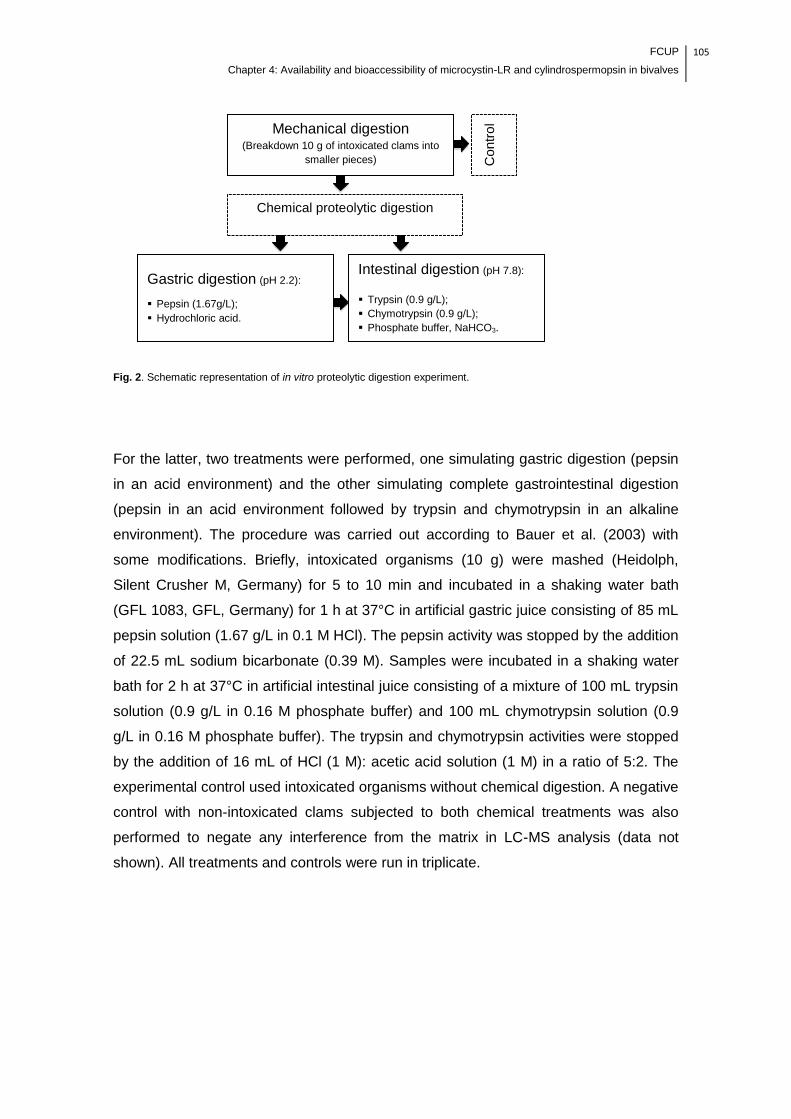
FCUP
Chapter 4: Availability and bioaccessibility of microcystin-LR and cylindrospermopsin in bivalves
105
Fig. 2. Schematic representation of in vitro proteolytic digestion experiment.
For the latter, two treatments were performed, one simulating gastric digestion (pepsin
in an acid environment) and the other simulating complete gastrointestinal digestion
(pepsin in an acid environment followed by trypsin and chymotrypsin in an alkaline
environment). The procedure was carried out according to Bauer et al. (2003) with
some modifications. Briefly, intoxicated organisms (10 g) were mashed (Heidolph,
Silent Crusher M, Germany) for 5 to 10 min and incubated in a shaking water bath
(GFL 1083, GFL, Germany) for 1 h at 37°C in artificial gastric juice consisting of 85 mL
pepsin solution (1.67 g/L in 0.1 M HCl). The pepsin activity was stopped by the addition
of 22.5 mL sodium bicarbonate (0.39 M). Samples were incubated in a shaking water
bath for 2 h at 37°C in artificial intestinal juice consisting of a mixture of 100 mL trypsin
solution (0.9 g/L in 0.16 M phosphate buffer) and 100 mL chymotrypsin solution (0.9
g/L in 0.16 M phosphate buffer). The trypsin and chymotrypsin activities were stopped
by the addition of 16 mL of HCl (1 M): acetic acid solution (1 M) in a ratio of 5:2. The
experimental control used intoxicated organisms without chemical digestion. A negative
control with non-intoxicated clams subjected to both chemical treatments was also
performed to negate any interference from the matrix in LC-MS analysis (data not
shown). All treatments and controls were run in triplicate.
Mechanical digestion (Breakdown 10 g of intoxicated clams into
smaller pieces)
Chemical proteolytic digestion
Intestinal digestion (pH 7.8):
Trypsin (0.9 g/L);
Chymotrypsin (0.9 g/L);
Phosphate buffer, NaHCO3.
Gastric digestion (pH 2.2):
Pepsin (1.67g/L);
Hydrochloric acid.
Contr
ol
106 FCUP
Chapter 4: Availability and bioaccessibility of microcystin-LR and cylindrospermopsin in bivalves
2.4. MC-LR determination
2.4.1. MC-LR extraction and clean-up
To avoid interference in the final concentration of the toxin through sample
freezing/thawing, free MC-LR was immediately extracted after each treatment,
according to the method of Amorim and Vasconcelos (1999), with some modifications.
Briefly, all samples were extracted in 50% MeOH at room temperature, then submitted
to ultrasonication (50 MHz) (Vibra-Cell 50, Sonics & Materials Inc. Danbury, CT, USA)
in an ice bath for 10 min and then centrifuged (Thermo Scientific - LegendTM T/RT
QUIKsetTM, Germany) (20 min, 4°C, 4495 g). The pellet was re-extracted with fresh
50% MeOH overnight. Samples were centrifuged again, and both supernatants were
pooled together. Extracts from samples were then concentrated and cleaned in SPE
(solid-phase extraction) cartridges (10g/60mL C-18-E, Strata® Phenomenex, USA),
which were preconditioned with 100% MeOH, followed by MilliQwater and 20% MeOH.
After loading the sample, the SPE cartridge was washed with 20% MeOH, and target
compounds were eluted in 80% MeOH. The supernatant was evaporated to dryness
under vacuum in a rotary evaporator (RotoquímicaBüch, Switzerland), and the residue
was resuspended in 1 mL 50% MeOH. Samples were frozen at -80°C until later LC-MS
analysis.
2.4.2. LC-MS analysis
The LC-MS system was a LCQ Fleet ion trap MSn (ThermoScientific, USA) with an
electrospray (ESI) interface, including a Surveyor LC pump, a Surveyor autosampler
and a Surveyor photoelectric diode array (PDA) detector. Separation was achieved on
a C18 Hypersil Gold column (100×4.6mm I.D., 5μm, ThermoScientific, USA) kept at
25°C with a flow rate of 0.8 mL/min. The injected volume was 25 µL. A gradient elution
was used with mobile phase A, MeOH, and B, water, both acidified with 0.1% formic
acid. Mobile phase A was linearly increased from 55 to 90% in 12 min, then increased
to 100% in 0.5 min and held for 2.5 min, and finally brought back to 55% and held for
10 min until the next injection. The retention time of MC-LR was 5.8 min. The mass
spectrometer was operated in a multiple-reaction monitoring mode (MRM) with a
collision energy of 35 eV. The capillary voltage and tube lens were maintained at 22
and 120 V, respectively. Nitrogen was used as the sheath and auxiliary gas. Helium
was used as the collision gas in the ion trap. The sheath gas flow rate was set at 80

FCUP
Chapter 4: Availability and bioaccessibility of microcystin-LR and cylindrospermopsin in bivalves
107
(arb units), and the capillary temperature was held at 350°C. Samples were analyzed
using the mass-to-charge ratio (m/z) transition of 995>599 at 23 V collision energy. The
MC-LR transition was monitored over one microscan.
2.5. Statistical analysis
Data from the experiments were analyzed using sample means comparison by one-
way analysis of variance (ANOVA) followed by Tukey‘s post-hoc test (SPSS® version
18.0 for Windows). Statistical analysis was performed between three individual groups:
1) samples of the control group and samples of the stored clams; 2) samples of the
control group and samples of the cooked clams; 3) samples of the control group and
samples subjected to gastric and gastrointestinal digestion.
The significance level was set at P<0.05. Results are expressed in ng/g (ww) as the
mean ± standard deviation (SD) for three replicates of each treatment. Bioaccessibility
(%) after in vitro proteolytic digestion was calculated as follows: [(MC-LR extracted
from the fraction after proteolytic digestion) ÷ (MC-LR extracted from the food matrix
before proteolytic digestion)] × 100.
3. Results and Discussion
The ecologically relevant concentration of toxic M. aeruginosa used in the experiment
did not result in mortality in C. fluminea. The Asian clam C. fluminea is an important
invasive species in aquatic ecosystems, because it is supposedly a substantial food
source for higher trophic levels and therefore a vehicle of MC-LR. The concentration of
free MC-LR quantified in C. fluminea (whole organism) after four days of exposure to
MC-LR-producing M. aeruginosa (1x10⁵ cell/mL) was between 26.17 and 100.33 ng/g.

108 FCUP
Chapter 4: Availability and bioaccessibility of microcystin-LR and cylindrospermopsin in bivalves
3.1. Effects of storage processes on the free MC-LR concentration
Understanding the effects of thermal storage on the available concentration of MC-LR
in clams is of particular relevance because this practice is extensively applied to extend
the shelf life and maintain the quality of the shellfish. Fig. 3 shows the changes in the
free MC-LR concentration after clams are stored under unrefrigerated (25°C),
refrigerated (4°C) and freezing (–20°C) conditions.
Fig. 3. The MC-LR concentration (ng/g) in intoxicated C. fluminea after different thermal storage conditions. Values are
expressed as the mean ± SD (n=3). Different letters (a, b, c, d, e, and f) indicate significant differences (P<0.05).
Columns that share the same letter are not significantly different.
The concentration of the free MC-LR in clams stored at unrefrigerated and refrigerated
conditions decreased over the storage time and was significantly lower after 48 h at
25°C (69.9%, P<0.05) and after 48 h and 72 h at 4°C (42.8% and 51.1%, respectively,
P<0.05), than the control group. Clams stored frozen (-20°C), had a significantly higher
concentration of free MC-LR from 48 h to one week and one month (58.2% and 69.9%,
respectively, P<0.05). The pattern of decrease in the free MC-LR concentration in
clams over time in both the unrefrigerated and refrigerated storage conditions can be
related to the state of deterioration of fresh clams. Shellfish is extremely perishable,
needing to be chilled immediately after removal from shells to delay autolytic and
microbial spoilage. It is likely that, as the storage time increases, cell breakage by
aab
ac
d
ac
bde deabe
f
f
0
20
40
60
80
100
12h 24h 48h 24h 48h 72h 48h 1 week 1 month
Control 25 °C 4 °C -20 °C
MC
-LR
co
nc
en
tra
tio
n n
g/g
(w
w)

FCUP
Chapter 4: Availability and bioaccessibility of microcystin-LR and cylindrospermopsin in bivalves
109
autolytic reactions may have occurred, allowing the interaction of MC-LR and internal
cellular constituents, including thiols. The conjugation of MC-LR to glutathione,
catalyzed by GSTs, is considered the major detoxification pathway of MCs
(Pflugmacher et al., 1998 Vasconcelos et al., 2007), and this binding makes the MC-LR
undetectable by MeOH extraction. Temperature variation (storage at 25ºC or 4ºC) can
alter the affinity conformation for glutathione binding and especially GST activity (the
physiological temperature for fish enzymes is 28ºC) (Pflugmacher et al., 1998), which
may be related to the lowest concentration of free MC-LR being detected in clams
stored at 25ºC for 48h. However, to confirm this hypothesis, further studies should be
performed in an attempt to identify and quantify the metabolite corresponding to the
glutathione conjugate of MC-LR (Ion m/z 1302.8 [M + H]+) (Pflugmacher et al., 1998).
Furthermore, the hypothesis that some MC-LR could have been removed by bacterial
action of natural flora and contaminant microorganisms should not be discarded.
Although the microbial flora of mollusks may vary due to many factors, namely the
water quality, some genera of bacteria have been recognized as ubiquitous, such as
Pseudomonas spp. and lactic acid bacteria (Manousaridis et al., 2005), whose ability to
degrade and to remove MC-LR from aqueous solutions has been shown (Takenaka
and Watanabe 1997; Nybom et al., 2007; Surono et al., 2008).
Storage by freezing is more effective in spoilage control, so autolytic reactions and
microbial growth are unlikely to occur. Furthermore, freezing may cause considerable
protein denaturation with corresponding changes in protein structure, which increase
over time. Thus, it may also be hypothesized that MC-LR is more efficiently extractable
and available after freezing storage for one week and one month. To our knowledge,
only Morais et al. (2008) has studied the effects of unrefrigerated, refrigerated and
freezing storage on changes in the available MCs in an edible organism (Mytilus
galloprovincialis). The authors found a significantly lower (P<0.05) concentration of
available MCs than in control group in all conditions, although, in contrast to our
results, there were no significant differences between different periods of storage.
In any case, as clams were exposed to whole cyanobacterial cells, is likely that some
cells have been accumulated in its digestive tract (Amorim and Vasconcelos, 1999;
Vasconcelos, 1995). It is important to point out that if the clams were submitted to
depuration after exposure, free MC-LR concentration after storage could be quite
different due to the emptiness of the digestive tract.
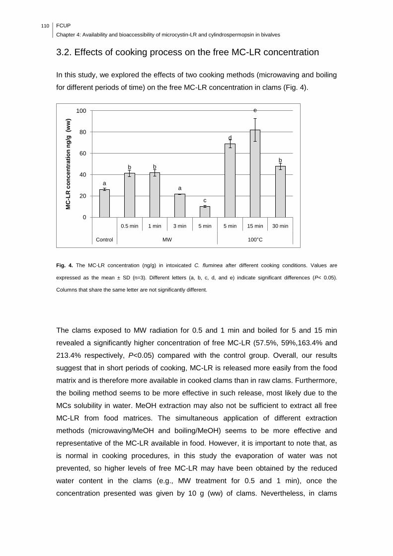
110 FCUP
Chapter 4: Availability and bioaccessibility of microcystin-LR and cylindrospermopsin in bivalves
3.2. Effects of cooking process on the free MC-LR concentration
In this study, we explored the effects of two cooking methods (microwaving and boiling
for different periods of time) on the free MC-LR concentration in clams (Fig. 4).
Fig. 4. The MC-LR concentration (ng/g) in intoxicated C. fluminea after different cooking conditions. Values are
expressed as the mean ± SD (n=3). Different letters (a, b, c, d, and e) indicate significant differences (P< 0.05).
Columns that share the same letter are not significantly different.
The clams exposed to MW radiation for 0.5 and 1 min and boiled for 5 and 15 min
revealed a significantly higher concentration of free MC-LR (57.5%, 59%,163.4% and
213.4% respectively, P<0.05) compared with the control group. Overall, our results
suggest that in short periods of cooking, MC-LR is released more easily from the food
matrix and is therefore more available in cooked clams than in raw clams. Furthermore,
the boiling method seems to be more effective in such release, most likely due to the
MCs solubility in water. MeOH extraction may also not be sufficient to extract all free
MC-LR from food matrices. The simultaneous application of different extraction
methods (microwaving/MeOH and boiling/MeOH) seems to be more effective and
representative of the MC-LR available in food. However, it is important to note that, as
is normal in cooking procedures, in this study the evaporation of water was not
prevented, so higher levels of free MC-LR may have been obtained by the reduced
water content in the clams (e.g., MW treatment for 0.5 and 1 min), once the
concentration presented was given by 10 g (ww) of clams. Nevertheless, in clams
a
b b
a
c
d
e
b
0
20
40
60
80
100
0.5 min 1 min 3 min 5 min 5 min 15 min 30 min
Control MW 100°C
MC
-LR
co
nc
en
tra
tio
n n
g/g
(w
w)
FCUP
Chapter 4: Availability and bioaccessibility of microcystin-LR and cylindrospermopsin in bivalves
111
exposed to longer periods of MW radiation (3 and 5 min), where major water
evaporation occurred, there was a decrease in the free MC-LR concentration (16.8%
and 61.3%, respectively), compared with the control group. Similarly, in clams boiled
for 30 min, although the free MC-LR concentration had been significantly higher than
uncooked clams (82.5%, P<0.05), it was significantly reduced in comparison to clams
boiled for 5 and 15 min. These results also suggest that the cooking time is critical for
the stability of MC-LR. So far, few studies have tested changes in the MC-LR
concentration in the matrices of raw and cooked edible aquatic organisms. Morais et al.
(2008) reported a significant decrease in the MCs concentration of toxic M. aeruginosa-
fed mussels (M. galloprovincialis) after MW radiation treatment for 1 and 5 min.
Similarly, Guzmán-Guillén et al. (2011) found a decrease of 36% in the MC-LR
concentration of fish muscle (Tilapia, Oreochromis niloticus) spiked with a solution of
pure MC-LR, MC-YR and MC-RR, after treatment with MW radiation by 5 min, although
after 1 min, the MC-LR concentration remained stable relative to the control group. Our
results are in line with previous studies in which a significant decrease of the free MC-
LR concentration occurred after 5 min of MW treatment. MW radiation for 5 min may
allow that the temperature of the clams to cause the evaporation of almost all the
water, leading to a high dehydration state and consequent matrix modification by
protein degradation. This process might have made MC-LR inaccessible for extraction
or promoted its degradation. However, the reason for the decrease in the free MC-LR
concentration in food matrices after longer periods of cooking is not fully understood
and should be further studied, because previous studies have shown that the stability
of pure MC-LR was not affected by extraction using a domestic microwave oven (650
W) for 9 min (Metcalf and Codd, 2000), or by boiling several hours (Van Apeldoorn et
al., 2007). Unfortunately, although MW radiation for 5 min seems to lead to lower levels
of MC-LR concentration in clams, the dehydration and the physical damage caused by
this treatment renders its organoleptic properties unappealing for human consumption.
Regarding to boiling treatments, in our study, we analyzed boiled clams together with
the water in which they were boiled, so the free MC-LR concentration determined is the
combination of the contents of both matrices. The results of previous studies in this
field are not in agreement. Zhang et al. (2010) generated intraperitoneal intoxication of
MC-RR and MC-LR in bighead carp (Aristichthys nobilis) and found a significant
increase in MCs concentration in boiled muscle (5 min) compared with lyophilized
muscle (P<0.01). Furthermore, Zhang et al. (2010) found a substantial amount of MCs
in the water in which the muscle fish was boiled. However, Morais et al. (2008) boiled
M. galloprovincialis fed with toxic M. aeruginosa for 5 and 30 min and did not find any
112 FCUP
Chapter 4: Availability and bioaccessibility of microcystin-LR and cylindrospermopsin in bivalves
differences between the control and boiling treatments. Guzmán-Guillén et al. (2011)
boiled Tilapia muscle spiked with a solution of MC-LR, MC-YR and MC-RR
continuously for 2 min and found a decrease in the MC-LR concentration (59.3%),
although the boiling water also had a substantial amount of MCs. In fact, MC-LR
transference to boiling water may constitute a risk to human health if the water and
animal tissue are consumed. However, this process could be applied to reduce the free
MC-LR concentration in food if the boiling water is discarded. Ibelings and Chorus
(2007) suggest that in industrially processed ―seafood‖, the removal of parts in which
the cyanotoxins can accumulate (e.g., viscera and liver of fishes or guts and
hepatopancreas of macrofauna) prior to processing could be effective in avoiding the
hazard of MCs. However, this is impracticable in mussels. Considering the HACCP
(Hazard Analysis Critical Control Points) approach set by the Codex Alimentarius
Commission, our results present a new insight to set critical limits for food
hypothetically contaminated with MC-LR, in which the boiling process is a critical
control point. Control of the cooking time and the rejection of cooking water may be
suitable procedures to reduce MC-LR to an acceptable level in food. Our results also
suggest that direct comparison between the MC-LR concentration determined in raw
edible aquatic organisms and the TDI set for MC-LR may be erroneous and may
underestimate the risk of exposure by contaminated food consumption.
3.3. Bioaccessibility of MC-LR after proteolytic digestion
Because the ingestion of contaminated food is one of the most important routes for
chronic exposure to MC-LR, it is of major importance to estimate the true exposure
after food proteolytic digestion. The changes in the free MC-LR concentration in
intoxicated clams after simulating gastric and gastrointestinal proteolytic digestion are
shown in Fig. 5.

FCUP
Chapter 4: Availability and bioaccessibility of microcystin-LR and cylindrospermopsin in bivalves
113
Fig. 5. The MC-LR concentration (ng/g) after in vitro proteolytic gastric (G) and gastrointestinal (GI) digestion. The
column filled with black points represents the bioaccessibility. Values are expressed as the mean ± SD (n=3). Different
letters (a, b, and c) indicate significant differences (P<0.05). Columns that share the same letter are not significantly
different.
The simulation of gastric digestion resulted in a significant increase (47.3%, P<0.05) of
the free MC-LR concentration, suggesting that pepsin and the acidic conditions found
in the stomach may allow easier release of MC-LR from the food matrix. The
denaturation of proteins with low pH may increase their digestibility, enhancing MC-LR
extraction. The results of previous studies on the degradation of MCs by digestive
enzymes are not in agreement. Moreno et al. (2004) observed that MC-LR was
degraded either by the action of pepsin or the acidic environment found in the stomach,
while Smith et al. (2010) found no digestion of MCs under gastric conditions. In the
present study, even if some degradation occurred at this stage, the overall result was
an increase in free MC-LR concentration, indicating that the major impact of gastric
conditions was the enhancement of MC-LR availability. When gastric digestion was
followed by intestinal digestion, the concentration of free toxin significantly decreases
(P<0.05) to levels below the control group, which seems to suggest the degradation of
free MC-LR by the pancreatic enzymes trypsin and chymotrypsin. It is likely that, with
sequential digestion, a prolonged incubation of MC-LR with proteases could make it
more susceptible to hydrolysis; however, this issue should be further studied.
Controversial results have also been reported regarding the effect of pancreatic
proteases on MC-LR, such as resistance to degradation by pancreatin (an enzyme
mix), trypsin and chymotrypsin (Moreno et al., 2004; Smith et al., 2010) in opposition to
a
b
c
0
50
100
150
200
Control G GIMC
-LR
co
nc
en
tra
tio
n n
g/g
(w
w)
114 FCUP
Chapter 4: Availability and bioaccessibility of microcystin-LR and cylindrospermopsin in bivalves
significant digestion by trypsin and chymotrypsin (Kankaanpää et al., 2005). It should
be emphasized that in all these studies, MC was used in its free form in solution and
alternatively exposed to gastric or intestinal digestive conditions. The results from these
studies should be therefore considered carefully when evaluating the effect of digestion
on MC toxicity because they do not take into consideration the bonds between the toxin
and molecules (such as PP, glutathione) from food items or the effect of the previous
gastric digestion on the intestinal digestion of MCs. In the present study, MC-LR-
contaminated food was sequentially subjected to gastric and intestinal conditions to
simulate complete gastrointestinal digestion, therefore providing the first, much more
realistic, insight on the fate of MC-LR after entering the human digestive tract. The
sequential proteolytic digestion allowed the calculation of the bioaccessibility of MC-LR,
which was 83.1%. Bioaccessibility of less than 100% suggests that the true exposure
to the contaminant is lower than expected, i.e., when raw food is analyzed
(Versantvoort et al., 2005). It has been recognized that the toxicity of MCs by oral route
is approximately 100-fold lower than by the intraperitoneal route. According to our
findings, a decrease in MC-LR bioaccessibility and thus bioavailability after proteolytic
digestion may be an important factor. However, it is surely not the only process that
can decrease the toxicity. Indeed, several factors may contribute, including the
following: MC-LR uptake dependent on the bile acid transport system;
biotransformation; excretion; degradation by intestinal microflora; and potential
interaction with different nutrients, minerals and antioxidants in food. However, this may
be not considered enough to evidence for the assurance of food safety. As shown
previously, frozen and undercooked clams generally have a higher available
concentration of MC-LR. Furthermore, heat treatment usually increases the digestibility
of the proteins, so it is imperative to study the influence of different cooking treatments
on the bioaccessibility of MC-LR. Finally, because there may be specific compounds
(e.g., MC-LR bound to a small residue of peptide) derived from the digestion of the MC-
LR-PP, that have half the toxicity of the parent toxin (Smith et al., 2010), they should
also be considered in the evaluation of the total toxicity of MCs after the proteolytic
digestion of food.
4. Conclusions
In conclusion, this study provides an enhancement of knowledge on the MC-LR
concentration available in food after employing techniques commonly used for their
preservation and processing. This might be of particular interest in setting acceptable
FCUP
Chapter 4: Availability and bioaccessibility of microcystin-LR and cylindrospermopsin in bivalves
115
thresholds in the industry, because the TDI can be exceeded in undercooked food.
Treatments applied in this study could also be considered for the definition of critical
control limits, considering the HACCP approach as a promising tool for risk
management. Furthermore, the combination of these methods (freezing, boiling, and
microwaving) with MeOH extraction may provide a more accurate determination of MC-
LR concentration available in food. The bioaccessibility of MC-LR after proteolytic
digestion is significantly reduced likely due to degradation by the pancreatic enzymes,
trypsin and chymotrypsin. However, many other properties of proteolytic digestion
should be further studied. To generate a more representative determination of
bioaccessibility in ready-to-eat food products, future research should combine food
storage and processing methods with in vitro enzymatic digestion.
Conflict of Interest
The authors declare that there are no conflicts of interest.
Acknowledgments
Marisa Freitas thanks the Fundação para a Ciência e Tecnologia (FCT) for the Ph.D.
Grant SFRH/BD/85490/2012. This work was partially funded by the FCT project PesT-
C/MAR/LA0015/2011.
5. References
Amorim, A., Vasconcelos, V.M., 1999. Dynamics of microcystins in the mussel Mytilus
galloprovincialis. Toxicon. 37, 1041-1052.
Bauer, E., Williams, B.A., Voigt, C., Mosenthin, R., Verstegen, M.W.A., 2003. Impact of
mammalian enzyme pretreatment on the fermentability of carbohydrate-rich feedstuffs.
J. Sci. Food Agric. 83, 207–214.
Cabañero, A.I., Madrid, Y., Cámara C., 2004. Selenium and mercury bioaccessibility in
fish samples: an in vitro digestion method. Anal. Chim. Acta. 526, 51–61.
116 FCUP
Chapter 4: Availability and bioaccessibility of microcystin-LR and cylindrospermopsin in bivalves
Chen, J., Xie, P., 2005. Seasonal Dynamics of the Hepatotoxic Microcystins in Various
Organs of Four Freshwater Bivalves from the Large Eutrophic Lake Taihu of
Subtropical China and the Risk to Human Consumption. Environ Toxicol. 20, 572–584.
Craig, M., Luu, H.A., McCready, T.L., Williams, D., Andersen, R.J., Holmes, C.F.B.,
1996. Molecular mechanisms underlying the interaction of motuporin and microcystins
with type-1 and type-2A protein phosphatases. Biochem. Cell Biol. 74, 569–578.
Fischer, W.J., Altheimer, S., Cattori, V., Meier, P.J., Dietrich, D.R., Hagenbuch, B.,
2005. Organic anion transporting polypeptides expressed in liver and brain mediate
uptake of microcystin. Toxicol. Appl. Pharmacol. 203, 257–263.
Grosse, Y., Baan, R., Straif, K., Secretan, B., Ghissassi, F.El., Cogliano, V., 2006.
Carcinogenicity of nitrate, nitrite, and cyanobacterial peptide toxins. Lancet Oncol. 7,
628–629.
Guzmán-Guillén, R., Prieto, A.I., Moreno, I., Soria, M.E., Cameán, A.M., 2011. Effects
of thermal treatments during cooking, microwave oven and boiling, on the
unconjugated microcystin concentration in muscle of fish (Oreochromis niloticus). Food
Chem. Toxicol. 49, 2060–2067.
Ibelings, B.W., Chorus I., 2007. Accumulation of cyanobacterial toxins in freshwater
‗‗seafood‘‘ and its consequences for public health: A review. Environ. Pollut. 150, 177-
192.
Kankaanpää, H.T., Holliday, J., Schröder, H., Goddard, T.J., von Fister, R.,
Carmichael, W.W., 2005. Cyanobacteria and prawn farming in northern New South
Wales, Australia—a case study on cyanobacteria diversity and hepatotoxin
bioaccumulation. Toxicol. Appl. Pharmacol. 203, 243–256.
Kotai, J., 1972. Instruction for preparation of modified nutrient solution Z8 for algae.
Norwegian Institute for Water Research, Oslo, Norway, Report nr B-11/69, 1-5.
MacKintosh, C., Beattie, K.A., Klumpp, S., Cohen, P., Codd, G.A., 1990.
Cyanobacterial microcystin-LR is a potent and specific inhibitor of protein
phosphatases 1 and 2A from both mammals and higher plants. FEBS Lett. 264, 187–
192.
MacKintosh, R.W., Dalby, K.N., Campbell, D.G., Cohen, P.T.W., Cohen, P.,
MacKintosh, C., 1995. The cyanobacterial toxin microcystin binds covalently to
FCUP
Chapter 4: Availability and bioaccessibility of microcystin-LR and cylindrospermopsin in bivalves
117
cysteine-273 on protein phosphatase 1. FEBS Lett. 371, 236–240.
Manousaridis, G., Nerantzaki, A., Paleologos, E.K., Tsiotsias, A., Savvaidis, I.N.,
Kontominas, M.G., 2005. Effect of ozone on microbial, chemical and sensory attributes
of shucked mussels. Food Microbiol. 22, 1–9.
Metcalf, J.S., Codd, G.A., 2000. Microwave oven and boiling waterbath extraction of
hepatotoxins from cyanobacterial cells. FEMS Microbiol. Lett. 184, 241-246.
Morais, J., Augusto, M., Carvalho, A.P., Vale, M., Vasconcelos, V.M., 2008.
Cyanobacteria hepatotoxins, microcystins: bioavailability in contaminated mussels
exposed to different environmental conditions. Eur. Food Res. Technol. 227, 949–952.
Moreno, I.M., Maraver, J., Aguete, E.C., Leao, M., Gago-Martínez, A., Cameán, A.M.,
2004. Decomposition of Microcystin-LR, Microcystin-RR, and Microcystin-YR in water
samples submitted to in Vitro dissolution tests. J. Agric. Food Chem. 52, 5933−5938.
Nybom, S.M.K., Salminen, S.J., Meriluoto, J.A.O., 2007. Removal of microcystin-LR by
strains of metabolically active probiotic bacteria. FEMS Microbiol. Lett. 270, 27–33.
Pflugmacher, S., Wiegand, C., Oberemm, A., Beattie, K.A. Krause, E., Codd, G.A.
Steinberg, C.E.W., 1998. Identification of an enzymatically formed glutathione
conjugate of the cyanobacterial hepatotoxin microcystin-LR: the first step of
detoxication. Biochim. Biophys. Acta. 1425, 527-533.
Sivonen, K.,Jones, G., 1999. Cyanobacterial toxins, in: Chorus, I., Bartram, J. (Eds.),
Toxic cyanobacteria in water: A guide to their public health consequences, monitoring
and management. World Health Organization, Taylor and Francis, London and New
York, pp. 41–111.
Smith, J.L., Schulz, K.L., Zimba, P.V., Boyer, G.L., 2010. Possible mechanism for the
foodweb transfer of covalently bound microcystins. Ecotoxicol. Environ. Saf. 73(5),
757-761.
Surono, I.S., Collado, M.C., Salminen, S., Meriluoto, J., 2008. Effect of glucose and
incubation temperature on metabolically active Lactobacillus plantarum from dadih in
removing microcystin-LR. Food Chem. Toxicol. 46, 502–507.
Takenaka, S., Watanabe, M.F., 1997. Microcystin-LR degradation by Pseudomonas
aeruginosa alkaline protease. Chemosphere. 34 (4), 749-757.

118 FCUP
Chapter 4: Availability and bioaccessibility of microcystin-LR and cylindrospermopsin in bivalves
Ueno, Y., Nagata, S., Tsutsumi, T., Hasegawa, A., Watanabe, M.F., Park, H.D., Chen,
G-C., Chen, G., Yu S-Z., 1996. Detection of microcystins, a blue-green algal
hepatotoxin, in drinking water sampled in Haimen and Fusui, endemic areas of primary
liver cancer in China, by highly sensitive immunoassay. Carcinogenesis. 17, 1317-
1321.
Van Apeldoorn, M.E., Van Egmond, H.P., Speijers, G.J., Bakker, G.J., 2007. Toxins of
cyanobacteria. Mol. Nutr. Food Res. 51, 7–60.
Vasconcelos, V.M., Wiegand C., Pflugmacher S., 2007. Dynamics of glutathione-S-
transferases in Mytilus galloprovincialis exposed to toxic Microcystis aeruginosa cells,
extracts and pure toxins. Toxicon. 50, 740-745.
Vasconcelos, V.M., 1995. Uptake and depuration of the heptapeptide toxin microcystin-
LR in Mytilus galloprovincialis. Aquat. Toxicol. 32, 227–237.
Versantvoort, C.H.M., Oomen, A.G., Van de Kamp, E., Rompelberg, C.J.M., Sips,
A.J.A.M., 2005. Applicability of an in vitro digestion model in assessing the
bioaccessibility of mycotoxins from food. Food Chem. Toxicol. 43, 31–40.
Williams, D.E., Dawe, S.C., Kent, M.L., Andersen, R.J., Craig, M., Holmes, C.F.B.,
1997. Bioaccumulation and clearance of microcystins from salt water mussels, Mytilus
edulis, and in vivo evidence for covalently bound microcystins in mussel tissues.
Toxicon. 35, 1617–1625.
Xing, G.H., Yang, Y., Chan, J.K.Y., Tao, S., Wong, M.H., 2008. Bioaccessibility of
polychlorinated biphenyls in different foods using an in vitro digestion method. Environ.
Pollut. 156, 1218–1226.
Zhang, D., Xie, P., Chen, J., 2010. Effects of temperature on the stability of
microcystins in muscle of fish and its consequences for food safety. Bull. Environ.
Contam. Toxicol.84, 202–207.
Zhou, L., Yu, H., Chen, K., 2002. Relationship between microcystin in drinking water
and colorectal cancer. Biomed. Environ. Sci. 15, 166-171.

FCUP
Chapter 4: Availability and bioaccessibility of microcystin-LR and cylindrospermopsin in bivalves
119
Bioaccessibility and changes in the cylindrospermopsin
concentration in edible mussels with storage and processing
time
Marisa Freitas1,2,3, Joana Azevedo1, António Paulo Carvalho1,2, Vera Mendes4, Bruno
Manadas4, Alexandre Campos1, Vitor Vasconcelos1,2
1 CIIMAR/CIMAR - Interdisciplinary Centre of Marine and Environmental Research,
University of Porto, Rua dos Bragas 289, P 4050-123 Porto, Portugal.
2 Faculty of Sciences, Porto University, Rua do Campo Alegre, 4169-007 Porto,
Portugal.
3 Polytechnic Institute of Porto. Escola Superior de Tecnologia da Saúde do Porto.
CISA/Research Center in Environment and Health, Rua de Valente Perfeito, 322,
4400-330 Gaia, Portugal.
4 Center for Neuroscience and Cell Biology, University of Coimbra, Portugal.
Abstract
The cyanobacterial alkaloid cylindrospermopsin (CYN) is recognized as being of
increased concern due to the global expansion of its main producer,
Cylindrospermopsis raciborskii. Previous studies have shown that aquatic organisms,
especially bivalves, can accumulate high levels of CYN without lethal effect. Based on
the potential for human health risks, a provisional tolerable daily intake of 0.03 µg/kg
body weight has been recommended. However, the human exposure assessment has
been based on the CYN concentration in raw food items. Thus, this study aimed to
assess the changes in CYN concentration in edible mussels with storage and
processing time as well as CYN bioaccessibility. Mussels (Mytilus galloprovincialis) fed
CYN-producing C. raciborskii were subjected to storage and processing treatments and
were then analyzed by LC-MS/MS. Mussels stored frozen allowed a significantly higher
recovery of CYN (52.5% in 48 h and 57.7% in 1 week, p < 0.05). The cooking
treatments did not produce significant differences in the CYN concentration in the

120 FCUP
Chapter 4: Availability and bioaccessibility of microcystin-LR and cylindrospermopsin in bivalves
mussel matrices (flesh); however, CYN was found in the cooking water, suggesting that
heat processing can be used to reduce the availability of CYN in this food item. The in
vitro digestion with salivary, gastric and duodenal juices considerably decreased the
CYN availability in uncooked and steamed mussels. The digestion of purified CYN (100
ng/mL) confirmed its removal by salivary and gastrointestinal juices, highlighting the
importance of integrating the bioaccessibility of CYN into the human health risk
assessment.
Keywords: Bioaccessibility, cylindrospermopsin, food storage, food processing, in vitro
digestion, Mytilus galloprovincialis.
Abbreviations: ACN, acetonitrile; CYN, cylindrospermopsin; CYP450, cytochrome
P450; FA, formic acid; FAO, Food and Agriculture Organization of the United Nations;
GSH, glutathione; HACCP, Hazard Analysis Critical Control Points; LC-MS/MS, liquid
chromatography coupled to tandem mass spectrometry; LOD, limit of detection; LOQ,
limit of quantification; MRM, multiple reaction monitoring; SD, standard deviation; TDI,
tolerable daily intake; ww, wet weight.
1. Introduction
The occurrence and progressive proliferation of harmful cyanobacteria in freshwater,
estuarine and marine ecosystems have been recognized as a potential consequence of
eutrophication and climate change (O‘Neil et al., 2012). During the few last decades,
the expansion of Cylindrospermopsis raciborskii (a planktonic freshwater
cyanobacterium) from its tropical origin to temperate waterbodies has been reported
(Poniedziałek et al., 2012). The cyanotoxin cylindrospermopsin (CYN), an alkaloid
consisting of a hydroxymethyluracil moiety linked to a tricyclic guanidine (Ohtani et al.,
1992) (Fig. 1), was first isolated from a culture of Cylindrospermopsis raciborskii
(Ohtani et al., 1992).
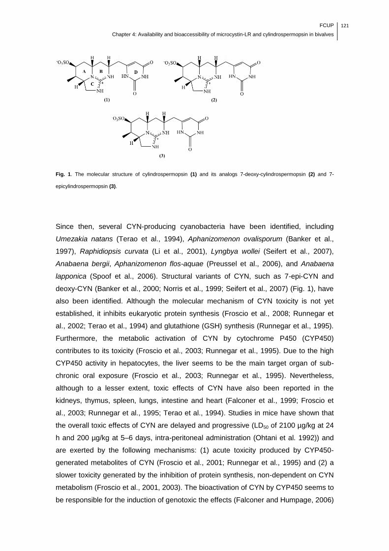
FCUP
Chapter 4: Availability and bioaccessibility of microcystin-LR and cylindrospermopsin in bivalves
121
Fig. 1. The molecular structure of cylindrospermopsin (1) and its analogs 7-deoxy-cylindrospermopsin (2) and 7-
epicylindrospermopsin (3).
Since then, several CYN-producing cyanobacteria have been identified, including
Umezakia natans (Terao et al., 1994), Aphanizomenon ovalisporum (Banker et al.,
1997), Raphidiopsis curvata (Li et al., 2001), Lyngbya wollei (Seifert et al., 2007),
Anabaena bergii, Aphanizomenon flos-aquae (Preussel et al., 2006), and Anabaena
lapponica (Spoof et al., 2006). Structural variants of CYN, such as 7-epi-CYN and
deoxy-CYN (Banker et al., 2000; Norris et al., 1999; Seifert et al., 2007) (Fig. 1), have
also been identified. Although the molecular mechanism of CYN toxicity is not yet
established, it inhibits eukaryotic protein synthesis (Froscio et al., 2008; Runnegar et
al., 2002; Terao et al., 1994) and glutathione (GSH) synthesis (Runnegar et al., 1995).
Furthermore, the metabolic activation of CYN by cytochrome P450 (CYP450)
contributes to its toxicity (Froscio et al., 2003; Runnegar et al., 1995). Due to the high
CYP450 activity in hepatocytes, the liver seems to be the main target organ of sub-
chronic oral exposure (Froscio et al., 2003; Runnegar et al., 1995). Nevertheless,
although to a lesser extent, toxic effects of CYN have also been reported in the
kidneys, thymus, spleen, lungs, intestine and heart (Falconer et al., 1999; Froscio et
al., 2003; Runnegar et al., 1995; Terao et al., 1994). Studies in mice have shown that
the overall toxic effects of CYN are delayed and progressive (LD50 of 2100 µg/kg at 24
h and 200 µg/kg at 5–6 days, intra-peritoneal administration (Ohtani et al. 1992)) and
are exerted by the following mechanisms: (1) acute toxicity produced by CYP450-
generated metabolites of CYN (Froscio et al., 2001; Runnegar et al., 1995) and (2) a
slower toxicity generated by the inhibition of protein synthesis, non-dependent on CYN
metabolism (Froscio et al., 2001, 2003). The bioactivation of CYN by CYP450 seems to
be responsible for the induction of genotoxic the effects (Falconer and Humpage, 2006)
122 FCUP
Chapter 4: Availability and bioaccessibility of microcystin-LR and cylindrospermopsin in bivalves
because CYP450 inhibitors appear to prevent DNA damage in in vitro assays
(Humpage et al., 2005; Runnegar et al., 1995). A preliminary study suggests that
tumors are generated in mice by oral exposure to CYN (Falconer and Humpage, 2001);
however, it remains to be elucidated whether the genotoxic effects might increase the
risk of carcinogenicity in vivo. Despite the lack of consistent epidemiological data,
consumption of contaminated water and food is the major source of chronic human
exposure to CYN. The first human intoxication associated with CYN was in Palm Island
(Queensland, Australia in 1979), where 149 people supplied with drinking water from a
reservoir with CYN-producing C. raciborskii suffered a hepatoenteritis-like illness
(Griffiths & Saker, 2003). Since then, public health concerns over CYN exposure have
increased, and the risks associated with this toxin are still under investigation. CYN is
highly water soluble due to its zwitterionic character, and contrary to the majority of
cyanotoxins, as much as 90% of the total CYN is outside of the cells dissolved in the
surrounding water (Chiswell et al., 1999; Rücker et al., 2007). Furthermore, CYN can
persist in the water because its photodegradation is very low under natural conditions
(Wörmer et al., 2010). Thus, the high levels and persistence of CYN in water can
potentiate its accumulation in a wide range of aquatic organisms. Previous studies
have shown that aquatic organisms, especially bivalves, can accumulate high levels of
CYN without lethal effect (Gutiérrez-Praena et al., 2013; Ibelings and Chorus, 2007;
Kinnear, 2010; Saker et al., 2004). Based on the potential for human health risks, a
provisional tolerable daily intake (TDI) of 0.03 µg/kg of body weight has been proposed
by Humpage and Falconer (2003). The human exposure assessment due to the
consumption of CYN-contaminated food has been based on the direct comparison
between the CYN concentration in raw edible organisms and TDI (Ibelings and Chorus,
2007). However, edible organisms are usually stored and processed before
consumption, and these practices may change the concentration of CYN available in
the food. Recent studies with microcystin-LR, the most studied cyanotoxin, have shown
that its recovery from food matrices is changed after applying common food storage
and cooking practices, as well as proteolytic digestion (Freitas et al., 2014; Guzmán-
Guillén et al., 2011; Morais et al., 2008; Zhang et al., 2010). According to our
knowledge, and as was recently reviewed by Gutiérrez-Praena et al. (2013) there are
no studies regarding the influence of different food storage and cooking procedures on
the CYN concentration in edible organisms. Furthermore, from the health risk point of
view, the oral bioavailability of CYN is an important parameter to consider because it
can vary from the CYN contained in food matrices. The bioavailability of a contaminant
depends on its (1) bioaccessibility, (2) transport across the intestinal epithelium into the
portal vein and (3) metabolism, mainly in the liver (Versantvoort et al., 2005). The
FCUP
Chapter 4: Availability and bioaccessibility of microcystin-LR and cylindrospermopsin in bivalves
123
bioaccessibility corresponds to the fraction of a contaminant that is released from the
food matrix by digestive juices and can be seen as an appropriate indicator of the
maximal oral bioavailability. Several in vitro digestion models can be used to determine
bioaccessibility by simulating, in a simplified manner, the digestion processes in the
mouth, stomach and small intestine (Guerra et al., 2012).
CYN is highly water-soluble and stable to extreme temperatures and pH (Chiswell et
al., 1999); thus, knowledge of the influence of common food storage and processing
practices, as well as human digestion, is required to achieve a more accurate human
health risk assessment. Therefore, the aim of this study was to assess the changes in
the CYN concentration in edible mussels with storage and processing time and to
assess the bioaccessibility of CYN in raw and processed (steamed) edible mussels.
2. Material and methods
2.1. Reagents and chemicals
The mammalian enzymes α-amylase (A-3176), pepsin (P7000), trypsin (T0303),
chymotrypsin (C4129), pancreatin (P1750) and bile salts (B-8756) were purchased
from Sigma-Aldrich (St. Louis, MO, USA). Potassium chloride (KCl, 99.5-100%),
sodium chloride (NaCl, ≥98%), sodium phosphate monobasic (NaH2PO4, 98-102%),
sodium hydrogen carbonate (NaHCO3, 99%), ammonium chloride (NH4Cl, A-0171),
calcium chloride dihydrate (CaCl22H2O, >99%), hydrochloric acid (HCl, 37%, g/g),
glucosamine hydrochloride (>99%), bovine serum albumin (BSA, 98%), magnesium
chloride (MgCl2, 99%), sodium sulfate (Na2SO4, ≥99%), potassium phosphate
monobasic (KH2PO4, ≥99%), glucuronic acid (97.5-102.5%), glucose (99.5%), uric acid
(99%) and urea were of analytical grade and were purchased from Sigma-Aldrich (St.
Louis, MO, USA). Analytical grade potassium thiocyanate (KSCN, 99%) was
purchased from Merck (Germany). All solutions were prepared with ultrapure water
supplied by a Millipore water purification system (0.0054 µS/cm) (MilliQ water). The
acetonitrile (ACN) and water used for CYN extraction were analytical grade, and all
solvents used in liquid chromatography coupled to tandem mass spectrometry (LC-
MS/MS) analysis were high-purity LC-MS grade (HiPerSolv CHROMANORM® for LC-
MS, VWR). The reagents used in the Z8 medium were analytical grade. Analytical
grade purity CYN was used as the reference standard (Lot# 20050531, CAS No.:

124 FCUP
Chapter 4: Availability and bioaccessibility of microcystin-LR and cylindrospermopsin in bivalves
143545-90-8, National Research Council, Canada).
2.2. Biological material – cyanobacterial culture and mussels
The exposure experiment was conducted with CYN-producing cells of
Cylindrospermopsis raciborskii (LEGE 97047). Cyanobacteria were cultured to the
death phase in Z8 medium (Kotai, 1972) (6-L flask) under fluorescent light (light/dark
cycle of 14/10 h) and 25 ± 1 °C. Live mussels, Mytilus galloprovincialis (Lamark, 1819)
with a mean shell length of 6.50 ± 0.56 cm (n=90), were purchased from a Portuguese
local market as for human consumption. The mussels were acclimated for 2 weeks
prior to the experiment in 20-L aquaria with seawater. During this period, the mussels
were fed twice a week with Chlorella vulgaris (105 cells/mL). The water was renewed
weekly.
2.3. Exposure and experiment
2.3.1. Mussels intoxication
The presence of CYN and the occurrence and growth of C. raciborskii in marine
ecosystems have not yet been documented. However, recently, Vareli et al. (2012)
found microcystins (freshwater cyanotoxins) in concentrations ranging from 45 ± 2 to
141.5 ± 13.5 ng/g in specimens of M. galloprovincialis collected from the Mediterranean
Sea, NW Greece. This raised attention for the potential negative impacts of freshwater
cyanotoxins in marine aquaculture in Mediterranean estuaries. Therefore, this study
attempted to simulate the contamination of estuarine or seashore ecosystems with
senescent toxic cyanobacterial blooms. Mussels were intoxicated with cyanobacterial
crude extract (cells and culture medium) for 4 days in static conditions (≈ 10 to 15 μg/L
of CYN per day). During the exposure experiment, the physical and chemical
conditions of the seawater were as follows: temperature 17.5 ± 1.3°C, salinity 32.84 ±
0.58 ‰ and pH 8.25 ± 0.23.
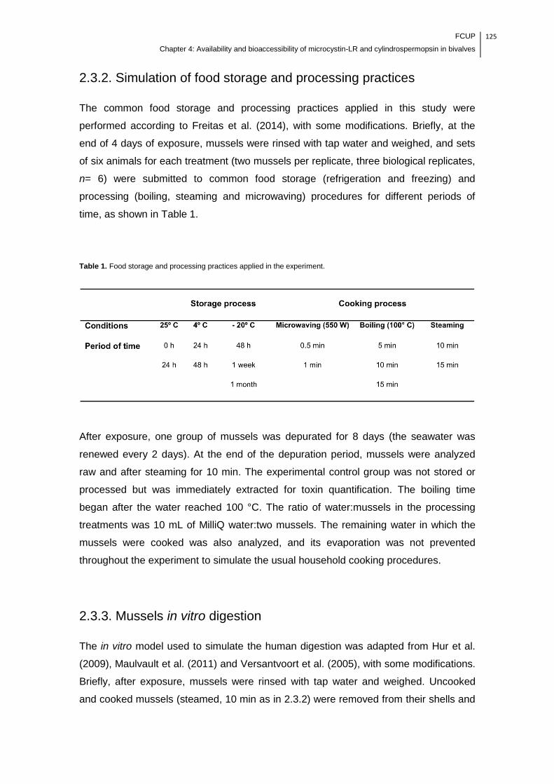
FCUP
Chapter 4: Availability and bioaccessibility of microcystin-LR and cylindrospermopsin in bivalves
125
2.3.2. Simulation of food storage and processing practices
The common food storage and processing practices applied in this study were
performed according to Freitas et al. (2014), with some modifications. Briefly, at the
end of 4 days of exposure, mussels were rinsed with tap water and weighed, and sets
of six animals for each treatment (two mussels per replicate, three biological replicates,
n= 6) were submitted to common food storage (refrigeration and freezing) and
processing (boiling, steaming and microwaving) procedures for different periods of
time, as shown in Table 1.
Table 1. Food storage and processing practices applied in the experiment.
After exposure, one group of mussels was depurated for 8 days (the seawater was
renewed every 2 days). At the end of the depuration period, mussels were analyzed
raw and after steaming for 10 min. The experimental control group was not stored or
processed but was immediately extracted for toxin quantification. The boiling time
began after the water reached 100 °C. The ratio of water:mussels in the processing
treatments was 10 mL of MilliQ water:two mussels. The remaining water in which the
mussels were cooked was also analyzed, and its evaporation was not prevented
throughout the experiment to simulate the usual household cooking procedures.
2.3.3. Mussels in vitro digestion
The in vitro model used to simulate the human digestion was adapted from Hur et al.
(2009), Maulvault et al. (2011) and Versantvoort et al. (2005), with some modifications.
Briefly, after exposure, mussels were rinsed with tap water and weighed. Uncooked
and cooked mussels (steamed, 10 min as in 2.3.2) were removed from their shells and

126 FCUP
Chapter 4: Availability and bioaccessibility of microcystin-LR and cylindrospermopsin in bivalves
homogenized with a blender for 1 to 2 min. Approximately 2 g (ww) of the homogenate
from both treatments was transferred to a 50-mL sterile centrifuge tubes and then
submitted to a chemical digestion, as shown in Fig. 2.
Fig. 2. Schematic representation of the in vitro digestion model used in the experiment.
The digestive procedure was performed in triplicate, and samples were sequentially
incubated in a shaking water bath (GFL 1083, GFL, Germany) at 37 ºC for (1) 5 min
with salivary juice (6 mL; 12 tubes), followed by (2) 1 h with gastric juice (12 mL; 9
tubes) and (3) 2 h with duodenal juices, which were individually tested, simulating
digestion in the small intestine by the proteolytic enzymes trypsin and chymotrypsin (12

FCUP
Chapter 4: Availability and bioaccessibility of microcystin-LR and cylindrospermopsin in bivalves
127
mL and 2 mL HCO3 (1 M); 3 tubes) or by whole pancreatic juice, containing pancreatic
proteolytic enzymes and pancreatic amylase and lipase (12 mL, 6 mL bile juice and 2
mL HCO3 (1 M); 3 tubes). The composition of the digestive juices is shown in Table 2.
Table 2. Composition (constituents and concentration) of digestive juices used in the in vitro digestion, representing fed
conditions. Adapted from Hur et al. (2009), Maulvault et al. (2011) and Versantvoort et al. (2005).
At the end of each digestive stage (mouth, stomach and small intestine), samples were
stored at 4 °C to stop the enzymatic reactions. The soluble and non-soluble fractions
from each digestive phase were obtained by centrifugation (Thermo Scientific –
LegendTM T/RT QUIKsetTM, Germany) (20 min, 4 °C, 4495 g). Groups of intoxicated
mussels, either raw or cooked, without chemical digestion were used as experimental
controls. Purified CYN in solution (100 ng/mL) was also subjected to the same
enzymatic treatments.
128 FCUP
Chapter 4: Availability and bioaccessibility of microcystin-LR and cylindrospermopsin in bivalves
2.4. CYN determination
2.4.1. CYN extraction and clean-up
To avoid any interference in the final concentration of the toxin through sample
freezing/thawing, the CYN from all samples (except the soluble fractions from the
digestion experiments, which were lyophilized) was immediately extracted after each
treatment.
The mussels of section 2.3.2 and solid the fractions of section 2.3.3 were weighed,
removed from their shells and homogenized with a blender for 1 to 2 min.
Approximately 1 g (ww) of the homogenate was transferred to 50-mL sterile centrifuge
tubes, and CYN was extracted twice in 5 mL of 90% ACN by ultrasonication (750 Watt,
20 KHz) (BioBlock Scientific, Vibracell 75041) in an ice bath for 1 min (pulse 1 second,
amplitude 40) and was then centrifuged (Eppendorf, 5810R) (5 min, 4 °C, 3200 g). For
the clean-up of the extracts, both supernatants were pooled in 0.5 g of C18 powder
(Bakerbond Octadecyl 40 µm, Prep LC Packing, J.T. Backer) and homogenized for 1
min at 25 °C and 1300 rpm (thermomixer comfort Eppendorf MTP). The supernatant (9
mL) was recovered by centrifugation (1 min, 20 °C, 2000 g), and 900 µL was then
evaporated to dryness in a SpeedVac (Eppendorf). The residue was re-suspended in
200 µL 0.1% formic acid (FA) in water (v/v). Samples were stored at -80 ºC until further
LC-MS/MS analysis.
2.4.2. LC-MS/MS analysis
The quantification of CYN was conducted by the linear regression of six-point
calibration curves of mussel matrix spiked (in triplicate) with a standard solution of CYN
(50, 100, 200, 400, 800 and 1600 ng/g). The lowest standard of the calibration curve
(50 ng/g) was the limit of quantification. The potential interference of matrix
components in the LC-MS/MS chromatograms was also assessed (in three
independent pools in triplicate), and the method was selective for CYN quantification
(data not shown).
Samples were analyzed on an Ultimate™3000 LC system (LC Packings, Dionex)
coupled to an ESI Turbo V ion source and a hybrid triple quadrupole/linear ion-trap
4000 QTrap mass spectrometer operated by Analyst 1.6.1 (AB Sciex). The
chromatographic separation was performed in a 3-μm Luna NH2 column (150 × 2.0
FCUP
Chapter 4: Availability and bioaccessibility of microcystin-LR and cylindrospermopsin in bivalves
129
mm, 100 Å, Phenomenex) with a 4 × 2.0 mm NH2 guard-column (Phenomenex). The
flow rate was set to 150 µL/min, and mobile phases A and B were 0.1% FA in water
and 0.1% FA in ACN, respectively. The LC program started with a linear gradient from
50% to 5% B (0 - 0.5 min), and it was maintained at 5% B for 6 minutes. After each
sample analysis, a 6-minute run was performed in isocratic mode with 50% B for
column equilibration. The ionization source was operated in the positive mode set to an
ion spray voltage of 5500 V, 30 psi for nebulizer gas 1 (GS1), 20 psi for nebulizer gas 2
(GS2), 30 psi for the curtain gas (CUR), and the temperature was 450 °C.
Cylindrospermopsin was quantified by multiple reaction monitoring (MRM) triple
quadrupole scan mode at unit resolution both in Q1 and Q3, and the MRM transitions
were 416.4/194.3 (used for quantification), 416.4/176.1 and 416.4/336.5 (both used for
compound confirmation). The MRM parameters were 10 eV for the entrance potential
(EP), 15 eV for the collision cell exit potential (CXP), and 100 V for the declustering
potential (DP), and the collision gas (CAD) was set to 8 psi. The dwell time was 100
ms, and the collision energies were 50 eV for transition 416.4/176.1, 60 eV for
transition 416.4/176.1 and 32 eV for transition 416.4/336.5. The peak areas were
integrated using MultiQuant software (version 2.1, AB Sciex).
2.5. Statistical analysis
Statistical analysis of the data was performed by one-way ANOVA after checking the
homogeneity of variance with the Levene test. When necessary, data were transformed
to achieve the variance homogeneity assumption. The Tukey multiple range test was
used to compare means whenever differences were detected by ANOVA at the
significance level of 0.05. All analyses were performed with IBM SPSS Statistics,
version 22. Four individual data groups were considered for statistical analysis: (1)
CYN concentration in the control group and CYN concentration after applying the
storage practices; (2) CYN concentration in the control group and CYN concentration
after applying the cooking practices; (3) CYN concentration in the control group and
CYN concentration in mussels depurated either raw or cooked; and (4) CYN in solution
before and after in vitro digestion. Significant differences were also tested between
different periods of time in the groups belonging to the storage and processing
treatments. The results are expressed in ng/g (ww) as the mean ± standard deviation
(SD) for three replicates for each treatment.
130 FCUP
Chapter 4: Availability and bioaccessibility of microcystin-LR and cylindrospermopsin in bivalves
3. Results and Discussion
The estimation of human exposure to CYN by food consumption is an essential
element for quantifying the risk; thus, data for the concentration of CYN in edible
organisms are required. In this study, marine mussels (M. galloprovincialis) were able
to accumulate CYN (28.1 ± 4.9 to 41.6 ± 5.7 ng/g; whole organism) when exposed to
ecologically relevant concentrations (≈ 10 to 15 μg/L) for 4 days, showing neither
mortality nor apparent detrimental effects on their organoleptic characteristics. As
mentioned above, Vareli et al. (2012) recently found microcystins in M. galloprovincialis
collected from the Mediterranean Sea, indicating that this edible aquatic organism
could contribute to chronic human exposure to cyanotoxins. Shellfish is an important
component of the human diet; thus, the study of the influence of common food storage
and processing practices and the human digestion on CYN availability in these edible
organisms is of major significance to provide more accurate data on human exposure
to this cyanotoxin.
3.1. Effects of storage on the CYN concentration in edible mussels
The storage process is a critical step to provide safe and high-quality shellfish for
human consumption. Thus, in this study, we intended to determine if different storage
conditions lead to changes in the levels of CYN in edible mussels. The concentration of
CYN in mussels stored unrefrigerated (25 ºC) for 24 h (43.1 ± 4.1 ng/g) and stored
refrigerated (4 ºC) for 24 h (45.3 ± 0.8 ng/g) or 48 h (55.7 ± 14.6 ng/g) did not
significantly differ from the control group (41.6 ± 5.7 ng/g) (Fig. 3).
The chemical stability of CYN is an important factor associated with its environmental
persistence and toxicity. In aqueous media, the stability of CYN has already been
demonstrated at temperatures ranging from 4 to 50 ºC for up to 4 weeks (Chiswell et
al., 1999).

FCUP
Chapter 4: Availability and bioaccessibility of microcystin-LR and cylindrospermopsin in bivalves
131
Fig. 3. The concentration of CYN (ng/g) in the mussel matrix submitted to different storage treatments at different
periods of time. Values are expressed as the mean ± SD (n = 3). Different letters (a, b and c) indicate significant
differences (p < 0.05). Columns that share the same letter are not significantly different.
In mussels stored frozen (-20 ºC), the recovery of CYN increased, especially after 48 h
(87.6 ± 15.6 ng/g) and 1 week (98.2 ± 23.6 ng/g) of storage, where the concentration of
CYN was significantly higher, 52.5% and 57.7%, respectively, (p < 0.05) than the
control group (Fig. 3). Froscio et al. (2008) showed that in reticulocyte lysates [14C],
CYN was partially released by incubation with an excess of unlabeled CYN, suggesting
that the toxin binds reversibly. Thus, our results can be associated with a more efficient
extraction of CYN from mussel tissues due to the potential cell disruption and protein
denaturation caused by freezing/thawing. These results may have relevance for
monitoring programs of CYN in edible organisms in which the frozen storage of
samples before analysis is typical. Furthermore, this increase in CYN recovery from the
mussel matrix may suggest that higher levels of the toxin are available when mussels
are consumed after being frozen.
3.2. Effects of cooking on the CYN concentration in edible mussels
Shellfish is generally cooked prior to consumption to enhance its microbiological safety
and flavor. However, cooking treatments can also alter the availability of chemical
contaminants in food (Domingo, 2011). As recently reviewed by Gutiérrez-Praena et al.
(2013) and Kinnear (2010), several studies have assessed the concentration of CYN in
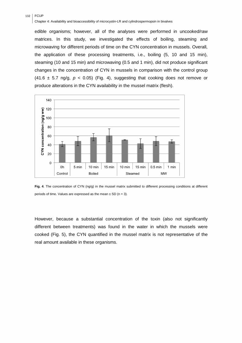
132 FCUP
Chapter 4: Availability and bioaccessibility of microcystin-LR and cylindrospermopsin in bivalves
edible organisms; however, all of the analyses were performed in uncooked/raw
matrices. In this study, we investigated the effects of boiling, steaming and
microwaving for different periods of time on the CYN concentration in mussels. Overall,
the application of these processing treatments, i.e., boiling (5, 10 and 15 min),
steaming (10 and 15 min) and microwaving (0.5 and 1 min), did not produce significant
changes in the concentration of CYN in mussels in comparison with the control group
(41.6 ± 5.7 ng/g, p < 0.05) (Fig. 4), suggesting that cooking does not remove or
produce alterations in the CYN availability in the mussel matrix (flesh).
Fig. 4. The concentration of CYN (ng/g) in the mussel matrix submitted to different processing conditions at different
periods of time. Values are expressed as the mean ± SD (n = 3).
However, because a substantial concentration of the toxin (also not significantly
different between treatments) was found in the water in which the mussels were
cooked (Fig. 5), the CYN quantified in the mussel matrix is not representative of the
real amount available in these organisms.

FCUP
Chapter 4: Availability and bioaccessibility of microcystin-LR and cylindrospermopsin in bivalves
133
Fig. 5. The concentration of CYN (ng/mL) in the water in which the mussels were cooked. Values are expressed as the
mean ± SD (n = 3).
In fact, heat processing causes a loss of water containing soluble compounds from the
mussel matrix into the intervalval fluid, which leaks into the cooking water due to the
shell opening shortly after the beginning (approximately 3 min) of the treatment.
Therefore, it is likely that this fluid is the source of the CYN found in the cooking water.
In view of this, the potential human exposure to CYN due to the consumption of
contaminated mussels based on the amount of CYN in raw mussels will be under-
evaluated. On the other hand, because the heat processing enhances the CYN
extraction from the mussel tissues, this practice can be applied to reduce its availability
in this food item by simply disposing of the cooking water. This can be relevant for
other industrial processes with mussels, such as canning and brine. According to the
Food and Agriculture Organization of the United Nations (FAO), canning represents
approximately 40 to 50% of the mussel market. Normally, mussels are precooked in
steam or boiling water to separate the edible portion from the shells, and the remaining
liquor is kept for reuse in canning or brine. The latter procedure should be avoided
because the sauce is usually ingested together with the mussels, which may potentially
increase the human exposure to CYN. Considering the Hazard Analysis Critical Control
Points (HACCP) system set by the Codex Alimentarius Commission, our results
provide new insights to identify critical control points and to determine critical limits for
the culinary preparation of mussels. The heat processing and the disposal of the
cooking water are suitable procedures to at least reduce the availability of CYN in
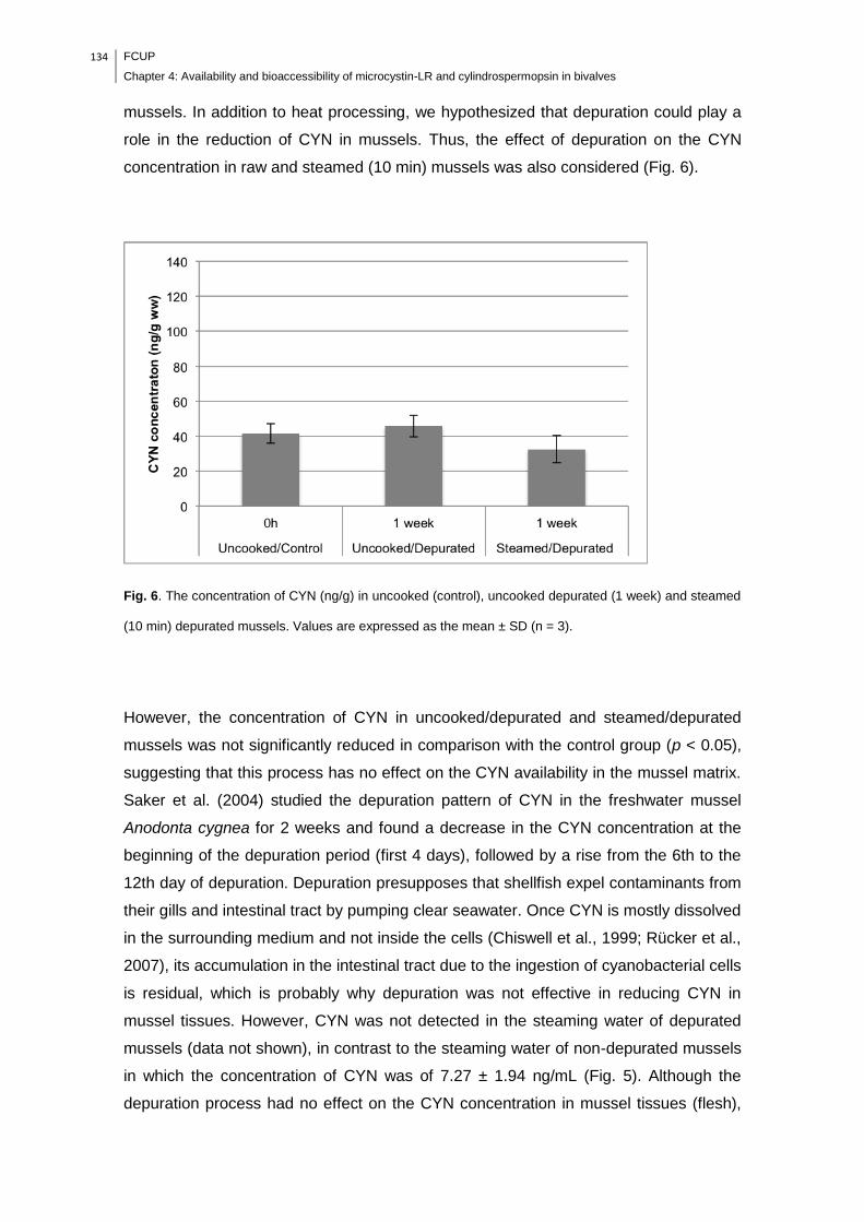
134 FCUP
Chapter 4: Availability and bioaccessibility of microcystin-LR and cylindrospermopsin in bivalves
mussels. In addition to heat processing, we hypothesized that depuration could play a
role in the reduction of CYN in mussels. Thus, the effect of depuration on the CYN
concentration in raw and steamed (10 min) mussels was also considered (Fig. 6).
Fig. 6. The concentration of CYN (ng/g) in uncooked (control), uncooked depurated (1 week) and steamed
(10 min) depurated mussels. Values are expressed as the mean ± SD (n = 3).
However, the concentration of CYN in uncooked/depurated and steamed/depurated
mussels was not significantly reduced in comparison with the control group (p < 0.05),
suggesting that this process has no effect on the CYN availability in the mussel matrix.
Saker et al. (2004) studied the depuration pattern of CYN in the freshwater mussel
Anodonta cygnea for 2 weeks and found a decrease in the CYN concentration at the
beginning of the depuration period (first 4 days), followed by a rise from the 6th to the
12th day of depuration. Depuration presupposes that shellfish expel contaminants from
their gills and intestinal tract by pumping clear seawater. Once CYN is mostly dissolved
in the surrounding medium and not inside the cells (Chiswell et al., 1999; Rücker et al.,
2007), its accumulation in the intestinal tract due to the ingestion of cyanobacterial cells
is residual, which is probably why depuration was not effective in reducing CYN in
mussel tissues. However, CYN was not detected in the steaming water of depurated
mussels (data not shown), in contrast to the steaming water of non-depurated mussels
in which the concentration of CYN was of 7.27 ± 1.94 ng/mL (Fig. 5). Although the
depuration process had no effect on the CYN concentration in mussel tissues (flesh),
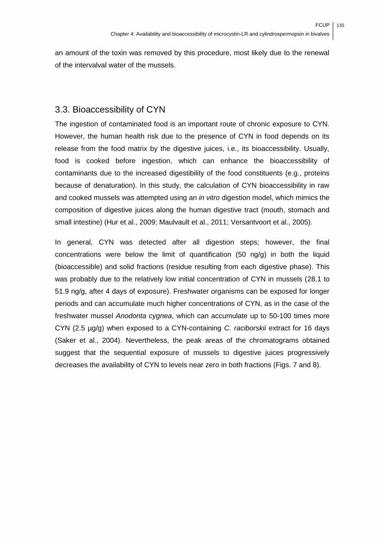
FCUP
Chapter 4: Availability and bioaccessibility of microcystin-LR and cylindrospermopsin in bivalves
135
an amount of the toxin was removed by this procedure, most likely due to the renewal
of the intervalval water of the mussels.
3.3. Bioaccessibility of CYN
The ingestion of contaminated food is an important route of chronic exposure to CYN.
However, the human health risk due to the presence of CYN in food depends on its
release from the food matrix by the digestive juices, i.e., its bioaccessibility. Usually,
food is cooked before ingestion, which can enhance the bioaccessibility of
contaminants due to the increased digestibility of the food constituents (e.g., proteins
because of denaturation). In this study, the calculation of CYN bioaccessibility in raw
and cooked mussels was attempted using an in vitro digestion model, which mimics the
composition of digestive juices along the human digestive tract (mouth, stomach and
small intestine) (Hur et al., 2009; Maulvault et al., 2011; Versantvoort et al., 2005).
In general, CYN was detected after all digestion steps; however, the final
concentrations were below the limit of quantification (50 ng/g) in both the liquid
(bioaccessible) and solid fractions (residue resulting from each digestive phase). This
was probably due to the relatively low initial concentration of CYN in mussels (28.1 to
51.9 ng/g, after 4 days of exposure). Freshwater organisms can be exposed for longer
periods and can accumulate much higher concentrations of CYN, as in the case of the
freshwater mussel Anodonta cygnea, which can accumulate up to 50-100 times more
CYN (2.5 µg/g) when exposed to a CYN-containing C. raciborskii extract for 16 days
(Saker et al., 2004). Nevertheless, the peak areas of the chromatograms obtained
suggest that the sequential exposure of mussels to digestive juices progressively
decreases the availability of CYN to levels near zero in both fractions (Figs. 7 and 8).

136 FCUP
Chapter 4: Availability and bioaccessibility of microcystin-LR and cylindrospermopsin in bivalves
Fig. 7. Chromatographic peak area of CYN detection from uncooked and steamed mussels before (CYN C) and after
digestion (for liquid and solid fractions). Letters represent each digestive step: mouth (M), gastric (G) and
gastrointestinal (GI) with proteolytic enzymes (PE). Values are expressed as the mean ± SD (n = 3). A.U., Arbitrary
Units.
Fig. 8. Chromatographic peak area of CYN detection from uncooked and steamed mussels before (CYN C) and after
digestion (for liquid and solid fractions). Letters represent each digestive step: mouth (M), gastric (G) and
gastrointestinal (GI) with pancreatic juice (P). Values are expressed as the mean ± SD (n = 3). A.U., Arbitrary Units.
To confirm the decrease in the CYN content after being submitted to digestive juices
and to exclude any interference of the food matrix on CYN detection in the
bioaccessible fraction (toxin bound to proteins or other molecules), purified CYN in
solution (100 ng/mL) was submitted to the same in vitro digestion model. The results
show the same trend of the intoxicated mussels, where the digestive juices sequentially
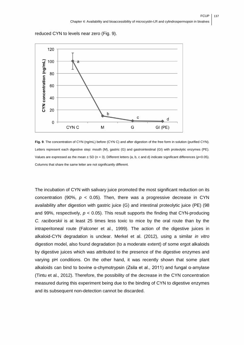
FCUP
Chapter 4: Availability and bioaccessibility of microcystin-LR and cylindrospermopsin in bivalves
137
reduced CYN to levels near zero (Fig. 9).
Fig. 9. The concentration of CYN (ng/mL) before (CYN C) and after digestion of the free form in solution (purified CYN).
Letters represent each digestive step: mouth (M), gastric (G) and gastrointestinal (GI) with proteolytic enzymes (PE).
Values are expressed as the mean ± SD (n = 3). Different letters (a, b, c and d) indicate significant differences (p<0.05).
Columns that share the same letter are not significantly different.
The incubation of CYN with salivary juice promoted the most significant reduction on its
concentration (90%, p < 0.05). Then, there was a progressive decrease in CYN
availability after digestion with gastric juice (G) and intestinal proteolytic juice (PE) (98
and 99%, respectively, p < 0.05). This result supports the finding that CYN-producing
C. raciborskii is at least 25 times less toxic to mice by the oral route than by the
intraperitoneal route (Falconer et al., 1999). The action of the digestive juices in
alkaloid-CYN degradation is unclear. Merkel et al. (2012), using a similar in vitro
digestion model, also found degradation (to a moderate extent) of some ergot alkaloids
by digestive juices which was attributed to the presence of the digestive enzymes and
varying pH conditions. On the other hand, it was recently shown that some plant
alkaloids can bind to bovine α-chymotrypsin (Zsila et al., 2011) and fungal α-amylase
(Tintu et al., 2012). Therefore, the possibility of the decrease in the CYN concentration
measured during this experiment being due to the binding of CYN to digestive enzymes
and its subsequent non-detection cannot be discarded.
138 FCUP
Chapter 4: Availability and bioaccessibility of microcystin-LR and cylindrospermopsin in bivalves
To our knowledge, this is the first report presenting the potential effects of human
digestion on CYN contained in a food item. According to our findings, it is reasonable to
assume that at the tested concentrations, CYN itself would not represent a risk to
human health. The assumption that the oral bioavailability of CYN is similar to the
levels quantified in raw and cooked mussels is erroneous; thus, the comparison of TDI
with CYN extracted from food items is not recommended. In this way, the
bioaccessibility of CYN must be integrated into the health risk assessment. In fact, as
referred to above, Falconer et al. (1999), by comparing the effects of various batches of
C. raciborskii in mice, found that oral toxicity is at least 25-fold lower than
intraperitoneal toxicity. Associated with the potential effects of digestive juices, some
other important factors may reduce the bioavailability of CYN, such as (1) uptake being
potentially dependent on a transporter; (2) hepatic metabolism (Norris et al., 2002); (3)
removal by intestinal microflora (Nybom et al., 2008); and (4) urinary and fecal
excretion (Norris et al., 2001). Finally, although human digestion substantially reduces
the CYN availability, the absence of health risks due to consumption of contaminated
mussels should be considered cautiously because the presence of additional toxins in
the C. raciborskii extracts has been reported (Falconer et al., 1999). Thus, the study of
the effects of other toxins, as well as the enzymatic-generated metabolites of CYN,
should be developed in the future.
4. Conclusion
In conclusion, this study fills a gap in the knowledge of the influence of food storage
and cooking practices on the levels of CYN in edible organisms. Our results show that
the recovery of CYN can be enhanced with frozen storage, and its concentration in raw
mussels does not represent the total amount of toxin available. Although there were no
significant differences in CYN concentration in raw, cooked and depurated mussel
matrices (flesh), heat processing and depuration are suitable procedures to reduce the
toxin in this edible organism through the removal/renewal of the intervalval fluid. More
importantly, in the industry, these practices can be easily applied and should be
considered in the implementation of HACCP systems.
This study also provides the first insights into the effects of human digestion on CYN.
At the concentrations tested, the digestive juices were effective in the reduction of CYN
content, whether present in solution or in the raw and steamed mussel matrices.
FCUP
Chapter 4: Availability and bioaccessibility of microcystin-LR and cylindrospermopsin in bivalves
139
According to these findings, CYN itself would not represent a risk to human health,
which highlights the importance in integrating the bioaccessibility of CYN as a part of
the risk assessment because the amount of toxin effectively available for absorption is
much lower than the amount of toxin quantified in raw and cooked mussels. Further
studies should be developed to identify the contributing factors of CYN removal,
particularly the role of digestive juices.
Conflict of Interest
The authors declare that there are no conflicts of interest.
Acknowledgments
Marisa Freitas thanks the Fundação para a Ciência e Tecnologia (FCT) for the Ph.D.
Grant SFRH/BD/85490/2012. This work was partially funded by the FCT project PesT-
C/MAR/LA0015/2011, and Rede Nacional de Espectometria de Massa (grant number
REDE/1506/REM/2005).
5. References
Banker, R., Carmeli, S., Hadas, O., Teltsch, B., Porat, R., Sukenik, A., 1997.
Identification of cylindrospermopsin in Aphanizomenon ovalisporum (cyanophyceae)
isolated from Lake Kinneret, Israel. J. Phycol. 33, 613–616.
Banker, R., Teltsch, B., Sukenik, A., Carmeli, S., 2000. 7-Epicylindrospermopsin, a
toxic minor metabolite of the cyanobacterium Aphanizomenon ovalisporum from Lake
Kinneret, Israel. J. Nat. Prod. 63, 387–389.
Chiswell, R.K., Shaw, G.R., Eaglesham, G., Smith, M.J., Norris, R.L., Seawright, A.A.,
Moore, M.R., 1999. Stability of cylindrospermopsin, the toxin from the cyanobacterium.
Cylindrospermopsis raciborskii: Effect of pH, temperature, and sunlight on
decomposition. Environ. Toxicol. 14, 155–161.
Domingo, J.L., 2011. Influence of cooking processes on the concentrations of toxic

140 FCUP
Chapter 4: Availability and bioaccessibility of microcystin-LR and cylindrospermopsin in bivalves
metals and various organic environmental pollutants in food: a review of the published
literature. Crit. Rev. Food Sci. Nutr. 51, 29–37.
Falconer, I., Humpage, A.R., 2006. Cyanobacterial (blue-green algal) toxins in water
supplies: cylindrospermopsins. Environ. Toxicol. 21, 299–304.
Falconer, I.R., Hardy, S.J., Humpage, A.R., Froscio, S.M., Tozer, G.J., Hawkins, P.R.,
1999. Hepatic and renal toxicity of the blue-green alga (cyanobacterium)
Cylindrospermopsis raciborskii in male Swiss albino mice. Environ. Toxicol. 14, 143–
150.
Falconer, I.R., Humpage, A.R., 2001. Preliminary evidence for in vivo tumour initiation
by oral administration of extracts of the blue-green alga Cylindrospermopsis raciborskii
containing the toxin cylindrospermopsin. Environ. Toxicol. 16, 192–195.
Freitas, M., Azevedo J., Carvalho A.P., Campos A., Vasconcelos V., 2014. Effects of
storage, processing and proteolytic digestion on microcystin-LR concentration in edible
clams. Food Chem. Toxicol. 66, 217- 223.
Froscio, S.M., Humpage, A.R., Burcham, P.C., Falconer, I.R., 2001. Cell-free protein
synthesis inhibition assay for the cyanobacterial toxin cylindrospermopsin. Environ.
Toxicol. 16, 408–412.
Froscio, S.M., Humpage, A.R., Burcham, P.C., Falconer, I.R., 2003.
Cylindrospermopsin-induced protein synthesis inhibition and its dissociation from acute
toxicity in mouse hepatocytes. Environ. Toxicol. 18, 243–251.
Froscio, S.M., Humpage, A.R., Wickramasinghe, W., Shaw, G., Falconer, I.R., 2008.
Interaction of the cyanobacterial toxin cylindrospermopsin with the eukaryotic protein
synthesis system. Toxicon 51, 191–198.
Griffiths, D.J., Saker, M.L., 2003. The Palm Island mystery disease twenty years on: a
review of research on the cyanotoxin cylindrospermopsin. Environ. Toxicol. 18, 78–93.
Guerra, A., Etienne-Mesmin, L., Livrelli. V., Denis, S., Blanquet-Diot, S., Alric, M., 2012.
Relevance and challenges in modelinghuman gastric and small intestinal digestion.
Trends Biotechnol. 30, 591-600.
Gutiérrez-Praena, D., Jos, Á., Pichardo, S., Moreno, I.M., Cameán, A.M., 2013.
Presence and bioaccumulation of microcystins and cylindrospermopsin in food and the
FCUP
Chapter 4: Availability and bioaccessibility of microcystin-LR and cylindrospermopsin in bivalves
141
effectiveness of some cooking techniques at decreasing their concentrations: A review.
Food Chem. Toxicol. 53, 139–152.
Guzmán-Guillén, R., Prieto, A.I., Moreno, I., Soria, M.E., Cameán, A.M., 2011. Effects
of thermal treatments during cooking, microwave oven and boiling, on the
unconjugated microcystin concentration in muscle of fish (Oreochromis niloticus). Food
Chem. Toxicol. 49, 2060–2067.
Humpage, A.R., Falconer, I.R., 2003. Oral toxicity of the cyanobacterial toxin
cylindrospermopsin in male swiss albino mice: Determination of no observed adverse
effect level for deriving a drinking water guideline value. Environ. Toxicol. 18, 94–103.
Humpage, A.R., Fontaine, F., Froscio, S., Burcham, P., Falconer, I.R., 2005.
Cylindrospermopsin genotoxicity and cytotoxicity: role of cytochrome P-450 and
oxidative stress. J. Toxicol. Environ. Health Part A 68, 739–753.
Hur, S.J., Decker, E.A., McClements, D.J., 2009. Influence of initial emulsifier type on
microstructural changes occurring in emulsified lipids during in vitro digestion. Food
Chem. 114, 253–262.
Ibelings, B.W., Chorus I., 2007. Accumulation of cyanobacterial toxins in freshwater
‗‗seafood‘‘ and its consequences for public health: A review. Environ. Pollut. 150, 177-
192.
Kinnear, S., 2010. Cylindrospermopsin: a decade of progress on bioaccumulation
research. Mar. Drugs 8, 542-564.
Kotai, J., 1972. Instruction for preparation of modified nutrient solution Z8 for algae.
Norwegian Institute for Water Research, Oslo, Norway, Report nr B-11/69, 1-5.
Li, R., Carmichael, W.W., Brittain, S., Eaglesham, G.K., Shaw, G.R., Liu, Y.,
Watanabe, M.M., 2001. First report of the cyanotoxins cylindrospermopsin and
deoxycylindrospermopsin from Raphidiopsis curvata (cyanobacteria.). J. Phycol. 3,
1121–1126.
Maulvault, A.L., Machado, R., Afonso C., Lourenço, H.M., Nunes, M.L., Coelho, I.,
Langerholc T., Marques, A., 2011. Bioaccessibility of Hg, Cd and As in cooked black
scabbard fish and edible crab. Food Chem. Toxicol. 49, 2808–2815.
Merkel, S., Dib, B., Maul, R., Köppen, R., Koch, M., Nehls, I., 2012. Degradation and
142 FCUP
Chapter 4: Availability and bioaccessibility of microcystin-LR and cylindrospermopsin in bivalves
epimerization of ergot alkaloids after baking and in vitro digestion. Anal. Bioanal.
Chem. 404, 2489–2497.
Morais, J., Augusto, M., Carvalho, A.P., Vale, M., Vasconcelos, V.M., 2008.
Cyanobacteria hepatotoxins, microcystins: bioavailability in contaminated mussels
exposed to different environmental conditions. Eur. Food Res. Technol. 227, 949–952.
Norris, R.L.G., Eaglesham, G.K., Pierens, G., Shaw, G.R., Smith, M.J., Chiswell, R.K.,
Seawright, A.A., Moore, M.R., 1999. Deoxycylindrospermopsin, an analog of
cylindrospermopsin from Cylindrospermopsis raciborskii. Environ. Toxicol. 14, 163–
165.
Norris R.L., Seawright A.A., Shaw G.R., Smith M.J., Chiswell R.K., Moore M.R., 2001.
Distribution of 14C-cylindrospermopsin in vivo in the mouse. Environ. Toxicol. 16, 498–
505.
Norris R.L., Seawright A.A., Shaw G.R., Senogles P., Eaglesham G.K., Smith M.J.,
Chiswell R.K., Moore M.R., 2002. Hepatic xenobiotic metabolism of cylindrospermopsin
in vivo in the mouse. Toxicon 40, 471–476.
Nybom, S.M.K., Salminen S.J., Meriluoto, J.A.O., 2008. Specific strains of probiotic
bacteria are efficient in removal of several different cyanobacterial toxins from solution.
Toxicon 52, 214–220.
O‘Neil, J.M.,
Davis, T.W., Burford, M.A., Gobler, C.J., 2012. The rise of harmful
cyanobacteria blooms: The potential roles of eutrophication and climate change.
Harmful Algae 14, 313–334.
Ohtani, I., Moore, R.E., Runnegar, M.T.C., 1992. Cylindrospermopsin: A potent
hepatotoxin from the blue-green alga Cylindrospermopsis raciborskii. J. Am. Chem.
Soc. 114, 7941-7942.
Poniedziałek, B., Rzymski, P., Kokociński, M., 2012. Cylindrospermopsin: Water-linked
potential threat to human health in Europe. Environ. Toxicol. Phar. 34, 651–660.
Preussel, K., Stüken, A., Wiedner, C., Chorus, I., Fastner, J., 2006. First report on
cylindrospermopsin producing Aphanizomenon flos-aquae (Cyanobacteria) isolated
from two German lakes. Toxicon 47, 156–162.
Rücker, J., Stüken, A., Nixdorf, B., Fastner, J., Chorus, I., Wiedner, C., 2007.
FCUP
Chapter 4: Availability and bioaccessibility of microcystin-LR and cylindrospermopsin in bivalves
143
Concentrations of particulate and dissolved cylindrospermopsin in 21 Aphanizomenon-
dominated temperate lakes. Toxicon 50, 800–809.
Runnegar, M.T., Kong, S.M., Zhong, Y.Z., Lu, S.C., 1995. Inhibition of reduced
glutathione synthesis by cyanobacterial alkaloid cylindrospermopsin in cultured rat
hepatocytes. Biochem. Pharmacol. 49, 219–225.
Runnegar, M.T., Xie, C., Snider, B.B., Wallace, G.A., Weinreb, S.M., Kuhlenkamp, J.,
2002. In vitro hepatotoxicity of the cyanobacterial alkaloid cylindrospermopsin and
related synthetic analogues. Toxicol. Sci. 67, 81–87.
Saker, M.L., Metcalf, J.S., Codd, G.A., Vasconcelos, V.M., 2004. Accumulation and
depuration of the cyanobacterial toxin cylindrospermopsin in the freshwater mussel
Anodonta cygnea. Toxicon 43, 185–194.
Seifert, M., Mcgregor, G., Eaglesham, G., Wickramasinghe, W., Shaw, G., 2007. First
evidence for the production of cylindrospermopsin and deoxy-cylindrospermopsin by
the freshwater benthic cyanobacterium, Lyngbya wollei (Farlow ex Gomont) Speziale
and Dyck. Harmful Algae 6, 73–80.
Spoof, L., Berg, K.A., Rapala, J., Lahti, K., Lepisto, L., Metcalf, J.S., Codd, G.A.,
Meriluoto, J., 2006. First observation of cylindrospermopsin in Anabaena lapponica
isolated from the boreal environment (Finland). Environ. Toxicol. 21, 552–560.
Terao, K., Ohmori, S., Igarashi, K., Ohtani, I., Watanabe, M.F., Harada, K.I., Ito, E.,
Watanabe, M., 1994. Electron microscopic studies on experimental poisoning in mice
induced by cylindrospermopsin isolated from blue-green alga Umezakia natans.
Toxicon 32, 833–843.
Tintu, I., Dileep, K.V., Augustine, A., Sadasivan, C., 2012. An isoquinoline alkaloid,
berberine, can inhibit fungal alpha amylase: enzyme kinetic and molecular modeling
studies. Chem. Biol. Drug Des. 80, 554–560.
Vareli, K., Zarali, E., Zacharioudakis,
G.S.A., Vagenas, G., Varelis, V., Pilidis, G.,
Briasoulis, E., Sainis, I., 2012. Microcystin producing cyanobacterial communities in
Amvrakikos Gulf (Mediterranean Sea, NW Greece) and toxin accumulation in mussels
(Mytilus galloprovincialis). Harmful Algae 15, 109–118.
Versantvoort, C.H.M., Oomen, A.G., Van de Kamp, E., Rompelberg, C.J.M., Sips,
A.J.A.M., 2005. Applicability of an in vitro digestion model in assessing the
144 FCUP
Chapter 4: Availability and bioaccessibility of microcystin-LR and cylindrospermopsin in bivalves
bioaccessibility of mycotoxins from food. Food Chem. Toxicol. 43, 31–40.
Wörmer, L., Huerta-Fontela, M., Cirés, S., Carrasco, D., Quesada, A., 2010. Natural
photodegradation of the cyanobacterial toxins microcystin and cylindrospermopsin.
Environ. Sci. Technol. 44, 3002–3007.
Zhang, D., Xie, P., Chen, J., 2010. Effects of temperature on the stability of
microcystins in muscle of fish and its consequences for food safety. Bull. Environ.
Contam. Toxicol. 84, 202–207.
Zsila, F., Kámán, J., Bogányi, B., Józsvai, D., 2011. Binding of alkaloids into the S1
specificity pocket of α-chymotrypsin: Evidence from induced circular dichroism spectra.
Org. Biomol. Chem. 9, 4127–4137.

FCUP
Chapter 5: General conclusions and future research
145
Chapter 5
General conclusions and future research

146 FCUP
Chapter 5: General conclusions and future research
5.1. General discussion and conclusions
The occurrence of toxic cyanobacterial blooms is expected to become increasingly
recurrent, which creates an important challenge for agricultural productivity,
environmental management and human health.
The toxic effects of cyanotoxins on crop plants are poorly studied, especially for CYN
and its mixture with MC-LR. In the present work (Chapter 3) the effects of ecologically
relevant concentrations of MC-LR, CYN and their mixture were determined in an
important crop plant, lettuce. The overall effects on the parameters assessed are
summarized in Table 1.
Table 1. The general results obtained regarding to the effects of MC-LR, CYN and the MC-LR/CYN mixture in lettuce
plants.
Concentration of
exposure (µg/L)
Growth (Fw) GST activity GPx activity Mineral content Protein abundance
Roots Leaves Roots Leaves Roots Leaves Roots Leaves Roots Leaves
MC-LR 1 ⁄ ⁄ ⁄ ⁄ ⁄ ⁄ - ⁄ - -
10 ⁄ ⁄ ⁄ ⁄ ⁄ ⁄ - ⁄ - -
100 ⁄ ⁄ ⁄ ⁄ ⁄ ⁄ - ⁄ - -
CYN 1 ⁄ ⁄ ⁄ ⁄ ⁄ ⁄ - ⁄ -
10 ⁄ ⁄ ⁄ ⁄ ⁄ ⁄ - ⁄ -
100 ⁄ ⁄ ⁄ ⁄ ⁄ ⁄ - ⁄ -
MC-LR/CYN
1 ⁄ ⁄ ⁄ ⁄ ⁄ ⁄ - ⁄ -
10 ⁄ ⁄ ⁄ ⁄ ⁄ ⁄ - ⁄ -
100 ⁄ ⁄ ⁄ ⁄ ⁄ ⁄ - ⁄ -
Increased; Decreased; No effects; - Not measured; 5 Days/ 10 Days of exposure.
Lettuce plants in non-early stage of development were able to cope with MC-LR, CYN
and their mixture, especially at the lower concentrations and at the lowest time of
exposure, ensuring the maintenance of mineral uptake/translocation and the oxidative
stress under control, as indicated by the increased activity of GST in roots.
The study of lettuce proteome allowed the understanding of the metabolic pathways
activated in response to cyanotoxins, providing new insights into potential protein
markers of exposure (e.g., SOD and APX) and the pathways that may confer tolerance
to lettuce plants (enhancement of the capacity to perform photosynthesis and to
synthesize ATP). With this study we hypothesize that the potential mechanism of
tolerance of lettuce plants to CYN, at 5 days of exposure, can be promoted by the
activation of mineral uptake and improving photosynthesis capacity.
FCUP
Chapter 5: General conclusions and future research
147
However, the concentration and the time of exposure are preponderant factors for the
toxic effects of these cyanotoxins in lettuce plants. The exposure of lettuce plants to
100 µg/L, especially of MC-LR and the MC-LR/CYN mixture, can compromise the crop
yield and productivity, as demonstrated by the decrease of the growth and the mineral
content in leaves (Table 1). In the exposure to the higher concentrations of MC-
LR/CYN mixture, the impairment of photosynthesis and carbon metabolism;
stress/defense response, and protein synthesis and signal transduction were also
observed at proteome level. Thus, the use of water for irrigation contaminated with
these cyanotoxins at concentrations ≥ 100 µg/L can lead to potential economic losses
in crop production.
This study led also to the discovery of some traits associated with the quality and
safety of leaves of lettuce plants exposed to environmentally relevant concentrations of
MC-LR, CYN and the mixture of MC-LR/CYN. Among other nutrients, lettuce provides
an important source of minerals and antioxidants for human nutrition. Leaves of lettuce
plants exposed to CYN had an enhanced content of minerals and probably
antioxidants, and this can be of major significance facing the challenge of food security,
once lettuce was able to cope with abiotic stress and also maximize its nutritional
quality.
On the other hand, lettuce accumulated putative allergenic proteins and probably also
cyanotoxins. Hence, the covering of various parameters allowed a broader
understanding of the mechanisms of action of these cyanotoxins in crop plants, and
this can be an asset to take more targeted measures in a field poorly regulated, such
as the use of surface water for agricultural production. Even so, the findings of this
work indicate the importance to conduct further studies to evaluate the health
significance and the risks of the utilization of irrigation waters contaminated with
cyanotoxins in agriculture.

148 FCUP
Chapter 5: General conclusions and future research
Regarding to human exposure to cyanotoxins, obviously, the best measure to reduce
the occurrence of cyanotoxins in edible aquatic organisms is to control the toxic
cyanobacterial blooms. However, the proliferation of toxic cyanobacterial blooms has
been forecasted as increasingly recurrent as a consequence of the intensification of
anthropogenic pressure and climate change. Furthermore, bivalves may contain
unpredictable amounts of cyanotoxins in their tissues due to bioaccumulation and long-
term elimination.
This work fills also an information gap concerning the influence of food storage and
cooking practices as well as human digestion on the levels of MC-LR and CYN in
edible bivalves (Chapter 4). The Table 2 summarizes the results obtained.
Table 2. The general results obtained on the changes of MC-LR and CYN in bivalves after applying different practices
of food storage and processing as well as the effects of digestive juices.
Condition Time MC-LR CYN
Storage 25° C 12h -
24 h
48 h -
4° C 24 h
48 h
72 h -
- 20 ° C 48 h
1 week
1 month
Cooking Boiling 5 min
10 min -
15 min
30 min -
Steaming 10 min -
15 min -
Microwaving 0.5 min
1 min
3 min -
5 min -
Toxin in the cooking water - X
Bioaccessibility Raw
Steamed 10 min -
Increased in comparison to control group; Decreased in comparison to control group;
There were no effects in comparison to control group; - Not measured; X The toxin was found in cooking water.
Overall the results show that the recovery of MC-LR and CYN in clams and mussels,
respectively, are enhanced with frozen storage. This indicates that the toxin measured
from fresh clam and mussel tissues can underestimate the real amount available.
FCUP
Chapter 5: General conclusions and future research
149
Thus, the extraction of MC-LR and CYN can be improved with this procedure and this
may have relevance for the monitoring programs and human health risk assessment.
The cooking procedures influenced the availability of MC-LR and CYN in edible
bivalves. It is important to highlight that although there were no differences in the
concentration of CYN in processed mussel matrices in comparison to control group, the
water in which mussels were cooked contained a significant amount of CYN. Thus, it is
likely that the overall increase of the MC-LR availability after processing is related to
the joint determination of free MC-LR content in both matrices, i.e., boiled clams
together with the water in which they were boiled.
Despite the differences in the physico-chemical properties of these two cyanotoxins,
both are hydrophilic molecules, thus, it was expected that a considerable amount would
be leached into the cooking water. Thus, the combination of processing methods with
MeOH and ACN extraction of MC-LR and CYN, respectively, could result in a more
effective and representative determination of these cyanotoxins in food products.
Under this framework, once MC-LR and CYN are recognized as relevant chemical
hazards in edible aquatic organisms, the development and implementation of risk
management strategies are of critical importance. Thus, the treatments applied in this
study can also be considered for the definition of critical control limits, considering the
HACCP approach as a promising tool for risk management. In this way, the control of
the processing time and the discarding of cooking water may be suitable procedures to
reduce both cyanotoxins in edible aquatic organisms.
The bioaccessibility of MC-LR and especially CYN is highly reduced due to digestion
with pancreatic proteolytic enzymes and salivary juice, respectively. This strongly
reduces the potential toxicological risks because the concentration determined in fresh
clams and mussels might considerably overestimate the the amount of toxin available
for assimilation after consumption. Further studies should be developed to identify the
contributing factors of the removal of cyanotoxins from the edible tissues of these
aquatic organisms, i.e., if they are related to the degradation of cyanotoxins or, for
instance, to the binding of these toxins with digestive enzymes.
Because the food consumption is one of the most important routes of exposure to
cyanotoxins, the incorporation of the bioaccessibility in the human exposure estimation
would be of particular relevance to achieve a more realistic human health risk
assessment.

150 FCUP
Chapter 5: General conclusions and future research
In conclusion, this dissertation provides substantial contributions towards a more
rational identification of the relevant hazards as well as the application of more
assertive management measures in the field of the human health and environmental
risks associated to toxic cyanobacterial blooms.
FCUP
Chapter 5: General conclusions and future research
151
5.2. Future research
Several hypotheses and questions were brought to light in this thesis, requiring further
supporting evidences and scientific clarification. The following matters should be
addressed in future research:
1 - To analyze the concentration of MC-LR and CYN in the edible tissues of lettuce
plants. This work is already in progress.
In this topic it would be also of particular interest exploring the effects of these
cyanotoxins in the accumulation of other constituents that compromise food safety
such as allergenic proteins.
2 - To analyze non-enzymatic substances of antioxidative system, such as carotenoids,
tocopherols and phenolic compounds to better characterize the nutritional quality of
lettuce plants exposed to cyanotoxins.
3 – To analyze the activity of other antioxidant enzymes, which seem to be active (by
proteomic analysis) in lettuce response to cyanotoxins exposure, such as SOD, CAT,
APX.
4 – To characterize and validate the function of some identified proteins, that seems to
confer tolerance to lettuce plants against CYN and MC-LR/CYN.
5 – To analyze the MC-LR bioaccessibility after applying different food storage and
processing methods.
6 – To characterize the toxic potential of the products of digestion of MC-LR and CYN.
152 FCUP
Chapter 6: Supporting information
Chapter 6
Supporting information
From the manuscript: Marisa Freitas, Alexandre Campos, Joana Azevedo, Aldo
Barreiro, Sébastien Planchon, Jenny Renaut, Vitor Vasconcelos. Lettuce (Lactuca
sativa L.) leaf-proteome profiles after exposure to cylindrospermopsin and a
microcystin-LR/cylindrospermopsin mixture: a concentration-dependent response.
Accepted for publication in the Journal Phytochemistry.
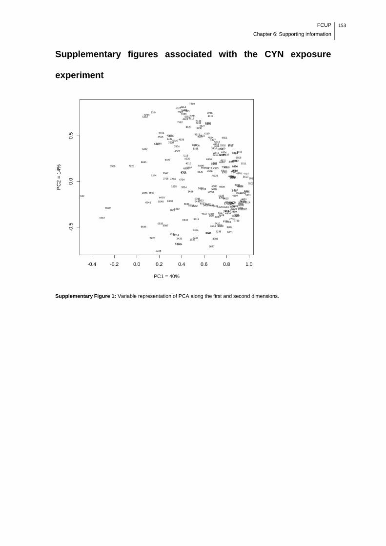
FCUP
Chapter 6: Supporting information
153
Supplementary figures associated with the CYN exposure
experiment
Supplementary Figure 1: Variable representation of PCA along the first and second dimensions.
-0.4 -0.2 0.0 0.2 0.4 0.6 0.8 1.0
-0.5
0.0
0.5
PC1 = 40%
PC
2 =
14
%
1327
1609
3244
3310
3312
3313
3325 3410
3416
3419
3435 3436
3438
3508
3509
3510
3511
3706
3707
4110
42074214
4216
4217
4301
4308
4320
4323
4327
4328
4401
4409
44104412
4423
4424
4431
4432
4504
45054514
4516
4521
4522
4526
4527
4528
4529
4531
45334534
4535
4536
4603
4604
4606
4607
4610
4612
4651
47044706
4707
5110
5206
5209
5210
521152125213
5214
5302
5305
5307
5310
5312
5314
5315
54045408
54175427
5505
5510
5513
5525
5547
5550
5610
5618
5620
56385705
5706
5707
6304
6310
6318
6327 6506
6524 6536
6635
7216
7218
7219
7501
7504
7508
7513
7518
7520
7522
1326
1612
2226
2228
2235
2430
3212
3225
3309
3314
3319
3409
3414
3425
3433
3501
350435073606
4303
4304
4307
4313
4326
4402
4524
4532
4601
4602
4609
46494654
4655
47024703
5301
54065407
5421
5448
5450
5455
5502
55145515
5537
5545
55465548
5602
5603
5605
5607
56095613
5623
5628
5634
5635
5636
56375639
5702
5708
5710
6301
6302
6303
6328
6401
6402
6403
6404
6405
6408
6410
6502
6505
6507
6511
65126513
6514
6516
6527
65316533
6535
6537
6538
6539
6601
6602
6637
6638
6640
6641
6642
6643
6701
6703
6704
7202
7203
73017303
7503
7602
6329 7225
1512
6532
6639
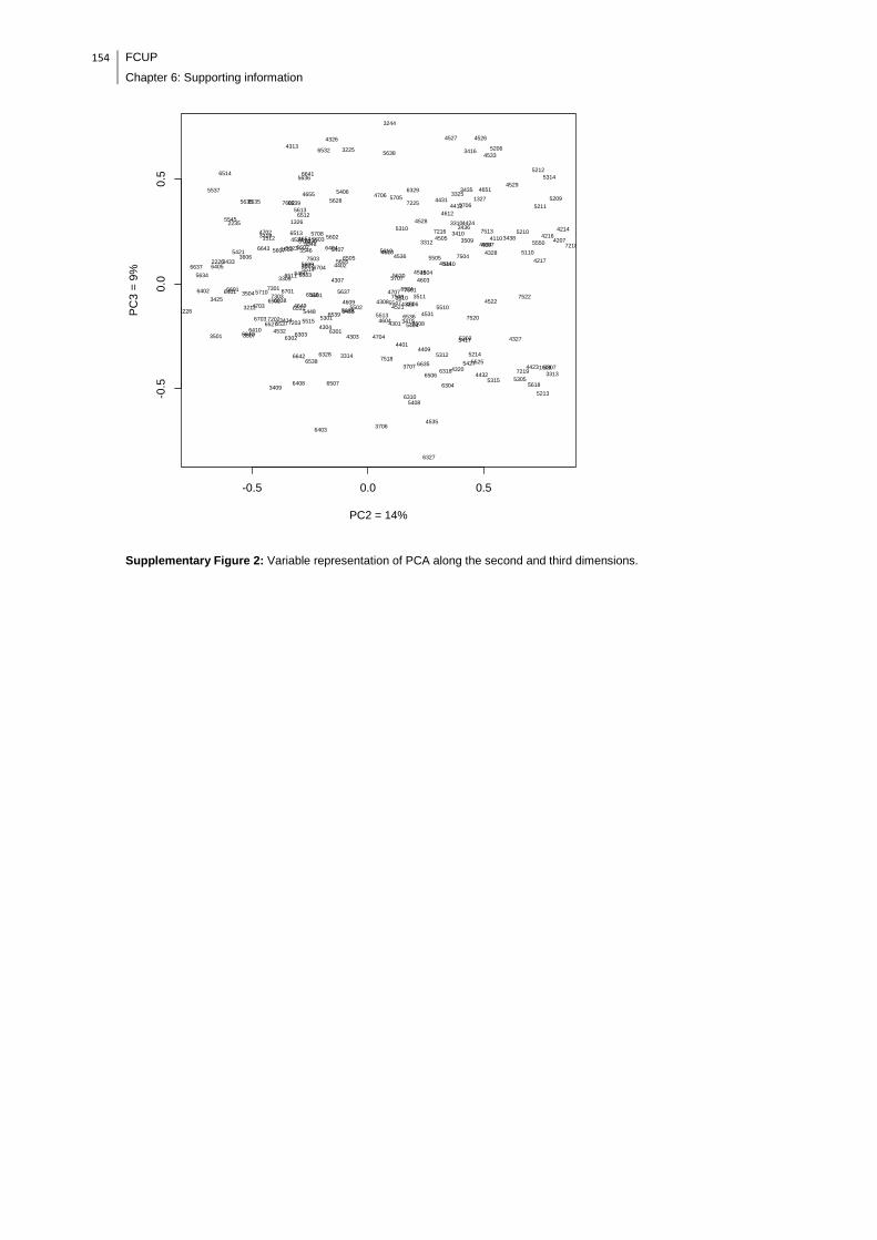
154 FCUP
Chapter 6: Supporting information
Supplementary Figure 2: Variable representation of PCA along the second and third dimensions.
-0.5 0.0 0.5
-0.5
0.0
0.5
PC2 = 14%
PC
3 =
9%
1327
3244
3310
3312
3325
3410
3416
3435
3436
34383509 4110 4207
4214
4216
4217
4328
4410
4412
4424
4431
4504
4505
4514
4516
45264527
4528
4529
4533
4534
4536
4603
4607
4610
4612
46514706
5110
5206
5209
5210
5211
5212
5310
5314
5505
5550
5610
5620
5638
5705
5706
5707
6329
7216
7218
7225
7504
7513
16093313
34193508
3510 3511
3706
3707
4301
4308
4320
4323
43274401
4409
4423
4432
45214522
4531
4535
4604
4606
4704
4707
5213
5214
5302
5305
5307
5312
5315
5404
5408
5417
5427
5510
5513
5525
5547
56186304
6310
6318
6327
6506
6524
6536
6635
7219
7501
7508
7518
7520
7522
1326
1512
1612
2226
2235
2430
3225
3309
3319
34333606
4307
4313
4326
4402
4524
4602
4654
4655
4702
5406
54075421
54505514
5537
5545
55465548
56025603
5605
5607
5609
5613
5623
5628
5634
5635
5636
5702
5708
6404
6405
6505
6511
6512
6513
6514
6532
6533
6535
6602
6637
6639
6641
6643
6704
7503
7602
22283212
3314
3409
3414
3425
3501
3504
3507 4303
43044532
4601
460946494703
53015448 5455
5502
5515
5637
5639
5710
63016302
6303
6328
64016402
6403
6408
6410
6502
6507
6516
6527
6531
6537
6538
6539
6601
6638
6640
6642
6701
6703 72027203
7301
7303
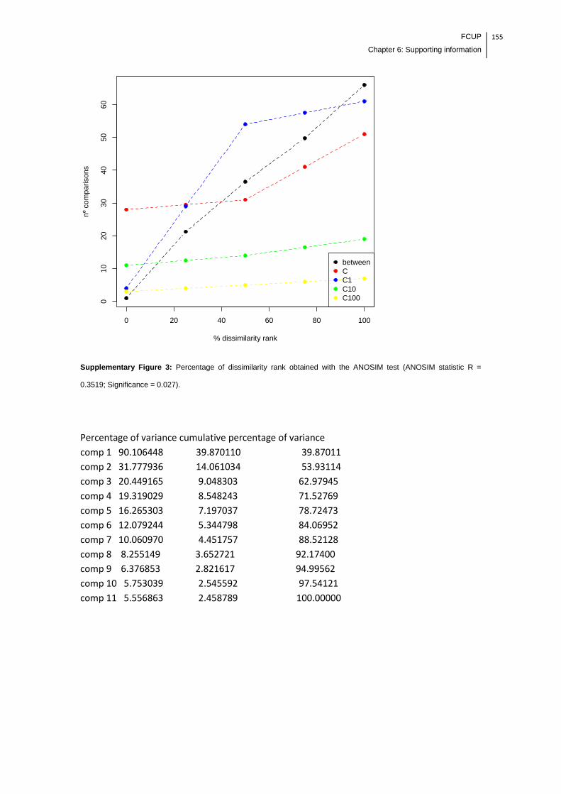
FCUP
Chapter 6: Supporting information
155
Supplementary Figure 3: Percentage of dissimilarity rank obtained with the ANOSIM test (ANOSIM statistic R =
0.3519; Significance = 0.027).
Percentage of variance cumulative percentage of variance
comp 1 90.106448 39.870110 39.87011
comp 2 31.777936 14.061034 53.93114
comp 3 20.449165 9.048303 62.97945
comp 4 19.319029 8.548243 71.52769
comp 5 16.265303 7.197037 78.72473
comp 6 12.079244 5.344798 84.06952
comp 7 10.060970 4.451757 88.52128
comp 8 8.255149 3.652721 92.17400
comp 9 6.376853 2.821617 94.99562
comp 10 5.753039 2.545592 97.54121
comp 11 5.556863 2.458789 100.00000
0 20 40 60 80 100
010
20
30
40
50
60
% dissimilarity rank
nº
com
pari
sons
between
C
C1
C10
C100

156 FCUP
Chapter 6: Supporting information
Supplementary Figure 4: 2-DE gel of lettuce leaf-protein spots that changed in abundance after CYN exposure. A
protein mass of 400 µg was loaded on each Immobiline IEF gel strip (17 cm, pH 4-7) for isoelectric focusing. The SDS-
PAGE was performed in 12% (w/v) polyacrylamide gels, which were stained with Colloidal Coomassie G-250. The
differentially abundant proteins were identified by MALDI-TOF/TOF MS. The spots surrounded by a green color
correspond to group C1, the spots surrounded by a blue color correspond to group C10, and the spots surrounded by a
purple color correspond to group C100. The spot numbers labeled red indicate that the same protein exhibited changes
in abundance in multiple groups.

FCUP
Chapter 6: Supporting information
157
Supplementary figures associated with the MC-LR/CYN
exposure experiment
Supplementary Figure 5: Variable representation of PCA along the first and second dimensions.
-0.5 0.0 0.5 1.0
-0.5
0.0
0.5
PC1 = 33%
PC
2 =
25%
X105X206
X1109
X1113
X1114
X1121
X1202
X1301
X1304
X1305
X1307X1309
X1310
X1311
X1312
X1314
X1315
X1316
X1321
X13139
X13140
X1407
X1408
X1561
X1614
X1674
X1702
X1704
X1804
X2108
X2110
X2113
X2206
X2210
X2216
X22109
X2304
X2305
X2307
X2308
X2313X23264
X2402
X2501
X2504
X2505
X2605
X2701
X2706
X2708
X2720
X27426
X2801
X2802
X2808
X3203
X32116
X32118
X32119X3301
X3303
X3305
X3316
X3323
X3401
X3407
X34102X34103
X3507
X3508
X3510
X36127
X36128X3702
X3703X3704
X3803
X3804
X41100
X41101X4210
X4273
X4301
X4377
X4380
X4402
X4491
X4492
X4495
X4496
X4504
X4508
X4585
X4586
X4587
X4606
X46120
X46121
X4703
X4706
X4711
X47164
X47166
X47167
X4804
X5201
X5216
X5226
X5309
X5405
X5407
X5494
X5503
X5604
X5607X5712
X5714
X5722X5723
X5724
X5801
X5802
X5805
X6365
X6367
X6410X6412
X6490
X6503
X6506
X6509
X6602
X6604
X66103
X66105
X66108
X66109X6707
X6711
X6713
X6716
X6717
X7306
X7405
X7406
X74109
X7514
X76147
X7704
X7709
X8101
X8104
X8105
X81138
X8204
X8207
X82105
X8301
X8302
X8304
X8305X8401
X8410
X8412
X8418
X8503
X8504
X8508
X8607
X8609
X8706
X8709
X9202X9503
X1203
X1204
X1205
X12146
X1313
X1317
X1402
X1405
X1406
X1501X2106
X2201
X2202
X2204
X2209
X2212
X2214
X2217
X2218X2219
X2301
X2306
X2309
X2310
X2314
X2325X24110X26150
X2703
X2705X2707
X3103
X3108
X3112
X31133
X3201
X3204
X3207
X3208
X3250
X32117
X32120
X3302
X3304X3306
X3309
X3310
X3312
X3315
X3317
X33119
X3402 X3403
X3404
X3504X3509
X3583
X36126
X3701X3809
X3810
X4018
X4106
X4204
X4205
X4206
X4208
X4253
X4256
X4274
X4275
X4307
X4308
X4309
X4310
X4376
X4378
X4379
X4382
X4401
X4403
X4405X4407
X4410
X4412
X4413X4415
X4502
X4503
X4507
X4515
X4616
X4704
X4707
X47168
X4811
X5101
X5102
X5202
X5204
X5206
X5208
X5209
X5212
X5214
X5215
X5218X5246
X5301
X5302
X5305
X5307
X5370X5371
X5402X5404
X5408
X5438
X5490
X5492
X5493X5504 X5511
X5512
X5521
X5560
X56105
X5704
X5710
X5719
X5720
X57149
X6108
X6202
X6203
X6204
X6206
X6207
X6262
X6305
X6366
X6401
X6403
X6404
X6405
X6406
X6413
X6415
X6492
X6495
X6502X6507
X6510
X6517
X6693
X66106
X6701
X6702
X6730
X6811
X7103X7104
X7107
X7201
X7204
X7205
X7301
X7304
X7402
X7677X7807
X8102
X8407
X8414
X9108
X1703X21229 X2302
X2303
X2403
X2702
X3210
X3211
X3308
X3406X3408
X3409
X3501
X3603
X3607
X3801
X3812
X4406X4408
X4409
X4497
X4509
X4510
X4511
X4512
X4514
X4602
X4607
X4612
X4701
X47165
X5306
X5372
X5401
X5406
X5488
X5489
X5495
X5502
X5506
X5510X5516X5605 X5606
X5706
X5707
X5708
X5803
X5808X5820
X5821
X6208
X6209
X6210
X6302
X6304
X6363
X6370
X6372
X6402X6409
X6411
X6436
X6487X6488
X6512
X6513X6514
X65108
X65109
X65110
X6603
X6607
X6608
X66107
X6705X6718
X6719
X6731
X6752
X67111
X6803
X6805
X6807
X6808
X6810
X7101
X7210
X7299X7403
X7410
X74106
X7501
X7504
X75116
X75117
X76149X7701
X7702
X7716X77192X77193
X8404X8405
X8413X8416
X8501
X8505
X8506
X8583
X8605
X8701X8713
X8809
X1127
X1446
X2118
X2123
X2405
X2803
X3811
X41102
X4381
X4404
X4498
X4506
X4613
X4809X4814
X4817
X5145
X5184X5185
X5219
X5278
X5308
X5333
X5373
X5491
X5496
X5520
X5721 X57148
X57154
X5810
X6101
X6103
X6106
X6107X6263
X6264
X6265X6266
X6267X6301
X6303X6306
X6307
X6368
X6369
X6371
X6408
X6489
X6491
X6493
X6494
X6496
X6497
X6601
X6605X6606
X6626
X66110
X66111
X6724
X6732
X6734
X67110
X6809
X6821
X7102
X7206
X72100
X72101
X7302
X7305
X7395
X7399
X7408
X74107
X74108
X7512
X7513
X75118
X75119
X7604
X7606
X7611
X7612
X7708
X7711
X7713
X7715
X77189
X7802X7803 X7810
X7812
X8201
X8202
X8203
X8208
X8303X8415
X8419X8604
X8804

158 FCUP
Chapter 6: Supporting information
Supplementary Figure 6: Variable representation of PCA along the second and third dimensions.
-0.5 0.0 0.5
-0.6
-0.4
-0.2
0.0
0.2
0.4
0.6
0.8
PC2 = 25%
PC
3 =
11%
X206
X1109
X1114
X1121
X1301
X1304
X1305
X1307
X1309
X1310
X1311
X1312
X1314
X13139
X13140
X1408
X1561
X1614
X1674
X1703
X1804
X2108
X2110
X21229
X2210
X2302
X2303
X2304
X2308
X23264
X2504
X2701
X2708
X2802
X2808
X3203X3211
X32119
X3301
X3303
X3305
X3308
X3407
X34102
X3507
X3702
X3703
X41100
X41101
X4273
X4301
X4380
X4402
X4406
X4409
X4491
X4495
X4496
X4497
X4504
X4586
X4587
X4602
X4612
X46120
X46121
X4701
X4703
X47167
X4804
X5201
X5216
X5372
X5405
X5407
X5494
X5502
X5503X5604
X5607
X5706
X5708
X5723
X5724
X5803
X6208
X6209X6302
X6304
X6363
X6365
X6367
X6370
X6372X6409
X6412
X6436
X6487
X6490
X6514
X6604
X66108
X6716X6717
X6719
X6731
X6752
X6808
X6810
X7101
X7299
X74106
X75116
X76149
X77193
X8104
X8105
X81138
X8204
X8207
X8305
X8405X8410
X8413X8416
X8501
X8504
X8505
X8506
X8583
X8713X8809
X9503
X105
X1113X1202
X1315
X1316
X1321
X1407X1702
X1704
X2113
X2206
X2216
X22109
X2305
X2307
X2313
X2402
X2403
X2501
X2505
X2605
X2702
X2706
X2720
X27426
X2801X3210
X32116
X32118
X3316
X3323
X3401
X3406
X3408
X3409 X34103
X3501
X3508
X3510
X3603
X3607X36127
X36128
X3704
X3801
X3803
X3804
X3812
X4210
X4377
X4408
X4492
X4508
X4509X4510
X4511
X4512
X4514
X4585
X4606
X4607
X4706
X4711
X47164
X47165
X47166
X5226
X5306
X5309
X5401
X5406
X5488
X5489
X5495X5506
X5510
X5516
X5605
X5606
X5707X5712 X5714
X5722
X5801
X5802
X5805
X5808
X5820
X5821
X6210
X6402
X6410X6411
X6488
X6503 X6506X6509
X6512X6513
X65108X65109
X65110
X6602X6603X6607
X6608
X66103X66105
X66107
X66109
X6705X6707
X6711
X6713
X6718
X67111
X6803X6805
X6807
X7210
X7306
X7403
X7405
X7406
X7410
X74109
X7501
X7504
X7514
X75117X76147
X7701
X7702
X7704 X7709
X7716X77192
X8101
X82105
X8301
X8302
X8304
X8401
X8404
X8412
X8418X8503
X8508
X8605
X8607X8609
X8701
X8706
X8709
X9202
X1127X1204
X1313 X1402
X1406
X2118
X2123
X2201
X2202
X2204
X2209
X2212
X2214X2217
X2218
X2219
X2301
X2310
X2325
X2703
X2707
X2803
X3103
X3108
X3112
X31133X3201
X3204
X3207
X3208
X32120
X3309
X3315
X33119
X3403
X36126
X3809X4018
X4106
X41102
X4205
X4206 X4208
X4253
X4256X4274
X4275
X4309
X4376
X4379
X4382
X4401
X4403
X4404
X4405
X4407
X4412
X4498
X47168
X4814
X5101
X5102
X5184
X5185
X5202
X5206
X5208
X5209
X5214
X5215
X5219X5246
X5278
X5301
X5302
X5307
X5333
X5370
X5373
X5404
X5408
X5490
X5491
X5492 X5511
X5704
X5719
X5720
X57148
X57154
X5810
X6101
X6103
X6106
X6107
X6202
X6204
X6206X6262
X6263
X6264
X6265
X6266
X6267
X6301
X6303
X6306
X6307X6368
X6369
X6371
X6401
X6404X6413X6415
X6489
X6491
X6492
X6494
X6495
X6496
X6497
X6606
X6626X66110
X66111
X6734
X67110
X6809
X6811
X6821X7102
X7103X7104
X7107X7201
X7204
X7206
X72100
X72101
X7301
X7304 X7305
X7395
X7399X7408
X74107
X74108
X7512
X7513
X75118
X75119
X7611
X7612
X7677
X7708X7711
X7713
X7715 X7802X7803
X7807
X7810
X7812
X8102
X8202
X8203
X8208
X8303
X8414
X8604
X8804
X1203
X1205
X12146
X1317
X1405
X1446
X1501
X2106
X2306X2309
X2314
X2405
X24110X26150
X2705
X3250
X32117
X3302 X3304
X3306
X3310
X3312X3317
X3402
X3404
X3504
X3509
X3583
X3701
X3810
X3811
X4204
X4307X4308
X4310
X4378
X4381
X4410
X4413
X4415
X4502
X4503
X4506
X4507
X4515
X4613
X4616
X4704
X4707
X4809
X4811
X4817
X5145
X5204
X5212
X5218
X5305
X5308X5371
X5402
X5438
X5493
X5496
X5504
X5512
X5520
X5521
X5560
X56105X5710
X5721X57149
X6108
X6203
X6207
X6305
X6366
X6403
X6405
X6406
X6408
X6493
X6502X6507
X6510
X6517
X6601
X6605
X6693
X66106
X6701
X6702 X6724
X6730X6732
X7205X7302X7402
X7604
X7606
X77189
X8201
X8407
X8415
X8419
X9108

FCUP
Chapter 6: Supporting information
159
Supplementary Figure 7: Percentage of dissimilarity rank obtained with the ANOSIM test (ANOSIM statistic R =
0.6636; Significance = 0.002).
Percentage of variance cumulative percentage of variance
comp 1 184.040890 32.516058 32.51606
comp 2 142.833375 25.235579 57.75164
comp 3 63.703200 11.254982 69.00662
comp 4 43.745250 7.728843 76.73546
comp 5 31.651982 5.592223 82.32768
comp 6 28.372424 5.012796 87.34048
comp 7 22.878164 4.042078 91.38256
comp 8 16.114324 2.847054 94.22961
comp 9 12.191268 2.153934 96.38355
comp 10 11.198727 1.978574 98.36212
comp 11 9.270396 1.637879 100.00000
0 20 40 60 80 100
010
20
30
40
50
60
% dissimilarity rank
nº
co
mpa
rison
s
betweenCC1C10C100

160 FCUP
Chapter 6: Supporting information
Supplementary Figure 8: 2-DE gel of lettuce leaf-protein spots that changed in abundance after MC-LR/CYN
exposure. A protein mass of 400 µg was loaded on each Immobiline IEF gel strip (17 cm, pH 4-7) for isoelectric
focusing. The SDS-PAGE was performed in 12% (w/v) polyacrylamide gels, which were stained with Colloidal
Coomassie G-250. The differentially abundant proteins were identified by MALDI-TOF/TOF MS. The spots surrounded
by a green color correspond to group C1, the spots surrounded by a blue color correspond to group C10, and the spots
surrounded by a purple color correspond to group C100. The spot numbers labeled in red indicate that the same protein
exhibited changes in abundance in multiple groups.

FCUP
Chapter 6: Supporting information
161
162 FCUP
Chapter 6: Supporting information
Supplemental table 1: Full data regarding to the proteins identification of the CYN exposure experiment with lettuce plants.
Condition/Function
Spot number
Proteins identified Homologous
protein Species
Acession number
Score Expect No. of unique
peptides
MW (Da)
Spot intensity
Fold change
variation
Relative protein
abundance
Average ± SD C
Average ± SD C1
Average ± SD C10
Average ± SD C100
Photosynthesis and carbon metabolism
C1 3212 RecName: Full=Plastocyanin Lactuca sativa
gi|130270 323 5.80E-27 3 10410 8130.3 ± 3298.6
1716.7 ± 491.8
NA NA 4.7 Decreased
5545 EST QGF6C23.yg.ab1 QG_EFGHJ lettuce serriola Lactuca serriola cDNA clone QGF6C23, mRNA sequence
Chain A, Chloroplast Nadp-Dependent Malate Dehydrogenase
Flaveria bidentis
gi|22413587|gb|BQ994052.1|
294 7.00E-24 4 26207 90 ± 41.8
18.7 ± 11.63
NA NA 4.8 Decreased
C1 and C100
5307 EST QGF5O18.yg.ab1 QG_EFGHJ lettuce serriola Lactuca serriola cDNA clone QGF5O18, mRNA sequence
PREDICTED: chlorophyll a-b binding protein 8, chloroplastic-like
Cucumis sativus
gi|22413490|gb|BQ993955.1|
208 2.80E-15 4 27024 NA 44.1 ± 16.4
NA 106.7 ± 65.4
Protein spot absent in C
NA
C10 5315 EST CLRY6253.b1_J03.ab1 CLR(XYZ) lettuce serriola Lactuca serriola cDNA clone CLRY6253, mRNA sequence
Chlorophyll a/b-binding protein
Tagetes erecta
gi|83915463|gb|DW117543.1|
208 2.80E-15 3 31074
5.7 ± 3.2 NA 289.2 ± 145.1
NA 50.7 Increased
C10 and C100
4423 Chlorophyll a/b binding protein precursor Euphorbia esula
gi|6716783 112 7.30E-06 2 28542 NA NA 119 ± 72.1 441.4 ± 126.8
Protein spot absent in C
NA
4531 EST CLSS13408.b1_P16.ab1 CLS(LMS) lettuce sativa Lactuca sativa cDNA clone CLSS13408, mRNA sequence
PREDICTED: quinone oxidoreductase-like protein At1g23740, chloroplastic-like
Fragaria vesca subsp. Vesca
gi|90517876|gb|DY979734.1|
162 1.10E-10 2 27498 2.3 ± 1.4 NA 29 ± 15.9 49.2 ± 3.5 12.8 (C10) and 21.7 (C100)
Increased
5305 EST QGF5O18.yg.ab1 QG_EFGHJ lettuce serriola Lactuca serriola cDNA clone QGF5O18, mRNA sequence
PREDICTED: chlorophyll a-b binding protein 8, chloroplastic-like
Cucumis sativus
gi|22413490|gb|BQ993955.1|
171 1.40E-11 5 27024 NA NA 188.6 ± 111.8
224.5 ± 59.2
Protein spot absent in C
NA
6318 EST CLPX5655.b1_M22.ab1 CLP(XYZ) lettuce perennis Lactuca perennis cDNA clone CLPX5655, mRNA sequence
Oxygen-evolving enhancer protein 2, chloroplastic
NA gi|83880790|gb|DW082870.1|
260 1.80E-20 4 24725 NA NA 210.5 ± 27.5
344.2 ± 76.7
Protein spot absent in C
NA
C100 4535 Sedoheptulose-1,7-bisphosphatase Cucumis sativus
gi|229597543
143 5.80E-09 3 42532 15.3 ± 7.2
NA NA 180.5 ± 79.9
11.8 Increased
4601 EST CLPX707.b1_F10.ab1 CLP(XYZ) lettuce perennis Lactuca perennis cDNA clone CLPX707, mRNA sequence
Ribulose bisphosphate carboxylase/oxygenase activase 1, chloroplast precursor, putative
Ricinus communis
gi|83881931|gb|DW084011.1|
623 8.80E-57 7 31547 382.5 ± 83.5
NA NA 932.8 ± 168.6
2.4 Increased
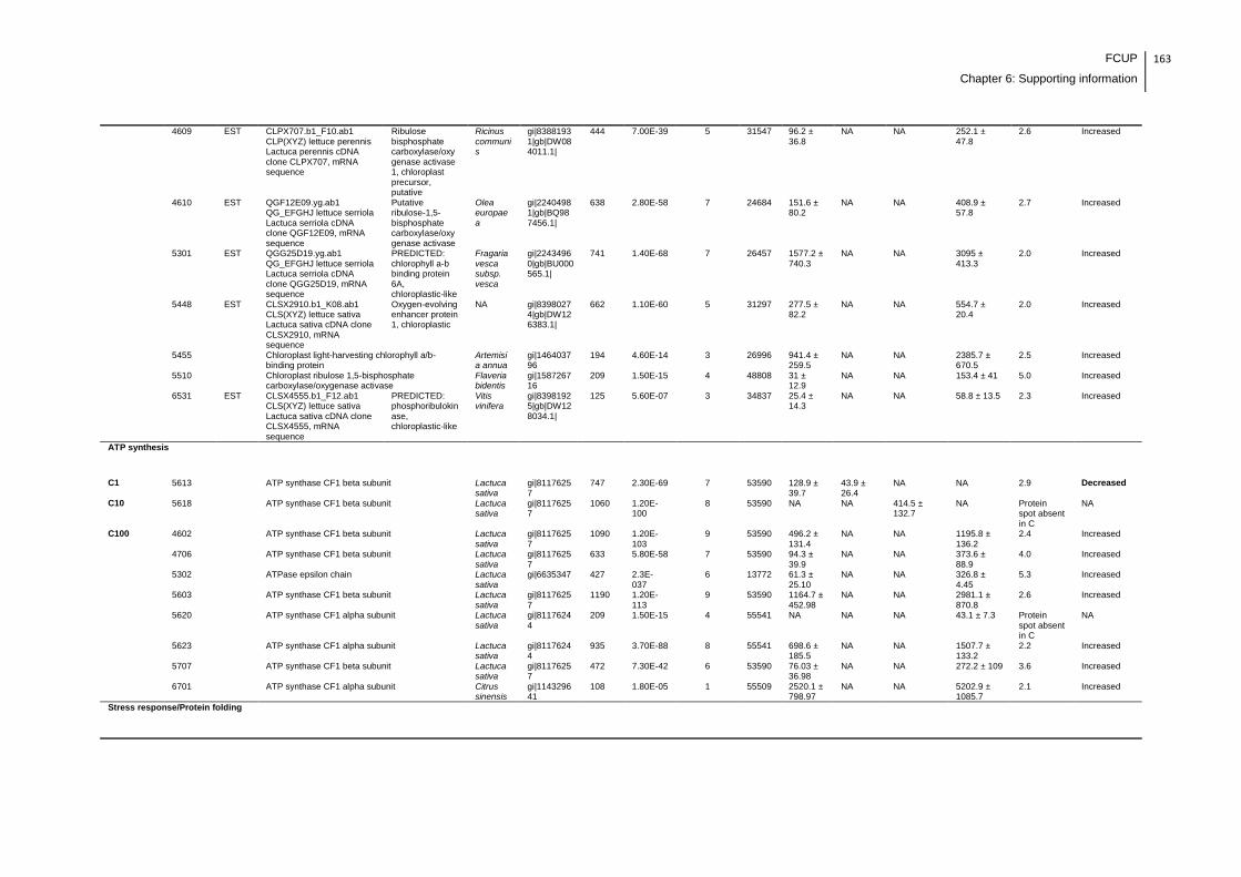
FCUP
Chapter 6: Supporting information
163
4609 EST CLPX707.b1_F10.ab1 CLP(XYZ) lettuce perennis Lactuca perennis cDNA clone CLPX707, mRNA sequence
Ribulose bisphosphate carboxylase/oxygenase activase 1, chloroplast precursor, putative
Ricinus communis
gi|83881931|gb|DW084011.1|
444 7.00E-39 5 31547 96.2 ± 36.8
NA NA 252.1 ± 47.8
2.6 Increased
4610 EST QGF12E09.yg.ab1 QG_EFGHJ lettuce serriola Lactuca serriola cDNA clone QGF12E09, mRNA sequence
Putative ribulose-1,5-bisphosphate carboxylase/oxygenase activase
Olea europaea
gi|22404981|gb|BQ987456.1|
638 2.80E-58 7 24684 151.6 ± 80.2
NA NA 408.9 ± 57.8
2.7 Increased
5301 EST QGG25D19.yg.ab1 QG_EFGHJ lettuce serriola Lactuca serriola cDNA clone QGG25D19, mRNA sequence
PREDICTED: chlorophyll a-b binding protein 6A, chloroplastic-like
Fragaria vesca subsp. vesca
gi|22434960|gb|BU000565.1|
741 1.40E-68 7 26457 1577.2 ± 740.3
NA NA 3095 ± 413.3
2.0 Increased
5448 EST CLSX2910.b1_K08.ab1 CLS(XYZ) lettuce sativa Lactuca sativa cDNA clone CLSX2910, mRNA sequence
Oxygen-evolving enhancer protein 1, chloroplastic
NA gi|83980274|gb|DW126383.1|
662 1.10E-60 5 31297 277.5 ± 82.2
NA NA 554.7 ± 20.4
2.0 Increased
5455 Chloroplast light-harvesting chlorophyll a/b-binding protein
Artemisia annua
gi|146403796
194 4.60E-14 3 26996
941.4 ± 259.5
NA NA 2385.7 ± 670.5
2.5 Increased
5510 Chloroplast ribulose 1,5-bisphosphate carboxylase/oxygenase activase
Flaveria bidentis
gi|158726716
209 1.50E-15 4 48808
31 ± 12.9
NA NA 153.4 ± 41 5.0 Increased
6531 EST CLSX4555.b1_F12.ab1 CLS(XYZ) lettuce sativa Lactuca sativa cDNA clone CLSX4555, mRNA sequence
PREDICTED: phosphoribulokinase, chloroplastic-like
Vitis vinifera
gi|83981925|gb|DW128034.1|
125 5.60E-07 3 34837 25.4 ± 14.3
NA NA 58.8 ± 13.5 2.3 Increased
ATP synthesis
C1 5613 ATP synthase CF1 beta subunit Lactuca sativa
gi|81176257
747 2.30E-69 7 53590 128.9 ± 39.7
43.9 ± 26.4
NA NA 2.9 Decreased
C10 5618 ATP synthase CF1 beta subunit Lactuca sativa
gi|81176257
1060 1.20E-100
8 53590
NA NA 414.5 ± 132.7
NA Protein spot absent in C
NA
C100 4602 ATP synthase CF1 beta subunit Lactuca sativa
gi|81176257
1090 1.20E-103
9 53590 496.2 ± 131.4
NA NA 1195.8 ± 136.2
2.4 Increased
4706 ATP synthase CF1 beta subunit Lactuca sativa
gi|81176257
633 5.80E-58 7 53590 94.3 ± 39.9
NA NA 373.6 ± 88.9
4.0 Increased
5302 ATPase epsilon chain Lactuca sativa
gi|6635347 427 2.3E-037
6 13772 61.3 ± 25.10
NA NA 326.8 ± 4.45
5.3 Increased
5603 ATP synthase CF1 beta subunit Lactuca sativa
gi|81176257
1190 1.20E-113
9 53590 1164.7 ± 452.98
NA NA 2981.1 ± 870.8
2.6 Increased
5620 ATP synthase CF1 alpha subunit Lactuca sativa
gi|81176244
209 1.50E-15 4 55541 NA NA NA 43.1 ± 7.3 Protein spot absent in C
NA
5623 ATP synthase CF1 alpha subunit Lactuca sativa
gi|81176244
935 3.70E-88 8 55541 698.6 ± 185.5
NA NA 1507.7 ± 133.2
2.2 Increased
5707 ATP synthase CF1 beta subunit Lactuca sativa
gi|81176257
472 7.30E-42 6 53590 76.03 ± 36.98
NA NA 272.2 ± 109 3.6 Increased
6701 ATP synthase CF1 alpha subunit Citrus sinensis
gi|114329641
108 1.80E-05 1 55509
2520.1 ± 798.97
NA NA 5202.9 ± 1085.7
2.1 Increased
Stress response/Protein folding
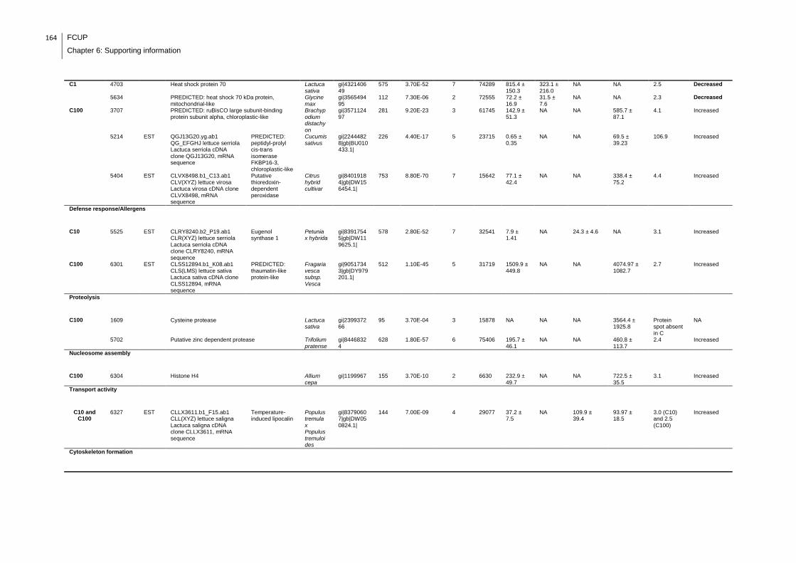
164 FCUP
Chapter 6: Supporting information
C1 4703 Heat shock protein 70 Lactuca sativa
gi|432140649
575 3.70E-52 7 74289 815.4 ± 150.3
323.1 ± 216.0
NA NA 2.5 Decreased
5634 PREDICTED: heat shock 70 kDa protein, mitochondrial-like
Glycine max
gi|356549495
112 7.30E-06 2 72555 72.2 ± 16.9
31.5 ± 7.6
NA NA 2.3 Decreased
C100 3707 PREDICTED: ruBisCO large subunit-binding protein subunit alpha, chloroplastic-like
Brachypodium distachyon
gi|357112497
281 9.20E-23 3 61745 142.9 ± 51.3
NA NA 585.7 ± 87.1
4.1 Increased
5214 EST QGJ13G20.yg.ab1 QG_EFGHJ lettuce serriola Lactuca serriola cDNA clone QGJ13G20, mRNA sequence
PREDICTED: peptidyl-prolyl cis-trans isomerase FKBP16-3, chloroplastic-like
Cucumis sativus
gi|22444828|gb|BU010433.1|
226 4.40E-17 5 23715 0.65 ± 0.35
NA NA 69.5 ± 39.23
106.9 Increased
5404 EST CLVX8498.b1_C13.ab1 CLV(XYZ) lettuce virosa Lactuca virosa cDNA clone CLVX8498, mRNA sequence
Putative thioredoxin-dependent peroxidase
Citrus hybrid cultivar
gi|84019184|gb|DW156454.1|
753 8.80E-70 7 15642 77.1 ± 42.4
NA NA 338.4 ± 75.2
4.4 Increased
Defense response/Allergens
C10 5525 EST CLRY8240.b2_P19.ab1 CLR(XYZ) lettuce serriola Lactuca serriola cDNA clone CLRY8240, mRNA sequence
Eugenol synthase 1
Petunia x hybrida
gi|83917545|gb|DW119625.1|
578 2.80E-52 7 32541
7.9 ± 1.41
NA 24.3 ± 4.6 NA 3.1 Increased
C100 6301 EST CLSS12894.b1_K08.ab1 CLS(LMS) lettuce sativa Lactuca sativa cDNA clone CLSS12894, mRNA sequence
PREDICTED: thaumatin-like protein-like
Fragaria vesca subsp. Vesca
gi|90517343|gb|DY979201.1|
512 1.10E-45 5 31719
1509.9 ± 449.8
NA NA 4074.97 ± 1082.7
2.7 Increased
Proteolysis
C100 1609 Cysteine protease Lactuca sativa
gi|239937266
95 3.70E-04 3 15878
NA NA NA 3564.4 ± 1925.8
Protein spot absent in C
NA
5702 Putative zinc dependent protease Trifolium pratense
gi|84468324
628 1.80E-57 6 75406 195.7 ± 46.1
NA NA 460.8 ± 113.7
2.4 Increased
Nucleosome assembly
C100 6304 Histone H4 Allium cepa
gi|1199967 155 3.70E-10 2 6630 232.9 ± 49.7
NA NA 722.5 ± 35.5
3.1 Increased
Transport activity
C10 and C100
6327 EST CLLX3611.b1_F15.ab1 CLL(XYZ) lettuce saligna Lactuca saligna cDNA clone CLLX3611, mRNA sequence
Temperature-induced lipocalin
Populus tremula x Populus tremuloides
gi|83790607|gb|DW050824.1|
144 7.00E-09 4 29077 37.2 ± 7.5
NA 109.9 ± 39.4
93.97 ± 18.5
3.0 (C10) and 2.5 (C100)
Increased
Cytoskeleton formation

FCUP
Chapter 6: Supporting information
165
C10 6514 Actin Stevia rebaudiana
gi|23955912
823 5.80E-77 8 41943 134.2 ± 52.3
NA 39.1 ± 20.4 NA 3.4 Decreased
C100 4536 Plastid-dividing ring protein Solanum lycopersicum
gi|350539589
109 1.50E-05 3 44069 107.8 ± 37.7
NA NA 302.93 ± 69.0
2.8 Increased
4603 Plastid-dividing ring protein Solanum lycopersicum
gi|350539589
167 2.30E-11 3 44069 61.9 ± 24.7
NA NA 313.3 ± 67.4
5.1 Increased
Unknown/Miscellaneous
C10 5213 EST CLLY5506.b1_D09.ab1 CLL(XYZ) lettuce saligna Lactuca saligna cDNA clone CLLY5506, mRNA sequence
PREDICTED: thylakoid lumenal 15 kDa protein 1, chloroplastic-like
Solanum lycopersicum
gi|83805737|gb|DW065954.1|
238 2.80E-18 5 31379 2.1 ± 0.9 NA 78.5 ± 39.3 NA 37.4 Increased
C10 and C100
6310 EST CLRX6025.b2_A20.ab1 CLR(XYZ) lettuce serriola Lactuca serriola cDNA clone CLRX6025, mRNA sequence
PREDICTED: auxin-binding protein ABP19a-like
Cucumis sativus
gi|83906681|gb|DW108761.1|
204 7.00E-15 2 30319
45.1 ± 14.8
NA 274.3 ± 90.8
202.6 ± 81.4
6.1 (C10) and 4.5 (C100)
Increased
7218 EST CLSS2149.b1_I10.ab1 CLS(LMS) lettuce sativa Lactuca sativa cDNA clone CLSS2149, mRNA sequence
Lettuce sativa Lactuca sativa cDNA clone CLSS2149, mRNA sequence
NA gi|90518319|gb|DY980177.1|
592 1.10E-53 8 28094 51.97 ± 18.9
NA 206.5 ± 55.4
237.7 ± 67.9
4.0 (C10) and 4.6 (C100)
Increased
C100 4320 EST QGA10P22.yg.ab1 QG_ABCDI lettuce salinas Lactuca sativa cDNA clone QGA10P22, mRNA sequence
Thylakoid lumen 18.3 kDa protein
Arabidopsis lyrata subsp. lyrata
gi|22223341|gb|BQ843556.1|
677 3.50E-62 6 27549 129.8 ± 53.3
NA NA 439.2 ± 81.4
3.4 Increased
4432 EST CLLY7947.b1_E20.ab1 CLL(XYZ) lettuce saligna Lactuca saligna cDNA clone CLLY7947, mRNA sequence
PREDICTED: unknown protein DS12 from 2D-PAGE of leaf, chloroplastic-like
Solanum lycopersicum
gi|83807970|gb|DW068187.1|
677 3.50E-62 6 27549 16.2 ± 1.9
NA NA 138.9 ± 24.7
8.6 Increased
5417 EST CLRX2340.b1_G09.ab1 CLR(XYZ) lettuce serriola Lactuca serriola cDNA clone CLRX2340, mRNA sequence
Lettuce serriola Lactuca serriola cDNA clone CLRX2340, mRNA sequence
NA gi|83902453|gb|DW104533.1|
669 2.20E-61 6 30931 67.6 ± 36.7
NA NA 291.5 ± 84 4.3 Increased
7303 EST CLRX6025.b2_A20.ab1 CLR(XYZ) lettuce serriola Lactuca serriola cDNA clone CLRX6025, mRNA sequence
PREDICTED: auxin-binding protein ABP19a-like
Cucumis sativus
gi|83906681|gb|DW108761.1|
510 1.80E-45 4 30319 1051.5 ± 456.2
NA NA 4514.2 ± 639.9
4.3 Increased
NOTES
gi| Proteins identified using NCBI databasw
gi|gb| Proteins identified using EST database
NA: not applicable

166 FCUP
Chapter 6: Supporting information
Supplemental table 2: Full data regarding to the proteins identification of the MC-LR/CYN exposure experiment with lettuce plants.
Condition/Function
Spot number
Proteins identified Homologous protein
Species Acession number
Score Expect No. of unique
peptides
MW (Da)
Spot intensity Fold change
variation
Relative protein
abundance Average ± SD C
Average ± SD C1
Average ± SD C10
Average ± SD C100
Photosynthesis and carbon metabolism
C1 2206 EST CLVX6829.b1_J04.ab1 CLV(XYZ) lettuce virosa Lactuca virosa cDNA clone CLVX6829, mRNA sequence
PREDICTED: heme-binding protein 2-like
Fragaria vesca subsp. Vesca
gi|84017490|gb|DW154760.1|
329 2.20E-27 4 25580 170.8 ± 89.4
540.1 ± 211.7
NA NA 3.2 Increased
2212 EST QGH5D12.yg.ab1 QG_EFGHJ lettuce serriola Lactuca serriola cDNA clone QGH5D12, mRNA sequence
PREDICTED: chlorophyll a-b binding protein 13, chloroplastic-like
Cucumis sativus
gi|22442095|gb|BU007700.1|
321 1.40E-26 3 25497 625.9 ± 194.5
1649 ± 208.8
NA NA 2.6 Increased
2216 EST CLLX13763.b1_F10.ab1 CLL(XYZ) lettuce saligna Lactuca saligna cDNA clone CLLX13763, mRNA sequence
PREDICTED: probable ribose-5-phosphate isomerase-like
Solanum lycopersicum
gi|83786877|gb|DW047094.1|
291 1.40E-23 5 30124 366.3 ± 144.02
814.1 ± 48.4
NA NA 2.2 Increased
2219 Chlorophyll a/b-binding protein Medicago truncatula
gi|357452427
118 1.80E-06 3 28592 254.5 ± 28.4
562.3 ± 107.2
NA NA 2.2 Increased
2505 EST CLPX707.b1_F10.ab1 CLP(XYZ) lettuce perennis Lactuca perennis cDNA clone CLPX707, mRNA sequence
Ribulose bisphosphate carboxylase/oxygenase activase 1, chloroplast precursor, putative
Ricinus communis
gi|83881931|gb|DW084011.1|
334 7.00E-28 6 31547 188.4 ± 77.7
554.7 ± 123.5
NA NA 2.9 Increased
3306 EST CLSY1255.b1_N01.ab1 CLS(XYZ) lettuce sativa Lactuca sativa cDNA clone CLSY1255, mRNA sequence
Oxygen-evolving enhancer protein 1, chloroplastic
NA gi|83987671|gb|DW133780.1|
263 8.80E-21 2 31446 157.6 ± 84.9
554.03 ± 122.2
NA NA 3.5 Increased
3312 EST CLSX2910.b1_K08.ab1 CLS(XYZ) lettuce sativa Lactuca sativa cDNA clone CLSX2910, mRNA sequence
Oxygen-evolving enhancer protein 1, chloroplastic
NA gi|83980274|gb|DW126383.1|
796 4.40E-74 7 31297 4049.6 ± 1060.7
7353 ± 610.2
NA NA 2.0 Increased
3404 EST CLSY9694.b1_K23.ab1 CLS(XYZ) lettuce sativa Lactuca sativa cDNA clone CLSY9694, mRNA sequence
PREDICTED: phosphoribulokinase, chloroplastic-like
Vitis vinifera
gi|83995525|gb|DW141634.1|
757 3.50E-70 8 35619 1127.8 ± 486.1
2591.8 ± 689.1
NA NA 2.3 Increased
3704 EST QGG12O20.yg.ab1 QG_EFGHJ lettuce serriola Lactuca serriola cDNA clone QGG12O20, mRNA sequence
PREDICTED: beta-xylosidase/alpha-L-arabinofuranosidase 2-like
Vitis vinifera
gi|22430866|gb|BQ996470.1|
241 1.40E-18 2 24887 128.6 ± 63.98
313.5 ± 8.7
NA NA 2.4 Increased
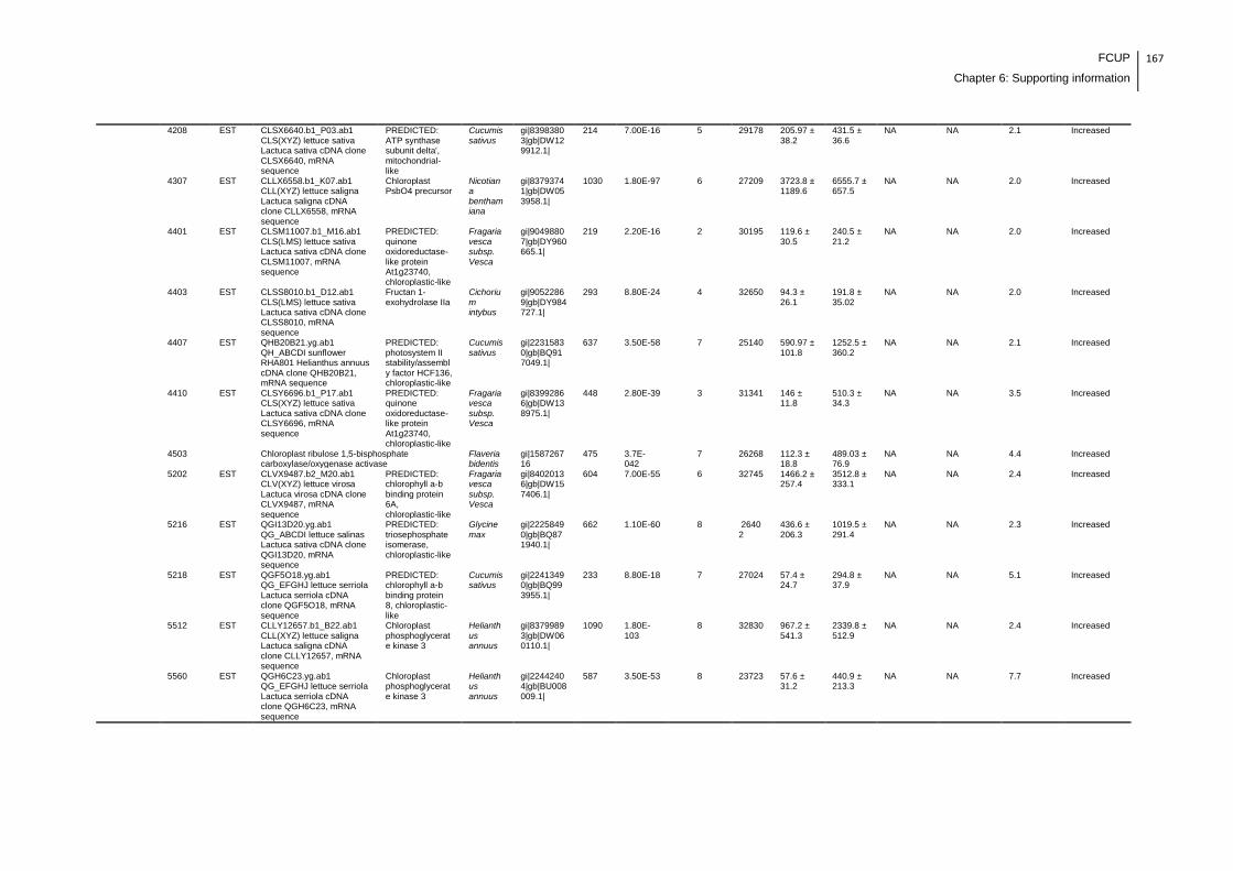
FCUP
Chapter 6: Supporting information
167
4208 EST CLSX6640.b1_P03.ab1 CLS(XYZ) lettuce sativa Lactuca sativa cDNA clone CLSX6640, mRNA sequence
PREDICTED: ATP synthase subunit delta', mitochondrial-like
Cucumis sativus
gi|83983803|gb|DW129912.1|
214 7.00E-16 5 29178 205.97 ± 38.2
431.5 ± 36.6
NA NA 2.1 Increased
4307 EST CLLX6558.b1_K07.ab1 CLL(XYZ) lettuce saligna Lactuca saligna cDNA clone CLLX6558, mRNA sequence
Chloroplast PsbO4 precursor
Nicotiana benthamiana
gi|83793741|gb|DW053958.1|
1030 1.80E-97 6 27209 3723.8 ± 1189.6
6555.7 ± 657.5
NA NA 2.0 Increased
4401 EST CLSM11007.b1_M16.ab1 CLS(LMS) lettuce sativa Lactuca sativa cDNA clone CLSM11007, mRNA sequence
PREDICTED: quinone oxidoreductase-like protein At1g23740, chloroplastic-like
Fragaria vesca subsp. Vesca
gi|90498807|gb|DY960665.1|
219 2.20E-16 2 30195 119.6 ± 30.5
240.5 ± 21.2
NA NA 2.0 Increased
4403 EST CLSS8010.b1_D12.ab1 CLS(LMS) lettuce sativa Lactuca sativa cDNA clone CLSS8010, mRNA sequence
Fructan 1-exohydrolase IIa
Cichorium intybus
gi|90522869|gb|DY984727.1|
293 8.80E-24 4 32650 94.3 ± 26.1
191.8 ± 35.02
NA NA 2.0 Increased
4407 EST QHB20B21.yg.ab1 QH_ABCDI sunflower RHA801 Helianthus annuus cDNA clone QHB20B21, mRNA sequence
PREDICTED: photosystem II stability/assembly factor HCF136, chloroplastic-like
Cucumis sativus
gi|22315830|gb|BQ917049.1|
637 3.50E-58 7 25140 590.97 ± 101.8
1252.5 ± 360.2
NA NA 2.1 Increased
4410 EST CLSY6696.b1_P17.ab1 CLS(XYZ) lettuce sativa Lactuca sativa cDNA clone CLSY6696, mRNA sequence
PREDICTED: quinone oxidoreductase-like protein At1g23740, chloroplastic-like
Fragaria vesca subsp. Vesca
gi|83992866|gb|DW138975.1|
448 2.80E-39 3 31341 146 ± 11.8
510.3 ± 34.3
NA NA 3.5 Increased
4503 Chloroplast ribulose 1,5-bisphosphate carboxylase/oxygenase activase
Flaveria bidentis
gi|158726716
475 3.7E-042
7 26268 112.3 ± 18.8
489.03 ± 76.9
NA NA 4.4 Increased
5202 EST CLVX9487.b2_M20.ab1 CLV(XYZ) lettuce virosa Lactuca virosa cDNA clone CLVX9487, mRNA sequence
PREDICTED: chlorophyll a-b binding protein 6A, chloroplastic-like
Fragaria vesca subsp. Vesca
gi|84020136|gb|DW157406.1|
604 7.00E-55 6 32745 1466.2 ± 257.4
3512.8 ± 333.1
NA NA 2.4 Increased
5216 EST QGI13D20.yg.ab1 QG_ABCDI lettuce salinas Lactuca sativa cDNA clone QGI13D20, mRNA sequence
PREDICTED: triosephosphate isomerase, chloroplastic-like
Glycine max
gi|22258490|gb|BQ871940.1|
662 1.10E-60 8 26402
436.6 ± 206.3
1019.5 ± 291.4
NA NA 2.3 Increased
5218 EST QGF5O18.yg.ab1 QG_EFGHJ lettuce serriola Lactuca serriola cDNA clone QGF5O18, mRNA sequence
PREDICTED: chlorophyll a-b binding protein 8, chloroplastic-like
Cucumis sativus
gi|22413490|gb|BQ993955.1|
233 8.80E-18 7 27024 57.4 ± 24.7
294.8 ± 37.9
NA NA 5.1 Increased
5512 EST CLLY12657.b1_B22.ab1 CLL(XYZ) lettuce saligna Lactuca saligna cDNA clone CLLY12657, mRNA sequence
Chloroplast phosphoglycerate kinase 3
Helianthus annuus
gi|83799893|gb|DW060110.1|
1090 1.80E-103
8 32830 967.2 ± 541.3
2339.8 ± 512.9
NA NA 2.4 Increased
5560 EST QGH6C23.yg.ab1 QG_EFGHJ lettuce serriola Lactuca serriola cDNA clone QGH6C23, mRNA sequence
Chloroplast phosphoglycerate kinase 3
Helianthus annuus
gi|22442404|gb|BU008009.1|
587 3.50E-53 8 23723 57.6 ± 31.2
440.9 ± 213.3
NA NA 7.7 Increased
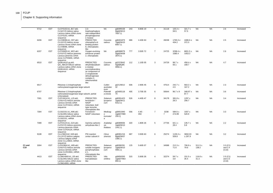
168 FCUP
Chapter 6: Supporting information
5712 EST CLSY5153.b1_B17.ab1 CLS(XYZ) lettuce sativa Lactuca sativa cDNA clone CLSY5153, mRNA sequence
2,3-bisphosphoglycerate-independent phosphoglycerate mutase
NA gi|83991588|gb|DW137697.1|
243 8.80E-19 4 31118 125.3 ± 59.5
321.7 ± 57.5
NA NA 2.6 Increased
6206 EST CLVX8096.b1_O07.ab1 CLV(XYZ) lettuce virosa Lactuca virosa cDNA clone CLVX8096, mRNA sequence
PREDICTED: chlorophyll a-b binding protein 8, chloroplastic-like
Cucumis sativus
gi|84018797|gb|DW156067.1|
686 4.40E-63 9 28238 1765.3 ± 434.2
4486.6 ± 372.3
NA NA 2.5 Increased
6207 EST CLPX5655.b1_M22.ab1 CLP(XYZ) lettuce perennis Lactuca perennis cDNA clone CLPX5655, mRNA sequence
Oxygen-evolving enhancer protein 2, chloroplastic
NA gi|83880790|gb|DW082870.1|
777 3.50E-72 7 24725 3298.4 ± 1085.4
8651.5 ± 1053.2
NA NA 2.6 Increased
6510 EST QGB14A10.yg.ab1 QG_ABCDI lettuce salinas Lactuca sativa cDNA clone QGB14A10, mRNA sequence
PREDICTED: dihydrolipoyllysine-residue succinyltransferase component of 2-oxoglutarate dehydrogenase complex 2, mitochondrial-like
Cucumis sativus
gi|22236425|gb|BQ850956.1|
112 1.10E-05 3 24729 98.7 ± 56.1
458.4 ± 168.7
NA NA 4.7 Increased
6517 Ribulose-1,5-bisphosphate carboxylase/oxygenase large subunit
Cullen australasicum
gi|212901232
506 2.90E-45 6 47514 242.7 ± 4.9
563.2 ± 167.2
NA NA 2.3 Increased
6707 Ribulose-1,5-bisphosphate carboxylase/oxygenase large subunit, partial (chloroplast)
Potentilla erecta
gi|340511870
405 3.70E-35 6 50644 96.7 ± 9 348.97 ± 87.7
NA NA 3.6 Increased
7301 EST CLRY4414.b1_L23.ab1 CLR(XYZ) lettuce serriola Lactuca serriola cDNA clone CLRY4414, mRNA sequence
PREDICTED: ferredoxin--NADP reductase, leaf-type isozyme, chloroplastic-like
Solanum lycopersicum
gi|83914232|gb|DW116312.1|
526 4.40E-47 8 34178 381.8 ± 28.3
1297 ± 206.7
NA NA 3.4 Increased
7304 EST CLSS2231.b1_N06.ab1 CLS(LMS) lettuce sativa Lactuca sativa cDNA clone CLSS2231, mRNA sequence
Ferredoxin-NADP reductase
Medicago truncatula
gi|90518401|gb|DY980259.1|
556 4.4E-050
7 32387
584.6 ± 274.99
1937 ± 520.9
NA NA 3.3 Increased
7399 EST CLPZ4124.b1_G24.ab1 CLP(XYZ) lettuce perennis Lactuca perennis cDNA clone CLPZ4124, mRNA sequence
Gamma carbonic anhydrase-like 2
Arabidopsis thaliana
gi|83899361|gb|DW101441.1|
320 1.80E-26 5 27735 63.1 ± 28.4
135.7 ± 9.8
NA NA 2.2 Increased
9108 EST CLLZ3823.b2_N20.ab1 CLL(XYZ) lettuce saligna Lactuca saligna cDNA clone CLLZ3823, mRNA sequence
PSI reaction center subunit II
Citrus sinensis
gi|83812168|gb|DW072385.1|
687 3.50E-63 6 25274 1135.3 ± 509.8
3650.03 ± 297.9
NA NA 3.2 Increased
C1 and C10
3304 EST CLRX2804.b1_H05.ab1 CLR(XYZ) lettuce serriola Lactuca serriola cDNA clone CLRX2804, mRNA sequence
PREDICTED: soluble inorganic pyrophosphatase 1, chloroplastic-like
Solanum lycopersicum
gi|83902911|gb|DW104991.1|
125 5.60E-07 2 34588 212.3 ± 71.6
706.9 ± 79.6
511.5 ± 138.2
NA 3.3 (C1) and 2.4 (C10)
Increased
4413 EST CLSM14093.b1_I20.ab1 CLS(LMS) lettuce sativa Lactuca sativa cDNA clone CLSM14093, mRNA
PREDICTED: transaldolase-like
Vitis vinifera
gi|90502001|gb|DY963859.1|
315 5.60E-26 4 32274 59.7 ± 3.8
151.5 ± 35.4
116.8 ± 31.3
NA 2.5 (C1) and 2.0 (C10)
Increased
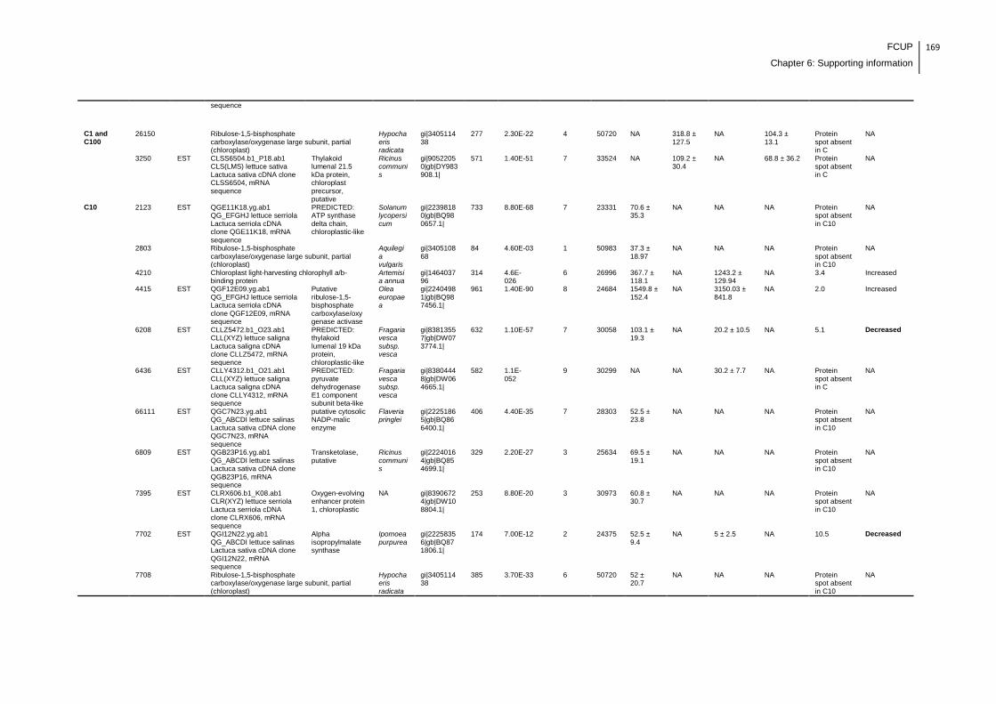
FCUP
Chapter 6: Supporting information
169
sequence
C1 and C100
26150 Ribulose-1,5-bisphosphate carboxylase/oxygenase large subunit, partial (chloroplast)
Hypochaeris radicata
gi|340511438
277 2.30E-22 4 50720 NA 318.8 ± 127.5
NA 104.3 ± 13.1
Protein spot absent in C
NA
3250 EST CLSS6504.b1_P18.ab1 CLS(LMS) lettuce sativa Lactuca sativa cDNA clone CLSS6504, mRNA sequence
Thylakoid lumenal 21.5 kDa protein, chloroplast precursor, putative
Ricinus communis
gi|90522050|gb|DY983908.1|
571 1.40E-51 7 33524 NA 109.2 ± 30.4
NA 68.8 ± 36.2 Protein spot absent in C
NA
C10 2123 EST QGE11K18.yg.ab1 QG_EFGHJ lettuce serriola Lactuca serriola cDNA clone QGE11K18, mRNA sequence
PREDICTED: ATP synthase delta chain, chloroplastic-like
Solanum lycopersicum
gi|22398180|gb|BQ980657.1|
733 8.80E-68 7 23331 70.6 ± 35.3
NA NA NA Protein spot absent in C10
NA
2803 Ribulose-1,5-bisphosphate carboxylase/oxygenase large subunit, partial (chloroplast)
Aquilegia vulgaris
gi|340510868
84 4.60E-03 1 50983 37.3 ± 18.97
NA NA NA Protein spot absent in C10
NA
4210 Chloroplast light-harvesting chlorophyll a/b-binding protein
Artemisia annua
gi|146403796
314 4.6E-026
6 26996 367.7 ± 118.1
NA 1243.2 ± 129.94
NA 3.4 Increased
4415 EST QGF12E09.yg.ab1 QG_EFGHJ lettuce serriola Lactuca serriola cDNA clone QGF12E09, mRNA sequence
Putative ribulose-1,5-bisphosphate carboxylase/oxygenase activase
Olea europaea
gi|22404981|gb|BQ987456.1|
961 1.40E-90 8 24684 1549.8 ± 152.4
NA 3150.03 ± 841.8
NA 2.0 Increased
6208 EST CLLZ5472.b1_O23.ab1 CLL(XYZ) lettuce saligna Lactuca saligna cDNA clone CLLZ5472, mRNA sequence
PREDICTED: thylakoid lumenal 19 kDa protein, chloroplastic-like
Fragaria vesca subsp. vesca
gi|83813557|gb|DW073774.1|
632 1.10E-57 7 30058 103.1 ± 19.3
NA 20.2 ± 10.5 NA 5.1 Decreased
6436 EST CLLY4312.b1_O21.ab1 CLL(XYZ) lettuce saligna Lactuca saligna cDNA clone CLLY4312, mRNA sequence
PREDICTED: pyruvate dehydrogenase E1 component subunit beta-like
Fragaria vesca subsp. vesca
gi|83804448|gb|DW064665.1|
582 1.1E-052
9 30299 NA NA 30.2 ± 7.7 NA Protein spot absent in C
NA
66111 EST QGC7N23.yg.ab1 QG_ABCDI lettuce salinas Lactuca sativa cDNA clone QGC7N23, mRNA sequence
putative cytosolic NADP-malic enzyme
Flaveria pringlei
gi|22251865|gb|BQ866400.1|
406 4.40E-35 7 28303 52.5 ± 23.8
NA NA NA Protein spot absent in C10
NA
6809 EST QGB23P16.yg.ab1 QG_ABCDI lettuce salinas Lactuca sativa cDNA clone QGB23P16, mRNA sequence
Transketolase, putative
Ricinus communis
gi|22240164|gb|BQ854699.1|
329 2.20E-27 3 25634 69.5 ± 19.1
NA NA NA Protein spot absent in C10
NA
7395 EST CLRX606.b1_K08.ab1 CLR(XYZ) lettuce serriola Lactuca serriola cDNA clone CLRX606, mRNA sequence
Oxygen-evolving enhancer protein 1, chloroplastic
NA gi|83906724|gb|DW108804.1|
253 8.80E-20 3 30973 60.8 ± 30.7
NA NA NA Protein spot absent in C10
NA
7702 EST QGI12N22.yg.ab1 QG_ABCDI lettuce salinas Lactuca sativa cDNA clone QGI12N22, mRNA sequence
Alpha isopropylmalate synthase
Ipomoea purpurea
gi|22258356|gb|BQ871806.1|
174 7.00E-12 2 24375 52.5 ± 9.4
NA 5 ± 2.5 NA 10.5 Decreased
7708 Ribulose-1,5-bisphosphate carboxylase/oxygenase large subunit, partial (chloroplast)
Hypochaeris radicata
gi|340511438
385 3.70E-33 6 50720 52 ± 20.7
NA NA NA Protein spot absent in C10
NA
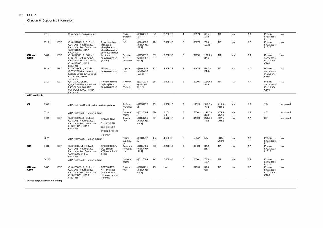
170 FCUP
Chapter 6: Supporting information
7711 Succinate dehydrogenase Litchi chinensis
gi|326467055
325 3.70E-27 4 69574 66.5 ± 19.3
NA NA NA Protein spot absent in C10
NA
7715 EST CLSM11592.b1_O17.ab1 CLS(LMS) lettuce sativa Lactuca sativa cDNA clone CLSM11592, mRNA sequence
Pyrophosphate--fructose 6-phosphate 1-phosphotransferase subunit beta
NA gi|90499383|gb|DY961241.1|
114 7.00E-06 2 32973 70.9 ± 10.05
NA NA NA Protein spot absent in C10
NA
C10 and C100
6409 EST CLSM12308.b1_G05.ab1 CLS(LMS) lettuce sativa Lactuca sativa cDNA clone CLSM12308, mRNA sequence
Isocitrate dehydrogenase (NAD+)
Nicotiana tabacum
gi|90500129|gb|DY961987.1|
639 2.20E-58 6 32255
102.2 ± 37.6
NA NA NA Protein spot absent in C10 and C100
NA
8413 EST CLVX7336.b1_O09.ab1 CLV(XYZ) lettuce virosa Lactuca virosa cDNA clone CLVX7336, mRNA sequence
Malate dehydrogenase
Glycine max
gi|84018031|gb|DW155301.1|
303 8.80E-25 5 29634 52.7 ± 24.94
NA NA NA Protein spot absent in C10 and C100
NA
8416 EST QGF20O02.yg.ab1 QG_EFGHJ lettuce serriola Lactuca serriola cDNA clone QGF20O02, mRNA sequence
Glyceraldehyde-3-phosphate dehydrogenase
Helianthus annuus
gi|22410236|gb|BQ990701.1|
513 8.80E-46 5 22206 124.4 ± 53.4
NA NA NA Protein spot absent in C10 and C100
NA
ATP synthesis
C1 4106 ATP synthase D chain, mitochondrial, putative Ricinus communis
gi|255577651
309 1.50E-25 5 19728 316.6 ± 71.4
618.8 ± 108.6
NA NA 2.0 Increased
5719 ATP synthase CF1 alpha subunit Lactuca sativa
gi|81176244
920 1.2E-086
9 55541 247.3 ± 39.8
674.5 ± 257.4
NA NA 2.7 Increased
7402 EST CLSM20020.b1_G13.ab1 CLS(LMS) lettuce sativa Lactuca sativa cDNA clone CLSM20020, mRNA sequence
PREDICTED:
ATP synthase
gamma chain,
chloroplastic-like
isoform 1
Glycine max
gi|90507111|gb|DY968969.1|
727 3.50E-67 8 34799 216.2 ± 79.8
797 ± 305.2
NA NA 3.7 Increased
7677 ATP synthase CF1 alpha subunit Lilium superbum
gi|338825720
134 4.60E-08 2 55342 NA 78.5 ± 25.98
NA NA Protein spot absent in C
NA
C10 6489 EST CLSM9831.b1_M10.ab1 CLS(LMS) lettuce sativa Lactuca sativa cDNA clone CLSM9831, mRNA sequence
PREDICTED: V-type proton ATPase subunit C-like
Solanum lycopersicum
gi|90514256|gb|DY976114.1|
239 2.20E-18 3 33428 32.2 ±8.7
NA NA NA Protein spot absent in C10
NA
66105 ATP synthase CF1 alpha subunit Lactuca sativa
gi|81176244
147 2.30E-09 3 55541 79.3 ± 11.7
NA NA NA Protein spot absent in C10
NA
C10 and C100
6487 EST CLSM20020.b1_G13.ab1 CLS(LMS) lettuce sativa Lactuca sativa cDNA clone CLSM20020, mRNA sequence
PREDICTED: ATP synthase gamma chain, chloroplastic-like isoform 1
Glycine max
gi|90507111|gb|DY968969.1|
102 NA 2 34799
55.9 ± 6.8
NA NA NA Protein spot absent in C10 and C100
NA
Stress response/Protein folding
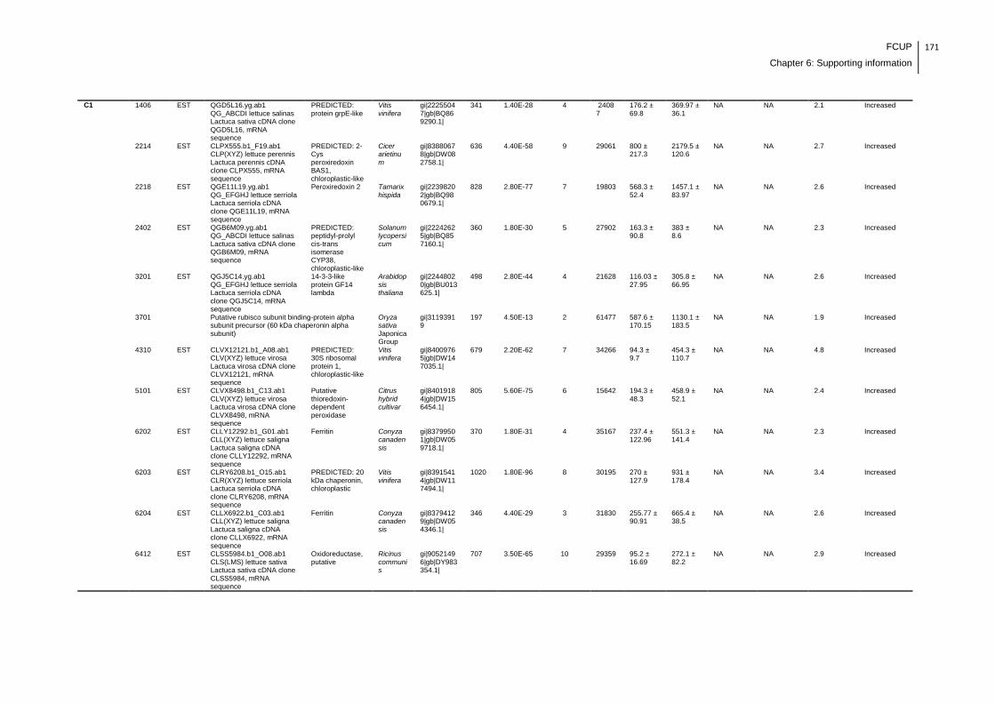
FCUP
Chapter 6: Supporting information
171
C1 1406 EST QGD5L16.yg.ab1 QG_ABCDI lettuce salinas Lactuca sativa cDNA clone QGD5L16, mRNA sequence
PREDICTED: protein grpE-like
Vitis vinifera
gi|22255047|gb|BQ869290.1|
341 1.40E-28 4 24087
176.2 ± 69.8
369.97 ± 36.1
NA NA 2.1 Increased
2214 EST CLPX555.b1_F19.ab1 CLP(XYZ) lettuce perennis Lactuca perennis cDNA clone CLPX555, mRNA sequence
PREDICTED: 2-Cys peroxiredoxin BAS1, chloroplastic-like
Cicer arietinum
gi|83880678|gb|DW082758.1|
636 4.40E-58 9 29061 800 ± 217.3
2179.5 ± 120.6
NA NA 2.7 Increased
2218 EST QGE11L19.yg.ab1 QG_EFGHJ lettuce serriola Lactuca serriola cDNA clone QGE11L19, mRNA sequence
Peroxiredoxin 2 Tamarix hispida
gi|22398202|gb|BQ980679.1|
828 2.80E-77 7 19803 568.3 ± 52.4
1457.1 ± 83.97
NA NA 2.6 Increased
2402 EST QGB6M09.yg.ab1 QG_ABCDI lettuce salinas Lactuca sativa cDNA clone QGB6M09, mRNA sequence
PREDICTED: peptidyl-prolyl cis-trans isomerase CYP38, chloroplastic-like
Solanum lycopersicum
gi|22242625|gb|BQ857160.1|
360 1.80E-30 5 27902 163.3 ± 90.8
383 ± 8.6
NA NA 2.3 Increased
3201 EST QGJ5C14.yg.ab1 QG_EFGHJ lettuce serriola Lactuca serriola cDNA clone QGJ5C14, mRNA sequence
14-3-3-like protein GF14 lambda
Arabidopsis thaliana
gi|22448020|gb|BU013625.1|
498 2.80E-44 4 21628 116.03 ± 27.95
305.8 ± 66.95
NA NA 2.6 Increased
3701 Putative rubisco subunit binding-protein alpha subunit precursor (60 kDa chaperonin alpha subunit)
Oryza sativa Japonica Group
gi|31193919
197 4.50E-13 2 61477 587.6 ± 170.15
1130.1 ± 183.5
NA NA 1.9 Increased
4310 EST CLVX12121.b1_A08.ab1 CLV(XYZ) lettuce virosa Lactuca virosa cDNA clone CLVX12121, mRNA sequence
PREDICTED: 30S ribosomal protein 1, chloroplastic-like
Vitis vinifera
gi|84009765|gb|DW147035.1|
679 2.20E-62 7 34266 94.3 ± 9.7
454.3 ± 110.7
NA NA 4.8 Increased
5101 EST CLVX8498.b1_C13.ab1 CLV(XYZ) lettuce virosa Lactuca virosa cDNA clone CLVX8498, mRNA sequence
Putative thioredoxin-dependent peroxidase
Citrus hybrid cultivar
gi|84019184|gb|DW156454.1|
805 5.60E-75 6 15642 194.3 ± 48.3
458.9 ± 52.1
NA NA 2.4 Increased
6202 EST CLLY12292.b1_G01.ab1 CLL(XYZ) lettuce saligna Lactuca saligna cDNA clone CLLY12292, mRNA sequence
Ferritin Conyza canadensis
gi|83799501|gb|DW059718.1|
370 1.80E-31 4 35167 237.4 ± 122.96
551.3 ± 141.4
NA NA 2.3 Increased
6203 EST CLRY6208.b1_O15.ab1 CLR(XYZ) lettuce serriola Lactuca serriola cDNA clone CLRY6208, mRNA sequence
PREDICTED: 20 kDa chaperonin, chloroplastic
Vitis vinifera
gi|83915414|gb|DW117494.1|
1020 1.80E-96 8 30195 270 ± 127.9
931 ± 178.4
NA NA 3.4 Increased
6204 EST CLLX6922.b1_C03.ab1 CLL(XYZ) lettuce saligna Lactuca saligna cDNA clone CLLX6922, mRNA sequence
Ferritin Conyza canadensis
gi|83794129|gb|DW054346.1|
346 4.40E-29 3 31830 255.77 ± 90.91
665.4 ± 38.5
NA NA 2.6 Increased
6412 EST CLSS5984.b1_O08.ab1 CLS(LMS) lettuce sativa Lactuca sativa cDNA clone CLSS5984, mRNA sequence
Oxidoreductase, putative
Ricinus communis
gi|90521496|gb|DY983354.1|
707 3.50E-65 10 29359 95.2 ± 16.69
272.1 ± 82.2
NA NA 2.9 Increased
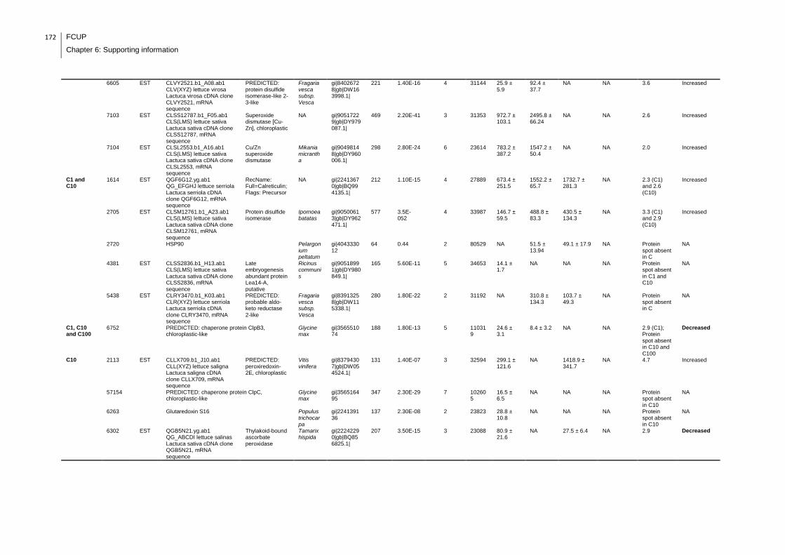
172 FCUP
Chapter 6: Supporting information
6605 EST CLVY2521.b1_A08.ab1 CLV(XYZ) lettuce virosa Lactuca virosa cDNA clone CLVY2521, mRNA sequence
PREDICTED: protein disulfide isomerase-like 2-3-like
Fragaria vesca subsp. Vesca
gi|84026728|gb|DW163998.1|
221 1.40E-16 4 31144 25.9 ± 5.9
92.4 ± 37.7
NA NA 3.6 Increased
7103 EST CLSS12787.b1_F05.ab1 CLS(LMS) lettuce sativa Lactuca sativa cDNA clone CLSS12787, mRNA sequence
Superoxide dismutase [Cu-Zn], chloroplastic
NA gi|90517229|gb|DY979087.1|
469 2.20E-41 3 31353 972.7 ± 103.1
2495.8 ± 66.24
NA NA 2.6 Increased
7104 EST CLSL2553.b1_A16.ab1 CLS(LMS) lettuce sativa Lactuca sativa cDNA clone CLSL2553, mRNA sequence
Cu/Zn superoxide dismutase
Mikania micrantha
gi|90498148|gb|DY960006.1|
298 2.80E-24 6 23614 783.2 ± 387.2
1547.2 ± 50.4
NA NA 2.0 Increased
C1 and C10
1614 EST QGF6G12.yg.ab1 QG_EFGHJ lettuce serriola Lactuca serriola cDNA clone QGF6G12, mRNA sequence
RecName: Full=Calreticulin; Flags: Precursor
NA gi|22413670|gb|BQ994135.1|
212 1.10E-15 4 27889 673.4 ± 251.5
1552.2 ± 65.7
1732.7 ± 281.3
NA 2.3 (C1) and 2.6 (C10)
Increased
2705 EST CLSM12761.b1_A23.ab1 CLS(LMS) lettuce sativa Lactuca sativa cDNA clone CLSM12761, mRNA sequence
Protein disulfide isomerase
Ipomoea batatas
gi|90500613|gb|DY962471.1|
577 3.5E-052
4 33987 146.7 ± 59.5
488.8 ± 83.3
430.5 ± 134.3
NA 3.3 (C1) and 2.9 (C10)
Increased
2720 HSP90 Pelargonium peltatum
gi|404333012
64 0.44 2 80529 NA 51.5 ± 13.94
49.1 ± 17.9 NA Protein spot absent in C
NA
4381 EST CLSS2836.b1_H13.ab1 CLS(LMS) lettuce sativa Lactuca sativa cDNA clone CLSS2836, mRNA sequence
Late embryogenesis abundant protein Lea14-A, putative
Ricinus communis
gi|90518991|gb|DY980849.1|
165 5.60E-11 5 34653 14.1 ± 1.7
NA NA NA Protein spot absent in C1 and C10
NA
5438 EST CLRY3470.b1_K03.ab1 CLR(XYZ) lettuce serriola Lactuca serriola cDNA clone CLRY3470, mRNA sequence
PREDICTED: probable aldo-keto reductase 2-like
Fragaria vesca subsp. Vesca
gi|83913258|gb|DW115338.1|
280 1.80E-22 2 31192 NA 310.8 ± 134.3
103.7 ± 49.3
NA Protein spot absent in C
NA
C1, C10 and C100
6752 PREDICTED: chaperone protein ClpB3, chloroplastic-like
Glycine max
gi|356551074
188 1.80E-13 5 110319
24.6 ± 3.1
8.4 ± 3.2 NA NA 2.9 (C1); Protein spot absent in C10 and C100
Decreased
C10 2113 EST CLLX709.b1_J10.ab1 CLL(XYZ) lettuce saligna Lactuca saligna cDNA clone CLLX709, mRNA sequence
PREDICTED: peroxiredoxin-2E, chloroplastic
Vitis vinifera
gi|83794307|gb|DW054524.1|
131 1.40E-07 3 32594
299.1 ± 121.6
NA 1418.9 ± 341.7
NA 4.7 Increased
57154 PREDICTED: chaperone protein ClpC, chloroplastic-like
Glycine max
gi|356516495
347 2.30E-29 7 102605
16.5 ± 6.5
NA NA NA Protein spot absent in C10
NA
6263 Glutaredoxin S16 Populus trichocarpa
gi|224139136
137 2.30E-08 2 23823 28.8 ± 10.8
NA NA NA Protein spot absent in C10
NA
6302 EST QGB5N21.yg.ab1 QG_ABCDI lettuce salinas Lactuca sativa cDNA clone QGB5N21, mRNA sequence
Thylakoid-bound ascorbate peroxidase
Tamarix hispida
gi|22242290|gb|BQ856825.1|
207 3.50E-15 3 23088 80.9 ± 21.6
NA 27.5 ± 6.4 NA 2.9 Decreased
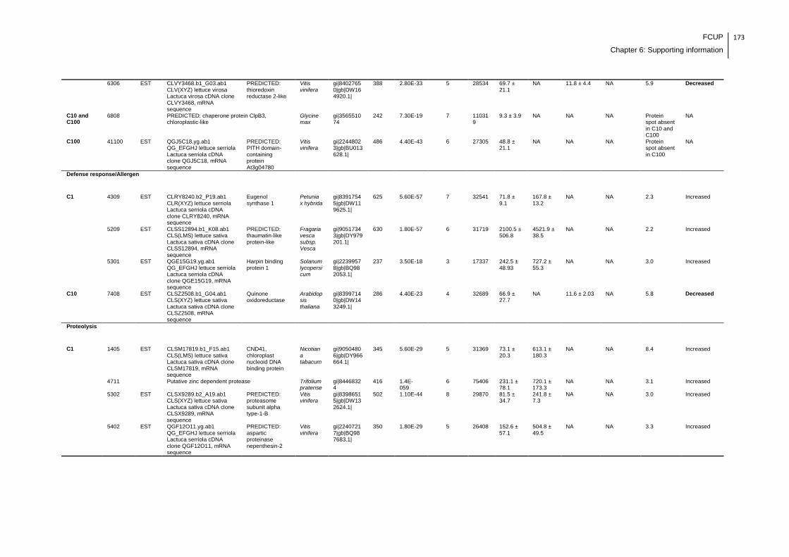
FCUP
Chapter 6: Supporting information
173
6306 EST CLVY3468.b1_G03.ab1 CLV(XYZ) lettuce virosa Lactuca virosa cDNA clone CLVY3468, mRNA sequence
PREDICTED: thioredoxin reductase 2-like
Vitis vinifera
gi|84027650|gb|DW164920.1|
388 2.80E-33 5 28534 69.7 ± 21.1
NA 11.8 ± 4.4 NA 5.9 Decreased
C10 and C100
6808 PREDICTED: chaperone protein ClpB3, chloroplastic-like
Glycine max
gi|356551074
242 7.30E-19 7 110319
9.3 ± 3.9 NA NA NA Protein spot absent in C10 and C100
NA
C100 41100 EST QGJ5C18.yg.ab1 QG_EFGHJ lettuce serriola Lactuca serriola cDNA clone QGJ5C18, mRNA sequence
PREDICTED: PITH domain-containing protein At3g04780
Vitis vinifera
gi|22448023|gb|BU013628.1|
486 4.40E-43 6 27305
48.8 ± 21.1
NA NA NA Protein spot absent in C100
NA
Defense response/Allergen
C1 4309 EST CLRY8240.b2_P19.ab1 CLR(XYZ) lettuce serriola Lactuca serriola cDNA clone CLRY8240, mRNA sequence
Eugenol synthase 1
Petunia x hybrida
gi|83917545|gb|DW119625.1|
625 5.60E-57 7 32541 71.8 ± 9.1
167.8 ± 13.2
NA NA 2.3 Increased
5209 EST CLSS12894.b1_K08.ab1 CLS(LMS) lettuce sativa Lactuca sativa cDNA clone CLSS12894, mRNA sequence
PREDICTED: thaumatin-like protein-like
Fragaria vesca subsp. Vesca
gi|90517343|gb|DY979201.1|
630 1.80E-57 6 31719 2100.5 ± 506.8
4521.9 ± 38.5
NA NA 2.2 Increased
5301 EST QGE15G19.yg.ab1 QG_EFGHJ lettuce serriola Lactuca serriola cDNA clone QGE15G19, mRNA sequence
Harpin binding protein 1
Solanum lycopersicum
gi|22399578|gb|BQ982053.1|
237 3.50E-18 3 17337 242.5 ± 48.93
727.2 ± 55.3
NA NA 3.0 Increased
C10 7408 EST CLSZ2508.b1_G04.ab1 CLS(XYZ) lettuce sativa Lactuca sativa cDNA clone CLSZ2508, mRNA sequence
Quinone oxidoreductase
Arabidopsis thaliana
gi|83997140|gb|DW143249.1|
286 4.40E-23 4 32689 66.9 ± 27.7
NA 11.6 ± 2.03 NA 5.8 Decreased
Proteolysis
C1 1405 EST CLSM17819.b1_F15.ab1 CLS(LMS) lettuce sativa Lactuca sativa cDNA clone CLSM17819, mRNA sequence
CND41, chloroplast nucleoid DNA binding protein
Nicotiana tabacum
gi|90504806|gb|DY966664.1|
345 5.60E-29 5 31369 73.1 ± 20.3
613.1 ± 180.3
NA NA 8.4 Increased
4711 Putative zinc dependent protease Trifolium pratense
gi|84468324
416 1.4E-059
6 75406 231.1 ± 78.1
720.1 ± 173.3
NA NA 3.1 Increased
5302 EST CLSX9289.b2_A19.ab1 CLS(XYZ) lettuce sativa Lactuca sativa cDNA clone CLSX9289, mRNA sequence
PREDICTED: proteasome subunit alpha type-1-B
Vitis vinifera
gi|83986515|gb|DW132624.1|
502 1.10E-44 8 29870 81.5 ± 34.7
241.8 ± 7.3
NA NA 3.0 Increased
5402 EST QGF12O11.yg.ab1 QG_EFGHJ lettuce serriola Lactuca serriola cDNA clone QGF12O11, mRNA sequence
PREDICTED: aspartic proteinase nepenthesin-2
Vitis vinifera
gi|22407217|gb|BQ987683.1|
350 1.80E-29 5 26408 152.6 ± 57.1
504.8 ± 49.5
NA NA 3.3 Increased
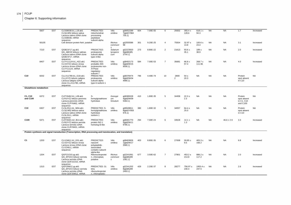
174 FCUP
Chapter 6: Supporting information
5607 EST CLSS8038.b1_L18.ab1 CLS(LMS) lettuce sativa Lactuca sativa cDNA clone CLSS8038, mRNA sequence
PREDICTED: mitochondrial-processing peptidase subunit alpha
Vitis vinifera
gi|90522897|gb|DY984755.1|
604 7.00E-55 6 29302 355.4 ± 128.6
618.1 ± 94.3
NA NA 1.7 Increased
56105 Cell division protein ftsH, putative Ricinus communis
gi|255558698
301 9.20E-25 4 75504 32.97 ± 13.8
100.6 ± 23.4
NA NA 3.1 Increased
7210 EST QGB21F17.yg.ab1 QG_ABCDI lettuce salinas Lactuca sativa cDNA clone QGB21F17, mRNA sequence
PREDICTED: proteasome subunit alpha type-3-like
Solanum lycopersicum
gi|22239209|gb|BQ853744.1|
273 8.80E-22 2 21613 93.6 ± 35.1
189 ± 38.9
NA NA 2.0 Increased
8407 EST CLVY11124.b1_H22.ab1 CLV(XYZ) lettuce virosa Lactuca virosa cDNA clone CLVY11124, mRNA sequence
PREDICTED: probable 26S proteasome non-ATPase regulatory subunit 7
Vitis vinifera
gi|84021736|gb|DW159006.1|
584 7.00E-53 7 35681 66.8 ± 57.4
268.7 ± 111.96
NA NA 4.0 Increased
C10 8202 EST CLLX11780.b1_G18.ab1 CLL(XYZ) lettuce saligna Lactuca saligna cDNA clone CLLX11780, mRNA sequence
PREDICTED: proteasome subunit alpha type-6 isoform 1
Vitis vinifera
gi|83784749|gb|DW044966.1|
796 4.40E-74 10 28002
54 ± 20.3
NA NA NA Protein spot absent in C10
NA
Glutathione metabolism
C1, C10 and C100
6372 EST CLPY6462.b1_L08.ab1 CLP(XYZ) lettuce perennis Lactuca perennis cDNA clone CLPY6462, mRNA sequence
S-formylglutathione hydrolase
Gossypium hirsutum
gi|83893284|gb|DW095364.1|
410 1.80E-35 5 34458 22.3 ± 2.3
NA NA NA Protein spot absent in C1, C10 and C100
NA
C10 6307 EST CLSL2511.b1_M04.ab1 CLS(LMS) lettuce sativa Lactuca sativa cDNA clone CLSL2511, mRNA sequence
PREDICTED: S-formylglutathione hydrolase isoform 1
Vitis vinifera
gi|90498118|gb|DY959976.1|
380 1.80E-32 5 34557 64.4 ± 25.0
NA NA NA Protein spot absent in C10
NA
C100 5371 EST CLRY8401.b2_B14.ab1 CLR(XYZ) lettuce serriola Lactuca serriola cDNA clone CLRY8401, mRNA sequence
PREDICTED: protein IN2-1 homolog B-like
Vitis vinifera
gi|83917705|gb|DW119785.1|
294 7.00E-24 6 33528 14.1 ± 1.3
NA NA 46.6 ± 2.9 3.3 Increased
Protein synthesis and signal transduction (Transcription, RNA processing and translocation, and translation)
C1 1203 EST CLVZ4611.b1_E01.ab1 CLV(XYZ) lettuce virosa Lactuca virosa cDNA clone CLVZ4611, mRNA sequence
PREDICTED: nascent polypeptide-associated complex subunit alpha-like
Vitis vinifera
gi|84038261|gb|DW173580.1|
403 8.80E-35 6 27008 59.85 ± 9.5
405.3 ± 166.2
NA NA 6.8 Increased
1204 EST QGF21O10.yg.ab1 QG_EFGHJ lettuce serriola Lactuca serriola cDNA clone QGF21O10, mRNA sequence
Ribonucleoprotein, chloroplast, putative
Ricinus communis
gi|22410616|gb|BQ991081.1|
677 3.50E-62 7 27801 453.2 ± 214.8
886.2 ± 117.2
NA NA 2.0 Increased
1313 EST QGF25M12.yg.ab1 QG_EFGHJ lettuce serriola Lactuca serriola cDNA clone QGF25M12, mRNA
PREDICTED: 31 kDa ribonucleoprotein, chloroplastic-
Vitis vinifera
gi|22412028|gb|BQ992493.1|
429 2.20E-37 8 28277 756.97 ± 230.4
1953.4 ± 247.5
NA NA 2.6 Increased
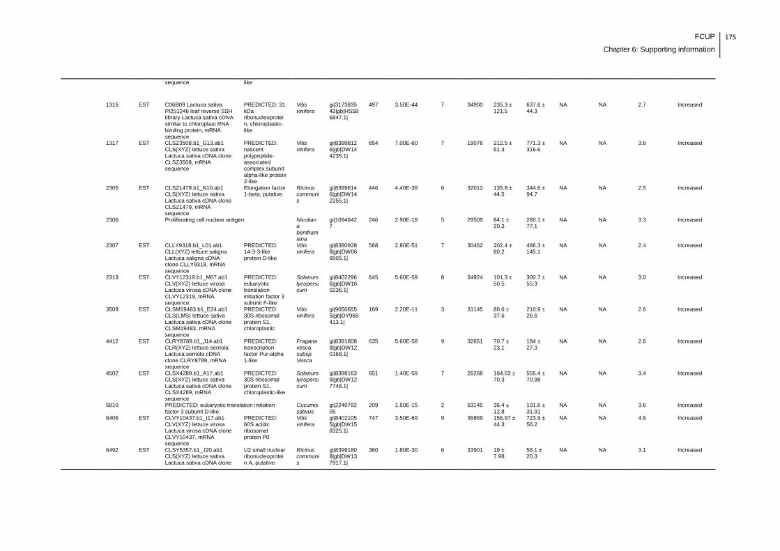
FCUP
Chapter 6: Supporting information
175
sequence like
1315 EST C06B09 Lactuca sativa PI251246 leaf reverse SSH library Lactuca sativa cDNA similar to chloroplast RNA binding protein, mRNA sequence
PREDICTED: 31 kDa ribonucleoprotein, chloroplastic-like
Vitis vinifera
gi|317383543|gb|HS586847.1|
497 3.50E-44 7 34900 235.3 ± 121.5
637.6 ± 44.3
NA NA 2.7 Increased
1317 EST CLSZ3508.b1_G13.ab1 CLS(XYZ) lettuce sativa Lactuca sativa cDNA clone CLSZ3508, mRNA sequence
PREDICTED: nascent polypeptide-associated complex subunit alpha-like protein 2-like
Vitis vinifera
gi|83998126|gb|DW144235.1|
654 7.00E-60 7 19076 212.5 ± 51.3
771.3 ± 316.6
NA NA 3.6 Increased
2305 EST CLSZ1479.b1_N10.ab1 CLS(XYZ) lettuce sativa Lactuca sativa cDNA clone CLSZ1479, mRNA sequence
Elongation factor 1-beta, putative
Ricinus communis
gi|83996146|gb|DW142255.1|
446 4.40E-39 6 32012 135.8 ± 44.5
344.6 ± 94.7
NA NA 2.5 Increased
2306 Proliferating cell nuclear antigen Nicotiana benthamiana
gi|10946427
246 2.90E-19 5 29509 84.1 ± 20.3
280.1 ± 77.1
NA NA 3.3 Increased
2307 EST CLLY9318.b1_L01.ab1 CLL(XYZ) lettuce saligna Lactuca saligna cDNA clone CLLY9318, mRNA sequence
PREDICTED: 14-3-3-like protein D-like
Vitis vinifera
gi|83809288|gb|DW069505.1|
568 2.80E-51 7 30462 202.4 ± 80.2
486.3 ± 145.1
NA NA 2.4 Increased
2313 EST CLVY12319.b1_M07.ab1 CLV(XYZ) lettuce virosa Lactuca virosa cDNA clone CLVY12319, mRNA sequence
PREDICTED: eukaryotic translation initiation factor 3 subunit F-like
Solanum lycopersicum
gi|84022966|gb|DW160236.1|
645 5.60E-59 8 34924 101.3 ± 50.5
300.7 ± 55.3
NA NA 3.0 Increased
3509 EST CLSM19483.b1_E24.ab1 CLS(LMS) lettuce sativa Lactuca sativa cDNA clone CLSM19483, mRNA sequence
PREDICTED: 30S ribosomal protein S1, chloroplastic
Vitis vinifera
gi|90506555|gb|DY968413.1|
169 2.20E-11 3 31145 80.6 ± 37.6
210.9 ± 26.6
NA NA 2.6 Increased
4412 EST CLRY8789.b1_J14.ab1 CLR(XYZ) lettuce serriola Lactuca serriola cDNA clone CLRY8789, mRNA sequence
PREDICTED: transcription factor Pur-alpha 1-like
Fragaria vesca subsp. Vesca
gi|83918088|gb|DW120168.1|
635 5.60E-58 9 32651 70.7 ± 23.1
184 ± 27.3
NA NA 2.6 Increased
4502 EST CLSX4289.b1_A17.ab1 CLS(XYZ) lettuce sativa Lactuca sativa cDNA clone CLSX4289, mRNA sequence
PREDICTED: 30S ribosomal protein S1, chloroplastic-like
Solanum lycopersicum
gi|83981639|gb|DW127748.1|
651 1.40E-59 7 26268 164.03 ± 70.3
555.4 ± 70.98
NA NA 3.4 Increased
5810 PREDICTED: eukaryotic translation initiation factor 3 subunit D-like
Cucumis sativus
gi|224079205
209 1.50E-15 2 63145 36.4 ± 12.8
131.6 ± 31.91
NA NA 3.6 Increased
6406 EST CLVY10437.b1_I17.ab1 CLV(XYZ) lettuce virosa Lactuca virosa cDNA clone CLVY10437, mRNA sequence
PREDICTED: 60S acidic ribosomal protein P0
Vitis vinifera
gi|84021055|gb|DW158325.1|
747 3.50E-69 9 36869 156.97 ± 44.3
723.9 ± 56.2
NA NA 4.6 Increased
6492 EST CLSY5357.b1_J20.ab1 CLS(XYZ) lettuce sativa Lactuca sativa cDNA clone
U2 small nuclear ribonucleoprotein A, putative
Ricinus communis
gi|83991808|gb|DW137917.1|
360 1.80E-30 6 33901 19 ± 7.98
58.1 ± 20.3
NA NA 3.1 Increased
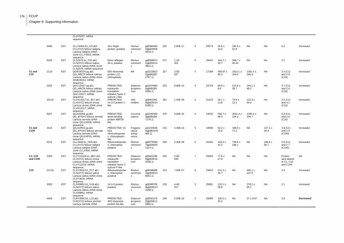
176 FCUP
Chapter 6: Supporting information
CLSY5357, mRNA sequence
6495 EST CLLY4453.b1_I10.ab1 CLL(XYZ) lettuce saligna Lactuca saligna cDNA clone CLLY4453, mRNA sequence
Zinc finger protein, putative
Ricinus communis
gi|83804602|gb|DW064819.1|
225 5.60E-17 3 29073 29.8 ± 10.3
185.9 ± 52.8
NA NA 6.2 Increased
8204 EST CLSZ675.b1_F02.ab1 CLS(XYZ) lettuce sativa Lactuca sativa cDNA clone CLSZ675, mRNA sequence
Minor allergen Alt a, putative
Ricinus communis
gi|83998732|gb|DW144841.1|
572 1.1E-051
6 28441 163.2 ± 30.7
358.7 ± 20.43
NA NA 2.2 Increased
C1 and C10
2110 EST QGB18O24.yg.ab1 QG_ABCDI lettuce salinas Lactuca sativa cDNA clone QGB18O24, mRNA sequence
50S ribosomal protein L12, chloroplastic
NA gi|22238232|gb|BQ852767.1|
327 3.5E-027
4 17289 468.97 ± 90.3
1610.1 ± 164.5
1381.4 ± 246.4
NA 3.4 (C1) and 2.9 (C10)
Increased
3302 EST QGC12N17.yg.ab1 QG_ABCDI lettuce salinas Lactuca sativa cDNA clone QGC12N17, mRNA sequence
PREDICTED: eukaryotic translation initiation factor 3 subunit J-like
Solanum lycopersicum
gi|22244849|gb|BQ859384.1|
203 8.80E-15 4 24723 58.9 ± 9.8
215.6 ± 35.7
144.1 ± 48.2
NA 3.7 (C1) and 2.4 (C10)
Increased
33119 EST CLVX13217.b1_B17.ab1 CLV(XYZ) lettuce virosa Lactuca virosa cDNA clone CLVX13217, mRNA sequence
PREDICTED: 14-3-3 protein 1-like
Vitis vinifera
gi|84010918|gb|DW148188.1|
351 1.40E-29 5 31315 30.1 ± 13.8
74.9 ± 13.1
122.3 ± 54.0
NA 2.5 (C1) and 4.1 (C10)
Increased
8207 EST QGJ15D06.yg.ab1 QG_EFGHJ lettuce serriola Lactuca serriola cDNA clone QGJ15D06, mRNA sequence
PREDICTED: auxin-binding protein ABP20-like
Cucumis sativus
gi|22404803|gb|BQ987278.1|
475 5.60E-42 4 23032 543.7 ± 66.3
2041.4 ± 234.2
1395.4 ± 194.7
NA 3.8 (C1) and 2.6 (C10)
Increased
C1 and C100
2314 EST QGJ14P03.yg.ab1 QG_EFGHJ lettuce serriola Lactuca serriola cDNA clone QGJ14P03, mRNA sequence
PREDICTED: 31 kDa ribonucleoprotein, chloroplastic-like
Fragaria vesca subsp. vesca
gi|22445364|gb|BU010969.1|
216 4.40E-16 5 18596 50.2 ± 15.6
189.9 ± 72.2
NA 127.4 ± 30.3
3.8 (C1) and 2.5 (C100)
Increased
5212 EST CLLX400.b1_O03.ab1 CLL(XYZ) lettuce saligna Lactuca saligna cDNA clone CLLX400, mRNA sequence
Ribonucleoprotein, chloroplast, putative
Ricinus communis
gi|83790997|gb|DW051214.1|
549 2.20E-49 5 34181 128.4 ± 42.3
739.8 ± 186.1
NA 348.8 ± 90.7
5.8 (C1) and 2.7 (C100)
Increased
C1, C10 and C100
2303 EST CLVY12319.b1_M07.ab1 CLV(XYZ) lettuce virosa Lactuca virosa cDNA clone CLVY12319, mRNA sequence
PREDICTED: eukaryotic translation initiation factor 3 subunit F-like
Solanum lycopersicum
gi|84022966|gb|DW160236.1|
545 5.6E-049
7 34924 77.8 ± 27.8
NA NA NA Protein spot absent in C1, C10 and C100
NA
C10 32118 EST CLSY3618.b1_D17.ab1 CLS(XYZ) lettuce sativa Lactuca sativa cDNA clone CLSY3618, mRNA sequence
Ribonucleoprotein, chloroplast, putative
Ricinus communis
gi|83989952|gb|DW136061.1|
324 7.00E-27 6 24824 101.3 ± 26.7
NA 495.1 ± 107.9
NA 4.9 Increased
3303 EST CLSX9852.b1_G16.ab1 CLS(XYZ) lettuce sativa Lactuca sativa cDNA clone CLSX9852, mRNA sequence
14-3-3 protein, putative
Ricinus communis
gi|83987059|gb|DW133168.1|
226 4.4E-017
3 25891 133.1 ± 58.8
NA 278.2 ± 35.1
NA 2.1 Increased
4404 EST CLRY3990.b2_L13.ab1 CLR(XYZ) lettuce serriola Lactuca serriola cDNA
PREDICTED: 40S ribosomal protein SA-like
Solanum lycopersicum
gi|83913780|gb|DW115860.1|
245 5.60E-19 3 28095
105.5 ± 33.4
NA 27 ± 9.97 NA 3.9 Decreased
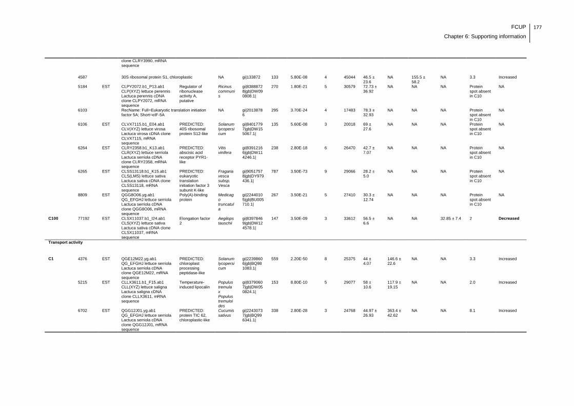
FCUP
Chapter 6: Supporting information
177
clone CLRY3990, mRNA sequence
4587 30S ribosomal protein S1, chloroplastic NA gi|133872 133 5.80E-08 4 45044 46.5 ± 23.6
NA 155.5 ± 58.2
NA 3.3 Increased
5184 EST CLPY2072.b1_P13.ab1 CLP(XYZ) lettuce perennis Lactuca perennis cDNA clone CLPY2072, mRNA sequence
Regulator of ribonuclease activity A, putative
Ricinus communis
gi|83888728|gb|DW090808.1|
270 1.80E-21 5 30579 72.73 ± 36.92
NA NA NA Protein spot absent in C10
NA
6103 RecName: Full=Eukaryotic translation initiation factor 5A; Short=eIF-5A
NA gi|20138786
295 3.70E-24 4 17483 78.3 ± 32.93
NA NA NA Protein spot absent in C10
NA
6106 EST CLVX7115.b1_E04.ab1 CLV(XYZ) lettuce virosa Lactuca virosa cDNA clone CLVX7115, mRNA sequence
PREDICTED: 40S ribosomal protein S12-like
Solanum lycopersicum
gi|84017797|gb|DW155067.1|
135 5.60E-08 3 20018 69 ± 27.6
NA NA NA Protein spot absent in C10
NA
6264 EST CLRY2358.b1_K13.ab1 CLR(XYZ) lettuce serriola Lactuca serriola cDNA clone CLRY2358, mRNA sequence
PREDICTED: abscisic acid receptor PYR1-like
Vitis vinifera
gi|83912166|gb|DW114246.1|
238 2.80E-18 6 26470 42.7 ± 7.07
NA NA NA Protein spot absent in C10
NA
6265 EST CLSS13118.b1_K15.ab1 CLS(LMS) lettuce sativa Lactuca sativa cDNA clone CLSS13118, mRNA sequence
PREDICTED: eukaryotic translation initiation factor 3 subunit K-like
Fragaria vesca subsp. Vesca
gi|90517578|gb|DY979436.1|
787 3.50E-73 9 29066 28.2 ± 5.0
NA NA NA Protein spot absent in C10
NA
8809 EST QGG8O06.yg.ab1 QG_EFGHJ lettuce serriola Lactuca serriola cDNA clone QGG8O06, mRNA sequence
Poly(A)-binding protein
Medicago truncatula
gi|22440105|gb|BU005710.1|
267 3.50E-21 5 27410 30.3 ± 12.74
NA NA NA Protein spot absent in C10
NA
C100 77192 EST CLSX11037.b1_I24.ab1 CLS(XYZ) lettuce sativa Lactuca sativa cDNA clone CLSX11037, mRNA sequence
Elongation factor 2
Aegilops tauschii
gi|83978469|gb|DW124578.1|
147 3.50E-09 3 33612 56.5 ± 6.6
NA NA 32.85 ± 7.4 2 Decreased
Transport activity
C1 4376 EST QGE12M22.yg.ab1 QG_EFGHJ lettuce serriola Lactuca serriola cDNA clone QGE12M22, mRNA sequence
PREDICTED: chloroplast processing peptidase-like
Solanum lycopersicum
gi|22398606|gb|BQ981083.1|
559 2.20E-50 8 25375 44 ± 4.07
146.6 ± 22.6
NA NA 3.3 Increased
5215 EST CLLX3611.b1_F15.ab1 CLL(XYZ) lettuce saligna Lactuca saligna cDNA clone CLLX3611, mRNA sequence
Temperature-induced lipocalin
Populus tremula x Populus tremuloides
gi|83790607|gb|DW050824.1|
153 8.80E-10 5 29077 58 ± 10.6
117.9 ± 19.15
NA NA 2.0 Increased
6702 EST QGG12J01.yg.ab1 QG_EFGHJ lettuce serriola Lactuca serriola cDNA clone QGG12J01, mRNA sequence
PREDICTED: protein TIC 62, chloroplastic-like
Cucumis sativus
gi|22430737|gb|BQ996341.1|
338 2.80E-28 3 24768 44.97 ± 26.93
363.4 ± 42.62
NA NA 8.1 Increased

178 FCUP
Chapter 6: Supporting information
7201 EST CLSS5192.b1_O02.ab1 CLS(LMS) lettuce sativa Lactuca sativa cDNA clone CLSS5192, mRNA sequence
Apolipoprotein d, putative
Ricinus communis
gi|90521021|gb|DY982879.1|
20311 3.50E-54 8 20311 87.3 ± 31.2
216.9 ± 22.25
NA NA 2.5 Increased
C10 8208 RecName: Full=GTP-binding nuclear protein Ran1A
NA gi|1710007 489 1.50E-43 7 24114 118.5 ± 56.8
NA NA NA Protein spot absent in C10
NA
C10 and C100
66106 EST CLLY11583.b1_M15.ab1 CLL(XYZ) lettuce saligna Lactuca saligna cDNA clone CLLY11583, mRNA sequence
PREDICTED: protein TIC 62, chloroplastic-like
Cucumis sativus
gi|83798738|gb|DW058955.1|
182 1.10E-12 3 32021 13.7 ± 1.4
NA 46.7 ± 4.1 62.5 ± 20.9 3.4 (C10) increased and 4.6 (C100) decreased
Decreased
Cytoskeleton formation
C1 3403 Plastid-dividing ring protein Solanum tuberosum
gi|47156057
409 1.50E-35 4 44089 103.9 ± 23.36
245.4 ± 16.29
NA NA 2.4 Increased
C1 and C10
2301 EST CLVY7509.b1_I06.ab1 CLV(XYZ) lettuce virosa Lactuca virosa cDNA clone CLVY7509, mRNA sequence
Fibrillin Coffea canephora
gi|84033889|gb|DW169208.1|
267 3.5E-021
4 31814 146.6 ± 73.7
385.6 ± 84.3
415.6 ± 137.7
NA 2.6 (C1) and 2.8 (C10)
Increased
C10 6107 EST QGE6O14.yg.ab1 QG_EFGHJ lettuce serriola Lactuca serriola cDNA clone QGE6O14, mRNA sequence
Actin depolymerizing factor 2
Gossypium hirsutum
gi|22402995|gb|BQ985470.1|
744 7.00E-69 8 20038 96.2 ± 10.0
NA NA NA Protein spot absent in C10
NA
Cell wall biogenesis/degradation
C1 8401 EST CLPY6271.b1_N07.ab1 CLP(XYZ) lettuce perennis Lactuca perennis cDNA clone CLPY6271, mRNA sequence
PREDICTED: UDP-arabinopyranose mutase 1
Vitis vinifera
gi|83893082|gb|DW095162.1|
181 1.40E-12 4 32689 62.6 ± 16.6
235.4 ± 103.9
NA NA 3.8 Increased
C1 and C100
8305 EST CLSS6180.b1_G09.ab1 CLS(LMS) lettuce sativa Lactuca sativa cDNA clone CLSS6180, mRNA sequence
Xyloglucan endotransglucosylase/hydrolase
Tagetes patula
gi|90521702|gb|DY983560.1|
557 3.50E-50 8 36297
295.7 ± 66.5
924.2 ± 266.6
NA 107.4 ± 53.60
3.1 (C1 )increased and 2.8 (C100) decreased
Decreased
Amino acid metabolism
C1 6701 EST CLVY7678.b1_L24.ab1 CLV(XYZ) lettuce virosa Lactuca virosa cDNA clone CLVY7678, mRNA sequence
ketol-acid reductoisomerase
Elaeis guineensis
gi|84034071|gb|DW169390.1|
674 7.00E-62 6 31229 189.3 ± 78.10
635.4 ± 105.4
NA NA 3.4 Increased
C10 6497 EST QGB27B24.yg.ab1 QG_ABCDI lettuce salinas Lactuca sativa cDNA clone QGB27B24, mRNA sequence
PREDICTED: N-carbamoylputrescine amidase-like
Fragaria vesca subsp. Vesca
gi|22241060|gb|BQ855595.1|
212 1.10E-15 4 26768 39.9 ± 5.2
NA NA NA Protein spot absent in C10
NA
6732 Acetohydroxyacid synthase 1
Helianthus annuus
gi|46948852
348 1.80E-29 4 71154 29.3 ± 4.5
NA 9.5 ± 2.7 NA 3.1 Decreased

FCUP
Chapter 6: Supporting information
179
7513 EST CLRY2510.b1_K04.ab1 CLR(XYZ) lettuce serriola Lactuca serriola cDNA clone CLRY2510, mRNA sequence
PREDICTED: diaminopimelate decarboxylase 2, chloroplastic isoform 1
Vitis vinifera
gi|83912316|gb|DW114396.1|
368 2.8E-031
6 31648 50.4 ± 24.3
NA NA NA Protein spot absent in C10
NA
7803 Vitamin-b12 independent methionine synthase, 5-methyltetrahydropteroyltriglutamate-homocysteine
Populus trichocarpa
gi|224104961
210 1.20E-15 4 85033 51.5 ± 19.5
NA NA NA Protein spot absent in C10
NA
C100 6490 EST CLSS4535.b2_N06.ab1 CLS(LMS) lettuce sativa Lactuca sativa cDNA clone CLSS4535, mRNA sequence
Thiosulfate sulfertansferase, putative
Ricinus communis
gi|90520693|gb|DY982551.1|
301 1.40E-24 5 26299 55 ± 21.94
NA NA NA Protein spot absent in C100
NA
Pigment metabolism
C1 6405 EST CLRY6323.b1_F21.ab1 CLR(XYZ) lettuce serriola Lactuca serriola cDNA clone CLRY6323, mRNA sequence
PREDICTED: coproporphyrinogen-III oxidase, chloroplastic-like
Vitis vinifera
gi|83915540|gb|DW117620.1|
436 4.40E-38 9 33519 145.9 ± 49.8
452.7 ± 119.43
NA NA 3.1 Increased
C10 74107 EST CLRX3506.b1_C13.ab1 CLR(XYZ) lettuce serriola Lactuca serriola cDNA clone CLRX3506, mRNA sequence
Uroporphyrinogen decarboxylase, chloroplastic
NA gi|83903592|gb|DW105672.1|
232 1.10E-17 5 28920 103.1 ± 35.33
NA NA NA Protein spot absent in C10
NA
C100 66108 EST QGB6J15.yg.ab1 QG_ABCDI lettuce salinas Lactuca sativa cDNA clone QGB6J15, mRNA sequence
Polyphenol oxidase precursor (chloroplast)
Taraxacum officinale
gi|22242562|gb|BQ857097.1|
147 3.50E-09 2 26774 83.2 ± 19.54
NA NA NA Protein spot absent in C100
NA
Lipid metabolism
C1 4495 EST CLSS2981.b1_J02.ab1 CLS(LMS) lettuce sativa Lactuca sativa cDNA clone CLSS2981, mRNA sequence
PREDICTED: GDSL esterase/lipase At5g45670
Vitis vinifera
gi|90519132|gb|DY980990.1|
634 7.00E-58 7 33181 47.6 ± 16.28
104.7 ± 8.9
NA NA 2.2 Increased
6413 EST CLLY11826.b1_D06.ab1 CLL(XYZ) lettuce saligna Lactuca saligna cDNA clone CLLY11826, mRNA sequence
Enoyl-ACP reductase 1
Helianthus annuus
gi|83799000|gb|DW059217.1|
527 3.50E-47 6 33037 106.5 ± 10.9
339.4 ± 38.9
NA NA 3.2 Increased
6494 EST CLSM18468.b1_G09.ab1 CLS(LMS) lettuce sativa Lactuca sativa cDNA clone CLSM18468, mRNA sequence
PREDICTED: GDSL esterase/lipase LTL1-like
Glycine max
gi|90505494|gb|DY967352.1|
267 3.50E-21 4 34501 46.3 ± 21.6
141.1 ± 27.5
NA NA 3.0 Increased
7305 EST CLSY7797.b1_J05.ab1 CLS(XYZ) lettuce sativa Lactuca sativa cDNA clone CLSY7797, mRNA sequence
PREDICTED: bifunctional epoxide hydrolase 2-like
Solanum lycopersicum
gi|83994039|gb|DW140148.1|
522 1.10E-46 8 32235 76.8 ± 22.2
127.7 ± 9.1
NA NA 1.7 Increased
C10 6734 EST QGC4h07.yg.ab1 QG_ABCDI lettuce salinas Lactuca sativa cDNA clone QGC4h07, mRNA sequence
PREDICTED: 2-hydroxyacyl-CoA lyase-like
Glycine max
gi|22250919|gb|BQ865454.1|
335 5.60E-28 4 23074 29.3 ± 5.8
NA NA NA Protein spot absent in C10
NA

180 FCUP
Chapter 6: Supporting information
75118 EST CLSM7866.b1_D23.ab1 CLS(LMS) lettuce sativa Lactuca sativa cDNA clone CLSM7866, mRNA sequence
Acetyl-CoA C-acetyltransferase protein
Camellia oleifera
gi|90512340|gb|DY974198.1|
356 4.40E-30 4 28386 95.9 ± 28.6
NA NA NA Protein spot absent in C10
NA
Ascorbic acid biosynthesis
C1, C10 and C100
6363 EST CLPY2939.b1_E16.ab1 CLP(XYZ) lettuce perennis Lactuca perennis cDNA clone CLPY2939, mRNA sequence
Phosphomannomutase
Glycine max
gi|83889580|gb|DW091660.1|
138 2.80E-08 2 33644 24.3 ± 7.01
NA NA NA Protein spot absent in C1, C10 and C100
NA
C10 6267 EST CLPY3087.b1_M03.ab1 CLP(XYZ) lettuce perennis Lactuca perennis cDNA clone CLPY3087, mRNA sequence
Phosphomannomutase
Glycine max
gi|83889719|gb|DW091799.1|
453 8.80E-40 5 33650 32.8 ± 2.5
NA NA NA Protein spot absent in C10
NA
Vitamin B1 biosynthesis
C1 3309 EST CLLX1473.b1_B10.ab1 CLL(XYZ) lettuce saligna Lactuca saligna cDNA clone CLLX1473, mRNA sequence
PREDICTED: thiamine thiazole synthase, chloroplastic-like
Fragaria vesca subsp. Vesca
gi|83787896|gb|DW048113.1|
194 7.00E-14 2 29123 158.5 ± 5.5
327.8 ± 89.95
NA NA 2.1 Increased
4308 EST CLLX1473.b1_B10.ab1 CLL(XYZ) lettuce saligna Lactuca saligna cDNA clone CLLX1473, mRNA sequence
PREDICTED: thiamine thiazole synthase, chloroplastic-like
Fragaria vesca subsp. Vesca
gi|83787896|gb|DW048113.1|
297 3.50E-24 4 29123 118.8 ± 37.8
417 ± 154.7
NA NA 3.5 Increased
Inositol biosynthesis
C1 5720 L-myo-inositol-1-phosphate synthase Actinidia deliciosa
gi|409194966
528 1.80E-47 5 56733 156.5 ± 103.94
538.03 ± 101.08
NA NA 3.4 Increased
Unknown/Miscellaneous
C1 1501 EST QGB19G06.yg.ab1 QG_ABCDI lettuce salinas Lactuca sativa cDNA clone QGB19G06, mRNA sequence
TGB12K interacting protein 2
Nicotiana tabacum
gi|22238401|gb|BQ852936.1|
238 2.8E-018
4 21973 99.7 ± 39.92
378.8 ± 128.28
NA NA 3.8 Increased
2108 EST CLRY589.b1_I04.ab1 CLR(XYZ) lettuce serriola Lactuca serriola cDNA clone CLRY589, mRNA sequence
Drought-induced protein SDi-6 - common sunflower (fragment)
NA gi|83915068|gb|DW117148.1|
397 3.50E-34 4 16020
1273.6 ± 452.4
2375.9 ± 62.7
NA NA 2.0 Increased
2310 EST CLLY12528.b1_O12.ab1 CLL(XYZ) lettuce saligna Lactuca saligna cDNA clone CLLY12528, mRNA sequence
PREDICTED: probable plastid-lipid-associated protein 13, chloroplastic-like
Glycine max
gi|83799755|gb|DW059972.1|
427 3.50E-37 4 32179 172.4 ± 32.43
345.8 ± 81.15
NA NA 2.0 Increased
2325 EST QGB15I23.yg.ab1 QG_ABCDI lettuce salinas Lactuca sativa cDNA clone QGB15I23, mRNA sequence
Alpha chain of nascent polypeptide associated complex
Nicotiana benthamiana
gi|22236981|gb|BQ851516.1|
466 4.40E-41 5 24262 NA 243.5 ± 53.43
NA NA Protein spot absent in C
NA
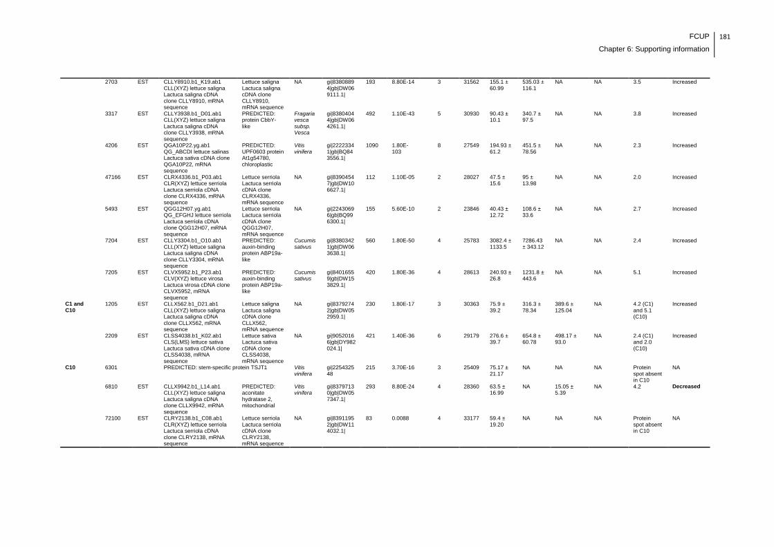
FCUP
Chapter 6: Supporting information
181
2703 EST CLLY8910.b1_K19.ab1 CLL(XYZ) lettuce saligna Lactuca saligna cDNA clone CLLY8910, mRNA sequence
Lettuce saligna Lactuca saligna cDNA clone CLLY8910, mRNA sequence
NA gi|83808894|gb|DW069111.1|
193 8.80E-14 3 31562 155.1 ± 60.99
535.03 ± 116.1
NA NA 3.5 Increased
3317 EST CLLY3938.b1_D01.ab1 CLL(XYZ) lettuce saligna Lactuca saligna cDNA clone CLLY3938, mRNA sequence
PREDICTED: protein CbbY-like
Fragaria vesca subsp. Vesca
gi|83804044|gb|DW064261.1|
492 1.10E-43 5 30930 90.43 ± 10.1
340.7 ± 97.5
NA NA 3.8 Increased
4206 EST QGA10P22.yg.ab1 QG_ABCDI lettuce salinas Lactuca sativa cDNA clone QGA10P22, mRNA sequence
PREDICTED: UPF0603 protein At1g54780, chloroplastic
Vitis vinifera
gi|22223341|gb|BQ843556.1|
1090 1.80E-103
8 27549 194.93 ± 61.2
451.5 ± 78.56
NA NA 2.3 Increased
47166 EST CLRX4336.b1_P03.ab1 CLR(XYZ) lettuce serriola Lactuca serriola cDNA clone CLRX4336, mRNA sequence
Lettuce serriola Lactuca serriola cDNA clone CLRX4336, mRNA sequence
NA gi|83904547|gb|DW106627.1|
112 1.10E-05 2 28027 47.5 ± 15.6
95 ± 13.98
NA NA 2.0 Increased
5493 EST QGG12H07.yg.ab1 QG_EFGHJ lettuce serriola Lactuca serriola cDNA clone QGG12H07, mRNA sequence
Lettuce serriola Lactuca serriola cDNA clone QGG12H07, mRNA sequence
NA gi|22430696|gb|BQ996300.1|
155 5.60E-10 2 23846 40.43 ± 12.72
108.6 ± 33.6
NA NA 2.7 Increased
7204 EST CLLY3304.b1_O10.ab1 CLL(XYZ) lettuce saligna Lactuca saligna cDNA clone CLLY3304, mRNA sequence
PREDICTED: auxin-binding protein ABP19a-like
Cucumis sativus
gi|83803421|gb|DW063638.1|
560 1.80E-50 4 25783 3082.4 ± 1133.5
7286.43 ± 343.12
NA NA 2.4 Increased
7205 EST CLVX5952.b1_P23.ab1 CLV(XYZ) lettuce virosa Lactuca virosa cDNA clone CLVX5952, mRNA sequence
PREDICTED: auxin-binding protein ABP19a-like
Cucumis sativus
gi|84016559|gb|DW153829.1|
420 1.80E-36 4 28613 240.93 ± 26.8
1231.8 ± 443.6
NA NA 5.1 Increased
C1 and C10
1205 EST CLLX562.b1_D21.ab1 CLL(XYZ) lettuce saligna Lactuca saligna cDNA clone CLLX562, mRNA sequence
Lettuce saligna Lactuca saligna cDNA clone CLLX562, mRNA sequence
NA gi|83792742|gb|DW052959.1|
230 1.80E-17 3 30363 75.9 ± 39.2
316.3 ± 78.34
389.6 ± 125.04
NA 4.2 (C1) and 5.1 (C10)
Increased
2209 EST CLSS4038.b1_K02.ab1 CLS(LMS) lettuce sativa Lactuca sativa cDNA clone CLSS4038, mRNA sequence
Lettuce sativa Lactuca sativa cDNA clone CLSS4038, mRNA sequence
NA gi|90520166|gb|DY982024.1|
421 1.40E-36 6 29179 276.6 ± 39.7
654.8 ± 60.78
498.17 ± 93.0
NA 2.4 (C1) and 2.0 (C10)
Increased
C10 6301 PREDICTED: stem-specific protein TSJT1 Vitis vinifera
gi|225432548
215 3.70E-16 3 25409 75.17 ± 21.17
NA NA NA Protein spot absent in C10
NA
6810 EST CLLX9942.b1_L14.ab1 CLL(XYZ) lettuce saligna Lactuca saligna cDNA clone CLLX9942, mRNA sequence
PREDICTED: aconitate hydratase 2, mitochondrial
Vitis vinifera
gi|83797130|gb|DW057347.1|
293 8.80E-24 4 28360 63.5 ± 16.99
NA 15.05 ± 5.39
NA 4.2 Decreased
72100 EST CLRY2138.b1_C08.ab1 CLR(XYZ) lettuce serriola Lactuca serriola cDNA clone CLRY2138, mRNA sequence
Lettuce serriola Lactuca serriola cDNA clone CLRY2138, mRNA sequence
NA gi|83911952|gb|DW114032.1|
83 0.0088 4 33177
59.4 ± 19.20
NA NA NA Protein spot absent in C10
NA

182 FCUP
Chapter 6: Supporting information
74108 EST CLLY3754.b1_D04.ab1 CLL(XYZ) lettuce saligna Lactuca saligna cDNA clone CLLY3754, mRNA sequence
PREDICTED: uncharacterized oxidoreductase At4g09670-like
Vitis vinifera
gi|83803854|gb|DW064071.1|
181 1.40E-12 4 30511 50.8 ± 23.64
NA NA NA Protein spot absent in C10
NA
C10 and C100
41101 EST QGA2d11.yg.ab1 QG_ABCDI lettuce salinas Lactuca sativa cDNA clone QGA2d11, mRNA sequence
Lettuce salinas Lactuca sativa cDNA clone QGA2d11, mRNA sequence
NA gi|22231705|gb|BQ847436.1|
649 2.20E-59 8 25396
44.1 ± 13.17
NA NA NA Protein spot absent in C10 and C100
NA
7101 PREDICTED: 40S ribosomal protein S12-like isoform 2
Vitis vinifera
gi|225465502
179 1.50E-12 2 15359 98.83 ± 1.89
NA NA NA Protein spot absent in C10 and C100
NA
76149 EST CLSM8393.b1_B12.ab1 CLS(LMS) lettuce sativa Lactuca sativa cDNA clone CLSM8393, mRNA sequence
PREDICTED: U-box domain-containing protein 72
Vitis vinifera
gi|90512855|gb|DY974713.1|
160 1.80E-10 5 33215 33.37 ± 10.98
NA NA NA Protein spot absent in C10 and C100
NA
C100 1408 EST QGC21I20.yg.ab1 QG_ABCDI lettuce salinas Lactuca sativa cDNA clone QGC21I20, mRNA sequence
Plastid transcriptionally active 6
Arabidopsis thaliana
gi|22248068|gb|BQ862603.1|
421 1.40E-36 5 23478 105.8 ± 4.02
NA NA 61.83 ± 8.28
2.0 Decreased
1446 EST CLSX9917.b1_J08.ab1 CLS(XYZ) lettuce sativa Lactuca sativa cDNA clone CLSX9917, mRNA sequence
PREDICTED: probable plastid-lipid-associated protein 3, chloroplastic-like
Vitis vinifera
gi|83987123|gb|DW133232.1|
138 2.80E-08 4 31742 NA NA NA 202.97 ± 44.35
Protein spot absent in C
NA
NOTES
gi| Proteins identified using NCBI databasw
gi|gb| Proteins identified using EST database
NA: not applicable
FCUP
References
183
References
Abe, T., Lawson, T., Weyers, J.D.B., Codd, G.A., 1996. Microcystin-LR inhibits
photosynthesis of Phaseolus vulgaris primary leaves: imphcations for current spray
irrigation practice. New Phytol. 133, 651-658.
Abreu, I.A., Farinha, A.P., Negrão, S., Gonçalves, N., Fonseca, C., Rodrigues, M.,
Batista, R., Saibo, N.J.M., Oliveira, M.M., 2013. Coping with abiotic stress: Proteome
changes for crop improvement. J. Proteomics 93, 145-168.
Afroz, A., Ali, G.M., Mir, A., Komatsu, S., 2011. Application of proteomics to investigate
stress-induced proteins for improvement in crop protection. Plant Cell Rep. 30, 745–
763.
Amorim, A., Vasconcelos, V.M., 1999. Dynamics of microcystins in the mussel Mytilus
galloprovincialis. Toxicon 37, 1041-1052.
Azevedo, C.C., Azevedo, J., Osório, H., Vasconcelos, V., Campos, A., 2014. Early
physiological and biochemical responses of rice seedlings to low concentration of
microcystin-LR. Ecotoxicology 23, 107–121.
Babica P., Blaha L., Marsalek B., 2006. Exploring the natural role of microcystins -
review of effects on photoautotrophic organisms. J. Phycol. 42, 9-20.
Banker, R., Carmeli, S., Hadas, O., Teltsch, B., Porat, R., Sukenik, A., 1997.
Identification of cylindrospermopsin in Aphanizomenon ovalisporum (cyanophyceae)
isolated from Lake Kinneret, Israel. J. Phycol. 33, 613–616.
Banker, R., Teltsch, B., Sukenik, A., Carmeli, S., 2000. 7-Epicylindrospermopsin, a
toxic minor metabolite of the cyanobacterium Aphanizomenon ovalisporum from Lake
Kinneret, Israel. J. Nat. Prod. 63, 387–389.
Bauer, E., Williams, B.A., Voigt, C., Mosenthin, R., Verstegen, M.W.A., 2003. Impact of
mammalian enzyme pretreatment on the fermentability of carbohydrate-rich feedstuffs.
J. Sci. Food Agric. 83, 207–214.
Beyer, D., Surányi, G., Vasas, G., Roszik, J., Erdődi, F., M-Hamvas, H., Bácsi, I.,

184 FCUP
References
Bátori, R., Serfőző, Z., Szigeti, Z.M., Vereb, G., Demeter, Z., Gonda, S., Máthé, C.,
2009. Cylindrospermopsin induces alterations of root histology and microtubule
organization in common reed (Phragmites australis) plantlets cultured in vitro. Toxicon
54, 440–449.
Bradford, M.M., 1976. A rapid and sensitive method for the quantitation of microgram
quantities of protein utilizing the principle of protein-dye binding. Anal. Biochem. 72,
248-254.
Brient, L., Lengronne, M., Bormans, M., Fastner, J., 2008. First occurrence of
cylindrospermopsin in freshwater in France. Environ. Toxicol. 24, 415–420.
Cabañero, A.I., Madrid, Y., Cámara C., 2004. Selenium and mercury bioaccessibility in
fish samples: an in vitro digestion method. Anal. Chim. Acta. 526, 51–61.
Chen, J., Song, L., Dai, J., Gan, N., Liu, Z., 2004. Effects of microcystins on the growth
and the activity of superoxide dismutase and peroxidase of rape (Brassica napus
L.) and rice (Oryza sativa L.). Toxicon 43, 393–400.
Chen, J., Xie, P., 2005. Seasonal Dynamics of the Hepatotoxic Microcystins in Various
Organs of Four Freshwater Bivalves from the Large Eutrophic Lake Taihu of
Subtropical China and the Risk to Human Consumption. Environ Toxicol. 20, 572–584.
Chen, J., Zhang, H-Q., Hu, L-B., Shi, Z-Q., 2013. Microcystin-LR-induced phytotoxicity
in rice crown root is associated with the cross-talk between auxin and nitric oxide.
Chemosphere 93, 283–293.
Chiswell, R.K., Shaw, G.R., Eaglesham, G., Smith, M.J., Norris, R.L., Seawright, A.A.,
Moore, M.R., 1999. Stability of cylindrospermopsin, the toxin from the cyanobacterium.
Cylindrospermopsis raciborskii: Effect of pH, temperature, and sunlight on
decomposition. Environ. Toxicol. 14, 155–161.
Cho, W.K., Chen, X-Y., Uddin, N.M., Rim, Y., Moon, J., Jung, J-H., Shi, C., Chu, H.,
Kim, S., Kim, S-W., Park, Z-Y., Kim, J-Y., 2009. Comprehensive proteome analysis of
lettuce latex using multidimensional protein-identification technology. Phytochemistry
70, 570–578.
Choi, J-S., Cho, S-W., Kim, T-S., Cho, K., Han, S-S., Kim, H-K., Woo, S-H., Chung, K-
Y., 2008. Proteome analysis of greenhouse-cultured lettuce with the natural soil
mineral conditioner illite. Soil Biol. Biochem. 40, 1370–1378.
FCUP
References
185
Chong, M.W.K., Wong, B.S.F., Lam, P.K.S., Shaw, G.R., and Seawright, A.A., 2002.
Toxicity and uptake mechanism of cylindrospermopsin and lophyrotomin in primary rat
hepatocytes. Toxicon 40, 205–211.
Corbel, S., Mougin, C., Bouaicha, N., 2014. Cyanobacterial toxins: Modes of actions,
fate in aquatic and soil ecosystems, phytotoxicity and bioaccumulation in agricultural
crops. Chemosphere 96, 1–15.
Craig, M., Luu, H.A., McCready, T.L., Williams, D., Andersen, R.J., Holmes, C.F.B.,
1996. Molecular mechanisms underlying the interaction of motuporin and microcystins
with type-1 and type-2A protein phosphatases. Biochem. Cell Biol. 74, 569–578.
Crush, J.R., Briggs, L.R., Sprosen, J.M., Nichols, S.N., 2008. Effect of irrigation with
lake water containing microcystins on microcystin content and growth of ryegrass,
clover, rape, and lettuce. Environ. Toxicol. 23, 246-252.
Denison, F.C., Paul, A-L., Zupanska, A.K., Ferl, R.J., 2011. 14-3-3 proteins in plant
physiology. Semin. Cell Dev. Biol. 22, 720–727.
Domingo, J.L., 2011. Influence of cooking processes on the concentrations of toxic
metals and various organic environmental pollutants in food: a review of the published
literature. Crit. Rev. Food Sci. Nutr. 51, 29–37.
El Khalloufi, F., El Ghazali, I., Saqrane, S., Oufdou, K., Vasconcelos, V., Oudra, B.,
2012. Phytotoxic effects of a natural bloom extract containing microcystins on
Lycopersicon esculentum. Ecotoxicol. Environ. Saf. 79, 199–205.
El Khalloufi, F., Oufdou, K., Lahrouni, M., El Ghazali, I., Saqrane, S., Vasconcelos, V.,
Oudra, B., 2011. Allelopatic effects of cyanobacteria extracts containing microcystins
on Medicago sativa-Rhizobia symbiosis. Ecotoxicol. Environ. Saf. 74, 431-438.
El Khalloufi, F., Oufdou, K., Lahrouni, M., Faghire, M., Peix, A., Ramírez-Bahena, M.H.,
Vasconcelos, V., Oudra, B., 2013. Physiological and antioxidant responses of
Medicago sativa- rhizobia symbiosis to cyanobacterial toxins (Microcystins) exposure.
Toxicon 76, 167–177.
Elliott, J.A., 2012. Is the future blue-green? A review of the current model predictions of
how climate change could affect pelagic freshwater cyanobacteria. Water Res. 46,
1364-1371.
186 FCUP
References
Falconer, I., Humpage, A.R., 2006. Cyanobacterial (blue-green algal) toxins in water
supplies: cylindrospermopsins. Environ. Toxicol. 21, 299–304.
Falconer, I.R., Hardy, S.J., Humpage, A.R., Froscio, S.M., Tozer, G.J., Hawkins, P.R.,
1999. Hepatic and renal toxicity of the blue-green alga (cyanobacterium)
Cylindrospermopsis raciborskii in male Swiss albino mice. Environ. Toxicol. 14, 143–
150.
Falconer, I.R., Humpage, A.R., 2001. Preliminary evidence for in vivo tumour initiation
by oral administration of extracts of the blue-green alga Cylindrospermopsis raciborskii
containing the toxin cylindrospermopsin. Environ. Toxicol. 16, 192–195.
Fessard, V., Bernard, C., 2003. Cell alterations but no DNA strand breaks induced In
Vitro by cylindrospermopsin in CHO K1 cells. Environ. Toxicol. 18, 353–359.
Fischer, W.J., Altheimer, S., Cattori, V., Meier, P.J., Dietrich, D.R., Hagenbuch, B.,
2005. Organic anion transporting polypeptides expressed in liver and brain mediate
uptake of microcystin. Toxicol. Appl. Pharmacol. 203, 257–263.
Freitas, M., Azevedo J., Carvalho A.P., Campos A., Vasconcelos V., 2014. Effects of
storage, processing and proteolytic digestion on microcystin-LR concentration in edible
clams. Food Chem. Toxicol. 66, 217- 223.
Froscio, S.M., Humpage, A.R., Burcham, P.C., Falconer, I.R., 2001. Cell-free protein
synthesis inhibition assay for the cyanobacterial toxin cylindrospermopsin. Environ.
Toxicol. 16, 408–412.
Froscio, S.M., Humpage, A.R., Burcham, P.C., Falconer, I.R., 2003.
Cylindrospermopsin-induced protein synthesis inhibition and its dissociation from acute
toxicity in mouse hepatocytes. Environ. Toxicol. 18, 243–251.
Froscio, S.M., Humpage, A.R., Wickramasinghe, W., Shaw, G., Falconer, I.R., 2008.
Interaction of the cyanobacterial toxin cylindrospermopsin with the eukaryotic protein
synthesis system. Toxicon 51, 191–198.
Funari, E., Testai, E., 2008. Human Health Risk Assessment Related to Cyanotoxins
Exposure. Crit. Rev. Toxicol. 38, 97-125.
Gehringer, M.M., Kewada, V., Coates, N., Downing, T.G., 2003. The use of Lepidium
sativum in a plant bioassay system for the detection of microcystin-LR. Toxicon 41,
FCUP
References
187
871–876.
Gill, S.S., Tuteja, N., 2010. Reactive oxygen species and antioxidant machinery in
abiotic stress tolerance in crop plants. Plant Physiol Biochem. 48, 909-930.
Griffiths, D.J., Saker, M.L., 2003. The Palm Island mystery disease twenty years on: a
review of research on the cyanotoxin cylindrospermopsin. Environ. Toxicol. 18, 78–93.
Grosse, Y., Baan, R., Straif, K., Secretan, B., Ghissassi, F.El., Cogliano, V., 2006.
Carcinogenicity of nitrate, nitrite, and cyanobacterial peptide toxins. Lancet Oncol. 7,
628–629.
Guerra, A., Etienne-Mesmin, L., Livrelli. V., Denis, S., Blanquet-Diot, S., Alric, M., 2012.
Relevance and challenges in modelinghuman gastric and small intestinal digestion.
Trends Biotechnol. 30, 591-600.
Gutiérrez-Praena, D., Campos, A., Azevedo, J., Neves, J., Freitas, M., Guzmán-
Guillén, R., Cameán, A.M., Renaut, J., Vasconcelos, V., 2014. Exposure of
Lycopersicon esculentum to microcystin-LR: Effects in the leaf proteome and toxin
translocation from water to leaves and fruits. Toxins 6, 1837-1854.
Gutiérrez-Praena, D., Jos, Á., Pichardo, S., Moreno, I.M., Cameán, A.M., 2013.
Presence and bioaccumulation of microcystins and cylindrospermopsin in food and the
effectiveness of some cooking techniques at decreasing their concentrations: A review.
Food Chem. Toxicol. 53, 139–152.
Guzmán-Guillén, R., Prieto, A.I., Moreno, I., Soria, M.E., Cameán, A.M., 2011. Effects
of thermal treatments during cooking, microwave oven and boiling, on the
unconjugated microcystin concentration in muscle of fish (Oreochromis niloticus). Food
Chem. Toxicol. 49, 2060–2067.
Habig, W.H., Pabst, M.J., Jakoby, W.B., 1974. Glutathione S-transferases. The first
enzymatic step in mercapturic acid formation. J. Biol. Chem. 249, 7130–7139.
Humpage, A.R., Falconer, I.R., 2003. Oral toxicity of the cyanobacterial toxin
cylindrospermopsin in male swiss albino mice: Determination of no observed adverse
effect level for deriving a drinking water guideline value. Environ. Toxicol. 18, 94–103.
Humpage, A.R., Fontaine, F., Froscio, S., Burcham, P., Falconer, I.R., 2005.
Cylindrospermopsin genotoxicity and cytotoxicity: role of cytochrome P-450 and
188 FCUP
References
oxidative stress. J. Toxicol. Environ. Health Part A 68, 739–753.
Hur, S.J., Decker, E.A., McClements, D.J., 2009. Influence of initial emulsifier type on
microstructural changes occurring in emulsified lipids during in vitro digestion. Food
Chem. 114, 253–262.
Ibelings, B.W., Chorus I., 2007. Accumulation of cyanobacterial toxins in freshwater
‗‗seafood‘‘ and its consequences for public health: A review. Environ. Pollut. 150, 177-
192.
Järvenpää, S., Lundberg-Niinistö, C., Spoof, L., Sjövall, O., Tyystjärvi, E., Meriluoto, J.,
2007. Effects of microcystins on broccoli and mustard, and analysis of accumulated
toxin by liquid chromatography–mass spectrometry. Toxicon 49, 865–874.
Jensen, M.H., Malter, A.J., 1995. Protected Agriculture: A Global Review. Chapter 7,
Water supply, water quality and mineral nutrition. World Bank Publications.
Kankaanpää, H.T., Holliday, J., Schröder, H., Goddard, T.J., von Fister, R.,
Carmichael, W.W., 2005. Cyanobacteria and prawn farming in northern New South
Wales, Australia—a case study on cyanobacteria diversity and hepatotoxin
bioaccumulation. Toxicol. Appl. Pharmacol. 203, 243–256.
Kaushik, G., Satya, S., Naik, S.N., 2009. Food processing a tool to pesticide residue
Kinnear, S., 2010. Cylindrospermopsin: a decade of progress on bioaccumulation
research. Mar. Drugs. 8, 542-564.
Kinnear, S., 2010. Cylindrospermopsin: a decade of progress on bioaccumulation
research. Mar. Drugs 8, 542-564.
Kinnear, S., Fabbro, L., Duivenvoorden, L., 2008. Variable growth responses of water
thyme (Hydrilla verticillata); to whole-cell extracts of Cylindrospermopsis raciborskii.
Arch. Environ. Contam. Toxicol. 54, 187–194.
Kittler, K., Schreiner, M., Krumbein, A., Manzei, S., Koch, M., Rohn, S., Maul, R., 2012.
Uptake of the cyanobacterial toxin cylindrospermopsin in Brassica vegetables. Food
Chem. 133, 875–879.
Kosová, K., Vítámvás, P., Prášil, I.T., Renaut, J., 2011. Plant proteome changes under
abiotic stress — Contribution of proteomics studies to understanding plant stress
response. J. Proteomics 74, 1301-1322.
FCUP
References
189
Kotai, J., 1972. Instruction for preparation of modified nutrient solution Z8 for algae.
Norwegian Institute for Water Research, Oslo, Norway, Report nr B-11/69, 1-5.
Lahrouni, M., Oufdou, K., El Khalloufi, F., Baz, M., Lafuente, A., Dary, M., Pajuelo, E.,
Oudra, B., 2013. Physiological and biochemical defense reactions of Vicia faba L.-
Rhizobium symbiosis face to chronic exposure to cyanobacterial bloom extract
containing microcystins. Environ. Sci. Pollut. Res. Int. 20, 5405-5415.
Lawrence, R.A., Burk, R.F., 1976. Glutathione peroxidase activity in selenium- deficient
rat liver. Biochem. Biophys. Res. Commun. 71, 952–958.
Lefebvre, S., Lawson, T., Fryer, M., Zakhleniuk, O.V., Lloyd, J.C., Raines, C.A., 2005.
Increased sedoheptulose-1,7-bisphosphatase activity in transgenic tobacco plants
stimulates photosynthesis and growth from an early stage in development. Plant
Physiol. 138, 451-460.
Li, R., Carmichael, W.W., Brittain, S., Eaglesham, G.K., Shaw, G.R., Liu, Y.,
Watanabe, M.M., 2001. First report of the cyanotoxins cylindrospermopsin and
deoxycylindrospermopsin from Raphidiopsis curvata (cyanobacteria.). J. Phycol. 3,
1121–1126.
MacKintosh, C., Beattie, K.A., Klumpp, S., Cohen, P., Codd, G.A., 1990.
Cyanobacterial microcystin-LR is a potent and specific inhibitor of protein
phosphatases 1 and 2A from both mammals and higher plants. FEBS Lett. 264, 187–
192.
MacKintosh, R.W., Dalby, K.N., Campbell, D.G., Cohen, P.T.W., Cohen, P.,
MacKintosh, C., 1995. The cyanobacterial toxin microcystin binds covalently to
cysteine-273 on protein phosphatase 1. FEBS Lett. 371, 236–240.
Malécot, M., Guével, B., Pineau, C., Holbech, B.F., Bormans, M., Wiegand, C., 2013.
Specific proteomic response of Unio pictorum mussel to a mixture of glyphosate and
microcystin-LR. J. Proteome Res. 12, 5281–5292.
Manousaridis, G., Nerantzaki, A., Paleologos, E.K., Tsiotsias, A., Savvaidis, I.N.,
Kontominas, M.G., 2005. Effect of ozone on microbial, chemical and sensory attributes
of shucked mussels. Food Microbiol. 22, 1–9.
Martins, J.C., Leão, P.N., Vasconcelos, V., 2009. Differential protein expression in
190 FCUP
References
Corbicula fluminea upon exposure to a Microcystis aeruginosa toxic strain. Toxicon 53,
409–416.
Máthé, C., Beyer, D., Erdodi, F., Serfozo, Z., Székvölgyi, L., Vasas, G., M-Hamvas, M.,
Jámbrik, K., Gonda, S., Kiss, A., Szigeti, Z.M., Surányi, G., 2009. Microcystin-LR
induces abnormal root development by altering microtubule organization in tissue-
cultured common reed (Phragmites australis) plantlets. Aquat. Toxicol. 92, 122-130.
Máthé, C., M-Hamvas, M., Vasas, G., 2013. Microcystin-LR and cylindrospermopsin
induced alterations in chromatin organization of plant cells. Mar. Drugs.11, 3689-3717.
Maulvault, A.L., Machado, R., Afonso C., Lourenço, H.M., Nunes, M.L., Coelho, I.,
Langerholc T., Marques, A., 2011 Bioaccessibility of Hg, Cd and As in cooked black
scabbard fish and edible crab. Food Chem. Toxicol. 49. 2808–2815
McElhiney, J., Lawton, L.A., Leifert, C., 2001. Investigations into the inhibitory effects of
microcystins on plant growth, and the toxicity of plant tissues following exposure.
Toxicon 39, 1411-1420.
Merel, S., Walker, D., Chicana, R., Snyder, S., Baurès, E., Thomas, O., 2013. State of
knowledge and concerns on cyanobacterial blooms and cyanotoxins. Environ. Int. 59,
303–327.
Merkel, S., Dib, B., Maul, R., Köppen, R., Koch, M., Nehls, I., 2012. Degradation and
epimerization of ergot alkaloids after baking and in vitro digestion. Anal. Bioanal.
Chem. 404, 2489–2497.
Metcalf, J.S., Barakate, A., Codd, G.A., 2004. Inhibition of plant protein synthesis by
the cyanobacterial hepatotoxin, cylindrospermopsin. FEMS Microbiol. Lett. 235, 125–
129.
Metcalf, J.S., Codd, G.A., 2000. Microwave oven and boiling waterbath extraction of
hepatotoxins from cyanobacterial cells. FEMS Microbiol. Lett. 184, 241-246.
Mitrovic, S.M., Allis, O., Furey, A., James, K.J., 2005. Bioaccumulation and harmful
effects of microcystin-LR in the aquatic plants Lemna minor and Wolffia arrhiza and the
filamentous alga Chladophora fracta. Ecotoxicol. Environ. Saf. 61, 345–352.
FCUP
References
191
Morais, J., Augusto, M., Carvalho, A.P., Vale, M., Vasconcelos, V.M., 2008.
Cyanobacteria hepatotoxins, microcystins: bioavailability in contaminated mussels
exposed to different environmental conditions. Eur. Food Res. Technol. 227, 949–952.
Moreno, I.M., Maraver, J., Aguete, E.C., Leao, M., Gago-Martínez, A., Cameán, A.M.,
2004. Decomposition of Microcystin-LR, Microcystin-RR, and Microcystin-YR in water
samples submitted to in Vitro dissolution tests. J. Agric. Food Chem. 52, 5933−5938.
Murphy J., Riley, J., 1962. A modified single solution method for the determination of
phosphate in natural waters. Anal. Chim. Acta. 27: 31–36.
Neuhoff, V., Arold, N., Taube, D., Ehrhardt, W., 1988. Improved staining of proteins in
polyacrylamide gels including isoelectric focusing gels with clear background at
nanogram sensitivity using Coomassie Brilliant Blue G-250 and R-250. Electrophoresis
9, 255-262.
Norris R.L., Seawright A.A., Shaw G.R., Senogles P., Eaglesham G.K., Smith M.J.,
Chiswell R.K., Moore M.R., 2002. Hepatic xenobiotic metabolism of cylindrospermopsin
in vivo in the mouse. Toxicon 40, 471–476.
Norris R.L., Seawright A.A., Shaw G.R., Smith M.J., Chiswell R.K., Moore M.R., 2001.
Distribution of 14C-cylindrospermopsin in vivo in the mouse. Environ. Toxicol. 16, 498–
505.
Norris, R.L.G., Eaglesham, G.K., Pierens, G., Shaw, G.R., Smith, M.J., Chiswell, R.K.,
Seawright, A.A., Moore, M.R., 1999. Deoxycylindrospermopsin, an analog of
cylindrospermopsin from Cylindrospermopsis raciborskii. Environ. Toxicol. 14, 163–
165.
Nybom, S.M.K., Salminen S.J., Meriluoto, J.A.O., 2008. Specific strains of probiotic
bacteria are efficient in removal of several different cyanobacterial toxins from solution.
Toxicon 52, 214–220.
Nybom, S.M.K., Salminen, S.J., Meriluoto, J.A.O., 2007. Removal of microcystin-LR by
strains of metabolically active probiotic bacteria. FEMS Microbiol. Lett. 270, 27–33.
O‘Neil, J.M.,
Davis, T.W., Burford, M.A., Gobler, C.J., 2012. The rise of harmful
cyanobacteria blooms: The potential roles of eutrophication and climate change.
Harmful Algae 14, 313–334.
192 FCUP
References
Ohtani, I., Moore, R.E., Runnegar, M.T.C., 1992. Cylindrospermopsin: A potent
hepatotoxin from the blue-green alga Cylindrospermopsis raciborskii. J. Am. Chem.
Soc. 114, 7941-7942.
Paerl, H.W., Paul, V.J., 2012. Climate change: Links to global expansion of harmful
cyanobacteria. Water Res. 46, 1349-1363.
Palacín, A., Tordesillas, L., Gamboa, P., Sanchez-Monge, R., Cuesta-Herranz, J.,
Sanz, M.L., Barber, D., Salcedo, G., Díaz-Perales, A., 2010. Characterization of peach
thaumatin-like proteins and their identification as major peach allergens. Clin. Exp.
Allergy. 40, 1422–1430.
Pereira, S., Saker, M.L., Vale, M., Vasconcelos, V.M., 2009., Comparison of sensitivity
of grasses (Lolium perenne L. and Festuca rubra L.) and lettuce (Lactuca sativa L.)
exposed to water contaminated with microcystins. Bull Environ. Contam. Toxicol. 83,
81-84.
Peuthert, A., Pflugmacher, S., 2010 Influence of the cyanotoxin microcystin-LR on
tocopherol in Alfalfa seedlings (Medicago sativa). Toxicon 56, 411–417.
Pflugmacher, S., 2002. Possible allelopathic effects of cyanotoxins, with reference to
microcystin-LR, in aquatic ecosystems. Environ. Toxicol. 17, 407–413.
Pflugmacher, S., 2004. Promotion of oxidative stress in the aquatic macrophyte
Ceratophyllum demersum during biotransformation of the cyanobacterial toxin
microcystin-LR. Aquat. Toxicol. 70, 169–178.
Pflugmacher, S., Codd, G.A., Steinberg, C.E.W., 1999. Effects of the cyanobacterial
toxin microcystin-LR on detoxication enzymes in aquatic plants. Environ. Toxicol. 14,
111-115.
Pflugmacher, S., Hofmann, J., Hübner, B., 2007. Effects on growth and physiological
parameters in wheat (Triticum aestivum L.) grown in soil and irrigated with
cyanobacterial toxin contaminated water. Environ. Toxicol. Chem. 26, 2710-2716.
Pflugmacher, S., Jung, K., Lundvall, L., Neumann, S., Peuthert, A., 2006. Effects of
cyanobacterial toxins and cyanobacterial cell-free crude extract on germination of
alfalfa (Medicago sativa) and induction of oxidative stress. Environ. Toxicol. Chem. 25,
2381–2387.
FCUP
References
193
Pflugmacher, S., Wiegand, C., Beattie, K.A., Krause, E., Steinberg, C.E.W., Codd,
G.A., 2001. Uptake, effects, and metabolism of cyanobacterial toxins in the emergent
reed plant Phragmites australis (cav.) trin. ex steud. Environ. Toxicol. Chem. 20, 846–
852.
Pflugmacher, S., Wiegand, C., Oberemm, A., Beattie, K.A., Krause, E., Codd, G.A.,
Steinberg C.E.W., 1998. Identification of an enzymatically formed glutathione conjugate
of the cyanobacterial hepatotoxin microcystin-LR: the first step of detoxication.
Biochim. Biophys. Acta.1425, 527-533.
Pichardo, S., Pflugmacher, S., 2011. Study of the antioxidant response of several bean
variants to irrigation with water containing MC-LR and cyanobacterial crude extract.
Environ. Toxicol. 26, 300-306.
Pietsch, C., Wiegand, C., Amé, M.V., Nicklisch, A., Wunderlin, D., Pflugmacher, S.,
2001. The effects of a cyanobacterial crude extract on different aquatic organisms:
evidence for cyanobacterial toxin modulating factors. Environ. Toxicol. 16, 535–542.
Pinheiro, C., Sergeant, K., Machado, C.M., Renaut, J., Ricardo, C.P., 2013. Two
traditional maize inbred lines of contrasting technological abilities are discriminated by
the seed flour proteome. J. Proteome Res. 12, 3152-3165.
Pinto, E., Almeida, A. A., Aguiar, A.A.R.M., Ferreira, I.M.P.L.V.O., 2014. Changes in
macrominerals, trace elements and pigments content during lettuce (Lactuca sativa L.)
growth: Influence of soil composition. Food Chem. 152, 603–611.
Poniedziałek, B., Rzymski, P., Kokociński, M., 2012. Cylindrospermopsin: Water-linked
potential threat to human health in Europe. Environ. Toxicol. Phar. 34, 651–660.
Pouria, S., de Andrade, A., Barbosa, J., Cavalcanti, R.L., Barreto, V.T.S., Ward, C.J.,
Preiser, W., Poon, G.K., Neild, G.H., Codd, G.A., 1998. Fatal microcystin intoxication in
haemodialysis unit in Caruaru, Brazil. Lancet 352, 21–26.
Preussel, K., Stüken, A., Wiedner, C., Chorus, I., Fastner, J., 2006. First report on
cylindrospermopsin producing Aphanizomenon flos-aquae (Cyanobacteria) isolated
from two German lakes. Toxicon 47, 156–162.
Prieto, A., Campos, A., Cameán, A., Vasconcelos, V., 2011. Effects on growth and
oxidative stress status of rice plants (Oryza sativa) exposed to two extracts of toxin-

194 FCUP
References
producing cyanobacteria (Aphanizomenon ovalisporum and Microcystis aeruginosa).
Ecotoxicol. Environ. Saf. 74, 1973-1980.
Printz, B., Sergeant, K., Lutts, S., Guignard, C., Renaut, J., Hausman, J.F., 2013. From
tolerance to acute metabolic deregulation: contribution of proteomics to dig into the
molecular response of alder species under a polymetallic exposure. J. Proteome Res.
12, 5160-5179.
Puerto, M., Campos, A., Prieto, A.I., Cameán, A.M., Almeida, A., Varela Coelho, A.,
Vasconcelos, V.M., 2011. Differential protein expression in two bivalve species; Mytilus
galloprovincialis and Corbicula fluminea; exposed to Cylindrospermopsis raciborskii
cells. Aquat. Toxicol. 101, 109–116.
Ramanan, S., Tang, J., Velayudhan, A., 2000. Isolation and preparative purification of
microcystin variants. J. Chromatogr A. 883,103-112.
Rücker, J., Stüken, A., Nixdorf, B., Fastner, J., Chorus, I., Wiedner, C., 2007.
Concentrations of particulate and dissolved cylindrospermopsin in 21 Aphanizomenon-
dominated temperate lakes. Toxicon 50, 800–809.
Runnegar, M.T., Kong, S.M., Zhong, Y.Z., Lu S.C., 1995. Inhibition of reduced
glutathione synthesis by cyanobacterial alkaloid cylindrospermopsin in cultured rat
hepatocytes. Biochem. Pharmacol. 49, 219–225.
Runnegar, M.T., Xie, C., Snider, B.B., Wallace, G.A., Weinreb, S.M., Kuhlenkamp, J.,
2002. In vitro hepatotoxicity of the cyanobacterial alkaloid cylindrospermopsin and
related synthetic analogues. Toxicol. Sci. 67, 81–87.
Saker, M.L., Metcalf, J.S., Codd, G.A., Vasconcelos, V.M., 2004. Accumulation and
depuration of the cyanobacterial toxin cylindrospermopsin in the freshwater mussel
Anodonta cygnea. Toxicon 43, 185–194.
Saqrane, S., El ghazali, I., Ouahid, Y., El Hassni, M., El Hadrami, I., Bouarab, L., del
Campo, F.F., Oudra, B., Vasconcelos, V., 2007. Phytotoxic effects of cyanobacteria
extract on the aquatic plant Lemna gibba: Microcystin accumulation, detoxication and
oxidative stress induction. Aquat. Toxicol. 83, 284–294.
Saqrane, S., Ouahid, Y., El Ghazali, I., Oudra, B., Bouarab, L., del Campo, F.F., 2009.
Physiological changes in Triticum durum, Zea mays, Pisum sativum and Lens
FCUP
References
195
esculenta cultivars, caused by irrigation with water contaminated with microcystins: a
laboratory experimental approach. Toxicon 53, 786-796.
Seifert, M., Mcgregor, G., Eaglesham, G., Wickramasinghe, W., Shaw, G., 2007. First
evidence for the production of cylindrospermopsin and deoxy-cylindrospermopsin by
the freshwater benthic cyanobacterium, Lyngbya wollei (Farlow ex Gomont) Speziale
and Dyck. Harmful Algae 6, 73–80.
Sheoran, I.S., Ross, A.R.S., Olson, D.J.H., Sawhney, V.K., 2007. Proteomic analysis of
tomato (Lycopersicon esculentum) pollen. J. Exp. Bot. 58, 3525–3535.
Sivonen, K.,Jones, G., 1999. Cyanobacterial toxins, in: Chorus, I., Bartram, J. (Eds.),
Toxic cyanobacteria in water: A guide to their public health consequences, monitoring
and management. World Health Organization, Taylor and Francis, London and New
York, pp. 41–111.
Smith, J.L., Schulz, K.L., Zimba, P.V., Boyer, G.L., 2010. Possible mechanism for the
foodweb transfer of covalently bound microcystins. Ecotoxicol. Environ. Saf. 73(5),
757-761.
Spoof, L., Berg, K.A., Rapala, J., Lahti, K., Lepisto, L., Metcalf, J.S., Codd, G.A.,
Meriluoto, J., 2006. First observation of cylindrospermopsin in Anabaena lapponica
isolated from the boreal environment (Finland). Environ. Toxicol. 21, 552–560.
Stüven, J., Pflugmacher, S., 2007. Antioxidative stress response of Lepidium sativum
due to exposure to cyanobacterial secondary metabolites. Toxicon 50, 85–93.
Surono, I.S., Collado, M.C., Salminen, S., Meriluoto, J., 2008. Effect of glucose and
incubation temperature on metabolically active Lactobacillus plantarum from dadih in
removing microcystin-LR. Food Chem. Toxicol. 46, 502–507.
Taiz, L., Zeiger, E., 2002. Plant Physiology, Third Edition. Sinauer Associates,
Sunderland, MA. 690 pps.
Takenaka, S., Watanabe, M.F., 1997. Microcystin-LR degradation by Pseudomonas
aeruginosa alkaline protease. Chemosphere 34 (4), 749-757.
Terao, K., Ohmori, S., Igarashi, K., Ohtani, I., Watanabe, M.F., Harada, K.I., Ito, E.,
Watanabe, M., 1994. Electron microscopic studies on experimental poisoning in mice
induced by cylindrospermopsin isolated from blue-green alga Umezakia natans.
196 FCUP
References
Toxicon 32, 833–843.
Tintu, I., Dileep, K.V., Augustine, A., Sadasivan, C., 2012. An isoquinoline alkaloid,
berberine, can inhibit fungal alpha amylase: enzyme kinetic and molecular modeling
studies. Chem. Biol. Drug Des. 80, 554–560.
Toivola, D.M., Eriksson J.E., 1999. Toxins affecting cell signalling and alteration of
cytoskeletal structure. Toxicol. In Vitro 13, 521-530.
Ueno, Y., Nagata, S., Tsutsumi, T., Hasegawa, A., Watanabe, M.F., Park, H.D., Chen,
G-C., Chen, G., Yu S-Z., 1996. Detection of microcystins, a blue-green algal
hepatotoxin, in drinking water sampled in Haimen and Fusui, endemic areas of primary
liver cancer in China, by highly sensitive immunoassay. Carcinogenesis 17, 1317-1321.
Van Apeldoorn, M.E., Van Egmond, H.P., Speijers, G.J., Bakker, G.J., 2007. Toxins of
cyanobacteria. Mol. Nutr. Food Res. 51, 7–60.
Vareli, K., Zarali, E., Zacharioudakis,
G.S.A., Vagenas, G., Varelis, V., Pilidis, G.,
Briasoulis, E., Sainis, I., 2012. Microcystin producing cyanobacterial communities in
Amvrakikos Gulf (Mediterranean Sea, NW Greece) and toxin accumulation in mussels
(Mytilus galloprovincialis). Harmful Algae 15, 109–118.
Vasas, G., Gáspár, A., Surányi, G., Batta, G., Gyémánt, G., M-Hamvas, M., Máthé, C.,
Grigorszky, I., Molnár, E., Borbély, G., 2002. Capillary electrophoretic assay and
purification of cylindrospermopsin, a cyanobacterial toxin from Aphanizomenon
ovalisporum, by plant test (blue-green Sinapis test). Anal. Biochem. 302, 95–103.
Vasconcelos, V.M., 1995. Uptake and depuration of the heptapeptide toxin microcystin-
LR in Mytilus galloprovincialis. Aquat. Toxicol. 32, 227–237.
Vasconcelos, V.M., Wiegand, C., Pflugmacher, S., 2007. Dynamics of glutathione-S-
transferases in Mytilus galloprovincialis exposed to toxic Microcystis aeruginosa cells,
extracts and pure toxins. Toxicon 50, 740–745.
Versantvoort, C.H.M., Oomen, A.G., Van de Kamp, E., Rompelberg, C.J.M., Sips,
A.J.A.M., 2005. Applicability of an in vitro digestion model in assessing the
bioaccessibility of mycotoxins from food. Food Chem. Toxicol. 43, 31–40.
Wang, Y., Frei, M., 2011. Stressed food – The impact of abiotic environmental stresses
on crop quality. Agr. Ecosyst. Environ. 141, 271–286.
FCUP
References
197
Welker, M., Bickel, H., Fastner, J., 2002. HPLC-PDA detection of cylindrospermopsin -
opportunities and limits. Water Res. 36, 4659-4663.
WHO. 1998. Guidelines for drinking-water quality. Addendum to volume 2, health
criteria and other supporting information. Second ed. Geneva: World Health
Organization.
Williams, D.E., Dawe, S.C., Kent, M.L., Andersen, R.J., Craig, M., Holmes, C.F.B.,
1997. Bioaccumulation and clearance of microcystins from salt water mussels, Mytilus
edulis, and in vivo evidence for covalently bound microcystins in mussel tissues.
Toxicon 35, 1617–1625.
Wörmer, L., Huerta-Fontela, M., Cirés, S., Carrasco, D., Quesada, A., 2010. Natural
photodegradation of the cyanobacterial toxins microcystin and cylindrospermopsin.
Environ. Sci. Technol. 44, 3002–3007.
Xing, G.H., Yang, Y., Chan, J.K.Y., Tao, S., Wong, M.H., 2008. Bioaccessibility of
polychlorinated biphenyls in different foods using an in vitro digestion method. Environ.
Pollut. 156, 1218–1226.
Zhang, D., Xie, P., Chen, J., 2010. Effects of temperature on the stability of
microcystins in muscle of fish and its consequences for food safety. Bull. Environ.
Contam. Toxicol. 84, 202–207.
Zhou, L., Yu, H., Chen, K., 2002. Relationship between microcystin in drinking water
and colorectal cancer. Biomed. Environ. Sci. 15, 166-171.
Zsila, F., Kámán, J., Bogányi, B., Józsvai, D., 2011. Binding of alkaloids into the S1
specificity pocket of α-chymotrypsin: Evidence from induced circular dichroism spectra.
Org. Biomol. Chem. 9, 4127–4137.


