Bucknell University Bucknell Digital Commons Faculty Journal Articles Faculty Scholarship Spring 2013 Fluorescent Chrysotile From Sterling Hill, New Jersey James A. Van Fleet Bucknell University, vanfl[email protected]Earl R. Verbeek Sterling Hill Mining Museum Follow this and additional works at: hp://digitalcommons.bucknell.edu/fac_journ Part of the Geology Commons is Article is brought to you for free and open access by the Faculty Scholarship at Bucknell Digital Commons. It has been accepted for inclusion in Faculty Journal Articles by an authorized administrator of Bucknell Digital Commons. For more information, please contact [email protected]. Recommended Citation Van Fleet, James A. and Verbeek, Earl R.. "Fluorescent Chrysotile From Sterling Hill, New Jersey." e Picking Table: Journal of the Franklin-Ogdensburg Mineralogical Society 54, no. 1 (2013) : 17-21.
Transcript
Bucknell UniversityBucknell Digital Commons
Faculty Journal Articles Faculty Scholarship
Spring 2013
Fluorescent Chrysotile From Sterling Hill, NewJerseyJames A. Van FleetBucknell University, [email protected]
Earl R. VerbeekSterling Hill Mining Museum
Follow this and additional works at: http://digitalcommons.bucknell.edu/fac_journ
Part of the Geology Commons
This Article is brought to you for free and open access by the Faculty Scholarship at Bucknell Digital Commons. It has been accepted for inclusion inFaculty Journal Articles by an authorized administrator of Bucknell Digital Commons. For more information, please contact [email protected].
Recommended CitationVan Fleet, James A. and Verbeek, Earl R.. "Fluorescent Chrysotile From Sterling Hill, New Jersey." The Picking Table: Journal of theFranklin-Ogdensburg Mineralogical Society 54, no. 1 (2013) : 17-21.
Fluorescent Chrysotile From Sterling Hill, New Jersey
JAMES VAN FLEET 222 MARKET STREET
MIFFLINBURG, PA 17844
Figure 1. Granular calcite-franklinite ore, from Sterling Hill, cut by a vein of fibrous chrysotile intergrown with calcite. The adjacent rock contains additional chrysotile in the form of equant, rounded , reddish-brown and greenish-brown to dark brown grains, presumably pseudomorphous after willemite. Specimen measures 7 x 6.5 x 6.5 em. Franklin Mineral Museum specimen via Mark Boyer.
Figure 2. Same specimen as in Figure 1, shown under medium-wave ultraviolet light. Chrysotile in vein fluoresces weak pale yellow; granular chrysotile in adjacent wall rock shows no noticeable response. The tiny orange- and blue-fluorescing grains along the vein margins are sphalerite; patchy blue fluorescence in calcite host rock is hydrozincite.
EARL R. VERBEEK, PhD STERLING HI LL MINING MUSEUM
30 PLANT STREET
OGDENSBURG, NJ 07439
INTRODUCTION
Minerals of the serpentine group, notably chrysotile and to a lesser extent lizardite, are widely present at both Franklin and Sterling Hill. They are late-stage hydrous magnesium silicate minerals that formed by hydrothermal alteration of earlier species, among them willemite and tephroite, and are also common components of hydrothermal veins cutting the orebodies and the enclosing marble (Dunn, 1995). Although long recognized in the area (Fowler, 1825), local serpentine was not documented as afluorescent mineral until 2004, when a brief description of a fluorescent serpentine from Franklin appeared in The Picking Table (Cianciulli, 2004). In the present paper, we describe additional examples offluorescent serpentine, most from Sterling Hill.
ANALYTICAL TECHNIQUES
The general appearance of local serpentine under a Ioupe (pale to dark brown color, resinous luster, exceedingly fine grain size, no cleavage, irregular to conchoidal fracture) are useful clues to its identity. Unless otherwise indicated, however, all serpentine samples discussed in this paper have been confirmed as such by X-ray diffraction (XRD). The instrument used was a Philips (now PANalytical) X'Pert Pro MPD powder diffractometer with a Cu K-alpha radiation source. X-ray settings were 45 kV and 40 rnA. The analysis software used was X'Pert Highscore, which matches the resulting diffraction peaks to mineral IDs in an internal library. In all specimens X-rayed to date, the serpentine has proved to be chrysotile.
TWO EXAMPLES FROM STERLING HILL
On May 7, 2004, one of us (ERV) recovered a specimen of fluorescent serpentine, not then recognized as such, from a boulder that had been excavated the day before by John KoJic from the footwall of the east limb of ore along the west wall of the fill quarry at Sterling Hill, about 50 ft south of the ore pillar. Approximate mine coordinates for this occurrence are 540 N, 1500 W. The specimen consists of an irregular vein
THE PICKING TABLE, VOLUME 54, NO. 1- SPRING 2013 17
FLUORESCENT CHRYSOTILE FROM STERLING HILL, NEW JERSEY JAMES VAN FLEET and EARL R. VERBEEK, PhD
Figure 3. A thin coating of translucent to nearly transparent, honey brown chrysotile coating a fracture surface in lean ore composed of mediumgray calcite, abundant grains of black franklinite, reddish-brown grains of serpentinized willemite(?), and small, scattered grains of sphalerite. James Van Fleet collection ; 9 x 5 x 4 em.
0.6 to 1.7 em thick of fibrous , dark gray to greenish-brown calcite from a fault opening in lean, medium-gray, granular franklinite-calcite ore. The vein material was noted to fluoresce dim, pale yellow under longwave (LW) ultraviolet (UV) light, a response in strong contrast to the moderately bright, orangered fluorescence of the host rock. Both responses were, at the time, attributed to calcite. Also noted in the rock were scattered rusty-looking grains of a mineral that resembled willemite but showed no fluorescence, and was tentatively identified as serpentinized willemite or an altered humite-group mineral. The fault zone from which this specimen came is well exposed along nearly the entire length of the west wall of the fill quarry and consists of multiple irregular fault strands of shallow dip. At least two phases of movement have occurred along this fault zone, the second of which is recorded by the fibrous calcite noted above.
Several years later, in 2008, Mark Boyer recovered additional specimens from this same occurrence, one of which was subsequently X-rayed by JVF. The XRD data showed the vein material to beamixtureofcalcite and chrysotile; a representative example is shown in Figures 1 and 2. Dissolution of the calcite component in dilute hydrochloric acid left a residue that fluoresces identically to the vein material , revealing the source of its fluorescence to be the chrysotile, rather than calcite as originally supposed. Additional chrysotile forms thin coatings on fracture surfaces in some of the specimens collected by Mr. Boyer, and these too show a pale yellow fluorescence under LW UV (Figs. 3 and 4). Numerous specimens exist from this find and are now widely dispersed among collectors. One such specimen is currently (20 13) in the fluorescent displays of the Franklin Mineral Museum.
Figure 4. Same specimen as in Figure 3, shown under longwave ultraviolet light. The coating of chrysotile fluoresces ghostly white, the underlying calcite weak red (bright red under SW UV), and a few grains of sphalerite (top of photo) pink to blue.
A second find of fluorescent chrysotile dates from thespring 2009 digs at Sterling Hill, when collectors removed specimens of fluorescent calcite intergrown with dolomite (the material locally nicknamed "crazy calcite") from a large boulder in the Passaic Pit. Under SW ultraviolet light, these specimens show the typical intricate intergrowth of red-fluorescent calcite and nonfluorescent to weakly fluorescent dolomite, but some contain multiple layers that show no fluorescence. These layers, in daylight, are of dark appearance and contain more franklinite than the adjacent rock. Longwave UV revealed the rock also contained sphalerite, both as disseminated grains and as heavier concentrations in the dark, franklinite-rich layers. In several specimens collected, an additional mineral , one that fluoresces weak greenish-yellow LW, was noted in these same layers.
Examination with a I4x Ioupe revealed that the yellowfluorescing mineral was amber brown in daylight, in anhedral grains with a resinous luster. A steel dental tool was used to scrape some of this material from the surface of the rock to provide a sample for X-ray determination. The resulting scan revealed the presence of calcite, dolomite, sphalerite, franklinite, magnetite, and chrysotile. Clearly the initial sample was not very "pure" and included every constituent of the rock. Accordingly, a second sample was taken, using a hammer and nail to selectively chip out grains of the amberbrown mineral. The fragments were tested under LW UV to confirm their yellow fluorescence. They were then crushed and the franklinite and magnetite removed using a strong magnet. The resultant, purified sample was X-rayed and proved to be chrysotile, along with a little calcite and dolomite.
18 THE PICKING TABLE, VOLUME 54, NO. 1- SPRING 2013
FLUORESCENT CHRYSOTILE FROM STERLING HILL, NEW JERSEY JAMES VAN FLEET and EARL R. VERBEEK, PhD
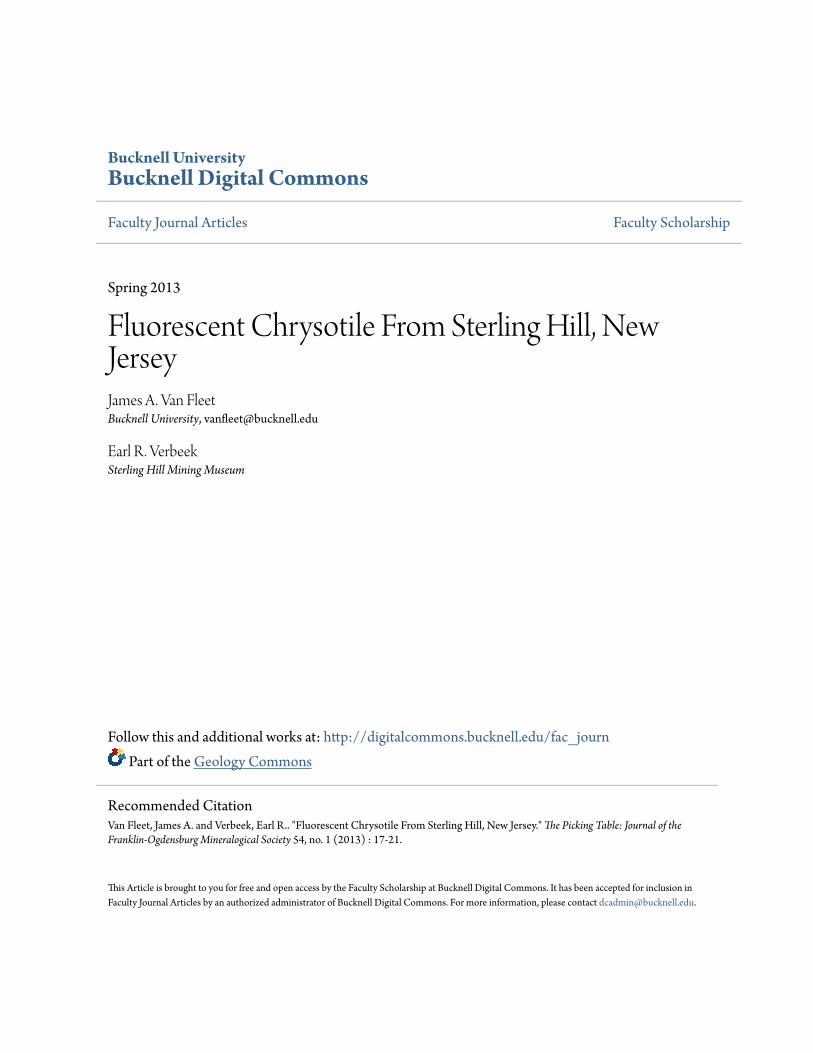
Figure 5. Altered granular ore from the 1750 level at Sterling Hill, consisting of abundant black franklinite, white calcite (stained red by hematite where in contact with franklinite) , and abundant honey brown to ochre brown, translucent chrysotile with resinous luster and conchoidal fracture. A layered vein of tan to reddish brown, fine-grained sphalerite cuts the specimen at a low angle to the photographed face. James Van Fleet specimen, 8.5 x 7 x 4 em.
Figure 7. Vuggy, ivory-colored, microcrystalline to fibrous chrysotile cementinggrainsoffranklinite, chlorite, calcite, and rounded, brownish-green grains of an unidentified mineral, probably serpentine pseudomorphous after willemite. James Van Fleet collection; 14 x 8 x 5 em.
THE SEARCH FOR MORE
The results documented above prompted one of us (JVF) to search for more examples in local collections, at mineral shows, and from online mineral vendors. It now appears that fluorescent serpentines from the local area are at least modestly common, but their generally dim fluorescence and common admixture with other minerals, chiefly calcite, have largely prevented their recognition. Figures 5 through 8 show some of the specimens obtained thus far. In each of them,
Figure 6. Same specimen as in Figure 5, shown under longwave ultraviolet light. Grains of chrysotile, likely pseudomorphous after willemite, fluoresce weak pale yellow, while the sphalerite in the vein fluoresces orange through pink to blue.
Figure 8. Same specimen as in Figure 7, shown under longwave ultraviolet light. The matrix chrysotile fluoresces weak to moderate yellowish white. Scattered grains showing dull red fluorescence are residual calcite.
XRD of the fluorescent material confirmed chrysotile as the chief mineral constituent. Additional specimens (Figs. 1 0-14) were made known to us by Richard Bostwick as this article was in preparation; these have not been X-rayed and thus are sight-identified only as serpentine. To date, we have examined nearly two dozen specimens of fluorescent serpentine from the local area and feel confident that many more exist. Fluorescent serpentine has now been recognized from Sterling Hill (Figs. 1-1 2), Franklin (Figs. 13 and 14), and from one ofthe local quarries (not shown).
THE PICKING TABLE, VOLUME 54, NO.1 - SPRING 2013 19
FLUORESCENT CHRYSOTJLE FROM STERLING HILL, NEW JERSEY JAMES VAN FLEET and EARL R. VERBEEK, PhD
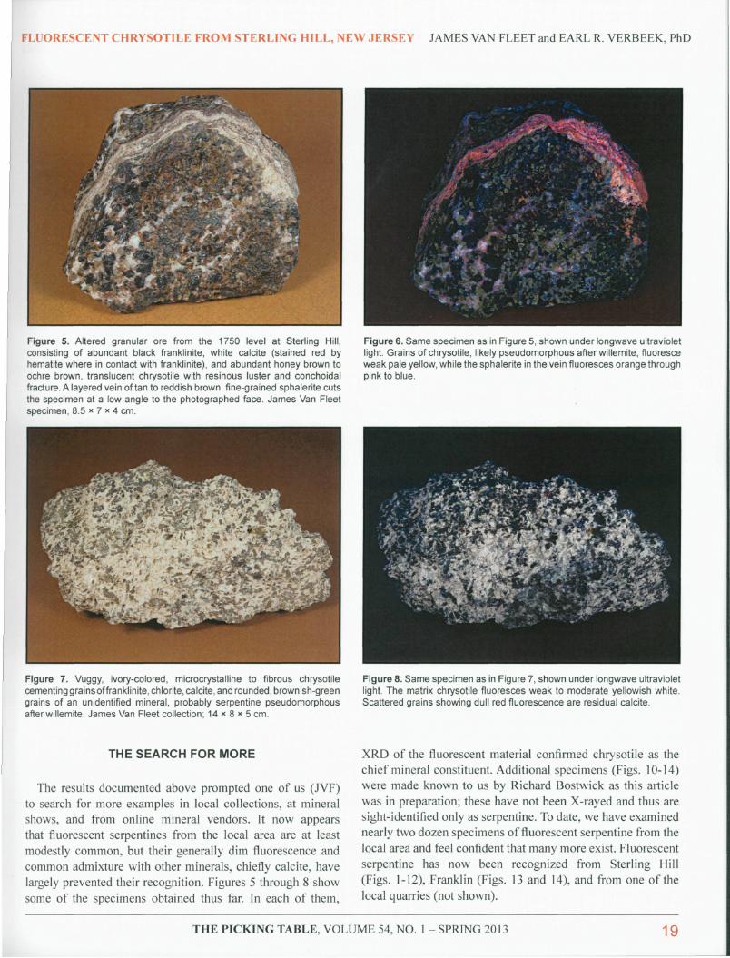
Figure 9. Massive tan serpentine bordered by "cherty" pink sussexite and white calcite, the whole in contact with altered granular ore at left, and minor secondary zincite (orange) at top. The specimen measures 17 x 12 x 9 em and is from the east limb of the North Ore Body at Sterling Hill. Richard Bostwick collection; formerly no. GG2707 in the collection of Gary Grenier.
Figure 11. A vein consisting of translucent to locally transparent dark brown serpentine and white calcite, both in thin, irregular layers, upon a matrix (not visible, on back of specimen) of altered granular franklinitecalcite-willemite (serpentinized) zinc ore. The photograph was taken perpendicular to one of the serpentine layers in the vein. The specimen, 5 x 4 x 3 em, is from the North Ore Body at Sterling Hill. Richard Bostwick specimen, formerly in the AI Smith and "Sunny" Cook collections.
Among the local serpentine specimens examined to date, chrysotile that occurs as a component of chrysotile-calcite veins and as coatings on fracture surfaces is most likely to fluoresce; the occurrences recognized to date come mostly from Sterling Hill. The most common associated minerals are calcite, dolomite, sphalerite, and franklinite. Massive material
Figure 10. Same specimen as in Figure 9, photographed under mediumwave ultraviolet light to show the dim orange-tan fluorescence of the serpentine. Pink areas indicate calcite; green is willemite.
Figure 12. Same specimen as in Figure 11, photographed under longwave ultraviolet light to show the dim yellow to orange fluorescence of the serpentine.
that formed as replacements of precursor minerals in the rock matrix is less likely to show any response. Collectors might profit from examining Sterling Hill serpentine specimens in their own collections with a longwave UV lamp and some patience, preferably in a very dark room. The fluorescent response is "mild" at best.
20 THE PICKING TABLE, VOLUME 54, NO.1 - SPRING 201 3
t I
I
I I I I
I
I [
FLUORESCENT CHRYSOTILE FROM STERLING HILL, NEW JERSEY JAMES VAN FLEET and EARL R. VERBEEK, PhD
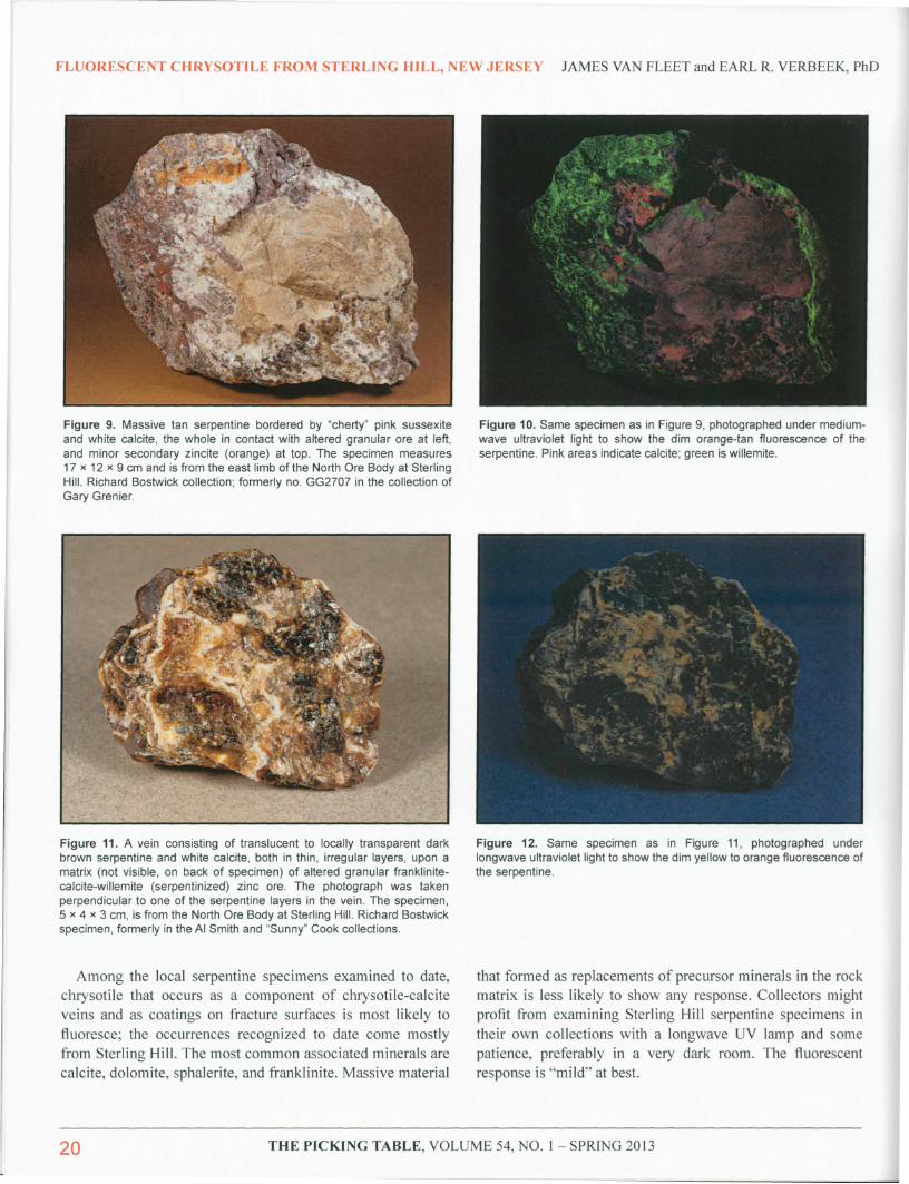
Figure 13. Orange-tan serpentine (probably serpentinized willemite) hosting myriad tiny grains of franklinite in contact with larger grains of franklinite and coarse-grained white calcite. The specimen is labeled Franklin rather than Sterling Hill and measures 6 x 5 x 3.5 em. Richard Bostwick collection; formerly no. DW721 in the collection of Dru Wilbur.
fncidentally, fluorescent serpentine is not especially rare on a worldwide basis, as shown in part by the number oflocalities (II) listed by Dr. Gerhard Henkel (Verbeek and Modreski, 1989). The most common fluorescent response recorded by Dr. Henkel for fluorescing serpentines was cream to yellow or white LW, similar to that of our local material. The common massive green serpentines that form the main component of the rock serpentinite, often quarried as a decorative facing or carving stone, are the least likely to fluoresce, probably because the same iron (substituting for magnesium) that gives most such serpentine its green color also deters its fluorescence. Much more likely to show fluorescence are serpentines that in daylight are of pale to medium brown color, like our local examples and the "deweylite" from the State Line chromite district along the Pennsylvania-Maryland border.
All photos by Earl R. Verbeek
Figure 14. Same specimen as in Figure 13, photographed under longwave ultraviolet light. Serpentine fluoresces pale orange-tan to ivory, calcite dull red, and scattered grains of willemite dull green.
ACKNOWLEDGEMENTS
The authors wish to thank Brad Jordan, Laboratory Director for the Geology Department at Bucknell University, for his assistance in obtaining the X-ray diffraction data that made this paper possible. We are also grateful to Mark Boyer and Richard Bostwick for commenting on serpentine specimens in · our possession, bringing others to our attention, and providing their expertise throughout.
REFERENCES
Cianciulli, J .C. (2004), Clinochrysotile - A fluorescent serpentine from Franklin, New Jersey: The Picking Table, vol. 45, no. 2, pp. 37-39.
Fowler, S. (1825), An account of some new and extraordinary minerals discovered in Warwick, Orange County, N.Y. : American Journal of Science, 1st series, vol. 9, pp. 242-245.
Dunn, P.J . (1995), Franklin and Sterling Hill, New Jersey - The World's Most Magnificent Mineral Deposits. Privately published, 755 pp.
Verbeek, E.R. and Modreski, P.J ., eds. (1989), The Henkel glossary of fluorescent minerals: Journal of the Fluorescent Mineral Society, vol. 15, 9 1 pp. ~
THE PICKING TABLE, VOLUME 54, NO.1 - SPRING 201 3 21