- 67 - Imaging Science in Dentistry 2018; 48: 67-72 https://doi.org/10.5624/isd.2018.48.1.67 The styloid process is a bony prominence of the petrous portion of the temporal bone that projects anteriorly, inferi- orly, and slightly medially. 1-4 The normal length of the sty- loid process is approximately 20 mm, although it can vary on an individual basis. 5-7 When a styloid process exceeds 30 mm in length, it is considered to be elongated. 1,3-5,8-10 Approximately 4% of the population has an elongated sty- loid process, 1,6 and the majority of these patients are diag- nosed based on an incidental radiographic finding. 8 Among patients with elongated styloid processes, 4%- 10% are estimated to have a symptomatic elongated sty- loid process, known as Eagle syndrome. 1,3,5,8 The most common symptoms include throat pain, dysphagia, for- eign body sensation, facial pain, tinnitus, otalgia, and limitation of mandibular movements. 3,6,10-14 However, the broad range of symptoms presents a diagnostic challenge. 3 Eagle syndrome is characterized by facial pain arising from the compression of adjacent anatomical structures due to an elongated styloid process or a calcified stylo- hyoid ligament. 8,13 Eagle syndrome has a 3:1 female-to- male predilection, and affected patients most commonly present with an elongated styloid process between the ages of 30 and 50. 1,15 The presence of symptoms is essen- tial for Eagle syndrome to be diagnosed, and clearly marks the distinction from a patient with an asymptomat- ic elongated styloid process. 3,8,16 Simply identifying a cal- cified stylohyoid ligament or an elongated styloid process radiographically does not warrant the diagnosis of Eagle syndrome. 3 Although uncommon, the timely and proper diagnosis of Eagle syndrome is imperative in all medical fields. 15,17 To confirm the physical examination findings, a panoramic radiograph can be used to identify the elon- gated styloid process. However, because panoramic imag- Fractured styloid process masquerading as neck pain: Cone-beam computed tomography investigation and review of the literature Hassan M. Khan 1 , Andrew D. Fraser 2 , Steven Daws 1 , Jaisri Thoppay 3 , Mel Mupparapu 1, * 1 Division of Radiology, Department of Oral Medicine, University of Pennsylvania School of Dental Medicine, Philadelphia, PA, USA 2 Division of Growth and Development, Section of Orthodontics, School of Dentistry, University of California, Los Angeles, Los Angeles, CA, USA 3 Department of Oral and Maxillofacial Surgery, School of Dentistry, Virginia Commonwealth University, Richmond, VA, USA ABSTRACT Historically, Eagle syndrome is a term that has been used to describe radiating pain in the orofacial region, foreign body sensation, and/or dysphagia due to a unilateral or bilateral elongated styloid process impinging upon the tonsillar region. Because elongated styloid processes-with or without associated Eagle syndrome-can present with various symptoms and radiographic findings, it can be challenging for healthcare practitioners to formulate an accurate diagnosis. Abnormal styloid anatomy can lead to a multitude of symptoms, including chronic orofacial/neck pain, thus masquerading as more commonly diagnosed conditions. In this report, we describe a patient who presented to our department with styloid process elongation and fracture. A careful history, physical examination, and a cone- beam computed tomography (CBCT) investigation led to the diagnosis. The patient was then referred for appropriate care. This case report demonstrates the utilization of CBCT in differentiating a fracture site from a pseudo-joint that might mimic a fracture. (Imaging Sci Dent 2018; 48: 67-72) KEY WORDS: Cone-Beam Computed Tomography; Neck Pain Copyright ⓒ 2018 by Korean Academy of Oral and Maxillofacial Radiology This is an Open Access article distributed under the terms of the Creative Commons Attribution Non-Commercial License (http://creativecommons.org/licenses/by-nc/3.0) which permits unrestricted non-commercial use, distribution, and reproduction in any medium, provided the original work is properly cited. Imaging Science in Dentistry·pISSN 2233-7822 eISSN 2233-7830 Received December 5, 2017; Revised January 2, 2018; Accepted January 13, 2018 *Correspondence to : Prof. Mel Mupparapu Division of Oral and Maxillofacial Radiology, University of Pennsylvania, School of Dental Medicine, Robert Schattner Building, 240 S. 40th St., Philadelphia, PA 19104, USA Tel) 1-215-746-8869, Fax) 215-573-7853, E-mail) [email protected]
Transcript
- 67 -
Imaging Science in Dentistry 2018; 48: 67-72https://doi.org/10.5624/isd.2018.48.1.67
The styloid process is a bony prominence of the petrous portion of the temporal bone that projects anteriorly, inferi-orly, and slightly medially.1-4 The normal length of the sty-loid process is approximately 20 mm, although it can vary on an individual basis.5-7 When a styloid process exceeds 30 mm in length, it is considered to be elongated.1,3-5,8-10 Approximately 4% of the population has an elongated sty-loid process,1,6 and the majority of these patients are diag-nosed based on an incidental radiographic finding.8
Among patients with elongated styloid processes, 4%-10% are estimated to have a symptomatic elongated sty-loid process, known as Eagle syndrome.1,3,5,8 The most common symptoms include throat pain, dysphagia, for-eign body sensation, facial pain, tinnitus, otalgia, and
limitation of mandibular movements.3,6,10-14 However, the broad range of symptoms presents a diagnostic challenge.3 Eagle syndrome is characterized by facial pain arising from the compression of adjacent anatomical structures due to an elongated styloid process or a calcified stylo-hyoid ligament.8,13 Eagle syndrome has a 3:1 female-to-male predilection, and affected patients most commonly present with an elongated styloid process between the ages of 30 and 50.1,15 The presence of symptoms is essen-tial for Eagle syndrome to be diagnosed, and clearly marks the distinction from a patient with an asymptomat-ic elongated styloid process.3,8,16 Simply identifying a cal-cified stylohyoid ligament or an elongated styloid process radiographically does not warrant the diagnosis of Eagle syndrome.3 Although uncommon, the timely and proper diagnosis of Eagle syndrome is imperative in all medical fields.15,17 To confirm the physical examination findings, a panoramic radiograph can be used to identify the elon-gated styloid process. However, because panoramic imag-
Fractured styloid process masquerading as neck pain: Cone-beam computed tomography investigation and review of the literature
Hassan M. Khan1, Andrew D. Fraser2, Steven Daws1, Jaisri Thoppay3, Mel Mupparapu1,*1Division of Radiology, Department of Oral Medicine, University of Pennsylvania School of Dental Medicine, Philadelphia, PA, USA 2Division of Growth and Development, Section of Orthodontics, School of Dentistry, University of California, Los Angeles, Los Angeles, CA, USA 3Department of Oral and Maxillofacial Surgery, School of Dentistry, Virginia Commonwealth University, Richmond, VA, USA
AbstrACt
Historically, Eagle syndrome is a term that has been used to describe radiating pain in the orofacial region, foreign body sensation, and/or dysphagia due to a unilateral or bilateral elongated styloid process impinging upon the tonsillar region. Because elongated styloid processes-with or without associated Eagle syndrome-can present with various symptoms and radiographic findings, it can be challenging for healthcare practitioners to formulate an accurate diagnosis. Abnormal styloid anatomy can lead to a multitude of symptoms, including chronic orofacial/neck pain, thus masquerading as more commonly diagnosed conditions. In this report, we describe a patient who presented to our department with styloid process elongation and fracture. A careful history, physical examination, and a cone-beam computed tomography (CBCT) investigation led to the diagnosis. The patient was then referred for appropriate care. This case report demonstrates the utilization of CBCT in differentiating a fracture site from a pseudo-joint that might mimic a fracture. (Imaging Sci Dent 2018; 48: 67-72)
Copyright ⓒ 2018 by Korean Academy of Oral and Maxillofacial RadiologyThis is an Open Access article distributed under the terms of the Creative Commons Attribution Non-Commercial License (http://creativecommons.org/licenses/by-nc/3.0)
which permits unrestricted non-commercial use, distribution, and reproduction in any medium, provided the original work is properly cited.Imaging Science in Dentistry·pISSN 2233-7822 eISSN 2233-7830
Received December 5, 2017; Revised January 2, 2018; Accepted January 13, 2018*Correspondence to : Prof. Mel MupparapuDivision of Oral and Maxillofacial Radiology, University of Pennsylvania, School of Dental Medicine, Robert Schattner Building, 240 S. 40th St., Philadelphia, PA 19104, USATel) 1-215-746-8869, Fax) 215-573-7853, E-mail) [email protected]
Fractured styloid process masquerading as neck pain: Cone-beam computed tomography investigation and review of the literature
- 68 -
ing does not always reliably capture the area of the styloid process, cone-beam computed tomography (CBCT) may be utilized to visualize the styloid anatomy.2,6,9,16,18 Pain management is critical for some patients who have both Eagle syndrome and other associated findings; therefore, the proper diagnosis is important. In some cases, the co-morbid factors may lead to facial or neck pain, rather than Eagle syndrome itself.
It is important to recognize that other conditions, such as a fractured styloid process, may present similarly to Eagle syndrome. The signs and symptoms of an elongat-ed styloid process or a stylohyoid ligament calcification as originally described by Eagle are very similar to the clinical presentation of a fractured styloid process.10-12 The differential diagnosis for a patient presenting with a
fractured styloid process may include temporomandibular joint dysfunction, tonsillitis, pharyngitis, mastoiditis, pain secondary to unerupted or impacted third molars, glosso-pharyngeal neuralgia, sphenopalatine neuralgia, trigeminal neuralgia, pharyngeal foreign body or tumor, migraine, degenerative disc disease, chronic laryngopharyngeal re-flux, cluster headache, or myofascial pain dysfunction syndrome.15,19-21
This report presents a 46-year-old patient with cervical pain due to a fractured styloid process, which was origi-nally suspected to be a parotid pathology. Evaluating the patient both clinically and radiographically aided in diag-nosing the correct source of pain.
Fig. 1. A panoramic reconstruction cone-beam computed tomography image of the patient shows the elongated styloid processes bilat-erally and the fractured left styloid process (arrow).
Fig. 2. A sagittal cone-beam computed tomography image of the skull shows only the left styloid process (arrows).
Fig. 3. A coronal cone-beam computed tomography image shows the styloid processes bilaterally and identifies the fractured left styloid process (arrows).
- 69 -
Hassan M. Khan et al
Case reportA 46-year-old African-American male presented to the
admissions clinic at Penn Dental Medicine with the chief complaint of needing a check-up due to left neck tender-ness. The patient’s medical history revealed that he had been involved in a brawl 4 months previously, which resulted in an injury in the left infraorbital and malar re-gions. The patient went to the emergency room, where he received a consultation from an otorhinolaryngologist and a primary care physician. The medical team diagnosed the
patient with left orbital fracture, which was reduced and surgically treated with bone plating and screws.
Following surgery, the patient complained of unilateral left-sided jaw and neck pain along with mild swelling for a duration of 3 weeks. The patient reported constant, dull, pressure-like pain that transformed to sharp pain on per-cussion. The pain was elicited by chewing, swallowing, and lateral neck movements. These sharp pain episodes lasted for a few minutes, and the patient reported that cracking his neck relieved the pain. The patient experi-enced swelling and tenderness in his left cervical area, which was also associated with similar pain episodes occurring for the past few months. Due to this pain, the patient revisited the otorhinolaryngologist at the local hospital. He was sent for magnetic resonance imaging
(MRI) of the cervical spine. MRI showed intact vertebrae with mild degenerative changes, as well as mild canal stenosis. In addition, a parotid salivary gland biopsy and submandibular fine needle aspiration were performed on the left submandibular salivary gland tissue. These proce-dures were done because it was thought that the pain orig-inated from the salivary glands, specifically the left pa-rotid, as the pain was radiating to that region. The biopsy results revealed normal glandular tissue and no evidence of malignancy. Aspiration of the swelling only managed to provide short-term symptom relief for a period of 2 weeks.
When the patient presented to Penn Dental Medicine for a re-evaluation, he reported a long history of xerostomia, pain with deglutition, and dysphagia. He denied dysgeu-sia. His further medical history included hypertension, hepatitis C, HIV infection, and epileptic seizures, all of which were medically managed. He reported that although
Fig. 4. An axial cone-beam computed tomography image shows the styloid processes bilaterally and identifies the fractured left styloid process (arrows).
Fig. 5. Multiplanar reconstruction cone-beam computed tomography images show the fractured styloid process (arrows) in all 3 views.
Fractured styloid process masquerading as neck pain: Cone-beam computed tomography investigation and review of the literature
- 70 -
his most recent seizure was after the altercation, his last unprovoked seizure was in 2007.
A repeated clinical exam showed mild diffuse swelling with tenderness on the left posterior auricular area, tender-ness on the left side of the submandibular and parotid area, and pinpoint tenderness near the gonial angle at the level of the C3/C4 vertebrae. No tenderness or salivary gland enlargement was noted on bimanual manipulation of the right submandibular and parotid glands. The range of mo-tion for jaw opening was within normal limits and without myofascial pain on the right side. The temporomandibular joint exam was bilaterally within normal limits. However, due to multifocal tenderness and diffuse swelling, it was difficult to appreciate definitive objective findings on the left side. Cranial nerves II-XII were intact. Intraoral exam-ination showed dental caries on tooth #15 with sensitivity to percussion, mild oral dryness, and clear saliva that was scant when milked from the parotid and submandibular ducts.
A panoramic radiograph was obtained to rule out any odontogenic pathology and to visualize the immediate vicinity of the submandibular region. The radiograph showed bilaterally elongated styloid processes and what appeared to be a horizontal lucency right through the left styloid process at the level of C3 and C4. CBCT was then performed to evaluate the extent of the mineralization and to review the suspected fracture 3-dimensionally. The CBCT images (Figs. 1-5) revealed multiple pseudo-jointed bilateral elongated styloid processes, a well-mineralized stylohyoid process with an almost uniform thickness of 11
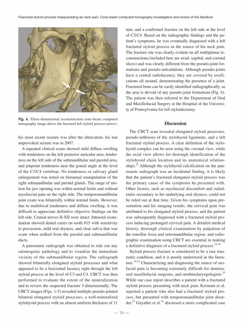
mm, and a confirmed fracture on the left side at the level of C3/C4. Based on the radiographic findings and the pa-tient’s symptoms, he was eventually diagnosed with a left fractured styloid process as the source of his neck pain. The fracture site was clearly evident on all multiplanar re-constructions (included here are axial, sagittal, and coronal slices) and was clearly different from the pseudo-joint for-mations and pseudo-articulations. Although pseudo-joints have a central radiolucency, they are covered by ossifi-cations all around, demonstrating the presence of a joint. Fractured bone can be easily identified radiographically, as the area is devoid of any pseudo-joint formations (Fig. 6). The patient was then referred to the Department of Oral and Maxillofacial Surgery at the Hospital of the Universi-ty of Pennsylvania for left styloidectomy.
pseudo-arthroses of the stylohyoid ligaments, and a left fractured styloid process. A clear definition of the stylo-hyoid complex can be seen using the coronal view, while the axial view allows for thorough identification of the stylohyoid chain location and its anatomical relation-ships.22 Although the stylohyoid calcification on the pan-oramic radiograph was an incidental finding, it is likely that the patient’s fractured elongated styloid process was the primary cause of the symptoms he presented with. Other factors, such as myofascial discomfort and sialad-enitis secondary to his underlying oral dryness, could not be ruled out at that time. Given his symptoms upon pre-sentation and his imaging results, the cervical pain was attributed to his elongated styloid process, and the patient was subsequently diagnosed with a fractured styloid pro-cess inducing prolonged cervical pain. A detailed medical history, thorough clinical examination by palpation of the tonsillar fossa and retromandibular region, and radio-graphic examination using CBCT are essential in making a definitive diagnosis of a fractured styloid process.16,19
Styloid process fracture is considered to be a rare trau-matic condition, and it is poorly understood in the litera-ture.19,23 Characterizing and diagnosing the source of oro-facial pain is becoming extremely difficult for dentists, oral maxillofacial surgeons, and otorhinolaryngologists.23 While our case report describes a patient with a fractured styloid process presenting with neck pain, Kermani et al. reported a patient who also had a fractured styloid pro-cess, but presented with temporomandibular joint disor-der.23 Gayathri et al.24 discussed a more complicated case
Fig. 6. Three-dimensional reconstruction cone-beam computed tomography image shows the fractured left styloid process (arrow).
- 71 -
Hassan M. Khan et al
in which a patient was diagnosed with a styloid fracture co-existing with a mandibular fracture. It is evident that the symptoms associated with a styloid fracture are easy to misdiagnose, making a careful examination a critical aspect of deciphering the source of pain.
Dental outpatient settings are not the only healthcare site where a fractured styloid process can be diagnosed and treated.17 Due to the varying, non-specific, symptom-atic presentations of a fractured styloid process, it is not uncommon for patients with this condition to present to a wide range of healthcare professionals, such as prima-ry care physicians, otorhinolaryngologists, neurologists, neurosurgeons, and psychiatrists.17 Having a thorough understanding of the treatment modalities for styloid pro-cess fracture and guiding the patient in the appropriate direction to help manage their condition are critical steps. The management of styloid process fracture can include conservative therapy, medical management, or surgical treatment.25,26 A conservative approach typically includes heat, rest, liquid diet, non-steroidal anti-inflammatory agents, and muscle relaxants.25,27 Medical management consists of steroidal injections, administration of long-act-ing local anesthetics in the tonsillar fossa region, and oral administration of carbamazepine.12,25 Surgical manage-ment can be done through either the intraoral or extraoral approach.2,5,6,28,29 If the symptoms can be relieved with conservative treatment, that is the preferred route;25 how-ever, surgery was prudent for the patient in our case due to his presenting and ongoing symptoms. The patient was initially prescribed analgesics for pain management, but the relief was only temporary, so he was referred for surgi-cal intervention due to the limitations of the conservative regimen. Intraoral surgical management of this condition is via an incision on the periosteum at the tip of the sty-loid process, followed by an appropriate excision of the bony projection.6,28 For the extraoral method, the incision is more extensive and starts from the mastoid process, ex-tends to the hyoid bone, and then extends to the midline of the chin.2,17,29,30 The advantages of the intraoral technique are that it is safe, simple, less time-consuming, and leaves no scar. However, deep neck infection, injury to the major vessels, and poor visualization are its drawbacks.6,28,30 In contrast, the external approach provides adequate visual-ization and reduces the risk of a deep cervical infection. Scarring, the length of the procedure, and the relatively high likelihood of injuring the facial nerve are disadvan-tages of the extraoral surgical procedure.2,8,29,31
It is inherently difficult to properly diagnose a fractured styloid process due to conflicting factors and the obscure
clinical presentation. While an elongated styloid process is a condition that can be easily diagnosed, it is still import-ant to include styloid process fracture within the differen-tial diagnosis for applicable cases to provide patients with the appropriate treatment. Due to similarities in the clini-cal presentation, a clinician can easily mistake a fractured styloid process for a number of other commonly identified conditions, such as facial neuralgias and craniofacial dis-eases associated with dental, oral, or temporomandibular joint regions.31,32 By reviewing this case study, all health-care practitioners have been alerted of the appropriate strategies for managing patients with symptoms associated with a fractured styloid process or an elongated styloid process, as well as how to differentiate these possibilities from other conditions that can present similarly. Styloidec-tomy is the treatment of choice for a fractured styloid pro-cess.
Eagle syndrome-like symptoms, when masquerading as chronic neck pain, are challenging for clinicians to diag-nose. The patient described herein was initially suspected to have having a salivary gland pathosis; therefore, MRI and a parotid gland biopsy were obtained unnecessarily. A proper history, physical examination, and utilization of CBCT led to the correct diagnosis in our case.
references 1. Rechtweg JS, Wax MK. Eagle’s syndrome: a review. Am J
drome: a review of 15 cases in KVG Medical College Sullia. Oman Med J 2011; 26: 122-6.
3. Mupparapu M, Robinson MD. The mineralized and elongated styloid process: a review of current diagnostic criteria and evaluation strategies. Gen Dent 2005; 53: 54-9.
4. Feldman VB. Eagle’s syndrome: a case of symptomatic cal-cification of the stylohyoid ligaments. J Can Chiropr Assoc 2003; 47: 21-7.
5. Han MK, Kim DW, Yang JY. Non surgical treatment of Ea-gle’s syndrome - a case report -. Korean J Pain 2013; 26: 169-72.
6. Politi M, Toro C, Tenani G. A rare cause for cervical pain: Ea-gle’s syndrome. Int J Dent 2009; 2009: 781297.
7. Chandler JR. Anatomical variations of the stylohyoid complex and clinical significance. Laryngoscope 1977; 87: 1692-701.
8. Murtagh RD, Caracciolo JT, Fernandez G. CT findings associ-ated with Eagle syndrome. AJNR Am J Neuroradiol 2001; 22: 1401-2.
9. Gokce C, Sisman Y, Sipahioglu M. Styloid process elongation or Eagle’s syndrome: is there any role for ectopic calcifica-tion? Eur J Dent 2008; 2: 224-8.
10. Eagle WW. Symptomatic elongated styloid process: report of two cases of styloid process-carotid artery syndrome with op-
Fractured styloid process masquerading as neck pain: Cone-beam computed tomography investigation and review of the literature
- 72 -
eration. Arch Otolaryngol 1949; 49: 490-503.11. Eagle WW. Elongated styloid process: report of two cases.
ment. AMA Arch Otolaryngol 1958; 67: 172-6.13. Balbuena L Jr, Hayes D, Ramirez SG, Johnson R. Eagle’s
syndrome (elongated styloid process). South Med J 1997; 90: 331-4.
14. Langlais RP, Miles DA, Van Dis ML. Elongated and mineral-ized stylohyoid ligament complex: a proposed classification and report of a case of Eagle’s syndrome. Oral Surg Oral Med Oral Pathol 1986; 61: 527-32.
15. Mendelsohn AH, Berke GS, Chhetri DK. Heterogeneity in the clinical presentation of Eagle’s syndrome. Otolaryngol Head Neck Surg 2006; 134: 389-93.
16. Balasubramanian S. The ossification of styloid ligament and its relation to facial pain. Br Dent J 1964; 116: 108-11.
17. Khandelwal S, Hada YS, Harsh A. Eagle’s syndrome - a case report and review of the literature. Saudi Dent J 2011; 23: 211-5.
18. Keur JJ, Campbell JP, McCarthy JF, Ralph WJ. The clinical significance of the elongated styloid process. Oral Surg Oral Med Oral Pathol 1986; 61: 399-404.
19. Tiwary P, Sahoo N, Thakral A, Ranjan U. Styloid process fracture associated with maxillofacial trauma: incidence, dis-tribution, and management. J Oral Maxillofac Surg 2017; 75: 2177-82.
21. Gossman JR Jr, Tarsitano JJ. The styloid-stylohyoid syn-drome. J Oral Surg 1977; 35: 555-60.
22. Alcalde RE, Ueyama Y, Nishiyama A, Mizuguchi T, Matsu-
mura T, Kishi K. Diagnostic imaging of Eagle’s syndrome: report of three cases. Oral Radiol 1994; 10: 63-8.
23. Kermani H, Dehghani N, Aghdashi F, Esmaeelinejad M. Non-syndromic isolated temporal bone styloid process fracture. Trauma Mon 2016; 21: e24395.
24. Gayathri G, Elavenil P, Sasikala B, Pathumai M, Krishnaku-mar Raja VB. ‘Stylo-mandibular complex’ fracture from a maxillofacial surgeon’s perspective-review of the literature and proposal of a management algorithm. Int J Oral Maxillofac Surg 2016; 45: 297-303.
25. Dubey KN, Bajaj A, Kumar I. Fracture of the styloid process associated with the mandible fracture. Contemp Clin Dent 2013; 4: 116-8.
26. Blythe JN, Matthews NS, Connor S. Eagle’s syndrome after fracture of the elongated styloid process. Br J Oral Maxillofac Surg 2009; 47: 233-5.
27. Smith GR, Cherry JE. Traumatic Eagle’s syndrome: report of a case and review of the literature. J Oral Maxillofac Surg 1988; 46: 606-9.
28. Chase DC, Zarmen A, Bigelow WC, McCoy JM. Eagle’s syndrome: a comparison of intraoral versus extraoral surgical approaches. Oral Surg Oral Med Oral Pathol 1986; 62: 625-9.
29. Chrcanovic BR, Custódio AL, de Oliveira DR. An intraoral surgical approach to the styloid process in Eagle’s syndrome. Oral Maxillofac Surg 2009; 13: 145-51.
30. Strauss M, Zohar Y, Laurian N. Elongated styloid process syndrome: intraoral versus external approach for styloid sur-gery. Laryngoscope 1985; 95: 976-9.
31. Baseer MA, Alenazy MS. Eagle’s syndrome: a rare case of young female. Dent Res J (Isfahan) 2013; 10: 568-70.
32. Mayrink G, Figueiredo EP, Sato FR, Moreira RW. Cervicofa-cial pain associated with Eagle’s syndrome misdiagnosed as trigeminal neuralgia. Oral Maxillofac Surg 2012; 16: 207-10.