Page 1
From Spikes to Ripples:
The Evolving and Expanding Role of
Electroencephalography in the Diagnosis
and Treatment of Epilepsy
December 3, 2011
Gregory K. Bergey, M.D.
Johns Hopkins University School of Medicine
American Epilepsy Society | Annual Meeting
Page 2
Disclosures
Name of Commercial
Interest
Eisai
Pfizer
Supernus
UCB
NeuroPace
NIH
Type of Financial
Relationship
Advisory Board
Fellowship Selection
Advisory Board
Consultant
Site PI
Co-Investigator
American Epilepsy Society | Annual Meeting
(Last 12 months)
Page 3
Learning Objectives
• At the conclusion of this presentation
attendees will understand the early
development and applications of EEG.
• At the conclusion of the presentation
attendees will appreciate the current
utility of EEG and potential future uses.
American Epilepsy Society | Annual Meeting
Page 4
Before Hans Berger:
Precursors to the Human EEG
• Caton (1842-1926) recorded electrical
activity from brain cortex of living
animals using galvanometer
(“electrical current of grey matter”)
• Pravdic-Neminsky (1879-1952)
“electrocerebrogram,” recorded
electrical activity from the animal
brain (1912).
• Kauffman – recorded experimentally
induced seizures in dogs (1912).
Richard Caton
Page 5
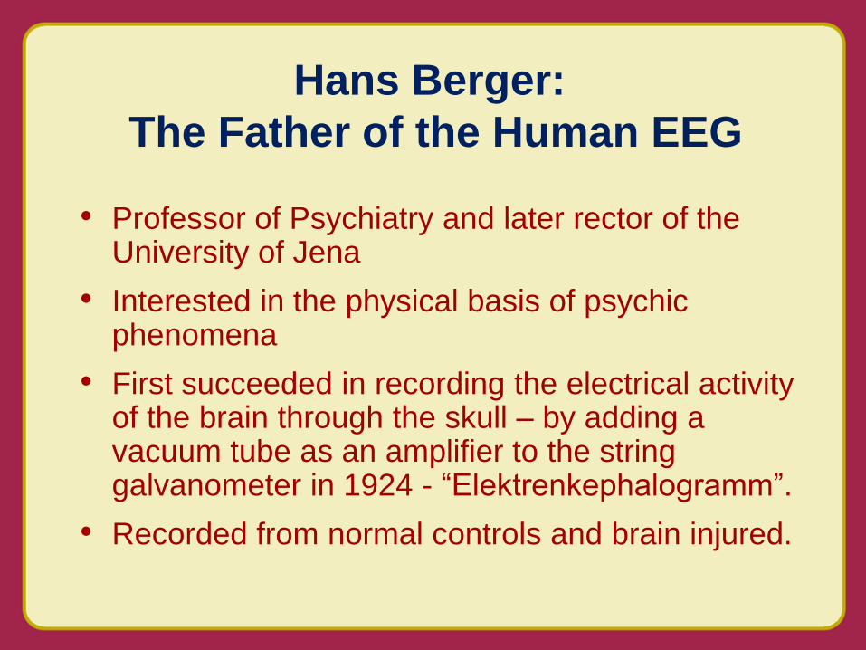
• Professor of Psychiatry and later rector of the University of Jena
• Interested in the physical basis of psychic phenomena
• First succeeded in recording the electrical activity of the brain through the skull – by adding a vacuum tube as an amplifier to the string galvanometer in 1924 - “Elektrenkephalogramm”.
• Recorded from normal controls and brain injured.
Hans Berger:
The Father of the Human EEG
Page 6
Hans Berger – 1873-1941
1929
Page 7
• Described two rhythms – alpha and beta
• Demonstrated that wave characteristics could be
used as index of brain disease
• Described reactivity of alpha rhythm
• First described the electroencephalgram of an
epileptic patient
• At first ignored and even ridiculed, his
discovery was lauded in 1937 at a symposium
with Adrian in Paris.
Hans Berger:
The Father of the EEG
Berger’s First EEG – single
channel with time code
Page 8
• Determination of normal background rhythms
• Detection of generalized and focal (e.g.
slowing) abnormal activity
• Detection of epileptiform activity (spikes,
spike-wave discharges)
• Used widely in neurologic evaluations for
headaches, tumors, stroke, in the absence of
modern imaging techniques
Early Applications of the EEG
Page 9
• Earliest surgery (Horsley, Penfield) did not
rely much on EEG seizure localization
Epilepsy Surgery: Increased
Need for Seizure Localization
1936
Page 10
Early Use of EEG for Seizure
Localization
1936
3 channel EEG – a major advance
by Albert Grass in 1935
Page 11
Early Use of EEG for Seizure
Localization
1938
Page 12
Early Electrocorticography
Jasper HH. Electroencephalogr Clin Neurophysiol. 1949;1:11-18.
Page 13
• Increased numbers of scalp
electrodes
• Delineation of partial vs generalized
epileptiform activity
• Identification of epileptic syndromes
which incorporate EEG patterns (e.g.
petit mal, Lennox-Gastaut)
Later Applications of the EEG
Page 14
Early Use of EEG for Seizure
Classification
Jasper HH and Hawke WA
Arch Neurol Psych 1938;
39:885-9011938
Gibbs FA, Gibbs EL, Lennox
WG. Arch Neurol Psych
1939;41:1111-1116
Page 15
• Educated at Yale and Johns Hopkins (1929)
• Studied with William G. Lennox
• Married Erna Leonhardt (Lennox’s technician) in 1930
• Published first edition of Atlas of EEG in 1941 (year of Berger’s death)
• Awarded (with William Lennox) Albert Lasker Award in 1951
Frederic A Gibbs (1903-1992)
Page 16
Eight channel EEG from Albert
Einstein during exercise where
he is asked to think about the
theory of relativity
Life Magazine April 9, 1951
Page 17
• Use for non-epilepsy diagnoses appropriately diminished by the advances in neuroimaging
• Digital storage
• Ready reformatting
• Remote reading over networks
• Spike and seizure detection software
• Spike and frequency mapping
Current State of Scalp EEG
Page 18
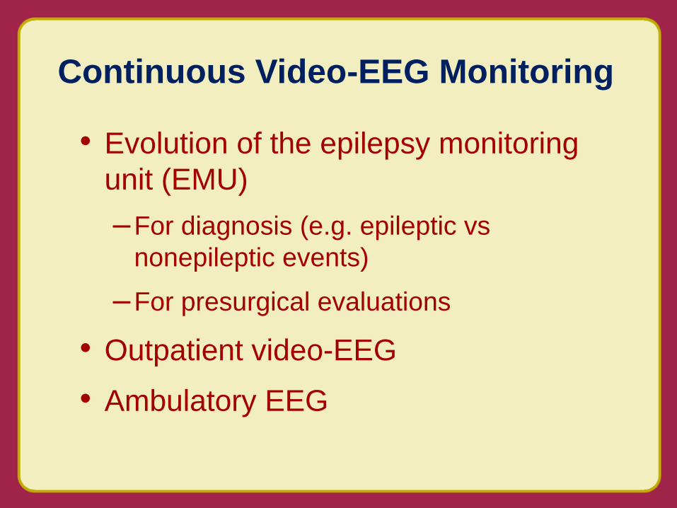
• Evolution of the epilepsy monitoring
unit (EMU)
–For diagnosis (e.g. epileptic vs
nonepileptic events)
–For presurgical evaluations
• Outpatient video-EEG
• Ambulatory EEG
Continuous Video-EEG Monitoring
Page 19
• Earliest arrays used paper and
ink recording and unsophisticated
video-EEG correlation
• Later digital + VCR technology
• Later digital video with digital
EEG
Continuous Video-EEG Monitoring
Page 20
• Dedicated unit staffed with technologists and nurses
• Capability for continuous video and EEG monitoring and storage of data
• Various spike and seizure detection software
• Epilepsy fellows
Components of an EMU
Page 21
• Current digital technology for video and EEG
• Excellent synchronization of video and EEG
• Large storage demands
– Video (5 GB for 24 hr)
– Directly related to channel number, sampling
frequency (e.g. 24 hrs of 124 channels sampled at
2000 Hz requires 20 GB without video)
Continuous Video-EEG Monitoring
Page 22
• Expansion outside EMU
–NICU monitoring (seizures,
hypothermia)
–NCCU monitoring
• Treatment of refractory status
epilepticus
• Detection and treatment of
subtle status
Continuous Video-EEG Monitoring
Page 23
• Need for improved seizure
localization in patients with
inconclusive scalp ictal recordings
• Need for improved function
mapping of eloquent cortex
–Foerster and Penfield began
functional mapping in 1920s
Evolution of Intracranial Monitoring
Penfield and Roberts
1959
Page 24
• Depth electrode arrays – 1960s
• Subdural grid arrays – 1970s
Evolution of Intracranial Monitoring
Page 25
• Increased numbers of
channels with intracranial
arrays
• Increased sampling
frequencies for higher
frequencies
• Increased storage needs
Evolution of Intracranial Monitoring
Page 26
• High density scalp arrays
• Dipole localization
• Spike mapping
• fMRI
• Functional mapping
Recent Applications of the EEG
Page 27
• High frequency and ultrahigh
frequency recording
• Functional mapping without cortical
stimulation
• Therapeutic responsive stimulation
triggered by EEG activity
• Brain/machine; brain body interface
Future applications
(the future is now)
Page 28
• High frequency and ultrahigh
frequency recording
Future applications
(the future is now)
Jouny CC and Bergey GK, unpublished
Page 29
• Old terminology – alpha, beta, delta,
theta, spikes
• New terminology
– Irritative zone (IZ)
–Seizure onset zone (SOZ)
–Epileptogenic zone (EZ)
EEG Terminology
Page 30
• New terminology
– Gamma (30-80 Hz), high gamma (80-150 Hz)
– High frequency oscillations (HFO; 40-500 Hz))
– Ripples (80-250 Hz), fast ripples (250-500 Hz)
EEG Terminology
Page 31
• Functional mapping without cortical stimulation
Future applications
(the future is now)
Functional-anatomic and temporal specificity of high gamma augmentation
Flinker et al. 2010 J Neurosci
Page 32
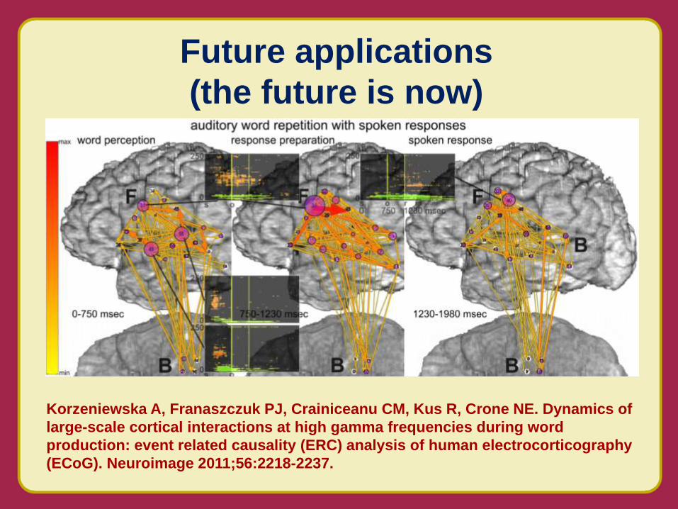
Future applications
(the future is now)
Korzeniewska A, Franaszczuk PJ, Crainiceanu CM, Kus R, Crone NE. Dynamics of
large-scale cortical interactions at high gamma frequencies during word
production: event related causality (ERC) analysis of human electrocorticography
(ECoG). Neuroimage 2011;56:2218-2237.
Page 33
• Responsive stimulation triggered by
epileptiform EEG activity
Future applications
(the future is now)
Seizure activity triggers detector
early in seizure. Closed-loop
therapy delivered and seizure terminated
Page 34
• The EEG remains important in the evaluation of patients with seizure disorders.
• Development of digital technology has led to dramatic improvements in video-EEG recording technology.
• New recording arrays and high frequency sampling hold promise for improving presurgical evaluations.
• Closed loop systems offer promise for advances in therapeutic neurostimulation.
Impact on Clinical Care
Page 35
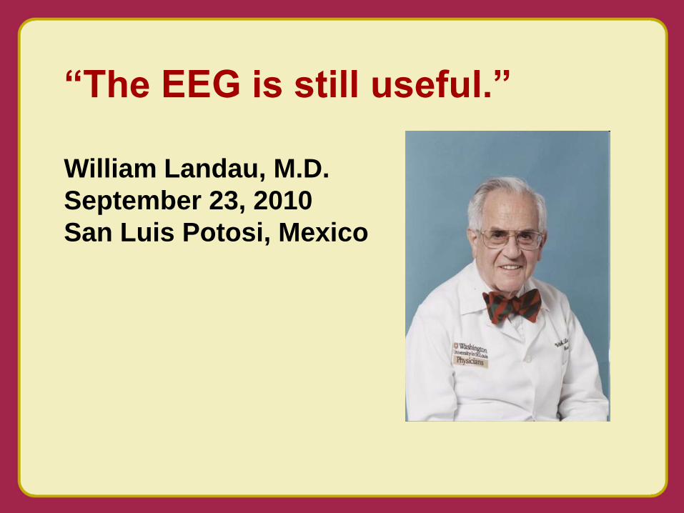
“The EEG is still useful.”
William Landau, M.D.
September 23, 2010
San Luis Potosi, Mexico