

From Spikes to Ripples:
The Evolving and Expanding Role of
Electroencephalography in the Diagnosis
and Treatment of Epilepsy
December 3, 2011
Gregory K. Bergey, M.D.
Johns Hopkins University School of Medicine
American Epilepsy Society | Annual Meeting

Disclosures
Name of Commercial
Interest
Eisai
Pfizer
Supernus
UCB
NeuroPace
NIH
Type of Financial
Relationship
Advisory Board
Fellowship Selection
Advisory Board
Consultant
Site PI
Co-Investigator
American Epilepsy Society | Annual Meeting
(Last 12 months)

Learning Objectives
• At the conclusion of this presentation
attendees will understand the early
development and applications of EEG.
• At the conclusion of the presentation
attendees will appreciate the current
utility of EEG and potential future uses.
American Epilepsy Society | Annual Meeting

Before Hans Berger:
Precursors to the Human EEG
• Caton (1842-1926) recorded electrical
activity from brain cortex of living
animals using galvanometer
(“electrical current of grey matter”)
• Pravdic-Neminsky (1879-1952)
“electrocerebrogram,” recorded
electrical activity from the animal
brain (1912).
• Kauffman – recorded experimentally
induced seizures in dogs (1912).
Richard Caton
• Professor of Psychiatry and later rector of the University of Jena
• Interested in the physical basis of psychic phenomena
• First succeeded in recording the electrical activity of the brain through the skull – by adding a vacuum tube as an amplifier to the string galvanometer in 1924 - “Elektrenkephalogramm”.
• Recorded from normal controls and brain injured.
Hans Berger:
The Father of the Human EEG

Hans Berger – 1873-1941
1929

• Described two rhythms – alpha and beta
• Demonstrated that wave characteristics could be
used as index of brain disease
• Described reactivity of alpha rhythm
• First described the electroencephalgram of an
epileptic patient
• At first ignored and even ridiculed, his
discovery was lauded in 1937 at a symposium
with Adrian in Paris.
Hans Berger:
The Father of the EEG
Berger’s First EEG – single
channel with time code

• Determination of normal background rhythms
• Detection of generalized and focal (e.g.
slowing) abnormal activity
• Detection of epileptiform activity (spikes,
spike-wave discharges)
• Used widely in neurologic evaluations for
headaches, tumors, stroke, in the absence of
modern imaging techniques
Early Applications of the EEG

• Earliest surgery (Horsley, Penfield) did not
rely much on EEG seizure localization
Epilepsy Surgery: Increased
Need for Seizure Localization
1936

Early Use of EEG for Seizure
Localization
1936
3 channel EEG – a major advance
by Albert Grass in 1935

Early Use of EEG for Seizure
Localization
1938

Early Electrocorticography
Jasper HH. Electroencephalogr Clin Neurophysiol. 1949;1:11-18.

• Increased numbers of scalp
electrodes
• Delineation of partial vs generalized
epileptiform activity
• Identification of epileptic syndromes
which incorporate EEG patterns (e.g.
petit mal, Lennox-Gastaut)
Later Applications of the EEG

Early Use of EEG for Seizure
Classification
Jasper HH and Hawke WA
Arch Neurol Psych 1938;
39:885-9011938
Gibbs FA, Gibbs EL, Lennox
WG. Arch Neurol Psych
1939;41:1111-1116

• Educated at Yale and Johns Hopkins (1929)
• Studied with William G. Lennox
• Married Erna Leonhardt (Lennox’s technician) in 1930
• Published first edition of Atlas of EEG in 1941 (year of Berger’s death)
• Awarded (with William Lennox) Albert Lasker Award in 1951
Frederic A Gibbs (1903-1992)

Eight channel EEG from Albert
Einstein during exercise where
he is asked to think about the
theory of relativity
Life Magazine April 9, 1951

• Use for non-epilepsy diagnoses appropriately diminished by the advances in neuroimaging
• Digital storage
• Ready reformatting
• Remote reading over networks
• Spike and seizure detection software
• Spike and frequency mapping
Current State of Scalp EEG
• Evolution of the epilepsy monitoring
unit (EMU)
–For diagnosis (e.g. epileptic vs
nonepileptic events)
–For presurgical evaluations
• Outpatient video-EEG
• Ambulatory EEG
Continuous Video-EEG Monitoring

• Earliest arrays used paper and
ink recording and unsophisticated
video-EEG correlation
• Later digital + VCR technology
• Later digital video with digital
EEG
Continuous Video-EEG Monitoring

• Dedicated unit staffed with technologists and nurses
• Capability for continuous video and EEG monitoring and storage of data
• Various spike and seizure detection software
• Epilepsy fellows
Components of an EMU

• Current digital technology for video and EEG
• Excellent synchronization of video and EEG
• Large storage demands
– Video (5 GB for 24 hr)
– Directly related to channel number, sampling
frequency (e.g. 24 hrs of 124 channels sampled at
2000 Hz requires 20 GB without video)
Continuous Video-EEG Monitoring

• Expansion outside EMU
–NICU monitoring (seizures,
hypothermia)
–NCCU monitoring
• Treatment of refractory status
epilepticus
• Detection and treatment of
subtle status
Continuous Video-EEG Monitoring

• Need for improved seizure
localization in patients with
inconclusive scalp ictal recordings
• Need for improved function
mapping of eloquent cortex
–Foerster and Penfield began
functional mapping in 1920s
Evolution of Intracranial Monitoring
Penfield and Roberts
1959

• Depth electrode arrays – 1960s
• Subdural grid arrays – 1970s
Evolution of Intracranial Monitoring

• Increased numbers of
channels with intracranial
arrays
• Increased sampling
frequencies for higher
frequencies
• Increased storage needs
Evolution of Intracranial Monitoring

• High density scalp arrays
• Dipole localization
• Spike mapping
• fMRI
• Functional mapping
Recent Applications of the EEG

• High frequency and ultrahigh
frequency recording
• Functional mapping without cortical
stimulation
• Therapeutic responsive stimulation
triggered by EEG activity
• Brain/machine; brain body interface
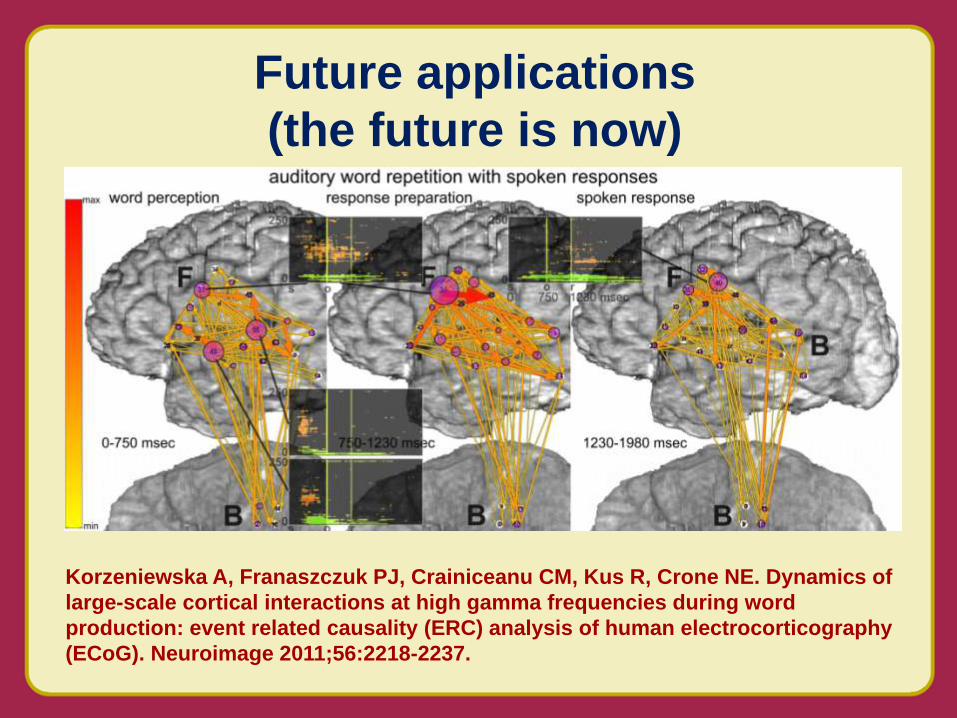
Future applications
(the future is now)

• High frequency and ultrahigh
frequency recording
Future applications
(the future is now)
Jouny CC and Bergey GK, unpublished

• Old terminology – alpha, beta, delta,
theta, spikes
• New terminology
– Irritative zone (IZ)
–Seizure onset zone (SOZ)
–Epileptogenic zone (EZ)
EEG Terminology

• New terminology
– Gamma (30-80 Hz), high gamma (80-150 Hz)
– High frequency oscillations (HFO; 40-500 Hz))
– Ripples (80-250 Hz), fast ripples (250-500 Hz)
EEG Terminology

• Functional mapping without cortical stimulation
Future applications
(the future is now)
Functional-anatomic and temporal specificity of high gamma augmentation
Flinker et al. 2010 J Neurosci
Future applications
(the future is now)
Korzeniewska A, Franaszczuk PJ, Crainiceanu CM, Kus R, Crone NE. Dynamics of
large-scale cortical interactions at high gamma frequencies during word
production: event related causality (ERC) analysis of human electrocorticography
(ECoG). Neuroimage 2011;56:2218-2237.

• Responsive stimulation triggered by
epileptiform EEG activity
Future applications
(the future is now)
Seizure activity triggers detector
early in seizure. Closed-loop
therapy delivered and seizure terminated

• The EEG remains important in the evaluation of patients with seizure disorders.
• Development of digital technology has led to dramatic improvements in video-EEG recording technology.
• New recording arrays and high frequency sampling hold promise for improving presurgical evaluations.
• Closed loop systems offer promise for advances in therapeutic neurostimulation.
Impact on Clinical Care
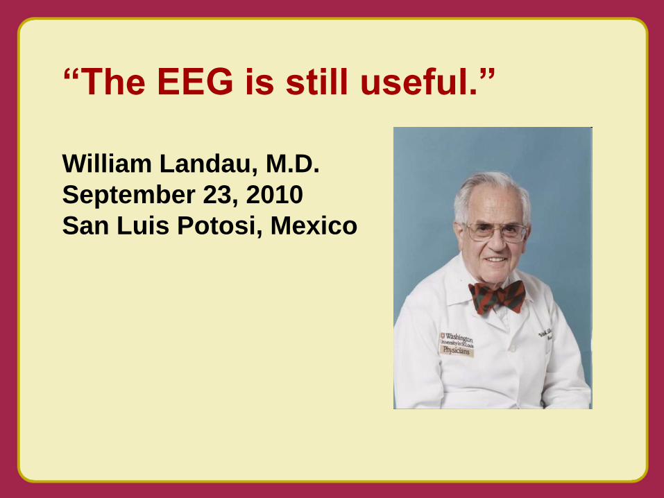
“The EEG is still useful.”
William Landau, M.D.
September 23, 2010
San Luis Potosi, Mexico


