A METHOD TO DETERMINE THE PERIPHERAL ARTERIAL BLOOD PRESSURE IN THE MOUSE BY PHILIP D. McMASTER, M.D. (From the Laboratories of Tke Rockefeller Institutefor Medical Researck) PLATES 4 AND 5 (Received for publication, January 22, 1941) A method for the determination of the blood pressure of the mouse without injury to the animal will be briefly described in the present paper, together with certain observations upon the changes of pressure under various physio- logical conditions. The method has the advantage that it enables the pres- sure to be taken while injections into a vein of the tail are in progress. Previous Work.--Recently Bonsmann (1) reported a method to determine the blood pressure in the tails of mice and rats. A specially designed cuff enclosed the tail except for its tip which lay under a photoelectric cell. Blood was driven out of the tail by pressure in the cuff, and when it was allowed to return the resulting change of color in the tip of the tail was registered by the cell and the pressure was read off. More will be said of Bonsmann's findings below. As the method employed the ani- maps tail, it was not suited to our purposes. Still more recently Diaz and Levy (2) and Williams, Harrison, and Grollman (3) have published excellent methods to de- termine the blood pressure in the rat, but their methods also involve utilization of the animal's tail. Griffith (4) has reported a method to measure the blood pressure in the legs of the rat. In his work the blood vessels of the skin of the foot were observed while pressure was exerted on the thigh by means of a cuff. We have modified the method of Griffith, observing the return of blood to the claws of the hind feet of mice. The Metkod The daws of the hind legs of mice were found to be transparent to the focused and cooled beam of a carbon arc light. Under these circumstances, as will be shown below, the entire circulation of the nail bed, including the afferent and efferent vessels and the capillaries, becomes visible under a bin- ocular microscope. One can see with ease the moment at which peripheral circulation ceases in the claw, if pressure is exerted higher up on the leg by a cuff, as in clinical methods for determining blood pressure. One can note too with great accuracy the moment that arterial circulation begins anew when pressure is gradually released. Readings can be made more accurately by 29 Downloaded from http://rupress.org/jem/article-pdf/74/1/29/1182239/29.pdf by guest on 18 March 2022
Transcript
A METHOD TO D E T E R M I N E THE PERIPHERAL ARTERIAL BLOOD PRESSURE IN THE MOUSE
BY PHILIP D. McMASTER, M.D.
(From the Laboratories of Tke Rockefeller Institute for Medical Researck)
PLATES 4 AND 5
(Received for publication, January 22, 1941)
A method for the determination of the blood pressure of the mouse without injury to the animal will be briefly described in the present paper, together with certain observations upon the changes of pressure under various physio- logical conditions. The method has the advantage that it enables the pres- sure to be taken while injections into a vein of the tail are in progress.
Previous Work.--Recently Bonsmann (1) reported a method to determine the blood pressure in the tails of mice and rats. A specially designed cuff enclosed the tail except for its tip which lay under a photoelectric cell. Blood was driven out of the tail by pressure in the cuff, and when it was allowed to return the resulting change of color in the tip of the tail was registered by the cell and the pressure was read off. More will be said of Bonsmann's findings below. As the method employed the ani- maps tail, it was not suited to our purposes. Still more recently Diaz and Levy (2) and Williams, Harrison, and Grollman (3) have published excellent methods to de- termine the blood pressure in the rat, but their methods also involve utilization of the animal's tail.
Griffith (4) has reported a method to measure the blood pressure in the legs of the rat. In his work the blood vessels of the skin of the foot were observed while pressure was exerted on the thigh by means of a cuff. We have modified the method of Griffith, observing the return of blood to the claws of the hind feet of mice.
The Metkod
The daws of the hind legs of mice were found to be transparent to the focused and cooled beam of a carbon arc light. Under these circumstances, as will be shown below, the entire circulation of the nail bed, including the afferent and efferent vessels and the capillaries, becomes visible under a bin- ocular microscope. One can see with ease the moment at which peripheral circulation ceases in the claw, if pressure is exerted higher up on the leg by a cuff, as in clinical methods for determining blood pressure. One can note too with great accuracy the moment that arterial circulation begins anew when pressure is gradually released. Readings can be made more accurately by
29
Dow
nloaded from http://rupress.org/jem
/article-pdf/74/1/29/1182239/29.pdf by guest on 18 March 2022
30 PERIPB-ERALARTERIALBLOOD PRESSURE INMOUSE
observing the circulation in the claw than by watching the circulation in the skin of the leg, for, as Griffith (4) has pointed out for the rat , there is difficulty in de termining whether or not the smaller cutaneous vessels seen under the microscope are ar ter ia l or venous. 1
Mice of 16 to 35 gm. body weight were anesthetized by a single intraperitoneal injection of nembutal or luminal. Nembutal was given as a 1 per cent solution, 0.5 cc. per 25 gin. of body weight, luminal as a 2 per cent solution in doses of 0.125 cc. for every 10 gm. of body weight. In the dosages employed, the luminal usually pro- duced a deeper and longer anesthesia, but, as will be seen below, the depth of anes- thesia varied greatly from animal to animal at any given time after the injection.
The Pressure Apparatus.--It is difficult to arrange a blood pressure cuff about the upper part of the leg of small mice, for the leg is so shaped that the cuff when inflated tends to slip toward the foot. I t was necessary to prevent this in some way. The following means was e m p l o y e d : -
The board on which the anesthetized mouse lay consisted of pieces of cork cemented together. I t was hollowed out, as shown in Fig. 1, and two movable strips of celluloid were laid over the hollow. These strips could be shifted to accommodate animals of different sizes. Two smaller celluloid plates, 6 cm. long and 2 cm. in height, were set upright and parallel to each other into other smaller pieces of cork board, as shown in the figure. These celluloid strips stood 1.5 cm. apart. Through each plate of cellu- loid three holes were bored, 1 cm. in diameter, large enough to allow the anesthetized animal's hind legs and tail to pass through without meeting obstruction. When the animal was placed on its back, as shown in the photograph, and one leg was drawn through the holes, a segment of the thigh 1.5 cm. in length lay between the two cellu- loid plates and about 0.7 cm. above that portion of cork board shown in the figure at A.
A small rubber blood pressure cuff had been constructed, of such size that when deflated it could be passed about the animal's thigh, just filling the space between the parallel pieces of celluloid and the board at A. A ribbon-like strip of steel, just wide enough to fit between the celluloid plates and bent in a semicircle, was placed over the cuff while the latter was still deflated, as indicated at C in Fig. 1. Through holes bored in both celluloid plates pins, not shown in the figure, were passed horizontally just above the metal strip. When inflation of the cuff was begun, the pins prevented the metal strip from rising. As result, the cuff was held against the leg by the metal strip above, by the cork board below, and by the celluloid plates at each side, and was so retained by these structures that pressure came upon that portion of the leg which lay between the celluloid plates. The latter, in addition, prevented the cuff from slipping up or down the leg. The cuff was connected with a mercury or water ma- nometer in the usual way and with an inflating bulb.
To make visible the circulation in the nail, the rays of a Leitz carbon arc light were cooled by passage through 5 cm. of water and focused, by an adjustable concave mirror, upon the claw to be examined. For simplicity this part of the apparatus has been omitted from the photograph in Fig. 1. The brilliant light transilluminated the
1 I t may be noted in passing that the circulation in the claw of rats, even of young ones, can be seen only with difficulty.
Dow
nloaded from http://rupress.org/jem
/article-pdf/74/1/29/1182239/29.pdf by guest on 18 March 2022
rmLn, D. ~cMASTER 31
claw and when the room was in darkness the circulation of blood and movement of the individual red ceils in the capillaries could be easily seen with the aid of a microscope. In each experiment the light was focused in turn upon each of the five claws and the circulation in each examined. The claw yielding the clearest picture of its blood vessels was selected. The blood vessels were best seen when the rays of light entered on the convex surface of the claw.
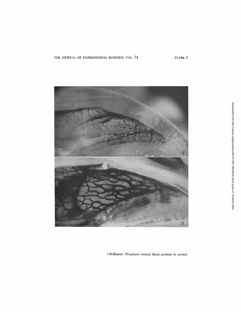
The vascularization of two claws is shown in Figs. 2 and 3. To obtain Fig. 2 an anesthetized mouse, 27 gm. in body weight, was slowly injected into a vein with 1 cc. of a solution of I-Iiggins India ink diluted six times with a 0.9 per cent NaC1 solution containing 1.3 per cent of gelatin. After this had entered the circulation, the animal was bled from the jugular vein while a second cubic centimeter of the ink solution was injected. A few minutes after the injection the foot was removed and placed in ice water for 15 minutes and then in cold alcohol for half an hour, after which it was partially cleared in methyl salicylate for 24 hours. The vessels, magnified 100 times as seen in the photograph, have approximately the same caliber as in life and appear much as they do during the measurement of blood pressure. In order to avoid dilatation of the vessels it was necessary to make an incomplete injec- tion, and as result the picture does not bring out all the vessels present. Further, in a photograph it is impossible to include all the vessels since they cannot be brought into one plane. The richness of the circulatory bed of the claw is better demonstrated in Fig. 3, which was obtained under the same circumstances as Fig. 2, except for the fact tha t the injection material con- sisted of 4 cc. of a mixture of 8 per cent gelatin solution with equal parts of undiluted Higgins India ink. I t will be seen on comparison with Fig. 2 tha t the smaller vessels are much dilated. During life blood flows toward the pe- riphery in the smaller vessels of the plexus at the center of the picture and returns in the two large marginal vessels.
The*measurement of blood pressure was carried out in the following manner. The cuff was rapidly inflated to a pressure estimated to be just above the systolic blood pressure, which will be seen below to vary somewhat with the stage of anesthesia and the condition of the animal. If the movement of blood continued in the claw, the pressure in the cuff was raised slowly until it ceased. When cessation of circulation was attained, the pressure in the cuff was lowered by stages of 10 ram. of mercury, allowing it to remain at each level for 2 minutes. When flow first appeared in any of the vessels of the claw, the pressure was recorded and the cuff deflated. After 2 or 3 minutes the cuff was again blown up, this time to a pressure a few millimeters of mercury higher than that just recorded. When all flow in the claw ceased, the pressure was lowered, 2 or 3 mm. of mercury at a time, with a wait of a minute or two at each new pressure, until a slight movement of red cells appeared in one or two of the small vessels in the central portion of the plexus near the base of the claw, the region indicated in Fig. 3 by an arrow. The slight movement usually lasted for only
Dow
nloaded from http://rupress.org/jem
/article-pdf/74/1/29/1182239/29.pdf by guest on 18 March 2022
32 P E R I P H E R A L A R T E R I A L BLOOD PRESSURE I N MOUSE
a few seconds and then ceased, as though it had been caused by a readjustment of fluid in the vessels and not by true blood flow. In most instances no further flow took place and a minute or two later the pressure in the cuff was lowered 2 or 3 ram. of mercury. As result, a pulsating flow of cells, toward the extremity of the toe, usually made its appearance, first in one or two channels, then in most of the vessels of the plexus. In some instances, without any lowering of the pressure, the slight initial movement of cells was followed by the pulsating flow, while in rare cases this failed to appear until the pressure had been lowered several times by 2 or 3 mm. of mercury. As a rule, a few seconds after the pulsating flow of cells began the flow became con- tinuous through all the small channels of the plexus. For a little while, as the pressure was maintained in the cuff, the cells accumulated in the larger collecting vessels at the edge of the plexus, but after half a minut.e, or slightly more, blood flow usually es- tablished itself in these vessels too. If the circulation failed to become complete within one or two minutes the pressure in the cuff was reduced by 2 or 3 ram. of mercury and invariably flow appeared in all the vessels. If the final reduction of pressure was not made, the circulation eventually established itself, but sometimes only after several minutes.
For reasons to be discussed below, we have taken as the systolic blood pres- sure in the leg that pressure found in the cuff at the moment when the pul- sating flow of blood first appeared in the small vessels of the claw and became continuous. As routine, to avoid undue congestion of the foot, pressure in the cuff was released as soon as the systolic pressure had been determined. In all instances two pressure determinations were regularly made in the manner just described and the average of the readings was taken, if agreement was good. When the readings varied by more than 5 mm. of mercury, a third determination was made and the three readings averaged.
Control Experiments
The question arose, were we measuring the true systolic pressure in the large arteries of the thigh or did the apparatus merely obstruct blood flow in the skin and claw? To test this point two series of control experiments were made. In the first, dye was injected into a tail vein while various pres- sures were maintained in the pressure cuff. In the second series of control experiments blood pressure was determined directly from the carotid ar tery and compared with simultaneous measurements obtained from the leg by the method just described.
Results of the Injection of Dye into the Circulation during Measurement of the Blood Pressure.--In twelve experiments, after the systolic blood pressure had been measured in the leg as just described, the cuff was inflated to a pressure higher by 2 to 4 ram. of mercury. After ascertaining that flow in the claw had ceased, 0.05 cc. of a 5.4 per cent, isotonic solution of a vital dye, pontamine sky blue, was injected into the tail vein. This dye solution, the preparation of which has been described (5), has been used by us in larger or smaller doses in much previous work (5-8). The injections
Dow
nloaded from http://rupress.org/jem
/article-pdf/74/1/29/1182239/29.pdf by guest on 18 March 2022
l,m~xP D. MC.MAS~'ER 33
employed here colored the animals well except in the portion of the leg and foot below the pressure cuff. After a few minutes, during which color still failed to appear in the occluded foot, the pressure in the sphygmomanometer cuff was lowered by a few millimeters of mercury until the pulsating flow of red cells made its appearance in the minute vessels of the claw.
This procedure invariably was at tended by blue coloration of the leg and foot. This had been absent previously, showing that there was no blood flow to the tissues of the foot or lower leg at pressures above the one we accepted as the peripheral systolic pressure.
The second series of control experiments involved simultaneous blood pres- sure readings in the leg by the method described above, and in the carotid ar tery by direct cannulation of the latter.
Direct Measurement of the Carotid Blood Pressure of the Mo~e.--Mice anesthetized with nembutal or luminal were injected intravenously with 0.1 co. of a heparin solu- tion, 10 units to the co. They were placed on the board shown in Fig. 1, covered with light layers of cotton, and kept warm by means of electric lights placed near the body. Half an hour later one carotid artery was exposed and cannulated with a gauge No. 27 hypodermic needle, employing a binocular microscope for its insertion. During this manipulation the artery was occluded with a rubber-tipped bulldog clamp. To prevent all loss of blood during the measurement of blood pressure, the apparatus shown in Fig. 1 was employed. The cannulating needle, previously filled with heparin solution, was connected with a three-way stopcock and this in turn was affixed to a 0.2 co. Bureau of Standards pipette bent at right angles, as shown in the figure. The pipette was also filled with heparin solution but contained a minute droplet of mercury in the center of the graduated portion. A manometer in circuit with a device by which any desired pressure could be brought upon the contents of the pipette was connected with the latter, as shown in the figure. The device need not be described in detail here as that has already been done in earlier work (9), while furthermore, the principles of its operation are clear from the photograph (Fig. 1).
During the insertion of the needle into the artery and while it was being tied in with very fine silk thread, the stopcock was turned in such a way that the contents of the needle was shut off from that of the pipette. The clamp on the artery was then released, and a pressure of about 100 ram. of mercury was put upon the contents of the pipette. The stopcock was then turned for a second or two to allow communica- tion between the needle and the pipette-manometer system. Since the pressures in the pipette and the carotid artery were usually not equal, the droplet of mercury in the pipette began to move either toward or away from the artery. The stopcock was immediately closed, and the pressure in the pipette-manometer circuit either increased or decreased by raising or lowering the leveling bulb B, shown in Fig. 1. Again the stopcock was opened to permit communication between the needle and the pipette and the droplet of mercury allowed to return to its original position. These adjust- ments were repeated as often as necessary until the pressure in the pipette just bal- anced that in the carotid artery and the mercury droplet merely pulsated in the pipette but did not flow in either direction.
Dow
nloaded from http://rupress.org/jem
/article-pdf/74/1/29/1182239/29.pdf by guest on 18 March 2022
34 PERIPH~P~AL ARTEI~AL BLOOD PRESSURE IN MOUSE
A Comparison of the Findings Obtained by the Two Methods Just Described.-- In fourteen experiments simultaneous readings of blood pressure in the carotid artery and in the leg were successfully obtained. As a rule, the reading of the manometer attached to the carotid artery was a few millimeters higher than that attached to the cuff when the cells in the minute vessels of the claw first manifested the pulsating movement already described, which later, and without change in cuff pressure, became continuous. Better agreement be- tween the two methods was sometimes found if the pressure in the cuff was read at the moment when the first irregular movement of cells occurred in the smallest vessels of the obstructed claw. But, as already mentioned, the first irregular movement of ceils in the small vessels of the claw sometimes ceased and was not resumed again unless the cuff pressure was lowered by a few millimeters of mercury. I t was felt that this movement might simply be the expression of readjustment of blood in the vessels following the early re- lease of pressure in the cuff and the phenomenon was not as dear-cut as the appearance of the pulsating flow.
In some experiments many readings were taken by observation of the claw and while doing so the cuff was repeatedly inflated or deflated. In these in- stances pronounced edema of the foot and lower leg developed. Under these circumstances the first appearance in the capillaries of the pulsating flow which later became continuous occurred at cuff pressures which became progressively lower than the direct carotid blood pressure readings, eventually by as much as 10 to 15 mm. of mercury. The finding will be discussed below. I t was evidently advisable to make as few blood pressure determinations as possible in any one experiment and none was attempted if swelling or edema of the foot appeared.
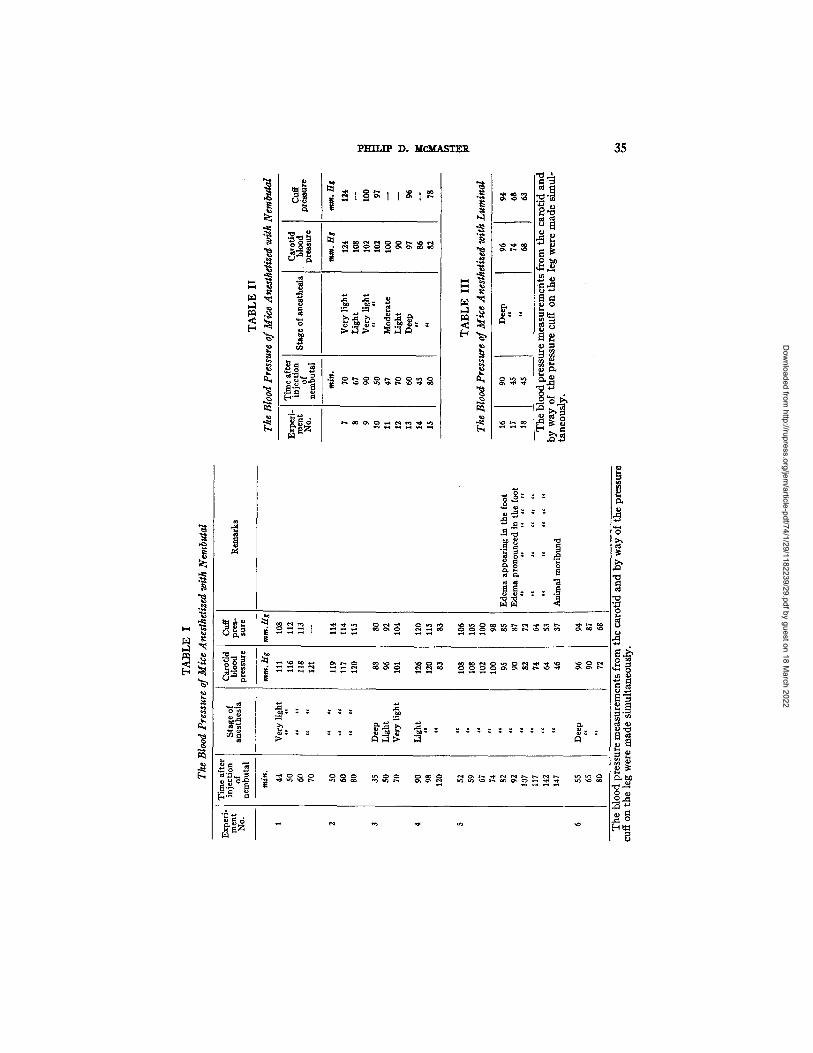
A comparison of the blood pressure findings obtained by the two methods appears in Tables I to III, which will be discussed below. For the present it will suffice to say that a comparison of the figures in the 4th and 5th columns indicates a remarkably good agreement. From this it is dear that the method for determining blood pressure by observation of the circulation in the claw is adequate for most purposes.
Variations in the Blood Pressure of Mice
Under the circumstances of our studies the blood pressure of the mice dif- fered greatly, and the pressure in individual animals also varied much from time to time. Because of the lack of data on the blood pressure in mice, it seems necessary to present a record of the variations we have encountered, in order that others may know of them who wish to employ the method de- scribed.
I t is generally known that animals under deep anesthesia have a lower blood pressure than those lightly anesthetized, and furthermore, that blood pressure
Dow
nloaded from http://rupress.org/jem
/article-pdf/74/1/29/1182239/29.pdf by guest on 18 March 2022
pl=r r r~ D. McMASTER 35
• ~,~ .~ O o ~
o~ .~ ~
~ o ~ "~ ~ ~ .~'~ ~
.c~ "o
~ o ~ o
o ~
~ o ,~
D~
)
m~~ m ~
o , ~
u
Dow
nloaded from http://rupress.org/jem
/article-pdf/74/1/29/1182239/29.pdf by guest on 18 March 2022
36 PERIPHERAL ARTERIAL BLOOD PRESSURE IN MOUSE
varies grea t ly with changes in the physiological state. Our findings in the mouse are in accord with this knowledge (Tables I to I I I ) .
The physiological state of the animals varied much from instance to instance. In some animals cannulation of the carotid artery was done rapidly and with case, in others slowly and with diflSculty. The trauma of the operation and manipulation of the needle in the tissues of the neck must have much influenced matters. Further, we desired to measure the blood pressure in animals as lightly anesthetized as possible.
TABLE IV
Experi- ment NO.
Time after injection
of the anesthetic
~d ~d 1 1½
½
1
1½
Blood pressure in
the leg "Cuff
method"
~m. H t
76 84
105 109
80 82 98
104 106 114
72 88 98
118
Comment on depth of anesthesia
No response on pricking tail: deep anesthesia Response on pricking tail: moderate anesthesia Occasional movement of legs: light anesthesia Frequent movement of legs: very light anesthesia; at
1 hr. 40 rain. withdrew leg from cuff and ran off
Response to pricking tail: moderate anesthesia ct ~ t¢ ~c t t t c
¢ t t t t t ~t t t t t
Occasional movement of legs: light anesthesia t t t t t c e t t t t t
Frequent movement of legs: very light anesthesia At 1 hr. 44 rain. withdrew leg from cuff and ran off
No response on pricking tail: deep anesthesia Slight response on pricking tail: moderate anesthesia Occasional movement of legs: light anesthesia Frequent movement of legs: very light anesthesia; at
1 hr. 32 rain. withdrew leg from cuff and ran off
Blood pressure readings made by the "cuff method" alone in mice anesthetized with nem- butal and subjected to no trauma. As anesthesia became lighter the blood pressure rose.
The amount of nembutal given was just sufficient to maintain anesthesia during the average time required for cannulation of the carotid artery. As the time required to perform the cannulation varied much, the initial blood pressure readings were made in some instances as early as 35 minutes after giving the anesthetic (Experiment 3, Table I), while the animals were still deeply under its influence. In other instances, as in Experiments 4 (Table I) and 9 (Table II) , the initial blood pressure readings were not obtained until 1½ hours after giving the nembutal, and the animals were in a state of light narcosis rather than anesthesia. Apart from these differences, the response of some of the animals to the uniform dose of nembutal varied much. Some remained deeply anesthetized for periods of approximately 1½ hours, Experiments 6 (Table I)
Dow
nloaded from http://rupress.org/jem
/article-pdf/74/1/29/1182239/29.pdf by guest on 18 March 2022
PHILIP D. MC..MASTER
TABLE V
37
Experi- ment No.
Time after injection of the
snesthetic
~gf$.
q 2 21 2~ 3
i 3-I
1-1 2
3 3-1
' 1 i II
q 2-1 3
2-1 3
1
1½ 2
1
1-1 2
2-1 3
1¼ 2
i l i
Blood ) r ~ s u r e in
the leg "Cuff
method"
ram. Hg
70
100 106 109
62 62 62 95
102
72 72 82 82 93
69 76 87 94
96 90 72
81 81 92
60 62
78 80
71
Depth of anesthesia
Deep Moderate Light
Very light
Deep Moderate
~c
Light Very light
Deep
Moderate Light Very light
Deep
Moderate
Deep
Ex ., imeafter] Blood p ment injection pressure in of the the leg Depth of anesthesia
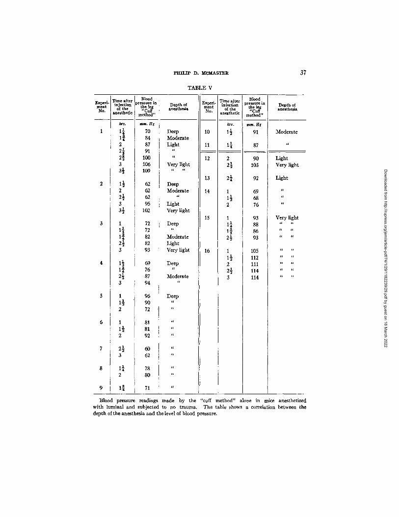
Blood pressure readings made by the "cuff method" alone in mice anesthetized with luminal and subjected to no trauma. The table shows a correlation between the depth of the anesthesia and the level of blood pressure.
Dow
nloaded from http://rupress.org/jem
/article-pdf/74/1/29/1182239/29.pdf by guest on 18 March 2022
38 PERIPI:[ERAL ARTERIAL BLOOD PRESSURE IN MOUSE
and 15 (Table II). The three animals given luminal (Table III) also remained deeply anesthetized.
In all the experiments the depth of anesthesia or narcosis was judged from time to time by the animal's response to a light puff of air into the nostrils or a slight pin prick in the skin of the tail. In the tables the depth of anesthesia at the time of each read- ing has been recorded as "deep," "moderate," "light," or "very light." By deep anesthesia is meant surgical anesthesia with loss of reflexes, by moderate anesthesia a stage in which the animal responded to the stimuli mentioned above by a momentary twitch. During light anesthesia mice without direct stimulation made occasional spontaneous movements tending to withdraw the leg from the cuff, and when the state of anesthesia was very light these movements became frequent.
In Table I, Experiments 1 to 3 summarize the findings from three of six animals which recovered from the anesthetic sufficiently to withdraw the leg from the pressure cuff a few minutes after the last readings were taken. I t is clear from this fact that the anesthesia had almost entirely worn off. In all instances, the lighter the anesthesia the higher were the blood pressure readings. In three other experiments (4 to 6, Table I) the blood pressure fell as time passed. The data of Experiment 5 iUustrate the fact mentioned earlier, that repeated estimations of blood pressure by the cuff method sometimes led to the development of a discrepancy between the pressure readings taken from the leg and those obtained from the carotid artery. In this instance the ex- periment lasted for 2½ hours, and the animal obviously suffered from shock and exposure. Ten measurements of blood pressure in the leg were made, each in duplicate or triplicate. The foot showed visible edema after the fourth determination, as indicated in the table, and with the passage of time the edema increased. Similar findings appeared in other experiments, which need not be included in the table.
Table I I gives the data from nine experiments in which single pressure readings only were attempted. I t will be seen that there was a rough correla- tion between the level of the blood pressure and the depth of anesthesia when judged as described earlier. Finally, Table I I I shows the blood pressure read- ings that were obtained from three animals anesthetized with luminal Though all were in the state of deep anesthesia, the first animal 1½ hours after giving the anesthetic showed a higher blood pressure than the others ~ of an hour after receiving luminal
Tables IV and V show well the variations in blood pressure that have been found in mice which were deeply, moderately, or lightly anesthetized with nembutal or luminal. In these experiments the animals were not operated upon and they lay quietly on a warming pad with one leg in the pressure cuff. In most of the experiments blood pressure determinations were made from time to time while the animals recovered from the anesthetic. The three mice of Table IV recovered sufficiently to withdraw the leg from the pressure
Dow
nloaded from http://rupress.org/jem
/article-pdf/74/1/29/1182239/29.pdf by guest on 18 March 2022
Pm~iP D. Mc~sxER 39
cuff and run off. Inspection of Table V shows that in most of the lightly anesthetized mice the blood pressure was relatively high 1 to 2 hours after injecting the anesthetic (Experiments 12 to 16, inclusive). In the animals deeply anesthetized (Experiments 1 to 9) the initial readings were low in com- parison with the later ones.
COM'MENT
The data of the tables indicate, as might have been expected, that the blood pressure of mice lightly anesthetized with nembutal or luminal was higher than that of animals deeply anesthetized.
Bonsmann (1), studying the blood pressure in the tail of mice by means of a pressure cuff and photoelectric cell, found a 20 to 45 per cent reduction in the pressure after administration of 30 per cent of the fatal dose of chloral hydrate and morphine. The blood pressure of the mouse as reported by him varied from 70 to 100 mm. of Hg, figures lower than those of the present work in which it varied from 60 to 126 mm. of Hg. We attribute the difference to the accuracy of the present method.
S U M ~ R Y
Advantage has been taken of the relative transparency of the claw of the mouse to devise a method, here described, to measure the blood pressure in the animal's leg. Direct measurements of the systolic blood pressure from the carotid arteries of anesthetized mice have also been made. Simultaneous blood pressure readings by both these methods applied to the same animal showed close agreement.
The systolic pressure ranged from 60 to 126 mm. Hg, according to the conditions.
BIBLIOGRAPHY
1. Bonsmann, M. R., Arch. exp. Path. u. Pkarmakol., 1934, 176, 460. 2. Diaz, J. T., and Levy, S. E., Proc. Soc. Exp. Biol. and Med., 1939, 40, 402. 3. Wilfiams, J. R., Jr., Harrison, T. R., and Grollman, A., Y. Clin. Inv., 1939,18, 373. 4. Griffith, J. Q., Jr., Proc. Soc. Exp. Biol. and Med., 1934-35, 32, 394. 5. Parsons, R. J., and McMaster, P. D., J. Exp. Med., 1938, 68, 869. 6. McMaster, P. D., and ttudaek, S. S., Y. Exp. Med., 1934, 60, 479. 7. MeMaster, P. D., and Hudack, S. S., J. Exp. Med., 1932, 55, 417. 8. Hudack, S. S., and McMaster, P. D., Y. Exp. Med., 1932, 55, 431. 9. Me_Master, P. D., a r. Exp. Med., 1941, 73, 67.
Dow
nloaded from http://rupress.org/jem
/article-pdf/74/1/29/1182239/29.pdf by guest on 18 March 2022
40 PERIPHERAL ARTERIAL BLOOD PRESSURE IN MOUSE
EXPLANATION OF PLATES
These photographs were made by Mr. Joseph B. Haulenbeek.
PLATE 4
FIO. 1. Apparatus for the determination of blood pressure in the mouse by way of a pressure cuff on the leg and the carotid artery (see text).
Dow
nloaded from http://rupress.org/jem
/article-pdf/74/1/29/1182239/29.pdf by guest on 18 March 2022
THE JOURNAL OF EXPERIMENTAL MEDICINE VOL. 74 PLATE 4
(McMaster: Peripheral arterial blood pressure in mouse)
Dow
nloaded from http://rupress.org/jem
/article-pdf/74/1/29/1182239/29.pdf by guest on 18 March 2022
PLATE 5
FI6S. 2 and 3. Blood vesse|s in the claw of the mouse after injection of a gelatin mass containing India ink. X 100.
Dow
nloaded from http://rupress.org/jem
/article-pdf/74/1/29/1182239/29.pdf by guest on 18 March 2022
TtIE JOURNAL OF EXPERIMENTAL MEDICINE VOL. 74 PLATE 5
(McMaster: Peripheral arterial blood pressure in mouse)
Dow
nloaded from http://rupress.org/jem
/article-pdf/74/1/29/1182239/29.pdf by guest on 18 March 2022