110
Functional Characterization of the LCMV GP-C Signal Peptide Sabrina Schrempf 2008

Functional Characterization
of the LCMV GP-C Signal Peptide
Sabrina Schrempf
2008

INAUGURAL - DISSERTATION
submitted to the
Combined Faculties for the Natural Sciences and for Mathematics
of the Ruperto-Carola University of Heidelberg
for the degree of
Doctor of Natural Sciences
presented by
Diplom-Biochemikerin Sabrina Schrempf
from Gelnhausen
Date of oral examination: . . . . . . . . . . . . . . .

Functional Characterization
of the LCMV GP-C Signal Peptide
Referees:
Prof. Dr. Bernhard Dobberstein
Prof. Dr. Irmgard Sinning

Fur Oliver,
der mich immer auffangt
und
fur meine Eltern,
die mich stets unterstutzen.

Abstract
N-terminal signal sequences of secretory and membrane proteins mediate targeting to
and translocation across the endoplasmic reticulum (ER) membrane. After membrane
insertion, signal sequences are in most cases cleaved from the precursor protein by signal
peptidase (SPase). Signal sequences are usually 15 to 25 amino acid residues in length
and have a typical tripartite structure with a central hydrophobic core of about 7 to 10
residues, a polar N-terminal region, and a short C-terminal region which contains the
SPase cleavage site.
Insertion of the lymphocytic choriomeningitis virus (LCMV) precursor glycoprotein C
(pGP-C) into the membrane of the ER is mediated by an unusual signal sequence. It
comprises 58 amino acid residues and contains an extended N-terminal region including a
myristoylation consensus site and two hydrophobic regions separated by a lysine residue.
After cleavage by SPase, the resulting signal peptide (SPGP−C) accumulates in cells and
virus particles.
The aim of this study was to characterize the post-targeting functions of the LCMV
SPGP−C. It could be shown that the LCMV SPGP−C is an essential component of the
glycoprotein complex and that different regions of SPGP−C are required for distinct steps
in glycoprotein maturation and virus infectivity.
The investigation of SPGP−C deletion mutants showed that one hydrophobic region of
LCMV SPGP−C is sufficient for ER membrane insertion of GP-C, while both hydrophobic
regions are required for GP-C processing into its subunits and cell surface expression of
the glycoprotein complex. The N-terminal region of SPGP−C and its myristoylation are
dispensable for these steps in GP-C maturation, however, were found to be essential for
viral infection of target cells. The analysis of a possible association of LCMV SPGP−C
with GP-C by co-immunoprecipitation revealed that the LCMV SPGP−C is part of the
glycoprotein complex and interacts with the membrane-anchored GP-2 subunit. For this
non-covalent interaction the hydrophobic regions of SPGP−C are sufficient and essential,
whereas the N-terminal region is not required.
As the LCMV SPGP−C possesses two hydrophobic regions, different topologies across
the membrane are conceivable. The membrane topology of SPGP−C was investigated
using point mutations introducing potential N-glycosylation sites throughout the SPGP−C.
It could be shown that unmyristoylated SPGP−C exposes its N-terminal region to the
exoplasmic side of the membrane. This SPGP−C can promote GP-C maturation but is
defective in viral infection. Myristoylation and SPGP−C membrane topology may thus hold
the key to unravel the role of LCMV SPGP−C in GP-C complex assembly and function.

Zusammenfassung
N-terminale Signalsequenzen von sekretorischen Proteinen und von Membranproteinen
vermitteln den zielgerichteten Transport zum endoplasmatischen Retikulum (ER) und
die Translokation der Proteine durch die ER-Membran. In den meisten Fallen wird
die Signalsequenz nach der Membraninsertion durch die Signalpeptidase (SPase) vom
Praprotein abgespalten. Signalsequenzen besitzen ublicherweise eine Gesamtlange von 15
bis 25 Aminosauren und sind in drei Regionen unterteilt: einen zentralen hydrophoben
Kern bestehend aus 7 bis 10 Aminosauren, einer polaren N-terminalen Region und einer
kurzen C-terminalen Region, die die SPase Spaltstelle beinhaltet.
Die Insertion des lymphozytaren Choriomeningitis Virus (LCMV) Glykoproteins (pGP-C)
in die Membran des ER wird durch eine ungewohnliche Signalsequenz vermittelt. Diese
Signalsequenz besteht aus 58 Aminosauren und besitzt eine erweiterte N-terminale Region
einschließlich einer Konsensussequenz fur Myristoylierung und zwei hydrophobe Regionen,
die durch ein Lysin getrennt sind. Nach der Abspaltung durch die SPase akkumuliert das
resultierende Signalpeptid (SPGP−C) in Zellen und in Viruspartikeln.
Ziel der vorliegenden Arbeit war die Charakterisierung der Funktionen des LCMV SPGP−C
uber den zielgerichteten Transport hinaus. Es konnte gezeigt werden, dass das LCMV
SPGP−C ein essentieller Bestandteil des Glykoproteinkomplexes ist und dass die unter-
schiedlichen Regionen des SPGP−C fur verschiedene Schritte wahrend der GP-C-Reifung
und fur die Infektiositat des Virus benotigt werden.
Die Untersuchung von SPGP−C Deletionsmutanten zeigte, dass eine hydrophobe Region
des LCMV SPGP−C fur die Insertion von GP-C in die ER-Membran genugt, wohingegen
beide hydrophobe Regionen fur die Prozessierung von GP-C in seine Untereinheiten und
fur den Transport des Glykoproteinkomplexes zur Zelloberflache benotigt werden. Die
N-terminale Region des SPGP−C und dessen Myristoylierung werden fur diese Schritte der
GP-C-Reifung nicht benotigt, sind jedoch essentiell fur die virale Infektion von Zielzellen.
Die Analyse einer moglichen Assoziierung des SPGP−C mit GP-C durch Koimmuno-
prazipitation zeigte, dass das LCMV SPGP−C ein Bestandteil des Glykoproteinkomplexes
ist und dass es mit der membranverankerten GP-2 Untereinheit interagiert. Die hy-
drophoben Regionen des SPGP−C sind fur diese nichtkovalente Interaktion ausreichend
und essentiell, wohingegen die N-terminale Region nicht benotigt wird.
Aufgrund der zwei hydrophoben Regionen des LCMV SPGP−C sind verschiedene Topolo-
gien in der Membran vorstellbar. Zur Untersuchung wurden potenzielle N-Glykosylierungs-
stellen durch Punktmutationen in SPGP−C eingefugt. Es konnte gezeigt werden, dass sich
die N-terminale Region des nicht-myristoylierten SPGP−C auf der exoplasmatischen Seite
der Membran befindet. Dieses SPGP−C ist fahig die GP-C-Reifung zu begunstigen, ver-
hindert jedoch die virale Infektion. Die Myristoylierung und die Topologie des SPGP−C
konnten daher den Schlussel zur Funktion des SPGP−C innerhalb des Glykoproteinkom-
plexes darstellen.

Contents
1 Introduction 1
1.1 Signal sequences . . . . . . . . . . . . . . . . . . . . . . . . . . . . . . . . . 1
1.1.1 Protein sorting in eukaryotic cells . . . . . . . . . . . . . . . . . . . 1
1.1.2 Protein translocation across the ER membrane . . . . . . . . . . . . 2
1.1.3 The structure of N-terminal ER signal sequences . . . . . . . . . . . 4
1.1.4 Functions of signal sequences beyond ER targeting . . . . . . . . . 5
1.2 Viral glycoproteins . . . . . . . . . . . . . . . . . . . . . . . . . . . . . . . 7
1.2.1 Trafficking of viral glycoproteins and virus budding . . . . . . . . . 7
1.2.2 Membrane fusion mechanisms of viral glycoproteins . . . . . . . . . 8
1.3 The lymphocytic choriomeningitis virus . . . . . . . . . . . . . . . . . . . . 10
1.3.1 Genome organization and structure of the viral particle . . . . . . . 10
1.3.2 The LCMV infection cycle . . . . . . . . . . . . . . . . . . . . . . . 11
1.3.3 Synthesis and function of the LCMV glycoprotein C . . . . . . . . . 12
1.3.4 The signal peptide of the LCMV glycoprotein C . . . . . . . . . . . 13
1.4 Aim of this work . . . . . . . . . . . . . . . . . . . . . . . . . . . . . . . . 14
2 Materials and Methods 15
2.1 Materials . . . . . . . . . . . . . . . . . . . . . . . . . . . . . . . . . . . . 15
2.1.1 Chemicals . . . . . . . . . . . . . . . . . . . . . . . . . . . . . . . . 15
2.1.2 Standard stock solutions and buffers . . . . . . . . . . . . . . . . . 15
2.1.3 DNA and protein standards . . . . . . . . . . . . . . . . . . . . . . 15
2.1.4 Oligonucleotides . . . . . . . . . . . . . . . . . . . . . . . . . . . . . 16
2.1.5 Enzymes . . . . . . . . . . . . . . . . . . . . . . . . . . . . . . . . . 16
2.1.6 Plasmids . . . . . . . . . . . . . . . . . . . . . . . . . . . . . . . . . 17
2.1.7 Bacteria culture . . . . . . . . . . . . . . . . . . . . . . . . . . . . . 18
2.1.8 Cell culture . . . . . . . . . . . . . . . . . . . . . . . . . . . . . . . 19
I

Table of Contents
2.1.9 Antibodies . . . . . . . . . . . . . . . . . . . . . . . . . . . . . . . . 19
2.1.10 Kits . . . . . . . . . . . . . . . . . . . . . . . . . . . . . . . . . . . 20
2.1.11 Computer software . . . . . . . . . . . . . . . . . . . . . . . . . . . 20
2.2 Methods . . . . . . . . . . . . . . . . . . . . . . . . . . . . . . . . . . . . . 20
2.2.1 Biomolecular methods . . . . . . . . . . . . . . . . . . . . . . . . . 20
2.2.2 Cell culture techniques . . . . . . . . . . . . . . . . . . . . . . . . . 25
2.2.3 Metabolic labeling / Pulse-chase . . . . . . . . . . . . . . . . . . . . 27
2.2.4 SDS-PAGE . . . . . . . . . . . . . . . . . . . . . . . . . . . . . . . 29
2.2.5 Western blotting . . . . . . . . . . . . . . . . . . . . . . . . . . . . 30
2.2.6 Analysis of cell surface expression by flow cytometry . . . . . . . . 32
2.2.7 Production and analysis of LCMV pseudoviruses . . . . . . . . . . . 32
3 Results 35
3.1 The LCMV SPGP-C is essential for GP-C processing
and transport to the cell surface . . . . . . . . . . . . . . . . . . . . . . . . 35
3.2 Effects of C-terminal truncations of LCMV GP-C
on GP-C processing and transport . . . . . . . . . . . . . . . . . . . . . . . 37
3.3 The SPGP-C h-regions are required for distinct steps
in LCMV GP-C maturation . . . . . . . . . . . . . . . . . . . . . . . . . . 41
3.3.1 One SPGP-C h-region is sufficient for ER membrane insertion
of pGP-C while both are required for GP-C processing . . . . . . . 42
3.3.2 Both SPGP-C h-regions are needed for cell surface expression
of the GP complex . . . . . . . . . . . . . . . . . . . . . . . . . . . 43
3.4 Use of SPGP-C point mutants to investigate SPGP-C membrane topology
and effects on SPGP-C stability and GP-C maturation . . . . . . . . . . . . 44
3.4.1 Point mutations introduced in SPGP-C did not lead to SPGP-C
glycosylation but influence SPGP-C stability and GP-C maturation . 45
3.4.2 The unmyristoylated SPGP-C n-region is exposed to the ER lumen . 47
3.5 SPGP-C is part of the LCMV GP complex . . . . . . . . . . . . . . . . . . . 49
3.5.1 Co-immunoprecipitation of SPGP-C with GP-C
under different lysing conditions . . . . . . . . . . . . . . . . . . . . 50
II

Table of Contents
3.5.2 Interaction of SPGP-C with GP-C during maturation
of the GP complex . . . . . . . . . . . . . . . . . . . . . . . . . . . 52
3.5.3 SPGP-C requirements for the interaction with GP-C . . . . . . . . . 52
3.5.4 SPGP-C interacts with the GP-2 subunit . . . . . . . . . . . . . . . . 54
3.5.5 The cytoplasmic and transmembrane region of GP-C are not essen-
tial for the interaction with SPGP-C . . . . . . . . . . . . . . . . . . 56
3.5.6 SPGP-C is not disulfide linked to GP-C . . . . . . . . . . . . . . . . 58
3.5.7 ER retention of GP-C(WE) is not due to a lack of SPGP-C interaction 59
3.6 Influence of the interaction with GP-C on SPGP-C stability . . . . . . . . . 60
3.7 The myristoylated SPGP-C n-region is essential for virus infectivity . . . . . 63
3.8 Visualization of LCMV pseudoviruses during cell entry . . . . . . . . . . . 65
4 Discussion 69
4.1 Function of the LCMV SPGP-C during GP-C maturation . . . . . . . . . . 70
4.1.1 SPGP-C is essential for GP-C maturation . . . . . . . . . . . . . . . 70
4.1.2 Functional significance of the SPGP-C n-region and its myristoylation
for GP-C maturation . . . . . . . . . . . . . . . . . . . . . . . . . . 71
4.1.3 Functional significance of the SPGP-C h-regions for GP-C maturation 72
4.2 Membrane topology of LCMV SPGP-C . . . . . . . . . . . . . . . . . . . . . 74
4.3 SPGP-C as part of the LCMV GP complex . . . . . . . . . . . . . . . . . . 76
4.4 Requirements of SPGP-C for LCMV infection . . . . . . . . . . . . . . . . . 79
4.5 Towards the analysis of LCMV pseudovirus cell entry
using fluorescent labeling . . . . . . . . . . . . . . . . . . . . . . . . . . . . 81
Publications derived from this thesis 82
References 83
List of Figures 96
List of Abbreviations 98
III

1 Introduction
1.1 Signal sequences
1.1.1 Protein sorting in eukaryotic cells
A typical eukaryotic cell is compartmentalized into functionally distinct, membrane-
enclosed organelles, like the nucleus, the endoplasmic reticulum (ER), the Golgi appa-
ratus, mitochondria, chloroplasts, peroxisomes, and lysosomes. All of these organelles
contain a characteristic set of proteins in order to fulfill their specific functions. Almost
all of these proteins are synthesized on cytosolic ribosomes and have to cross at least one
membrane to reach their final destination in the cell (Blobel, 1980; Verner and Schatz,
1988; Schatz and Dobberstein, 1996). Proteins that are destined for a certain compart-
ment in the cell are targeted to these compartments by specific sorting signals that are
recognized by complementary receptor proteins (Gierasch, 1989). Proteins that do not
have a sorting signal remain in the cytosol.
After synthesized in the cytosol, nuclear proteins are selectively imported into the nucleus
through the nuclear pore complexes which function as selective gates between topologically
equivalent spaces. The selectivity of this nuclear import process resides in nuclear local-
ization signals. In many nuclear proteins these signals consist of one or two short amino
acid sequences that are rich in the positively charged amino acids lysine and arginine
(Gorlich and Kutay, 1999; Kalderon et al., 1984; Lanford and Butel, 1984).
Protein import into mitochondria, chloroplasts, peroxisomes and into the ER involves
membrane-bound protein translocators that transport proteins across a membrane. Most
of the mitochondrial and chloroplast proteins have an N-terminal signal that is recognized
by cytosolic chaperones which prevent proteins from aggregation or folding and guide the
proteins to their respective organelle. Peroxisomal proteins contain short signal sequences
and are selectively imported in a fully folded conformation (Alberts et al., 2002; Dalbey
and von Heijne, 2003). Proteins targeted to the mitochondria, chloroplasts or peroxi-
somes are synthesized on free ribosomes in the cytosol before being post-translationally
translocated into their target organelle. In contrast, proteins targeted to the ER can be
translocated co-translationally, i.e. during synthesis on ER membrane-bound ribosomes,
or post-translationally (Rapoport, 2007; Rapoport et al., 1999). The proteins initially
targeted to the ER include secretory proteins and proteins destined for the ER, Golgi
apparatus, lysosomes, or the plasma membrane. The co-translational translocation of
proteins into the ER through the translocon, formed by the Sec61 complex, is usually
1

1. Introduction
mediated by an N-terminal signal sequence that is recognized by the signal recognition
particle (SRP) (Johnson and van Waes, 1999; Rapoport, 2007; Walter and Blobel, 1981;
Walter and Johnson, 1994). While the polypeptide chain passes through the translocon,
the N-terminal signal sequences of soluble proteins and type I membrane proteins are
cleaved by signal peptidase (SPase) (Blobel and Dobberstein, 1975; Paetzel et al., 2002).
Protein targeting to the ER is not exclusively done by N-terminal signal sequences. Mem-
brane proteins can also be targeted to the ER by internal signal sequences. These internal
signal sequences are also recognized by SRP and inserted into the translocon but in con-
trast to N-terminal signal sequences they are not cleaved by SPase. Like a transmembrane
domain, the internal signal sequences exit the translocon laterally thereby anchoring the
protein in the ER membrane (Johnson and van Waes, 1999; Martoglio et al., 1995).
From the ER, proteins are transported in vesicles to the Golgi apparatus, where they are
sorted for transport to lysosomes, the plasma membrane, or secretion from the cell. Pro-
teins that function within the ER or the Golgi apparatus were retained in the respective
organelle (Alberts et al., 2002; Cooper, 2000).
1.1.2 Protein translocation across the ER membrane
In mammalian cells, most proteins translocate into the ER in a co-translational and
SRP-dependent manner. The co-translational translocation is initiated by binding of
SRP to the signal sequence of a growing polypeptide chain emerging from the ribosome
(Luirink and Sinning, 2004). SRP is a ribonucleoprotein consisting of a single small
RNA molecule (7S RNA) and six different polypeptides (Keenan et al., 2001; Walter
and Blobel, 1982). The signal sequence recognition is mediated by the SRP54 subunit
of SRP. Formation of the ribosome-nascent-chain-SRP complex leads to an elongation
arrest mediated by binding of the SRP Alu domain at the ribosomal subunit interface
(Terzi et al., 2004). The entire complex is targeted to the rough ER via interaction
with the SRP receptor (Gilmore et al., 1982; Meyer and Dobberstein, 1980). The SRP
receptor is composed of a peripheral (SRα) and a membrane-anchored subunit (SRβ)
(Tajima et al., 1986). The GTP-dependent interaction of SRP with the SRP receptor
results in the release of SRP and transfer of the ribosome-nascent-chain complex to the
translocon (Connolly and Gilmore, 1989; Pool et al., 2002). This allows translation to
resume and translocation of the elongating nascent chain through the channel across the
ER membrane (Johnson and van Waes, 1999; Pool, 2003). Co-translationally the signal
sequence is usually cleaved from the preprotein by SPase (Paetzel et al., 2002) and the
growing polypeptide chain can get glycosylated at asparagine residues in the consensus
sequence N-X-T/S by the oligosaccharyl transferase in the ER lumen. As translation
proceeds and after the release of the polypeptide chain into the ER lumen, the polypeptide
is folded in its three-dimensional conformation with the help of molecular chaperones like
2
1. Introduction
BiP and the protein disulfide isomerase which catalyzes the formation of disulfide bonds.
In addition to protein folding, the assembly of multisubunit proteins takes place within
the ER lumen. Correctly folded proteins are subsequently exported from the ER, further
modified in the Golgi apparatus and transported in vesicles to their final destination
(Alberts et al., 2002; Cooper, 2000; Ellgaard and Helenius, 2003). ER resident proteins
contain an ER retention or retrieval signal to ensure their proper localization (Michelsen
et al., 2005; Nilsson and Warren, 1994).
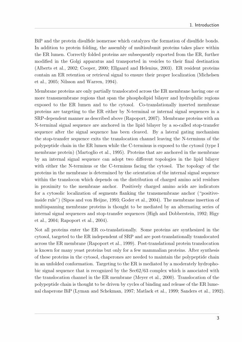
Membrane proteins are only partially translocated across the ER membrane having one or
more transmembrane regions that span the phospholipid bilayer and hydrophilic regions
exposed to the ER lumen and to the cytosol. Co-translationally inserted membrane
proteins are targeting to the ER either by N-terminal or internal signal sequences in a
SRP-dependent manner as described above (Rapoport, 2007). Membrane proteins with an
N-terminal signal sequence are anchored in the lipid bilayer by a so-called stop-transfer
sequence after the signal sequence has been cleaved. By a lateral gating mechanism
the stop-transfer sequence exits the translocation channel leaving the N-terminus of the
polypeptide chain in the ER lumen while the C-terminus is exposed to the cytosol (type I
membrane protein) (Martoglio et al., 1995). Proteins that are anchored in the membrane
by an internal signal sequence can adopt two different topologies in the lipid bilayer
with either the N-terminus or the C-terminus facing the cytosol. The topology of the
proteins in the membrane is determined by the orientation of the internal signal sequence
within the translocon which depends on the distribution of charged amino acid residues
in proximity to the membrane anchor. Positively charged amino acids are indicators
for a cytosolic localization of segments flanking the transmembrane anchor (“positive-
inside rule”) (Sipos and von Heijne, 1993; Goder et al., 2004). The membrane insertion of
multispanning membrane proteins is thought to be mediated by an alternating series of
internal signal sequences and stop-transfer sequences (High and Dobberstein, 1992; Higy
et al., 2004; Rapoport et al., 2004).
Not all proteins enter the ER co-translationally. Some proteins are synthesized in the
cytosol, targeted to the ER independent of SRP and are post-translationally translocated
across the ER membrane (Rapoport et al., 1999). Post-translational protein translocation
is known for many yeast proteins but only for a few mammalian proteins. After synthesis
of these proteins in the cytosol, chaperones are needed to maintain the polypeptide chain
in an unfolded conformation. Targeting to the ER is mediated by a moderately hydropho-
bic signal sequence that is recognized by the Sec62/63 complex which is associated with
the translocation channel in the ER membrane (Meyer et al., 2000). Translocation of the
polypeptide chain is thought to be driven by cycles of binding and release of the ER lume-
nal chaperone BiP (Lyman and Schekman, 1997; Matlack et al., 1999; Sanders et al., 1992).
3

1. Introduction
1.1.3 The structure of N-terminal ER signal sequences
N-terminal signal sequences that target secretory and membrane proteins to the ER show
great variations in their amino acid sequence and overall length (Martoglio and Dobber-
stein, 1998). In general, they consist of about 15 to 25 amino acid residues and have a
tripartite structure with a central hydrophobic core that is flanked by a polar N-terminal
region and a short C-terminal region (Figure 1.1) (von Heijne, 1990). The hydrophobic
core is the most characteristic feature of N-terminal signal sequences and is about 7 to 10
amino acid residues in length. This so-called h-region is the recognition site for SRP and
essential for ER targeting and membrane translocation (von Heijne, 1985). The region
N-terminally of the hydrophobic core, the n-region, is extremely variable in its length and
amino acid composition, but usually has a positive net charge. On its C-terminal side, the
h-region is flanked by the short c-region that contains the consensus sequence for SPase
cleavage. The c-region usually consists of about 2 to 9 amino acids and often contains
helix breaking proline and glycine residues. The SPase cleavage site is determined by
small, uncharged amino acid residues at the -3 and -1 positions relative to the cleavage
site. A proline residue at the +1 position is not tolerated (von Heijne, 1983).
n h cNH2
SPase++
Figure 1.1: Schematic representation of an N-terminal signal sequence for ER targeting
N-terminal ER signal sequences have a typical tripartite structure with a polar N-terminal region (n-region),
a central hydrophobic region (h-region), and a short C-terminal region (c-region). The overall length of a
typical signal sequence is about 15 - 25 amino acid residues containing an h-region of about 7 - 10 residues.
The SPase cleavage site and the positive net charge of the n-region (+) are indicated.
After the N-terminal signal sequence emerges from the ribosome, SRP binds to the
h-region and translation is stopped (Walter and Blobel, 1981). Upon binding to the
SRP receptor, the ribosome-nascent-chain complex is transferred to the translocon and
translation resumes (Johnson and van Waes, 1999). Membrane insertion in the translo-
cating channel is thought to occur in a loop-like fashion such that the N-terminus of the
signal sequence is exposed on the cytoplasmic side and the cleavage site for SPase on the
lumenal side of the ER membrane (Walter et al., 1984). The SPase of mammalian cells
consists of five different subunits with the active site located near the membrane surface
on the lumenal side of the ER membrane (Dalbey and von Heijne, 1992; Paetzel et al.,
2002). After co-translational cleavage of the signal sequence by SPase, the N-terminus of
the resulting signal peptide (SP) thus would face the cytosol while the c-region remains
in the ER lumen.
4

1. Introduction
1.1.4 Functions of signal sequences beyond ER targeting
Cleavage of N-terminal ER signal sequences by SPase results in the release of the so-
called signal peptides (SPs) which are supposed to be directly degraded by a yet unknown
mechanism. A growing number of signal peptides, however, are found not to be degraded
but to stay membrane-inserted, to be liberated from the ER membrane or to be further
processed by an intramembrane cleaving protease, which results in the release of SP
fragments from the lipid bilayer (Martoglio, 2003). These released SP fragments as well
as membrane-inserted or liberated full length signal peptides are known to have diverse
functions beyond ER targeting (Hegde and Bernstein, 2006).
Signal peptide processing and the release of bioactive signal peptide fragments
After the initial cleavage by SPase, signal peptides can undergo intramembrane proteo-
lysis. This processing is mediated by the signal peptide peptidase (SPP) which belongs
to the family of intramembrane cleaving proteases (I-Clip) (Weihofen et al., 2002). SPP
cleavage within the transmembrane region of signal peptides results in the release of
SP fragments from the ER membrane (Lemberg and Martoglio, 2002). Some of these
released SP fragments are known to have post-targeting functions and thus act as so-called
bioactive peptides (Martoglio, 2003; Weihofen and Martoglio, 2003).
The first known process that depends on SPP processing is the generation of HLA-E epi-
topes in humans (Lemberg et al., 2001). The signal sequences of the MHC class I molecules
HLA-A, -B, -C, and -G contain a highly conserved segment within their N-terminal por-
tion, which serves as an epitope for the presentation by the non-classical antigen presenting
molecule HLA-E (Braud et al., 1997). During biosynthesis of MHC class I molecules the
signal sequence is first cleaved by SPase and then further processed by SPP. The generated
epitope-containing N-terminal fragment is subsequently released from the ER membrane
into the cytosol and after further trimming transported into the ER lumen via the TAP
transporter where it is loaded on to the HLA-E molecules (Lemberg et al., 2001). The
presentation of peptide-loaded HLA-E molecules on the cell surface serves as an inhibitory
signal to natural killer cells reporting proper biosynthesis of MHC class I molecules
(Braud et al., 1998).
Another well characterized substrate for SPP is the signal peptide of prolactin, a peptide
hormone. Upon processing by SPP, the N-terminal SP fragment is released into the cy-
tosol where it binds to calmodulin in a calcium-dependent manner in vitro (Lyko et al.,
1995; Martoglio et al., 1997). Similar to prolactin, a SP fragment derived from the human
immunodeficiency virus 1 (HIV-1) envelope protein gp160 interacts with calmodulin after
release from the ER membrane (Martoglio et al., 1997).
5

1. Introduction
A further example for SPP processing is the maturation of the hepatitis C virus (HCV)
core protein. The core protein is synthesized as the most N-terminal component of the
HCV polyprotein and is followed by the signal sequence of the envelope glycoprotein. This
signal sequence targets the polyprotein to the ER and is subsequently cleaved by SPase
leaving the core protein anchored in the ER membrane by the signal peptide. Further
processing by SPP results in the release of the mature core protein from the ER membrane
(McLauchlan et al., 2002).
So far the physiological role of SPP is not completely understood (Crawshaw et al., 2004;
Loureiro et al., 2006; Weihofen et al., 2002). It is conceivable that the major function
of SPP is the release of bioactive peptides from the ER membrane (Martoglio, 2003;
Weihofen and Martoglio, 2003). However, one cannot exclude a role for SPP in signal
peptide and membrane protein degradation (Loureiro et al., 2006).
Further functions of full length signal peptides
Besides the post-targeting role of signal peptides after SPP processing, some signal pep-
tides remain membrane-inserted or are liberated from the ER membrane without fur-
ther processing to promote their post-targeting functions. In this context mainly signal
peptides of viral proteins are so far found to have functions beyond ER targeting.
The signal peptide of the envelope protein of the mouse mammary tumor virus (MMTV),
for example, was found to accumulate in nucleoli (Hoch Marchaim et al., 2003). The
MMTV envelope signal peptide comprises 98 amino acid residues and contains a nuclear
localization signal within its extended n-region. After ER insertion and SPase cleavage,
the signal peptide initially accumulates in the ER membrane and is subsequently released
into the cytosol in an SPP-independent manner (Dultz et al., 2008). Due to the nuclear
localization signal, the signal peptide of the MMTV envelope protein is able to enter the
nucleus and was shown to be sufficient to mediate the export of intron-containing RNA
transcripts (Mertz et al., 2005).
Other viral signal peptides mediate post-targeting functions that are essential for the viral
life cycle as membrane-inserted signal peptides. The signal peptide of the foamy virus
envelope glycoprotein, for example, is an essential component of infectious viral particles
and is required for particle budding (Lindemann et al., 2001). The need for the signal pep-
tide during foamy virus morphogenesis is due to an interaction between the signal peptide
n-region with the N-terminal region of the viral capsid protein Gag (Wilk et al., 2001).
While the N-terminus of the extended signal peptide n-region mediates the interaction
with the foamy virus capsid, it is dispensable for ER targeting of the envelope glycoprotein
(Lindemann et al., 2001).
6

1. Introduction
The unusually long signal peptides of several arenaviral glycoproteins (LCMV, Lassa
and Junın virus) also remain membrane-inserted after SPase cleavage and mediate post-
targeting functions (Eichler et al., 2003b; Froeschke et al., 2003; York et al., 2004). These
highly conserved signal peptides contain an extended n-region with a myristoylation con-
sensus site and two h-regions separated by basic amino acid residues. The signal peptide of
the Lassa virus glycoprotein, for example, was shown to be essential for proteolytic matu-
ration of the glycoprotein and performs this function even in trans (Eichler et al., 2003a).
For the signal peptide of the lymphocytic choriomeningitis virus (LCMV) glycoprotein it
was shown that it is actually incorporated into virus particles (Froeschke et al., 2003).
The function of the signal peptide in the virus particle as well as the role of the LCMV
signal peptide during glycoprotein maturation were not known so far. The post-targeting
functions of the signal peptide of the LCMV glycoprotein are subject of this study.
1.2 Viral glycoproteins
Enveloped viruses enter their target cell by fusing the viral membrane with a host cell
membrane (Kielian and Rey, 2006; Sollner, 2004). This initial step in the viral replication
cycle is mediated by the viral envelope glycoproteins and includes receptor recognition and
membrane fusion activity. In addition to the function of the viral glycoproteins during
virus entry, they determine for most enveloped viruses the location within the cell at
which budding takes place (Garoff et al., 1998; Knipe and Howley, 2001). To achieve the
correct localization within a cell, the viral glycoproteins take advantage of the cellular
sorting pathways. For this purpose they have adopted many targeting signals found in
cellular proteins (Compans et al., 2004).
1.2.1 Trafficking of viral glycoproteins and virus budding
Viral glycoproteins are targeted to the ER by N-terminal or internal signal sequences
and are co-translationally inserted into the ER membrane. During synthesis, the poly-
peptide chain can get glycosylated at asparagine residues in the consensus sequence
N-X-T/S by the oligosaccharyl transferase in the ER lumen. Correctly folded and assem-
bled proteins are subsequently transported to the site in the cell where budding occurs
(Garoff et al., 1998). This transport may include further protein maturation events, for
example, the modification of glycosylation or proteolytic processing. In many cases virus
assembly takes place at the plasma membrane, but in others intracellular membranes, e.g.
of the ER, the Golgi apparatus or the multivesicular bodies, are the sites where budding
is initiated (Compans et al., 2004; Knipe and Howley, 2001).
7

1. Introduction
The glycoproteins of viruses that bud from the plasma membrane travel along the se-
cretory pathway to reach the cell surface. After protein synthesis, folding, and com-
plex assembly, the viral glycoproteins have to pass the quality control in the ER before
transit to the Golgi apparatus (Ellgaard and Helenius, 2003). The trafficking between
the membrane compartments is mediated by specific transport vesicles. These vesicles
are transiently coated with protein complexes that allow the selective transfer of the
cargo proteins (Bonifacino and Glick, 2004; Bonifacino and Lippincott Schwartz, 2003;
Kirchhausen, 2000). Transport vesicles that bud from the ER membrane are associated
with the COP II coat complex, whereas COP I coats are associated with retrograde
transport vesicles that bud from the Golgi apparatus membrane (Barlowe et al., 1994;
Letourneur et al., 1994; Robinson, 1987). The viral glycoproteins enter the Golgi ap-
paratus at the cis Golgi network and are subsequently transported through the Golgi
stacks and end up in the trans Golgi network where further protein sorting takes place
(Keller and Simons, 1997; Matsuura Tokita et al., 2006; Pelham, 2001). As the proteins
pass through the Golgi the N-linked oligosaccharides added to the viral glycoproteins in
the ER are further modified. In addition, some viral glycoproteins are proteolytically pro-
cessed before they are transported to the plasma membrane. This processing is crucial
for the generation of a functional glycoprotein complex (Kido et al., 1996; Kunz et al.,
2003).
Virus assembly at the plasma membrane results in the release of the virus particles directly
in the extracellular space. Viruses that bud on intracellular membranes are delivered into
the lumen of the respective organelle and finally exit the cell by exocytosis (Compans
et al., 2004; Knipe and Howley, 2001). For most enveloped viruses, with the exception of
the retroviruses, the efficiency of virus budding depends on the presence of the viral glyco-
proteins (Klein et al., 2007; Resh, 2005). Budding is driven by the interaction of the viral
glycoproteins with internal viral structures, e.g. viral nucleoproteins or matrix proteins,
and/or by a lateral interaction between glycoprotein subunits (Garoff et al., 1998).
1.2.2 Membrane fusion mechanisms of viral glycoproteins
Enveloped viruses use different entry pathways to deliver their genome into the cytosol
(Marsh and Helenius, 2006). Cell entry is a stepwise process where receptor binding is fol-
lowed by the fusion of the viral and a host cell membrane. Virus membrane fusion can take
place either at the plasma membrane or at intracellular membranes. Membrane fusion at
the plasma membrane is triggered by the binding of the viral membrane glycoproteins to
a specific cell surface receptor at neutral pH. By contrast, fusion at an intracellular mem-
brane, after virus internalization by receptor-mediated endocytosis, is frequently induced
by the low pH within the respective organelle (Jahn et al., 2003; Kielian and Rey, 2006;
Sollner, 2004).
8

1. Introduction
So far, two distinct classes of viral fusion proteins have been defined, class I (e.g. influenza
and HIV) and class II (e.g. Semliki Forest virus and dengue virus), which differ in key
structural features, but follow a similar membrane fusion mechanism. The functions of
fusion proteins include pulling the fusing membranes towards one another, dehydrating
the membranes, and creating membrane defects that lower the energy barrier for pore
formation (Earp et al., 2005).
Class I fusion proteins are synthesized as fusion-inactive precursors that are processed
by host-cell proteases. The two generated subunits, a peripheral and a transmembrane
subunit, remain associated and are incorporated in the viral membrane as trimeric spikes
(Colman and Lawrence, 2003). These prefusion trimers are maintained in a metastable,
high-energy state at the virus surface. Upon initiation of membrane fusion the so-called
fusion peptide, a critical hydrophobic sequence of the transmembrane subunit, inserts into
the target membrane (Epand, 2003). Conformational rearrangements of the fusion protein
bring the viral and the host cell membrane in close proximity, which results in membrane
fusion (Earp et al., 2005; Eckert and Kim, 2001; Harrison, 2005). In the final lowest-
energy form (postfusion form), class I fusion proteins contain six-helix-bundles (Bullough
et al., 1994; Carr and Kim, 1993). The postfusion forms are often referred to as “trimers
of hairpins” (Eckert and Kim, 2001).
The general structure of class II fusion proteins is quite different from that of class I fusion
proteins. Class II fusion proteins exist in their prefusion state as dimers and undergo an
oligomeric rearrangement during fusion. In addition, an internal fusion loop instead of a
fusion peptide is inserted into the host cell membrane. Insertion of the fusion loop triggers
the irreversible trimerization of the fusion proteins. Refolding of the trimers then pulls
both membranes towards one another which is followed by membrane fusion (Kielian,
2006; Sollner, 2004).
In general, viral fusion is driven by protein refolding that is triggered by interactions with
the target cell and/or by low pH (Earp et al., 2005). The energy needed to pull the fus-
ing lipid bilayers towards one another seems to be stored in the metastable, high-energy
state of the prefusion trimers respectively dimers and is released upon protein refolding
(Carr et al., 1997; Kielian, 2006; Stiasny et al., 2001). Insertion of the fusion peptides/
loops into the target cell membrane links the two opposite membranes and the protein fold-
ing reactions brings the membranes in close proximity. Viral fusion peptides/loops are hy-
drophobic sequences usually enriched in alanine and glycine residues and have the capacity
to destabilize the lipid bilayer (Earp et al., 2005; Epand, 2003; Nieva and Agirre, 2003).
As the viral fusion peptides/loops do not penetrate the lipid bilayer it is thought that
a fusion intermediate, the hemifusion stalk, is formed as a transition to the fusion pore
(Sollner, 2004). The fusion peptide may not only be needed for anchoring the fusion pro-
tein to the target membrane but also assist in creating the hemifusion stalk and function
in fusion pore opening (Tamm and Han, 2000).
9

1. Introduction
1.3 The lymphocytic choriomeningitis virus
The lymphocytic choriomeningitis virus (LCMV) belongs to the very large family of are-
naviruses which are rodent-borne, enveloped RNA viruses. The arenavirus family is sub-
divided into two major groups, the Old World species (e.g. LCMV and Lassa virus) and
the New World species (e.g. Junın virus and Pichinde virus) (Clegg, 2002). Arenaviruses
cause, usually asymptomatic, persistent infections of their natural rodent hosts. Infec-
tions of humans after contact with infected rodents are common and in some cases cause
hemorrhagic fever syndromes (Buchmeier, 2002; Knipe and Howley, 2001).
1.3.1 Genome organization and structure of the viral particle
The arenavirus genome consists of two single-stranded RNA molecules, the S and L seg-
ment (Figure 1.2). Each segment is arranged in an ambisense orientation which directs
the synthesis of two polypeptides in opposite orientation. The S RNA encodes the nucleo-
capsid protein (NP) in negative sense at the 3′-end and the viral glycoprotein C (GP-C) in
genomic sense at the 5′-end (Auperin et al., 1984; Southern et al., 1987). Post-translational
processing of GP-C yields the viral glycoproteins GP-1 and GP-2. The L RNA encodes
the viral RNA-dependent RNA polymerase (L) in negative sense and a zinc-binding RING
finger protein (Z) in genomic sense (Iapalucci et al., 1989; Salvato et al., 1989). The two
proteins on each segment are separated by a non-coding intergenic region (IGR) which has
the potential to form relatively stable stem-loop structures (Knipe and Howley, 2001).
NPGP-C
LZ
IGR
IGR
5'
5'
3'
3'
S RNA
L RNA
GP-1 GP-2
Figure 1.2: Arenavirus genome organization
The arenaviral S RNA encodes the glycoprotein (GP-C) and the nucleocapsid protein (NP); the L RNA
encodes the Z protein and the RNA polymerase (L). The proteins on each RNA segment are separated by a
non-coding intergenic region (IGR).
Arenaviral particles are composed of a nucleocapsid which is surrounded by a lipid en-
velope (Figure 1.3 A and B). The particles were observed to have a roughly spherical
appearance with great variations in size (Neuman et al., 2005). The spikes that decorate
10

1. Introduction
the surface of the viral particles are formed by GP-1/GP-2 oligomers that mediate the
interaction with host cell receptors and virus membrane fusion (Rojek and Kunz, 2008).
The Z protein, which interacts with the viral glycoproteins, is located just below the lipid
envelope and is thought to act as a matrix protein (Capul et al., 2007; Perez et al., 2003).
The genomic RNA is associated with the nucleocapsid proteins (NP) in form of ribonu-
cleoprotein (RNP) complexes (Buchmeier, 2002). The viral polymerase (L) is a minor
component of these RNP complexes.
L
NP
Z
GP-C
RNP complex
A B
Figure 1.3: Structure of the arenavirus particle
(A) Schematic representation of an arenaviral particle. The nucleocapsid is surrounded by a lipid envelope
containing the viral glycoprotein spikes. The genomic RNA, the NP and L proteins form the ribonucleoprotein
(RNP) complex. The Z protein is located just below the viral envelope. (B) Electron cryomicroscopy of
LCMV particles (Neuman et al., 2005).
1.3.2 The LCMV infection cycle
The entry of LCMV particles into target cells is initiated by binding of GP-1, the pe-
ripheral component of the viral glycoprotein spikes, to α-dystroglycan, a ubiquitously
expressed cell surface receptor for extracellular matrix proteins (Borrow and Oldstone,
1992; Cao et al., 1998). α-dystroglycan is non-covalently associated with the membrane-
anchored β-dystroglycan, which binds to a variety of cytoskeletal proteins and signal
transduction molecules (Barresi and Campbell, 2006). Docking of the viral particles
to the cell is followed by endocytosis into smooth-walled vesicles (Borrow and Oldstone,
1994). LCMV does not appear to use the clathrin-mediated endocytosis and cellular entry
was shown to be sensitive to cholesterol depletion (Rojek et al., 2008; Shah et al., 2006).
In addition, LCMV entry seems to be independent of caveolin and does not require the
GTPase dynamin or the actin cytoskeleton (Borrow and Oldstone, 1994; Rojek et al., 2008).
The detailed entry pathway of LCMV, however, is not known until now. Release of the
viral RNA into the cell cytoplasm occurs after pH-dependent membrane fusion mediated
11

1. Introduction
by the viral glycoproteins upon an acid-induced conformational change (Borrow and Old-
stone, 1994; Di Simone et al., 1994). The pH at which membrane fusion occurs (5.3 to 5.5)
suggests delivery of the LCMV particles to late endosomes. Membrane fusion is promoted
by structural rearrangements in the transmembrane-containing GP-2 subunit forming a
six-helix-bundle characteristic for class I viral fusion proteins (Eschli et al., 2006; Gal-
laher et al., 2001). After unpacking of the viral RNA in the cytoplasm, replication and
transcription is initiated. Newly synthesized viral RNAs, nucleocapsid proteins, and vi-
ral polymerases are subsequently assembled into ribonucleoprotein complexes, which are
incorporated into budding viral particles. The main driving force for LCMV budding
is the myristoylated Z protein, which is thought to function as a matrix protein (Perez
et al., 2004). Budding of LCMV particles takes place at the cell surface of infected cells
and requires the interaction of the Z protein with the viral glycoprotein spikes that are
localized to the plasma membrane (Capul et al., 2007; Knipe and Howley, 2001).
1.3.3 Synthesis and function of the LCMV glycoprotein C
The LCMV glycoprotein C is synthesized as a precursor protein (pGP-C) with an N-
terminal signal sequence that targets the protein to the ER (Buchmeier and Parekh, 1987)
(Figure 1.4). After pGP-C insertion into the ER membrane the signal sequence is cleaved
off by SPase. The cleaved signal sequence is called signal peptide (SPGP−C). After cleavage
of SPGP−C, GP-C undergoes extensive N-linked glycosylation and is thought to oligomerize
within the ER before being further processed in a late-Golgi or post-Golgi compartment
(Wright et al., 1990). Proteolytic processing of GP-C is mediated by the cellular subtilase
SKI-1/S1P (subtilisin-kexin isoenzyme 1/ site 1 protease) and yields the glycoprotein
subunits GP-1 and GP-2 (Beyer et al., 2003; Borrow and Oldstone, 1992).
SPGP-C GP-1 GP-2Y Y Y Y YY YYY
1 58 265 498 aa
TM
SPase SKI-1/S1P
pGP-C
Figure 1.4: The LCMV precursor glycoprotein C
The LCMV pGP-C is composed of a signal sequence (SPGP−C; aa 1-58), the GP-1 subunit (aa 59-265), and
the GP-2 subunit containing the transmembrane (TM) region (aa 266-498). Putative N-glycosylation sites
(Y), the SPase and SKI-1/S1P cleavage sites are indicated.
GP-1 is a highly glycosylated peripheral protein that is non-covalently attached to the
membrane-anchored GP-2 (Burns and Buchmeier, 1991). Together they build up the gly-
coprotein (GP) spikes in the viral membrane (Figure 1.3). Each GP spike is build of three
GP-1/GP-2 heterodimers (Eschli et al., 2006). The GP-1 subunit forms the globular head
of the GP complex and mediates the interaction with the cellular receptor α-dystroglycan
12

1. Introduction
(Cao et al., 1998; Neuman et al., 2005). The stem of the LCMV glycoprotein spike
is formed by the membrane-spanning GP-2 subunit. In the current model, the GP-2
ectodomain is thought to be composed of two α-helices separated by a disulfide-bonded
loop and a hydrophobic N-terminus (Eschli et al., 2006; Gallaher et al., 2001). Whereas
the C-terminal half of GP-2 is believed to build up the surface exposed stalk region
observed by cryo-EM (Neuman et al., 2005), the N-terminal half, including a coiled-coil
core and the N-terminal hydrophobic region, is buried in the interior of the GP com-
plex (Eschli et al., 2006). Exposure to low pH (5.3 to 5.5) triggers GP-1 dissociation
from GP-2 and induces irreversible conformational rearrangements in the GP-2 subunit
(Di Simone and Buchmeier, 1995; Di Simone et al., 1994). As a consequence it is thought,
that the hydrophobic N-terminus of GP-2, the so-called fusion peptide (Glushakova et al.,
1990), is inserted into the target cell membrane. The conformational rearrangements in
GP-2 presumably lead to the formation of a six-helix bundle, thereby pulling the viral
and host cell membranes together giving rise to the fusion pore. Although the LCMV
glycoproteins share most of the characteristics of class I fusion proteins (Eschli et al.,
2006; Gallaher et al., 2001), further work is needed to understand the detailed mechanism
for LCMV glycoprotein-mediated membrane fusion.
1.3.4 The signal peptide of the LCMV glycoprotein C
Insertion of the LCMV precursor glycoprotein C into the ER membrane is mediated
by an unusual signal sequence (Froeschke et al., 2003). The LCMV signal sequence is
longer than average signal sequences comprising 58 amino acid residues. After cleavage
by SPase, the resulting signal peptide, SPGP−C, was found to accumulate in cells and
virus particles and therefore proposed to have further functions in the virus life cycle
(Froeschke et al., 2003). SPGP−C contains an extended hydrophilic N-terminal region
(n-region) including a myristoylation consensus site and two hydrophobic regions (h1-
and h2-region) separated by a basic amino acid residue (Figure 1.5).
n h1 h2 c GP-CK
+- - - + +MGQIVTMFEALPHIIDEVINIVIIVLIIITSIKAVYNFATCGILALVSFLFLAGRSCG SPGP-C
SPase
NH2
myr
Figure 1.5: The signal peptide of LCMV GP-C
The LCMV signal peptide (SPGP−C) is depicted as amino acid sequence in one-letter code and as schematic
outline of the SPGP−C regions. SPGP−C comprises a hydrophilic N-terminal region (n), two hydrophobic
regions (h1 and h2) and a C-terminal region (c) containing the SPase cleavage site. The N-terminal amino
acid residues of SPGP−C match the myristoylation consensus sequence MGxxxT/S indicated by a line (myr).
Charged amino acid residues are marked.
13

1. Introduction
The amino acid sequence of the LCMV SPGP−C shows a high level of conservation among
the precursor glycoproteins of arenaviruses. For the Old World Lassa arenavirus it
was shown that SPGP−C cleavage is a prerequisite for GP-C processing into GP-1 and
GP-2 by SKI-1/S1P (Eichler et al., 2003b; Lenz et al., 2001). Furthermore, the prote-
olytic processing of GP-C was abolished by substitution of SPGP−C with an unrelated
signal sequence or by deletion of the SPGP−C N-terminus including the first hydropho-
bic region (Eichler et al., 2003a, 2004). Whereas both hydrophobic regions of the Lassa
virus SPGP−C were needed for proteolytic processing, each hydrophobic region alone
was shown to have the potential to mediate insertion of pGP-C into the ER membrane
(Eichler et al., 2004). As the Lassa virus SPGP−C is essential for GP-C processing and was
shown to perform this function even in trans, SPGP−C was proposed to be a maturation
factor for GP-C (Eichler et al., 2003a). Likewise, the New World Junın arenavirus SPGP−C
was found to be N-terminally myristoylated and required for pH-dependent cell-cell fu-
sion (York et al., 2004). So far, the responsible mechanisms and the functional regions
required for the distinct roles of the arenaviral SPsGP−C are mostly unknown.
1.4 Aim of this work
The aim of this work was to discover and analyze the post-targeting functions of LCMV
SPGP−C. More precisely, to investigate SPGP−C requirements for ER membrane insertion,
processing and cell surface expression of LCMV GP-C and the role of SPGP−C in virus
infection. Furthermore, the question whether SPGP−C is part of the GP complex and the
topology of SPGP−C in the membrane were addressed.
In order to determine possible functions of the different SPGP−C regions, SPGP−C deletion
mutants were analyzed for their ability to target LCMV pGP-C to the ER membrane
and to promote processing and intracellular transport of the GP complex in transiently
transfected cells. To understand the function of SPGP−C, it is also important to know
the orientation of SPGP−C in the membrane. As the LCMV SPGP−C has two h-regions, of
which one or both might span the membrane, the topology of SPGP−C was analyzed by
using potential N-glycosylation sites introduced throughout the SPGP−C. The potential
involvement of SPGP−C myristoylation in SPGP−C function was investigated by using a
SPGP−C mutant in which the N-terminal myristoylation consensus sequence was disrupted.
To study a possible complex formation of SPGP−C with GP-C a co-immunoprecipitation
protocol was established. The role of SPGP−C in virus infection was investigated using
LCMV pseudoviruses, where the LCMV GP complex (wild type or mutant) is embedded
in the membrane of a replication-deficient retrovirus encoding eGFP.
14

2 Materials and Methods
2.1 Materials
2.1.1 Chemicals
All standard chemicals were purchased from Sigma (Taufkirchen), Merck (Darmstadt),
Serva (Heidelberg) or Roche (Mannheim) unless otherwise indicated. The source of
specific chemicals is mentioned at the corresponding position.
The radiochemical 35S-Met/Cys (cell labeling mix) was purchased from Amersham-Pharmarcia
(Braunschweig).
2.1.2 Standard stock solutions and buffers
Solution/ Buffers Composition
10x PBS 27 mM KCl
14 mM KH2PO4
1.37 M NaCl
78 mM Na2HPO4
10x TBS 1.37 M NaCl
0.25 M Tris/HCl pH 7.4
TBST 1x TBS
0.05 % (v/v) Tween 20
Other used solutions and buffers are mentioned together with the corresponding method
in section 2.2. If necessary sterilization was carried out by autoclaving at 121◦C for 20 min
or by filtering through a 0.22 µm filter (Millipore, Schwalbach).
2.1.3 DNA and protein standards
A 100 bp DNA standard for estimation of the molecular size of DNA fragments was pur-
chased from New England Biolabs (Schwalbach). The 1 kb molecular size DNA standard
was obtained from Invitrogen (Karlsruhe).
15

2. Materials and Methods
For Western blotting the prestained broad range protein marker (6 - 175 kDa) purchased
from New England Biolabs (Schwalbach) was used. The radioactive labeled 14C molecular
weight markers (MW) for low MW range (2.35 - 30 kDa) and high MW range (14.3 -
200 kDa) were obtained from Amersham-Pharmarcia (Braunschweig).
2.1.4 Oligonucleotides
All oligonucleotides were purchased from MWG-Biotech (Ebersberg).
Name Sequence (5′ → 3′) Annotation
5′-pHCMV-EcoRI atcattttggcaaagaattcctcg pHCMV primer
3′-pHCMV-EcoRI gcacctgaggagtgaattcctcg pHCMV primer
3′-∆C cggatccttatcatctatgtgttggtatc pGP-C/∆C primer
3′-HA agcgtaatctggaacatcgtatgg sequencing
5′-GP-2 gctggtaggtcctgtggcggcacattcacctggacc overlap PCR
3′-GP-2 ggtccaggtgaatgtgccgccacaggacctaccagc overlap PCR
5′-∆TM-mut gacaagggagtactcctttagccttaatggattacc mutagenesis
3′-∆TM-mut ggtaatccattaaggctaaaggagtactcccttgtc mutagenesis
5′-SPVSV-G-mut cgatgcccaatgcgtgctcagccaacaactc mutagenesis
3′-SPVSV-G-mut gagttgttggctgagcacgcattgggcatcg mutagenesis
2.1.5 Enzymes
Restriction enzymes were purchased from New England Biolabs (NEB) (Schwalbach) or
Roche (Mannheim). The deglycosylation enzyme peptide-N-glycosidase F (PNGase F)
was obtained from NEB. Further enzymes purchased from Roche are the calf intestine
phosphatase (CIP), the Taq-Polymerase, and the T4-DNA-Ligase. The Pfu-Turbo DNA
polymerase was obtained from Stratagene (La Jolly, USA).
16

2. Materials and Methods
2.1.6 Plasmids
Plasmids used in this study are listed below.
Name Features Reference
pGP-C-HA LCMV pGP-C M. Froschke
pGP-C/∆C deletion of cytoplasmic region this thesis
pGP-C-HA/∆TMC deletion of cytoplasmic and TM region M. Froschke
SPGP−C-VSV-G-HA LCMV SPGP−C fused to VSV-G M. Froschke
SPVSV−G-GP-C-HA SPVSV−G fused to LCMV GP-C M. Froschke
SPVSV−G-GP-C/∆C deletion of cytoplasmic region this thesis
SPVSV−G-GP-C-HA/∆TMC deletion of cytoplasmic and TM region this thesis
SPGP−C deletion mutants: M. Froschke/Schrempf et al. (2007)
∆nME, ∆nMK deletion of the SPGP−C n-region
with Glu (E) or Lys (K) in front of the h1-region
∆h1 deletion of the SPGP−C h1-region
∆h2 deletion of the SPGP−C h2-region
SPGP−C glycosylation mutants: M. Froschke/Schrempf et al. (2007)
I4N in SPGP−C n-region
V22T, I29N in SPGP−C h1-region
A39T, L46N in SPGP−C h2-region
G54N in SPGP−C c-region
SPGP−C myristoylation mutants: M. Froschke/Schrempf et al. (2007)
G2A prevention of myristoylation
G2A/I4N combined mutant
17

2. Materials and Methods
Name Features Reference
SPGP−C LCMV SPGP−C Froeschke et al. (2003)
pGP-C-HA(WE) LCMV GP-C mutant M. Froschke/Beyer et al. (2001)
pGP-1-HA LCMV GP-1 subunit M. Froschke
pGP-2-HA LCMV GP-2 subunit this thesis
pMP71-eGFP-pre retroviral expression vector Beyer et al. (2002)
encoding eGFP
pSV-Mo-MLVgagpol MLV Gag-Pol Beyer et al. (2002)
MLV Gag-YFP MLV Gag Sherer et al. (2003)
C-terminally YFP tagged
The LCMV pGP-C was derived from the cDNA sequence of the recloned GP(WE-HPI)
(accession number AJ297484) (Beyer et al., 2001). The coding regions of the constructs
obtained from M. Froschke were recloned in the pHCMV expression vector (Yee et al., 1994)
and contain the LCMV GP-C 5′UTR. Plasmids generated in this thesis descend from
these constructs. All proteins expressed from the listed plasmids are C-terminally HA
tagged except of pGP-C/∆C, SPVSV−G-GP-C/∆C, SPGP−C, pMP71-eGFP-pre, pSV-Mo-
MLVgagpol and MLV Gag-YFP. Point mutations in SPVSV−G-GP-C-HA and pGP-C-
HA/∆TMC were mutagenized to the GP-C wild type sequence using the Stratagene
Mutagenesis Kit. Sequencing was performed at 4base lab (Reutlingen) or MWG-Biolabs
(Ebersberg).
2.1.7 Bacteria culture
The E. coli strains used in this study are DH5α and TOP10 (Invitrogen, Karlsruhe).
Bacteria were cultivated in standard LB-medium (1 % (w/v) Bacto tryptone, 0.5 % (w/v)
yeast extract, 1 % (w/v) NaCl) containing 100 µg/ml ampicillin. LB plates additionally
contained 1.5 % (w/v) agar.
18

2. Materials and Methods
2.1.8 Cell culture
Cell lines
In this study the following mammalian cell lines were used:
• HeLa: human cervix carcinoma (DSMZ No. ACC 57)
• HEK 293T: human embryonic kidney (ATCC No. CRL-11268)
• TE671: human rhabdomyosarcoma (DSMZ No. ACC 263)
Cell culture media
HeLa cells were grown in DMEM supplemented with 10 % fetal calf serum (FCS), 2 mM glu-
tamine and 1 mM pyruvate. Cultivation of HEK 293T cells was done in DMEM/F-12
supplemented with 10 % FCS and 2 mM glutamine. Cells cultivated at the Georg-Speyer-
Haus (GSH, Frankfurt a.M.) in the research group of D. von Laer: HEK 293T cells
were grown in DMEM with 4 mM glutamine, 10 % FCS and penicillin-streptomycin;
TE671 were grown in DMEM supplemented with 10 % FCS. All media, supplements and
Trypsin-EDTA were obtained from Gibco (Invitrogen, Karlsruhe).
2.1.9 Antibodies
Primary antibodies:
Antibody Properties Reference
anti-HA rabbit, polyclonal Santa Cruz Biotechnology (USA)
detection of HA-tag
anti-SP7 rabbit, polyclonal Froeschke et al. (2003)
detection of LCMV SPGP−C
KL25 mouse, monoclonal Bruns et al. (1983)
detection of LCMV GP-1
anti-B23 rabbit, polyclonal Santa Cruz Biotechnology (USA)
detection of B23
Anti-B23 was used as an unrelated control antibody for co-immunoprecipitations.
19

2. Materials and Methods
Secondary antibodies:
Antibody Properties Reference
anti-rabbit IgG goat, HRPO-conjugated Dianova (Hamburg)
anti-mouse IgG goat, phycoerythrin-conjugated Dianova (Hamburg)
anti-mouse IgG goat, Alexa Fluor 488 Molecular Probes (Karlsruhe)
2.1.10 Kits
• QIAprep Spin Miniprep Kit (QIAGEN, Hilden)
• QIAquick Gel Extraction Kit (QIAGEN, Hilden)
• QIAquick PCR-Purification Kit (QIAGEN, Hilden)
• Nucleobond AX Plasmid-Purification-Kit (Machery-Nagel, Duren)
• ECL Western-Blot Detection-Kit (Roche, Mannheim)
• TOPO TA Cloning R© Kit (Invitrogen, Karlsruhe)
• in vitro Mutagenesis Kit (Stratagene, La Jolly, USA)
2.1.11 Computer software
Figure editing was done with Adobe Photoshop and figure labeling with Adobe Illustrator.
ImageJ was used for the quantification of Western blots. The bar diagrams were prepared
with Microsoft Excel. Autoradiography analyses were accomplished using MacBas2.0
software for Fuji BAS1000 phosphoimager (Fuji, Japan) or the FLA-3000 phosphoimager
(Fuji, Japan). Construction of plasmid maps and analysis of nucleic acid and protein
sequences were done with the Gene Construction Kit (Textco, USA).
2.2 Methods
2.2.1 Biomolecular methods
General cloning strategy
DNA fragments destined for cloning were either cut out of existing plasmids and ligated
directly with the target vector or amplified from plasmids by polymerase chain reaction
(PCR). PCR products were digested by the appropriate enzymes and subsequently ligated
with a vector backbone. Existing and generated plasmids are listed in section 2.1.6.
20

2. Materials and Methods
Polymerase chain reaction
The principal of the polymerase chain reaction (PCR) is the enzymatic amplification of
defined DNA sequences between two oligonucleotide primers. The primers contain com-
plementary sequences to both ends of the DNA template. After heat denaturation of
the double-stranded DNA, the primers anneal to the DNA template during cooling of
the reaction. The primer hybridization is followed by the DNA synthesis catalyzed by a
heat-stable DNA polymerase, such as Taq or Pfu polymerase. The elongation time needed
to synthesize a new DNA strand complementary to the DNA template depends both on
the DNA polymerase used and the length of the DNA fragment to be amplified. Various
rounds of denaturation, annealing and elongation lead to an exponential amplification of
the DNA template.
A typical 50 µl PCR reaction contained:
• 10 ng DNA template
• 0.2 µM of each primer
• 0.2 mM ever dATP, dCTP, dGTP, dTTP
• 5 µl polymerase buffer (10x)
• 1 U Taq DNA polymerase (Roche, Mannheim)
As starting basis for the optimization of the PCR conditions the following PCR procedure
was used:
steps time temperature
1. denaturation 3 min 94◦C
2. denaturation 30 s 94◦C
3. annealing 30 s 58◦C
4. elongation 2 min 72◦C
5. final elongation 10 min 72◦C
pause → 4◦C
The steps 2 - 4 were repeated 29 times. The annealing temperature and the elongation
time were adapted to the used primers and the expected length of the PCR product. The
PCR reaction was done in the T3 Thermocycler (Biometra, Gottingen). Purification of
the PCR products was done with the QIAquick PCR-Purification Kit (QIAGEN, Hilden).
21

2. Materials and Methods
Site-directed mutagenesis
The site-directed mutagenesis can be used to insert or eliminate specific mutations in a
DNA sequence. If a plasmid contains a mutation in the coding region the sequence can
be changed by using two complementary oligonucleotides containing the desired sequence.
The used primers should be between 25 - 45 base pairs in length, have a melting temper-
ature above 78◦C and should contain the sequence change in the center. After successive
PCR cycles the primer-based strand outnumbers the mutated template plasmid. The re-
maining template DNA is eliminated by digestion with DpnI, a restriction enzyme which
cleaves specifically the methylated template DNA.
A typical 50 µl reaction contained:
• 20 ng DNA template
• 125 nM of each primer
• 0.2 mM ever dATP, dCTP, dGTP, dTTP
• 5 µl polymerase buffer (10x)
• 2.5 U Pfu-Turbo DNA polymerase (Stratagene, La Jolly, USA)
The PCR reaction was done with the protocol described below. The steps 2 - 4 were
repeated 18 times. After the addition of 10 U DpnI (Stratagene, La Jolly, USA) the PCR
reaction was incubated at 37◦C for 1 h.
steps time temperature
1. denaturation 90 s 95◦C
2. denaturation 30 s 95◦C
3. annealing 1 min 55◦C
4. elongation 7 min (1 min/kb) 68◦C
5. final elongation 10 min 68◦C
pause → 4◦C
Overlap extension PCR
The overlap extension PCR is a method to link two defined DNA fragments independent
of their sequence without using restriction endonucleases or DNA ligases. The two DNA
fragments designated for fusion were amplified in separate PCR reactions using the Pfu
22

2. Materials and Methods
polymerase to avoid the addition of adenine on the 3′ ends of the PCR products (see
Figure 2.1). The primers b and c covering the fusion sites were designed such that the
joining ends contain complementary sequences. The PCR products were purified and
quantified by an agarose gel and 100 ng of both fragments diluted in water were mixed in
an Eppendorf cup and put in boiling water for denaturation. After cooling of the water
bath to room temperature, 5 µl polymerase buffer (10x), 0.2 mM of each dNTP and 2.5 U
Pfu-Turbo DNA polymerase were added and an elongation reaction of 10 min was done
using the T3 Thermocycler. At last the primers a and d (0.1 µM of each), already used
in the first PCR reaction, were added and the second PCR was performed.
5'5'3'3'
a
d
5'5'3'3'
5'5'3'3' 5'
5'3'3'
b
c
5' 3'5'3'
denaturationannealing
5'3' 5'
3'
elongation
PCR with primers a and d
PCR PCR
Figure 2.1: Overlap extension PCR
Two DNA fragments were amplified in separate PCR reactions with the indicated primers. The primers b
and c are complementary to each other. After denaturation of the PCR products, annealing and elongation,
a second PCR with the primers a and d is performed.
Restriction digest
All restriction digests were done with the buffers recommended by the manufacturer. In a
standard 50 µl reaction for a preparative digest, 5 µg DNA were mixed with 5 µl 10x buffer
and 5 µl restriction enzyme and incubated at 37◦C for 1 - 2 h. To prevent religation of the
vector backbone the 5′-ends were dephosphorylated with the calf intestine phosphatase
(CIP) (Roche, Mannheim) at 37◦C for 30 min.
23

2. Materials and Methods
Agarose gel electrophoresis
To analyze and purify PCR products and digestion reactions typically a 1 % agarose gel
was used. For a 1 % agarose gel 3 g Agarose were heated in 300 ml TAE buffer until
a clear solution was gained. Before pouring the gel, ethidium bromide was added to an
end concentration of 1 µg/ml. When the gel had cooled down and become solid it was
covered with TAE buffer. The DNA samples were mixed with the DNA loading buffer in
a 1/10 ratio and injected into the slots. As size markers a 1 kb or 500 bp DNA latter was
used. For documentation the stained DNA was visualized with a UV transilluminator
(366 nm). Purification of DNA fragments was done with the QIAquick Gel Extraction
Kit (QIAGEN, Hilden).
TAE: 90 mM Tris-acetate, 2 mM EDTA
DNA loading buffer: (10x) 0.1 % (w/v) bromphenol blue, 50 % (v/v) glycerol
in 1x TAE
Ligation
The relative amounts of the DNA fragments destined for ligation were quantified by
agarose gel electrophoresis. In general, a four fold molar excess of the insert compared to
the vector was used. A typical 10 µl ligation reaction contained 1 µl dephosphorylated
vector, 4 µl insert, 1 µl 10 x T4 DNA ligase buffer, and 0.5 µl T4 DNA ligase. The
reaction was incubated at 16◦C for 2 h. If the pCRII-Topo-vector from the Topo TA
Cloning R© Kit from Invitrogen was used, 4 µl insert were incubated with 1 µl salt solution
and 1 µl Topo-vector for 5 min at room temperature.
Transformation of chemical competent cells
To produce chemical competent cells a single colony of the E. coli strain DH5α was
inoculated into LB-medium and grown to OD600 of 0.4 - 0.5 at 37◦C. After incubation of
the cell suspension on ice for 10 min the cells were pelleted at 2100 ×g for 10 min at 4◦C
and resuspended in ice-cold calcium chloride solution (100 mM CaCl2, 15 % glycerol).
After two rounds of centrifugation, resuspension in CaCl2 solution and incubation on ice
for 30 min for the first round and 60 min for the second round, aliquots of 100 µl were
snap freeze in liquid nitrogen and stored at -80◦C.
For transformation 50 µl chemical competent DH5α were incubated with 5 µl ligation
reaction on ice for 30 min followed by a heat shock at 42◦C for 45 s. Directly after the
heat shock the cells were placed on ice for at least 2 min. Afterwards 200 µl of LB-medium
without antibiotics were added and the cell suspension was incubated at 37◦C for 30 min.
24

2. Materials and Methods
At last the cell suspension was plated on a LB agar plate with ampicillin and incubated
over night at 37◦C. If the Topo TA Cloning R© Kit was used, the whole ligation reaction
was incubated with TOP10 chemical competent cells for 30 min on ice followed by a heat
shock at 42◦C for 30 s. Immediately after the heat shock the tubes were transferred onto
ice. After the addition of 250 µl S.O.C medium (Invitrogen, Karlsruhe) and incubation
at 37◦C for 1 h the cells were spread on a selective plate with X-Gal and incubated over
night at 37◦C.
Amplification and isolation of plasmid DNA from E. coli
For plasmid amplification a single colony of transformed cells was inoculated into LB-
medium containing 100 µg/ml ampicillin and grown over night at 37◦C. To store the
transformed cells 850 µl of an overnight culture were mixed with 150 µl 87 % glycerol and
stored at -80◦C. Purification of large amounts of plasmid DNA (100 - 500 µg) from 500 ml
E. coli culture was done with the Nucleobond Plasmid-Purification-Kit (Machery-Nagel,
Duren) according to the instruction manual. For plasmid isolation from cultures up to a
volume of 5 ml the QIAprep Spin Miniprep Kit (QIAGEN, Hilden) was used. The purified
plasmid DNA was dissolved in water, adjusted to a concentration of 1 µg/µl and stored
at -20◦C.
2.2.2 Cell culture techniques
Cultivation of eukaryotic cell lines
Mammalian cells were cultivated in their appropriate medium (see 2.1.8) at 37◦C in a
5 % CO2 atmosphere with a high air humidity in a Heraeus incubator. At regular intervals
the adherent cell monolayers were detached from the cell culture dish and seeded in a new
plate under sterile working conditions. To detach the cells the medium was removed and
the cells were washed with PBS followed by an incubation with Trypsin-EDTA at 37◦C.
Trypsin is a proteolytic enzyme that cleaves proteins on the cell surface that mediate
cell-cell interaction and the contact to the bottom of the dish. As some cell adhesion
molecules (for example the cadherins) depend on calcium ions to function, the calcium
chelator EDTA together with trypsin make the cells detach from the bottom of the dish.
Detached cells were resuspended in fresh media and seeded into a new culture dish in a
particular dilution depending on the growing behavior of the cells.
25

2. Materials and Methods
Freezing and thawing of cells
Cultivated cell lines can be stored frozen for many years using cryoprotective agents such
as DMSO (dimethylsulphoxide). For that purpose adherent cells were washed with PBS,
trypsinized, resuspended in fresh medium, and pelleted at 200 ×g for 2 min. After aspi-
ration of the supernatant the cell pellet was gently resuspended in freezing medium (10 %
DMSO / 90 % FCS). 1 ml aliquots of the cell suspension were distributed in cryo-tubes
and placed over night in the -80◦C freezer. For long time storage the cryo-tubes were
transferred to liquid nitrogen.
Unfreezing of cells was done at 37◦C in a water bath. When the cells were almost thawed,
the cell suspension was gently resuspended in the appropriate culture medium and cen-
trifuged at 200 ×g for 2 min. The cell pellet was again resuspended in fresh medium and
transferred into a culture dish.
Transient transfection with calcium phosphate
For Western blot and pulse-chase, cells were transiently transfected using the calcium
phosphate precipitation method. If DNA is mixed with a solution of CaCl2 the negatively
charged phosphate groups of the DNA interact with the positively charged calcium ions.
With the addition of a phosphate-buffered solution, calcium phosphate co-precipitates
with the DNA. The insoluble precipitates attach to the plasma membrane of cells and
are taken up into the cells by endocytosis. After several cell divisions the DNA gets lost
because of the lack of a centrosome in the transfected DNA. For that reason the highest
expression level is achieved after 48 - 72 hours of transfection.
For transfection 2.5 × 10⁵ HeLa cells in 2 ml medium were seeded in a 6 cm dish. After
20 - 24 h the cells were washed two times with PBS to remove dead cells and 4.5 ml
fresh medium was added. To generate the calcium phosphate precipitates, 7.5 µg DNA
(3 µg plasmid DNA and 4.5 µg carrier DNA (herring sperm DNA)) were mixed with
25 µl CaCl2 solution (2.5 M stock solution) and adjusted to 250 µl with dH2O. This
CaCl2/DNA solution was dropwise added to 250 µl HeBS solution on a vortexer within
30 s. After incubation at room temperature for 20 - 30 min, the transfection mixture
was slowly pipetted onto the cells. Transfected cells were cultivated at 37◦C for 20 h,
washed twice with PBS and supplemented with fresh medium. 24 hours later the cells
were harvested.
HeBS solution: 280 mM NaCl, 50 mM HEPES, 1.48 mM Na2HPO4,
adjusted to pH 7.05 with NaOH
26

2. Materials and Methods
Transient transfection with Lipofectamine 2000
Lipofectamine 2000 from Invitrogen is a cationic lipid-based transfection reagent with
a very high transfection efficiency and little toxicity to cells. This transfection method
was used to investigate the cell surface expression of LCMV GP-C in HEK 293T cells.
For transfection, cells were seeded in a 6 well plate in 1 ml medium (1/3 DMEM/F-
12 and 2/3 DMEM) per well so that the cells will be 90 - 95 % confluent at the time
of transfection. For each transfection sample 4 µg DNA were diluted in 250 µl serum-
free medium. In a separate Eppendorf cup 6 µl Lipofectamine 2000 were diluted in
250 µl serum-free medium and incubated for 5 min at room temperature. Afterwards
the Lipofectamine solution was added to the DNA solution, incubated for 20 min and
pipetted onto the cells. After incubation of the cells at 37◦C for 5 h, the medium was
changed to the growth medium (DMEM/F-12). Cells were harvested 24 h later.
Cell surface expression of the LCMV SPGP−C mutants was analyzed in cooperation with
the research group of D. von Laer (GSH, Frankfurt a.M.) using 800 ng plasmid DNA per
well (24-well plate) for the transient transfection with Lipofectamine 2000.
2.2.3 Metabolic labeling / Pulse-chase
40 h after transfection, HeLa cells were washed with PBS and starved for 2 h at 37◦C
with depletion medium lacking methionine and cysteine, which was supplemented with
2 mM glutamine and 10 % amino acid free FCS (dialyzed). Metabolic labeling (pulse) was
performed with 72 µCi/ml 35S-Met/Cys for 30 min. Subsequently, cells were washed with
PBS and lysed directly or were incubated in normal growth medium for the indicated time
periods (chase). After cell lysis, proteins of interest were immunoprecipitated, separated
by SDS-PAGE and analyzed by autoradiography.
Cell lysis for immunoprecipitation
Cell lysis was performed directly after the metabolic labeling or after the chase. For ly-
sis, cells were washed two times with ice-cold PBS, covered with PBS containing protease
inhibitors (1 mM PMSF; 10 ng/µl aprotinin), scraped from the cell culture dish and trans-
ferred in an Eppendorf cup. Cells were pelleted by centrifugation at 1000 ×g for 4 min
at 4◦C. The cell pellet was resuspended in 200 µl ice-cold lysis buffer supplemented with
protease inhibitors (1 mM PMSF; Complete Protease Inhibitor Cocktail Tablets (Roche,
Mannheim)) and incubated for the appropriate time period on ice (indicated below the
respective lysis buffer). After cell lysis, un-soluble cell debris was removed by centrifuga-
tion (16000 ×g; 5 min; 4◦C). If not otherwise indicated the 1 % Triton lysis buffer with
150 mM NaCl was used. For co-immunoprecipitations a 2 % digitonin lysis buffer with
150 mM NaCl (Co-IP lysis buffer) was used.
27

2. Materials and Methods
Digitonin lysis buffer: 10 % glycerol, 1.5 mM MgCl2, 50 mM HEPES pH 7.5,
(lysis: 20 min on ice) 150 mM or 500 mM NaCl, 1 mM EGTA, 0.2 - 2 % digitonin
Triton lysis buffer: 10 % glycerol, 1.5 mM MgCl2, 50 mM HEPES pH 7.5,
(lysis: 10 min on ice) 150 mM or 500 mM NaCl, 1 mM EGTA, 0.5 - 2 % Triton X-100
ODG lysis buffer: 5 % glycerol, 50 mM Tris/HCl pH 8, 150 mM NaCl, 1 mM EGTA,
(lysis: 10 min on ice) 2 % Octyl-β-D-glucopyranoside (Calbiochem, Darmstadt)
RIPA lysis buffer: 50 mM Tris/HCl pH 8, 150 mM NaCl, 0.1 % SDS,
(lysis: 10 min on ice) 0.5 % deoxycholate, 1 % Triton X-100
Immunoprecipitation
Immunoprecipitation (IP) of proteins from cell lysates (200 µl) was carried out in lysis
buffer with 30 µl of a 1:1 slurry protein A-sepharose (Amersham-Pharmarcia (Braun-
schweig) and the indicated antibody over night at 4◦C. After centrifugation at 3500 ×g
for 2 min at 4◦C, the sedimented protein A-sepharose with the bound proteins were
washed to remove unspecifically bound proteins. In general, immunoprecipitated samples
were washed two times in 1 ml IP-buffer A, two times in IP-buffer B and two times in
IP-buffer C. The washing buffers used for the test of different lysing conditions for co-
immunoprecipitation are listed below. Where indicated, the immunoprecipitated proteins
were deglycosylated with peptide-N-glycosidase F (PNGase F) as described by the man-
ufacturer. After the deglycosylation or directly after the last IP washing step, 30 - 40 µl
Schagger loading buffer was added to the sample followed by an incubation for 10 min at
95◦C before loading onto a Schagger gradient gel.
Antibody amounts: 4 µl anti-HA
4 µl anti-SP7 (1:1 diluted with glycerol)
2 µl KL25
4 µl B23 (unrelated control antibody for Co-IP)
IP-buffer A: 150 mM NaCl, 10 mM Tris/HCl (pH 7.5),
2 mM EDTA, 0.4 % Triton X-100
IP-buffer B: 500 mM NaCl, 10 mM Tris/HCl (pH 7.5),
2 mM EDTA, 0.2 % NP-40
IP-buffer C: 10 mM Tris/HCl (pH 7.5)
28

2. Materials and Methods
Lysis buffer: Washing buffer:
Digitonin low salt: IP-buffer A without detergent
Digitonin high salt: IP-buffer A and B without detergent
Triton low salt: IP-buffer A and C
Triton high salt: IP-buffer A, B and C
ODG: ODG lysis buffer
RIPA: IP-buffer A and C
2.2.4 SDS-PAGE
SDS-PAGE is the abbreviation for Sodium dodecyl sulfate (SDS) polyacrylamide gel elec-
trophoresis (PAGE) and is used to separate proteins according to their electrophoretic
mobility. To better separate proteins between 2 - 30 kDa the SDS-PAGE as described by
Schagger and von Jagow (1987) was used in this thesis. Gels were poured in 20 cm× 20 cm
format or 15 cm × 30 cm format between two glass plates separated by spacers (0.75 mm)
and sealed with a rubber tube. The separating gel was poured as a 10 % - 16 % gradient
gel with the help of a mixing chamber on top of a pure 16 % gel. After polymerization
of the separating gel, a 5 cm high stacking gel was layered on top. The acrylamide used
was purchased from AppliChem (Darmstadt). SDS-PAGE was performed in vertical elec-
trophoresis chambers with Schagger anode and cathode buffer. Samples were incubated
for 10 min at 95◦C in Schagger loading buffer before loading onto a gel. Gels were run
over night at 30 mA for 20 cm × 20 cm gradient gels or at 50 mA with the voltage limited
to 130 V for 15 cm × 30 cm gels.
Schagger loading buffer: (2x) 24 % (v/v) glycerol, 8 % (w/v) SDS,
0.02 % Serva Blue G, 150 mM Tris/HCl pH 6.8,
200 mM DTT (freshly added)
Schagger gel buffer: 3 M Tris/HCl pH 8.45, 0.3 % (w/v) SDS
Schagger anode buffer: (10x) 2 M Tris/HCl pH 8.9
Schagger cathode buffer: (10x) 1 M tricine, 1 M Tris, 1 % (w/v) SDS
29

2. Materials and Methods
Schagger gradient gel 10 % 16 % 4 %
20 cm × 20 cm / 15 cm × 30 cm separating gel separating gel stacking gel
H2O 4.14 ml / 6.2 ml 2.52 ml / 3.8 ml 5 ml / 10 ml
Schagger gel buffer 3 ml / 4.5 ml 6 ml / 9 ml 1.8 ml / 3.6 ml
49.5 % acrylamide 1.8 ml/ 2.7 ml 6.36 ml / 9.5 ml 0.6 ml / 1.2 ml
(acrylamide/bisacrylamide) (48:1.5) (46.5:3) (48:1.5)
glycerol (80 %) - 3 ml / 4.5 ml -
TEMED 3 µl / 4.5 µl 2.5 µl + 3 µl / 6.25 µl / 12.5 µl
(gradient + pure gel) 3.8 µl + 4.5 µl
10 % APS 45 µl / 67.5 µl 37.5 µl + 45 µl / 75 µl / 150 µl
(gradient + pure gel) 56.5 µl + 67.5 µl
7.5 ml / 12 ml of the 16 % separating gel were used as a pure 16 % gel and 9 ml / 13.5 ml
were used for the gradient.
Gels with radioactively labeled proteins were fixed in Fix-Mix for 45 min and subsequently
dried for 2.5 h under vacuum on a Whatman paper at 70◦C. Dried gels were exposed on
phosphoimager plates (Fuji, Japan) for at least 24 h. The read out of the plates was done
with a Fuji BAS1000 phosphoimager (Fuji, Japan) or the FLA-3000 phosphoimager (Fuji,
Japan). Non-radioactive gels were analyzed by Western blotting.
Fix-Mix: 40 % (v/v) methanol, 10 % (v/v) acetic acid
2.2.5 Western blotting
Western blotting is a method to detect a specific protein after separation of the proteins
in a given sample using SDS-PAGE and transfer onto a membrane, by probing the mem-
brane with an antibody specific to the target protein.
40 - 48 h after transfection, cells were washed two times with ice-cold PBS, covered with
PBS containing protease inhibitors (1 mM PMSF; 10 ng/µl aprotinin), transferred in an
Eppendorf cup and pelleted by centrifugation at 1000 ×g for 4 min at 4◦C. For whole
cell lysates the pellets were resuspended in 1x Schagger loading buffer containing 100 mM
DTT if not otherwise indicated, followed by an incubation for 10 min at 95◦C, 15 min
sonification and a further incubation for 5 min at 95◦C. If the proteins were destined for
deglycosylated with peptide-N-glycosidase F (PNGase F) the cells were lysed in 100 µl of
the 1 % Triton lysis buffer containing 150 mM NaCl for 10 min on ice, followed by cen-
trifugation (16000 ×g; 5 min; 4◦C) to remove un-soluble cell debris. One half of the Triton
lysate was subsequently treated with PNGase F, while the other half was left untreated.
30

2. Materials and Methods
Deglycosylation was performed as described by the manufacturer. After the addition of
Schagger loading buffer the samples were incubated for 10 min at 95◦C.
Proteins of whole cell lysates or Triton lysates were separated using a Schagger gradient
gel. To transfer the proteins from the gel onto the nitrocellulose membrane (Protran R©,
Schleicher & Schuell, Dassel) a semi-dry blotting device (Schleicher & Schuell) was used.
For blotting, three Whatman papers (3 mm) soaked in anode buffer II and three What-
man papers soaked in anode buffer I were placed onto the anode of the blotting device
followed by the nitrocellulose membrane, the gel and three Whatman papers soaked in
cathode buffer. Gel and membrane were soaked in anode buffer I before put onto the
stack of Whatman papers. Electrotransfer was conducted for 2 h under constant current
of 1 mA/cm2 and a maximum of 20 V. To verify uniform blotting the membrane was
incubated in Ponceau red followed by destaining in TBST. The membrane was blocked in
blocking buffer for 1 h at RT. After incubation with the primary antibody for 1 h at RT,
unbound antibodies were removed by washing three times 5 min with TBST. Incubation
with the secondary antibody conjugated to horseradish peroxidase (HRPO) was carried
out for 1 h at RT followed by four washing steps with TBST for 5 min each. The blot was
developed with BM chemiluminscence blotting substrate (Roche, Mannheim) according
to the manufacturer and exposed on Super RX films (Fuji, Japan).
Anode buffer II: 300 mM Tris, 20 % (v/v) methanol
Anode buffer I: 30 mM Tris, 20 % (v/v) methanol
Cathode buffer: 40 mM 6-Aminohexanoic acid, 25 mM Tris,
0.01 % (w/v) SDS, 20 % (v/v) methanol
Ponceau red: 2 % acetic acid, 0.4% PonceauS
TBS: (10x) 1.37 M NaCl, 0.25 M Tris/HCl pH 7.4
TBST: 1x TBS, 0.05 % (v/v) Tween 20
Block buffer: 10 % powdered milk, 0.5 % (v/v) Tween 20 in 1x TBS
Primary antibody: block buffer diluted 1:5 with 1x TBS,
anti-HA 1:2500 or anti-SP7 1:1000
Secondary antibody: 2 % powdered milk in TBST
HRPO-conjugated anti-rabbit IgG 1:10000
31

2. Materials and Methods
2.2.6 Analysis of cell surface expression by flow cytometry
Flow cytometry is a technique for counting and examining fluorescently labeled cells sus-
pended in a stream of fluid. To investigate the cell surface expression of LCMV GP-C,
HEK 293T cells were transiently transfected with one of the GP-C expression plasmids
listed in section 2.1.6 using Lipofectamine 2000 or mock treated. After 48 h, transfected
cells were washed two times with PBS, resuspended in PBS/3 % FCS and incubated for
1 h at RT with the KL25 antibody, which recognizes an epitope in the LCMV GP-1
subunit. To remove the primary antibody the cells were pelleted at 400 ×g for 2 min at
4◦C and washed three times with ice-cold PBS/3 % FCS. After washing, the cells were
incubated with the secondary antibody for 30 - 60 min at RT in the dark. If not otherwise
indicated, the secondary antibody used was the Alexa Fluor 488 goat anti-mouse IgG. In
experiments done in cooperation with the research group of D. von Laer (GSH, Frankfurt
a.M.) the antibody incubation was done at 4◦C and the phycoerythrin (PE)-conjugated
goat anti-mouse IgG was used as secondary antibody. After incubation with the sec-
ondary antibody the cells were pelleted and washed four times with PBS/3 % FCS and
analyzed subsequently by flow cytometry on a FACS-Scan (Becton Dickinson, USA) or
a FACS-Calibur (Becton Dickinson, USA). To distinguish between living cells and dead
cells, 1 µl/ml propidium iodide (Invitrogen, Karlsruhe) was added to each sample.
Primary antibody: KL25 1:100
Secondary antibody: Alexa Fluor 488 goat anti-mouse IgG 1:400
PE-conjugated goat anti-mouse IgG 1:40
2.2.7 Production and analysis of LCMV pseudoviruses
MLV-based LCMV pseudoviruses
For the transient production of LCMV GP-C pseudoviruses, one 10 cm dish of subcon-
fluently grown HEK 293T cells was transfected with 7.5 µg of pMP71-eGFP, 12.5 µg
pSV-Mo-MLVgagpol, and 2 µg of LCMV pGP-C-HA (wt or mutant) using a calcium
phosphate transfection kit (Sigma, Taufkirchen). The medium was replaced 6 to 8 hours
after transfection. The supernatants containing the LCMV GP-C pseudoviral particles
were harvested 39 and 48 hours after transfection, filtered through a 0.45 µm Millex-HV
filter (Millipore, Schwalbach), and used for transduction of target cells and Western blot
analysis of GP-C.
For Western blotting, purified pseudoviruses were obtained by ultracentrifugation of 10 ml
supernatant of transfected cells through 1.5 ml of a 20 % sucrose cushion in a SW41 Beck-
man rotor (2 h, 25000 rpm, 4◦C). Pelleted pseudoviruses were lysed in Schagger loading
32

2. Materials and Methods
buffer and analyzed by Western blot. Glycoprotein amounts were quantified using the
ImageJ software (National Institute of Health).
The infectivity of pseudoviral particles was determined by flow cytometry of transduced
TE671 cells. TE671 cells were seeded at a density of 5 × 10⁴ cells in 24-well plates and
incubated over night. Dilutions of supernatants were added to the cells and plates were
centrifuged at 1000 ×g for 1 h. 24 h later, supernatants were removed and the cells were
cultivated in normal growth medium. The percentage of eGFP positive cells was deter-
mined 72 h post transduction with a FACS-Calibur (Becton Dickinson, USA). Infectivity
of pseudoviral particles was normalized to the amount of LCMV GP-C quantified from
the Western blot of virus lysates.
Fluorescently labeled LCMV pseudoviruses
To generate fluorescently labeled LCMV pseudoviruses, one 10 cm dish of subconfluently
grown HEK 293T cells was transfected with 4 µg LCMV pGP-C-HA (wt or mutant), 3 µg
pSV-Mo-MLVgagpol, 1 µg MLV Gag-YFP, 4 µg pMP71-eGFP, and 8 µg carrier DNA us-
ing Lipofectamine 2000. Viral particles lacking a glycoprotein in the viral membrane were
produced without LCMV pGP-C-HA. For transfection, the DNA was diluted in 1.25 ml
serum-free medium. In a separate Eppendorf cup 30 µl Lipofectamine 2000 was mixed
with 1.25 ml serum-free medium, incubated for 5 min and added to the DNA solution. Af-
ter 20 min incubation, the mixture was pipetted onto the cells. The medium was changed
to the growth medium (DMEM/F-12) after incubation of the cells at 37◦C for 5 hours.
Produced pseudoviruses released into the cell culture supernatant were collected 48 hours
after transfection and filtered through a 0.45 µm Millex-HA filter (Millipore, Schwalbach).
For transduction of target cells, the collected cell culture supernatant was added to HeLa
cells grown on a coverslip in a 12-well plate.
To analyze the binding and uptake of the fluorescently labeled pseudoviruses, HeLa cells
were incubated with the pseudovirus containing supernatant at 37◦C for 1 hour. After
extensive washing with PBS, the cells were fixed with 4 % formaldehyde (EM grade,
methanol free, Polysciences, Eppelheim). Coverslips with fixed cells were mounted with
Mowiol mounting medium containing 0.1 µg/ml DAPI.
In order to analyze the infectivity of the produced pseudoviruses, HeLa cells were incu-
bated with the collected supernatant of transfected HEK 293T cells over night at 37◦C. Af-
ter removal of the pseudovirus containing supernatant, the cells were cultivated in normal
growth medium for 3 to 4 days to allow for eGFP expression, fixed with 4 % formaldehyde
and mounted with Mowiol mounting medium containing 0.1 µg/ml DAPI.
Immunofluorescence microscopy was performed on a Leica DM IRE2 microscope (Leica,
Bensheim) with a 100 × oil immersion objective (HCX PL APO CS 100/1,4 0,7, Leica,
33

2. Materials and Methods
Bensheim). Pictures were taken with an ORCA ER digital camera (Hamamatsu Photon-
ics, Herrsching am Ammersee) using Openlab 5.0.2 (Improvision, Heidelberg). The used
filter cubes were purchased from AHF Analysentechnik (Tubingen) or Leica (Bensheim).
Mowiol mounting medium: 6 g water free glycerol, 2.4 g Mowiol 40-88,
6 ml H2O, 12 ml 0.2 M Tris/HCl pH 8.5
Filter Exciter Beamsplitter Emitter
YFP (AHF: F31-040) D 510/20 530 DCLP D 560/40
EGFP (AHF: F41-017) HQ 470/40 Q 495 LP HQ 525/50
DAPI (Leica: A) BP 340-380 LP 400 LP 425
34

3 Results
3.1 The LCMV SPGP-C is essential for GP-C processing
and transport to the cell surface
Most secretory and membrane proteins are synthesized as preproteins with an N-terminal
signal sequence that targets the protein to the ER. Signal sequences are usually 15 to 25
amino acid residues in length and contain a central hydrophobic core. The signal se-
quences of the arenavirus precursor glycoproteins (pGP-Cs) are longer than average sig-
nal sequences and share several well-conserved features, like an extended N-terminal re-
gion and two hydrophobic regions. After pGP-C insertion into the membrane of the
ER the signal sequence is cleaved off by the ER resident signal peptidase (SPase). For
the lymphocytic choriomeningitis virus (LCMV), the prototypic arenavirus, it was shown
that the resulting signal peptide, called SPGP−C, accumulates in cells and virus parti-
cles (Froeschke et al., 2003). After co-translational cleavage of SPGP−C, GP-C is further
processed into GP-1 and GP-2 while transported to the cell surface. Together the pe-
ripheral protein GP-1 and the membrane-anchored GP-2 build up the viral glycoprotein
(GP) complex. For the Lassa virus, a further member of the arenavirus family, it has
been shown that the cleaved signal peptide of GP-C is needed for the processing of the
glycoprotein into its subunits (Eichler et al., 2003a).
In order to investigate whether the LCMV SPGP−C is essential for GP-C processing and
transport of the GP complex to the cell surface, the SPGP−C was replaced by the signal
sequence of the vesicular stomatitis virus glycoprotein precursor (pVSV-G) (Figure 3.1).
The pVSV-G signal sequence contains the characteristic short N-terminal region and one
central hydrophobic core of a minimal signal sequence for ER targeting (von Heijne, 1985).
To allow for immunodetection the constructs were C-terminally HA-tagged.
pGP-C-HA
SPVSV-G-GP-C-HA
SPGP-C GP-1 GP-2Y Y Y Y YY YYY HA
1 58 265 507 aa
TM
SPVSV-G
GP-1 GP-21 16 223 465 aa
Y Y Y Y YY YYY HATM
Figure 3.1: Substitution of SPGP−C with SPVSV−G
Schematic representation of LCMV pGP-C-HA and GP-C-HA fused to the signal sequence of the VSV
glycoprotein (SPVSV−G-GP-C-HA). The SPGP−C (58 amino acids (aa)), the SPVSV−G (16 aa), and the
GP-C subunits (GP-1 and GP-2) are indicated. Putative N-glycosylation sites (Y), the transmembrane
region (TM), and the HA-tag are marked.
35

3. Results
To determine the effect of the SPGP−C substitution on GP-C maturation, the proteins were
expressed in HeLa cells and analyzed by Western blot. After cell lysis in a Triton lysis
buffer, the proteins were treated with the peptide-N-glycosidase F (PNGase F) to remove
N-linked oligosaccharide side chains or left untreated. Soluble proteins were separated by
SDS polyacrylamide gel electrophoresis (PAGE) using a Tris-Tricine gradient gel. After
transfer onto nitrocellulose, the membrane was probed with the anti-HA antibody. For
pGP-C-HA and SPVSV−G-GP-C-HA, the glycosylated GP-C-HA of about 70 kDa and,
after PNGase F treatment, its unglycosylated lower-molecular-mass form, GP-C-HA*, of
about 49 kDa were detected (Figure 3.2 A). As glycosylation requires membrane translo-
cation of GP-C-HA, the VSV-G signal sequence is able to target GP-C-HA to the ER,
leading to membrane insertion of the glycoprotein. In addition to the full length protein,
the glycosylated GP-2-HA (about 36 kDa) and, after PNGase F treatment, the unglycosy-
lated form (about 27 kDa) were detected for pGP-C-HA but not for SPVSV−G-GP-C-HA
expressing cells. Thus, an unrelated signal sequence still promotes insertion of LCMV
GP-C into the ER membrane, but proteolytic processing of GP-C into GP-1 and GP-2
depends on the presence of the authentic SPGP−C.
0
20
40
60
80
100
pGP-C-H
A
contro
l
rela
tive
cell
surf
ace
expr
essi
on [%
]
SPVSV-G -G
P-C-H
A
83
kDa
62
47.5
32.5GP-2-HA
GP-2-HA*
GP-C-HAGP-C-HA*
+- PNGase FV pGP-C
-HA
+ +-SP
VSV-G -GP-C
-HA
-
25
BA
Figure 3.2: Effect of SPGP−C substitution with SPVSV−G on GP-C processing and transport
(A) Analysis of ER insertion and processing of GP-C. HeLa cells transfected with pGP-C-HA,
SPVSV−G-GP-C-HA or vector control plasmid (V) were lysed with 1% Triton X-100 and analyzed by Western
blot using the anti-HA antibody. Where indicated, proteins were deglycosylated with PNGase F to remove
all N-linked carbohydrates. Deglycosylated proteins are marked by an asterisk. (B) Analysis of cell surface
expression. Transfected HEK 293T cells expressing pGP-C-HA or SPVSV−G-GP-C-HA as well as untrans-
fected control cells were analyzed by flow cytometry with the GP-1-specific KL25 antibody. The GP amount
detected for pGP-C-HA expressing cells was set to 100 %. Data represent three independent experiments
(± standard deviation).
Processing of the LCMV GP-C into GP-1 and GP-2 is not a prerequisite for cell surface
expression of the GP complex (Beyer et al., 2003; Kunz et al., 2003). To investigate the
36

3. Results
influence of the SPGP−C substitution on transport of the glycoprotein, the GP amount
on the cell surface of transfected cells was determined by flow cytometry using the KL25
antibody, which recognizes an epitope in the GP-1 subunit (Bruns et al., 1983). After
transient transfection of HEK 293T cells with pGP-C-HA or SPVSV−G-GP-C-HA, intact
cells were incubated with the KL25 antibody and subsequently with a fluorescently labeled
secondary antibody. Cell surface expression of the GPs was detected for pGP-C-HA
expressing cells and was set to 100 % (Figure 3.2 B). The detected amount of GP-C-HA
on the cell surface derived from the SPVSV−G-containing precursor protein was drastically
reduced compared to the amount seen with pGP-C-HA. Thus, the LCMV SPGP−C is
required for the cell surface expression of the GP complex. Substitution with the VSV-G
signal sequence results in an intracellular accumulation of the glycoprotein.
3.2 Effects of C-terminal truncations of LCMV GP-C
on GP-C processing and transport
Assembly of oligomeric complexes typically takes place in the endoplasmic reticulum
(Hurtley and Helenius, 1989). To ensure that only fully assembled complexes are trans-
ported to their destination, transmembrane proteins often encode specific ER localiza-
tion signals within the cytoplasmic domain (Ellgaard and Helenius, 2003; Teasdale and
Jackson, 1996). The deletion of the cytoplasmic domains of viral glycoproteins was also
shown to have profound effects on ectodomain structure and function (Edwards et al.,
2002; Krzyzaniak et al., 2007; de Zarate et al., 2004). In order to investigate the role of
the cytoplasmic domain of LCMV GP-C in glycoprotein maturation, the C-terminus of
GP-C was truncated leaving 6 amino acids after the transmembrane region (pGP-C/∆C)
(Figure 3.3 A). As the substitution of SPGP−C with SPVSV−G resulted in an intracellular
accumulation of the glycoprotein, a ∆C deletion mutant of SPVSV−G-GP-C was also an-
alyzed.
In order to determine the effect of the deletion on GP-C processing, HeLa cells expressing
pGP-C-HA or one of the GP-C ∆C deletion mutants were metabolically labeled for 30 min
with 35S-Met/Cys (pulse) and lysed directly or chased for 3 hours in non-radioactive
medium to allow for transport and processing of GP-C. Lysed proteins were immunopre-
cipitated using the KL25 antibody and analyzed by SDS-PAGE and autoradiography. To
show ER membrane insertion by glycosylation of the proteins and to better resolve the
GP-1 and GP-2 subunits, proteins were deglycosylated with PNGase F or left untreated.
Directly after the pulse, the glycosylated GP-C-HA was detected for the pGP-C-HA ex-
pressing cells and the glycosylated shorter GP-C/∆C (about 65 kDa) for the pGP-C/∆C
and the SPVSV−G-GP-C/∆C deletion mutants (Figure 3.3 B). The double banding pat-
tern in case of the pGP-C-HA and the pGP-C/∆C might be due to inefficient cleavage
of the SPGP−C (Froeschke et al., 2003). Expression of pGP-C-HA and a chase of 3 hours
resulted in the immunoprecipitation of two additional diffuse bands which resolve into
37

3. Results
distinct bands after PNGase F treatment (lanes 7 and 8). These bands represent GP-1*
(about 23 kDa (lane 8)) and the co-immunoprecipitated GP-2-HA* (about 27 kDa). For
the pGP-C/∆C and SPVSV−G-GP-C/∆C mutants only a weak band was detected in ad-
dition to the full length GP-C/∆C in the PNGase F treated samples after the 3 hours
chase (lanes 12 and 16). As the expected molecular weight of the truncated GP-2 subunit,
GP-2/∆C* (about 22 kDa), and the GP-1 subunit differ by only 1 kDa, both subunits
overlap leading to only one additional band. In the untreated sample the band might be to
diffuse to be detected. For pGP-C-HA and, to a lower amount, for pGP-C/∆C expressing
cells a further protein of about 6 kDa was detected (arrow). Due to the molecular weight,
this band most likely represents the cleaved SPGP−C (see section 3.5). Taken together,
the deletion of the cytoplasmic domain of LCMV GP-C drastically effects processing of
GP-C into GP-1 and GP-2. This effect is independent of the type of signal sequence that
mediates GP-C insertion into the ER membrane.
GP-2-HA*GP-1* GP-2/∆C*
GP-C-HA69
30
46
21.5
12.5
6.5
kDa
V pGP-C-HA
0 0 0 03 3 3
1 2 3 4 5 6 7 8 9 10
chase [h]
11 12 13 14 15 16
3PNGase F- + - + - + - + - + - + - + - +
pGP-C/∆C SPVSV-G-GP-C/∆C
GP-C/∆C
GP-C-HA*GP-C/∆C*
lanes
pGP-C/∆C SPGP-C GP-1 GP-2/∆C1 58 265 462 aa
SPVSV-G-GP-C/∆C
SPVSV-G
GP-1 GP-2/∆C430 aa1 16 223
Y Y Y Y YY YYY TM
Y Y Y Y YY YYY TM
pGP-C-HA SPGP-C GP-1 GP-2Y Y Y Y YY YYY HA
1 58 265 507 aa
TMA
B
{
Figure 3.3: ER insertion and processing of the GP-C ∆C deletion mutants
(A) Schematic representation of pGP-C-HA, pGP-C/∆C and SPVSV−G-GP-C/∆C. In both ∆C deletion
mutants the GP-C C-terminus was deleted 6 amino acids after the transmembrane region (TM). The
putative N-glycosylation sites (Y) are indicated. (B) HeLa cells expressing pGP-C-HA, pGP-C/∆C,
SPVSV−G-GP-C/∆C or mock treated cells (V) were metabolically labeled (pulse) and chased for the in-
dicated time periods. After solubilization with 1 % Triton X-100, proteins were immunoprecipitation with
the KL25 antibody and where indicated deglycosylated with PNGase F. Deglycosylated proteins are marked
by an asterisk. The position of an additional 6 kDa band is marked by an arrow.
38

3. Results
To see whether the deletion of the cytoplasmic domain of GP-C effects the intracellular
transport of the glycoprotein, the cell surface expression of the GP-C ∆C deletion mutants
was analyzed by flow cytometry using the KL25 antibody. Cell surface expression was
detected for pGP-C-HA, pGP-C/∆C and to a very low extent for SPVSV−G-GP-C/∆C
expressing cells (Figure 3.4). The detected amount for pGP-C/∆C, however, was reduced
compared to pGP-C-HA. The reduction of cell surface expression was even stronger in
SPVSV−G-GP-C/∆C expressing cells. Thus, deletion of the cytoplasmic domain of LCMV
GP-C has an influence on the transport efficiency of the glycoprotein to the cell surface.
The substitution of SPGP−C with SPVSV−G further decreased the amount of the glyco-
protein detected on the plasma membrane. Hence, SPGP−C is still required for the cell
surface expression of LCMV GP-C when the cytoplasmic domain of GP-C is deleted.
0
20
40
60
80
100
pGP-C-H
A
pGP-C/∆
C
contro
l
rela
tive
cell
surf
ace
expr
essi
on [%
]
SPVSV-G -G
P-C/∆
C
Figure 3.4: Cell surface expression of the GP-C ∆C deletion mutants
Transfected HEK 293T cells expressing pGP-C-HA, pGP-C/∆C or SPVSV−G-GP-C/∆C as well as untrans-
fected control cells were analyzed by flow cytometry using the KL25 antibody. Data represent cell surface ex-
pression of the glycoproteins normalized to the amount detected for pGP-C-HA expressing cells (set to 100 %)
from three independent experiments (± standard deviation).
Analysis of a secretory form of GP-C
To further analyze the requirements for a SPGP−C-dependent transport of GP-C to the
cell surface, the transmembrane region of GP-C was deleted in addition to the cytoplasmic
domain, generating the GP-C ∆TMC deletion mutants shown in Figure 3.5 A. Again the
signal sequence of LCMV pGP-C or that of pVSV-G was used to target the now secretory
form of GP-C to the ER.
39

3. Results
pGP-C-HA/∆TMCY Y Y Y YY YYY HA
SPGP-C GP-1 GP-2/∆TMC
1 58 265 447 aa
SPVSV-G-GP-C-HA/∆TMC405 aa223
GP-1 GP-2/∆TMC
SPVSV-G
1 16
A
B
Y Y Y Y YY YYY HA
30
pGP-C-H
A
pGP-C-H
A/∆TMC
V
1 2 3 4 5 6 7
69
kDa
46
21.5
12.5
6.5
GP-C-HAGP-C-HA/∆TMC
SPVSV-G -G
P-C-H
A/∆TMC
8
GP-2-HAGP-1
L L L LS S S S
lanes
GP-2-HA/∆TMC
pGP-C-HA SPGP-C GP-1 GP-2Y Y Y Y YY YYY HA
1 58 265 507 aa
TM
Figure 3.5: Expression and secretion of the GP-C ∆TMC deletion mutants
(A) Schematic representation of pGP-C-HA, pGP-C-HA/∆TMC, and SPVSV−G-GP-C-HA/∆TMC. The
C-terminal truncations include the cytoplasmic and the transmembrane (TM) region of GP-2. All constructs
are C-terminally HA-tagged. (B) Pulse-chase analysis of the GP-C ∆TMC deletion mutants. HeLa cells
transfected with pGP-C-HA, pGP-C-HA/∆TMC, SPVSV−G-GP-C-HA/∆TMC or vector control plasmid
(V) were metabolically labeled (pulse) and chased for 3 hours. The cell culture supernatant (S) as well as
the cell lysate (L) were used for immunoprecipitation with the KL25 antibody. The full length glycoproteins
(GP-C-HA and GP-C-HA/∆TMC), the subunits and an additional 6 kDa protein (arrow) are indicated.
HeLa cells expressing pGP-C-HA or one of the GP-C ∆TMC deletion mutants were
metabolically labeled (pulse) and chased in non-radioactive medium for 3 hours to al-
low for protein transport. Due to the lack of the transmembrane region, the truncated
proteins will be secreted into the medium instead of being expressed on the cell surface
if they were transported through the secretory pathway. To detect the secreted glyco-
proteins, the cell culture supernatant was collected before cell lysis. Both, cell lysate
and cell culture supernatant, were subjected to immunoprecipitation with the KL25 anti-
body (Figure 3.5 B). In the cell lysate (L) of pGP-C-HA expressing cells the glycosy-
lated GP-C-HA, the GP-1 and GP-2-HA subunits and a protein of about 6 kDa (arrow;
40

3. Results
see section 3.5) were detected (lane 3). The diffuse banding pattern for the GP-C sub-
units is due to the glycosylation of the proteins. In the cell culture supernatant (S) only
one diffuse band was detected (lane 4). It most likely corresponds to GP-1 released by
shedding from the non-covalently associated GP complex on the cell surface. For cells
expressing the pGP-C-HA/∆TMC or the SPVSV−G-GP-C-HA/∆TMC deletion mutant,
GP-C-HA/∆TMC (about 64 kDa) was detected in the cell lysate and the cell culture
supernatant. A 6 kDa protein was not detected. The processing products, GP-1 and the
truncated form of GP-2-HA (GP-2-HA/∆TMC), were for both GP-C deletion mutants
mainly found in the cell supernatant (lanes 6 and 8). Thus, transport and processing of
GP-C lacking the cytoplasmic domain and the transmembrane region (∆TMC) to the cell
surface is independent of the type of signal sequence that targets the protein to the ER.
3.3 The SPGP-C h-regions are required for distinct steps
in LCMV GP-C maturation
Signal sequences have a typical tripartite structure with one central hydrophobic core
(h-region), a polar N-terminal region (n-region) that is usually positively charged, and a
short C-terminal region (c-region) containing the SPase cleavage site (von Heijne, 1985).
The signal sequence of the LCMV pGP-C, however, contains two h-regions separated
by a lysine residue and an overall negatively charged n-region (Figure 3.6). In order to
investigate possible functions of the two h-regions either of them was deleted (∆h1 and
∆h2) and membrane insertion of pGP-C, processing into GP-1 and GP-2 and intracel-
lular transport of the GP complex was analyzed. To identify a possible contribution of
the SPGP−C n-region to these processes, the n-region was deleted leaving the initiating
methionine and the negatively charged glutamic acid in front of the h1-region (∆nME)
as in wild type (wt) SPGP−C, or replaced the glutamic acid by a positively charged lysine
residue (∆nMK).
wt
h1
h2
nME
nMK
n h1 h2 c GP-C-HAK
EM
KM
anti-SP7
+- - - + +MGQIVTMFEALPHIIDEVINIVIIVLIIITSIKAVYNFATCGILALVSFLFLAGRSCG SPGP-C
SPase
Figure 3.6: Outline of SPGP−C deletion mutants
On top the sequence of wt SPGP−C in one-letter amino acid code is shown. In the schematic representation
of SPGP−C (wt) and the SPGP−C deletion mutants, the N-terminal (n), hydrophobic (h1 and h2), and
C-terminal (c) regions are indicated. The region to which the SPGP−C antibody was raised (anti-SP7)
(Froeschke et al., 2003) and the SPase cleavage site are marked. Charged amino acid residues are indicated.
41

3. Results
3.3.1 One SPGP-C h-region is sufficient for ER membrane insertion of pGP-C
while both are required for GP-C processing
HeLa cells expressing pGP-C-HA (wt) or one of the SPGP−C deletion mutants (∆nME,
∆nMK, ∆h1, ∆h2) were lysed in a Triton lysis buffer and soluble proteins were treated
with PNGase F to remove N-linked oligosaccharide side chains or left untreated. After
transfer of the proteins onto nitrocellulose, the membrane was probed with the anti-HA
antibody (Figure 3.7). For the wt and all SPGP−C deletion mutants the glycosylated
GP-C-HA and, after PNGase F treatment, its unglycosylated smaller-molecular-mass
form, GP-C-HA*, were detected. The expression levels of the SPGP−C deletion mutants
were comparable to those of the wt, with the exception of ∆h2, where a reduced amount of
GP-C-HA was detected. As glycosylation requires membrane translocation of GP-C-HA,
the results show that only one SPGP−C h-region is required for ER membrane insertion of
pGP-C-HA. The detected double banding pattern of the PNGase F treated samples might
be due to inefficient deglycosylation of GP-C-HA. Processing of GP-C-HA into GP-1 and
GP-2-HA, indicated by the appearance of glycosylated GP-2-HA, was only detected for
the wt and the ∆nME mutant (Figure 3.7, lanes 2-5) but was not detected for the other
deletion mutants (lanes 6-11). Thus, both h-regions and a negatively charged amino acid
in front of the h1-region are required for processing of GP-C into its subunits. A positively
charged amino acid in the same position prevents GP-C processing.
GP-C-HA
GP-C-HA*
GP-2-HA
5 6 7 81 2 3 4
+ + - - PNGase F
V wt
83
62
47.5
32.5
kDa- + - + - + -
∆h1 ∆h2∆nME ∆nMK
GP-2-HA*
9 10 11 lanes
Figure 3.7: Effects of SPGP−C deletions on ER membrane insertion and processing of GP-C
HeLa cells transfected with vector control plasmid (V) or expression plasmids encoding for pGP-C-HA (wt)
or one of the SPGP−C deletion mutants were solubilized with 1 % Triton X-100 and analyzed by Western blot
using the anti-HA antibody. Where indicated, proteins were deglycosylated with PNGase F. The glycosylated
GP-C-HA and GP-2-HA as well as the deglycosylated proteins, marked by an asterisk, are indicated.
The possible effect of the deletions on SPGP−C stability was subsequently investigated.
As the epitope that is recognized by the anti-SP7 antibody is deleted in case of the
∆n SPGP−C, it was only possible to analyze the ∆h1 and ∆h2 deletion mutants. HeLa
42

3. Results
cells expressing pGP-C-HA (wt) or one of the SPGP−C h-region deletion mutants were
metabolically labeled (pulse) and lysed directly or chased for 3 hours. Proteins were im-
munoprecipitated using the anti-SP7 antibody and analyzed by SDS-PAGE and autora-
diography (Figure 3.8). For both time points the cleaved SPGP−C was detected for the wt
(lanes 1 and 2) and the shorter ∆h2 SPGP−C for the ∆h2 deletion mutant (lanes 5 and 6).
From cells expressing the SPGP−C ∆h1 deletion mutant, no ∆h1 SPGP−C was immunopre-
cipitated (lanes 3 and 4). As no precursor glycoprotein was found for any of the SPGP−C
deletion mutants using the anti-SP7 antibody in a Western blot (data not shown), it is
most likely that the ∆h1 SPGP−C is rapidly degraded after cleavage from the preprotein.
0 3 0 3 0 3 0 chase [h]V∆h2wt
kDa
6.5
3 ∆h2 SPGP-C
∆h1
1 2 3 4 5 6 7
SPGP-C
lanes
Figure 3.8: SPGP−C stability of the h-region deletion mutants
HeLa cells transfected with vector control plasmid (V) or plasmids expressing pGP-C-HA (wt) or one of the
SPGP−C h-region deletion mutants (∆h1 or ∆h2) were metabolically labeled (pulse) and chased for the times
indicated. After cell lysis, antigens were immunoprecipitated using the anti-SP7 antibody. The positions of
the SPGP−C and the ∆h2 SPGP−C are indicated.
3.3.2 Both SPGP-C h-regions are needed for cell surface expression
of the GP complex
To investigate whether deletion of one of the SPGP−C regions influences the cell surface
expression of the GPs, transfected cells were analyzed by flow cytometry. These experi-
ments were done in cooperation with T. Giroglou (research group of D. von Laer, GSH,
Frankfurt a.M.) (Schrempf et al., 2007). To monitor cell surface localization of the GPs,
transfected HEK 293T cells were incubated with the GP-1-specific KL25 antibody and
subsequently labeled with a PE-conjugated secondary antibody. Efficient cell surface
expression was detected for the wt and the ∆nME mutant (Figure 3.9). For the other
deletion mutants (∆nMK, ∆h1 and ∆h2) very low or no surface expression was detected.
The latter SPGP−C deletion mutants did also not promote processing of GP-C into GP-1
and GP-2 (see Figure 3.7). Thus, both h-regions of SPGP−C are required for the cell
surface expression of the GP complex, while the SPGP−C n-region is dispensable. The
positively charged lysine residue in front of the h1-region in the ∆nMK deletion mutant
negatively influences the cell surface expression.
43

3. Results
0
20
40
60
80
100
120
140
cell
surf
ace
expr
essi
on re
lativ
e to
wt [
%]
wt
∆nM
E
∆nM
K
∆h1
∆h2
cont
rol
Figure 3.9: Cell surface expression of SPGP−C deletion mutants
Untransfected HEK 293T cells (control) or cells expressing pGP-C-HA (wt) or one of the SPGP−C deletion
mutants were analyzed by flow cytometry. Cell surface expression of the GP complex was detected with the
KL25 antibody directed against the GP-1 subunit. Different shaded bars represent independent experiments.
Cell surface expression of wt was set to 100 %.
3.4 Use of SPGP-C point mutants to investigate SPGP-C membrane
topology and effects on SPGP-C stability and GP-C maturation
ER membrane insertion is thought to occur in a loop-like fashion such that the signal
sequence spans the membrane exposing the N-terminus to the cytosol and the C-terminus
with the SPase cleavage site to the ER lumen (Walter et al., 1984). In such a topology the
signal sequence can get co-translationally cleaved by the ER resident SPase. Minimal sig-
nal sequences contain only a single h-region and the SPase cleavage typically occurs 5 to 6
amino acids C-terminally of this region (von Heijne, 1985). As SPGP−C has two h-regions
it is unclear whether one or both of them span the membrane with the N-terminus ex-
posed on the cytosolic or the lumenal side of the ER membrane. In general, it is possible
that the N-terminus of the cleaved signal peptide is translocated into the lumen of the ER
(von Heijne, 1989). To analyze the topology of SPGP−C, potential N-glycosylation sites
were introduced throughout the signal peptide. The mutations were chosen such that
the overall properties of the different SPGP−C regions remained essentially unchanged
(Figure 3.10 A). Glycosylation of SPGP−C bearing one of the mutations would indicate
that the corresponding N-glycosylation consensus site is exposed on the lumenal side of
the ER membrane. In the next two sections the SPGP−C glycosylation mutants were used
to investigate the topology of SPGP−C. In addition, the influence of these point mutations
on SPGP−C stability and GP-C maturation was analyzed.
44

3. Results
3.4.1 Point mutations introduced in SPGP-C did not lead to SPGP-C glycosylation
but influence SPGP-C stability and GP-C maturation
In order to investigate the topology of SPGP−C, the SPGP−C point mutants as well as
pGP-C-HA (wt) were expressed in HeLa cells and analyzed by Western blot (Figure 3.10 B).
Probing the membrane with the anti-HA antibody (top panel) showed that for each point
mutant the glycosylated 70 kDa GP-C-HA was detected, indicating insertion into the ER.
Processing of GP-C-HA into GP-1 and GP-2-HA was seen for the wt and the I4N, V22T,
and A39T point mutants, however not for the I29N, L46N, and G54N point mutants.
To analyze SPGP−C glycosylation and stability, the Western blot membrane was probed
with the anti-SP7 antibody (bottom panel). Accumulation of SPGP−C was seen for the
wt and the I4N, V22T, and A39T point mutants, the same mutants that showed pro-
cessing into GP-1 and GP-2-HA. Very small amounts of SPGP−C were detected for the
I29N, L46N, and G54N point mutants. For none of the SPGP−C point mutants a 3-kDa-
higher-molecular-mass form of SPGP−C, indicative for glycosylation, was detected. Taken
together, even minor changes in the amino acid sequence of SPGP−C can result in drastic
effects on SPGP−C accumulation and GP-C processing. As for none of the investigated
SPGP−C point mutants a glycosylated form of SPGP−C was detected, no conclusion can be
drawn regarding the topology of SPGP−C.
n h1 h2 c GP-C-HAK
I4N
V22T
I29N
A39T
L46N
G54NA
B
SPGP-C
16.5
6.5
wtV22T
I29NA39T
L46NG54N
V I4N
GP-C-HA
GP-2-HA
83
62
47.5
32.5
kDa
anti-HA
anti-SP7
Figure 3.10: Analysis of SPGP−C point mutants by Western blot
(A) Schematic outline of SPGP−C with introduced point mutations to generate potential N-glycosylation
sites. (B) Western blot analysis of HeLa cells expressing pGP-C-HA (wt) or one of the SPGP−C point
mutants. Proteins of whole cell lysates were separated by SDS-PAGE and identified using the anti-HA
(top panel) or anti-SP7 antibody (bottom panel). Lane 3 derives from a different experiment.
45

3. Results
The Western blot of the SPGP−C point mutants showed that the detected amounts of
SPGP−C are very low for the I29N, L46N, and G54N point mutants. Therefore, the ques-
tion arises, whether these SPsGP−C are not generated or are instable and become degraded.
In order to answer this question, HeLa cells expressing the SPGP−C point mutants were
metabolically labeled for 30 min with 35S-Met/Cys and lysed directly. After cell lysis, pro-
teins were immunoprecipitated using the anti-SP7 antibody and analyzed by SDS-PAGE
and autoradiography. The cleaved SPsGP−C were detected for all SPGP−C point mutants
(Figure 3.11). A glycosylated, 3-kDa-higher-molecular-mass form of SPGP−C was detected
for none of the mutants. Thus, the introduced N-glycosylation sites in SPGP−C were either
not exposed to the ER lumen or are not accessible to the oligosaccharyl transferase. As
cleavage of SPGP−C from the preprotein was detected for all SPGP−C point mutants, the
reduced amount of SPGP−C detected in the Western blot is most likely due to SPGP−C
degradation.
SPGP-C6.5
3
wtV22T
I29NA39T
L46NG54N
VkDa
Figure 3.11: Identification of mutant SPsGP−C after metabolic labeling
HeLa cells transfected with vector control plasmid (V) or plasmids expressing pGP-C-HA (wt) or one of the
SPGP−C point mutants were metabolically labeled and lysed directly. Antigens were immunoprecipitated
with the anti-SP7 antibody. The cleaved SPsGP−C are indicated.
The SPGP−C point mutants that showed SPGP−C degradation (I29N, L46N, and G54N)
were also not processed into GP-1 and GP-2-HA. To investigate whether GP-C expressed
from the different SPGP−C point mutants is still transported to the plasma membrane, the
cell surface expression of the GPs were analyzed by flow cytometry (done in cooperation
with the research group of D. von Laer). Transfected HEK 293T cells were incubated
with the KL25 antibody and subsequently with the appropriate secondary antibody. Cell
surface expression of the GPs was detected for the I4N, V22T, and A39T point mutants
(Figure 3.12). These mutants were also processed into GP-1 and GP-2-HA. The detected
amounts on the cell surface were comparable to those detected for the wt or even higher.
The I29N, L46N, and G54N point mutants were not or only in a very low amount expressed
on the plasma membrane. Thus, the SPGP−C point mutants that were not processed into
GP-1 and GP-2 and show SPGP−C degradation were mainly not transported to the cell
surface.
46

3. Results
0
20
40
60
80
100
cell
surf
ace
expr
essi
on re
lativ
e to
wt [
%]
wt
I4N
cont
rol
V22T
I29N
A39
T
L46N
G54
N
120
Figure 3.12: Cell surface expression of SPGP−C point mutants
Transfected HEK 293T cells expressing pGP-C-HA (wt) or one of the SPGP−C point mutants were analyzed
by flow cytometry with the KL25 antibody. Different shaded bars represent independent experiments. Data
show percent cell surface expression relative to the wt (set to 100 %).
3.4.2 The unmyristoylated SPGP-C n-region is exposed to the ER lumen
The N-terminal amino acid sequence of the LCMV SPGP−C match the myristoylation
consensus sequence (MGxxxT/S) (Figure 3.13). This motif is conserved among all signal
sequences of arenavirus pGP-Cs (York et al., 2004). Myristoylation is a co-translational
event occurring after the initiating methionine has been removed. Myristate is then linked
to the glycine residue via an amide bond (Resh, 1999).
n h1 h2 c GP-C-HAK
G2A
G2A/I4Nmyristoylation consensus sequence
I4N
MMMMM
QQQQX
VVVVX
TTTTT/S
wtGAGAG
IINNX
Figure 3.13: Schematic representation of SPGP−C myristoylation mutants
Sequence of the extreme N-terminus of SPGP−C and outline of the myristoylation consensus site in one letter
amino acid code. Point mutations to prevent SPGP−C myristoylation (G2A) and to allow N-glycosylation
(I4N) are indicated.
Myristoylation of the LCMV SPGP−C was shown by M. Froschke, a former member of
our group. He determined SPGP−C myristoylation after incubation of transfected HeLa
cells with 3H-myristic acid and immunoprecipitation of soluble proteins with the anti-SP7
antibody. When myristoylation was inhibited by 2-Hydroxy myristic acid the amount of
myristoylated SPGP−C was drastically reduced. In addition, M. Froschke showed that mu-
tation of the glycine residue in position 2 to alanine (G2A) completely abolished SPGP−C
47

3. Results
myristoylation. These results are published in Schrempf et al. (2007). Myristoylation was
also reported for SPGP−C of the New World Junın arenavirus (York et al., 2004).
In this thesis the LCMV SPGP−C G2A mutant was used to determine whether myris-
toylation of SPGP−C is a prerequisite for proteolytic processing and transport of the GP
complex to the plasma membrane. In order to analyze the effect of myristoylation on
the orientation of the SPGP−C n-region, a double mutant (G2A/I4N) combining the G2A
myristoylation mutant and the I4N glycosylation mutant was investigated (Figure 3.13).
This G2A/I4N double mutant cannot be myristoylated, but the n-region of SPGP−C can
be N-glycosylated at the asparagine residue in position 4 if it is exposed to the ER lumen.
HeLa cells transfected with a plasmid encoding pGP-C-HA (wt), G2A, I4N or the G2A/I4N
double mutant were lysed and soluble proteins were analyzed by Western blot using the
anti-HA antibody (Figure 3.14, top panel). For the wt and all mutants, the glycosylated,
ER inserted GP-C-HA and the processing product GP-2-HA were detected. Probing the
membrane with the anti-SP7 antibody revealed SPGP−C accumulation for the G2A and
I4N mutants in an amount comparable to the wt (bottom panel). For the G2A/I4N dou-
ble mutant, a low amount of SPGP−C and, in addition, a 3-kDa-higher-molecular-mass
form of SPGP−C (gSPGP−C) were detected. This higher-molecular-mass form was identi-
fied as glycosylated SPGP−C as it disappeared at the expense of wt-sized SPGP−C after
deglycosylation with PNGase F or Endo H (M. Froschke, Schrempf et al. (2007)).
I4NV wt G2AG2A/I4
N
16.5
6.5SPGP-C
GP-C-HA
GP-2-HA
83
62
47.5
32.5
kDa
gSPGP-C
anti-HA
anti-SP7
Figure 3.14: Analysis of SPGP−C myristoylation mutants by Western blot
Whole cell lysates of HeLa cells transfected with pGP-C-HA (wt), the I4N glycosylation mutant or one of
the SPGP−C myristoylation mutants (G2A or G2A/I4N) were separated by SDS-PAGE and analyzed by
Western blot with the anti-HA (top panel) or anti-SP7 antibody (bottom panel). gSPGP−C indicates the
glycosylated 3-kDa-higher-molecular-mass form of SPGP−C.
48

3. Results
In order to determine whether the SPGP−C myristoylation mutants were transported to the
plasma membrane, HEK 293T cells were transfected with plasmids encoding pGP-C-HA
(wt), the G2A mutant or the G2A/I4N double mutant. Cell surface expression of the
GPs was analyzed by flow cytometry using the KL25 antibody (done in cooperation with
the research group of D. von Laer). For the G2A as well as for the G2A/I4N expressing
cells, the GP complex was detected on the cell surface in an amount comparable to the
amount detected for the wt (Figure 3.15).
0
20
40
60
80
100
cell
surf
ace
expr
essi
on re
lativ
e to
wt [
%]
wt
cont
rol
G2A
G2A
/I4N
120
Figure 3.15: Cell surface expression of SPGP−C myristoylation mutants
Transfected HEK 293T cells expressing pGP-C-HA (wt), the G2A or the G2A/I4N myristoylation mutant
were analyzed by flow cytometry using the KL25 antibody. Different shaded bars represent cell surface
expression from two independent experiments.
Taken together, the results show that the SPGP−C n-region can become glycosylated and
thus exposed to the ER lumen when myristoylation is prevented by the G2A mutation.
Furthermore, prevention of SPGP−C myristoylation and glycosylation at the asparagine
residue in position 4 do not interfere with processing of GP-C into GP-1 and GP-2 and
transport of the GP complex to the plasma membrane.
3.5 SPGP-C is part of the LCMV GP complex
As SPGP−C is required for LCMV GP-C maturation and was furthermore found to accu-
mulate in LCMV particles (Froeschke et al., 2003), it is likely that it assembles into the
particles as part of the GP complex. To investigate a possible interaction, it was tested
whether SPGP−C can be co-immunoprecipitated with the GPs.
49

3. Results
3.5.1 Co-immunoprecipitation of SPGP-C with GP-C
under different lysing conditions
In order to analyze a possible interaction between the LCMV SPGP−C and the GPs, it is
important to determine the conditions under which the proteins can be solubilized and
non-covalent protein complexes remain bound. As the forces that are involved in non-
covalent protein-protein interactions include electrostatic forces and hydrophobic effects,
non-ionic detergents in combination with low and high salt concentrations as well as
ionic detergents were used for protein solubilization. The non-ionic detergents used are
digitonin, Triton X-100, and Octyl-β-D-glucopyranoside (ODG). ODG is a detergent that
is known to efficiently extract proteins from Triton-resistant membranes (Brown and Rose,
1992). In order to solubilize most of the cellular proteins, while not affecting the antibody-
antigen interaction, the RIPA lysis buffer containing Triton X-100 and low amounts of
the ionic detergents sodium deoxycholate and SDS was used.
Transient transfected HeLa cells expressing pGP-C-HA were metabolically labeled with35S-Met/Cys for 30 min followed by protein solubilization using different lysis buffers as
above-mentioned. After immunoprecipitation with the KL25 antibody, proteins were se-
parated by SDS-PAGE and analyzed by autoradiography. The glycosylated GP-C-HA was
detected under all lysing conditions tested (Figure 3.16, top panel). The highest amount
of GP-C-HA was detected after lysis with the RIPA buffer. In comparison, cell lysis
with ODG, Triton X-100 or digitonin resulted in a slightly reduced amount of immuno-
precipitated GP-C-HA. The increase of the salt concentration from 150 mM to 500 mM
NaCl in combination with Triton X-100 and digitonin did not much alter the detected
amounts of GP-C-HA. Note that even the lowest amount of digitonin (0.2 %) was already
sufficient to solubilize GP-C-HA. In addition to GP-C-HA, a protein of about 6 kDa re-
presenting SPGP−C was detected in all samples to a different extent. Whereas nearly no
SPGP−C was co-immunoprecipitated with GP-C-HA after cell lysis with the RIPA buffer,
the highest amount of SPGP−C was detected using a digitonin-containing lysis buffer. The
two tested salt concentration combined with Triton X-100 or digitonin showed nearly no
difference in the amount of co-immunoprecipitated SPGP−C. Taken together, SPGP−C co-
immunoprecipitates with GP-C and cell lysis with digitonin is sufficient to solubilize the
GP complex.
To determine the total amount of SPGP−C and to investigate the solubility of SPGP−C
under the different lysing conditions, an aliquot of each sample was used for immunopre-
cipitation with the anti-SP7 antibody. SPGP−C was detected after cell lysis with the RIPA
buffer, ODG and Triton X-100 (Figure 3.16, bottom panel). No SPGP−C was immuno-
precipitated after protein solubilization with digitonin. The shift of SPGP−C after cell lysis
with the ODG buffer is most likely due to an incomplete displacement of the detergent by
SDS resulting in an altered mobility in the gel. In addition to SPGP−C, the precursor gly-
coprotein, pGP-C-HA, was detected in some samples. Comparing the amounts of SPGP−C
50

3. Results
detected with the anti-SP7 antibody and the amounts of SPGP−C co-immunoprecipitated
with GP-C-HA reveals that a high amount of co-immunoprecipitated SPGP−C correlates
with a low amount of immunoprecipitated SPGP−C and the other way around. Thus,
interaction of SPGP−C with GP-C-HA seems to impair binding of the anti-SP7 antibody.
The high amount of co-immunoprecipitated SPGP−C and the low accessibility of the anti-
SP7 antibody in the presence of digitonin, indicates a tight association of the complex.
The increased accessibility of the anti-SP7 antibody after cell lysis with Triton X-100 and
the lower amount of co-immunoprecipitated SPGP−C suggest a weaker interaction between
SPGP−C and GP-C-HA under these conditions.
Triton X-100 Digitonin
OD
G
RIPA
V150 mM
0.5 0.51 1 1 12 2 2 20.2 0.2
pGP-C-HA
% detergent150 mM500 mM 500 mM NaCl
69
30
46
21.5
12.5
6.5
kDa
GP-C-HA
SPGP-C
69
30
46
21.5
12.5
6.5
pGP-C-HA
SPGP-C
KL25
anti-SP7
Figure 3.16: Protein solubility and co-immunoprecipitation conditions
HeLa cells expressing pGP-C-HA were metabolically labeled and lysed with different detergents (RIPA lysis
buffer, Octyl-β-D-glucopyranoside (ODG), Triton X-100 or digitonin). The percentage of Triton X-100 and
digitonin and the used salt concentrations are indicated. Solubilized proteins were immunoprecipitated with
the KL25 antibody (top panel) or with the anti-SP7 antibody (bottom panel).
51

3. Results
3.5.2 Interaction of SPGP-C with GP-C during maturation of the GP complex
To analyze the interaction between SPGP−C and GP-C during GP maturation, HeLa
cells expressing pGP-C-HA were metabolically labeled (pulse) and chased for 3 or 6
hours to allow for GP-C processing and transport of the GP complex. As the highest
amount of co-immunoprecipitated SPGP−C was detected after cell lysis with digitonin, a
low salt digitonin lysis buffer (Co-IP lysis buffer) was used for protein solubilization.
After anti-HA immunoprecipitation the solubilized proteins were deglycosylated with
PNGase F to better resolve the GP-1 and GP-2-HA subunits. Directly after the pulse,
the deglycosylated GP-C-HA* and, in addition, SPGP−C were detected (Figure 3.17). The
co-immunoprecipitation of SPGP−C was still detectable after chase times of 3 and 6 hours,
when a large proportion of GP-C-HA was already processed into GP-1 and GP-2-HA.
Thus, the interaction of SPGP−C with the GPs persists during intracellular transport.
0 3 6
69
GP-2-HA*
kDa
46
30
21.5
14.3
6.5
V
SPGP-C
GP-1*
0 chase [h]
GP-C-HA*
pGP-C-HA
Figure 3.17: Co-immunoprecipitation of SPGP−C with GP-C during GP maturation
Transfected HeLa cells expressing pGP-C-HA were metabolically labeled (pulse) and chased for the times
indicated. Proteins were solubilized with 2 % digitonin (Co-IP lysis buffer), immunoprecipitated with the
anti-HA antibody, and treated with PNGase F. Deglycosylated proteins are marked by an asterisk.
3.5.3 SPGP-C requirements for the interaction with GP-C
To determine which parts of SPGP−C are required for the interaction of SPGP−C with
GP-C, the SPGP−C deletion and point mutants introduced in section 3.3 and 3.4 were an-
alyzed for their ability to interact with GP-C. Transfected HeLa cells were metabolically
labeled and lysed with the Co-IP lysis buffer. Immunoprecipitation was performed either
with the KL25 or the anti-HA antibody. To rule out unspecific co-immunoprecipitation
52

3. Results
an unrelated control antibody (anti-B23) was used. The KL25 antibody but not the unre-
lated control antibody efficiently co-immunoprecipitated SPGP−C from cells expressing wt
pGP-C-HA (Figure 3.18 A). For ∆nME and ∆nMK expressing cells the ∆n SPGP−C was
specifically co-immunoprecipitated with GP-C-HA (lanes 4 - 7). The positively charged
lysine residue in front of the SPGP−C h1-region (∆nMK mutant), however, negatively af-
fects the co-immunoprecipitation with GP-C-HA. This mutant also failed to promote
GP-C processing and transport to the cell surface (see section 3.3). No specific co-
immunoprecipitation was detected for the ∆h1 and ∆h2 deletion mutants (lanes 8 - 11).
This suggests that both SPGP−C h-regions are sufficient and essential for the interaction
of SPGP−C with GP-C, whereas the n-region of SPGP−C is not needed.
Co-immunoprecipitation of the SPGP−C point mutants showed that all mutated SPsGP−C
interact with GP-C-HA (Figure 3.18 B). Note that even the unmyristoylated and glyco-
sylated SPGP−C (gSPGP−C) of the G2A/I4N double mutant was co-immunoprecipitated
with GP-C-HA (lane 5).
6.5
3
SPGP-C
∆n SPGP-C
kDa
14.5
V wt ∆nME ∆nMK
1 2 3 4 5 6 7
KL25 c KL25 c KL25 cKL25
8 9 10 11
∆h1
KL25 c
∆h2KL
25 c antibody
wt V22TI29N
A39TL46N
G54NG2A
I4NG2A/I4
N
V14.3
6.5
kDa
SPGP-C
1 2 3 4 5 6 7 8 9 10
gSPGP-C
A
Blanes
lanes
Figure 3.18: Co-immunoprecipitation of SPGP−C mutants with GP-C
(A, B) pGP-C-HA (wt) and the SPGP−C mutants were expressed in HeLa cells. After metabolic labeling
and cell lysis with 2 % digitonin (Co-IP lysis buffer), solubilized proteins were immunoprecipitated with
the KL25 antibody, an unrelated control antibody (c (anti-B23)) (A) or the anti-HA antibody (B). The
positions of the co-immunoprecipitated SPsGP−C are indicated. gSPGP−C represents the glycosylated form
of SPGP−C. Last lanes in (A) derive from a different experiment.
53

3. Results
3.5.4 SPGP-C interacts with the GP-2 subunit
The LCMV GP-C is processed into GP-1 and GP-2 by the cellular subtilase SKI-1/S1P
during transport to the cell surface (Beyer et al., 2003). After processing, the peripheral
protein GP-1 non-covalently associates with the membrane-anchored GP-2. Together
they build up the viral glycoprotein spikes. To examine with which subunit SPGP−C
is interacting, constructs containing SPGP−C and only one of the GP-C subunits were
generated (Figure 3.19). In case of pGP-1-HA, the GP-2 subunit of pGP-C was deleted
and an HA-tag was added to the C-terminus of GP-1. To generate pGP-2-HA the GP-1
subunit was deleted such that SPGP−C is located directly in front of GP-2-HA.
pGP-C-HA SPGP-C GP-1 GP-2Y Y Y Y YY YYY HA
1 58 265 507 aa
TM
pGP-1-HA SPGP-C GP-1
1 58 274 aa
pGP-2-HA SPGP-C GP-2
1 58 300 aa
Y Y Y Y YY
YYY HATM
HA
Figure 3.19: Outline of pGP-C subunit deletion mutants
In order to express the GP-1 and GP-2 subunits of pGP-C-HA separately with SPGP−C, constructs where
either GP-1 or GP-2 are deleted were generated. To allow for their immunodetection an HA-tag was added
to the C-terminus of each construct. Potential N-glycosylation sites (Y) and the transmembrane region (TM)
are indicated.
Due to the deletion of GP-1 in the pGP-2-HA construct, the amino acid sequence C-
terminally of the SPase cleavage site has changed, which might affect the ER inser-
tion efficiency as well as the efficiency of SPGP−C cleavage. In pGP-1-HA, SPGP−C is
still in the same context as in the full length pGP-C-HA. To characterize the pGP-C
subunit deletion mutants concerning their ER insertion and SPGP−C cleavage efficiency,
pGP-C-HA, pGP-2-HA or pGP-1-HA were expressed in HeLa cells and proteins of whole
cell lysates were separated by SDS-PAGE. After transfer onto nitrocellulose the mem-
brane was probed with the anti-HA antibody (Figure 3.20, top panel). For pGP-C-HA
expressing cells, the glycosylated full length GP-C-HA of about 70 kDa and the gly-
cosylated GP-2-HA (about 36 kDa) were detected. For pGP-1-HA, one major protein
(about 42 kDa) and a low amount of a second protein (about 30 kDa) were detected
using the anti-HA antibody. The molecular weight of the detected proteins are consis-
tent with the calculated molecular weights of the glycosylated GP-1-HA and the ungly-
cosylated precursor protein (pGP-1-HA*). Transfection of pGP-2-HA resulted in a low
amount of a 36 kDa protein which co-migrates with GP-2-HA generated from pGP-C-HA.
In addition to GP-2-HA, one major protein of about 32 kDa was detected in pGP-2-HA
expressing cells. Due to the molecular weight, this protein most likely represents the
54

3. Results
unglycosylated pGP-2-HA precursor protein (pGP-2-HA*). Accumulation of pGP-2-HA*
and pGP-1-HA* was confirmed by probing the membrane with the anti-SP7 antibody
(data not shown). Thus, placing the SPGP−C directly in front of the GP-2 subunit reduces
the ER insertion efficiency of the preprotein, whereas deletion of GP-2 in the pGP-1-HA
construct has only a minor effect.
To analyze the cleavage of SPGP−C from the pGP-C subunit deletion mutants the Wes-
tern blot membrane was probed with the anti-SP7 antibody (Figure 3.20, bottom panel).
Accumulation of SPGP−C was detected for cells expressing pGP-C-HA, pGP-1-HA and
pGP-2-HA. The low amount of SPGP−C detected for pGP-2-HA is most likely due to the
observed low ER insertion efficiency of pGP-2-HA.
V pGP-C-H
A
pGP-1-HA
pGP-2-HA
83kDa
62
47.5
32.5
GP-1-HAGP-2-HApGP-2-HA*pGP-1-HA*
GP-C-HA
6.5SPGP-C
Figure 3.20: Analysis of pGP-1-HA and pGP-2-HA ER insertion and SPGP−C cleavage
HeLa cells expressing pGP-C-HA, pGP-1-HA or pGP-2-HA were lysed and soluble proteins were separated
by SDS-PAGE and analyzed by Western blot using the anti-HA (top panel) or anti-SP7 (bottom panel)
antibody. Unglycosylated preproteins (p) are marked by an asterisk.
In order to examine with which subunit of GP-C the SPGP−C is interacting, HeLa cells
expressing pGP-C-HA, pGP-1-HA or pGP-2-HA were metabolically labeled and lysed
with the Co-IP lysis buffer. After immunoprecipitation with the KL25 antibody, the
anti-HA antibody or an unrelated control antibody (anti-B23), the proteins were sep-
arated by SDS-PAGE and analyzed by autoradiography. SPGP−C was efficiently co-
immunoprecipitated with GP-C-HA using the KL25 antibody (Figure 3.21). Essen-
tially no co-immunoprecipitation of SPGP−C was detected with GP-1-HA. In contrast,
co-immunoprecipitation of SPGP−C was detected with GP-2-HA. The low amount of co-
immunoprecipitated SPGP−C is most likely due to the detected low amount of glycosylated,
ER-inserted GP-2-HA. In addition, a high amount of unglycosylated (p)GP-2-HA* was
detected. Thus, SPGP−C interacts with the membrane-anchored GP-2 subunit of GP-C.
55

3. Results
69
kDa
46
21.5
12.5
6.5
30
GP-1-HA
GP-C-HA
SPGP-C
V pGP-C-H
A
pGP-1-HA
KL25 pGP-2-HA
KL25KL25
HA
GP-2-HA(p)GP-2-HA*
antibodyc c c c
Figure 3.21: Co-immunoprecipitation of SPGP−C with GP-1-HA and GP-2-HA
Metabolically labeled proteins from cells transfected with pGP-C-HA, pGP-1-HA, pGP-2-HA or vector con-
trol plasmid (V) were solubilized with 2 % digitonin (Co-IP lysis buffer) and immunoprecipitated with the
KL25 antibody or the anti-HA antibody. As a control, an unrelated antibody (c (anti-B23)) was used.
Unglycosylated proteins are marked by an asterisk. The last two lanes derive from a different experiment.
3.5.5 The cytoplasmic and transmembrane region of GP-C are not essential
for the interaction with SPGP-C
The analysis of the pGP-C subunit deletion mutants, pGP-1-HA and pGP-2-HA, showed
that SPGP−C is interacting with GP-2. In order to determine whether the cytoplasmic
domain and/or the transmembrane region of GP-2 are needed for the interaction with
SPGP−C, the GP-C C-terminal deletion mutants pGP-C/∆C and pGP-C-HA/∆TMC
were analyzed for their ability to interact with SPGP−C. In the pGP-C/∆C deletion
mutant the cytoplasmic domain of GP-C was truncated leaving 6 amino acids after the
transmembrane region. To generate pGP-C-HA/∆TMC, a secretory form of pGP-C-HA,
the C-terminus of GP-C including the transmembrane region was deleted. Expression of
pGP-C/∆C or pGP-C-HA/∆TMC in HeLa cells showed that both mutants were inserted
into the ER and expressed on the cell surface, respectively, secreted into the medium (see
section 3.2).
To investigate whether the cytoplasmic and/or the transmembrane region of GP-C are
needed for the interaction with SPGP−C, pGP-C-HA or one of the GP-C C-terminal dele-
tion mutants were expressed in HeLa cells and metabolically labeled. After cell lysis
with the Co-IP lysis buffer, soluble proteins were immunoprecipitated using the KL25
56

3. Results
antibody (Figure 3.22 A). For pGP-C-HA expressing cells, the glycosylated GP-C-HA
and the co-immunoprecipitated SPGP−C were detected. Expression of pGP-C/∆C re-
sulted in the immunoprecipitation of the glycosylated form of GP-C/∆C with a molec-
ular weight of about 65 kDa. For pGP-C-HA/∆TMC expressing cells, a protein of
about 64 kDa, which corresponds to the calculated molecular weight of the glycosylated
GP-C-HA/∆TMC, was detected. SPGP−C was co-immunoprecipitated with GP-C/∆C
as well as with GP-C-HA/∆TMC. Unspecific co-immunoprecipitation was ruled out by
using an unrelated control antibody (anti-B23). Thus, the cytoplasmic domain and the
transmembrane region of GP-C are not essential for the interaction with SPGP−C.
As a control, the total amount of SPGP−C was determined after expression of the GP-C
C-terminal deletion mutants in HeLa cells, metabolic labeling and cell lysis. Immuno-
precipitation with the anti-SP7 antibody showed that the amount of SPGP−C in pGP-C/∆C
and pGP-C-HA/∆TMC expressing cells is comparable to the amount detected for pGP-C-HA
(Figure 3.22 B).
12.5
6.5
SPGP-C
A BpGP-C
/∆C
pGP-C-H
A/∆TMC
SPGP-C
69
kDa
46
30
21.5
12.5
6.5
GP-C-HAGP-C/∆CGP-C-HA/∆TMC
V pGP-C-H
A
pGP-C/∆
C
pGP-C-H
A/∆TMC
kDa
VpGP-C
-HA
KL25KL25
KL25antibodyc c cKL25
Figure 3.22: Co-immunoprecipitation of SPGP−C with the GP-C C-terminal deletion mutants
(A) Co-immunoprecipitation of SPGP−C. HeLa cells expressing pGP-C-HA, pGP-C/∆C or pGP-C-
HA/∆TMC were metabolically labeled and lysed with 2 % digitonin (Co-IP lysis buffer). Soluble proteins
were immunoprecipitated with the KL25 or an unrelated control antibody (c (anti-B23)). The immuno-
precipitated glycoproteins as well as the co-immunoprecipitated SPGP−C are indicated. The last two lanes
derive from a different experiment. (B) Total amount of SPGP−C. HeLa cells transfected with vector control
plasmid (V), pGP-C-HA, pGP-C/∆C or pGP-C-HA/∆TMC were metabolically labeled and solubilized with
1 % Triton X-100. Antigens were immunoprecipitated with the anti-SP7 antibody.
57

3. Results
3.5.6 SPGP-C is not disulfide linked to GP-C
The LCMV SPGP−C contains two highly conserved cysteine residues. One cysteine residue
is located within the h2-region (C41) and the second one is found in the c-region (C57)
of SPGP−C. As the LCMV SPGP−C interacts with the GP-2 subunit of GP-C, the ques-
tion arises whether SPGP−C is linked to GP-C via disulfide bonds. For the Lassa virus
SPGP−C it was shown that the C41 is involved in dimerization of SPGP−C via disulfide
bond formation if SPGP−C is expressed without GP-C (Eichler et al., 2004). To determine
whether the LCMV SPGP−C forms disulfide linked oligomers, SPGP−C was expressed alone
or together with GP-C and analyzed under non-reducing and reducing conditions.
HeLa cells expressing pGP-C-HA or SPGP−C alone were lysed under reducing condi-
tions (with DTT (Dithiothreitol)) or under non-reducing conditions (without DTT).
Soluble proteins were separated by SDS-PAGE and analyzed by Western blot using the
anti-HA antibody. For pGP-C-HA, the glycosylated GP-C-HA and the processing prod-
uct GP-2-HA were detected under both conditions showing no difference in their migra-
tion behavior (Figure 3.23 A). To analyze SPGP−C, the membrane was probed with the
anti-SP7 antibody. In pGP-C-HA expressing cells, SPGP−C was detected under reducing
and non-reducing conditions as a single peptide of 6 kDa (Figure 3.23 B). Additional bands
were not detected. When SPGP−C was expressed alone, in addition to the 6 kDa SPGP−C
detected under both conditions, a protein with a molecular weight of about 12 kDa was
detected only under non-reducing conditions. This may represent a homodimer of SPGP−C
or a crosslink to another small component. Taken together, SPGP−C is not disulfide linked
to GP-C and SPGP−C only forms homo- or heterodimers in the absence of GP-C.
+ DTTV pGP-C-HA
62
47.5
32.5
-SPGP-C
+- -
SPGP-C
SPGP-C
dimer
6.5
16.5
25
83
kDa
GP-C-HA
GP-2-HA
+- +- -V pGP-C-HA SPGP-C
DTT
62
47.5
32.5
83
kDa
A B
Figure 3.23: Disulfide bond formation of SPGP−C
Transfected HeLa cells expressing pGP-C-HA or SPGP−C alone were lysed under non-reducing conditions
(- DTT) or reducing conditions (+ DTT). Soluble proteins were separated by SDS-PAGE and analyzed by
Western blot using the anti-HA antibody (A) or the anti-SP7 antibody (B).
58

3. Results
3.5.7 ER retention of GP-C(WE) is not due to a lack of SPGP-C interaction
Beyer et al. (2001) showed that the protein encoded by the original LCMV(WE) GP-C
cDNA (Romanowski et al., 1985) is neither processed into GP-1 and GP-2 nor expressed
on the cell surface of transfected cells. Comparison of the amino acid sequence with the
GP-C cDNA recloned after LCMV infection, encoding a GP-C that is efficiently pro-
cessed and transported, revealed 12 mutated amino acids. Mutational analysis showed
that the exchange of a single amino acid (L110P) causes the block in GP-C maturation
(Beyer et al., 2001). One possible explanation for the maturation defect is that the muta-
tion may influence the correct assembly of the GP complex. As SPGP−C is part of the GP
complex and essential for GP-C processing and transport to the cell surface, interaction
of SPGP−C with GP-C(WE) was investigated. A schematic representation of pGP-C-HA
and pGP-C-HA(WE) is shown in Figure 3.24.
pGP-C-HA SPGP-C GP-1 GP-2Y Y Y Y YY YYY HA
1 58 265 507 aa
TM
pGP-C-HA(WE) SPGP-C
1 58 265 507 aa
Y Y Y Y YY YYY HATM
Figure 3.24: Schematic representation of GP-C-HA(WE)
Outline of pGP-C-HA and pGP-C-HA(WE). Mutations in the GP-C(WE) amino acid sequence compared to
pGP-C-HA are indicated by lines. Glycosylation sites (Y), the transmembrane region (TM) and the HA-tag
are marked.
To investigate whether pGP-C-HA(WE) interacts with SPGP−C, HeLa cells expressing
pGP-C-HA or pGP-C-HA(WE) were metabolically labeled and lysed with the Co-IP ly-
sis buffer. Soluble proteins were immunoprecipitated with the anti-HA antibody, sep-
arated by SDS-PAGE and analyzed by autoradiography. For pGP-C-HA as well as
for pGP-C-HA(WE) expressing cells, the glycosylated 70 kDa GP-C-HA respectively
GP-C-HA(WE) was detected (Figure 3.25). SPGP−C was co-immunoprecipitated with
both proteins. The amount of co-immunoprecipitated SPGP−C derived from pGP-C-HA(WE)
was comparable to the amount of SPGP−C detected in pGP-C-HA expressing cells. This
shows that the interaction of SPGP−C with GP-C is not disturbed by the amino acid
changes in GP-C-HA(WE). Hence, the ER retention of GP-C(WE) is not due to a lack
of SPGP−C interaction.
59

3. Results
V pGP-C-H
A
pGP-C-H
A(WE)
69
kDa
46
30
21.5
12.5
6.5SPGP-C
GP-C-HAGP-C-HA(WE)
Figure 3.25: Co-immunoprecipitation of SPGP−C with GP-C-HA(WE)
HeLa cells expressing pGP-C-HA or pGP-C-HA(WE) were metabolically labeled and lysed with 2 % digitonin
(Co-IP lysis buffer). Solubilized proteins were immunoprecipitated with the anti-HA antibody and analyzed
by SDS-PAGE and autoradiography.
3.6 Influence of the interaction with GP-C on SPGP-C stability
For the LCMV SPGP−C a half-life of more than 6 hours was calculated from a pulse-chase
of pGP-C expressing cells (Froeschke et al., 2003). In order to investigate the influence
of the interaction between SPGP−C and GP-C on SPGP−C stability, SPGP−C was fused
to an unrelated glycoprotein; the glycoprotein of the vesicular stomatitis virus (VSV-G)
(Figure 3.26).
SPGP-C GP-1 GP-2Y Y Y Y YY YYY HA
1 58 265 507 aa
TM
SPGP-C
1 58
Y YHA
562 aa
TM
VSV-G
pGP-C-HA
SPGP-C-VSV-G-HA
Figure 3.26: LCMV SPGP−C fused to the VSV glycoprotein
Schematic representation of LCMV pGP-C-HA and SPGP−C fused to VSV-G. To allow for immuno-
detection both constructs are C-terminally HA-tagged. SPGP−C and the glycoproteins with their putative
N-glycosylation sites (Y) and transmembrane region (TM) are marked. The two subunits of GP-C-HA, GP-1
and GP-2, are indicated.
60

3. Results
HeLa cells expressing pGP-C-HA or SPGP−C-VSV-G-HA were lysed and proteins were
separated by SDS-PAGE. After transfer onto nitrocellulose, the membranes were probed
with the anti-HA antibody (Figure 3.27 A, top panel) or the anti-SP7 antibody (bot-
tom panel). For pGP-C-HA expressing cells, the glycosylated GP-C-HA, GP-2-HA and
SPGP−C were detected. Expression of the SPGP−C-VSV-G-HA fusion protein resulted in
the detection of a protein with an estimated molecular weight of about 63 kDa using the
anti-HA antibody and a faint band detected with the anti-SP7 antibody, corresponding
to SPGP−C. As the expected molecular weight of the SPGP−C-VSV-G-HA precursor pro-
tein (about 62 kDa) is the same as for the fully glycosylated and signal-sequence-cleaved
VSV-G-HA, the two proteins cannot be distinguished in this Western blot. Thus, it
remains unclear whether SPGP−C is not cleaved from the SPGP−C-VSV-G-HA precursor
protein or if SPGP−C is degraded after cleavage.
GP-C-HA
GP-2-HA
V pGP-C-H
A
83
62
47.5
32.5
kDaSP
GP-C -VSV-G-H
A
SPGP-C
16.5
6
0 3 612.5
6.5
V
SPGP-C
0 chase [h]
pGP-C-HAkDa
0 3 6
SPGP-C-VSV-G-HA
A B
(p)VSV-G-HA
anti-HA
anti-SP7
Figure 3.27: Stability of SPGP−C fused to the VSV glycoprotein
HeLa cells transfected with pGP-C-HA, SPGP−C-VSV-G-HA or vector control plasmid (V) were analyzed by
Western blot (A) or by pulse-chase (B). (A) Proteins of whole cell lysates were separated by SDS-PAGE,
transferred onto a nitrocellulose membrane and identified with the anti-HA (top panel) or the anti-SP7
antibody (bottom panel). The N-glycosylated GP-C-HA and GP-2-HA as well as (p)VSV-G-HA (precursor
(p) and/or glycosylated VSV-G-HA) and the cleaved SPGP−C are indicated. (B) HeLa cells expressing
pGP-C-HA or SPGP−C-VSV-G-HA were metabolically labeled (pulse) and chased for the indicated time
periods. Soluble proteins were immunoprecipitated using the anti-SP7 antibody.
To analyze the signal sequence cleavage from the SPGP−C-VSV-G-HA preprotein and to
investigate the stability of SPGP−C, a pulse-chase experiment was performed. Transfected
HeLa cells were metabolically labeled (pulse) for 30 min with 35S-Met/Cys followed by
a chase for 3 or 6 hours in medium containing unlabeled amino acids. After cell lysis,
61

3. Results
proteins were immunoprecipitated using the anti-SP7 antibody, separated by SDS-PAGE
and visualized by autoradiography. The SPGP−C was detected for the pGP-C-HA and
the SPGP−C-VSV-G-HA fusion protein to all time points (Figure 3.27 B). At time point
0 the detected amounts of SPGP−C were comparable. After a chase of 3 and 6 hours the
amount of SPGP−C stayed nearly the same for pGP-C-HA expressing cells, whereas for
SPGP−C-VSV-G-HA lower amounts of SPGP−C were detected. Thus, the SPGP−C is able
to direct the unrelated VSV-G to the ER where it is cleaved from the preprotein. In com-
parison to pGP-C-HA, however, the stability of SPGP−C derived from SPGP−C-VSV-G-HA
is reduced.
GP-C-HA
GP-2-HA
5 6 7 81 2 3 4
V pGP-C-HA
83
62
47.5
32.5
kDa
9
(p)VSV-G-HA
SPGP-C-VSV-G-HA0 0 01 110 1020 20 lactacystin [µM]
16.5
6.5SPGP-C
(p)GP-C-HA*
lanes
anti-HA
anti-SP7
Figure 3.28: Lactacystin treatment of SPGP−C-VSV-G-HA expressing cells
HeLa cells expressing pGP-C-HA or the SPGP−C-VSV-G-HA fusion protein were incubated with different
amounts of lactacystin for 13 hours and analyzed by Western blot. Proteins were separated by SDS-PAGE and
identified with the anti-HA (top panel) or anti-SP7 antibody (bottom panel). Unglycosylated (p)GP-C-HA
is marked by an asterisk. (p)VSV-G-HA indicates the unglycosylated precursor (p) and/or the glycosylated
VSV-G-HA.
As the stability of SPGP−C was reduced in cells expressing SPGP−C-VSV-G-HA, the ques-
tion whether SPGP−C is degraded via the proteasome was addressed. Cells expressing
pGP-C-HA or SPGP−C-VSV-G-HA were treated with different concentrations of lacta-
cystin, a specific inhibitor of the 26S proteasome (Fenteany and Schreiber, 1998), for
13 hours. Analysis of whole cell lysates by Western blot with the anti-SP7 antibody re-
vealed that the SPGP−C derived from the SPGP−C-VSV-G-HA fusion protein accumulated
with increasing amounts of lactacystin (Figure 3.28, bottom panel). An increase in pro-
tein amounts, even though less compared to SPGP−C derived from SPGP−C-VSV-G-HA,
was also observed for the expressed glycoproteins, detected with the anti-HA antibody
62

3. Results
(top panel), and SPGP−C derived from pGP-C-HA. In addition, the unglycosylated and
most likely SPGP−C-containing (p)GP-C-HA* accumulated upon lactacystin treatment
(top panel, lanes 3-5). The precursor of the SPGP−C-VSV-G-HA and the fully glycosy-
lated and SPGP−C-cleaved VSV-G-HA cannot be distinguished as both are expected to
have a similar molecular weight, as mentioned above. Higher molecular weight forms of
the proteins due to ubiquitinylation were not detected.
3.7 The myristoylated SPGP-C n-region is essential for virus infectivity
LCMV particles consist of a nucleocapsid surrounded by a lipid envelope containing the
viral glycoprotein spikes (Neuman et al., 2005). Assembly of LCMV particles occurs at
the plasma membrane where budding of infectious virions takes place. The LCMV GP
complex is an essential component in this assembly process. Furthermore, the GP complex
promotes entry of the viral particles into target cells (Borrow and Oldstone, 1994; Lee
et al., 2002). Docking of LCMV particles onto target cells is mediated by binding of the
GP-1 subunits to cell surface receptors (α-dystroglycan), which is followed by endocytosis
of the viral particles (Cao et al., 1998). Acidification of endocytosed vesicles leads to
a conformational change of the GP complex and fusion with the vesicular membrane
(Di Simone et al., 1994). To study the effect of SPGP−C mutations on viral infectivity,
LCMV pseudoviruses were analyzed for their ability to infect target cells.
In cooperation with the research group of D. von Laer (GSH, Frankfurt a.M.), LCMV
pseudoviral particles were produced by transfection of HEK 293T cells with plasmids
encoding LCMV pGP-C-HA (wt or mutant), murine leukemia virus (MLV) Gag-Pol,
and a retroviral vector encoding eGFP (Beyer et al., 2002). The infectivity of LCMV
pseudoviral particles released from transfected cells was determined after incubation of
the cell culture supernatant with target cells by measuring the green fluorescence of target
cells expressing eGFP. As GP-C processing and cell surface expression of the GP complex
are prerequisites for the incorporation into budding viral particles to produce infectious
LCMV virions (Beyer et al., 2003; Kunz et al., 2003), only those SPGP−C mutants that
were processed and transported to the plasma membrane were investigated for pseudovirus
formation and infectivity.
To investigate the formation of LCMV pseudoviral particles, the cell culture supernatant
of transfected HEK 293T cells was collected and subjected to ultracentrifugation in order
to concentrate the produced pseudoviruses. The incorporation of the GP complex into the
released pseudoviral particles was analyzed by Western blot of pseudovirus lysates using
the anti-HA antibody (Figure 3.29 A). A high amount of GP-2-HA in the pseudovirus
lysates was detected for cells expressing pGP-C-HA (wt) and the unmyristoylated G2A
mutant (lanes 1 and 3). The other SPGP−C mutants, in particular the n-region mutants
(lanes 2, 4 and 5), showed less efficient pseudovirus particle formation.
63

3. Results
62
47.5
32.5
kDa
25
wtV22T
A39TG2A
I4NG2A/I4
N
GP-2-HA
∆nME
1 2 3 4 5 6 7 lanes
infe
ctiv
ity re
lativ
e to
wt [
%]
0
20
40
60
80
100
120
140
wt
∆nM
E
G2A I4
N
G2A
/I4N
V22T
A39
T
180
A B
Figure 3.29: Formation and infectivity of LCMV pseudoviruses with SPGP−C mutants
(A) Pseudovirus formation: HEK 293T cells were transfected with plasmids encoding LCMV pGP-C-HA
(wt) or one of the indicated SPGP−C mutants, MLV Gag-Pol, and a retroviral vector encoding eGFP. Pro-
duced LCMV pseudoviruses were collected from the cell supernatant by ultracentrifugation and lysed in
SDS-PAGE sample buffer. Solubilized proteins were separated by SDS-PAGE and analyzed by Western blot
using the anti-HA antibody. The amount of GP-2-HA was quantified by ImageJ and used for normalization
of pseudovirus infectivity. (B) Pseudovirus infectivity: TE671 cells were incubated with the cell culture
supernatant of HEK 293T cells producing LCMV pseudovirus. eGFP expression, indicative for pseudovirus
infection, was determined 72 hours after transduction by flow cytometry. Data represent mean infectivity of
LCMV pseudoviruses normalized to wt amount of GP-2-HA from three independent experiments (± standard
deviation). The infectivity of wt pseudoviruses was set to 100 %.
In order to analyze the infectivity of LCMV pseudoviral particles, TE671 cells were in-
cubated with the cell supernatant collected from transfected HEK 293T cells. 72 hours
post transduction, the amount of eGFP expressing cells was quantified by flow cytometry.
Infectivity of pseudoviral particles was normalized to the amount of GP-2-HA calculated
from the Western blot of pseudovirus lysates (Figure 3.29 A). Infection of target cells
was detected for LCMV pseudoviruses assembled from GP-C-HA (wt) and the SPGP−C
mutants V22T and A39T (Figure 3.29 B). Essentially no infectivity was determined for
pseudoviral particles collected from cells expressing the SPGP−C n-region deletion mutant
(∆nME) or SPGP−C carrying a point mutation in the n-region (G2A, I4N or G2A/I4N).
Note that despite the high amount in the Western blot, the pseudoviral particles from
cells expressing the unmyristoylated G2A SPGP−C mutant showed no infection of target
cells. Thus, the SPGP−C n-region and, specifically, its myristoylation are essential for
LCMV pseudovirus infectivity.
64

3. Results
3.8 Visualization of LCMV pseudoviruses during cell entry
The LCMV pseudovirus system used so far is based on the delivery of the viral RNA
encoding eGFP into the target cell to determine the infectivity of the pseudoviruses. This
system, however, does not allow to directly follow the LCMV pseudoviruses during cell
entry. The visualization of LCMV pseudoviruses during cell entry would allow to iden-
tify the intracellular compartments to which the LCMV pseudoviruses (wt and SPGP−C
mutants) localize within the target cells using organellar markers. In addition, it would
allow identifying the step at which the cell entry of mutant LCMV pseudoviruses that
were not able to infect target cells, for example LCMV pseudoviruses produced from cells
expressing the unmyristoylated SPGP−C G2A mutant, is blocked. Entry could be blocked
e.g. at the initial step of receptor binding or at the step of membrane fusion which would
presumably lead to an accumulation of the pseudoviruses in intracellular vesicles.
In order to follow the LCMV pseudoviruses during cell entry, a system was established
that allows to visualize the pseudoviruses by using a Gag-YFP (yellow fluorescent protein)
fusion protein to fluorescently label the viral capsid (Sherer et al., 2003). To determine
the infectivity of the fluorescently labeled LCMV pseudoviruses, a viral RNA encoding
eGFP was used. eGFP expression in the target cells indicates delivery of the viral RNA.
Fluorescently labeled LCMV pseudoviral particles were produced by transfection of HEK
293T cells with plasmids encoding LCMV pGP-C-HA (wt or mutant), MLV Gag-Pol, MLV
Gag-YFP (Sherer et al., 2003), and a retroviral vector encoding eGFP (Beyer et al., 2002).
Because attachment of YFP to Gag truncates Pol expression, the production of infectious
fluorescently labeled viruses requires the co-expression of the wild type Gag-Pol precursor
protein. A high infectivity of labeled viruses was reported when labeled Gag did not
exceed 25 % of all Gag molecules (Sherer et al., 2003).
In order to analyze the infectivity, HeLa cells were incubated with the cell culture su-
pernatant of transfected HEK 293T cells producing LCMV pseudoviruses with GP-C-HA
(wt) or with the SPGP−C G2A mutant over night at 37◦C. Particles produced without
a viral envelope glycoprotein (no GP) were used as a control. After removal of the
pseudovirus-containing supernatant, the cells were cultivated in normal growth medium
for 3 to 4 days to allow for eGFP expression, fixed and analyzed by immunofluores-
cence microscopy. eGFP expressing HeLa cells were only detected after incubation with
the supernatant of HEK 293T cells producing LCMV pseudoviruses with GP-C-HA (wt)
(Figure 3.30). As eGFP expression indicates delivery of the viral RNA into the cell, the
LCMV pseudoviruses produced with GP-C-HA (wt) are able to infect target cells. No
eGFP positive cells were detected after incubation with the cell culture supernatant of
HEK 293T cells producing LCMV pseudoviruses with the unmyristoylated SPGP−C G2A
mutant and for particles produced without a viral glycoprotein (no GP).
65

3. Results
GFP GFP/DAPI
no virus
GP-C-HA(wt)
G2A
no GP
Figure 3.30: Infectivity of fluorescent LCMV pseudoviruses
To analyze the infectivity of the fluorescent LCMV pseudoviruses, HeLa cells were incubated with the cell
culture supernatant of transfected HEK 293T cells over night at 37◦C. For infection, LCMV pseudoviruses
produced with GP-C-HA (wt) or with the SPGP−C G2A mutant and particles produced without a viral
glycoprotein (no GP) were used. The cell culture supernatant harvested from untransfected HEK 293T cells
was used as control (no virus). Expression of eGFP was analyzed 3 to 4 days after transduction in fixed
cells.
In order to determine whether the produced LCMV pseudoviruses are fluorescently labeled
and detectable during cell entry, HeLa cells were incubated with the supernatant of trans-
fected HEK 293T cells at 37◦C for 1 hour. After extensive washing, the cells were fixed
and analyzed by immunofluorescence microscopy. Fluorescent viral particles were de-
tected after the incubation with the supernatant of HEK 293T cells producing YFP-
labeled LCMV pseudoviruses with GP-C-HA (wt) and with pseudoviruses containing the
66

3. Results
SPGP−C G2A mutant (Figure 3.31). Fluorescently labeled particles produced without a
viral glycoprotein (no GP) were also detected. The observed YFP-labeled viral particles
represent either particles bound to the cell surface or internalized particles and are found
on the cell periphery as well as in the center of the cells.
YFP YFP/DAPI/Nomarski
no virus
GP-C-HA (wt)
G2A
no virus
GP-C-HA (wt)
G2A
no GP no GP
Figure 3.31: Fluorescently labeled viral particles
YFP-labeled LCMV pseudoviruses (GP-C-HA (wt) or SPGP−C G2A mutant) produced from transfected
HEK 293T cells or particles produced without a viral glycoprotein (no GP) were added to HeLa cells and
incubated at 37◦C for 1 hour. As a control, the cell supernatant from untransfected HEK 293T cells was
used. After the incubation, HeLa cells were washed extensively, fixed and analyzed by immunofluorescence
microscopy.
67

3. Results
Taken together, the produced fluorescently labeled LCMV pseudoviruses are able to infect
target cells and can be directly visualized by immunofluorescence microscopy. However,
fluorescent particles produced without a viral glycoprotein, although not able to infect
the target cells, were also detected on, respectively, within the cells.
68

4 Discussion
The signal sequence of LCMV pGP-C is unusually long, containing an extended n-region
and two h-regions. After insertion of pGP-C into the ER membrane, the signal sequence
is cleaved by SPase and the resulting signal peptide, SPGP−C, was shown to accumulate
in cells and virus particles (Froeschke et al., 2003). LCMV belongs to the arenaviruses,
a family of enveloped RNA viruses that includes highly pathogenic members that cause
hemorrhagic fever. The amino acid sequence of the LCMV signal sequence is highly
conserved among the arenavirus pGP-Cs. This conservation and the high stability of
LCMV SPGP−C points to a function of SPGP−C besides ER targeting.
In this work the post-targeting functions of LCMV SPGP−C were analyzed. It could be
shown that the LCMV SPGP−C is an essential component of the glycoprotein complex and
that different regions of SPGP−C are required for distinct steps in glycoprotein maturation
and virus infectivity (summarized in Figure 4.1). Investigation of SPGP−C mutants re-
vealed that one h-region of LCMV SPGP−C is sufficient for membrane insertion of pGP-C,
while both h-regions are required for GP-C processing and cell surface expression of the
GP complex. The n-region of SPGP−C and its myristoylation were not needed for these
steps in GP-C maturation but were found to be essential for pseudovirus formation and
viral infectivity. The LCMV SPGP−C mediates these functions as part of the GP com-
plex. For the non-covalent association of SPGP−C with GP-C the h-regions of SPGP−C
were shown to be sufficient and essential.
myr one su�cient for insertion
processing in GP-1 and GP-2 cell surface expressioninteraction with GP-C
pseudovirus formationpseudovirus infectivity
required for:
n h1 h2 c GP-CK
SPGP-C
+- -- + +
Figure 4.1: Functions of LCMV SPGP−C besides ER targeting
The different regions of LCMV SPGP−C mediate specific functions during GP-C maturation and viral in-
fection. One of the SPGP−C h-regions is sufficient for ER membrane insertion of pGP-C, while both are
required for processing of GP-C into GP-1 and GP-2, cell surface expression, and interaction with GP-C.
For pseudovirus formation and infectivity, the SPGP−C n-region and, specifically, its myristoylation (myr)
are required in addition to the h-regions.
69

4. Discussion
As the LCMV SPGP−C possesses two h-regions, different topologies across the membrane
are thinkable. Analysis of the SPGP−C topology revealed that the unmyristoylated SPGP−C
exposes its N-terminal region to the exoplasmic side of the membrane. This SPGP−C
can promote GP-C processing and transport to the cell surface but is defective in viral
infection. Myristoylation and membrane topology of SPGP−C may thus hold an important
key to an understanding of the role of SPGP−C in GP-C complex maturation and LCMV
infectivity.
4.1 Function of the LCMV SPGP-C during GP-C maturation
4.1.1 SPGP-C is essential for GP-C maturation
Replacing the authentic signal sequence of LCMV pGP-C with the signal sequence of
the unrelated vesicular stomatitis virus glycoprotein precursor (pVSV-G) still allows ER
membrane insertion of GP-C but prevents processing into GP-1 and GP-2 (Figure 3.2 A).
Similarly, the signal sequence of the Old World Lassa arenavirus pGP-C was shown to
be essential for proteolytic processing of Lassa GP-C. For the Lassa virus, this deficiency
in GP-C processing was reversed by coexpression of the authentic Lassa virus SPGP−C in
trans (Eichler et al., 2003a).
Processing of the LCMV GP-C by the cellular subtilase SKI-1/S1P takes place during
transport to the cell surface (Beyer et al., 2003). The absence of the GP-C processing
products, GP-1 and GP-2, upon substitution of the authentic pGP-C signal sequence
with the signal sequence of pVSV-G, may be due to ER retention of GP-C caused by
incorrect folding or incomplete complex assembly. A further possibility is that SPGP−C
is needed to keep GP-C in a conformation that is accessible to the processing enzyme,
while transport to the plasma membrane is not impaired. As the cell surface expression
of LCMV GP-C derived from the SPVSV−G-containing precursor protein was drastically
reduced compared to pGP-C, the authentic LCMV SPGP−C seems to be necessary for
the transport of GP-C (Figure 3.2 B). A similar observation was made for the Junın
virus GP-C. For this New World arenavirus it was shown that a recombinant pGP-C in
which the authentic signal sequence was replaced by the signal sequence of human CD4
was unable to promote efficient GP-C maturation (York et al., 2004). This recombinant
pGP-C was found to localize to the ER (Agnihothram et al., 2006). The reason for the
intracellular accumulation of the LCMV GP-C in absence of the authentic SPGP−C, might
be a folding or assembly defect such that the protein does not pass the quality control
mechanisms in the ER (Doms et al., 1993; Ellgaard and Helenius, 2003). The transport
competent GP complex might include the SPGP−C as an essential component. Complex
formation of LCMV SPGP−C with GP-C is discussed in section 4.3. The dependency of
LCMV GP-C maturation on the presence of the authentic SPGP−C clearly shows that the
70

4. Discussion
LCMV SPGP−C has additional functions besides ER targeting.
For the Lassa virus SPGP−C it is assumed that it functions as a chaperone for GP-C, as
the association of GP-C with the ER resident chaperone calnexin was prolonged in pGP-C
mutants containing an unrelated signal sequence (Eichler et al., 2003a).
4.1.2 Functional significance of the SPGP-C n-region and its myristoylation
for GP-C maturation
The analysis of LCMV SPGP−C mutants revealed that the n-region of the LCMV SPGP−C
is neither required for ER membrane insertion nor for processing and intracellular trans-
port of GP-C (Figure 4.1). Similarly, deletion of the n-region of the Lassa virus pGP-C
signal sequence had no effect on GP-C maturation (Eichler et al., 2004). Even though the
n-region of LCMV SPGP−C is not required for GP-C maturation, there are certain require-
ments with regard to the charged amino acid residues following the initiating methionine.
Leaving the negatively charged glutamic acid in front of the h1-region (∆nME mutant),
as in wild type SPGP−C, does not affect pGP-C membrane insertion, GP-C maturation or
complex formation with GP-C (summarized in Figure 4.2). Placing a positively charged
lysine residue at the same position (∆nMK mutant) does also not affect ER membrane
insertion of pGP-C, however, prevents processing of GP-C into GP-1 and GP-2 and neg-
atively influences the interaction of SPGP−C with GP-C as well as transport of the GP
complex to the cell surface. A similar result was recently reported for a LCMV SPGP−C
mutant in which both negatively charged amino acid residues in front of the h1-region
were replaced by lysine residues (Saunders et al., 2007). This substitution was shown to
result in a drastic reduction of GP-C processing and cell surface expression.
N-terminal regions of signal sequences for ER targeting are usually positively charged
(von Heijne, 1990), whereas the extended n-region of the LCMV SPGP−C has an overall
negative-charge character. Indeed, placing a positively charged amino acid residue in front
of the LCMV SPGP−C h1-region does not affect ER membrane insertion of pGP-C. For
GP-C maturation, however, a negatively charged amino acid residue is required. Thus,
the negative-charge character of the LCMV SPGP−C n-region seems to be needed for the
post-targeting functions of SPGP−C. The unexpected requirement for a negatively charged
amino acid residue in the n-region of SPGP−C might also be related to the fact that the
LCMV SPGP−C contains two h-regions, separated by a lysine residue, of which only one
might function as signal sequence for ER targeting.
The myristoylation consensus sequence at the N-terminus of the LCMV SPGP−C is con-
served among all signal sequences of arenavirus pGP-Cs (York et al., 2004). Myristoy-
lation of LCMV SPGP−C at its N-terminal glycine residue was shown by M. Froschke
(published in Schrempf et al. (2007)). To prevent SPGP−C myristoylation, the conserved
71

4. Discussion
Gly-2 was replaced by alanine (G2A mutant). This unmyristoylated SPGP−C is fully func-
tional in promoting GP-C processing into GP-1 and GP-2, cell surface expression of the
GP complex, and is still able to interact with GP-C (Figure 4.2). For the Junın virus
pGP-C signal sequence, myristoylation was shown by York et al. (2004). Myristoylation
of the Junın SPGP−C was also not required for GP-C maturation, complex assembly and
intracellular transport.
wt
∆nMK∆nME
∆h2∆h1
G2A
V22TG2A/I4N
I29NA39TL46NG54N
I4N
ER inse
rtion
of pGP-C
GP-C pro
cessi
ng
cell s
urface
expression
amount of
GP-2 in viru
s
infectivity
+++ +++
+++ +++
- +
- -
- -
+++ +++
+++ +++
+++ +++
+++ +++
- +
+++ +++
- +
- +
+++
+++
+++
+++
+++
++
+++
+
+++
+++
++
++
+
+++
+
n.d.
n.d.
n.d.
+++
+
+
++
n.d.
n.d.
n.d.
++
n.d.
n.d.
n.d.
n.d.
n.d.
n.d.
-
-
-
-
+++
+++
+++
interaction
+++
+++
+++
+++
+++
+++
+++
+++
+++
+++
-
-
++
Figure 4.2: Summarized results of LCMV SPGP−C deletion and point mutants
Functions of LCMV SPGP−C and steps in GP-C maturation are indicated on top of the figure. LCMV pGP-C
with the authentic signal sequence (wt) and pGP-Cs with mutant signal sequences are listed vertically.
Results are given in comparison to the wt: +++ as wt, ++ less, + drastically reduced, - lacking; n.d. not
determined.
4.1.3 Functional significance of the SPGP-C h-regions for GP-C maturation
The most characteristic feature of N-terminal signal sequences for ER targeting is a cen-
tral hydrophobic core required for SRP binding and ER membrane insertion (von Heijne,
1985; Martoglio and Dobberstein, 1995). The signal sequences of arenaviral pGP-Cs con-
tain two h-regions of which each could be used for ER targeting. The LCMV SPGP−C
with either the h1- or h2-region deleted is able to mediate ER membrane insertion of
pGP-C, although the h2-region is more efficient in mediating this process (Figure 4.2).
The h2-region with the positively charged lysine residue at its N-terminal side and its
proximity to the SPase cleavage site at its C-terminal side shows all characteristics of a
minimal signal sequence for ER targeting (Martoglio and Dobberstein, 1998). Therefore,
the h2-region may function in ER targeting and, in this function, may span the membrane
72

4. Discussion
during insertion and signal sequence cleavage (Figure 4.3). The orientation of signal se-
quences upon entering the translocon is determined, amongst others, by the distribution
of charged residues flanking the hydrophobic core, whereby the more positive segment is
predominantly localized on the cytoplasmic side of the membrane (“positive-inside rule”)
(Goder et al., 2004; Sipos and von Heijne, 1993).
The positively charged lysine residue in front of the LCMV SPGP−C h2-region is con-
served among all arenaviruses. The charge requirements in this position concerning GP-C
maturation were recently investigated by Saunders et al. (2007). They showed that the
exchange of the lysine residue for a negatively charged amino acid strongly reduced ac-
cumulation of LCMV GP-C, whereas the exchange for a different positive residue had no
negative effect. The reduced accumulation of GP-C might be caused be a lower insertion
efficiency and degradation of the protein. The positively charged lysine residue between
the h-regions might therefore be important for the function of the LCMV h2-region as
signal sequence.
Mutant LCMV SPsGP−C with one of the h-regions deleted neither mediate processing of
GP-C into GP-1 and GP-2 nor their cell surface expression (Figure 4.2). A similar result
was found previously for the Lassa virus SPGP−C. Eichler et al. (2004) showed that an
N-terminal deletion of the Lassa SPGP−C including the h1-region abolished processing of
GP-C by SKI-1/S1P. For the LCMV SPGP−C even single point mutations in either the h1-
or h2-region (I29N or L46N) prevent processing of GP-C and transport to the cell surface,
although they were still able to mediate ER membrane insertion of pGP-C (Figure 4.2).
As these mutant SPsGP−C are cleaved from the preprotein but do not accumulate, one can
speculate that the destabilization of the mutant SPsGP−C cause the block in GP-C trans-
port. It appears that the two h-regions of the LCMV SPGP−C are important structural
elements for intracellular transport of GP-C and its processing into GP-1 and GP-2.
N
SPase
+
ER membrane
cytosol
ER lumen
h1
h2
Figure 4.3: Postulated topology of LCMV SPGP−C during ER membrane insertion
During insertion of the nascent chain of LCMV pGP-C into the translocon, the SPGP−C h2-region most likely
spans the membrane with the N-terminus facing the cytosol and the SPGP−C c-region with the SPase cleavage
site facing the ER lumen. The positively charged lysine residue that N-terminally flanks the h2-region is
indicated by a filled circle (+).
73

4. Discussion
4.2 Membrane topology of LCMV SPGP-C
To understand the role of LCMV SPGP−C in GP-C complex maturation and LCMV in-
fectivity, the analysis of the SPGP−C topology in the membrane becomes of central im-
portance. As SPGP−C has two h-regions it is unclear whether one or both of them span
the membrane and to which side of the membrane the N- and C-terminus are exposed.
Membrane topology of LCMV SPGP−C was investigated by making use of N-glycosylation
sites introduced at different positions along SPGP−C. It was found that the n-region of
SPGP−C can be glycosylated when myristoylation is prevented (Figure 3.14). This clearly
shows that the n-region of SPGP−C has an intrinsic property to translocate to the exo-
plasmic side of the ER membrane with one or both h-regions spanning the membrane
(Figure 4.4). Glycosylated SPGP−C with an Nexo/Ccyt/exo (exoplasmic N- and cytoplasmic
or exoplasmic C-terminus) topology can still interact with GP-C and is able to promote
processing of GP-C into GP-1 and GP-2 and cell surface expression of the GP com-
plex. Thus, SPGP−C with an Nexo/Ccyt/exo topology has all the functional properties
required to promote maturation and intracellular transport of GP-C. This topology, how-
ever, does not promote infection of target cells (Figure 4.2). One possible explanation is
that myristoylation, a co-translational event occurring in the cytosol, prevents N-terminal
membrane translocation of SPGP−C and that it is the topological change in the unmyris-
toylated SPGP−C that prevents infection. An argument against this explanation is that
unmyristoylated and glycosylated SPGP−C with an Nexo/Ccyt/exo topology is fully func-
tional in promoting GP-C maturation and cell surface expression. Thus, another reason
why unmyristoylated SPGP−C is defective in viral infection might be that the myristoy-
lated n-region of SPGP−C has to be exposed on the exoplasmic side of the membrane.
In this case, myristoylation might then directly be required for GP-mediated infection of
target cells.
The fact that the glycosylation of the SPGP−C I4N mutant is seen only when myristoy-
lation is inhibited (M. Froschke; published in Schrempf et al. (2007)) or prevented by
the G2A mutation does not necessarily mean that myristoylation prevents translocation
of the SPGP−C n-region to the exoplasmic side of the membrane. It is conceivable that
myristoylation does not allow access of the oligosaccharyl transferase to the Asn-4 gly-
cosylation site (Nilsson and von Heijne, 2000; Silberstein and Gilmore, 1996). It should,
however, be mentioned that there is a precedent for glycosylation of an Asn-4 position
when the Gly-2 position is myristoylated (Utsumi et al., 2005).
As proteins that expose their myristoylated N-terminal regions on the exoplasmic side of
the membrane have been reported previously, myristoylation does not principally prevent
the N-terminal translocation of membrane proteins. Examples are the fusion-associated
small transmembrane (FAST) proteins of some reoviruses (Corcoran and Duncan, 2004;
Dawe et al., 2005) and the large envelope protein of the hepatitis B virus (HBV) (Gripon
74

4. Discussion
et al., 1995; Lambert and Prange, 2001; Urban and Gripon, 2002). Unfortunately, it was
not possible to directly determine the membrane topology of the myristoylated LCMV
SPGP−C.
membrane
cyto
exo
or
+
+C
C
N
N
h1
h1
h2 h2
+
+
-
-
Figure 4.4: Proposed membrane topologies of the unmyristoylated LCMV SPGP−C
The n-region of the LCMV SPGP−C can get N-glycosylated and thus can translocate to the exoplasmic (exo)
side of the membrane when myristoylation is prevented. As SPGP−C contains two h-regions one or both
might span the membrane. The SPGP−C h-regions (h1 and h2), the lysine residue that separates the two
h-regions (filled circle), and the glycosylation in the SPGP−C n-region (*) are indicated. Positive charged as
well as negative charged amino acid residues respectively regions are marked.
The fact that the n-region of LCMV SPGP−C is able to translocate to the exoplasmic
side of the membrane is certainly unusual, as signal sequences usually insert into the ER
membrane in a loop like fashion, exposing the N-terminal region to the cytoplasmic side
and the C-terminal region to the lumenal side of the ER membrane. Major determinants
for such an Ncyt/Cexo topology are the positively charged amino acid residues usually
found in the n-region of simple signal sequences (von Heijne, 1985; Sipos and von Heijne,
1993). The LCMV SPGP−C, however, can be considered as a complex signal sequence that
performs additional functions besides ER targeting and membrane insertion. Consistent
with an N-terminal translocation is the overall negative charge of the SPGP−C n-region
and the finding that a negatively charged amino acid residue in front of the h1-region is
required for post-targeting functions of SPGP−C, whereas a positive charge is not allowed.
Positively charged amino acids are indicators for a cytoplasmic localization of segments
in membrane proteins, a phenomenon known as “positive-inside rule” (Sipos and von
Heijne, 1993). It is also conceivable that SPGP−C does not exclusively adopt one topology
across the membrane. Examples for alternative topologies across membranes are well
established. The myristoylated N-terminus of the HBV large envelope protein is e.g. post-
translationally translocated across the membrane during HBV morphogenesis (Lambert
and Prange, 2001; Ostapchuk et al., 1994). Similarly, the internal signal sequences at
the C-termini of the transmembrane domains of the HCV (hepatitis C virus) envelope
glycoproteins are thought to reorient towards the cytosol after cleavage (Cocquerel et al.,
75

4. Discussion
2002). A further example for alternative topologies across membranes is the prion protein,
which is synthesized in multiple topological forms (Kim and Hegde, 2002; Ott et al., 2007).
Previously, the membrane topology of the Lassa virus SPGP−C has been analyzed by in-
troducing epitopes of different length containing N-glycosylation acceptor sites (Eichler
et al., 2004). The cytoplasmic or exoplasmic localization of these epitopes was determined
either by immunofluorescence analysis of permeabilized cells or by glycosylation within
the epitopes. For the modified Lassa SPGP−C, it was found that the N-terminus is lo-
calized on the cytoplasmic side of the membrane and that only the h1-region is used as
membrane anchor. For several reasons, this topological analysis may not be transferable
to the authentic SPGP−C of the Lassa virus or generally to SPsGP−C of arenavirus glyco-
proteins. The introduced epitopes are between 9 and 25 amino acid residues in length.
Such insertions could drastically affect the topology of SPGP−C. Even more serious is
the fact that only the mutated SPGP−C without its glycoprotein portion was investigated
in this topological analysis (Eichler et al., 2004). Therefore, not only were possible topo-
logical determinants added, but also the entire pathway for ER membrane insertion might
be different from that of the authentic pGP-C.
A recent study of the pGP-C signal sequence in the New World Junın arenavirus reported a
bitopic membrane topology of Junın SPGP−C with both the N- and the C-terminus residing
in the cytosol (Agnihothram et al., 2007). As tagged versions of the Junın SPGP−C were
used and SPGP−C was expressed in trans, it is questionable whether such a topology holds
true for the authentic SPGP−C cleaved from pGP-C. The different topology models for
the Junın virus and the LCMV SPGP−C may also reflect the differences between the New
World and Old World arenaviruses (Clegg, 2002). A further possibility is that the SPGP−C
can adopt alternative topologies in the membrane. Clearly, further studies are needed to
elucidate the topology of the arenaviral SPsGP−C and its functional significance.
4.3 SPGP-C as part of the LCMV GP complex
After cleavage from the preprotein, the LCMV SPGP−C was found to accumulate in cells
and virus particles (Froeschke et al., 2003). As it is most likely that SPGP−C assembles
into the particles as part of the GP complex the possible interaction between SPGP−C and
GP-C was analyzed.
Using co-immunoprecipitation, a stable interaction between the LCMV SPGP−C and GP-C
was discovered (Figure 3.17). Similarly, an interaction of the Lassa virus SPGP−C with its
glycoprotein was previously detected (Eichler et al., 2003a). The signal peptide of the New
World Junın arenavirus was also found to be part of the GP complex (York et al., 2004).
The association of the LCMV SPGP−C with GP-C does not involve disulfide bond for-
mation between the interacting partners (Figure 3.23) as it is true for the Junın virus
76
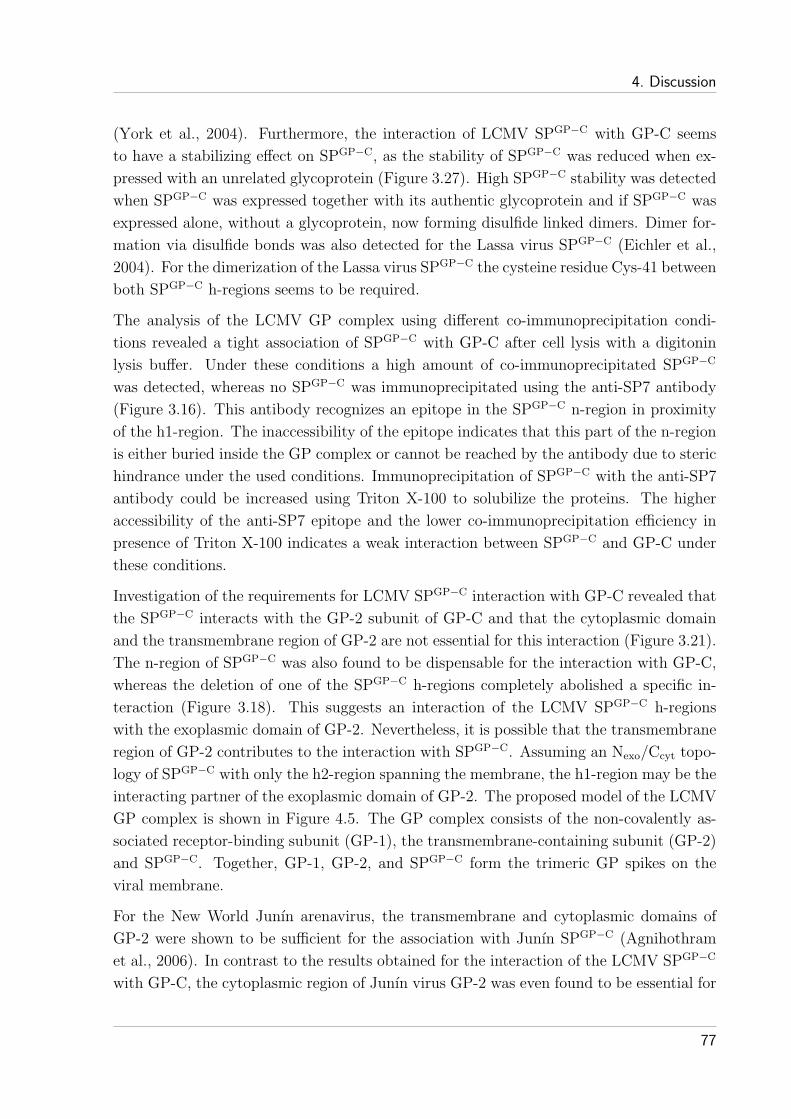
4. Discussion
(York et al., 2004). Furthermore, the interaction of LCMV SPGP−C with GP-C seems
to have a stabilizing effect on SPGP−C, as the stability of SPGP−C was reduced when ex-
pressed with an unrelated glycoprotein (Figure 3.27). High SPGP−C stability was detected
when SPGP−C was expressed together with its authentic glycoprotein and if SPGP−C was
expressed alone, without a glycoprotein, now forming disulfide linked dimers. Dimer for-
mation via disulfide bonds was also detected for the Lassa virus SPGP−C (Eichler et al.,
2004). For the dimerization of the Lassa virus SPGP−C the cysteine residue Cys-41 between
both SPGP−C h-regions seems to be required.
The analysis of the LCMV GP complex using different co-immunoprecipitation condi-
tions revealed a tight association of SPGP−C with GP-C after cell lysis with a digitonin
lysis buffer. Under these conditions a high amount of co-immunoprecipitated SPGP−C
was detected, whereas no SPGP−C was immunoprecipitated using the anti-SP7 antibody
(Figure 3.16). This antibody recognizes an epitope in the SPGP−C n-region in proximity
of the h1-region. The inaccessibility of the epitope indicates that this part of the n-region
is either buried inside the GP complex or cannot be reached by the antibody due to steric
hindrance under the used conditions. Immunoprecipitation of SPGP−C with the anti-SP7
antibody could be increased using Triton X-100 to solubilize the proteins. The higher
accessibility of the anti-SP7 epitope and the lower co-immunoprecipitation efficiency in
presence of Triton X-100 indicates a weak interaction between SPGP−C and GP-C under
these conditions.
Investigation of the requirements for LCMV SPGP−C interaction with GP-C revealed that
the SPGP−C interacts with the GP-2 subunit of GP-C and that the cytoplasmic domain
and the transmembrane region of GP-2 are not essential for this interaction (Figure 3.21).
The n-region of SPGP−C was also found to be dispensable for the interaction with GP-C,
whereas the deletion of one of the SPGP−C h-regions completely abolished a specific in-
teraction (Figure 3.18). This suggests an interaction of the LCMV SPGP−C h-regions
with the exoplasmic domain of GP-2. Nevertheless, it is possible that the transmembrane
region of GP-2 contributes to the interaction with SPGP−C. Assuming an Nexo/Ccyt topo-
logy of SPGP−C with only the h2-region spanning the membrane, the h1-region may be the
interacting partner of the exoplasmic domain of GP-2. The proposed model of the LCMV
GP complex is shown in Figure 4.5. The GP complex consists of the non-covalently as-
sociated receptor-binding subunit (GP-1), the transmembrane-containing subunit (GP-2)
and SPGP−C. Together, GP-1, GP-2, and SPGP−C form the trimeric GP spikes on the
viral membrane.
For the New World Junın arenavirus, the transmembrane and cytoplasmic domains of
GP-2 were shown to be sufficient for the association with Junın SPGP−C (Agnihothram
et al., 2006). In contrast to the results obtained for the interaction of the LCMV SPGP−C
with GP-C, the cytoplasmic region of Junın virus GP-2 was even found to be essential for
77

4. Discussion
SPGP−C binding. Whether this reflects differences between LCMV and Junın virus GPs
or different interactions at different stages of intracellular transport remains to be seen.
+
membrane
cyto
exo
C
N
+
myrmyr
+
h1
h2
GP-1
GP-
2Figure 4.5: Proposed model of the trimeric LCMV GP complex
The trimeric LCMV GP complex is formed by the globular head subunit GP-1, the transmembrane subunit
GP-2 and the myristoylated (myr) SPGP−C. Interaction of SPGP−C was observed with GP-2 alone and even
with GP-C lacking the transmembrane and cytoplasmic regions. Furthermore, deletion analysis revealed
that the SPGP−C h-regions are sufficient and essential for complex formation with the glycoprotein. These
findings and the topological consideration in section 4.2, suggest the SPGP−C h1-region and the exoplasmic
domain of GP-2 as interacting partners, although an additional interaction between the membrane spanning
regions cannot be excluded.
LCMV SPGP−C is an essential component of the GP complex. In absence of SPGP−C,
GP-C is neither processed into GP-1 and GP-2 nor expressed on the plasma membrane.
The ER retention of LCMV GP-C(WE), containing 12 mutated amino acids, was therefore
thought to be due to the inability to associate with SPGP−C. The analysis of pGP-C(WE),
however, revealed that the interaction of SPGP−C with GP-C(WE) is not disturbed by
the amino acid changes. Hence, the association of SPGP−C with GP-C seems not to be
sufficient to promote GP-C transport to the cell surface. Thus, not only the absence of
SPGP−C binding but also mutations within GP-C can result in ER retention of GP-C.
As above mentioned, the cytoplasmic domain of LCMV GP-C is dispensable for the
interaction with SPGP−C. The deletion of this domain (∆C), however, was shown to
drastically reduce GP-C processing and negatively influence the cell surface expression of
GP-C (Figures 3.3 and 3.4). In a previous study, it was shown that the deletion of the
last three amino acid residues of the LCMV pGP-C similarly results in a lack of GP-C
processing, whereas the cell surface expression was not affected (Kunz et al., 2003). For
this to occur, the effects of the C-terminal truncations must be transmitted through the
membrane. It is conceivable that truncations in the cytoplasmic domain, for example,
affect folding of the extracellular domain of GP-C. A partially misfolded GP-C might not
78

4. Discussion
be processed by SKI-1/S1P but may still get transported to the cell surface as GP-C
processing is not a prerequisite for its cell surface expression (Kunz et al., 2003). As mis-
folding of the glycoprotein might also affect complex formation, the transport of GP-C
with the cytoplasmic domain deleted could be impaired by affecting the interaction with
SPGP−C. Using digitonin to solubilize the GP complex, however, revealed no obvious
difference between the interaction of SPGP−C with GP-C or with GP-C lacking the cy-
toplasmic domain. In contrast, the co-immunoprecipitation of SPGP−C with the GP-C
∆C mutant was reduced compared to the amounts detected with GP-C in presence of
Triton X-100. This reduction suggests a very weak interaction of SPGP−C with GP-C if
the cytoplasmic domain is deleted and may thus be the reason for the reduced transport
to the cell surface.
The cell surface expression of LCMV GP-C lacking the cytoplasmic domain is still depen-
dent on the presence of the authentic SPGP−C as the substitution with an unrelated signal
sequence further decreased the amount of the GPs on the plasma membrane. In contrast,
C-terminal truncations in the cytoplasmic domain of the New World Junın arenavirus
GP-2 abrogates SPGP−C association yet now permit GP-C processing and transport to
the cell surface even in absence of SPGP−C (Agnihothram et al., 2006). Therefore, the
Junın SPGP−C is proposed to mask an endogenous ER localization signal within the cy-
toplasmic domain of GP-2. An SPGP−C-independent transport of LCMV GP-C to the
cell surface was only detected for a secretory form of LCMV GP-C, which lacks the cy-
toplasmic domain and the transmembrane region (∆TMC). During transport to the cell
surface the secretory form was processed into GP-1 and GP-2 even in absence of SPGP−C.
Processing of the GP-C ∆TMC mutant indicates correct protein folding of the extracel-
lular domain of GP-C. Although an interaction with SPGP−C is not needed for processing
of the secretory form, it was detected when cell lysis was performed with digitonin. This
interaction, however, seems to be very weak as it was not detectable in presence of Triton
X-100.
4.4 Requirements of SPGP-C for LCMV infection
LCMV infection of target cells is initiated by binding of the GP-1 subunit to its cellu-
lar receptor, α-dystroglycan (Cao et al., 1998). After internalization of bound virus into
large, smooth-walled, endocytic vesicles, the viral nucleocapsid is delivered into the cell
cytoplasm by pH-dependent membrane fusion (Borrow and Oldstone, 1994; Di Simone
et al., 1994). At an acidic pH, GP-1 dissociates from GP-2 and upon irreversible con-
formational rearrangements in GP-2, the hydrophobic N-terminus of GP-2, the putative
fusion peptide, gets exposed (Di Simone and Buchmeier, 1995; Di Simone et al., 1994).
79

4. Discussion
To analyze the effects of LCMV SPGP−C mutations on viral infectivity, LCMV pseu-
doviruses were generated. The pseudovirus system allows easy exchange of the glycopro-
teins incorporated into the viral membrane by transfection of the appropriate constructs
and a quantitative readout of infected cells by measuring the green fluorescence of target
cells expressing eGFP (Beyer et al., 2002).
With the exception of the unmyristoylated SPGP−C G2A mutant, all mutants tested
(∆nME, I4N, G2A/I4N, V22T, and A39T) showed a reduction in pseudovirus forma-
tion compared to the wt (Figure 4.2). As the mutations are located in the n-region as
well as in the h-regions of SPGP−C, this indicates that features of the entire SPGP−C con-
tribute to the efficiency of pseudovirus particle formation. The myristic acid moiety of
the SPGP−C (G2A mutant) seems not to be required for particle assembly unlike it is e.g.
for the retrovirus matrix protein and the poliovirus capsid protein (Ansardi et al., 1992;
Bryant and Ratner, 1990; Chow et al., 1987).
Analysis of pseudovirus infectivity revealed that pseudoviruses produced with one of the
two h-region mutants, V22T and A39T, were still able to infect target cells, whereas mu-
tations in the SPGP−C n-region completely inhibit the infection (Figure 4.2). This strongly
suggests that the n-region of SPGP−C and, specifically, the myristoylation are crucial for vi-
ral infection. The contribution of the LCMV SPGP−C to viral infectivity could be indirect
via modulating the conformation of the GP complex or a direct involvement of the SPGP−C
in viral entry or fusion. A role for SPGP−C in viral fusion is supported by the finding that
the SPGP−C of the New World Junın arenavirus is essential for pH-dependent membrane
fusion, which was tested by syncytium formation (York and Nunberg, 2006; York et al.,
2004). Furthermore, a recent study of the LCMV SPGP−C showed that the reduced in-
fectivity of virus-like particles (VLPs) produced with a G2A mutant of LCMV pGP-C
could be explained by the block of GP-C-mediated cell-cell fusion (Saunders et al., 2007).
Participation of a small myristoylated peptide in membrane fusion is not without prece-
dent. Considering size, topology, myristoylation, and sequence specific effects on viral
infectivity, the arenavirus SPsGP−C share striking similarities with the fusion-associated
small transmembrane (FAST) proteins. The FAST proteins are encoded by a subgroup
of the orthoreoviruses which are nonenveloped viruses that induce cell-cell fusion and
syncytium formation (Duncan, 1999; Shmulevitz and Duncan, 2000). The FAST pro-
teins of the reptilian reovirus and the baboon reovirus are small (14 to 15 kDa) integral
membrane fusion proteins that expose their N-terminally-myristoylated region to the ex-
oplasmic side of the plasma membrane (Corcoran and Duncan, 2004; Dawe et al., 2005).
The myristoylation of the exoplasmic N-terminus of these proteins is essential for the
fusion process. The FAST proteins are proposed to mediate cell-cell fusion in cooperation
with cellular adhesion proteins that promote close apposition of the cellular membranes
(Dawe et al., 2005). In the case of arenavirus membrane fusion, SPGP−C may similarly
80
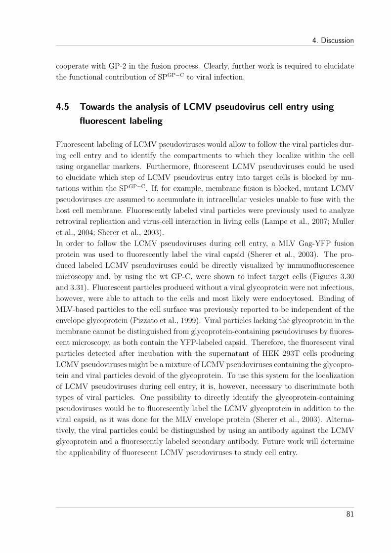
4. Discussion
cooperate with GP-2 in the fusion process. Clearly, further work is required to elucidate
the functional contribution of SPGP−C to viral infection.
4.5 Towards the analysis of LCMV pseudovirus cell entry using
fluorescent labeling
Fluorescent labeling of LCMV pseudoviruses would allow to follow the viral particles dur-
ing cell entry and to identify the compartments to which they localize within the cell
using organellar markers. Furthermore, fluorescent LCMV pseudoviruses could be used
to elucidate which step of LCMV pseudovirus entry into target cells is blocked by mu-
tations within the SPGP−C. If, for example, membrane fusion is blocked, mutant LCMV
pseudoviruses are assumed to accumulate in intracellular vesicles unable to fuse with the
host cell membrane. Fluorescently labeled viral particles were previously used to analyze
retroviral replication and virus-cell interaction in living cells (Lampe et al., 2007; Muller
et al., 2004; Sherer et al., 2003).
In order to follow the LCMV pseudoviruses during cell entry, a MLV Gag-YFP fusion
protein was used to fluorescently label the viral capsid (Sherer et al., 2003). The pro-
duced labeled LCMV pseudoviruses could be directly visualized by immunofluorescence
microscopy and, by using the wt GP-C, were shown to infect target cells (Figures 3.30
and 3.31). Fluorescent particles produced without a viral glycoprotein were not infectious,
however, were able to attach to the cells and most likely were endocytosed. Binding of
MLV-based particles to the cell surface was previously reported to be independent of the
envelope glycoprotein (Pizzato et al., 1999). Viral particles lacking the glycoprotein in the
membrane cannot be distinguished from glycoprotein-containing pseudoviruses by fluores-
cent microscopy, as both contain the YFP-labeled capsid. Therefore, the fluorescent viral
particles detected after incubation with the supernatant of HEK 293T cells producing
LCMV pseudoviruses might be a mixture of LCMV pseudoviruses containing the glycopro-
tein and viral particles devoid of the glycoprotein. To use this system for the localization
of LCMV pseudoviruses during cell entry, it is, however, necessary to discriminate both
types of viral particles. One possibility to directly identify the glycoprotein-containing
pseudoviruses would be to fluorescently label the LCMV glycoprotein in addition to the
viral capsid, as it was done for the MLV envelope protein (Sherer et al., 2003). Alterna-
tively, the viral particles could be distinguished by using an antibody against the LCMV
glycoprotein and a fluorescently labeled secondary antibody. Future work will determine
the applicability of fluorescent LCMV pseudoviruses to study cell entry.
81

Publications derived from this thesis
Freigang, S., Eschli, B., Harris, N., Geuking, M., Quirin, K., Schrempf, S., Zellweger, R.,
Weber, J., Hengartner, H., Zinkernagel, R. M., Aug 2007. A lymphocytic choriomenin-
gitis virus glycoprotein variant that is retained in the endoplasmic reticulum efficiently
cross-primes CD8(+) T cell responses. Proc Natl Acad Sci U S A 104 (33), 13426–13431.
Schrempf, S., Froeschke, M., Giroglou, T., von Laer, D., Dobberstein, B., Nov 2007. Signal
peptide requirements for lymphocytic choriomeningitis virus glycoprotein C maturation
and virus infectivity. J Virol 81 (22), 12515–12524.
82

References
Agnihothram, S. S., York, J., Nunberg, J. H., Jun 2006. Role of the stable signal peptide
and cytoplasmic domain of G2 in regulating intracellular transport of the Junın virus
envelope glycoprotein complex. J Virol 80 (11), 5189–5198.
Agnihothram, S. S., York, J., Trahey, M., Nunberg, J. H., Apr 2007. Bitopic mem-
brane topology of the stable signal peptide in the tripartite Junın virus GP-C envelope
glycoprotein complex. J Virol 81 (8), 4331–4337.
Alberts, B., Johnson, A., Lewis, J., Raff, M., Roberts, K., Walter, P., 2002. Molecular
Biology of the Cell. Garland Science.
Ansardi, D. C., Porter, D. C., Morrow, C. D., 1992. Myristylation of poliovirus capsid
precursor P1 is required for assembly of subviral particles. J Virol 66 (7), 4556–63.
Auperin, D. D., Romanowski, V., Galinski, M., Bishop, D. H., Dec 1984. Sequencing
studies of pichinde arenavirus S RNA indicate a novel coding strategy, an ambisense
viral S RNA. J Virol 52 (3), 897–904.
Barlowe, C., Orci, L., Yeung, T., Hosobuchi, M., Hamamoto, S., Salama, N., Rexach,
M. F., Ravazzola, M., Amherdt, M., Schekman, R., Jun 1994. COPII: a membrane coat
formed by Sec proteins that drive vesicle budding from the endoplasmic reticulum. Cell
77 (6), 895–907.
Barresi, R., Campbell, K. P., Jan 2006. Dystroglycan: from biosynthesis to pathogenesis
of human disease. J Cell Sci 119 (Pt 2), 199–207.
Beyer, W. R., Miletic, H., Ostertag, W., von Laer, D., 2001. Recombinant expression of
lymphocytic choriomeningitis virus strain WE glycoproteins: a single amino acid makes
the difference. J Virol 75 (2), 1061–4.
Beyer, W. R., Popplau, D., Garten, W., von Laer, D., Lenz, O., 2003. Endoproteolytic
processing of the lymphocytic choriomeningitis virus glycoprotein by the subtilase SKI-
1/S1P. J Virol 77 (5), 2866–72.
Beyer, W. R., Westphal, M., Ostertag, W., von Laer, D., 2002. Oncoretrovirus and
lentivirus vectors pseudotyped with lymphocytic choriomeningitis virus glycoprotein:
generation, concentration, and broad host range. J Virol 76 (3), 1488–95.
Blobel, G., Mar 1980. Intracellular protein topogenesis. Proc Natl Acad Sci U S A 77 (3),
1496–1500.
83

References
Blobel, G., Dobberstein, B., Dec 1975. Transfer of proteins across membranes. I. Presence
of proteolytically processed and unprocessed nascent immunoglobulin light chains on
membrane-bound ribosomes of murine myeloma. J Cell Biol 67 (3), 835–851.
Bonifacino, J. S., Glick, B. S., Jan 2004. The mechanisms of vesicle budding and fusion.
Cell 116 (2), 153–166.
Bonifacino, J. S., Lippincott Schwartz, J., May 2003. Coat proteins: shaping membrane
transport. Nat Rev Mol Cell Biol 4 (5), 409–414.
Borrow, P., Oldstone, M. B., 1992. Characterization of lymphocytic choriomeningitis
virus-binding protein(s): a candidate cellular receptor for the virus. J Virol 66 (12),
7270–81.
Borrow, P., Oldstone, M. B., 1994. Mechanism of lymphocytic choriomeningitis virus
entry into cells. Virology 198 (1), 1–9.
Braud, V., Jones, E. Y., McMichael, A., May 1997. The human major histocompatibility
complex class Ib molecule HLA-E binds signal sequence-derived peptides with primary
anchor residues at positions 2 and 9. Eur J Immunol 27 (5), 1164–1169.
Braud, V. M., Allan, D. S., O’Callaghan, C. A., Soderstrom, K., D’Andrea, A., Ogg,
G. S., Lazetic, S., Young, N. T., Bell, J. I., Phillips, J. H., Lanier, L. L., McMichael,
A. J., Feb 1998. HLA-E binds to natural killer cell receptors CD94/NKG2A, B and C.
Nature 391 (6669), 795–799.
Brown, D. A., Rose, J. K., Feb 1992. Sorting of GPI-anchored proteins to glycolipid-
enriched membrane subdomains during transport to the apical cell surface. Cell 68 (3),
533–544.
Bruns, M., Cihak, J., Muller, G., Lehmann Grube, F., 1983. Lymphocytic choriomenin-
gitis virus. VI. Isolation of a glycoprotein mediating neutralization. Virology 130 (1),
247–51.
Bryant, M., Ratner, L., 1990. Myristoylation-dependent replication and assembly of
human immunodeficiency virus 1. Proc Natl Acad Sci U S A 87 (2), 523–7.
Buchmeier, M. J., 2002. Arenaviruses: protein structure and function. Springer, Berlin,
Germany, pp. 159–173.
Buchmeier, M. J., Parekh, B. S., 1987. Protein structure and expression among
arenaviruses. Curr Top Microbiol Immunol 133, 41–57.
Bullough, P. A., Hughson, F. M., Skehel, J. J., Wiley, D. C., Sep 1994. Structure of
influenza haemagglutinin at the pH of membrane fusion. Nature 371 (6492), 37–43.
84

References
Burns, J. W., Buchmeier, M. J., 1991. Protein-protein interactions in lymphocytic
choriomeningitis virus. Virology 183 (2), 620–9.
Cao, W., Henry, M. D., Borrow, P., Yamada, H., Elder, J. H., Ravkov, E. V., Nichol,
S. T., Compans, R. W., Campbell, K. P., Oldstone, M. B., 1998. Identification of alpha-
dystroglycan as a receptor for lymphocytic choriomeningitis virus and Lassa fever virus.
Science 282 (5396), 2079–81.
Capul, A. A., Perez, M., Burke, E., Kunz, S., Buchmeier, M. J., Juan C de la Torre, Sep
2007. Arenavirus Z-glycoprotein association requires Z myristoylation but not functional
RING or late domains. J Virol 81 (17), 9451–9460.
Carr, C. M., Chaudhry, C., Kim, P. S., Dec 1997. Influenza hemagglutinin is spring-loaded
by a metastable native conformation. Proc Natl Acad Sci U S A 94 (26), 14306–14313.
Carr, C. M., Kim, P. S., May 1993. A spring-loaded mechanism for the conformational
change of influenza hemagglutinin. Cell 73 (4), 823–832.
Chow, M., Newman, J. F., Filman, D., Hogle, J. M., Rowlands, D. J., Brown, F., 1987.
Myristylation of picornavirus capsid protein VP4 and its structural significance. Nature
327 (6122), 482–486.
Clegg, J. C. S., 2002. Molecular phylogeny of the arenaviruses. Curr Top Microbiol
Immunol 262, 1–24.
Cocquerel, L., Op de Beeck, A., Lambot, M., Roussel, J., Delgrange, D., Pillez, A.,
Wychowski, C., Penin, F., Dubuisson, J., 2002. Topological changes in the trans-
membrane domains of hepatitis C virus envelope glycoproteins. Embo J 21 (12),
2893–902.
Colman, P. M., Lawrence, M. C., Apr 2003. The structural biology of type I viral
membrane fusion. Nat Rev Mol Cell Biol 4 (4), 309–319.
Compans, R. W., Cooper, M. D., Honjo, T., Koprowski, H., Melchers, F., Oldstone, M.
B. A., Olsnes, S., Potter, M., Vogt, P. K., Wagner H., Marsh M., 2004. Membrane
Trafficking in Viral Replication. Springer Berlin Heidelberg.
Connolly, T., Gilmore, R., May 1989. The signal recognition particle receptor medi-
ates the GTP-dependent displacement of SRP from the signal sequence of the nascent
polypeptide. Cell 57 (4), 599–610.
Cooper, G. M., 2000. The Cell: A Molecular Approach. Sinauer Associates.
Corcoran, J. A., Duncan, R., Apr 2004. Reptilian reovirus utilizes a small type III protein
with an external myristylated amino terminus to mediate cell-cell fusion. J Virol 78 (8),
4342–4351.
85

References
Crawshaw, S. G., Martoglio, B., Meacock, S. L., High, S., 2004. A misassembled trans-
membrane domain of a polytopic protein associates with signal peptide peptidase.
Biochem J Pt.
Dalbey, R., von Heijne, G. (Eds.), 2003. Protein Targeting, Transport & Translocation.
Academic Press.
Dalbey, R. E., von Heijne, G., Nov 1992. Signal peptidases in prokaryotes and eukaryotes–
a new protease family. Trends Biochem Sci 17 (11), 474–478.
Dawe, S., Corcoran, J. A., Clancy, E. K., Salsman, J., Duncan, R., May 2005. Unusual
topological arrangement of structural motifs in the baboon reovirus fusion-associated
small transmembrane protein. J Virol 79 (10), 6216–6226.
de Zarate, I. B. O., Kaelin, K., Rozenberg, F., Feb 2004. Effects of mutations in the cyto-
plasmic domain of herpes simplex virus type 1 glycoprotein B on intracellular transport
and infectivity. J Virol 78 (3), 1540–1551.
Di Simone, C., Buchmeier, M. J., 1995. Kinetics and pH dependence of acid-induced struc-
tural changes in the lymphocytic choriomeningitis virus glycoprotein complex. Virology
209 (1), 3–9.
Di Simone, C., Zandonatti, M. A., Buchmeier, M. J., 1994. Acidic pH triggers LCMV
membrane fusion activity and conformational change in the glycoprotein spike. Virology
198 (2), 455–65.
Doms, R. W., Lamb, R. A., Rose, J. K., Helenius, A., Apr 1993. Folding and assembly of
viral membrane proteins. Virology 193 (2), 545–562.
Dultz, E., Hildenbeutel, M., Martoglio, B., Hochman, J., Dobberstein, B., Kapp, K., Feb
2008. The signal peptide of the mouse mammary tumor virus rem protein is released
from the ER membrane and accumulates in nucleoli. J Biol Chem.
Duncan, R., Aug 1999. Extensive sequence divergence and phylogenetic relationships
between the fusogenic and nonfusogenic orthoreoviruses: a species proposal. Virology
260 (2), 316–328.
Earp, L. J., Delos, S. E., Park, H. E., White, J. M., 2005. The many mechanisms of viral
membrane fusion proteins. Curr Top Microbiol Immunol 285, 25–66.
Eckert, D. M., Kim, P. S., 2001. Mechanisms of viral membrane fusion and its inhibition.
Annu Rev Biochem 70, 777–810.
Edwards, T. G., Wyss, S., Reeves, J. D., Zolla Pazner, S., Hoxie, J. A., Doms, R. W.,
Baribaud, F., 2002. Truncation of the cytoplasmic domain induces exposure of conserved
regions in the ectodomain of human immunodeficiency virus type 1 envelope protein. J
Virol 76 (6), 2683–91.
86

References
Eichler, R., Lenz, O., Strecker, T., Eickmann, M., Klenk, H. D., Garten, W., Oct. 2003a.
Identification of Lassa virus glycoprotein signal peptide as a trans-acting maturation
factor. EMBO Rep 4 (11), 1084–8.
Eichler, R., Lenz, O., Strecker, T., Eickmann, M., Klenk, H. D., Garten, W., 2004.
Lassa virus glycoprotein signal peptide displays a novel topology with an extended
endoplasmic reticulum luminal region. J Biol Chem 279 (13), 12293–9.
Eichler, R., Lenz, O., Strecker, T., Garten, W., Feb. 2003b. Signal peptide of Lassa virus
glycoprotein GP-C exhibits an unusual length. FEBS Lett 538 (1-3), 203–6.
Ellgaard, L., Helenius, A., Mar 2003. Quality control in the endoplasmic reticulum. Nat
Rev Mol Cell Biol 4 (3), 181–191.
Epand, R. M., Jul 2003. Fusion peptides and the mechanism of viral fusion. Biochim
Biophys Acta 1614 (1), 116–121.
Eschli, B., Quirin, K., Wepf, A., Weber, J., Zinkernagel, R., Hans Hengartner, Jun 2006.
Identification of an N-terminal trimeric coiled-coil core within arenavirus glycoprotein
2 permits assignment to class I viral fusion proteins. J Virol 80 (12), 5897–5907.
Fenteany, G., Schreiber, S. L., Apr 1998. Lactacystin, proteasome function, and cell fate.
J Biol Chem 273 (15), 8545–8548.
Froeschke, M., Basler, M., Groettrup, M., Dobberstein, B., 2003. Long-lived signal pep-
tide of lymphocytic choriomeningitis virus glycoprotein pGP-C. J Biol Chem 278 (43),
41914–20.
Gallaher, W. R., DiSimone, C., Buchmeier, M. J., 2001. The viral transmembrane super-
family: possible divergence of Arenavirus and Filovirus glycoproteins from a common
RNA virus ancestor. BMC Microbiol 1 (1), 1.
Garoff, H., Hewson, R., Opstelten, D. J., Dec 1998. Virus maturation by budding.
Microbiol Mol Biol Rev 62 (4), 1171–1190.
Gierasch, L. M., Feb 1989. Signal sequences. Biochemistry 28 (3), 923–930.
Gilmore, R., Blobel, G., Walter, P., Nov 1982. Protein translocation across the endoplas-
mic reticulum. I. Detection in the microsomal membrane of a receptor for the signal
recognition particle. J Cell Biol 95 (2 Pt 1), 463–469.
Glushakova, S. E., Lukashevich, I. S., Baratova, L. A., 1990. Prediction of arenavirus
fusion peptides on the basis of computer analysis of envelope protein sequences. FEBS
Lett 269 (1), 145–7.
Goder, V., Junne, T., Spiess, M., 2004. Sec61p contributes to signal sequence orientation
according to the positive-inside rule. Mol Biol Cell 15 (3), 1470–8.
87

References
Gripon, P., Le Seyec, J., Rumin, S., Guguen Guillouzo, C., 1995. Myristylation of the
hepatitis B virus large surface protein is essential for viral infectivity. Virology 213 (2),
292–9.
Gorlich, D., Kutay, U., 1999. Transport between the cell nucleus and the cytoplasm. Annu
Rev Cell Dev Biol 15, 607–660.
Harrison, S. C., 2005. Mechanism of membrane fusion by viral envelope proteins. Adv
Virus Res 64, 231–261.
Hegde, R. S., Bernstein, H. D., Oct 2006. The surprising complexity of signal sequences.
Trends Biochem Sci 31 (10), 563–571.
High, S., Dobberstein, B., Aug 1992. Mechanisms that determine the transmembrane
disposition of proteins. Curr Opin Cell Biol 4 (4), 581–586.
Higy, M., Junne, T., Spiess, M., 2004. Topogenesis of membrane proteins at the
endoplasmic reticulum. Biochemistry 43 (40), 12716–22.
Hoch Marchaim, H., Weiss, A. M., Bar Sinai, A., Fromer, M., Adermann, K., Hochman,
J., 2003. The leader peptide of MMTV Env precursor localizes to the nucleoli in MMTV-
derived T cell lymphomas and interacts with nucleolar protein B23. Virology 313 (1),
22–32.
Hurtley, S. M., Helenius, A., 1989. Protein oligomerization in the endoplasmic reticulum.
Annu Rev Cell Biol 5, 277–307.
Iapalucci, S., Lopez, N., Rey, O., Zakin, M. M., Cohen, G. N., Franze Fernandez, M. T.,
Nov 1989. The 5’ region of Tacaribe virus L RNA encodes a protein with a potential
metal binding domain. Virology 173 (1), 357–361.
Jahn, R., Lang, T., Sudhof, T. C., Feb 2003. Membrane fusion. Cell 112 (4), 519–533.
Johnson, A. E., van Waes, M. A., 1999. The translocon: a dynamic gateway at the ER
membrane. Annu Rev Cell Dev Biol 15, 799–842.
Kalderon, D., Roberts, B. L., Richardson, W. D., Smith, A. E., Dec 1984. A short amino
acid sequence able to specify nuclear location. Cell 39 (3 Pt 2), 499–509.
Keenan, R. J., Freymann, D. M., Stroud, R. M., Walter, P., 2001. The signal recognition
particle. Annu Rev Biochem 70, 755–75.
Keller, P., Simons, K., Dec 1997. Post-Golgi biosynthetic trafficking. J Cell Sci 110 ( Pt
24), 3001–3009.
Kido, H., Niwa, Y., Beppu, Y., Towatari, T., 1996. Cellular proteases involved in the
pathogenicity of enveloped animal viruses, human immunodeficiency virus, influenza
virus A and Sendai virus. Adv Enzyme Regul 36, 325–347.
88

References
Kielian, M., Jan 2006. Class II virus membrane fusion proteins. Virology 344 (1), 38–47.
Kielian, M., Rey, F. A., Jan 2006. Virus membrane-fusion proteins: more than one way
to make a hairpin. Nat Rev Microbiol 4 (1), 67–76.
Kim, S. J., Hegde, R. S., Nov 2002. Cotranslational partitioning of nascent prion protein
into multiple populations at the translocation channel. Mol Biol Cell 13 (11), 3775–3786.
Kirchhausen, T., Dec 2000. Three ways to make a vesicle. Nat Rev Mol Cell Biol 1 (3),
187–198.
Klein, K. C., Reed, J. C., Lingappa, J. R., 2007. Intracellular destinies: degradation,
targeting, assembly, and endocytosis of HIV Gag. AIDS Rev 9 (3), 150–161.
Knipe, D. M., Howley, P. M. (Eds.), 2001. Fields Virology, 4th Edition. Lippincott
Williams & Wilkins.
Krzyzaniak, M., Mach, M., Britt, W. J., Oct 2007. The cytoplasmic tail of glycoprotein M
(gpUL100) expresses trafficking signals required for human cytomegalovirus assembly
and replication. J Virol 81 (19), 10316–10328.
Kunz, S., Edelmann, K. H., de la Torre, J. C., Gorney, R., Oldstone, M. B., 2003.
Mechanisms for lymphocytic choriomeningitis virus glycoprotein cleavage, transport,
and incorporation into virions. Virology 314 (1), 168–78.
Lambert, C., Prange, R., 2001. Dual topology of the hepatitis B virus large envelope
protein: determinants influencing post-translational pre-S translocation. J Biol Chem
276 (25), 22265–72.
Lampe, M., Briggs, J. A. G., Endress, T., Glass, B., Riegelsberger, S., Krausslich, H. G.,
Lamb, D. C., Brauchle, C., Muller, B., Mar 2007. Double-labelled HIV-1 particles for
study of virus-cell interaction. Virology 360 (1), 92–104.
Lanford, R. E., Butel, J. S., Jul 1984. Construction and characterization of an SV40
mutant defective in nuclear transport of T antigen. Cell 37 (3), 801–813.
Lee, K. J., Perez, M., Pinschewer, D. D., de la Torre, J. C., 2002. Identification of the
lymphocytic choriomeningitis virus (LCMV) proteins required to rescue LCMV RNA
analogs into LCMV-like particles. J Virol 76 (12), 6393–7.
Lemberg, M. K., Bland, F. A., Weihofen, A., Braud, V. M., Martoglio, B., 2001. In-
tramembrane proteolysis of signal peptides: an essential step in the generation of HLA-E
epitopes. J Immunol 167 (11), 6441–6.
Lemberg, M. K., Martoglio, B., 2002. Requirements for signal peptide peptidase-catalyzed
intramembrane proteolysis. Mol Cell 10 (4), 735–44.
89

References
Lenz, O., ter Meulen, J., Klenk, H. D., Seidah, N. G., Garten, W., 2001. The Lassa virus
glycoprotein precursor GP-C is proteolytically processed by subtilase SKI-1/S1P. Proc
Natl Acad Sci U S A 98 (22), 12701–5.
Letourneur, F., Gaynor, E. C., Hennecke, S., Demolliere, C., Duden, R., Emr, S. D.,
Riezman, H., Cosson, P., Dec 1994. Coatomer is essential for retrieval of dilysine-tagged
proteins to the endoplasmic reticulum. Cell 79 (7), 1199–1207.
Lindemann, D., Pietschmann, T., Picard Maureau, M., Berg, A., Heinkelein, M., Thurow,
J., Knaus, P., Zentgraf, H., Rethwilm, A., 2001. A particle-associated glycoprotein signal
peptide essential for virus maturation and infectivity. J Virol 75 (13), 5762–71.
Loureiro, J., Lilley, B. N., Spooner, E., Noriega, V., Tortorella, D., Ploegh, H. L.,
Jun 2006. Signal peptide peptidase is required for dislocation from the endoplasmic
reticulum. Nature 441 (7095), 894–897.
Luirink, J., Sinning, I., Nov 2004. SRP-mediated protein targeting: structure and function
revisited. Biochim Biophys Acta 1694 (1-3), 17–35.
Lyko, F., Martoglio, B., Jungnickel, B., Rapoport, T. A., Dobberstein, B., 1995. Signal
sequence processing in rough microsomes. J Biol Chem 270 (34), 19873–8.
Lyman, S. K., Schekman, R., Jan 1997. Binding of secretory precursor polypeptides to a
translocon subcomplex is regulated by BiP. Cell 88 (1), 85–96.
Marsh, M., Helenius, A., Feb 2006. Virus entry: open sesame. Cell 124 (4), 729–740.
Martoglio, B., 2003. Intramembrane proteolysis and post-targeting functions of signal
peptides. Biochem Soc Trans 31 (Pt 6), 1243–7.
Martoglio, B., Dobberstein, B., 1995. Protein insertion into the membrane of the endo-
plasmic reticulum: the architecture of the translocation site. Cold Spring Harb Symp
Quant Biol 60, 41–5.
Martoglio, B., Dobberstein, B., 1998. Signal sequences: more than just greasy peptides.
Trends Cell Biol 8 (10), 410–5.
Martoglio, B., Graf, R., Dobberstein, B., 1997. Signal peptide fragments of preprolactin
and HIV-1 p-gp160 interact with calmodulin. Embo J 16 (22), 6636–45.
Martoglio, B., Hofmann, M. W., Brunner, J., Dobberstein, B., 1995. The protein-
conducting channel in the membrane of the endoplasmic reticulum is open laterally
toward the lipid bilayer. Cell 81 (2), 207–14.
Matlack, K. E., Misselwitz, B., Plath, K., Rapoport, T. A., May 1999. BiP acts as a
molecular ratchet during posttranslational transport of prepro-alpha factor across the
ER membrane. Cell 97 (5), 553–564.
90

References
Matsuura Tokita, K., Takeuchi, M., Ichihara, A., Mikuriya, K., Nakano, A., Jun 2006.
Live imaging of yeast Golgi cisternal maturation. Nature 441 (7096), 1007–1010.
McLauchlan, J., Lemberg, M. K., Hope, G., Martoglio, B., 2002. Intramembrane prote-
olysis promotes trafficking of hepatitis C virus core protein to lipid droplets. Embo J
21 (15), 3980–8.
Mertz, J. A., Simper, M. S., Lozano, M. M., Payne, S. M., Jaquelin P Dudley, Dec 2005.
Mouse mammary tumor virus encodes a self-regulatory RNA export protein and is a
complex retrovirus. J Virol 79 (23), 14737–14747.
Meyer, D. I., Dobberstein, B., Nov 1980. Identification and characterization of a mem-
brane component essential for the translocation of nascent proteins across the membrane
of the endoplasmic reticulum. J Cell Biol 87 (2 Pt 1), 503–508.
Meyer, H. A., Grau, H., Kraft, R., Kostka, S., Prehn, S., Kalies, K. U., Hartmann, E.,
May 2000. Mammalian Sec61 is associated with Sec62 and Sec63. J Biol Chem 275 (19),
14550–14557.
Michelsen, K., Yuan, H., Schwappach, B., 2005. Hide and run. EMBO Rep 6 (8), 717–22.
Muller, B., Daecke, J., Fackler, O. T., Dittmar, M. T., Zentgraf, H., Krausslich, H. G.,
2004. Construction and characterization of a fluorescently labeled infectious human
immunodeficiency virus type 1 derivative. J Virol 78 (19), 10803–13.
Neuman, B. W., Adair, B. D., Burns, J. W., Milligan, R. A., Buchmeier, M. J., Yea-
ger, M., Mar 2005. Complementarity in the supramolecular design of arenaviruses and
retroviruses revealed by electron cryomicroscopy and image analysis. J Virol 79 (6),
3822–3830.
Nieva, J. L., Agirre, A., Jul 2003. Are fusion peptides a good model to study viral cell
fusion? Biochim Biophys Acta 1614 (1), 104–115.
Nilsson, I., von Heijne, G., Jun 2000. Glycosylation efficiency of Asn-Xaa-Thr sequons
depends both on the distance from the C terminus and on the presence of a downstream
transmembrane segment. J Biol Chem 275 (23), 17338–17343.
Nilsson, T., Warren, G., Aug 1994. Retention and retrieval in the endoplasmic reticulum
and the Golgi apparatus. Curr Opin Cell Biol 6 (4), 517–521.
Ostapchuk, P., Hearing, P., Ganem, D., 1994. A dramatic shift in the transmembrane
topology of a viral envelope glycoprotein accompanies hepatitis B viral morphogenesis.
Embo J 13 (5), 1048–57.
Ott, C. M., Akhavan, A., Lingappa, V. R., Apr 2007. Specific features of the prion pro-
tein transmembrane domain regulate nascent chain orientation. J Biol Chem 282 (15),
11163–11171.
91

References
Paetzel, M., Karla, A., Strynadka, N. C., Dalbey, R. E., 2002. Signal peptidases. Chem
Rev 102 (12), 4549–80.
Pelham, H. R., Dec 2001. Traffic through the Golgi apparatus. J Cell Biol 155 (7), 1099–
1101.
Perez, M., Craven, R. C., de la Torre, J. C., 2003. The small RING finger protein Z
drives arenavirus budding: implications for antiviral strategies. Proc Natl Acad Sci U
S A 100 (22), 12978–83.
Perez, M., Greenwald, D. L., De La Torre, J. C., 2004. Myristoylation of the RING Finger
Z Protein Is Essential for Arenavirus Budding. J Virol 78 (20), 11443–8.
Pizzato, M., Marlow, S. A., Blair, E. D., Takeuchi, Y., Oct 1999. Initial binding of murine
leukemia virus particles to cells does not require specific Env-receptor interaction. J
Virol 73 (10), 8599–8611.
Pool, M. R., Dec 2003. Getting to the membrane: how is co-translational protein targeting
to the endoplasmic reticulum regulated? Biochem Soc Trans 31 (Pt 6), 1232–1237.
Pool, M. R., Stumm, J., Fulga, T. A., Sinning, I., Dobberstein, B., Aug 2002. Distinct
modes of signal recognition particle interaction with the ribosome. Science 297 (5585),
1345–1348.
Rapoport, T. A., Nov 2007. Protein translocation across the eukaryotic endoplasmic
reticulum and bacterial plasma membranes. Nature 450 (7170), 663–669.
Rapoport, T. A., Goder, V., Heinrich, S. U., Matlack, K. E., 2004. Membrane-protein
integration and the role of the translocation channel. Trends Cell Biol 14 (10), 568–75.
Rapoport, T. A., Matlack, K. E., Plath, K., Misselwitz, B., Staeck, O., Oct 1999. Post-
translational protein translocation across the membrane of the endoplasmic reticulum.
Biol Chem 380 (10), 1143–1150.
Resh, M. D., 1999. Fatty acylation of proteins: new insights into membrane targeting of
myristoylated and palmitoylated proteins. Biochim Biophys Acta 1451 (1), 1–16.
Resh, M. D., 2005. Intracellular trafficking of HIV-1 Gag: how Gag interacts with cell
membranes and makes viral particles. AIDS Rev 7 (2), 84–91.
Robinson, M. S., Mar 1987. Coated vesicles and protein sorting. J Cell Sci 87 ( Pt 2),
203–204.
Rojek, J. M., Kunz, S., Jan 2008. Cell entry by human pathogenic arenaviruses. Cell
Microbiol.
Rojek, J. M., Perez, M., Kunz, S., Feb 2008. Cellular entry of lymphocytic choriomenin-
gitis virus. J Virol 82 (3), 1505–1517.
92

References
Romanowski, V., Matsuura, Y., Bishop, D. H., 1985. Complete sequence of the S
RNA of lymphocytic choriomeningitis virus (WE strain) compared to that of Pichinde
arenavirus. Virus Res 3 (2), 101–14.
Salvato, M., Shimomaye, E., Oldstone, M. B., Apr 1989. The primary structure of the lym-
phocytic choriomeningitis virus L gene encodes a putative RNA polymerase. Virology
169 (2), 377–384.
Sanders, S. L., Whitfield, K. M., Vogel, J. P., Rose, M. D., Schekman, R. W., Apr 1992.
Sec61p and BiP directly facilitate polypeptide translocation into the ER. Cell 69 (2),
353–365.
Saunders, A. A., Ting, J. P. C., Meisner, J., Neuman, B. W., Perez, M., de la Torre, J. C.,
Buchmeier, M. J., Jun 2007. Mapping the landscape of the lymphocytic choriomeningitis
virus stable signal peptide reveals novel functional domains. J Virol 81 (11), 5649–5657.
Schatz, G., Dobberstein, B., Mar 1996. Common principles of protein translocation across
membranes. Science 271 (5255), 1519–1526.
Schrempf, S., Froeschke, M., Giroglou, T., von Laer, D., Dobberstein, B., Nov 2007. Signal
peptide requirements for lymphocytic choriomeningitis virus glycoprotein C maturation
and virus infectivity. J Virol 81 (22), 12515–12524.
Shah, W. A., Peng, H., Carbonetto, S., 2006. Role of non-raft cholesterol in lymphocytic
choriomeningitis virus infection via alpha-dystroglycan. J Gen Virol 87 (Pt 3), 673–8.
Sherer, N. M., Lehmann, M. J., Jimenez Soto, L. F., Ingmundson, A., Horner, S. M.,
Cicchetti, G., Allen, P. G., Pypaert, M., Cunningham, J. M., Mothes, W., 2003. Visual-
ization of retroviral replication in living cells reveals budding into multivesicular bodies.
Traffic 4 (11), 785–801.
Shmulevitz, M., Duncan, R., Mar 2000. A new class of fusion-associated small transmem-
brane (FAST) proteins encoded by the non-enveloped fusogenic reoviruses. EMBO J
19 (5), 902–912.
Silberstein, S., Gilmore, R., Jun 1996. Biochemistry, molecular biology, and genetics of
the oligosaccharyltransferase. FASEB J 10 (8), 849–858.
Sipos, L., von Heijne, G., May 1993. Predicting the topology of eukaryotic membrane
proteins. Eur J Biochem 213 (3), 1333–1340.
Sollner, T. H., Aug 2004. Intracellular and viral membrane fusion: a uniting mechanism.
Curr Opin Cell Biol 16 (4), 429–435.
Southern, P. J., Singh, M. K., Riviere, Y., Jacoby, D. R., Buchmeier, M. J., Oldstone,
M. B., Mar 1987. Molecular characterization of the genomic S RNA segment from
lymphocytic choriomeningitis virus. Virology 157 (1), 145–155.
93

References
Stiasny, K., Allison, S. L., Mandl, C. W., Heinz, F. X., Aug 2001. Role of metastability
and acidic pH in membrane fusion by tick-borne encephalitis virus. J Virol 75 (16),
7392–7398.
Tajima, S., Lauffer, L., Rath, V. L., Walter, P., Oct 1986. The signal recognition particle
receptor is a complex that contains two distinct polypeptide chains. J Cell Biol 103 (4),
1167–1178.
Tamm, L. K., Han, X., Dec 2000. Viral fusion peptides: a tool set to disrupt and connect
biological membranes. Biosci Rep 20 (6), 501–518.
Teasdale, R. D., Jackson, M. R., 1996. Signal-mediated sorting of membrane proteins
between the endoplasmic reticulum and the golgi apparatus. Annu Rev Cell Dev Biol
12, 27–54.
Terzi, L., Pool, M. R., Dobberstein, B., Strub, K., Jan 2004. Signal recognition parti-
cle Alu domain occupies a defined site at the ribosomal subunit interface upon signal
sequence recognition. Biochemistry 43 (1), 107–117.
Urban, S., Gripon, P., 2002. Inhibition of duck hepatitis B virus infection by a
myristoylated pre-S peptide of the large viral surface protein. J Virol 76 (4), 1986–90.
Utsumi, T., Ohta, H., Kayano, Y., Sakurai, N., Ozoe, Y., 2005. The N-terminus
of B96Bom, a Bombyx mori G-protein-coupled receptor, is N-myristoylated and
translocated across the membrane. Febs J 272 (2), 472–81.
Verner, K., Schatz, G., Sep 1988. Protein translocation across membranes. Science
241 (4871), 1307–1313.
von Heijne, G., Jun 1983. Patterns of amino acids near signal-sequence cleavage sites. Eur
J Biochem 133 (1), 17–21.
von Heijne, G., 1985. Signal sequences. The limits of variation. J Mol Biol 184 (1), 99–105.
von Heijne, G., Oct 1989. Control of topology and mode of assembly of a polytopic
membrane protein by positively charged residues. Nature 341 (6241), 456–458.
von Heijne, G., Aug 1990. Protein targeting signals. Curr Opin Cell Biol 2 (4), 604–608.
Walter, P., Blobel, G., Nov 1981. Translocation of proteins across the endoplasmic retic-
ulum III. Signal recognition protein (SRP) causes signal sequence-dependent and site-
specific arrest of chain elongation that is released by microsomal membranes. J Cell Biol
91 (2 Pt 1), 557–561.
Walter, P., Blobel, G., Oct 1982. Signal recognition particle contains a 7S RNA essential
for protein translocation across the endoplasmic reticulum. Nature 299 (5885), 691–698.
94

References
Walter, P., Gilmore, R., Blobel, G., Aug 1984. Protein translocation across the
endoplasmic reticulum. Cell 38 (1), 5–8.
Walter, P., Johnson, A. E., 1994. Signal sequence recognition and protein targeting to
the endoplasmic reticulum membrane. Annu Rev Cell Biol 10, 87–119.
Weihofen, A., Binns, K., Lemberg, M. K., Ashman, K., Martoglio, B., 2002. Identification
of signal peptide peptidase, a presenilin-type aspartic protease. Science 296 (5576),
2215–8.
Weihofen, A., Martoglio, B., 2003. Intramembrane-cleaving proteases: controlled libera-
tion of proteins and bioactive peptides. Trends Cell Biol 13 (2), 71–8.
Wilk, T., Geiselhart, V., Frech, M., Fuller, S. D., Flugel, R. M., Lochelt, M., 2001.
Specific interaction of a novel foamy virus Env leader protein with the N-terminal Gag
domain. J Virol 75 (17), 7995–8007.
Wright, K. E., Spiro, R. C., Burns, J. W., Buchmeier, M. J., 1990. Post-translational
processing of the glycoproteins of lymphocytic choriomeningitis virus. Virology 177 (1),
175–83.
Yee, J. K., Miyanohara, A., LaPorte, P., Bouic, K., Burns, J. C., Friedmann, T., 1994.
A general method for the generation of high-titer, pantropic retroviral vectors: highly
efficient infection of primary hepatocytes. Proc Natl Acad Sci U S A 91 (20), 9564–8,
department of Pediatrics, University of California, San Diego, La Jolla 92093.
York, J., Nunberg, J. H., 2006. Role of the Stable Signal Peptide of Junin Arenavirus
Envelope Glycoprotein in pH-Dependent Membrane Fusion. J Virol 80 (15), 7775–80.
York, J., Romanowski, V., Lu, M., Nunberg, J. H., 2004. The signal peptide of the Junin
arenavirus envelope glycoprotein is myristoylated and forms an essential subunit of the
mature G1-G2 complex. J Virol 78 (19), 10783–92.
95

List of Figures
1.1 Schematic representation of an N-terminal signal sequence for ER targeting 4
1.2 Arenavirus genome organization . . . . . . . . . . . . . . . . . . . . . . . . 10
1.3 Structure of the arenavirus particle . . . . . . . . . . . . . . . . . . . . . . 11
1.4 The LCMV precursor glycoprotein C . . . . . . . . . . . . . . . . . . . . . 12
1.5 The signal peptide of LCMV GP-C . . . . . . . . . . . . . . . . . . . . . . 13
2.1 Overlap extension PCR . . . . . . . . . . . . . . . . . . . . . . . . . . . . . 23
3.1 Substitution of SPGP−C with SPVSV−G . . . . . . . . . . . . . . . . . . . . 35
3.2 Effect of SPGP−C substitution with SPVSV−G on GP-C processing and
transport . . . . . . . . . . . . . . . . . . . . . . . . . . . . . . . . . . . . 36
3.3 ER insertion and processing of the GP-C ∆C deletion mutants . . . . . . . 38
3.4 Cell surface expression of the GP-C ∆C deletion mutants . . . . . . . . . . 39
3.5 Expression and secretion of the GP-C ∆TMC deletion mutants . . . . . . 40
3.6 Outline of SPGP−C deletion mutants . . . . . . . . . . . . . . . . . . . . . . 41
3.7 Effects of SPGP−C deletions on ER membrane insertion
and processing of GP-C . . . . . . . . . . . . . . . . . . . . . . . . . . . . 42
3.8 SPGP−C stability of the h-region deletion mutants . . . . . . . . . . . . . . 43
3.9 Cell surface expression of SPGP−C deletion mutants . . . . . . . . . . . . . 44
3.10 Analysis of SPGP−C point mutants by Western blot . . . . . . . . . . . . . 45
3.11 Identification of mutant SPsGP−C after metabolic labeling . . . . . . . . . . 46
3.12 Cell surface expression of SPGP−C point mutants . . . . . . . . . . . . . . . 47
3.13 Schematic representation of SPGP−C myristoylation mutants . . . . . . . . 47
3.14 Analysis of SPGP−C myristoylation mutants by Western blot . . . . . . . . 48
3.15 Cell surface expression of SPGP−C myristoylation mutants . . . . . . . . . . 49
3.16 Protein solubility and co-immunoprecipitation conditions . . . . . . . . . . 51
3.17 Co-immunoprecipitation of SPGP−C with GP-C during GP maturation . . . 52
96

List of Figures
3.18 Co-immunoprecipitation of SPGP−C mutants with GP-C . . . . . . . . . . . 53
3.19 Outline of pGP-C subunit deletion mutants . . . . . . . . . . . . . . . . . 54
3.20 Analysis of pGP-1-HA and pGP-2-HA ER insertion and SPGP−C . . . . . . 55
3.21 Co-immunoprecipitation of SPGP−C with GP-1-HA and GP-2-HA . . . . . 56
3.22 Co-immunoprecipitation of SPGP−C with the
GP-C C-terminal deletion mutants . . . . . . . . . . . . . . . . . . . . . . 57
3.23 Disulfide bond formation of SPGP−C . . . . . . . . . . . . . . . . . . . . . 58
3.24 Schematic representation of GP-C-HA(WE) . . . . . . . . . . . . . . . . . 59
3.25 Co-immunoprecipitation of SPGP−C with GP-C-HA(WE) . . . . . . . . . . 60
3.26 LCMV SPGP−C fused to the VSV glycoprotein . . . . . . . . . . . . . . . . 60
3.27 Stability of SPGP−C fused to the VSV glycoprotein . . . . . . . . . . . . . 61
3.28 Lactacystin treatment of SPGP−C-VSV-G-HA expressing cells . . . . . . . . 62
3.29 Formation and infectivity of LCMV pseudoviruses with SPGP−C mutants . 64
3.30 Infectivity of fluorescent LCMV pseudoviruses . . . . . . . . . . . . . . . . 66
3.31 Fluorescently labeled viral particles . . . . . . . . . . . . . . . . . . . . . . 67
4.1 Functions of LCMV SPGP−C besides ER targeting . . . . . . . . . . . . . . 69
4.2 Summarized results of LCMV SPGP−C deletion and point mutants . . . . . 72
4.3 Postulated topology of LCMV SPGP−C during ER membrane insertion . . 73
4.4 Proposed membrane topologies of the unmyristoylated LCMV SPGP−C . . 75
4.5 Proposed model of the trimeric LCMV GP complex . . . . . . . . . . . . . 78
97

List of Abbreviations
aa amino acid
Ala alanine (A)
APS ammonium persulfate
Arg arginine (R)
Asn asparagine (N)
Asp aspartic acid (D)
ATP adenosine triphosphate
bp base pair
CIP calf intestine phosphatase
cryo-EM electron cryomicroscopy
COP coat protein
Co-IP co-immunoprecipitation
CTP cytosine triphosphate
Cys cysteine (C)
DAPI 4′,6′-diamidino-2-phenylindole
DMEM Dulbecco’s Modified Eagle’s Medium
DMSO dimethylsulphoxide
DNA deoxyribonucleic acid
dNTP deoxyribonucleotide triphosphate (dATP, dCTP, dGTP, dTTP)
DTT dithiothreitol
E.coli Escherichia coli
EDTA ethylenediaminetetraacetic acid
eGFP enhanced green fluorescent protein
Endo H Endoglycosidase H
ER endoplasmic reticulum
FACS fluorescence-activated cell sorter
FAST fusion-associated small transmembrane (FAST) proteins
FCS fetal calf serum
Gag group specific antigen
GFP green fluorescent protein
Gln glutamine (Q)
Glu glutamic acid (E)
Gly glycine (G)
GP glycoprotein
GTP guanosine triphosphate
98

List of Abbreviations
HA hemagglutinin
HBV hepatitis B virus
HCV hepatitis C virus
HeBS HEPES buffered saline
HEPES 4-(2-Hydroxyethyl)piperazine-1-ethanesulfonic acid
His histidine (H)
HIV human immunodeficiency virus
HLA human lymphocyte antigen
HRPO horseradish peroxidase
I-Clip intramembrane cleaving protease
IgG immunoglobulin G
IGR intergenic region
Ile isoleucine (I)
IP immunoprecipitation
kDa kilodalton
LB Luria-Bertani
LCMV lymphocytic choriomeningitis virus
Leu leucine (L)
Lys lysine (K)
Met methionine (M)
MHC major histocompatibility complex
MLV murine leukemia virus
MMTV mouse mammary tumor virus
ODG Octyl-β-D-glucopyranoside
PAGE polyacrylamide gel electrophoresis
PBS phosphate buffered saline
PCR polymerase chain reaction
PE phycoerythrin
pGP precursor glycoprotein
Phe phenylalanine (F)
PMSF phenylmethanesulphonylfluoride
PNGase F peptide-N-glycosidase F
Pro proline (P)
Pol polymerase
RIPA buffer Radio-Immunoprecipitation Assay buffer
RNA ribonucleic acid
RNP ribonucleoprotein
RT room temperature
SDS sodium dodecyl sulfate
99

List of Abbreviations
Ser serine (S)
SKI1/S1P subtilisin-kexin isoenzyme 1/site 1 protease
SP signal peptide
SPase signal peptidase
SPP signal peptide peptidase
SR SRP receptor
SRP signal recognition particle
TAE Tris-acetate/EDTA
TAP transporter associated with antigen presentation
TBS Tris buffered saline
TEMED Tetramethylethylendiamin
Thr threonine (T)
TM transmembrane
Tris Trishydroxymethylaminomethan
Trp tryptophane (W)
TTP thymidine triphosphate
Tyr tyrosine (Y)
UTR untranslated region
Val valine (V)
VLP virus-like particle
VSV vesicular stomatitis virus
VSV-G VSV glycoprotein
wt wild type
YFP yellow fluorescent protein
100

Danksagung
Ich danke all denen, die mir mit Rat und Tat zur Seite standen und somit zum Gelingen
dieser Arbeit beigetragen haben.
Allen voran geht mein Dank an Prof. Dr. Bernhard Dobberstein fur die Bereitstellung
des Themas und die gute wissenschaftliche Betreuung meiner Arbeit. Er hat mich stets
unterstutzt und war immer bereit auf Fragen und Probleme einzugehen.
Prof. Dr. Irmgard Sinning danke ich fur die Bereitschaft, meine Arbeit zu bewerten und
sie als Zweitgutachter vor der Fakultat zu vertreten.
Prof. Dr. med. Dorothee von Laer und Dr. Tsanan Giroglou vom Georg-Speyer-Haus
in Frankfurt a.M. mochte ich fur die gute Zusammenarbeit und die Analyse der LCMV
Pseudoviren danken.
Des Weiteren gilt mein Dank all meinen Kollegen in den Arbeitsgruppen Dobberstein
und Seedorf fur das angenehme und nie langweilig werdende Arbeitsklima, fur die vielen
wertvollen Diskussionen und Ratschlage und fur all die großen und kleinen Hilfestellungen.
101


