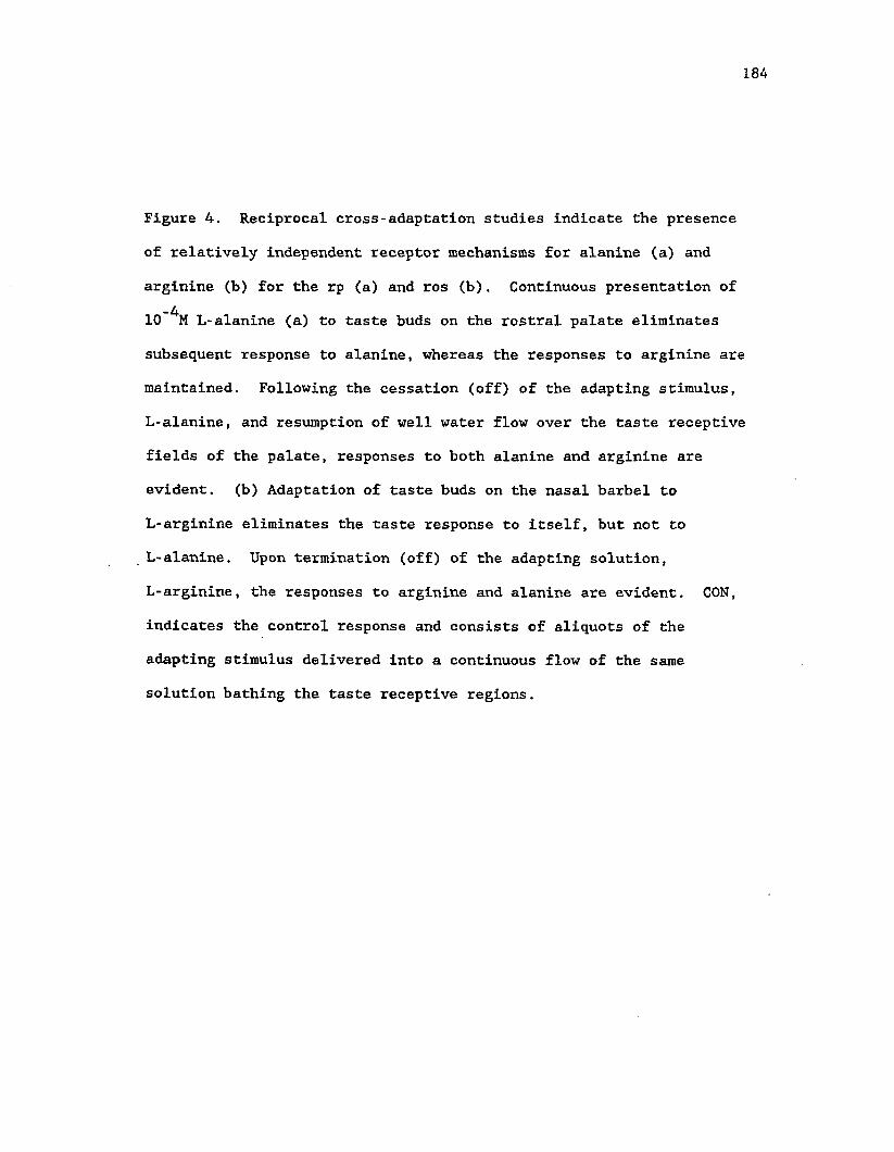
Louisiana State University LSU Digital Commons LSU Historical Dissertations and eses Graduate School 1986 Functional Organization of the Gustatory System in the Brains of Ictalurid Catfish: a Combined Electrophysiological and Neuroanatomical Study (Taste, Viscerotopic, Sensory Maps, Forebrain). Jagmeet Singh Kanwal Louisiana State University and Agricultural & Mechanical College Follow this and additional works at: hps://digitalcommons.lsu.edu/gradschool_disstheses is Dissertation is brought to you for free and open access by the Graduate School at LSU Digital Commons. It has been accepted for inclusion in LSU Historical Dissertations and eses by an authorized administrator of LSU Digital Commons. For more information, please contact [email protected]. Recommended Citation Kanwal, Jagmeet Singh, "Functional Organization of the Gustatory System in the Brains of Ictalurid Catfish: a Combined Electrophysiological and Neuroanatomical Study (Taste, Viscerotopic, Sensory Maps, Forebrain)." (1986). LSU Historical Dissertations and eses. 4244. hps://digitalcommons.lsu.edu/gradschool_disstheses/4244
Transcript
Louisiana State UniversityLSU Digital Commons
LSU Historical Dissertations and Theses Graduate School
1986
Functional Organization of the Gustatory Systemin the Brains of Ictalurid Catfish: a CombinedElectrophysiological and Neuroanatomical Study(Taste, Viscerotopic, Sensory Maps, Forebrain).Jagmeet Singh KanwalLouisiana State University and Agricultural & Mechanical College
Follow this and additional works at: https://digitalcommons.lsu.edu/gradschool_disstheses
This Dissertation is brought to you for free and open access by the Graduate School at LSU Digital Commons. It has been accepted for inclusion inLSU Historical Dissertations and Theses by an authorized administrator of LSU Digital Commons. For more information, please [email protected].
Recommended CitationKanwal, Jagmeet Singh, "Functional Organization of the Gustatory System in the Brains of Ictalurid Catfish: a CombinedElectrophysiological and Neuroanatomical Study (Taste, Viscerotopic, Sensory Maps, Forebrain)." (1986). LSU Historical Dissertationsand Theses. 4244.https://digitalcommons.lsu.edu/gradschool_disstheses/4244
This reproduction was made from a copy of a manuscript sent to us for publication and microfilming. While the most advanced technology has been used to photograph and reproduce this manuscript, the quality of the reproduction is heavily dependent upon the quality of the material submitted. Pages in any manuscript may have indistinct print. In all cases the best available copy has been filmed.
The following explanation of techniques is provided to help clarify notations which may appear on this reproduction.
1. Manuscripts may not always be complete. When it is not possible to obtain missing pages, a note appears to indicate this.
2. When copyrighted materials are removed from the manuscript, a note appears to indicate this.
3. Oversize materials (maps, drawings, and charts) are photographed by sectioning the original, beginning at the upper left hand comer and continuing from left to right in equal sections with small overlaps. Each oversize page is also filmed as one exposure and is available, for an additional charge, as a standard 35mm slide or in black and white paper format.*
4. Most photographs reproduce acceptably on positive microfilm or microfiche but lack clarity on xerographic copies made from the microfilm. Fbr an additional charge, all photographs are available in black and white standard 35mm slide format.*
*For more information about black and white slides or enlarged paper reproductions, please contact the Dissertations Customer Services Department.
T T A /T -T Dissertation L J 1 V 1 1 Information ServiceUniversity Microfilms InternationalA Bell & Howell Information Company300 N. Zeeb Road, Ann Arbor, Michigan 48106
8629178
K a n w a l, J a g m e e t S in g h
FUNCTIONAL ORGANIZATION OF THE GUSTATORY SYSTEM IN THE BRAINS OF ICTALURID CATFISH: A COMBINED ELECTROPHYSIOLOGICAL AND NEUROANATOMICAL STUDY
Thf5 Louisiana S ta te U niversity a n d A gricultural a n d M echanica l Col. Ph.D. 1986
UniversityMicrofilms
International 300 N. Zeeb Road, Ann Arbor, Ml 48106
PLEASE NOTE:
In all c a s e s this material h a s b een film ed in the b est p ossib le w ay from the available copy. Problem s encountered with this docu m en t h ave been identified here with a c h e c k mark V_
1. G lossy photographs or p a g e s . J2. Colored illustrations, paper or print_______
3. Photographs with dark background > /
4. Illustrations are p oor c o p y _______
5. P a g es with black marks, not original cop y _ _ * /
6 . Print sh o w s through a s there is text on both sides of p a g e .
7 . Indistinct, broken or small print on several p a g e s_________
8. Print ex c e e d s m argin requirem ents
9. Tightly bound c o p y with print lost in s p in e ________
10. Computer printout p a g es with indistinct print________
11. P a g e (s )_____________lacking w hen material received, and not available from sch o o l orauthor.
12. P a g e (s )_____________seem to b e m issing in numbering only as text fo llow s.
13. Two p a g e s n u m b ered . Text follow s.
14. Curling and wrinkled p a g e s _______
15. Dissertation con ta in s p a g es with print at a slant, film ed a s received .
16. Other _______________
UniversityMicrofilms
International
FUNCTIONAL ORGANIZATION OF THE GUSTATORY SYSTEM IN THE BRAINS OF ICTALURID CATFISH:. A combined electrophysiological and
neuroanatomical study
A Dissertation
submitted to the Graduate Faculty of the Louisiana State University and
Agricultural and Mechanical College in partial fulfillment of the requirements for the degree of
Doctor of Philosophy
in
Physiology
byJagmeet S. Kanwal
B.Sc.(hons.), Delhi University, 1977 B.S.(hons. sch.), Guru Nanak Dev University, 1979
M.S., Louisiana State University, 1982 May 1986
DEDICATED
to the 'hard working breed'
of graduate students
Much is the hope,
And many a despair.
An everlasting scope
And opportunities so rare.
ii
ACKNOWLEDGEMENTS
I am thankful to my thesis advisor, Dr. John Caprio, for always
lending a big helping hand and providing all the necessary guidance on
issues relating to as well as extending beyond the realm of science.
1 am also grateful to Dr. Thomas Finger for his constant encouragement
from afar as well as during my eventful trips to Denver. This thesis
is evidence of the marks left on me by the infectious nature of his
bubbling enthusiasm for neuroanatomy. My gratitude also extends to
all of the remaining members of my committee, Drs. George Strain,
Dennis Duffield, Albert Meier, Michael Fitzsimons and Harold
Silverman, for their useful suggestions and who along with my other
teachers succeeded in imparting precious knowledge and increasing my
awareness of the varied aspects of research in the biological
sciences. To Dr. Takayuki Marui, I thank for initiating and
encouraging me in my exploration■of the complexities of the vertebrate
central nervous system by means of electrophysiological techniques. I
am especially indebted to Dr. Dominique Homberger for all those
inspiring and thought provoking discussions which form the backbone of
good scientific research.
I feel great pleasure in acknowledging the critical role played
by my friends and family who were instrumental to the progress of this
work in many different ways. Without the help and companionship of my
colleagues, past and present, at L.S.U. it would have been difficult
to mix the pleasure with business. Their antics and attitudes added
ill
another dimension to graduate study and their memory will continue to
bring a smile on my face. My family's constant moral support was,
without doubt, the single big factor which helped me overcome many a
hardship during the course of this study.
Eventually, my acquaintance with Mini towards the later stages of
this study gave a new meaning to research and provided the much needed
'second wind' to complete my research projects.
TABLE OF CONTENTS
Page
ACKNOWLEDGEMENTS iii
LIST OF TABLES vii
LIST OF FIGURES viii
ABSTRACT xiv
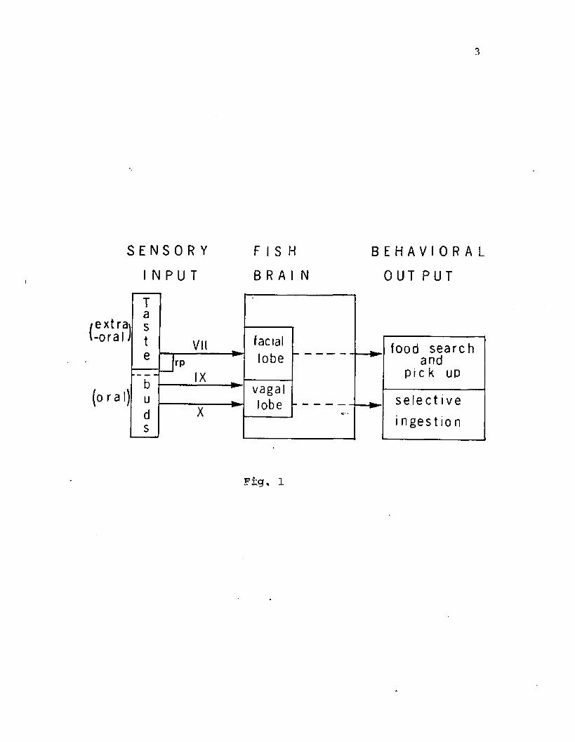
GENERAL INTRODUCTION .........................................I
recordings from facial (Caprio, '75, '78; Davenport and Caprio, '82),
glossopharyngeal and vagal (Kanwal and Caprio, '83) nerve branches
have shown that a few differences exist in the chemosensory inputs
from these two systems.
The glossopharyngeal-vagal (IX-X) complex has special
significance as it comprises the largest variety of functional fiber
types among the cranial nerves In vertebrates (Angevine and Cotman,
'81). Nerve trunks belonging to this complex transmit
exteroceptive-visceral information from regions of the oropharynx
(sensing environmental stimuli) and interoceptive-visceral Information
from organs in the abdominal cavity (Dart, *1922; Herrick, *1922). In
12
13
fish, the peripheral innervation of the glossopharyngeal nerve is
generally restricted to the anterior part of the oro-pharyngeal
region, whereas for the vagus nerve the field of innervation extends
from the pharynx to the visceral organs in the abdomen. Different
nerve branches of the IX-X complex, thus, carry different proportions
of general and special (taste) visceral sensory and motor fibers.
In the catfish, the IX-X taste system may have reciprocal
interactions with the VII (extra-oral) taste system for the
co-ordination of food search and ingestion. In order to elucidate the
neural substrate for the integration and coordination of
feeding-related behaviors it is essential to explore the pattern of
central projection of the primary sensory fibers belonging to the VII
as well as the IX-X cranial nerve complex. The peripheral innervation
and central distribution of the VII, IX and X cranial nerves in
catfish was described by Herrick ('01). Although experimental
confirmation of the central projections of the VII nerve was provided
recently (Finger, *76, 78; Morita et al., '80, '83), few reports
(Morita et al., '80; Morita and Finger, '85) exist on the experimental
determination of central projections of the IX and X nerves in fish.
Electrophysiological recordings from the glossopharyngeal nerve
in the catfish (Kanwal and Caprio, '83) confirms Herrick's (1901)
observation that this nerve innervates taste buds located on the gill
rakers of the first gill arch and in the anterior portion of the oral
cavity. Gross anatomical dissection also revealed that prior to its
entry into the gill arch, the glossophyarngeal nerve gives off a small
branch which rejoins the main trunk after making a short loop and
possibly sending some fibers to the mucosa on the roof of the oral
14
cavity (Fig. 1).
The vagal complex consists of several distinct nerve branches
with their ganglia grouped together and located outside the cranium,
adjacent to that of the glossopharyngeal nerve (Fig. 1). The vagal
nerve trunks, peripheral to the ganglia, are segregated
antero-posteriorly into branches innervating the second, third and
fourth gill arches and the corresponding portion of the floor of the
oral cavity. Separate branches also innervate other structures such
as the palatal organ in the oro-pharyngeal region. A distinct,
posterior branch of the vagus nerve complex turns caudally to
innervate visceral organs such as the stomach, heart and liver.
The purpose of this study was to examine the central pattern of
projection of individual branches of the IX and X nerves,
characterized on the basis of their peripheral distribution, in the channel catfish.
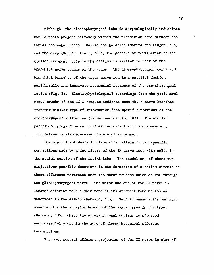
15
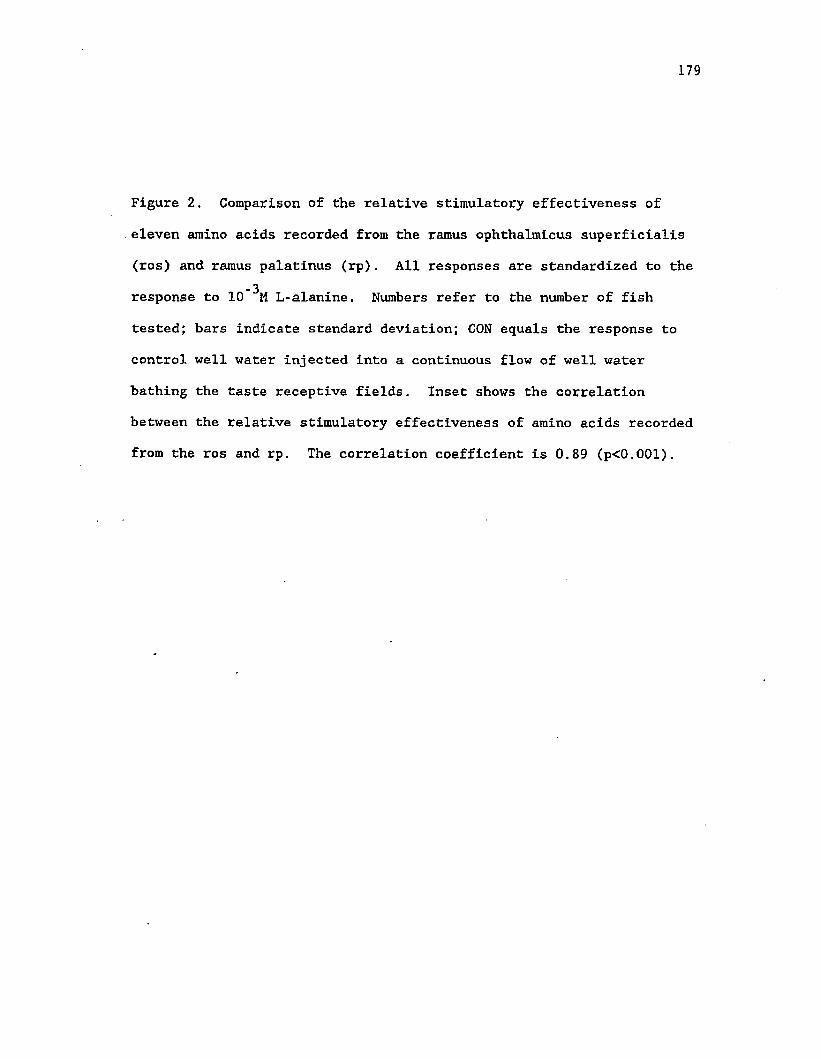
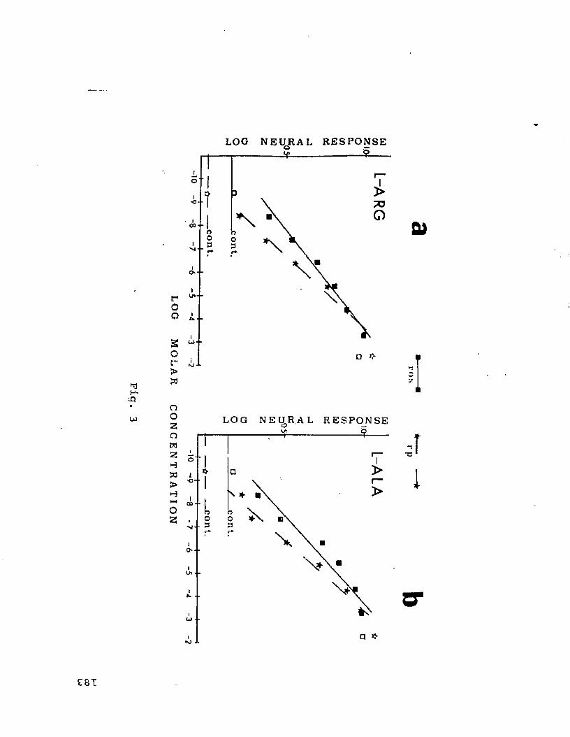
Figure 1. A diagrammatic saggital view of the peripheral pattern of
innervation of the glossopharyngeus (IX) and branchial branches and
interoceptive branch of the vagus (X) nerve in the oro-pharyngeal
region of the channel catfish.
glossopharyngeal
VN2-
GVN
■vagal nerve trunks -IVN
MATERIALS AND METHODS
Juvenile channel catfish, Ictalurus punctatus, 20-25 cm in
length, were obtained from a local fish farm and maintained in 15
gallon aquaria under a 12:12 h light-dark cycle. The fish were
anesthetized with tricaine methane sulfonate (MS-222) and clamped
horizontally in a fish holder prior to surgery. Water containing
MS-222 was perfused over the gills. The IX or X nerve branch was
dissected free of the surrounding tissue in the gill region,
transected and the central end sucked into a small section of PE-20 or
PE-90 tubing. Horseradish peroxidase (HRP) crystals (Sigma Type VI)
were placed next to the cut end of the nerve in the tubing after all
fluid inside was withdrawn with a cotton wick. The open end of the
tubing was sealed with Super Glue and the tube was glued to the
ventral surface of the cranium. This prevented displacement of the
cut nerve from within the tubing-as well as dilution and diffusion of
the HRP by tissue fluids. At the end of the operation vaseline was
applied to the region of the surgery, the wound sutured and the animal
returned to the tank.
The peripheral innervation of the labelled nerves was examined
from preserved specimens for purposes of nerve identification and
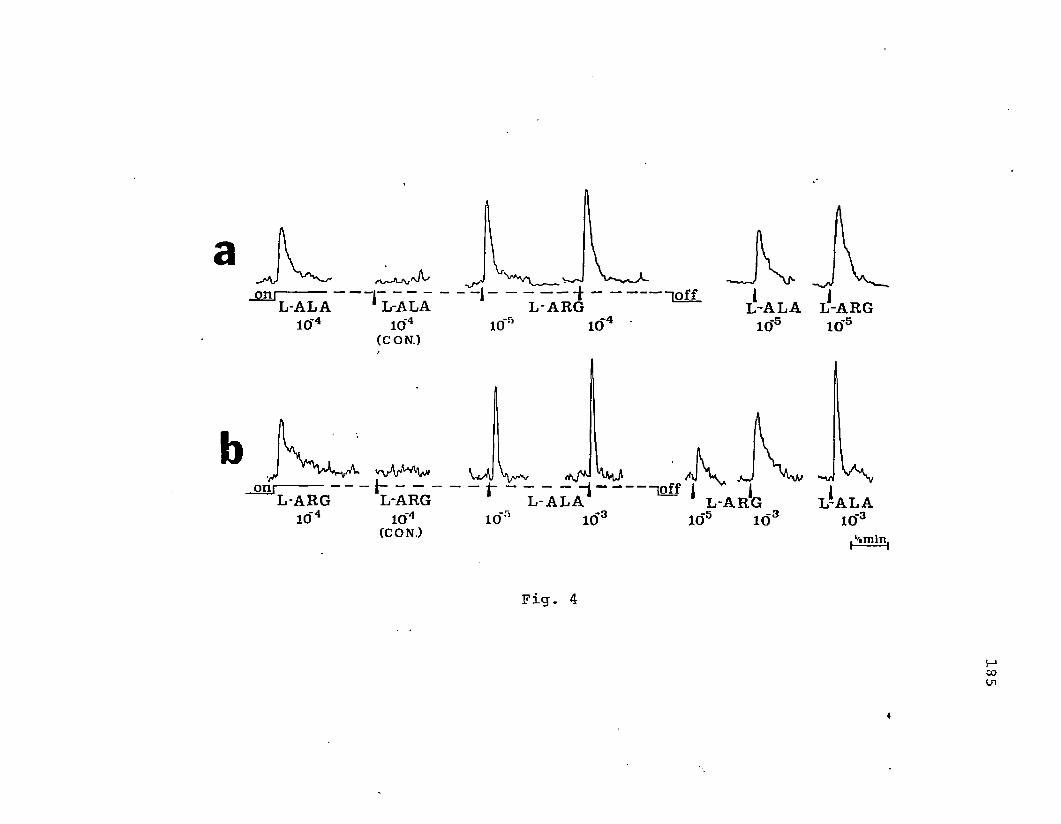
determination of the site of surgery and HRP application. The
specific nerves labelled were: I) the entire glossopharyngeal nerve
peripheral to its ganglion, ii) the anterior-most branch of the vagus
nerve innervating the second branchial cleft (VN2), Iii) the
17
18
posterior-most branch of the vagus nerve innervating the viscera
(IVN), iv) vagal branches located in the middle of VN2 and IVN and
identified as VN3, VN4 and VN5, according to their antero-posterior
sequence of innervation.
Following a survival period of 3 to 6 days, each animal was
reanesthetized with an overdose of MS-222 and perfused transcardially
with heparinized freshwater teleost Ringer's and a cold solution of 4%
glutaraldehyde in 0.1 M phosphate buffer (pH -7.2). After removal of
the fixed brain, the tissue was embedded in 20% gelatin or egg yolk
and fixed for an addition period of 4 to 6 hours in a cold solution of
4% buffered glutaraldehyde saturated with sucrose. The tissue was
sectioned either transversly or horizontally at 35 vim on a freezing
microtome. Sections were collected in 0.1 M phosphate buffer, reacted
with Hanker-Yates reagent (Bell et. al., '81) and mounted as two
alternating series onto chrome-alum-coated (subbed) slides. The
perfusion, cutting, reacting and mounting were generally performed
within a period of 3 to 5 days. The mounted and dried sections were
stained with thionin, dehydrated, cleared in xylene and mounted with
Permount.
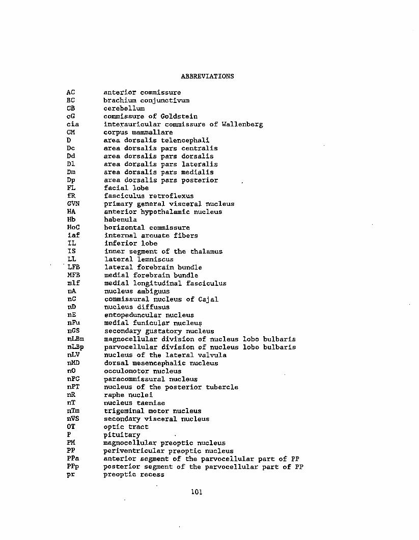
ABBREVIATIONS
BC brachium conjunctivumCB cerebellumcia interauricular commissure of WallenbergDC dorsal cap of the vagal lobedlf dorso-lateral fascicle of the vagusdtV descending tract of the trigeminal nerveFL facial lobeGL gustatory lemniscusGLN glossopharyngeal nerveGVN primary general visceral nucleushf horizontal fascicle of the vagusiaf internal arcuate fibersil intermediate lobule of the facial lobeIVN interoceptive branch of the vagus11 lateral lobule of the facial lobeml medial lobule of the facial lobemlf medial longitudinal fasciculusnA nucleus ambiguusnC nucleus commissuralis of CajalnD nucleus diffususnFm motor nucleus of the facial nervenFu medial funicular nucleusnGS secondary gustatory nucleusnIF nucleus intermedius of the facial lobenIV nucleus intermedius of the vagal lobenR raphe nucleinTm trigeminal motor nucleusnV vestibular nucleusOT optic tractRF reticular formationS spinal cordT telencephalonTeO optic tectumtSG(2G) secondary gustatory tractv ventriclevf ventral fasciculusVL vagal lobeVMC vagal motor columnVN vagal nerveVN2 vagal nerve branch innervating second branchial cleftVSC vagal sensory column
19
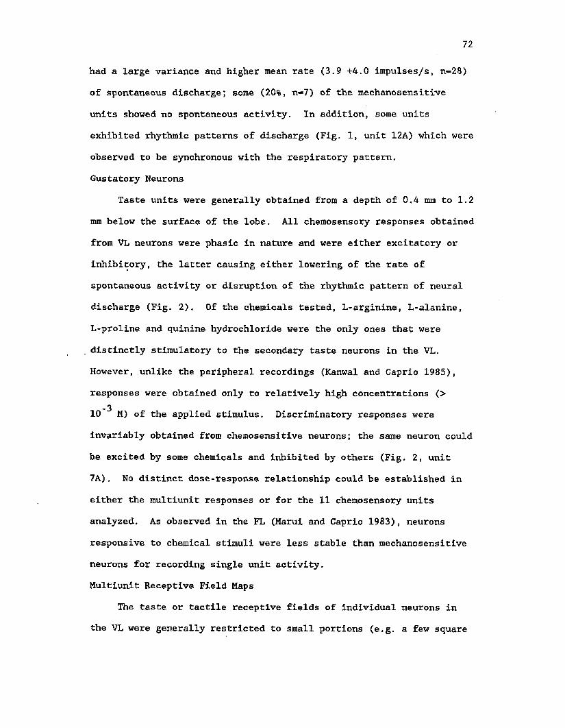
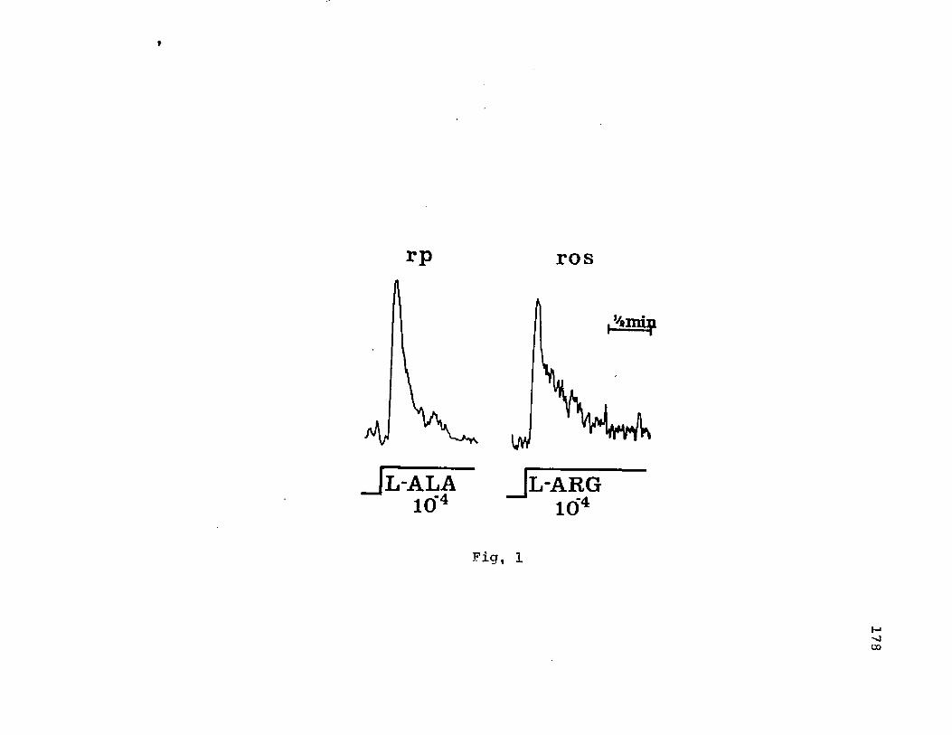
RESULTS,
Central organization of glossophyaryngeal afferents
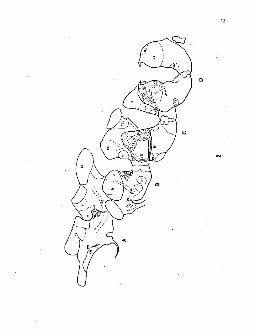
Afferent fibers of the glossopharyngeal nerve enter the
brainstem together with the vagal nerve complex. After traversing
rostro-dorsally along the lateral aspect of the vagal lobe, the fibers
split into two rootlets, a dorsolateral fascicle and a horizontal
fascicle (Fig. 2). The dorsolateral fascicle curves along the dorsal
surface of the vagal lobe, ventral to the dorsal cap, and terminates
heavily in the dorso-lateral portion of the vagal lobe, lateral to the
secondary gustatory tract. The horizontal fascicle proceeds medially
coursing through the bundles of the secondary gustatory tract. After
reaching the medial border of the vagal lobe these fibers diverge
dorsally and split into two components. Afferents of one component
continue posteriorly for a few hundred micra before terminating along
the medial edge of the vagal lobe, in the region of the nucleus
intermedius (nIV) of Herrick ('05). Other afferents turn anteriorly
and then extend laterally into the vagal lobe proper and intermingle
with the dorsal rootlet fibers before terminating. Most of the
glossopharyngeal afferents terminate diffusely in the anterior portion
of the vagal lobe, where it constricts before merging with the facial
lobe. In addition, a small root continues rostrally and eventually
splits into two fascicles, each fascile making a caudo- ventromedial
turn before terminating in small, separate areas in the nIF. Both
terminal fields are located along the ventral border of the fourth
ventricle. The most rostral branch turns ventrally, while the other
21
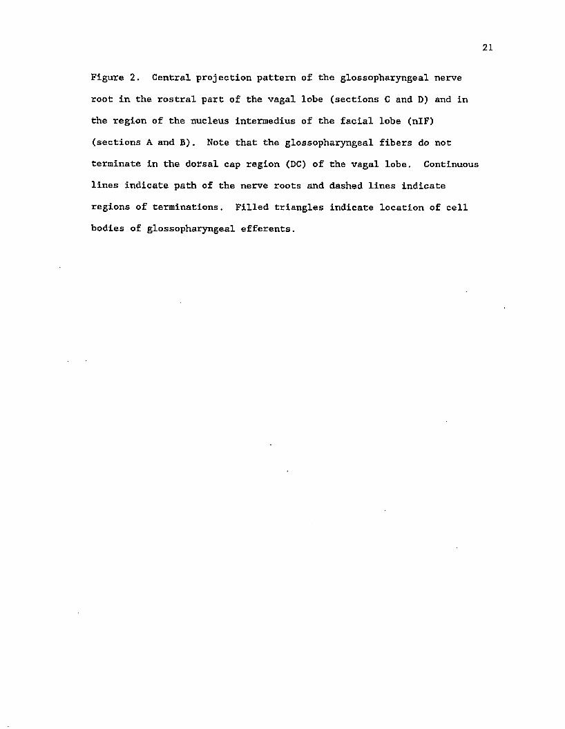
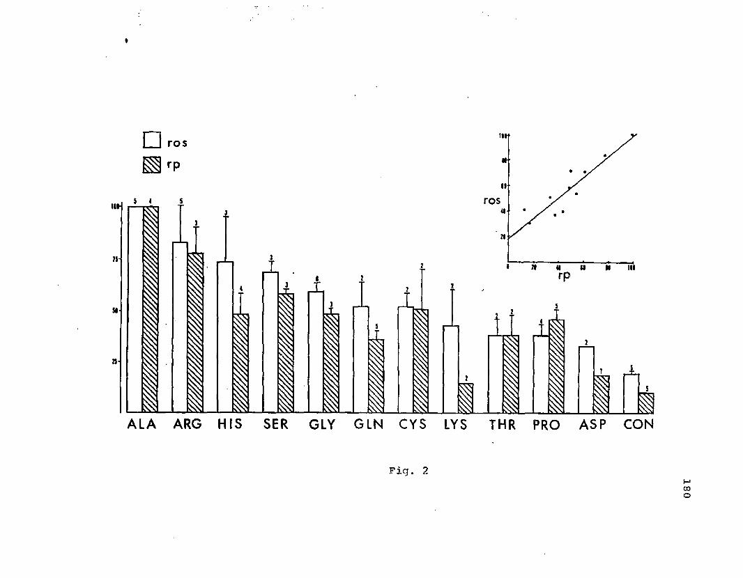
Figure 2. Central projection pattern of the glossopharyngeal nerve
root in the rostral part of the vagal lobe (sections C and D) and in
the region of the nucleus intermedius of the facial lobe (nIF)
(sections A and B). Note that the glossopharyngeal fibers do not
terminate in the dorsal cap region (DC) of the vagal lobe. Continuous
lines indicate path of the nerve roots and dashed lines indicate
regions of terminations. Filled triangles indicate location of cell
bodies of glossopharyngeal efferents.
22
23
turns dorsally prior to termination (Fig. 3a,b).
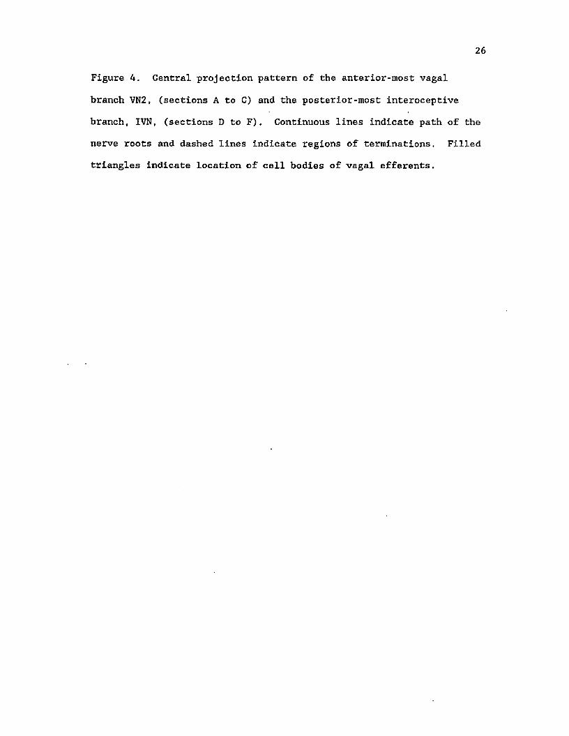
Central organization of vagal afferents. I. exteroceptive-branchial
roots
Vagal afferents can be grossly split into two parts: (i)
exteroceptive-branchial afferents, which innervate the gill arches and
posterior portions of the oral cavity and transmit sensory information
from the water flowing through the oro-pharyngeal region, and (ii) the
descending interoceptive-visceral branch. The exteroceptive-branchial
roots exhibit a pattern of termination in the vagal lobe similar to
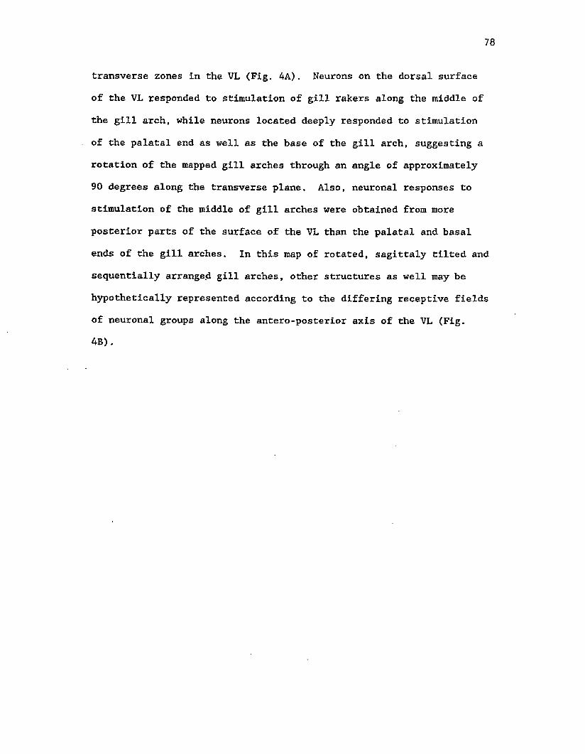
that described for the glossopharyngeal nerve (Fig. 4, sections A, B
and C). Thin fibers ascend obliquely and course in a rostro-dorsal
direction towards the area of termination of the dorsal fibers of the
IX nerve. The thicker fibers also ascend for a short distance, turn
medially over the spinal V tract and descend to the nIV near the
lateral wall of the fourth ventricle. A small fascicle of the dorsal
rootlet of the anterior branch of the vagus nerve continues dorsally
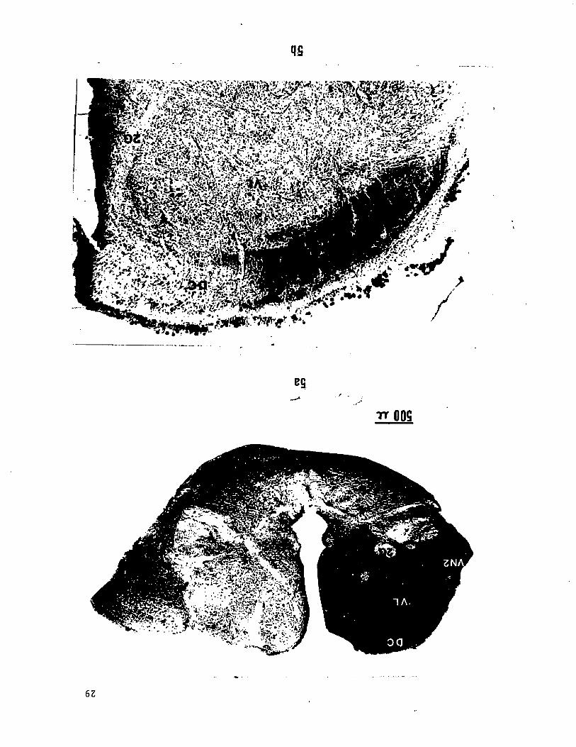
over the lobe and terminates along the medial half of the dorsal cap
of the vagal lobe (Fig. 5a). The dorsal cap is a dorsolateral nuclear
region which can be distinguished easily as a lamina separated from
the rest of the vagal lobe by a thin capsule of fiber fascicles. The
most posterior branchial root terminates extensively throughout the
caudal two-thirds of the vagal lobe even though the root enters the
lobe at its most caudal region. In addition, a small fascicle
continues for some distance in a rostro-dorsal direction and finally
terminates in the lateral half of the dorsal cap region (Fig. 5b).
24
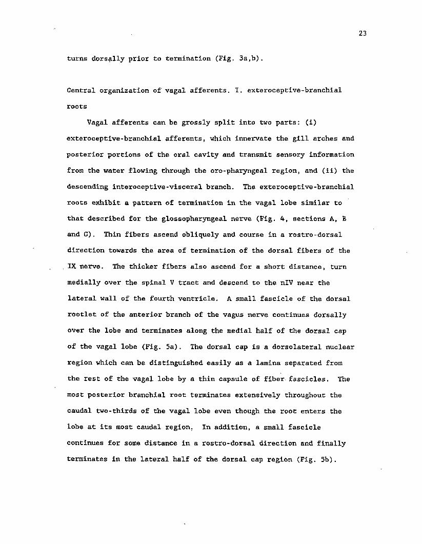
Figure 3. (a) Photomicrograph of a section caudal to figure 2B
showing the medial projection and caudal terminal zone of HRP labelled
glossopharyngeal fibers in the region of the nucleus intermedius of
the facial lobe (nIF). (b) Photomicrograph of a section caudal to
figure 2A showing HRP labelled fibers of the glossophyaryngeal nerves
coursing towards the region of the nIF in the facial lobe. It is not
known if the nIF proper ascends up to this level in the brainstem.
25
3b
Figure 4. Central projection pattern of the anterior-most vagal
branch VN2, (sections A to C) and the posterior-most interoceptive
branch, IVN, (sections D to F). Continuous lines indicate path of the
nerve roots and dashed lines indicate regions of terminations. Filled
triangles indicate location of cell bodies of vagal efferents.
27
u.
<
28
Figure 5. Photomicrographs of terminations in the dorsal cap region
(DC) of the vagal lobe of the branchial branches VN3 (a) and VN5 (b)
of the vagus labelled with HRP. The sections, taken from different
animals, represent approximately the same antero-posterior level in
the vagal lobe. The lateral portion of the dorsal cap labelled in (b)
extends towards the caudal end of the vagal lobe.
qg
fSfyX !&$Kh&&t''k?Xtf-.£ .■ > * * - S ’. ? ; r i - • . .? ^ j
B&.> " ■ _,TT QOS
62
*U
30
Central organization of vagal afferents. II. interoceptive-visceral
roots
The general visceral fibers innervating the viscera constitute a
unique pattern of central projection and termination. This root of
the vagus contains only general visceral sensory fibers innervating
the visceral organs and is thus referred to as the
interoceptive-visceral branch in order to make a clear distinction
from the exteroceptive-branchial branches of the vagus which contain
special visceral (taste) as well as general visceral and somatic
(tactile, proprioceptive, etc.) fibers (Herrick, '01, '06; Kanwal and
Caprio, 83). Unlike the branchial roots, this root does not split
into a dorsal and horizontal rootlet (Fig. 4 sections D, E and F).
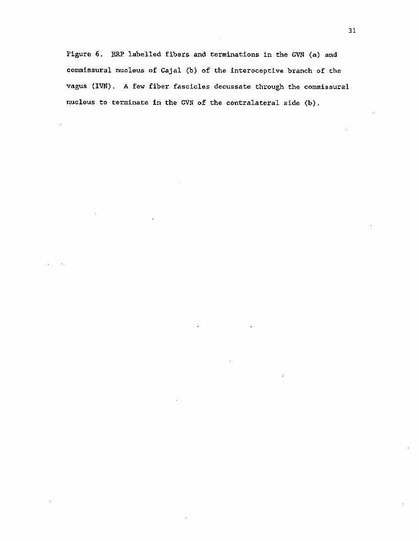
Instead, the entire root terminates just caudal to its point of entry
in the general visceral nucleus (GVN) with a few fibers continuing
rostrally into the intermediate vagal nucleus. The majority of fibers
terminate densely in the GVN as well as in the commissural nucleus of
Cajal. A few fibers cross through the commissure as several fascicles
and continue in a rostro-dorsal direction before terminating in the
GVN of the contralateral side. No terminations in the vagal lobe
proper of the ipsi- or contralateral side were observed for this vagal
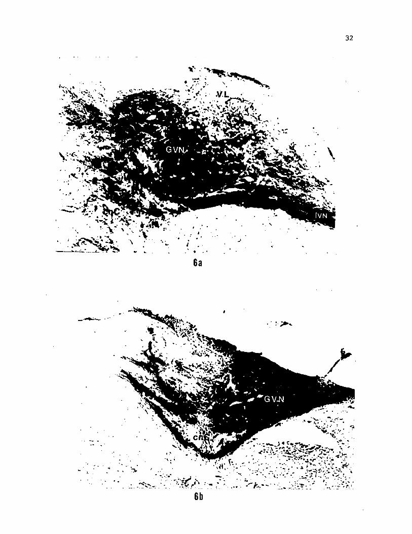
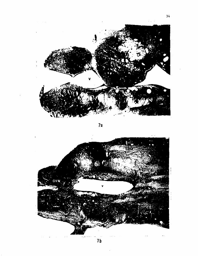
root (Fig. 6). The segmental pattern of projection of different vagal
roots and differences in the termination fields of the most anterior
and most posterior branches of the vagal roots are best seen in
photomicrographs of horizontal sections of the brainstem (Fig. 7a,b).
Central organization of glossopharyngeal and vagal efferents
All of the IX-X efferent roots originate from cell bodies located
31
Figure 6. HRP labelled fibers and terminations in the GVN (a) and
commissural nucleus of Cajal (b) of the interoceptive branch of the
vagus (IVN). A few fiber fascicles decussate through the commissural
nucleus to terminate in the GVN of the contralateral side (b).
32
6a
Figure 7. Photomicrographs of horizontal sections of the brainstem of
the catfish in which two vagal nerve branches, VN2 and IVN, were
simultaneously labelled with HRP. Only terminations of VN2 are seen
in a section through the vagal lobe proper (a). At levels ventral to
the vagal lobe (b), terminations of IVN in the GVN and portions of the
horizontal and dorsolateral fiber fascicles of VN2 are visible.
34
7b
35
in a continuous longitudinal column, bordering the fourth ventricle,
along the ventromedial portion of the medulla (Fig. 8). These motor
roots then travel caudo-laterally and join their respective afferent
roots before emerging from the cranium (Fig. 2, 4). The IX-X motor
column is broadened dorso-ventrally towards the caudal end of the
vagal lobe and tapers to a ventral location before terminating in the
region of the obex. As in Siluris (Berkelbach van der Sprenkel, '15;
Black, '17), this column is discontinuous with the facial motor
nucleus and terminates approximately 150 um before the appearance of
the VII motor nucleus rostrally. The cell bodies of glossopharyngeal
efferents are located only at the rostral extremity of the cell column
at the level of the caudal portion of the facial lobes (Fig. 8).
These cells are ovoid to conical in shape with the long axis directed
in a ventrolateral plane. The axons of these neurons proceed caudally
along the ventromedial margin of the ventricles before turning
dorso-laterally. These fibers travel through the nIF and loop around
the dorsal aspect of the spinal V tract before turning caudally to
exit the brain along with the afferent fibers (Fig. 2). This rootlet
then makes a sharp caudal turn and joins the main glossopharyngeal
root.
The most anterior branch of the vagus, which innervates the
second gill arch, has its cell bodies located in the anterior portion
of the vagal lobe at the level of termination of the glossopharyngeal
afferents (Fig, 8). The cell bodies are morphologically similar to
those of the glossopharyngeal efferents. As observed previously
(Herrick, '01; Morita and Finger, '85) the dendrites of these cells
extend well into the lateral portion of the reticular formation and
36
Figure 8. Diagramatic scheme of the visceral sensory (VSN) and vagal
motor column (VMN) projected onto a saggital plane of the medulla
(facial and vagal lobes). Arrows to the side-face of the sensory
column indicate the primary projection zone of the sensory fibers
while arrows to the front-face indicate the rostrocaudal extent of
fiber terminations for each nerve branch. The location of cell bodies
(CB) of motor neurons and the region of exit of efferent fibers (F)
from the medulla are indicated separately for each nerve labelled.
Both columns (VMC and VSC) are represented on the same scale and axis.
Numbers along the midline indicate the caudal distance in mm from the
rostral end of the vagal lobe. Motor neurons of the glossopharyngeal
nerve are located anterior to the vagal lobe. The
interoceptive-visceral branch of the vagus (IVN) projects to the
general visceral nucleus located caudal to the vagal lobe.
37
CENT RA L S EN SO RY
M O T O R F I BERS P R O J E C T IO N Z O N E S N.ERVE TRUNK
V M C FLGLN
' vsc <VL) GLi
VN 2VN2—
•VN3CBVL--- t.o
V N 4
I V N —
CB- ■VN4
IVN
GVN
8
38
the axon originates from the base of the dendrites. The next
branchial branch labelled (VN2), which innervates primarily the
palatal organ, is nearly devoid of efferents (Fig. 8). Only one cell
body was observed and it was located rostral to the level of its
afferent terminations. While the cell bodies of most branchial
branches appear to be arranged in a segmental fashion along the long
axis of the visceral motor column (Fig. 8), those of the most
posterior branch are distributed throughout the posterior two-thirds
of the motor column (Fig. 9a). These cell bodies are more rounded and
arranged more loosely than the motor neurons with efferents in the
branchial braches of the vagus (Fig, 9b). Also, dendrites belonging
to motor neurons of the IVN do not project laterally into the adjacent
reticular formation. In contrast to a restriction of the cell bodies
of efferents of the branchial branches of the vagus (VN2, VN3 and VN4)
to the anterior part of the vagal motor nucleus, those of the IVN form
the caudal extremity of the IX-X visceral motor column (Fig. 8).
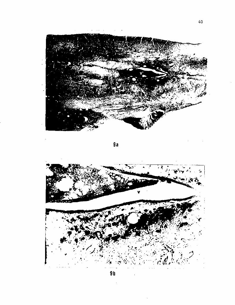
Figure 9. Photomicrographs showing the location (a) and cell
morphology (b) of parasympathetic neurons of the interoceptive branch
of the vagus in the caudal region of the medulla. The rostrally
extended distribution and circular shape of these neurons make them
distinct from the triangular, segmental arrangement of most other
motor neurons of the nucleus ambiguus whose efferent fibers project
peripherally in the branchial branches of the vagus.
40
9b
DISCUSSION
Several studies (Bardach et al., '67; Atema, '71; Johnsen and
Teeter, '80) have confirmed the role of gustation in the feeding
behavior of the catfish since Herrick first put forward his hypotheses
(Herrick, *04, '05). In keeping with Herrick's approach, the present
results are interpreted from a functional view point and delineate
further the neural substrate involved in feeding. A comparison of our
results with other anatomical studies in fishes as well as land
vertebrates also provides an evolutionary perspective on the pattern
of central projections of the IX and X nerves. The pattern of
projection of the IX-X complex in the brainstem of the channel
catfish, Ictalurus punctatus. generally conforms to Herrick's ('05)
description in the bullhead catfish, Ictalurus nebulosus. HRP
labelling of fibers and cell bodies, however, reveals some new and
important aspects of neural organization.
Phylogenetic Comparisons
Afferent and Efferent Roots of the Glossopharyngeus
The central organization of the visceral afferent and efferent
areas has been described in several groups of vertebrates. Among
mammals, the glossopharyngeal visceral afferents in the cat (Torvik,
'56; Kerr, '62; Kalia and Mesulam, '80), and the rat (Kalia and
Sullivan, '82; Hamilton and Norgren, '84) terminate extensively in the
nucleus of the solitary tract and extend from the caudomedial border
of the terminal zone of chorda tympani afferents to the region of the
41
42
obex. In Ranid frogs, the termination zone of the glossopharyngeal
nerve is quite similar to that of mammals (Steusse et al., 84);
however, a small contralateral projection of the IX nerve via the
commissural nucleus exists in some mammals (Kalia and Mesulam, '80;
Kalia and Sullivan, '82), whereas in amphibians (Matesz and Szekely,
'78; Hanamori and Ishiko, '83; Steusse et al., '84) this projection is
reported to he entirely ipsilateral. Among teleostean species, the
brainstem region corresponding to the nucleus of the solitary tract is
highly variable in its morphology. In general, this continuous column
can be divided morphologically into the facial, glossopharyngeal and
vagal lobes. In the ictalurid catfishes the glossopharyngeal lobe is
reduced and may not be visible from the surface. The present study,
however, confirms earlier reports (Herrick, ’05; Morita et al., '80,
83; Morita and Finger, '85) of the presence of glossopharyngeal
terminations in the transition zone of the facial and vagal lobes.
The restricted antero-posterior location (rostral part of the vagal
lobe and caudomedial region in the facial lobe) of this zone is unlike
the caudal medullary location of-glossopharyngeal terminations in
mammals (Kalia and Mesulam, '80; Kalia and Sullivan, '83; Hamilton and
Norgren, '84) and amphibians (Hanamori and Ishiko, '83; Steusse et
al., '84). Nevertheless, with respect to laterality,.the pattern is
like that observed in IL pipiens and esculenta (Steusse et al.,
'84). As in most vertebrate species studied, there is also some
overlap with the region of termination of vagal afferents (Fig. 8).
The general pattern of projection of the glossopharyngeal nerve
in the channel catfish is similar to previous descriptions in the
bullhead catfish (Herrick, '01; Morita and Finger, '85) and the carp
(Morita et al., '80). An important additional observation included in
this study relates to the distinct rostral projections of the
glossophyarngeal root seen after labelling the nerve with HRP. The
functional significance of these projections is discussed later. It
.is interesting that a similar rostral course of the glossopharyngeal
root was recently reported for the Ranid frogs (Steusse et al., '84).
A detailed investigation of the glossopharyngeal nerve was not
performed in the single experimental study on a fish (Morita et al.,
80). Moreover, previous reports are based on either staining or
degeneration techniques both of which are relatively insensitive and
less reliable than the HRP technique. In any case, it is hard to
generalize on the basis of observations in one species because of the
great variability of neuronal organization among teleosts.
The location of the glossopharyngeal motor nucleus in ictalurid
catfish is quite similar to that of other vertebrates studied. This
nucleus forms the rostral extremity of the ventromedial part of the
visceral motor column. The circuitous path taken by the motor root of
the IX is consistent with previous reports and is apparently a
characteristic feature of this nerve in all teleosts (Barnard, '35).
Afferent and Efferent Roots of the Vagus
The present results Indicate that exteroceptive-branchial and
interoceptive-visceral vagal roots exhibit two distinct patterns of
projection. The roots of all the exteroceptive-branchial branches of
the vagus contain general (tactile, proprioceptive, etc.) as well as
special (taste) visceral afferents (Herrick, '01, 06). These two
categories of fibers may separate centrally according to the observed
splitting of each root into a dorsolateral and ventral (horizontal)
m
44
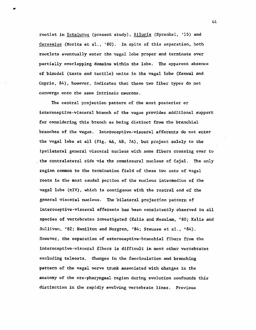
rootlet In Ictalurus (present study), Siluris (Sprenkel, '15) and
Carassius (Morita et al,, '80). In spite of this separation, both
rootlets eventually enter the vagal lobe proper and terminate over
partially overlapping domains within the lobe. The apparent absence
of bimodal (taste and tactile) units in the vagal lobe (Kanwal and
Caprio, 84), however, indicates that these two fiber types do not
converge onto the same intrinsic neurons.
The central projection pattern of the most posterior or
interoceptive-visceral branch of the vagus provides additional support
for considering this branch as being distinct from the branchial
branches of the vagus. Interoceptive-visceral afferents do not enter
the vagal lobe at all (Fig. 4A, 4B, 7A), but project solely to the
ipsilateral general visceral nucleus with some fibers crossing over to
the contralateral side via the commissural nucleus of Cajal. The only
region common to the termination field of these two sets of vagal
roots is the most caudal portion of the nucleus interraedius of the
vagal lobe (nlV), which is contiguous with the rostral end of the
general visceral nucleus. The bilateral projection pattern of
interoceptive-visceral afferents has been consistently observed in all
species of vertebrates investigated (Kalia and Mesulam, '80; Kalia and
Sullivan, '82; Hamilton and Norgren, '84; Steusse et al., '84).
However, the separation of exteroceptive-branchial fibers from the
interoceptive-visceral fibers is difficult in most other vertebrates
excluding teleosts. Changes in the fasciculation and branching
pattern of the vagal nerve trunk associated with changes in the
anatomy of the oro-pharyngeal region during evolution confounds this
distinction in the rapidly evolving vertebrate lines. Previous
studies on Ictalurus nebulosus (Herrick, '01, '05, '06) and Siluris
glanis (Sprenkel, '15) also report the presence of a general cutaneous
component (somatic afferents) in the vagal roots, which after
separating centrally, descends and terminates within the spinal V
nucleus. No such fibers were evident in the channel catfish although
they may be present in the few caudal branchial branches not labeled
in the present study.
Gross morphological evidence suggests that the posterior lateral
line nerve in fish, traditionally regarded as a branch of the vagus,
is a separate, phylogenetically primitive cranial nerve which has
disappeared with the advent of land vertebrates (Finger, '83). This
suggestion is supported by the uniqueness of its embryogenesis,
peripheral innervation, central projections and the nature of sensory
information transmitted centrally. For similar reasons, it may be
appropriate to regard the exteroceptive-branchial branches as forming
a separate cranial nerve trunk, distinct from the
interoceptive-visceral branch of the vagus. Such a clear separation
is not evident in the mammalian system because of the intermixing of
pharyngeal, laryngeal and visceral branches of the vagus In the course
of their peripheral and central paths. Previous studies have failed,
therefore, to delineate a functional organization in the nucleus
tractus solitarius (NTS) of mammals because of an apparent
intermingling of special (taste) and general visceral fibers in the
course of their terminations in this compact nucleus (Torvik, '56;
Kalia and Mesulam, *80; Kalia and Sullivan, '82), Nevertheless, the
single detailed study in the rat showed a minimal overlap between
terminals of the gustatory nerves and those of the cervical branch of
46
the vagus in the NTS (Hamilton and Norgren, '83).
The visceral motor column has been of considerable interest
classically as a model for the study of neurobiotaxis (Black, '17) as
well as recently with respect to the relationship of cellular topology
and architectonics with region and organ-specific representation in
mammals (Lawn, '66) and birds (Katz and Karten, '83, '85). Although
similar contemporary studies in fishes are lacking, the present
results do indicate a clear difference between the motor neuron
distribution in the root of the most caudal branchial branch and the
interoceptive-visceral branch of the vagus. The branchial motor
neurons of the most caudal root are distributed throughout the nucleus
ambiguus, rostral to its entry into the vagal lobe, whereas the
interoceptive-visceral neurons are restricted to a compact region at
the caudal end of the visceral motor column. In amniotes, two
populations of vagal motor neurons, the dorsal motor nucleus and the
nucleus ambiguus, are consistently observed (Brodal,'83). In
amphibia, the main portion of the vagal motor nucleus has been
homologized with the nucleus ambiguus of mammals (Matesz and Szekely,
'78). In fish, this distinction is not sufficiently clear. However,
the differing patterns of distribution of efferents in the various
roots may be evidence for the origin of the two motor, nuclei from a
single phylogenetically primitive nucleus containing an intermingled
population of two categories of neurons. The absence of direct
terminations of primary afferents onto vagal motor neurons is
consistent with previous observations in catfish (Herrick, '06;Barnard, '35).
47
Neuroethological Interpretations
The catfish is able to search for and localize a food source
primarily by means of its gustatory sense (Bardach et al., '67; Atema,
'71; Johnsen and Teeter, '80). This specialized ability for
monitoring the chemical stimuli in Its environment is correlated with
the relative enlargement of the facial lobe (Herrick, '05; 06; Atema,
'71). Once the food is pursued and captured, further assortment,
manipulation or selection Is generally unimportant and uneconomical
for an active predator such as the channel catfish. This is reflected
in the structure of the central nervous system by the small size of
the vagal lobe relative to the facial lobe while the glossopharyngeal
lobe is morphologically inconspicuous. In contrast, in the goldfish
which selects food from non-food after biting, the glossopharyngeal
lobe Is conspicuous and the vagal lobe is a large, highly derived
structure.
As described by Herrick ('05, '06), the glossopharyngeal and
vagal lobes do not show any kind of lobular or laminar organization
seen in the facial lobe of the catfish or the vagal lobe of the
goldfish, respectively. Lack of such a distinctive organization, in
the context of information theory (Campbell, '82), indicates that the
entropy of the system is high and feature extraction from the spatial
domain is probably not a prominent feature of neural processing in the
vagal lobe of the catfish (Kanwal and Caprio, chapter 2). Lack of
manipulation of food in the oral cavity during feeding may be taken as
support of this hypothesis. However, electrophysiological mapping of
receptive fields of individual intrinsic neurons of the vagal lobe can
provide important information to test this hypothesis.
48
Although, the glossopharyngeal lobe is morphologically indistinct
the IX roots project diffusely within the transition zone between the
facial and vagal lobes. Unlike the goldfish (Morita and Finger, *85)
and the carp (Morita et al., '80), the pattern of termination of the
glossopharyngeal roots in the catfish is similar to that of the
branchial nerve trunks of the vagus. The glossopharyngeal nerve and
branchial branches of the vagus nerve run in a parallel fashion
peripherally and innervate sequential segments of the oro-pharyngeal
region (Fig. 1). Electrophysiological recordings from the peripheral
nerve trunks of the IX-X complex indicate that these nerve branches
transmit similar type of information from specific portions of the
oro-pharyngeal epithelium (Kanwal and Gaprio, '83). The similar
pattern of projection may further indicate that the chemosensory
information is also processed in a similar manner.
One significant deviation from this pattern is two specific
connections made by a few fibers of the IX nerve root with cells in
the medial portion of the facial lobe. The caudal one of these two
projections possibly functions in the formation of a reflex circuit as
these afferents terminate near the motor neurons which course through
the glossopharyngeal nerve. The motor nucleus of the IX nerve is
located anterior to the main zone of its afferent termination as
described in the salmon (Barnard, '35). Such a connectivity was also
observed for the anterior branch of the vagus nerve in the trout
(Barnard, '35), where the efferent vagal nucleus is situated
ventro-medially within the-zone of glossopharyngeal afferent
terminations.
The most rostral afferent projection of the IX nerve is also of
special interest from a neuroethological perspective, because it may
constitute the neural substrate for mixing information in the central
nervous system. Gustatory information from the oral taste buds
converges onto neurons in the region of the nucleus intermedius of the
facial lobe (nlF) which also receives input from extra-oral taste buds
via the facial afferents. Electrophysiological mapping of the facial
lobe previously showed that neurons in this region have large tactile
receptive fields which extend from the oral to the extra-oral surface
(Marui and Caprio, '82). Some of these neurons are bimodal in
character and respond to oral chemical as well as tactile stimulation
(personal observation). Herrick regarded the nlF as a correlation
center (Herrick, '06). The present results indicate that a portion of
nlF may integrate extra-oral gustatory information relating to food
search and the consequent oral stimulation leading to food ingestion
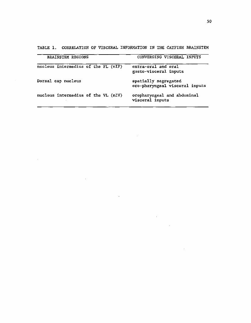
or rejection (Table 1).
The dorsal cap, also identified previously in the bullhead
catfish (Finger, '81), is another specific region of the vagal lobe
whose function has not been described adequately. The present results -
indicate that the dorsal cap may relate information from the anterior
and posterior portions of the oro-pharynx. Intrinsic neurons in this
region may therefore, have relatively large or dual receptive fields.
Also, - small HRP injections restricted to the dorsal cap region may
reveal a difference in its neuronal connectivity as compared to the
other parts of the vagal lobe.
Finally, the descending branch of the vagus or the
interoceptive-visceral branch is non-gustatory in function and
anatomically distinct from the exteroceptive-branchial branches of the
50
TABLE 1. CORRELATION OF VISCERAL INFORMATION IN THE CATFISH BRAINSTEM
BRAINSTEM REGIONS CONVERGING VISCERAL INPUTS
nucleus intermedius of the FL (nlF) extra-oral and oralgusto-visceral inputs
Dorsal cap nucleus spatially segregatedoro-pharyngeal visceral inputs
nucleus intermedius of the VL (nIV) oropharyngeal and abdominalvisceral inputs
vagus (Fig. 1, 4). The information provided by this branch is not
directly involved in feeding and is apparently processed differently
as it does not converge onto the secondary gustatory neurons.
Instead, it terminates in the general visceral nucleus (GVN) of the
ipsi- and contra-lateral side as well as in the commissural nucleus of
Cajal. Anatomically, the GVN is adjacent to the caudal end of the
nucleus intermedius of the vagal lobe (nIV) and has a terminal field
extending into this nucleus. The nIV also receives fibers from
branchial branches of the vagus and may thus constitute another
correlation center (Herrick, '05; Kanwal and Caprio, '84) which
integrates gustatory input determining feeding with
interoceptive-visceral input relating to the physiological state of
the animal.
Oro-pharyngeal sensory Input in mammals is also known to evoke a
variety of vagal-dependent physiological (Kuwahara, '83) and hormonal
(Brand et al., '76) responses including some relating to inititiation
of food Ingestion. Regulation of short term (Gonzalez and Deutsch,
'81; Lorenz and Goldman, '82; Alino et al., '83) and long term (Sharma
and Nasset, '62; Ch.inna and Bajaj , '72; Li and Anderson, '84) food
ingestion is further accomplished by the central influence of the
interoceptive-visceral sensory input via the descending branches of
the vagus. Some of these functions may be modulated by central
connections of neurons in the nIV.
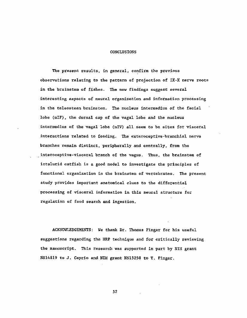
CONCLUSIONS
The present results, in general, confirm the previous
observations relating to the pattern of projection of IX-X nerve roots
in the brainstem of fishes. The new findings suggest several
interesting aspects of neural organization and information processing
in the teleostean brainstem. The nucleus intermedius of the facial
lobe (nlF), the dorsal cap of the vagal lobe and the nucleus
intermedius of the vagal lobe (nIV) all seem to be sites for visceral
interactions related to feeding. The exteroceptive-branchial nerve
branches remain distinct, peripherally and centrally, from the
interoceptive-visceral branch of the vagus. Thus, the brainstem of
ictalurid catfish is a good model to investigate the principles of
functional organization in the brainstem of vertebrates. The present
study provides important anatomical clues to the differential
processing of visceral information in this neural structure for
regulation of food search and ingestion.
ACKNOWLEDGEMENTS: We thank Dr. Thomas Finger for his useful
suggestions regarding the HRP technique and for critically reviewing
the manuscript. This research was supported in part by NIH grant
NS14819 to J. Caprio and NIH grant NS15258 to T. Finger.
52
REFERENCES
Alino, S.F., D. Garcia, and K. Uvnas-Moberg (1983) On the interaction
between intragastrie pH and electrical vagal stimulation in causing
gastric acid secretion and intraluminal release of gastrin and
somatostatin in anesthetized rats. Acta Physiol. Scand.
117:491-495.
Angevine, J.B., and Cotman C.W. (1981) Principles of Neuroanatomy.
Oxford University Press, New York. p.22.
Ariens Kappers, C.U. (1906) The structure of the teleostean and
selachian brain. J. Comp. Neurol, and Psychol. 16:1-113.
Atema, J. (1971) Stuctures and functions of the sense of taste
in the catfish (Ictalurus natalis). Brain Behav. and Evol.
4:273-294.
Bardach, J.E., J.H, Todd, and R. Crickmer (1967) Orientation by taste
in fish of the genus Ictalurus. Science 155:1276-1278.
Barnard, J.W. (1935) A phylogenetic study of the visceral afferent
areas associated with the facial, glossopharyngeal, and vagus
nerves, and their fiber connections. The effferent facial nucleus. J. Comp. Neurol. 65:503-602.
53
Bell, C.C., T.E. Finger, and C. Russel (1981) Central connections of
the posterior lateral line lobe In mormyrid fish. Exp. Brain Res.
42:9-22.
Berkelbach van der Sprenkel, H. (1915) The central relations of the
cranial nerves in Siluris glanis and Mormyrus caschive. J. Comp.
Neurol. 25:5-63.
Black, D, (1917) The motor nuclei of the cerebral nerves in
phylogeny: A study of the phenomena of neurobiotaxis. J. Comp.
Neurol. 27:467-558.
.Brand, J.G., R.H. Cagan, and M, Naim (1982) Chemical senses in the
release of gastric and pancreatic secretions. Ann, Rev. Nutr.
2:249-276.
Brodal, A. (1981) Neurological anatomy in relation to clinical
medicine. Oxford University Press, New York.
Campbell, J. (1982) Grammatical man. Simon & Schuster, New York.
Caprio, J. (1975) High sensitivity of catfish taste receptors
to amino acids. Comp. Biochem. Physiol. 52A:247-251.
Caprio, J. (1978) Olfaction and taste in the channel catfish:
An electrophysiological study of the responses to amino acids and
55
derivatives. J. Comp. Physiol. 123:357-371.
Caprio, J. (1982) High sensitivity and specificity of
olfactory and gustatory receptors of catfish to amino acids. In:
T.J. Hara (ed.) Chemoreception in Fishes. Elsevier Scientific
Publishing Co., Amsterdam pp. 109-133.
Chinna, G.S., and J.S. Bajaj (1972) Nervous regulation of glucose
homeostasis. J. Diabetic Assoc. India. 12:155-191.
Dart, R.A. (1922) The misuse of the term "visceral". J. Anat.
56:177-188.
. Davenport, C.J. and J. Caprio (1982) Taste and tactile recordings
from the ramus recurrens facialis innervating flank taste buds in
the catfish. J. Comp. Physiol. 147:217-229.
Finger, T.E. (1976) Gustatory pathways in the bullhead catfish.
I.Connections of the anterior ganglion. J. Comp. Neurol.
165:513-526.
Fitlger, T.E. (1981) Enkephalin-like immunoreactivity in the gustatory
lobes and visceral nuclei in the brains of goldfish and catfish.
Neuroscience 6(12):2747-2758.
Finger, T.E. (1983) The gustatory system in teleost fish. In: R.G.
Northcutt and R.E. Davis (eds.) Fish Neurobiology. University of
56
Ann Arbor: Michigan Press, pp. 285-310.
Finger, T.E. (1978) Gustatory pathways in the bullhead catfish.
II.Facial lobe connections. J. Comp, Neurol. 180:691-706.
Finger, T.E. and Y. Morita (1985) Two gustatory systems: Facial and
vagal gustatory nuclei have different brainstem connections.
Science 227:776-778.
Gonzalez, M.F. and J.A. Deutsch (1981) Vagotomy abolishes cues of
satiety produced by gastric distension. Science 212:1283-1284.
Hamilton, R.B. and R. Norgren (1984) Central projections of gustatory
nerves in the rat. J. Comp. Neurol. 222; 560-577.
Hanamori, T. and N. Ishiko (1983) Intraganglionic distribution of the
primary afferent neurons in the frog glossopharyngeal nerve and its
transganglionic projection to the rhombencephalon studied by HRP
method. Brain Res. 260:191-199.
Herrick, C.J. (1901) The cranial nerves and cutaneous sense
organs of the North American siluroid fishes. J. Comp. Neurol.
11:177-249.
Herrick, C.J. (1904) The organ and sense of taste in fishes.
Bull. U.S. Fish. Comm. 22:237-272.
57
Herrick, C.J. (1905) The central gustatory paths in the
brains of bony fishes. J. Comp. Neurol. 15:375-456.
Herrick, C.J. (1906) On the centers for taste and touch in the medulla
oblongata of fishes. J. Comp. Neurol. 16:403-456.
Herrick, C.J. (1922) What are viscera ? J. Anat. 56:167-176.
Johnsen, P.B., and J.H. Teeter (1980) Spatial gradient detection
of chemical cues by catfish. J, Comp. Physiol. 140:95-99.
Kalia, M., and Mesulam M.-M (1980) Brain stem projections of sensory
and motor components of the vagus complex in the cat: The cervical
vagus and nodose ganglion. J. Comp. Neurol. 193:435-465.
Kalia, M., and J.M. Sullivan (1982) Brainstem projections of sensory
and motor components of the vagus nerve in the rat. J. Comp.
Neurol. 211: 248-264.
Kanwal, J.S., and J. Caprio (1983) An electrophysiological
investigation of the oro-pharyngeal (IX-X) taste system of the
channel catfish, Ictalurus punctatus. J. Comp. Physiol.
150:345-357.
58
Kanwal, J.S., and J. Caprio (1984) Topographic arrangement and
response properties of gustatory neurons in the vagal lobe of the
catfish. Assoc. Cheraorecept. Sci. VI. Abstrt. #69.
Katz, D.M. , and H.J. Karten (1983) Subnuclear organization of the
dorsal motor nucleus of the vagus nerve in the pigeon, Columbia
livia. J. Comp. Neurol. 217:31-46.
Katz, D.M., and H.J. Karten (1985) Topographic representation of
visceral target organs within the dorsal motor nucleus of the vagus
nerve of the pigeon, Columbia livia. J. Comp. Neurol. 242:397-414.
Kerr, F.W.L. (1962) Facial, vagal and glossopharyngeal nerves in the
cat. Arch. Neurol. 6:24-41.
Kuwahara, A. (1983) Role of vagal and splanchnic nerves for gastric
motility changes in response.to chemical stimulation of canine
gastric mucosa. Jap. J. Physiol. 33:239-247.
Lawn, A.M. (1966) The localization, In the nucleus ambiguus of the
rabbit, of the cells of origin of motor nerve fibers In the
glossopharyngeal nerve and various branches of the vagus nerve by
means of retrograde degeneration. J. Comp. Neurol. 127:293-306.
59
Li, E.T.S., and H. Anderson (1984) A role for vagus nerve in
regulation of protein and carbohydrate intake. Am. J. Physiol.
:E815-E821.
Lorenz, D.N., and S.A. Goldman (1982) Vagal mediation of the
cholecystokinin satiety effect in rats. Physiol, and Behav.
29:599-604.
Marui, T. (1977) Taste responses in the facial lobe of the
carp, Cyprinus carpio L. Brain Res. 130:287-297.
Marui, T., and J. Caprio (1982) Electrophysiological evidence for
the topographical arrangement of taste and tactile neurons in the
facial lobe of the channel catfish. Brain Res 231:185-190.
Matesz, C., and G. Szekely (1978) The motor column and sensory
projections of the branchial nerves in the frog. J. Comp. Neurol.
178;157-176.
Morita, Y. , and T.E. Finger (1985) Reflex connections, of the facial
and vagal gustatory systems in the brainstem of the bullhead
catfish. J. Comp. Neurol. 231:547-558.
Morita, Y., H. Ito, and H. Masai (1980) Central gustatory paths in the
crucian carp, Carassius carassius. J. Comp. Neurol. 191:119-132.
Morita, Y,, T.’Murakami, and H. Ito (1983) Cytoarchitecture and
topographic projections of the gustatory centers in a teleost,
Carassius carassius. J. Comp. Neurol. 218:378-394.
Sharma, K.N., and E.S. Nasset (1962) Electrical activity in mesenteric
nerves after perfusion of gut lumen. Am. J. Physiol. 202:725-730.
Stuesse, S.L., W.L.R. Cruce and K.S, Powell (1984) Organization
within the cranial IX-X complex in Ranid frogs: A horseradish
peroxidase transport study. J. Comp. Neurol. 222:358-365.
Torvik, A. (1956) Afferent connections to the sensory trigeminal
nuclei, the nucleus of the solitary tract, and adjacent structures.
The anaesthetized animals were positioned with metal clamps over a
Plexiglass base and artificially respired by water containing MS 222
(approx. 90 mg/liter). The actual dose of the anaesthetic used varied
with the size and physiological condition of the animal. The relevant
98
portion of the cranium was removed by means of a dental drill and the
cerebrospinal fluid and mesenchymal tissues were manually aspirated
from the surface of the brain. HRP (Sigma, Type VI) injections were
accomplished either by an insect pin coated with a paste of HRP made
with millipore-filtered, distilled water (Finger, '76) or injected in
deeper regions using a micropipette. The micropipette, heat-pulled to
a tapered tip (approx. 20 um in diameter) was fixed in a
micromanipulator and aligned to a freely suspended weighted thread to
ensure a vertical penetration. Delivery of HRP (10-20% solution in
distilled water) was accomplished by means of pressure or
iontophoresis (15-20 uamps. for 10 to 15 mins.). HRP was applied with
or without 1% lysolecithin. The operated animals were allowed to
survive for 3 to 7 days before intra-cardial perfusion with
fresh-water teleost Ringer's followed by 4% glutaraldehyde in 0.1 M
phosphate buffer. The brain was removed from the cranium, embedded in
either gelatin or egg-yolk and post-fixed for an additional 2-6 hrs.
The embedded brain was refrigerated and left overnight in a
sucrose-buffer solution. Sectioning in the transverse plane was
generally done in a freezing microtome within a period of 2 to 5 days
from the time of fixation. Sections were collected in cold 0.1 M
phosphate buffer and treated according to a modified Hanker-Yates
protocol (Bell et al., '81) or the tetramethyl benzidine method
(Mesulam, '78).
For the principal experiment, HRP applications were made in
isthmic (6 animals), diencephalic (15 animals) and telencephalic (10
animals) nuclear regions related to gustation. The habenular and
anterior commissures provided the zero coordinates for the
diencephalic and telencephalic centers, respectively. In a few
animals, HRP was also applied to the medial and lateral regions of the
facial lobe and vagal lobe for stereotaxic localization of their
ascending projections. In addition, HRP injections were made in the
primary general visceral nucleus of Cajal (GVN) and the secondary
general visceral nucleus (nVS). These cases acted as controls for
tracing the general visceral pathway, which is known to ascend in
close proximity to the gustatory system in mammals (Ricardo and Koh,
'78).
Electrophysiological Experiments
Electrophysiological recordings of taste and tactile responses
were obtained from the anatomically determined diencephalic and
telencephalic gustatory areas. The nature of receptive fields and
spontaneous rates of activity of a sample of neurons in the gustatory
centers in the forebrain of the channel catfish were analyzed from the
recorded neural activity. Gustatory stimulation and recording
methodology was described previously (Kanwal and Caprio, Chapter 2).
A solution of bovine liver extract was used in addition to amino acids
and quinine hydrochloride for stimulation purposes.
ABBREVIATIONS
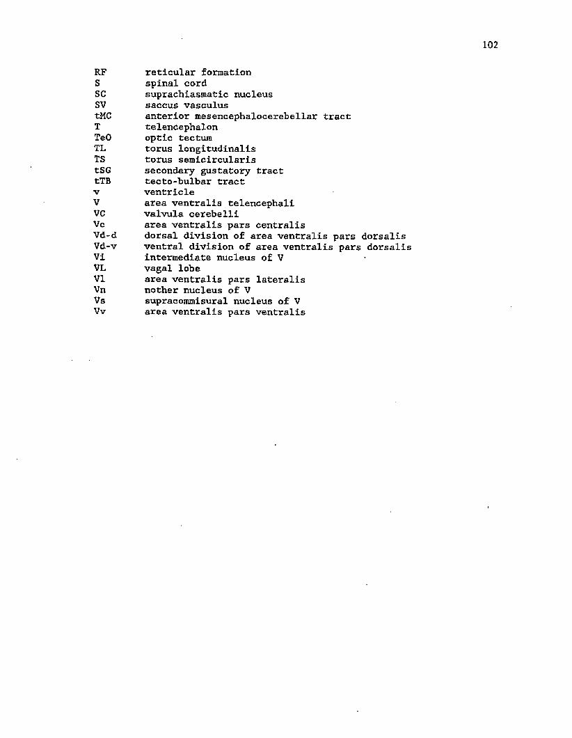
AC anterior commissureBC brachium conjunctivumCB cerebellumcG commissure of Goldsteincia intejrauricular commissure of WallenbergCM corpus mammallareD area dorsalis telencephaliDc area dorsalis pars centralisDd area dorsalis pars dorsalisD1 area dorsalis pars lateralisDm area dorsalis pars medialisDp area dorsalis pars posteriorFL facial lobefR fasciculus retroflexusGVN primary general visceral nucleusHA anterior hypothalamic nucleusHb habenulaHoC horizontal commissureiaf Internal arcuate fibersIL Inferior lobeIS inner segment of the thalamusLL lateral lemniscusLFB lateral forebrain bundleMFB medial forebrain bundlemlf medial longitudinal fasciculusnA nucleus ambiguusnC commissural nucleus of CajalnD nucleus diffususnE entopeduncular nucleusnFu medial funicular nucleusnGS secondary gustatory nucleusnLBm magnocellular division of nucleus lobo bulbarisnLBp parvocellular division of nucleus lobo bulbarisnLV nucleus of the lateral valvulanMD dorsal mesencephalic nucleusnO occulomotor nucleusnPC paracommissural nucleusnPT nucleus of the posterior tuberclenR raphe nucleinT nucleus taeniaenTm trigeminal motor nucleusriVS secondary visceral nucleusOT optic tractP pituitaryPM magnocellular preoptic nucleusPP periventricular preoptic nucleusPPa anterior segment of the parvocellular part of PPPPp posterior segment of the parvocellular part of PPPr preoptic recess
101
102
RF reticular formationS spinal cordSC suprachiasmatic nucleussv saccus vasculustMC anterior mesencephalocerebellar tractT telencephalonTeO optic tectumTL torus longitudinalisTS torus semicircularistSG secondary gustatory tracttTB tecto-bulbar tractV ventricleV area ventralis telencephaliVC valvula cerebelliVc area ventralis pars centralisVd-d dorsal division of area ventralis pars dorsalisVd-v ventral division of area ventralis pars dorsalisVi intermediate nucleus of VVL vagal lobeVI area ventralis pars lateralisVn nother nucleus of VVs supracommisural nucleus of VVv area ventralis pars ventralis
RESULTS
Nuclear Organization in the Catfish Forebrain
The occurance of eversion during development of the
actinopterygian forebrain and thus a nomenclature based on topology
(Neiuewenhuys, '62a) is now generally accepted. This nomenclature is
utilized for a general description of the nuclear domains in the
telencephalon of the channel catfish (Bass, '81a). In this study,
the description of the nuclear organization of the catfish forebrain
is extended to Include some of the unidentified regions of the
diencephalon in order to facilitate the analysis and unambiguous
communication of the results.
Transverse sections through the commissure of Goldstein (Fig.
la), through the anterior commissure (Fig. lb) and at the level of the
posterior end of the horizontal commissure, immediately anterior to
the habenular commissure (Fig. lc) indicate areas of the telencephalon
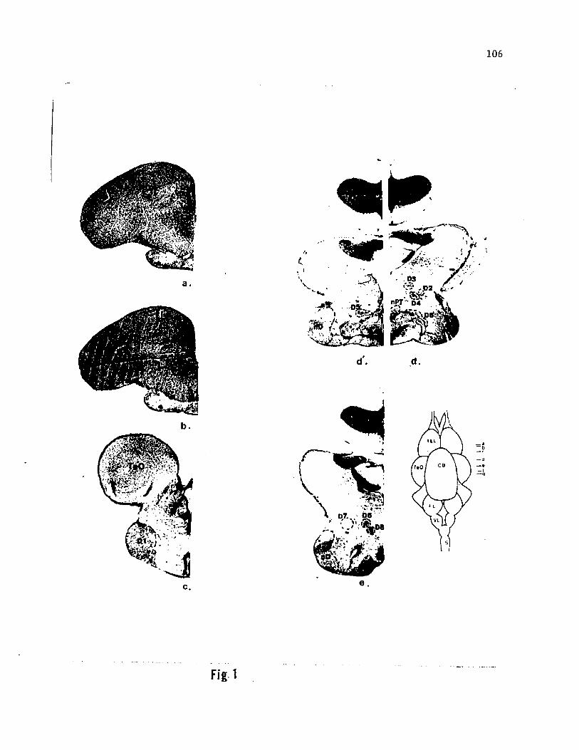
relevant to the present study. D1 (Fig. lc) is a large nucleus with
medium sized cells (approx.30 urn) arranged in clusters or glomeruli.
Rostrally, D1 appears as a lobule in the ventrolateral part of the
telencephalon at the level of the caudal edge of the entopeduncular
nucleus (see Bass, '81a). At the anterior diencephalic level (Fig.
lc), D1 enlarges into a lateral lobule. Cells in the ventral part of
the lobule are densely packed into a lamina, while in the dorsal
portion they remain well spaced. At more caudal levels, D1 is reduced
in size and displaced medially by the nucleus diffuses (nD). The
relative location and cellular arrangement within D1 is similar to the
103
104
nucleus preglomerulosus (nPG) of other teleosts (Schnitzlein, '62;
Braford and Northcutt, '83).
Sections through the anterior diencephalon (Fig. Id) in the
region of the nucleus of the posterior tubercle (nFT) (Finger, '75)
are characterized by the dorsomedial curvature of the lateral recess
which appears as a 'C' turned sideways. Dorsal to the lateral recess
two nuclei are seen to surround the caudal extension of Dl, here
labelled as D2. A small group of roughly triangular large cells
(approx. 70 urn) is present dorsomedial to D2 and is labelled as D3.
Also, a few well-spaced, small, round cells surround them and are
intermingled with the magnocellular D3, which is relatively compact
and may represent a ventrolateral migration of the magnocellular
preoptic neurons. This nucleus disappears quite suddenly at more
caudal levels. D4 is a group of small, well-spaced cells which
surround D2 and extend medially. D3 and D4 may be equivalent to the
outlying nuclei described by Braford and Northcutt ('83) for other
teleosts. At about the same level, a small nucleus (D5) is present
around the dorsolateral border of the lateral recess. This nucleus
has horizontally oriented, medium-sized, fusiform cells. Rostrally,
(left side of Fig. Id) these give way to a group of large cells
extending towards the medial border of the ventral diencephalon
(hypothalamus), while posteriorly (right side of Fig. Id) they are
reduced to a thin lamina curving around the dorsolateral border of the
lateral recess of the inferior lobe (IL).
The posterior diencephalon (Fig. le) is characterized by a
reduction of the lateral recess to a small lacuna in the middle of the
IL and the appearance of a group of three distinct nuclei in the
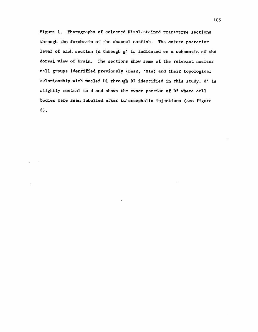
Figure 1. Photographs of selected Nissl-stained transverse sections
through the forebrain of the channel catfish. The antero-posterior
level of each section (a through g) is indicated on a schematic of the
dorsal view of brain. The sections show some of the relevant nuclear
cell groups identified previously (£ass, '81a) and their topological
relationship with nuclei D1 through D7 identified in this study, d' is
slightly rostral to d and shows the exact portion of D5 where cell
bodies were seen labelled after telencephalic injections (see figure
106
c.
t e l
CG
f I
V L
— d — •— I
Fig 1
107
Tf
Fig.1
ventral diencephalon (presumed thalamic region) dorsal to the 1L.
Caudal to the nPT and immediately rostral to the level of le, D6
appears as a large C-shaped cluster of cells with a fiber fascicle
extending into the concavity of the 'C'. In succeeding caudal
sections, D6 extends dorsomedially and becomes a small, compact
cluster of cells while retaining its characteristic shape. D7 is a
further caudal continuation of D1 and D2 and is topologically
equivalent to the posterior thalamic nucleus (nTP) of the bullhead
catfish (Finger, '78, '83). D7 consists of relatively large cells
distributed diffusely around the ventrolateral border of D6. At
levels caudal to that of le, D7 becomes quite compact and descends
ventrally to the level of the sulcus formed by the junction of the IL
with the torus. D8 is medial to D6 and D7 and is characterized at
rostral levels by a concentric cell free zone with a small compact
nucleus In the center. D8, however, becomes larger at caudal levels,
elongates in a caudomedial direction and may appear (depending upon
the exact plane of the section) to merge with D9 (Fig. If). D9 is a
small to medium sized cell group .which appears at the posterior
diencephalic level and descends caudomedially to the level of the
medial lobule of the IL. D9 is immediately dorsal to a distinct
crescent shaped periventricular nucleus that has been referred to as
the corpus mammallare or mammillary body (Marita and Finger, '85).
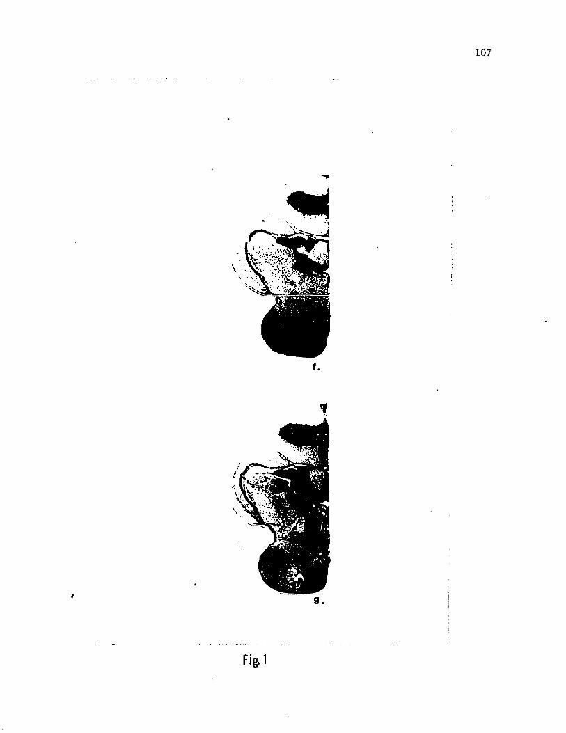
The nucleus lobo-bulbaris (nLB), another postero-ventral
diencephalic nucleus (Fig. Ig), is here redefined to represent two
adjacent and intermingling groups of neurons. The main part of the
nucleus consists of the large sized (magnocellular) neurons, while the
small, pear-shaped (parvocellular) neurons are mostly located
dorso-caudally of the magnocellular group. The magnocellular division
is named as nLBm and projects to the FL and VL as described by Herrick
{'05) and confirmed experimentally in I\_ nebulosus (Morita and Finger,
'85). The parvocellular division which is shown to project to the
telencephalon is referred to as nLBp.
HRP Experiments
Hindbrain Connections
Lateral and medial HRP injections made in the facial (FL) and
vagal lobes (VL) identified the stereotaxic location of the secondary
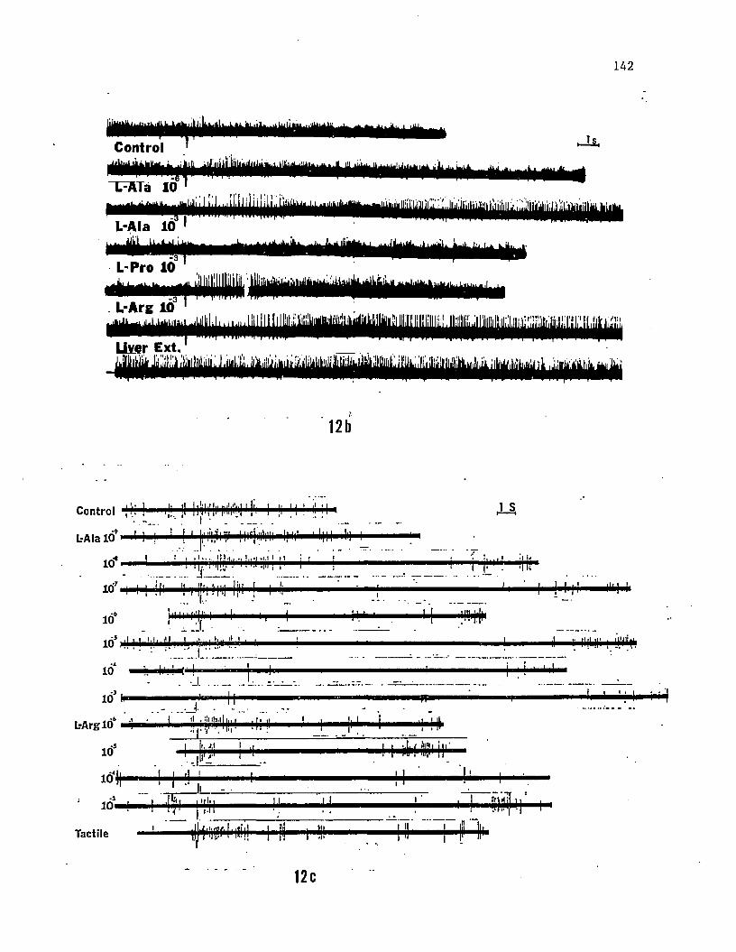
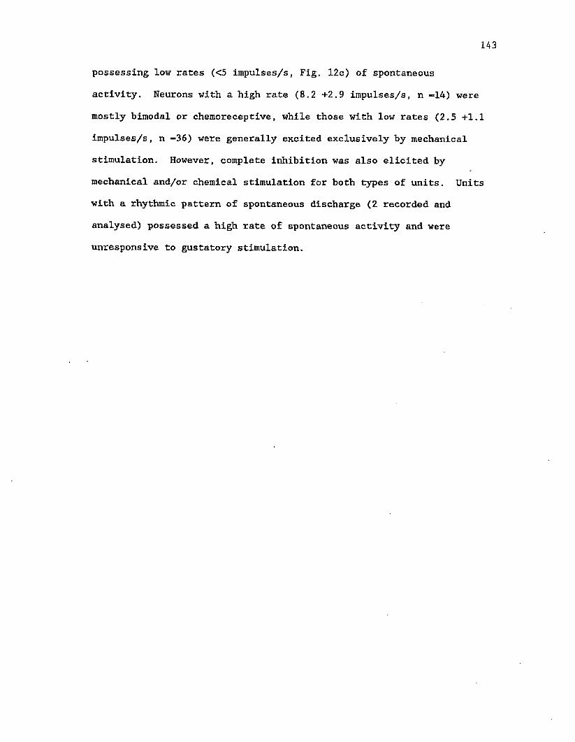
(isthmic) gustatory nucleus (nGS) in the brains of medium sized (15-20
cm) channel catfish (Fig. 2, sect. 6). The center of this spherical
nucleus was estimated to be 0.7 mm from the midline, 2.5 mm rostral to
the posterior edge of the cerebellum and 3.2 mm deep from the surface
of the cerebellum. Neurons in the FL and VL project bilaterally to
the nGS, although the ipsilateral projections are more dense. The FL
projects to the medial and caudal regions of nGS whereas the VL
projects to a semi-spherical portion in the dorsolateral part of the
nucleus. However, there is considerable overlap of FL and VL
projections in the caudal and lateral regions of the nucleus.
Anterograde and retrogarade transport of HRP after injections into the
FL and VL of the channel catfish confirmed the results obtained
previously for the bullhead catfish (see Finger, '78, 83; Morita and
Finger, '85). Both FL and VL have reciprocal connections with the
nucleus lobo-bulbaris (nLB), while the other diencephalic nucleus,
which sends fibers to the FL, is located in the pretectal region (see
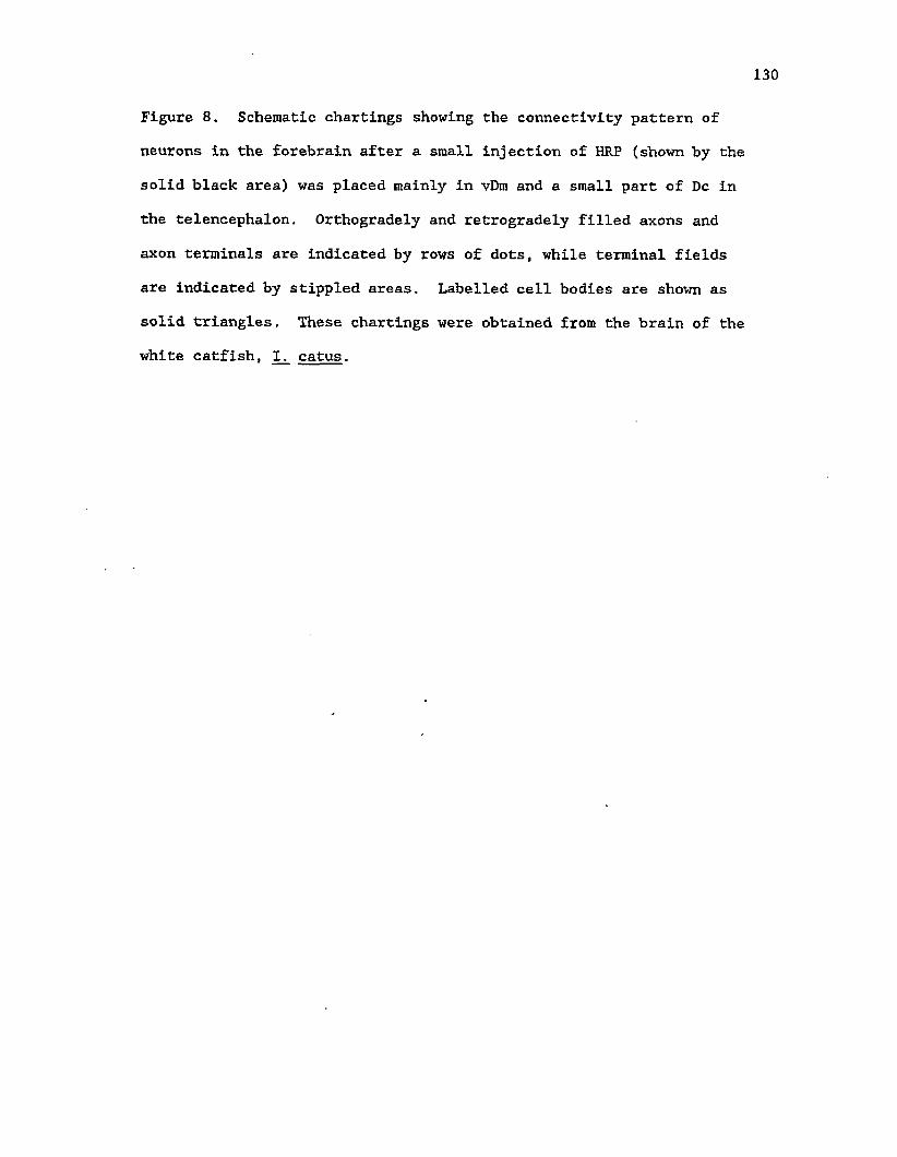
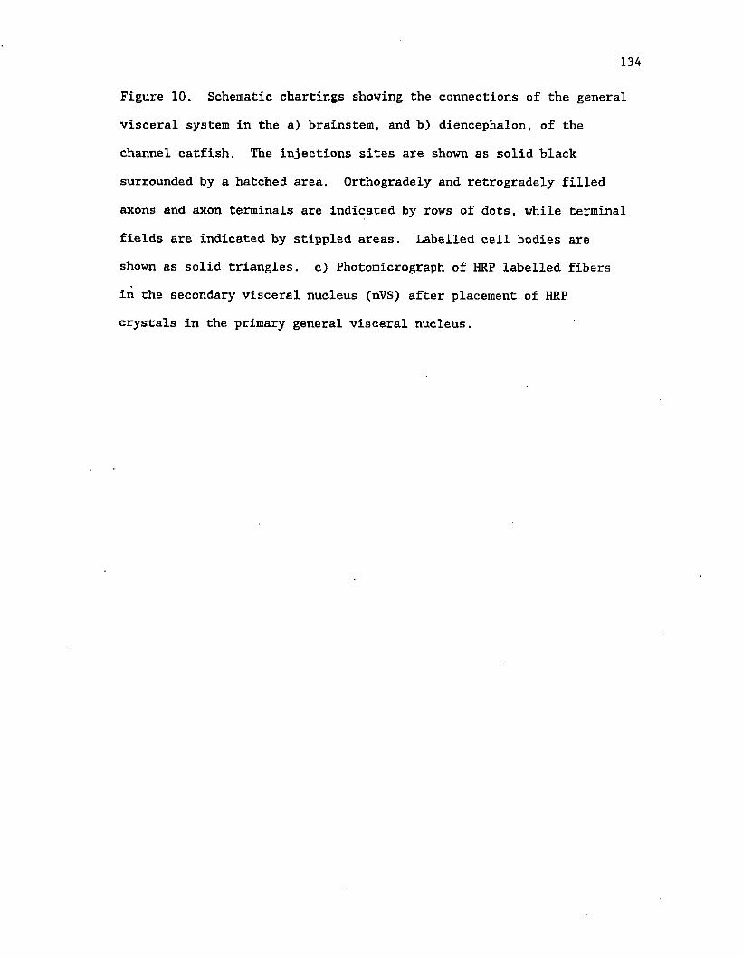
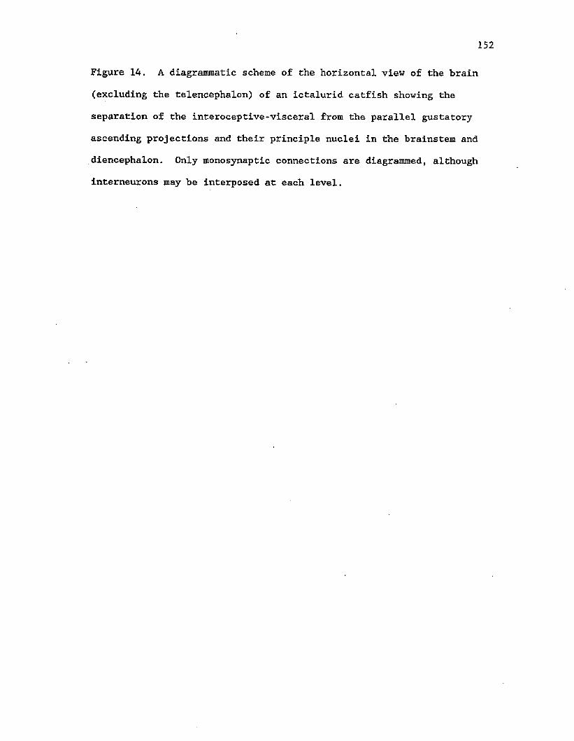
Figure 2, Schematic chartings of diencephalic connections of the
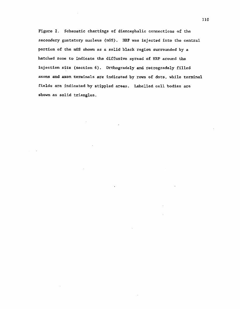
secondary gustatory nucleus (nGS). HRP was injected into the central
portion of the nGS shown as a solid black region surrounded by a
hatched zone to indicate the diffusive spread of HRP around the
injection site (section 6). Orthogradely and retrogradely filled
axons and axon terminals are indicated by rows of dots, while terminal
fields are indicated by stippled areas. Labelled cell bodies are
shown as solid triangles.
»
MRF
U1
112
Morita and Finger, '85).
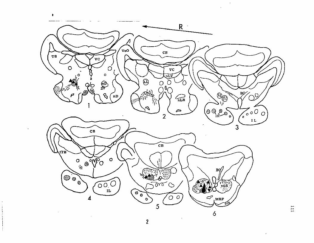
Isthmic Projections
Pressure injection of HRP into the nGS revealed its ascending
projections and the exact location of a reciprocally connected (Morita
and Finger, '85) diencephalic taste nucleus referred to previously
(Finger, '78, '83) as the posterior thalamic taste nucleus (Fig. 2).
A compact group of cell bodies were retrogradely labelled in the
ventro-lateral, posterior region of the diencephalon (D7 of fig. le),
slightly anterior and dorsal to nLB (Fig. 3a). D7 was estimated to be
1.2 mm posterior to the habenular commissure, 1.5 mm from the midline
and 5.0 mm deep from the surface of the cerebellum. This nucleus
sends its axons to the nGS and has fiber terminals distributed
diffusely in a small portion of the mesencephalic tegmentum (Fig. 2
sect. 1). A distinct group of axons extends laterally and terminates
in the nucleus diffusus (nD) at levels in the plane of and anterior to
D7 (Fig. 2 sects. 1 and 2). Injection of HRP into the lateral part of
the inferior lobe indicates that axons terminating in nD originate
from the cortical cells of nGS (Fig. 3b). Dense fiber terminals were
also labelled throughout the caudal region of the inferior lobe (XL).
These fiber terminals were restricted to a chain of six circular (in
the transverse plane) fields extending from the lateral to the medial
border of the IL (Fig. 2 sect. 3 to sect. 5, 3c). In the tapered,
caudal portion of the IL these fields converge to three large fields.
These terminal fields extend along the longitudinal axis of the IL and
are referred to as ltflL. Injections into the most rostral portion of
the nGS labelled the terminal fields only in the lateral portion of
the IL (Fig. 3d) indicating the possibility of a topographic
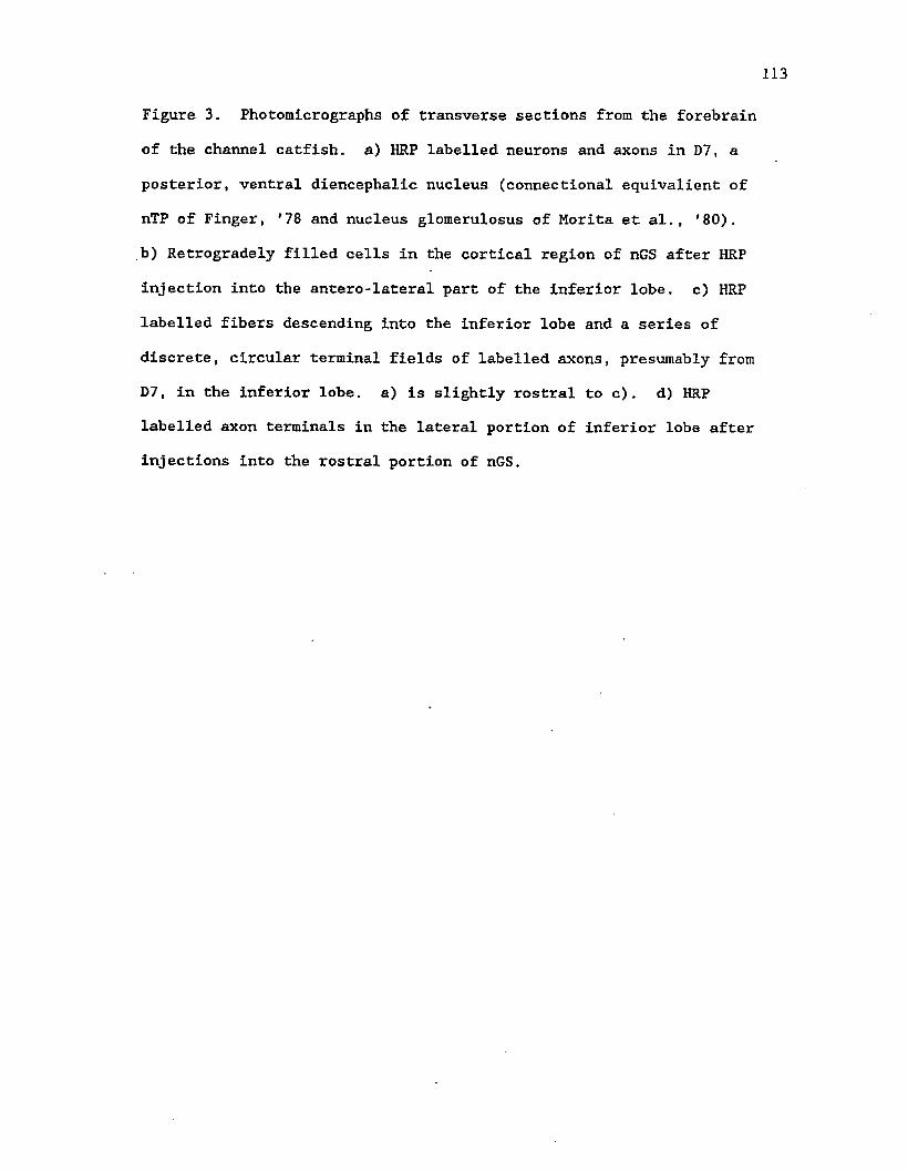
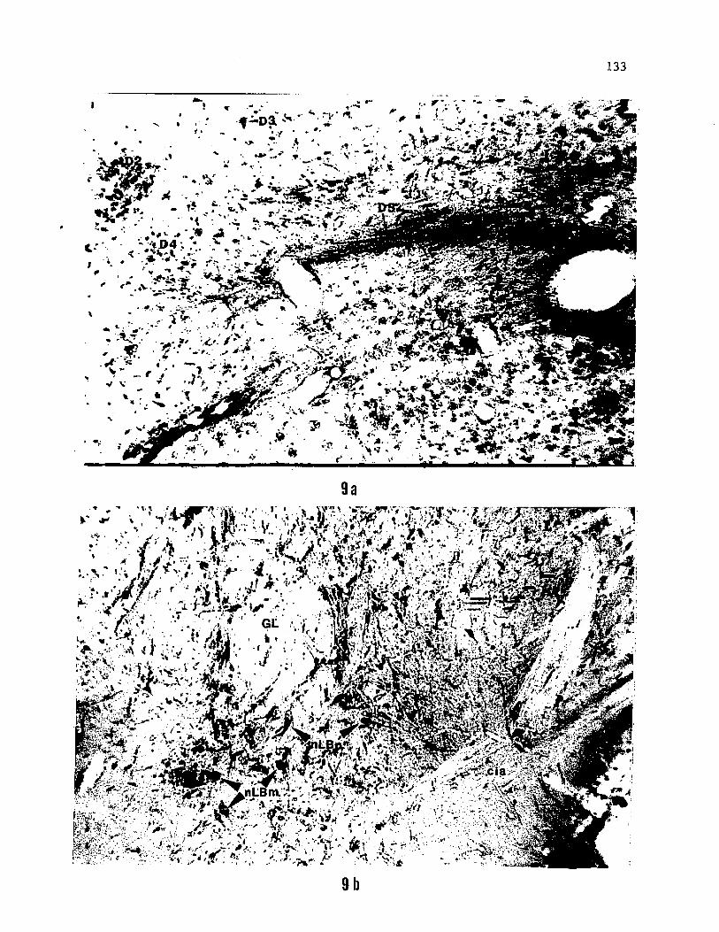
Figure 3. Photomicrographs of transverse sections from the forebrain
of the channel catfish, a) HRP labelled neurons and axons in D7, a
posterior, ventral diencephalic nucleus (connectional equivalient of
nTP of Finger, '78 and nucleus glomerulosus of Morita et al., '80).
b) Retrogradely filled cells in the cortical region of nGS after HRP
injection into the antero-lateral part of the inferior lobe, c) HRP
labelled fibers descending into the inferior lobe and a series of
discrete, circular terminal fields of labelled axons, presumably from
D7, in the inferior lobe, a) is slightly rostral to c). d) HRP
labelled axon terminals in the lateral portion of inferior lobe after
injections into the rostral portion of nGS.
114
§ ma vSafaW'
3b
V'
116
relationship between these terminal fields and the nGS. Cell bodies
in the FL and VL were also retrogradely labelled as observed in the
bullhead catfish (see Finger, '78, '83; Morita and Finger, '85).
Labelling was also observed in the cerebellum, but this was probably
due to leakage of HRP from the penetrating microelectrode as it was
inserted through the cerebellum for injecting HRP into the nGS. The
leakage apparently resulted in dense labelling of the ipsi and
contra-lateral brachium conjunctivum and retrogradely labelled cells
in the tectum. These results are not shown because forebrain
injections indicate that the cerebellar interactions of the gustatory
system (if any) are independent of its forebrain connections.
Forebrain Connections
Both large and restricted injections of HRP placed in the
posterior diencephalon identified the location of a gustatory area in
the telencephalon and specified connections of the gustatory system in
the forebrain of ictalurid catfish (Fig. 4). In addition, injections
were made in the presumed telencephalic gustatory area in separate
animals to confirm its connections with diencephalic nuclei-. Large
lateral and ventral injections (Fig. 4 sect. 11) covered regions of
the diencephalon including the nLB, D7 and sometimes D6. Labelled
cell bodies were observed ipsilaterally in the nGS (Fig. 5a), the area
dorsalis pars medialis (Dm) and the medial portion of area dorsalis
pars centralis (Dc) in the telencephalon (Fig. 5b). Fiber terminals
were observed in the FL and VL, in the nGS (Fig. 5a), in the ltfIL, in
the trigeminal motor nucleus, in Dm and medial part of Dc (Fig. 4
sect. 1, 5c). Fiber terminals were also observed in the pretectal
region, near the habenula and diffusely in the mesencephalic tegmentum
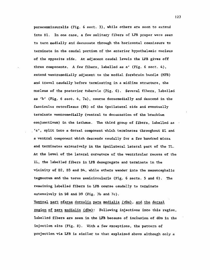
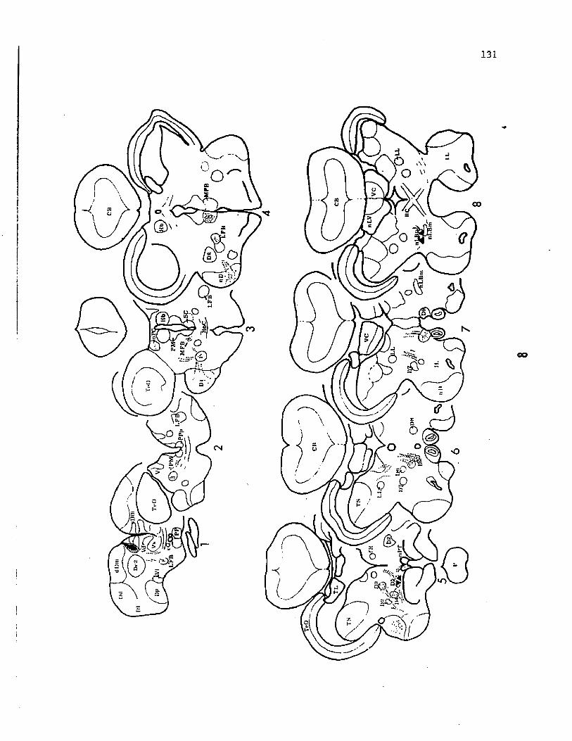
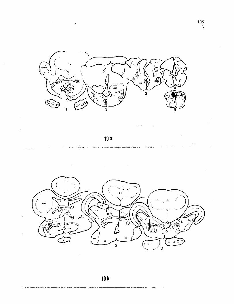
Figure 4. Schematic chartings of diencephalo-telencephalic
connections seen after placing relatively large quantities (80 to 100
nl) of HRP solution into the ventral diencephalon. Solid black and
hatched areas indicate the HRP-injected site and diffusion zone,
respectively. Portions of the injection site and diffusion zone
included the nucleus lobo-bulbaris, D7 and unidentified parts of the
ventral diencephalon. Orthogradely and retrogradely filled axons and
axon terminals are indicated by rows of dots, while terminal fields
are indicated by stippled areas. Labelled cell bodies are shown as
solid triangles.
dD
m
118
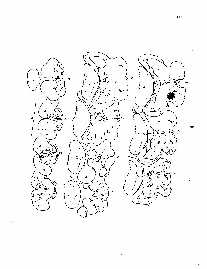
CD
CM
r\
»n
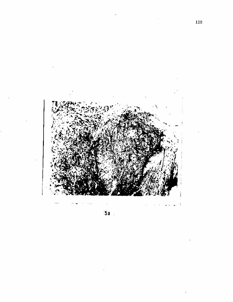
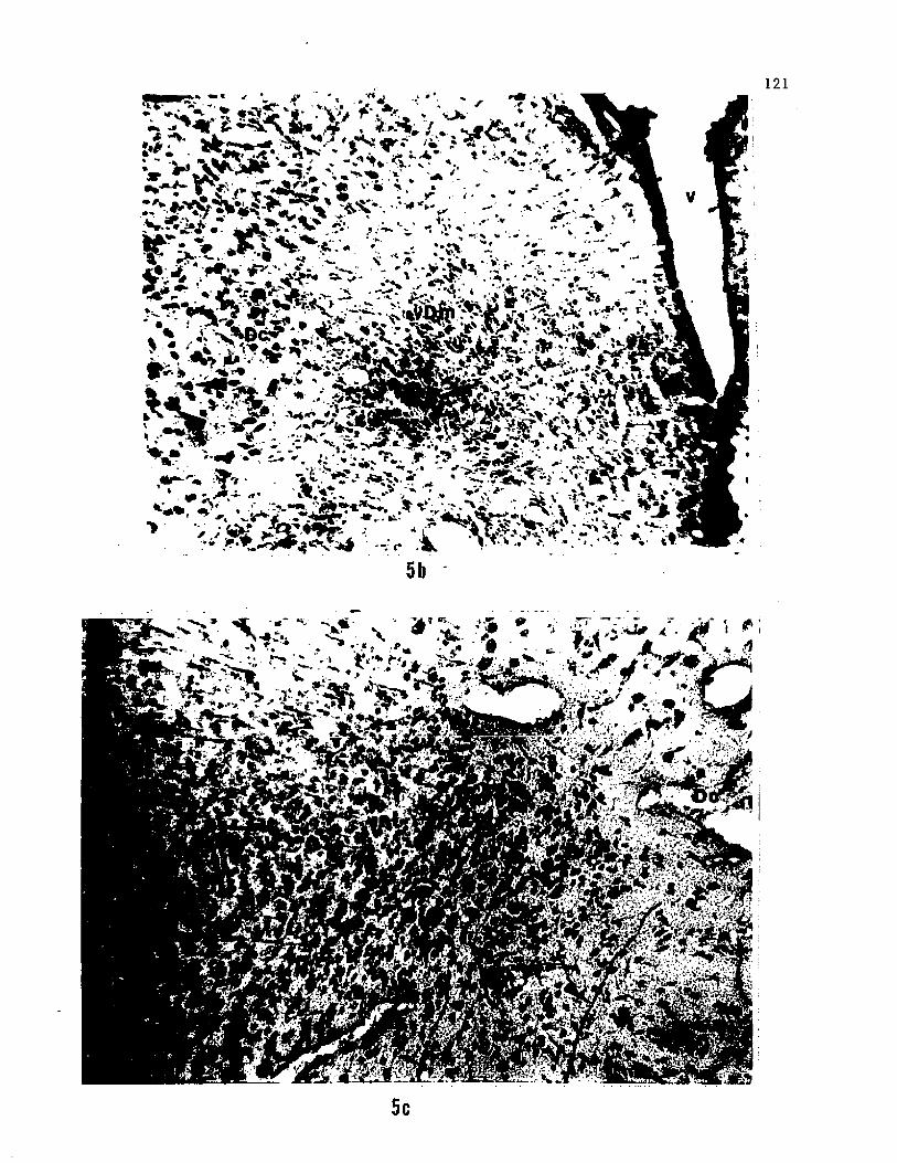
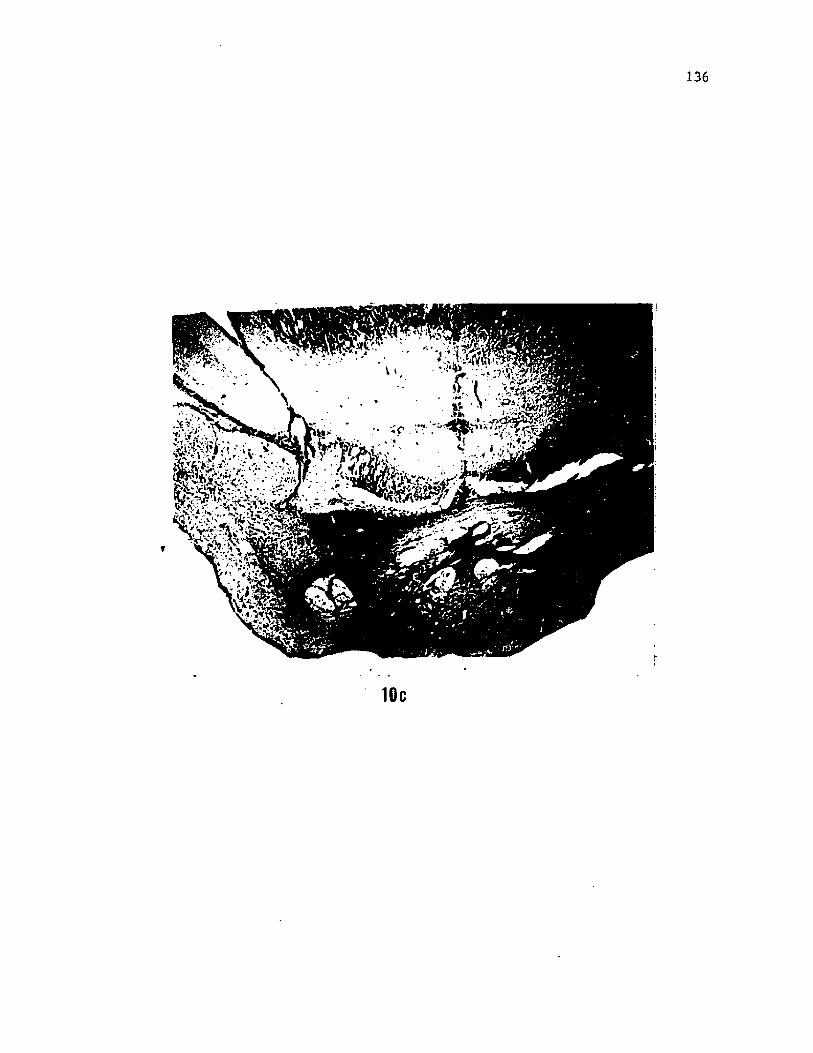
Figure 5. Photomicrographs showing a) HRP labelled axons and cell
bodies in the nGS, b) sparsely distributed, retrogradely filled cell
bodies in the telencephalon, and c) Diffusely distributed axons in the
telencephalon, after injections of HRP in the region of D7 (see arrows).
120
5 a
^ X ' fj* « V <
%
gffiKSSWi
121
122
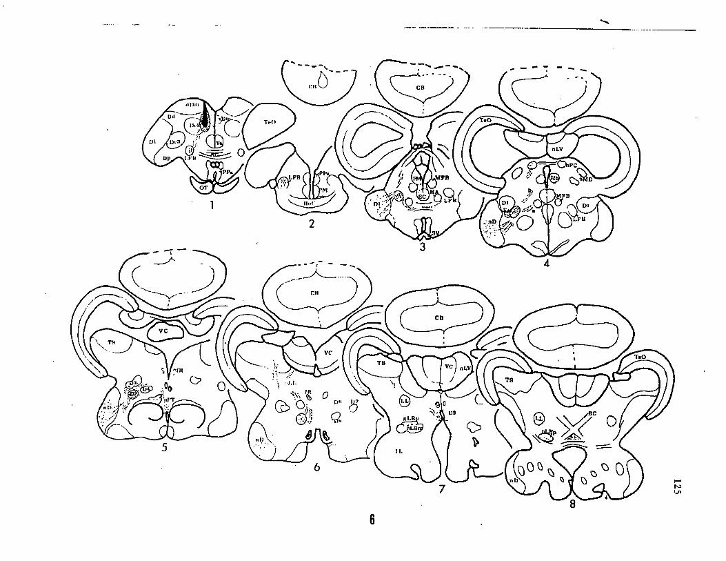
(Fig. 4 sects. 9 and 10). A few fibers crossed to the contra-lateral
side through the habenular commissure to terminate in the habenular
nucleus and ventral diencephalon (Fig. 4 sect. 10,11).
With restricted diencephalic injections, labelling in the nGS was
invariably accompanied by labelling of the longitudinal terminal
fields in the inferior lobe (ItfIL). Fiber terminals were observed in
the FL and VL only if the injection site extended to a portion of nLB.
No part of the gustatory system including the relevant telencephalic
areas were labelled in cases where injections were restricted to the
dorsal thalamus or otherwise excluded both D7 and the nLB.
Most telencephalic injections can be divided into two categories.
Large injections of HRP resulted in label extending through the dorsal
portion of Dm including the medial part of Dc, while small injections
were limited mainly to the ventromedial portion of Dm (known to
receive gustatory projections). This portion of Dm is here
arbitrarily referred to as the vDm (Fig. la,b), as no
cytoarchitectonic differences between different parts of Dm were
visible in a previous study (Bass, '80a). The remaining part of Dm
may be regarded as dDm. Since diencephalic injections indicated the
presence of gustatory neurons in vDm and a part of Dc, diencephalic
connections of these telencephalic regions are explained separately.
Area dorsalis pars centralis (Dc), and the dorsal region of pars
medialis (dDm): Following HRP injections, fibers were seen to course
ventrally in the plane of the anterior commissure (Fig. 6 sect. 1,2)
and enter the lateral forebrain bundle (LFB), which is easily traced
caudally up to the level of the habenula and epiphysis. At this
level, some fibers deviate dorsomedially and terminate in the nucleus
paracommissuralis (Fig. 6 sect. 3), while others are seen to extend
into Dl. In one case, a few solitary fibers of LFB proper were seen
to turn medially and decussate through the horizontal commissure to
terminate in the caudal portion of the anterior hypothalamic nucleus
of the opposite side. At adjacent caudal levels the LFB gives off
three components. A few fibers, labelled as a' (Fig. 6 sect. 4),
extend ventromedially adjacent to the medial forebrain bundle (MFB)
and travel caudally before terminating in a midline structure, the
nucleus of the posterior tubercle (Fig. 6). Several fibers, labelled
as 'b' (Fig. 6 sect. 4, 7a), course dorsomedially and descend in the
fasciculus retroflexus (fR) of the ipsilateral side and eventually
terminate ventromedially (ventral to decussation of the brachium
conjunctivum) in the isthmus. The third group of fibers, labelled as
'c', split into a dorsal component which terminates throughout Dl and
a ventral component which descends caudally for a few hundred micra
and terminates extensively in the ipsilateral lateral part of the IL.
At the level of the lateral curvature of the ventricular recess of the
IL, the labelled fibers in LFB desegregate and terminate in the
vicinity of D2, D3 and D4, while others wander into the mesencephalic
tegmentum and the torus semicircularis (Fig. 6 sects. 5 and 6). The
remaining labelled fibers in LFB course caudally to terminate
extensively in D8 and D9 (Fig. 7b and 7c).
Ventral part ofarea dorsalis pars medialis (vDm). and the dorsal
region of pars medialis (dDm): Following injections into this region,
labelled fibers are seen in the LFB because of inclusion of dDm in the
injection site (Fig. 8). With a few exceptions, the pattern of
projection via LFB is similar to that explained above although only a
Research: Chemoreception in the catfish, 4 semestersNeuroanatomical studies on the gustatory system of the catfish, 2 semesters (University of Colorado, School of Medicine)