Fundamental Components of Attention Eric I. Knudsen Department of Neurobiology, Stanford University School of Medicine, Stanford, California 94305-5125; email: [email protected]Annu. Rev. Neurosci. 2007. 30:57–78 First published online as a Review in Advance on April 6, 2007 The Annual Review of Neuroscience is online at neuro.annualreviews.org This article’s doi: 10.1146/annurev.neuro.30.051606.094256 Copyright c 2007 by Annual Reviews. All rights reserved 0147-006X/07/0721-0057$20.00 Key Words working memory, stimulus selection, prefrontal cortex, parietal cortex, superior colliculus, vision Abstract A mechanistic understanding of attention is necessary for the elu- cidation of the neurobiological basis of conscious experience. This chapter presents a framework for thinking about attention that fa- cilitates the analysis of this cognitive process in terms of underlying neural mechanisms. Four processes are fundamental to attention: working memory, top-down sensitivity control, competitive selec- tion, and automatic bottom-up filtering for salient stimuli. Each pro- cess makes a distinct and essential contribution to attention. Volun- tary control of attention involves the first three processes (working memory, top-down sensitivity control, and competitive selection) operating in a recurrent loop. Recent results from neurobiological research on attention are discussed within this framework. 57 Annu. Rev. Neurosci. 2007.30:57-78. Downloaded from arjournals.annualreviews.org by UNIVERSITY OF FLORIDA - Smathers Library on 09/01/09. For personal use only.
Transcript
ANRV314-NE30-03 ARI 7 May 2007 17:20
Fundamental Componentsof AttentionEric I. KnudsenDepartment of Neurobiology, Stanford University School of Medicine, Stanford,California 94305-5125; email: [email protected]
Annu. Rev. Neurosci. 2007. 30:57–78
First published online as a Review in Advance onApril 6, 2007
The Annual Review of Neuroscience is online atneuro.annualreviews.org
This article’s doi:10.1146/annurev.neuro.30.051606.094256
working memory, stimulus selection, prefrontal cortex, parietalcortex, superior colliculus, vision
AbstractA mechanistic understanding of attention is necessary for the elu-cidation of the neurobiological basis of conscious experience. Thischapter presents a framework for thinking about attention that fa-cilitates the analysis of this cognitive process in terms of underlyingneural mechanisms. Four processes are fundamental to attention:working memory, top-down sensitivity control, competitive selec-tion, and automatic bottom-up filtering for salient stimuli. Each pro-cess makes a distinct and essential contribution to attention. Volun-tary control of attention involves the first three processes (workingmemory, top-down sensitivity control, and competitive selection)operating in a recurrent loop. Recent results from neurobiologicalresearch on attention are discussed within this framework.
To behave adaptively in a complex world, ananimal must select, from the wealth of infor-mation available to it, the information that ismost relevant at any point in time. This in-formation is then evaluated in working mem-ory, where it can be analyzed in detail, de-cisions about that information can be made,and plans for action can be elaborated. Themechanisms of attention are responsible forselecting the information that gains access toworking memory.
Four component processes are funda-mental to attention: (a) working memory,(b) competitive selection, (c) top-down sen-sitivity control, and (d ) filtering for stim-uli that are likely to be behaviorally impor-tant (salience filters). Working memory is ahighly dynamic form of memory that oper-ates over periods of seconds and temporar-ily stores selected information for detailedanalysis (Baddeley 2003). Competitive selec-tion is the process that determines whichinformation gains access to working mem-ory (Desimone & Duncan 1995). Top-downsensitivity control is a process that regulatesthe relative signal strengths of the differentinformation channels that compete for ac-cess to working memory (Egeth & Yantis1997). Salience filters automatically enhanceresponses to stimuli that are infrequent inspace or time or are of instinctive or learned
biological importance (Koch & Ullman 1985).The engagement of these processes leads di-rectly to increased behavioral sensitivity andshortened response latencies (the traditionalmetrics of attention) as well as to the cogni-tive benefits that we associate with attention.
The past decade has witnessed an enor-mous surge in neurophysiological research onattention. Nearly all this research has focusedon various phenomena that are associated withattention. One major goal, however, is to un-derstand the neural mechanisms that underlieattention. Progress toward this goal would befacilitated by a model that accounts for thephenomena of attention in terms of neuro-biological components. This chapter estab-lishes the framework for such a model and dis-cusses recent results within the context of thisframework.
The proposed framework for attention isshown in Figure 1, a framework inspired bythe models of Desimone & Duncan (1995)and Miller & Cohen (2001). The central ner-vous system contains information about theworld, about stored memories, and about theinternal state of the animal. At any point intime, the information that gains access toworking memory is selected by a competitiveprocess from this repertoire of information onthe basis of its relative signal strength. Sig-nal strength reflects the combined effects ofthe quality of the encoded information, top-down bias signals, and bottom-up salience fil-ters. The information with the greatest sig-nal strength enters the circuitry for workingmemory and competes with existing informa-tion for control of working memory. The in-formation that controls working memory alsodirects top-down bias signals that modulatethe signal strengths of relevant ascending rep-resentations, forming a recurrent loop thatunderlies voluntary attention.
WORKING MEMORY
Working memory is a special form of mem-ory with extraordinary capabilities (Baddeley2003). Working memory holds a limited
58 Knudsen
Ann
u. R
ev. N
euro
sci.
2007
.30:
57-7
8. D
ownl
oade
d fr
om a
rjou
rnal
s.an
nual
revi
ews.
org
by U
NIV
ER
SIT
Y O
F FL
OR
IDA
- S
mat
hers
Lib
rary
on
09/0
1/09
. For
per
sona
l use
onl
y.
ANRV314-NE30-03 ARI 7 May 2007 17:20
Workingmemory(decision)
Competitiveselection
Gazecontrol
Neuralrepresentations
Sensitivitycontrol
Saliencefilters
World
Sensory
Motor
Stored memory
Internal state
Top-down
Bottom-up
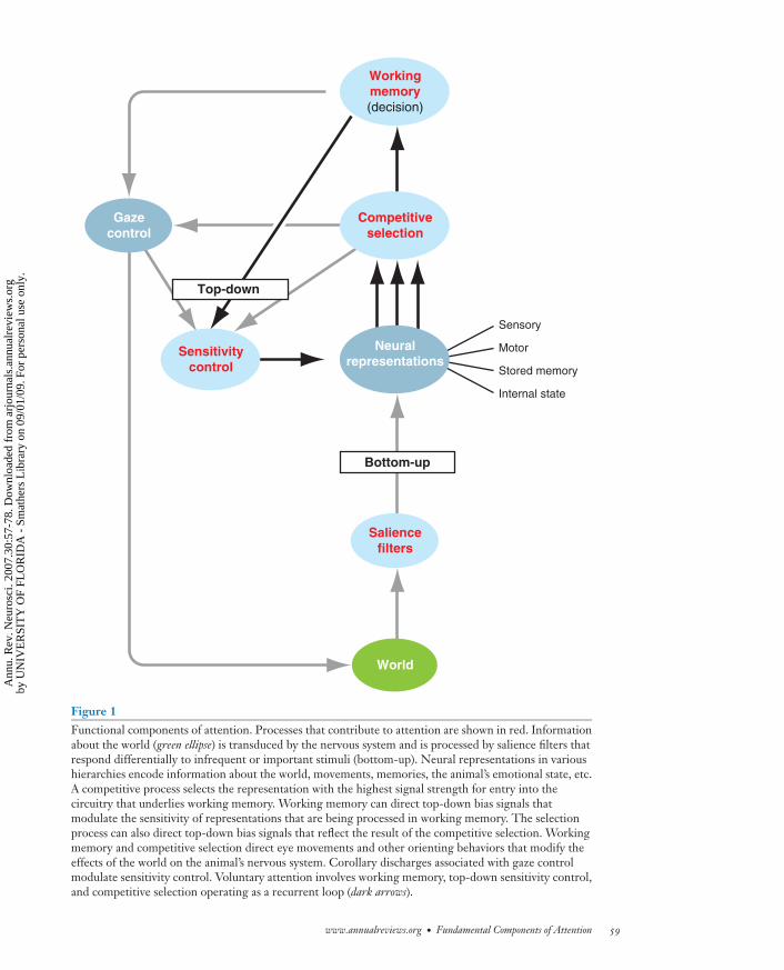
Figure 1Functional components of attention. Processes that contribute to attention are shown in red. Informationabout the world (green ellipse) is transduced by the nervous system and is processed by salience filters thatrespond differentially to infrequent or important stimuli (bottom-up). Neural representations in varioushierarchies encode information about the world, movements, memories, the animal’s emotional state, etc.A competitive process selects the representation with the highest signal strength for entry into thecircuitry that underlies working memory. Working memory can direct top-down bias signals thatmodulate the sensitivity of representations that are being processed in working memory. The selectionprocess can also direct top-down bias signals that reflect the result of the competitive selection. Workingmemory and competitive selection direct eye movements and other orienting behaviors that modify theeffects of the world on the animal’s nervous system. Corollary discharges associated with gaze controlmodulate sensitivity control. Voluntary attention involves working memory, top-down sensitivity control,and competitive selection operating as a recurrent loop (dark arrows).
www.annualreviews.org • Fundamental Components of Attention 59
Ann
u. R
ev. N
euro
sci.
2007
.30:
57-7
8. D
ownl
oade
d fr
om a
rjou
rnal
s.an
nual
revi
ews.
org
by U
NIV
ER
SIT
Y O
F FL
OR
IDA
- S
mat
hers
Lib
rary
on
09/0
1/09
. For
per
sona
l use
onl
y.
ANRV314-NE30-03 ARI 7 May 2007 17:20
PFC: prefrontalcortex
amount of information for periods of secondswhile the information is evaluated and ma-nipulated in a uniquely powerful and flexiblefashion on the basis of the animal’s internalstate and stored memories. Working mem-ory itself comprises competitive processes,and multiple types of information may com-pete for full control of the circuitry underly-ing working memory at any moment in time.The degree to which one type of informationgains full control of working memory reflectsthe relative strengths of the competing rep-resentations. The information that is held inworking memory serves as the basis for deci-sions and the planning of complex behaviors(Genovesio et al. 2006, Yoshida & Ishii 2006)and, most importantly for this discussion, con-trols top-down signals that modulate the sen-sitivity of neural representations that con-tribute to that information (Miller & Cohen2001).
Working memory and attention are inex-tricably inter-related. When an animal attendsto an object, information associated with thatobject enters working memory. Conversely,information in working memory is informa-tion that is associated with objects to which ananimal has attended (LaBar et al. 1999). Thus,working memory represents the objects ofattention.
The capacity of working memory to ma-nipulate information is limited at any one timeto a single domain (e.g., verbal, mathematical,visuospatial). The portions of the brain thatparticipate in working memory depend on theinformation being processed. For example,functional imaging studies on humans showthat verbal working memory tasks activate theventrolateral prefrontal cortex (PFC) and lan-guage areas in the temporal and inferior pari-etal cortex on the left side (Schumacher et al.1996). In contrast, visuospatial working mem-ory tasks activate the dorsolateral PFC, in-ferior parietal cortex on the right side, andhigh-order visual areas in the occipital cortex(Smith et al. 1996).
The PFC is one area of the brain that is ac-tivated consistently in working memory tasks.
Clinical reports in humans and lesion studiesin monkeys confirm a central role of the PFCin working memory (Miller & Cohen 2001).These studies indicate that lesions in the PFCcause general deficits in working memory,with no apparent deficits in sensory discrim-ination or motor performance (Diamond &Goldman-Rakic 1989, Duncan et al. 1996,Vendrell et al. 1995). In contrast, although le-sions in other areas of the brain can also leadto deficits in working memory, the effect ofthese lesions is specific for the sensory or mo-tor information represented in these areas andis accompanied by corresponding sensory ormotor deficits.
The data suggest that working memory isa function that is usually distributed widely inthe brain, with the PFC acting as an executivecontroller. During working memory tasks, thePFC engages with cortical and subcortical re-gions that process sensory information, motorinformation, information about internal state,or stored memories, depending on the task athand (Baddeley 2003, Constantinidis & Wang2004). The extensive, reciprocal anatomicalconnections of the PFC with most cortical andmany subcortical regions are consistent withthis view (Miller & Cohen 2001).
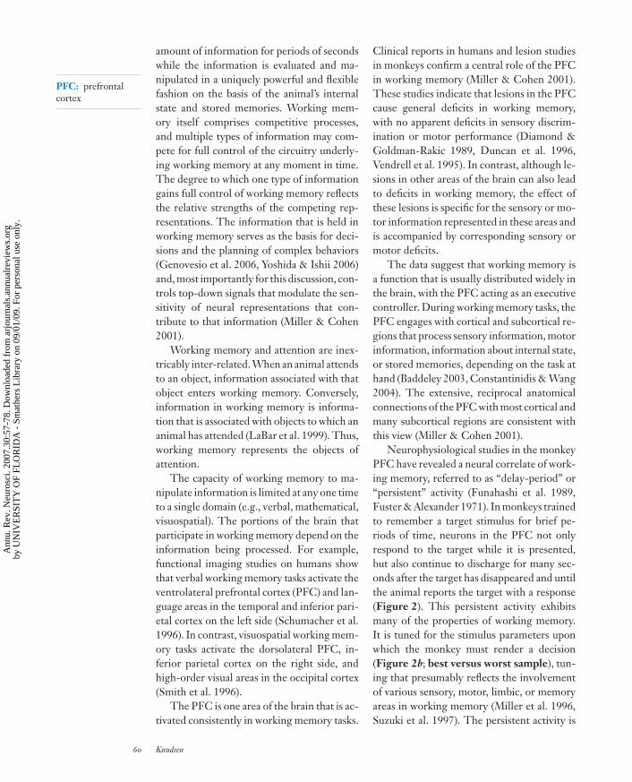
Neurophysiological studies in the monkeyPFC have revealed a neural correlate of work-ing memory, referred to as “delay-period” or“persistent” activity (Funahashi et al. 1989,Fuster & Alexander 1971). In monkeys trainedto remember a target stimulus for brief pe-riods of time, neurons in the PFC not onlyrespond to the target while it is presented,but also continue to discharge for many sec-onds after the target has disappeared and untilthe animal reports the target with a response(Figure 2). This persistent activity exhibitsmany of the properties of working memory.It is tuned for the stimulus parameters uponwhich the monkey must render a decision(Figure 2b; best versus worst sample), tun-ing that presumably reflects the involvementof various sensory, motor, limbic, or memoryareas in working memory (Miller et al. 1996,Suzuki et al. 1997). The persistent activity is
60 Knudsen
Ann
u. R
ev. N
euro
sci.
2007
.30:
57-7
8. D
ownl
oade
d fr
om a
rjou
rnal
s.an
nual
revi
ews.
org
by U
NIV
ER
SIT
Y O
F FL
OR
IDA
- S
mat
hers
Lib
rary
on
09/0
1/09
. For
per
sona
l use
onl
y.
ANRV314-NE30-03 ARI 7 May 2007 17:20
Sample Nonmatching test itemsMatching testBar release
Activity following:
Time from sample onset
Sp
ikes
pe
r s
ec
on
d
0 2000 4000 6000 8000
10
15
20
Best sample
Worst sample
Figure 2Delay-period unit activity in the PFC of a monkey performing a delayed match-to-sample task. (above)An example of a standard trial. The monkey was trained to release a bar when it saw an item that matchedthe first item (sample) in the trial. Time proceeds from left to right. The number of nonmatching testitems between the sample and the matching test item varied randomly from zero to 4. The actual stimuliwere color pictures. (below) Averaged responses for a population of 40 PFC neurons that exhibitedsample-selective delay-period activity. Responses are plotted separately for trials in which the mosteffective stimulus for each neuron was used as the sample (red ) and trials in which the least effectivestimulus for each neuron was used as the sample (blue). The delay-period activity distinguishes among thedifferent stimuli. Time is measured in ms. Bin width = 40 ms. Average baseline firing rate was10 spikes/s. The data are from Miller et al. (1996). Copyright 1996 by the Society for Neuroscience.
modulated according to decisions made bythe animal regarding the stimulus (Kim &Shadlen 1999). Moreover, the activity persistsuntil the animal reports the target stimulus,even though the target may be followed by aseries of “distracter” stimuli that are behav-iorally irrelevant (di Pellegrino & Wise 1993,Fuster 1995, Miller et al. 1996). An exception-ally high proportion of neurons in the PFC ex-hibits delay-period activity (Miller & Cohen
2001). Together, these data support the cen-tral role of the PFC in working memory.
All types of information about the worldand the organism are processed in workingmemory. However, the spatial location of astimulus in the world is a feature that is ana-lyzed in a special pathway, reflecting the im-portance of location as a parameter for makingdecisions and planning goal-oriented behav-iors. The processing in working memory of
www.annualreviews.org • Fundamental Components of Attention 61
Ann
u. R
ev. N
euro
sci.
2007
.30:
57-7
8. D
ownl
oade
d fr
om a
rjou
rnal
s.an
nual
revi
ews.
org
by U
NIV
ER
SIT
Y O
F FL
OR
IDA
- S
mat
hers
Lib
rary
on
09/0
1/09
. For
per
sona
l use
onl
y.
ANRV314-NE30-03 ARI 7 May 2007 17:20
PPC: posteriorparietal cortex
information according to location is referredto as spatial working memory.
Data from functional imaging studiesdemonstrate that spatial working memorytasks consistently activate two major corticalareas: the dorsolateral PFC and the posteriorparietal cortex (PPC) (Curtis 2006). Thesetwo structures are strongly interconnected byreciprocal pathways (Schwartz & Goldman-Rakic 1984). Both structures contain neuronswith delay-period activity that is tuned for thelocation of stimuli in space (Constantinidis& Wang 2004), and lesions of either struc-ture interfere with the monkey’s ability toplan responses using the remembered loca-tions of stimuli (Chafee & Goldman-Rakic2000).
As discussed above, the properties ofthe PFC demonstrate its critical role inworking memory, including spatial workingmemory (Constantinidis & Wang 2004). Incontrast, the functional properties and orga-nization of the PPC, which are discussed later,indicate that this region is likely to be moreinvolved in the processes of competitive selec-tion and top-down sensitivity control than inworking memory (Colby & Goldberg 1999).Persistent activity in the PPC is less preva-lent and more susceptible to interruption bydistracting stimuli (Powell & Goldberg 2000)than is the persistent activity in the PFC(Fuster 1995, Miller et al. 1996). Instead ofrobustly representing a behaviorally relevanttarget that is stored in working memory, activ-ity in the PPC represents the relative salienceof all stimuli (Bisley & Goldberg 2003) aswell as the goal locations of movements thata monkey intends to make (Andersen et al.2004, Batista & Andersen 2001, Ipata et al.2006).
TOP-DOWN SENSITIVITYCONTROL
In the context of attention, not only doesworking memory accept, store, and manipu-late information, but it also generates signalsthat improve the quality of the information
that it processes (Miller & Cohen 2001, Miller& D’Esposito 2005). One mechanism for im-proving information quality is to direct ori-enting movements toward targets (Figure 1,gaze control). For example, by directing ori-enting movements of the eyes toward an ob-ject, working memory optimizes the resolu-tion of visual information about the object(Andersen et al. 2004, Colby & Goldberg1999). The same principle applies to orient-ing movements of other appendages (e.g., thehand) and other sensory systems (e.g., somaticsensation).
A second strategy for improving informa-tion quality is to modulate the sensitivity ofneural circuits that represent the information(Figure 1, sensitivity control). This top-downmechanism can improve the signal-to-noise inall domains of information processing: sen-sory, motor, internal state, and memory. Ex-periments employing functional imaging inhumans have revealed the vast extent of brainareas that can be modulated by attention-related bias signals. Along with consistent ac-tivation of the PFC, attention tasks are ca-pable of enhancing activity in many regionsof the neocortex, limbic cortex, basal gan-glia, pulvinar nucleus, superior colliculus, andcerebellum. The regions of the brain that areactivated during a task depend specifically onwhat domain of information is being attended(Corbetta et al. 1991, Mesulam 1999, Mulleret al. 2006, Shomstein & Behrmann 2006,Shomstein & Yantis 2004, Summerfield et al.2006). These kinds of experiments have not,however, distinguished increased activity dueto information being processed in workingmemory (persistent activity) from increasedresponses due to top-down regulation of neu-ral sensitivity.
The effects of top-down bias signals havebeen observed neurophysiologically in mon-keys trained to discriminate among sensorystimuli. When monkeys make choices on thebasis of the properties of a stimulus, the re-sponses of neurons that represent the stimulusincrease compared with when the same stim-ulus is presented but is behaviorally irrelevant
62 Knudsen
Ann
u. R
ev. N
euro
sci.
2007
.30:
57-7
8. D
ownl
oade
d fr
om a
rjou
rnal
s.an
nual
revi
ews.
org
by U
NIV
ER
SIT
Y O
F FL
OR
IDA
- S
mat
hers
Lib
rary
on
09/0
1/09
. For
per
sona
l use
onl
y.
ANRV314-NE30-03 ARI 7 May 2007 17:20
(Desimone & Duncan 1995). Such increasesin responsiveness have been observed at manylevels in various information processing hier-archies (Maunsell & Cook 2002, McAdams &Reid 2005, McAlonan et al. 2006). The largestand most consistent increases are observed athigher levels in the hierarchies owing, at leastin part, to the fact that activity at these higherlevels reflects increases that have occurred atall lower levels.
Top-down modulations of neural respon-siveness are precise for the features uponwhich judgments will be made (Desimone &Duncan 1995, Maunsell & Treue 2006), aprecision that distinguishes attention-relatedmodulations from general arousal. Conse-quently, these top-down modulations im-prove the signal-to-noise of the encoded in-formation: Only neurons with receptive fieldsthat contain the stimulus and that are tunedfor the parameter values of the attended stim-ulus exhibit an increase in sensitivity. In con-trast, neurons tuned for different stimulusparameters often exhibit a decrease in sen-sitivity (Chelazzi et al. 1993, Reynolds &Desimone 2003, Treue & Martinez Trujillo1999). The inhibition of neurons that arenot tuned for the stimulus suggests that top-down bias signals activate local inhibitory cir-cuitry, as well as excitatory circuitry. This bal-anced influence on excitatory and inhibitorycircuitry increases responsiveness while main-taining sharp feature tuning (Shu et al.2003).
The effect of the top-down bias sig-nals on the responses of individual neuronsis multiplicative in high-order visual areastuned for line orientation (V4; Williford &Maunsell 2006) or the direction of stimu-lus motion (MT; Martinez-Trujillo & Treue2002); but see Reynolds et al. 2000: In both V4and MT, attention causes neural responses toincrease proportionately more as the stimulusmore closely matches the orientation or di-rection tuning of the neuron. In addition, theeffectiveness of the top-down signals is gradedwith the difficulty of the task. For example,when a monkey must discriminate among line
V4: extrastriatevisual cortex
MT: medialtemporal area
LIP: lateralintraparietal area
orientations and the difference between possi-ble orientations is small, the attention-relatedincreases in neural responses to a target aregreater than when the difference between pos-sible orientations is large (Boudreau et al.2006, Spitzer et al. 1988). Thus, the effective-ness of multiplicative bias signals increases asthe demand for resolution increases.
Top-down modulations of neural respon-siveness can be precise not only for featuresbut also in their timing (Khayat et al. 2006,Motter 1994). Neurons that represent an at-tended stimulus may exhibit elevated spikerates that decline rapidly once a monkey hasmade its decision but before it has made its re-sponse (Ghose & Maunsell 2002). This matchof elevated discharge rates with the period ofdecision indicates that top-down bias signalsmodulate rapidly (within tens of ms) and thatthey increase differentially during the deci-sion process.
Increases in neuronal sensitivity caused bythe task relevance of stimuli have been docu-mented in circuits at many levels of process-ing, from the thalamus and primary sensorycortex to the PFC (Khayat et al. 2006, McAlo-nan et al. 2006, Miller & D’Esposito 2005).These increases have been consistently inter-preted as reflecting solely attention-relatedprocesses. Maunsell (2004) has cautioned,however, that in many behavioral paradigms,response increases may include the effects ofvalue judgments associated with the stimuli(top-down signals) that may influence atten-tion, but may act independently of attentionprocesses. Neurons in the LIP increase thestrength of their responses to visual stim-uli depending on the magnitude and prob-ability of the reward associated with thestimulus (Platt & Glimcher 1997, Sugrueet al. 2005), and the paradigms used tostudy the effects of reward are essentially thesame as those that researchers have used tostudy attention-related effects. The effectsof reward on neural responses are likely toincrease at higher levels in processing hi-erarchies, and the effects should be strongparticularly in circuits that underlie working
www.annualreviews.org • Fundamental Components of Attention 63
Ann
u. R
ev. N
euro
sci.
2007
.30:
57-7
8. D
ownl
oade
d fr
om a
rjou
rnal
s.an
nual
revi
ews.
org
by U
NIV
ER
SIT
Y O
F FL
OR
IDA
- S
mat
hers
Lib
rary
on
09/0
1/09
. For
per
sona
l use
onl
y.
ANRV314-NE30-03 ARI 7 May 2007 17:20
memory, where the importance of informa-tion is the critical factor in determiningwhat information is maintained. In the fu-ture, behavioral tasks that seek to analyzeattention-related processes will need to dif-ferentiate between these two potential effectsby manipulating reward and attention loadindependently.
BOTTOM-UP SALIENCEFILTERS
Information does not need to be modu-lated by top-down bias signals to gain ac-cess to working memory (Egeth & Yantis1997, James 1890). Certain properties of theworld can evoke exceptionally strong neu-ral responses that may win access to work-ing memory (Itti & Koch 2001, Remingtonet al. 1992). Stimulus-driven access to workingmemory, commonly referred to as bottom-up attention, reflects the effects of saliencefilters (Figure 1) at many levels in the cen-tral nervous system that select for proper-ties of stimuli that are likely to be important.Typically, salient stimuli occur infrequently inspace or time, for example, a sudden sound,a flash of light, or a red dot in a field ofgreen dots. Salience filters may also select forstimuli of instinctive (e.g., looming stimuli)or learned (e.g., voice of a parent) biologi-cal importance. The nervous system respondsautomatically to such salient stimuli withunusually strong responses and/or with re-sponses distributed across large populations ofneurons.
A variety of neural mechanisms give riseto salience filters. Mechanisms of adaptationcan create filters for stimuli that occur in-frequently in time. Adaptation mechanisms,both intrinsic to cells as well as those gener-ated by network dynamics, cause neurons thatrespond strongly at first to reduce their re-sponses or to cease responding entirely to sus-tained or repeated stimuli. Network connec-tivity can create filters for stimuli that occurinfrequently in space. For example, networkscontaining widespread lateral inhibition, par-
ticularly divisive (shunting) inhibition, can de-tect isolated stimuli.
The unusually strong neural activationthat results from these filters gives the rep-resentations of salient stimuli an advantage inthe competition for access to working mem-ory. Such stimuli are perceived as “poppingout” from the scene (Egeth & Yantis 1997).In some cases, the advantage conferred on therepresentation of a salient stimulus is suffi-ciently great that the representation wins thecompetition for working memory, even whileworking memory is engaged in processingother kinds of information (Egeth & Yantis1997, James 1890).
Neural signals representing salient stimulimay influence working memory momentar-ily, for a period of less than a few hundredms (Bisley & Goldberg 2003). Once the in-formation enters working memory, its impor-tance can be evaluated and compared with theimportance of other information already be-ing processed in working memory (Baddeley2003). The information that is deemed tobe of greatest importance maintains controlof working memory and serves as the basisfor subsequent top-down sensitivity control(Miller & Cohen 2001, Miller & D’Esposito2005).
Unexpected or highly salient stimuli cantrigger top-down modulations of sensitiv-ity and orienting behaviors even before theneural activity representing the stimulus en-ters working memory. Although the infor-mation associated with unexpected or highlysalient stimuli also enters working memory(and, therefore, is attended), during the firstbrief period of time just after stimulus onset,competitive selection, sensitivity control, andgaze control operate independently of work-ing memory (Figure 1; arrows from compet-itive selection to sensitivity and gaze control).Highly salient stimuli can begin to modulatethe sensitivity of ascending circuits and cantrigger eye saccades within 120 ms of stim-ulus onset. Such short latency saccades (“ex-press saccades”) are mediated by the superiorcolliculus (and not by the motor cortex), they
64 Knudsen
Ann
u. R
ev. N
euro
sci.
2007
.30:
57-7
8. D
ownl
oade
d fr
om a
rjou
rnal
s.an
nual
revi
ews.
org
by U
NIV
ER
SIT
Y O
F FL
OR
IDA
- S
mat
hers
Lib
rary
on
09/0
1/09
. For
per
sona
l use
onl
y.
ANRV314-NE30-03 ARI 7 May 2007 17:20
occur only in response to salient stimuli, andthey never occur when judgments about prop-erties of the stimulus must be made to selecta correct endpoint for the saccade (McPeek& Keller 2004, Schiller et al. 1987). The lat-ter property indicates that express saccades areinitiated before they can be guided by workingmemory processes.
SPACE-SPECIFIC SENSITIVITYCONTROL
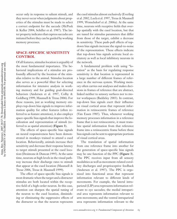
Of all features, stimulus location is arguably ofthe most fundamental importance. The be-havioral implications of a stimulus are pro-foundly affected by the location of the stim-ulus relative to the animal. Stimulus locationalso serves as a powerful filter for selectinginformation for intensive analysis in work-ing memory and for guiding goal-directedbehaviors (Andersen et al. 1997, Colby &Goldberg 1999, Maunsell & Treue 2006). Forthese reasons, just as working memory em-ploys top-down bias signals to improve infor-mation quality for other features (often re-ferred to as feature attention), it also employsspace-specific bias signals that improve the lo-calization and representation of stimuli (re-ferred to as spatial attention) (Figure 3).
The effects of space-specific bias signalson neural responsiveness have been demon-strated in monkeys trained to attend a cuedlocation. Behaviorally, animals increase theirsensitivity and decrease their response latencyto target stimuli presented at the cued loca-tion (Desimone & Duncan 1995). At the sametime, neurons at high levels in the visual path-way increase their discharge rates to stimulithat appear at the cued location (Maunsell &Treue 2006, Treue & Maunsell 1999).
The effect of space-specific bias signals ismost dramatic when the target and a distracterstimulus are both located within the recep-tive field of a high-order neuron. In this case,attention can sharpen the spatial tuning ofthe neuron to the cued location, diminish-ing or eliminating the suppressive effects ofthe distracter so that the neuron represents
the cued stimulus almost exclusively (Everlinget al. 2002, Luck et al. 1997, Treue & Maunsell1999, Womelsdorf et al. 2006a). At the sametime, neurons with receptive fields that over-lap spatially with the cued location, but thatare tuned for stimulus parameters that differfrom those of the target, exhibit a decreasein sensitivity. These push-pull effects of top-down bias signals increase the signal-to-noiseof the representation. These effects indicatethat top-down bias signals activate local ex-citatory as well as local inhibitory neurons inthe network.
A fundamental problem with using “lo-cation” as the basis for regulating responsesensitivity is that location is represented ina large number of different frames of refer-ence in the nervous system. Working mem-ory often carries out analyses and makes deci-sions in frames of reference that are abstract,linked neither to sensory surfaces nor to mo-tor workspaces (Baddeley 2003). In contrast,top-down bias signals exert their influenceon visual cortical areas that represent infor-mation in retinocentric frames of reference(Van Essen 1985). Thus, whenever workingmemory processes information in a referenceframe that is not retinocentric, it must trans-late spatial information from that referenceframe into a retinocentric frame before thesebias signals can be sent to appropriate portionsof visual cortical areas.
The translation of spatial informationfrom one reference frame into another forthe generation of space-specific bias signalsmay be one function of the PPC (Figure 3).The PPC receives input from all sensorymodalities as well as movement-related corol-lary discharges and proprioceptive feedback(Andersen et al. 1997). The PPC is orga-nized into functional areas that representinformation relevant to different kinds ofmovements. For example, the lateral intra-parietal (LIP) area represents information rel-evant to eye saccades; the medial intrapari-etal area represents information relevant toarm movements; and the ventral intraparietalarea represents information relevant to the
www.annualreviews.org • Fundamental Components of Attention 65
Ann
u. R
ev. N
euro
sci.
2007
.30:
57-7
8. D
ownl
oade
d fr
om a
rjou
rnal
s.an
nual
revi
ews.
org
by U
NIV
ER
SIT
Y O
F FL
OR
IDA
- S
mat
hers
Lib
rary
on
09/0
1/09
. For
per
sona
l use
onl
y.
ANRV314-NE30-03 ARI 7 May 2007 17:20
World
Nonspatial
Spatial
Workingmemory
Nonspatial aspects of:
• sensation• motor control• stored memory• internal state
Nonretinocentricspace
representations
Posteriorparietal cortex
(frames of reference)
Superiorcolliculus
(optic tectum)
Retinocentric
Frontal eyefields
Pulvinarnucleus
Mediodorsalthalamicnucleus
Visualcortex
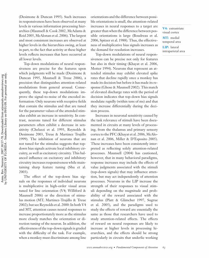
Figure 3Schema for top-down sensitivity control. Blue arrows: Bias signals that regulate neural responsiveness.Green arrows: bottom-up information filtered for salience in the superior colliculus and in visual corticalareas. Top-down bias signals from working memory that modulate the representations of nonspatialaspects of information are transmitted directly to the cognate representations (upper right). Top-downbias signals from working memory that modulate the representation of information on the basis of objectlocation are transmitted to the posterior parietal cortex (PPC) where they are represented in variousreference frames. Retinocentric bias signals are transmitted from working memory also to the frontal eyefields (FEF). Retinocentric bias signals are distributed from the PPC and FEF to retinocentric sensoryareas. Bias signals for modulating representations in other reference frames are transmitted from thePPC to the cognate representations. Bottom-up bias signals combine with top-down bias signals in allthese structures.
head (Colby & Goldberg 1999). These vari-ous representations of spatial information re-quire transformations from sensory frames ofreference into frames of reference appropri-
ate for different brain regions. Transforma-tions are accomplished by integrating sensoryspatial information with body or limb posi-tion information. For example, by integrating
66 Knudsen
Ann
u. R
ev. N
euro
sci.
2007
.30:
57-7
8. D
ownl
oade
d fr
om a
rjou
rnal
s.an
nual
revi
ews.
org
by U
NIV
ER
SIT
Y O
F FL
OR
IDA
- S
mat
hers
Lib
rary
on
09/0
1/09
. For
per
sona
l use
onl
y.
ANRV314-NE30-03 ARI 7 May 2007 17:20
eye-centered (retinocentric) visual informa-tion with eye position information, the LIPrepresents visual information relative to thehead (head-centered coordinates) (Andersenet al. 2004). Within the various functional ar-eas of the PPC, sensory information is trans-formed into a number of different coordinateframes (Figure 3; arrow to nonretinocentricspace representations.
The information about the relative po-sitions of the eyes, head, limbs, and bodycould also be used by the PPC to translatehigh-order spatial representations from work-ing memory into frames of reference that areappropriate for top-down control of sensoryprocessing areas. For example, the LIP mightcombine spatial information from workingmemory that is represented in an egocen-tric frame of reference with eye-positionand head-position information and, thereby,translate the spatial information into aretinocentric frame of reference. Once spatialbias signals are represented in a retinocentricframe of reference, they can be distributed toretinocentric sensory representations to reg-ulate their sensitivity (Figure 3; dashed box).
The distribution of retinocentric spatialbias signals may be carried out by both theLIP and the forebrain gaze control area, thefrontal eye fields (FEF) (Figure 3). The FEFis reciprocally connected with the LIP and thePFC and mediates voluntary control of gazedirection (Schiller et al. 1987, Stanton et al.1995). The role of the FEF in controllingorienting eye movements has been exploredextensively in the past, but only recently hasits role in distributing top-down bias signalsbecome appreciated (Awh et al. 2006, Mooreet al. 2003).
Psychophysicists were the first to discoverthe tight linkages that exist between gaze con-trol and spatial attention (Rizzolatti et al.1987). They found, for example, that eachtime we make a saccadic eye movement to anew location, our sensitivity to stimuli at thatlocation increases tens of ms before the eyesmove (Shepherd et al. 1986). Thus, orient-ing eye movements and spatial attention are
FEF: frontal eyefield
functionally linked (although separable) in thebrain.
The tight linkage between gaze controland spatial attention has been demonstrateddirectly by applying electrical microstimu-lation to gaze control areas in the mon-key (Moore & Fallah 2004). Monkeys weretrained to monitor a cued location in spacewithout moving the eyes (covert attentiontasks). When tested behaviorally, they ex-hibited increased sensitivity to luminancechanges of stimuli specifically at the cued lo-cation. An electrode for microstimulating thecortex was then placed in the FEF. Whenhigh current levels (50–150 μA) were deliv-ered through the electrode, the eyes made afixed-vector saccadic movement to a new lo-cation, defined in retinocentric coordinates asthe movement field for the site. When thesame FEF site was stimulated with low cur-rent levels, below the level required to evokeeye movements, monkeys demonstrated anincrease in behavioral sensitivity to stimulilocated specifically in the movement field ofthe microstimulation site, as though their at-tention had been directed to that location bythe focal activation of the FEF. Investigatorshave reported analogous results for the effectsof low-level microstimulation of the superiorcolliculus on behavioral detection of visualmotion or stimulus change (Cavanaugh et al.2006, Cavanaugh & Wurtz 2004, Muller et al.2005).
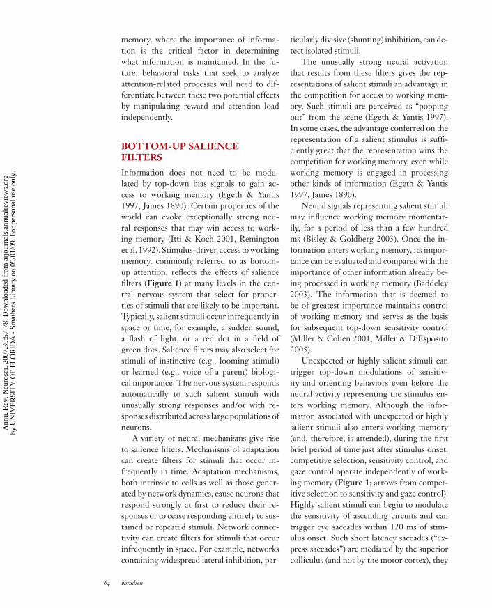
The same kind of weak electrical micros-timulation of the FEF also causes top-downbias signals to be distributed to retinotopi-cally matched areas in V4 (Armstrong et al.2006, Moore & Armstrong 2003). When theactivity of neurons in V4 was recorded duringFEF microstimulation, neurons with recep-tive fields that contained the movement fieldof the FEF site, and were tuned for the prop-erties of the stimulus, increased their respon-siveness to visual stimulation (Figure 4). Theresponse increases mimicked the response in-creases that occur in V4 when monkeys arecued to attend a location (Reynolds et al.1999).
www.annualreviews.org • Fundamental Components of Attention 67
Ann
u. R
ev. N
euro
sci.
2007
.30:
57-7
8. D
ownl
oade
d fr
om a
rjou
rnal
s.an
nual
revi
ews.
org
by U
NIV
ER
SIT
Y O
F FL
OR
IDA
- S
mat
hers
Lib
rary
on
09/0
1/09
. For
per
sona
l use
onl
y.
ANRV314-NE30-03 ARI 7 May 2007 17:20
1.00 0.5
Response(spikes/s)
80
Time fromvisual onset (s)
–8
–4
0
4
8
12
Change inresponse(spikes/s)
NonpreferredPreferred
V4 Recording
FEF Microstimulation
a b
FEF microstim RF stimulus
Figure 4Effect of FEF microstimulation on the responses of V4 neurons in the monkey. Microstimulation in theFEF and single-unit recordings in the V4 were carried out while the monkey fixated a central spot. Foreach FEF site, the current threshold for inducing an eye saccade and the direction and magnitude of thesaccades (movement field) were measured. For V4 neurons, the location of the visual receptive field andthe tuning for stimulus bar orientation were measured, and the preferred and nonpreferred barorientations were determined. (a) Microstimulation and recording sites are shown on a lateral view of themonkey brain. The horizontal bars indicate the timing of the appearance of the visual stimulus in the V4receptive field and the timing of the 50 ms FEF microstimulation. The peristimulus-time histogramshows responses of a single V4 neuron to a preferred stimulus with (red ) and without (black) low-levelFEF microstimulation; microstimulation current levels were always well below the threshold forinducing eye movement. Unit responses to the visual stimulus were enhanced immediately after FEFmicrostimulation. (b) Summary data of the average change in the responses of V4 neurons (n = 33) thatwas caused by FEF microstimulation. The data compare responses to preferred (open bars) andnonpreferred (solid bars) stimulus orientations. Error bars indicate the standard error of the mean. Whenthe movement field for the FEF site (red arrow) aligned with the V4 receptive field (dashed circle), FEFmicrostimulation induced increases in V4 responses that were much greater for the preferred than for thenonpreferred stimulus. The effect increased dramatically when a distracter stimulus was present in thevisual field (middle pair of bars). When the movement field of the FEF site did not align with the V4receptive field (right pair of bars), FEF microstimulation decreased responses to the preferred stimulus.FEF microstimulation altered V4 activity only when a stimulus was present in a unit’s receptive field, andthe direction of the effect depended specifically on the mutual alignment of the FEF movement field withthe V4 receptive field. The data are from Moore & Armstrong (2003).
AGF: arcopallialgaze field
The role of the forebrain gaze control areaas a distributor of top-down space-specificbias signals holds across species and acrosssensory modalities (Winkowski & Knudsen2006). The FEF equivalent in the avian brainis the arcopallial gaze field (AGF). In barnowls, electrical microstimulation applied tothe AGF evokes fixed-vector eye and headsaccades. Low-level microstimulation causesspace-specific increases in the responsiveness
of auditory neurons in the optic tectum (theavian equivalent of the mammalian supe-rior colliculus), similar to the effects of FEFmicrostimulation on visual responses in V4(Armstrong et al. 2006).
The bias signals elicited by FEF or AGFmicrostimulation are precise in both time andspace. The increase in neural sensitivity thatresults from microstimulation typically lasts100 ms after the end of stimulation (although
68 Knudsen
Ann
u. R
ev. N
euro
sci.
2007
.30:
57-7
8. D
ownl
oade
d fr
om a
rjou
rnal
s.an
nual
revi
ews.
org
by U
NIV
ER
SIT
Y O
F FL
OR
IDA
- S
mat
hers
Lib
rary
on
09/0
1/09
. For
per
sona
l use
onl
y.
ANRV314-NE30-03 ARI 7 May 2007 17:20
the effect lasts longer in Figure 4a) (Mooreet al. 2003, Winkowski & Knudsen 2006),about the same duration as the increase inbehavioral performance following FEF mi-crostimulation (Moore & Fallah 2004). In ad-dition, AGF microstimulation sharpens thespatial tuning of auditory neurons in the optictectum according to the spatial location rep-resented at the AGF stimulation site. Anal-ogously, FEF microstimulation enhances theresponsiveness of V4 neurons only for stim-uli located in the portion of the receptive fieldthat corresponds to the movement field of theFEF stimulation site (Armstrong et al. 2006).This sharpening of spatial tuning is analogousto the spatial sharpening of receptive fieldsthat researchers have observed in high-ordervisual areas during spatial attention tasks(Everling et al. 2002, Luck et al. 1997, Treue& Maunsell 1999, Womelsdorf et al. 2006a).Because visual receptive fields in most high-order visual areas are larger than movementfields in the FEF (Van Essen 1985), FEF mi-crostimulation is likely to sharpen visual spa-tial tuning in these areas, although this idearemains to be tested.
Single-unit recordings in the FEF indicatethat the neurons that encode top-down biassignals are different from the neurons thatexclusively encode eye saccades (Thompsonet al. 2005). In monkeys trained to suppresssaccades to specific targets, the responses ofvisual FEF neurons that encode the target arestrongly enhanced while movement-relatedFEF neurons that encode eye saccades to thatlocation are suppressed. Visual activity in theFEF represents salient stimuli and could bedistributed to other visual areas to act as biassignals for modulating sensory responsivenessin a space-specific manner (Figure 3). Thedata from this experiment are consistent witha common origin for retinocentric bias signalsand eye movement control signals, but thesesignals separate at the level of the FEF, whichcan distribute retinocentric bias signals evenwithout commanding an eye movement.
The FEF heavily projects back to the LIP,which, in turn, projects to a wide range of
cortical and subcortical sensory areas (Mooreet al. 2003, Schall et al. 1995, Stanton et al.1995). Hence, the effects of FEF microstim-ulation on space-specific modulations of neu-ronal responsiveness may well be mediatedby the LIP. However, the FEF also projectsextensively to these sensory areas and could,therefore, convey these bias signals directly.Moreover, spatial information from work-ing memory that is already represented in aretinocentric reference frame could pass di-rectly to the FEF and, from there, directly tovisual cortical areas, without being processedin the LIP. Indeed, these pathways may actin parallel (Figure 3). To determine whethereither the FEF or LIP is essential for medi-ating top-down spatial bias signals, the effectsof pharmacological inactivation of the FEF orLIP could be studied in animals trained to di-rect spatial attention based on nonspatial cues(e.g., green cue = attend to the right).
COMPETITIVE SELECTION
The selection of information for entry intoworking memory is a highly competitive pro-cess (Desimone & Duncan 1995). Informa-tion about the external world, from memorystores, and about the animal’s internal stateis processed extensively and automatically inparallel hierarchies of networks in the cen-tral nervous system. Competition for repre-sentation occurs at many levels in these hi-erarchies. The competition compares signalstrengths that result from the combined ef-fects of the quality of the encoded informa-tion, modulations by top-down bias signals,and the influences of bottom-up salience fil-ters (Figure 1). The competition at each levelhelps to eliminate the effects of distractingstimuli and to select the most salient stimulusin a given parameter space. At low levels in ahierarchy, the competition occurs within therepresentations of basic stimulus parameters(for example, stimulus location or sound fre-quency). At higher levels, the competition canoccur among neurons tuned for higher-orderfeatures (for example, shapes or types of
www.annualreviews.org • Fundamental Components of Attention 69
Ann
u. R
ev. N
euro
sci.
2007
.30:
57-7
8. D
ownl
oade
d fr
om a
rjou
rnal
s.an
nual
revi
ews.
org
by U
NIV
ER
SIT
Y O
F FL
OR
IDA
- S
mat
hers
Lib
rary
on
09/0
1/09
. For
per
sona
l use
onl
y.
ANRV314-NE30-03 ARI 7 May 2007 17:20
objects). A final competition takes place at theinterface with working memory, where dif-ferent domains of information (for example,vision, audition, or somatic sensation) com-pete for entry into working memory networks(Baddeley 2003).
Information on which the selection ofstimuli for working memory is based (i.e.,the relative salience of stimuli across the vi-sual field) is represented in the LIP (Bisley& Goldberg 2003). In monkeys trained todiscriminate visual targets in the presenceof visual distracters, neurons in the LIP en-code both the target and the distracter withelevated discharge rates that reflect the ef-fects of top-down bias signals and bottom-up salience filters, respectively. Moreover, therelative spike rates that represent the targetversus the distracter change over the courseof a trail. Spike rates are high for the targetbefore a distracter (a flashed dot) is presented.Immediately after the distracter is flashed,spike rates are higher for the distracter thanfor the target. Then, within 300 ms followingthe distracter, spike rates are again higher forthe target. The changes in the relative spikerates to the target and to the distracter cor-relate with changes in the monkey’s behav-ioral performance in discriminating the targetstimulus. These data are consistent with theproposition that the LIP contains a represen-tation of relative stimulus salience across theentire visual field and that the relative level ofactivity within this population predicts the in-formation that will gain access to the circuitryof working memory.
Thus, in addition to its proposed role intranslating top-down bias signals into variousreference frames (discussed previously), theLIP also appears to contribute importantlyto competitive selection. These functions aremutually compatible; indeed, competitive se-lection should act on networks that are modu-lated by both top-down and bottom-up mech-anisms. Although the circuits that mediatetop-down sensitivity control and competitiveselection may overlap, they are not the same.Top-down bias signals originate in a different
network and modulate sensitivity to specificinformation. In contrast, competitive selec-tion reflects a computation that is intrinsic toa network, a competition for representationthat is based on the relative strength of activ-ity (salience) across the entire network.
The competitions that contribute to theselection process take place at various hier-archical levels. These competitions have aspecial requirement: They must compare re-sponse strengths to multiple, simultaneousstimuli and select the strongest responses,whereas the information represented by eachregion of neural activation in a network is pre-served (i.e., neural activity representing dif-ferent stimuli must not be combined or av-eraged to arrive at a single solution). Thisdemand (to compare response strengths with-out altering information) indicates a specialclass of winner-take-all process. A neural net-work that could mediate such a competitioninvolves a special type of inhibitory neuronthat receives input from a restricted portion ofa network and extends inhibitory connectionsthroughout the entire network. Unlike typi-cal inhibitory neurons that operate locally forsuch purposes as contrast enhancement, regu-lation of excitability, or spike synchronization,these neurons would establish mutual inhi-bition, and therefore competition, among allchannels in a representation. A winner-take-all competition suggests that the inhibition isnonlinear (Lee et al. 1999). The gain of thenetwork could be increased with the additionof positive recurrent connections (Brody et al.2003, Major & Tank 2004, Shu et al. 2003).A network with these properties could medi-ate the final selection of information for entryinto working memory as a competition amongdifferent networks.
Inhibitory circuits that exhibit this unusualarchitecture have been described in the retinaand in the avian superior colliculus, called theoptic tectum (Famiglietti 1992, Stafford &Dacey 1997, Wang et al. 2004). The circuitin the optic tectum is of particular interestbecause this structure participates in stimu-lus selection (McPeek & Keller 2004). The
70 Knudsen
Ann
u. R
ev. N
euro
sci.
2007
.30:
57-7
8. D
ownl
oade
d fr
om a
rjou
rnal
s.an
nual
revi
ews.
org
by U
NIV
ER
SIT
Y O
F FL
OR
IDA
- S
mat
hers
Lib
rary
on
09/0
1/09
. For
per
sona
l use
onl
y.
ANRV314-NE30-03 ARI 7 May 2007 17:20
optic tectum represents the locations of visual,auditory, or somatosensory stimuli as a topo-graphic map of space. The responses of tectalneurons are suppressed by a special class ofinhibitory neuron that resides in the nucleusisthmi pars magnocellularis (Imc) (Wang et al.2004). Each Imc neuron is excited by inputfrom a discrete location in the tectal spacemap and projects back with inhibitory inputto the entire space map, except to the locationfrom which it received its excitatory input. Inaddition, Imc neurons also inhibit choliner-gic modulatory neurons that project to thosesame regions of the optic tectum and may pro-vide local positive recurrent input (Wang et al.2006). Although the unusual anatomy of thiscircuit suggests that it could mediate a winner-take-all, competitive selection for stimulus lo-cation, the function of this circuit has yet tobe determined.
Although stimulus selection is usuallydominated by cortical networks, subcorticalstructures exert a powerful influence on theselection process. This is true particularly forthe superior colliculus. As mentioned previ-ously, changes in gaze direction that are medi-ated by eye saccades cause momentary shifts inspatial attention to stimuli located at the targetof the impending eye saccade (Rizzolatti et al.1987, Shepherd et al. 1986). This implies thatchanges in gaze direction are accompanied byneural signals that cause the representation ofthe stimulus selected for the next eye saccade,to win the competition for entry into the cir-cuitry for working memory as an eye saccadeoccurs. Corollary discharges associated witheye saccades occur in the superior colliculusand propagate to the FEF, via the mediodorsalthalamic nucleus (Figure 3). In the FEF, thesecorollary discharges shift the locations of vi-sual receptive fields tens of ms before each eyesaccade so that FEF neurons represent stimuliat the future locations of their receptive fields(Sommer & Wurtz 2006). This influenceof the superior colliculus on the FEF likelycontributes to stabilization of the visual worldduring eye movements. Similar predictiveshifts of visual receptive fields occur in the
Imc: nucleus isthmipars magnocellularis
LIP before each eye saccade (Duhamel et al.1992, Umeno & Goldberg 1997). The samecorollary discharges, when they impinge onneurons with foveal receptive fields (whichrepresent the target of an impending eyesaccade) may also act as bias signals thatincrease the responsiveness of these neurons(Figure 1; arrow from gaze control to sen-sitivity control). The differentially increasedresponses of these neurons could confer amomentary competitive advantage on therepresentation of the target for an impendingeye saccade. Thus, corollary discharges fromthe superior colliculus could control stimulusselection during eye saccades.
In addition, the superior colliculus con-tributes to stimulus selection in certain vi-sual discrimination tasks (McPeek & Keller2004). In lower mammals, and even moreso in nonmammalian vertebrates, the supe-rior colliculus plays a major role in form vi-sion (Stein & Meredith 1993). In primates,however, the role of the superior colliculus inform vision has been largely usurped by thevisual cortex (Van Essen 1985). This findingmakes especially noteworthy the demonstra-tion by McPeek & Keller (2004) that the su-perior colliculus contributes to visual targetselection. In this experiment, monkeys weretrained to make an eye saccade to the oddball-colored dot in a four-dot display (e.g., to thered dot among three green dots). Both thecolor and location of the four dots were rep-resented in the visual cortex. In contrast, onlythe locations of the dots were represented inthe superior colliculus because collicular neu-rons are not selective for color. Nevertheless,when the superior colliculus was focally inac-tivated so that the representation of the target(red) stimulus was suppressed in the colliculus(but was still present in the cortex) the mon-key no longer discriminated the oddball color,but instead selected each of the 4 dots withapproximately equal probability. The condi-tions of this experiment (rapid responses toflashed stimuli) optimized the influence of thesuperior colliculus relative to that of the visualcortex because the colliculus is differentially
www.annualreviews.org • Fundamental Components of Attention 71
Ann
u. R
ev. N
euro
sci.
2007
.30:
57-7
8. D
ownl
oade
d fr
om a
rjou
rnal
s.an
nual
revi
ews.
org
by U
NIV
ER
SIT
Y O
F FL
OR
IDA
- S
mat
hers
Lib
rary
on
09/0
1/09
. For
per
sona
l use
onl
y.
ANRV314-NE30-03 ARI 7 May 2007 17:20
involved in short latency saccades, and col-licular neurons are particularly responsive tosalient (flashed) stimuli (Schiller et al. 1987,Stein & Meredith 1993). Nevertheless, the re-sults demonstrate a strong influence of the su-perior colliculus on target selection.
These results imply that information fromthe superior colliculus influences the selectionprocess in parallel with information from thevisual cortex (Figure 3). Information fromthe superior colliculus reaches the FEF viathe mediodorsal thalamic nucleus (Sommer& Wurtz 2006) and the LIP via the pulvinarnucleus, a thalamic nucleus known to play acritical role in spatial attention (Robinson &Petersen 1992). In addition, the FEF and theLIP are heavily interconnected. These path-ways provide the superior colliculus with ac-cess to representations of stimulus salience inthe FEF and LIP, and these pathways oper-ate in parallel with those that originate in thevisual cortex.
NEURAL CORRELATES OFSTIMULUS SELECTION
One difficulty in studying the neural mech-anisms that select information for workingmemory is identifying when information is,indeed, being gated into working memory.The problem is that access to working mem-ory may depend not on the absolute spike ratesof competing neurons, but rather on their rel-ative spike rates, as proposed for the LIP byBisley & Goldberg (2003). Therefore, unlessthe spike rates of all competing neurons aremonitored simultaneously, it may be impossi-ble to determine which neurons are providinginput to working memory at any point in time.
A potential solution to this problem is theobservation that neurons can exhibit a distinc-tive temporal discharge pattern when the in-
formation they encode gains access to workingmemory. Single-unit studies in monkeys, aswell as electroencephalographic studies in hu-mans, report that when an animal attends to aparticular target stimulus, neurons that repre-sent the target in high-order sensory areas, inthe PFC, and in the PPC exhibit synchronizeddischarges with a periodicity of 40–70 Hz,referred to as gamma frequencies (Baueret al. 2006, Bichot et al. 2005, Steinmetzet al. 2000, Taylor et al. 2005, Tiitinen et al.1993). The association of attentional selec-tion of stimuli with oscillations specifically inthe gamma-band is controversial, although ithas been replicated using a variety of atten-tion tasks. In a recent study (Womelsdorf et al.2006b), for example, monkeys were trained todetect a small change in the color of a tar-get at a cued location in the visual field inthe presence of a distracting stimulus at an-other location. Single-unit recordings in V4demonstrated that when the monkey attendedthe target, units tuned for the target stimulusincreased their discharge rates and synchro-nized their spikes with the local field poten-tial, which oscillated at gamma frequencies. Acomparison of the increase in discharge rateswith the increase in synchronization showedthat synchronization was more sensitive thandischarge rate as an indicator of behavioralperformance.
Oscillations at gamma frequencies occur ina wide range of networks under various con-ditions (Gray et al. 1989, Lee 2003, Liu &Newsome 2006). Clearly, they are not spe-cific for attention. However, it is temptingto hypothesize that when the synchroniza-tion of unit activity at gamma frequencies in-creases dramatically during attention tasks,the synchronized activity represents informa-tion that is entering the circuitry for workingmemory.
SUMMARY POINTSThe conceptual framework presented here proposes that attention reflects the combinedcontributions of four distinct processes: working memory, competitive selection, top-down sensitivity control, and automatic filtering for salient stimuli. Attention selects the
72 Knudsen
Ann
u. R
ev. N
euro
sci.
2007
.30:
57-7
8. D
ownl
oade
d fr
om a
rjou
rnal
s.an
nual
revi
ews.
org
by U
NIV
ER
SIT
Y O
F FL
OR
IDA
- S
mat
hers
Lib
rary
on
09/0
1/09
. For
per
sona
l use
onl
y.
ANRV314-NE30-03 ARI 7 May 2007 17:20
information that gains access to the circuitry for working memory. Access to workingmemory is determined by the relative signal strengths of competing representations ofinformation. Signal strength is modulated automatically by bottom-up salience filtersand is modulated top-down by bias signals that are controlled by working memory andby corollary discharges that accompany gaze changes. Voluntary control of attention ismediated by a recurrent loop comprising working memory, top-down sensitivity control,and competitive selection.
Information is evaluated and decisions are made in working memory. According tothe proposed framework, attention does not identify targets; working memory does. Inaddition, attention is not “deployed” but rather is an ongoing competition among infor-mation processing hierarchies vying for access to working memory. What is “deployed”are top-down bias signals based on decisions made in working memory. Top-down biassignals can selectively enhance representations of certain information so that that in-formation continues to have a high probability of gaining entry into working memory.Eye movements, along with other orienting movements, are also guided by decisionsmade in working memory and serve, together with top-down bias signals, to improvethe resolution of information provided to working memory.
FUTURE ISSUESThe framework for attention proposed in this review is intended to act as a heuristic toolto facilitate the study of neural mechanisms underlying attention. By identifying the keyfunctional components of attention, this framework allows studies of basic neural mech-anisms to be interpreted in the broader context of attention. For example, studies on theshort-term maintenance of information by persistent activity may provide insight to themechanisms of working memory. Such studies are being carried out on a wide range ofpreparations, from the entorhinal cortex to the brainstem and spinal cord (Constantinidis& Wang 2004, Fransen et al. 2006, Major & Tank 2004). Mechanisms that could un-derlie competitive selection can be explored in networks that perform winner-take-allcomputations on ther inputs. The mechanisms of top-down sensitivity control might beelucidated by studying networks that exhibit spatially and temporally precise regulationof neuronal sensitivity. Finally, a mechanistic understanding of salience filters may resultfrom examining intrinsic cellular and network mechanisms of adaptation.
A further benefit of this conceptual framework is in interpreting the symptoms ofdisease or dysfunction. Many disorders affect attention, but they do so in different ways.Different manifestations of attention disorders indicate that the components of attention,particularly working memory, competitive selection, and top-down sensitivity control,are differentially affected by disorders. For example, prominent among the symptoms ofschizophrenias is the inability to ignore irrelevant or imagined stimuli (Phillips & Silver-stein 2003), suggesting a particular problem with mechanisms of competitive selectioneither within, or for, working memory. In contrast, attention deficit disorder frequentlyincludes an inability to retain information in working memory and/or an inability tomaintain attention on a specific task (Biederman & Faraone 2005), suggesting problemswith working memory and top-down sensitivity control, respectively. These differ-ent components of attention are mediated by different, although potentially overlapping,
www.annualreviews.org • Fundamental Components of Attention 73
Ann
u. R
ev. N
euro
sci.
2007
.30:
57-7
8. D
ownl
oade
d fr
om a
rjou
rnal
s.an
nual
revi
ews.
org
by U
NIV
ER
SIT
Y O
F FL
OR
IDA
- S
mat
hers
Lib
rary
on
09/0
1/09
. For
per
sona
l use
onl
y.
ANRV314-NE30-03 ARI 7 May 2007 17:20
sets of neural mechanisms. Therefore, the development and selection of optimal ther-apies for ameliorating such disorders of attention require that we both greatly expandour knowledge of the neural mechanisms that underlie attention and diagnose the symp-toms of attention disorders precisely and in the context of this knowledge. Hopefullythe framework for attention presented here will be useful in this regard.
ACKNOWLEDGMENTS
I thank K. Armstrong, M. Goldberg, and W. Newsome for reviewing the manuscript andP. Knudsen for figure preparation. This work was supported by grants from the NationalInstitutes of Deafness and Other Communication Disorders.
LITERATURE CITED
Andersen R, Meeker D, Pesaran B, Brezen B, Buneo C, Scherberger H. 2004. Sensorimotortransformations in the posterior parietal portex. In The Cognitive Neurosciences III, ed. MSGazzaniga, pp. 463–74. Cambridge, MA: MIT Press
Andersen RA, Snyder LH, Bradley DC, Xing J. 1997. Multimodal representation of spacein the posterior parietal cortex and its use in planning movements. Annu. Rev. Neurosci.20:303–30
Armstrong KM, Fitzgerald JK, Moore T. 2006. Changes in visual receptive fields with micro-stimulation of frontal cortex. Neuron 50:791–98
Awh E, Armstrong KM, Moore T. 2006. Visual and oculomotor selection: links, causes andimplications for spatial attention. Trends Cogn. Sci. 10:124–30
Baddeley A. 2003. Working memory: looking back and looking forward. Nat. Rev. Neurosci.4:829–39
Batista AP, Andersen RA. 2001. The parietal reach region codes the next planned movementin a sequential reach task. J. Neurophysiol. 85:539–44
Bauer M, Oostenveld R, Peeters M, Fries P. 2006. Tactile spatial attention enhances gamma-band activity in somatosensory cortex and reduces low-frequency activity in parieto-occipital areas. J. Neurosci. 26:490–501
Bichot NP, Rossi AF, Desimone R. 2005. Parallel and serial neural mechanisms for visualsearch in macaque area V4. Science 308:529–34
Biederman J, Faraone SV. 2005. Attention-deficit hyperactivity disorder. Lancet 366:237–48Bisley JW, Goldberg ME. 2003. Neuronal activity in the lateral intraparietal area and spatial
attention. Science 299:81–86Boudreau CE, Williford TH, Maunsell JH. 2006. Effects of task difficulty and target likelihood
in area V4 of macaque monkeys. J. Neurophysiol. 96:2377–87Cavanaugh J, Wurtz RH. 2004. Subcortical modulation of attention counters change blindness.
J. Neurosci. 24:11236–43Chafee MV, Goldman-Rakic PS. 2000. Inactivation of parietal and prefrontal cortex re-
veals interdependence of neural activity during memory-guided saccades. J. Neurophysiol.83:1550–66
Chelazzi L, Miller EK, Duncan J, Desimone R. 1993. A neural basis for visual search in inferiortemporal cortex. Nature 363:345–47
74 Knudsen
Ann
u. R
ev. N
euro
sci.
2007
.30:
57-7
8. D
ownl
oade
d fr
om a
rjou
rnal
s.an
nual
revi
ews.
org
by U
NIV
ER
SIT
Y O
F FL
OR
IDA
- S
mat
hers
Lib
rary
on
09/0
1/09
. For
per
sona
l use
onl
y.
ANRV314-NE30-03 ARI 7 May 2007 17:20
Colby CL, Goldberg ME. 1999. Space and attention in parietal cortex. Annu. Rev. Neurosci.22:319–49
Constantinidis C, Wang XJ. 2004. A neural circuit basis for spatial working memory. Neurosci-entist 10:553–65
Corbetta M, Miezin FM, Dobmeyer S, Shulman GL, Petersen SE. 1991. Selective and dividedattention during visual discriminations of shape, color, and speed: functional anatomy bypositron emission tomography. J. Neurosci. 11:2383–402
Curtis CE. 2006. Prefrontal and parietal contributions to spatial working memory. Neuroscience139:173–80
Desimone R, Duncan J. 1995. Neural mechanisms of selective visual attention. Annu. Rev.Neurosci. 18:193–222
Diamond A, Goldman-Rakic PS. 1989. Comparison of human infants and rhesus monkeys onPiaget’s AB task: evidence for dependence on dorsolateral prefrontal cortex. Exp. BrainRes. 74:24–40
di Pellegrino G, Wise SP. 1993. Effects of attention on visuomotor activity in the premotorand prefrontal cortex of a primate. Somatosens. Mot. Res. 10:245–62
Duhamel JR, Colby CL, Goldberg ME. 1992. The updating of the representation of visualspace in parietal cortex by intended eye movements. Science 255:90–92
Duncan J, Emslie H, Williams P, Johnson R, Freer C. 1996. Intelligence and the frontal lobe:the organization of goal-directed behavior. Cognit. Psychol. 30:257–303
Egeth HE, Yantis S. 1997. Visual attention: control, representation, and time course. Annu.Rev. Psychol. 48:269–97
Everling S, Tinsley CJ, Gaffan D, Duncan J. 2002. Filtering of neural signals by focusedattention in the monkey prefrontal cortex. Nat. Neurosci. 5:671–76
Famiglietti EV. 1992. Polyaxonal amacrine cells of rabbit retina: morphology and stratificationof PA1 cells. J. Comp. Neurol. 316:391–405
Fransen E, Tahvildari B, Egorov AV, Hasselmo ME, Alonso AA. 2006. Mechanism of gradedpersistent cellular activity of entorhinal cortex layer v neurons. Neuron 49:735–46
Funahashi S, Bruce CJ, Goldman-Rakic PS. 1989. Mnemonic coding of visual space in themonkey’s dorsolateral prefrontal cortex. J. Neurophysiol. 61:331–49
Fuster JM. 1995. Memory in the Cerebral Cortex. Cambridge, MA: MIT PressFuster JM, Alexander GE. 1971. Neuron activity related to short-term memory. Science
173:652–54Genovesio A, Brasted PJ, Wise SP. 2006. Representation of future and previous spatial goals
by separate neural populations in prefrontal cortex. J. Neurosci. 26:7305–16Ghose GM, Maunsell JH. 2002. Attentional modulation in visual cortex depends on task timing.
Nature 419:616–20Gray CM, Konig P, Engel AK, Singer W. 1989. Oscillatory responses in cat visual cortex
exhibit intercolumnar synchronization which reflects global stimulus properties. Nature338:334–37
Ipata AE, Gee AL, Goldberg ME, Bisley JW. 2006. Activity in the lateral intraparietal areapredicts the goal and latency of saccades in a free-viewing visual search task. J. Neurosci.26:3656–61
Itti L, Koch C. 2001. Computational modelling of visual attention. Nat. Rev. Neurosci. 2:194–203
James W. 1890. Principles of Psychology. New York: HoltKhayat PS, Spekreijse H, Roelfsema PR. 2006. Attention lights up new object representations
before the old ones fade away. J. Neurosci. 26:138–42
www.annualreviews.org • Fundamental Components of Attention 75
Ann
u. R
ev. N
euro
sci.
2007
.30:
57-7
8. D
ownl
oade
d fr
om a
rjou
rnal
s.an
nual
revi
ews.
org
by U
NIV
ER
SIT
Y O
F FL
OR
IDA
- S
mat
hers
Lib
rary
on
09/0
1/09
. For
per
sona
l use
onl
y.
ANRV314-NE30-03 ARI 7 May 2007 17:20
Kim JN, Shadlen MN. 1999. Neural correlates of a decision in the dorsolateral prefrontalcortex of the macaque. Nat. Neurosci. 2:176–85
Koch C, Ullman S. 1985. Shifts in selective visual attention: towards the underlying neuralcircuitry. Hum. Neurobiol. 4:219–27
LaBar KS, Gitelman DR, Parrish TB, Mesulam M. 1999. Neuroanatomic overlap of workingmemory and spatial attention networks: a functional MRI comparison within subjects.Neuroimage 10:695–704
Lee D. 2003. Coherent oscillations in neuronal activity of the supplementary motor area duringa visuomotor task. J. Neurosci. 23:6798–809
Liu J, Newsome WT. 2006. Local field potential in cortical area MT: stimulus tuning andbehavioral correlations. J. Neurosci. 26:7779–90
Luck SJ, Chelazzi L, Hillyard SA, Desimone R. 1997. Neural mechanisms of spatial selectiveattention in areas V1, V2, and V4 of macaque visual cortex. J. Neurophysiol. 77:24–42
Major G, Tank D. 2004. Persistent neural activity: prevalence and mechanisms. Curr. Opin.Neurobiol. 14:675–84
Martinez-Trujillo J, Treue S. 2002. Attentional modulation strength in cortical area MT de-pends on stimulus contrast. Neuron 35:365–70
Maunsell JH, Cook EP. 2002. The role of attention in visual processing. Philos. Trans. R. Soc.London B Biol. Sci. 357:1063–72
Maunsell JH, Treue S. 2006. Feature-based attention in visual cortex. Trends Neurosci. 29:317–22
McAdams CJ, Reid RC. 2005. Attention modulates the responses of simple cells in monkeyprimary visual cortex. J. Neurosci. 25:11023–33
McAlonan K, Cavanaugh J, Wurtz RH. 2006. Attentional modulation of thalamic reticularneurons. J. Neurosci. 26:4444–50
McPeek RM, Keller EL. 2004. Deficits in saccade target selection after inactivation of superiorcolliculus. Nat. Neurosci. 7:757–63
Mesulam MM. 1999. Spatial attention and neglect: parietal, frontal and cingulate contributionsto the mental representation and attentional targeting of salient extrapersonal events.Philos. Trans. R Soc. London B Biol. Sci. 354:1325–46
Miller BT, D’Esposito M. 2005. Searching for “the top” in top-down control. Neuron 48:535–38
Miller EK, Cohen JD. 2001. An integrative theory of prefrontal cortex function. Annu. Rev.Neurosci. 24:167–202
Miller EK, Erickson CA, Desimone R. 1996. Neural mechanisms of visual working memoryin prefrontal cortex of the macaque. J. Neurosci. 16:5154–67
Moore T, Armstrong KM. 2003. Selective gating of visual signals by microstimulation of frontalcortex. Nature 421:370–73
Moore T, Armstrong KM, Fallah M. 2003. Visuomotor origins of covert spatial attention.Neuron 40:671–83
Moore T, Fallah M. 2004. Microstimulation of the frontal eye field and its effects on covertspatial attention. J. Neurophysiol. 91:152–62
Motter BC. 1994. Neural correlates of feature selective memory and pop-out in extrastriatearea V4. J. Neurosci. 14:2190–99
Muller JR, Philiastides MG, Newsome WT. 2005. Microstimulation of the superior colliculusfocuses attention without moving the eyes. Proc. Natl. Acad. Sci. USA 102:524–29
Phillips WA, Silverstein SM. 2003. Convergence of biological and psychological perspectiveson cognitive coordination in schizophrenia. Behav. Brain Sci. 26:65–82
76 Knudsen
Ann
u. R
ev. N
euro
sci.
2007
.30:
57-7
8. D
ownl
oade
d fr
om a
rjou
rnal
s.an
nual
revi
ews.
org
by U
NIV
ER
SIT
Y O
F FL
OR
IDA
- S
mat
hers
Lib
rary
on
09/0
1/09
. For
per
sona
l use
onl
y.
ANRV314-NE30-03 ARI 7 May 2007 17:20
Platt ML, Glimcher PW. 1997. Responses of intraparietal neurons to saccadic targets andvisual distractors. J. Neurophysiol. 78:1574–89
Powell KD, Goldberg ME. 2000. Response of neurons in the lateral intraparietal area to adistractor flashed during the delay period of a memory-guided saccade. J. Neurophysiol.84:301–10
Remington RW, Johnston JC, Yantis S. 1992. Involuntary attentional capture by abrupt onsets.Percept. Psychophys. 51:279–90
Reynolds JH, Chelazzi L, Desimone R. 1999. Competitive mechanisms subserve attention inmacaque areas V2 and V4. J. Neurosci. 19:1736–53
Reynolds JH, Pasternak T, Desimone R. 2000. Attention increases sensitivity of V4 neurons.Neuron 26:703–14
Reynolds JH, Desimone R. 2003. Interacting roles of attention and visual salience in V4. Neuron37:853–63
Rizzolatti G, Riggio L, Dascola I, Umilta C. 1987. Reorienting attention across the horizontaland vertical meridians: evidence in favor of a premotor theory of attention. Neuropsychologia25:31–40
Robinson DL, Petersen SE. 1992. The pulvinar and visual salience. Trends Neurosci. 15:127–32Schall JD, Morel A, King DJ, Bullier J. 1995. Topography of visual cortex connections with
frontal eye field in macaque: convergence and segregation of processing streams. J.Neurosci. 15:4464–87
Schiller PH, Sandell JH, Maunsell JH. 1987. The effect of frontal eye field and superiorcolliculus lesions on saccadic latencies in the rhesus monkey. J. Neurophysiol. 57:1033–49
Schumacher EH, Lauber E, Awh E, Jonides J, Smith EE, Koeppe RA. 1996. PET evidence foran amodal verbal working memory system. Neuroimage 3:79–88
Schwartz ML, Goldman-Rakic PS. 1984. Callosal and intrahemispheric connectivity of theprefrontal association cortex in rhesus monkey: relation between intraparietal and principalsulcal cortex. J. Comp. Neurol. 226:403–20
Shepherd M, Findlay JM, Hockey RJ. 1986. The relationship between eye movements andspatial attention. Q. J. Exp. Psychol. A 38:475–91
Shomstein S, Behrmann M. 2006. Cortical systems mediating visual attention to both objectsand spatial locations. Proc. Natl. Acad. Sci. USA 103:11387–92
Shomstein S, Yantis S. 2004. Control of attention shifts between vision and audition in humancortex. J. Neurosci. 24:10702–6
Shu Y, Hasenstaub A, McCormick DA. 2003. Turning on and off recurrent balanced corticalactivity. Nature 423:288–93
Smith EE, Jonides J, Koeppe RA. 1996. Dissociating verbal and spatial working memory usingPET. Cereb. Cortex 6:11–20
Sommer MA, Wurtz RH. 2006. Influence of the thalamus on spatial visual processing in frontalcortex. Nature 444:374–77
Stafford DK, Dacey DM. 1997. Physiology of the A1 amacrine: a spiking, axon-bearing in-terneuron of the macaque monkey retina. Vis. Neurosci. 14:507–22
Stanton GB, Bruce CJ, Goldberg ME. 1995. Topography of projections to posterior corticalareas from the macaque frontal eye fields. J. Comp. Neurol. 353:291–305
Stein BE, Meredith MA. 1993. The Merging of the Senses. Cambridge, MA: MIT PressSteinmetz PN, Roy A, Fitzgerald PJ, Hsiao SS, Johnson KO, Niebur E. 2000. Attention
modulates synchronized neuronal firing in primate somatosensory cortex. Nature 404:187–90
Sugrue LP, Corrado GS, Newsome WT. 2005. Choosing the greater of two goods: neuralcurrencies for valuation and decision making. Nat. Rev. Neurosci. 6:363–75
www.annualreviews.org • Fundamental Components of Attention 77
Ann
u. R
ev. N
euro
sci.
2007
.30:
57-7
8. D
ownl
oade
d fr
om a
rjou
rnal
s.an
nual
revi
ews.
org
by U
NIV
ER
SIT
Y O
F FL
OR
IDA
- S
mat
hers
Lib
rary
on
09/0
1/09
. For
per
sona
l use
onl
y.
ANRV314-NE30-03 ARI 7 May 2007 17:20
Suzuki WA, Miller EK, Desimone R. 1997. Object and place memory in the macaque entorhinalcortex. J. Neurophysiol. 78:1062–81
Taylor K, Mandon S, Freiwald WA, Kreiter AK. 2005. Coherent oscillatory activity in monkeyarea v4 predicts successful allocation of attention. Cereb. Cortex 15:1424–37
Thompson KG, Biscoe KL, Sato TR. 2005. Neuronal basis of covert spatial attention in thefrontal eye field. J. Neurosci. 25:9479–87
Tiitinen H, Sinkkonen J, Reinikainen K, Alho K, Lavikainen J, Naatanen R. 1993. Selectiveattention enhances the auditory 40-Hz transient response in humans. Nature 364:59–60
Treue S, Maunsell JH. 1999. Effects of attention on the processing of motion in macaque middletemporal and medial superior temporal visual cortical areas. J. Neurosci. 19:7591–602
Umeno MM, Goldberg ME. 1997. Spatial processing in the monkey frontal eye field. I. Pre-dictive visual responses. J. Neurophysiol. 78:1373–83
Van Essen DC. 1985. Functional organization of primate visual cortex. In Cerebral Cortex, ed.Peters A., Jones EG. New York: Plenum
Vendrell P, Junque C, Pujol J, Jurado MA, Molet J, Grafman J. 1995. The role of prefrontalregions in the Stroop task. Neuropsychologia 33:341–52
Wang Y, Luksch H, Brecha NC, Karten HJ. 2006. Columnar projections from the choliner-gic nucleus isthmi to the optic tectum in chicks (Gallus gallus): a possible substrate forsynchronizing tectal channels. J. Comp. Neurol. 494:7–35
Wang Y, Major DE, Karten HJ. 2004. Morphology and connections of nucleus isthmi parsmagnocellularis in chicks (Gallus gallus). J. Comp. Neurol. 469:275–97
Williford T, Maunsell JH. 2006. Effects of spatial attention on contrast response functions inmacaque area V4. J. Neurophysiol. 96:40–54
Winkowski DE, Knudsen EI. 2006. Top-down gain control of the auditory space map by gazecontrol circuitry in the barn owl. Nature 439:336–39
Womelsdorf T, Anton-Erxleben K, Pieper F, Treue S. 2006a. Dynamic shifts of visual receptivefields in cortical area MT by spatial attention. Nat. Neurosci. 9:1156–60
Womelsdorf T, Fries P, Mitra PP, Desimone R. 2006b. Gamma-band synchronization in visualcortex predicts speed of change detection. Nature 439:733–36
Yoshida W, Ishii S. 2006. Resolution of uncertainty in prefrontal cortex. Neuron 50:781–89
Visual Perception and Memory: A New View of Medial TemporalLobe Function in Primates and RodentsElisabeth A. Murray, Timothy J. Bussey, and Lisa M. Saksida � � � � � � � � � � � � � � � � � � � � � � � � 99
Mechanisms that Regulate Establishment, Maintenance, andRemodeling of Dendritic FieldsJay Z. Parrish, Kazuo Emoto, Michael D. Kim, and Yuh Nung Jan � � � � � � � � � � � � � � � � �399