27
Fundoscopic examination
| Date post: | 14-Dec-2015 |
| Category: |
Documents |
| Upload: | skylar-leedom |
| View: | 218 times |
| Download: | 0 times |

Fundoscopicexamination

Fundoscopic Examination Window to the
blood vessels Prerequisites-
Good ophthalmoscope
A large pupil A still field

Fundoscopic Examination
Diminish illumination in the room( to overcome light reflex)
Instruct the pt to look at a distant point, which is clearly defined( to overcome accomodation and keeping the eye still)
Rt eye for examining rt fundus, lt eye for left fundus
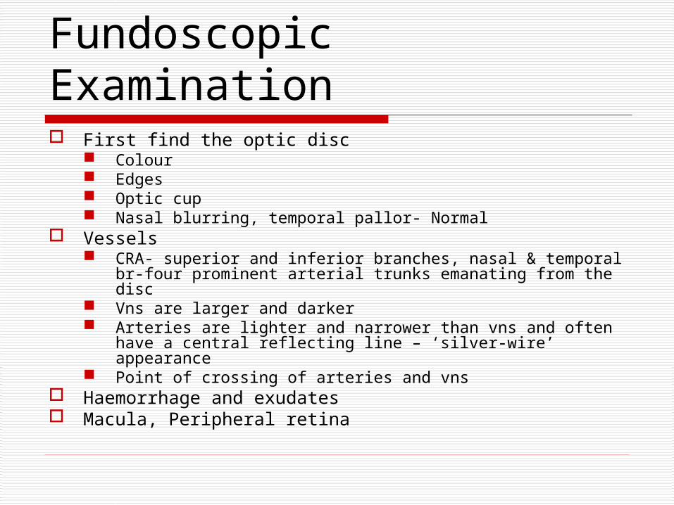
Fundoscopic Examination First find the optic disc
Colour Edges Optic cup Nasal blurring, temporal pallor- Normal
Vessels CRA- superior and inferior branches, nasal & temporal br-four
prominent arterial trunks emanating from the disc Vns are larger and darker Arteries are lighter and narrower than vns and often have a
central reflecting line – ‘silver-wire’ appearance Point of crossing of arteries and vns
Haemorrhage and exudates Macula, Peripheral retina

Optic disc
Optic cup
Vein
Arterioles
Fovea
Normal Ocular Fundus

Fundoscopic Examination Cup to Disk Ratio
Diameter of the cupped region of the optic nerve head
divided by the diameter of the optic nerve head.
Normal is ~0.3-0.5. Abnormal values are
higher and are associated with
glaucomaC/D = 0.6

Cotton Wool SpotsCotton Wool Spots Cotton wool spots result
from occlusion of retinal pre-capillary arterioles supplying the nerve fibre layer with concomitant swelling of local nerve fibre axons. Also called "soft exudates" or "nerve fibre layer infarctions" they are white, fluffy lesions in the nerve fibre layer.
Papilloedema, HTN, PAN, retinal embolism, severe anaemia

Hard exudates Hard exudates
Hard exudates ( Intra-retinal lipid exudates ) are yellow deposits of lipid and protein within the retina. Accumulations of lipids leak from surrounding capillaries and microaneurysms, they may form a circinate pattern.
Hyperlipidemia may correlate with the development of hard exudates.

Papilledema- Definition
Swelling of optic disc Arbitrarily, the term has been
reserved for the passive disc swelling associated with raised ICP
Usually bilateral, although it may be asymmetrical
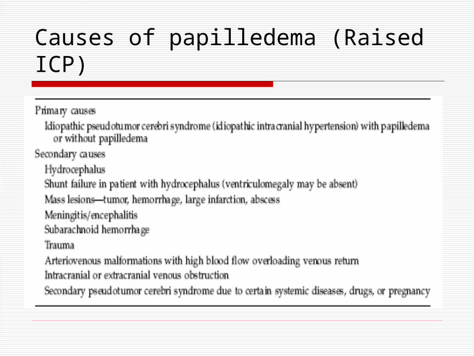
Causes of papilledema (Raised ICP)

Clinical Features of Papilledema Usually bilateral but may be unilateral or asymmetric Usually preserved visual acuity and color vision early May have transient visual loss lasting seconds
(obscurations of vision) Visual field defects
Enlarged blind spot Generalized constriction Glaucomatous-like defects Eventual peripheral constriction, especially nasally
No afferent pupillary defect
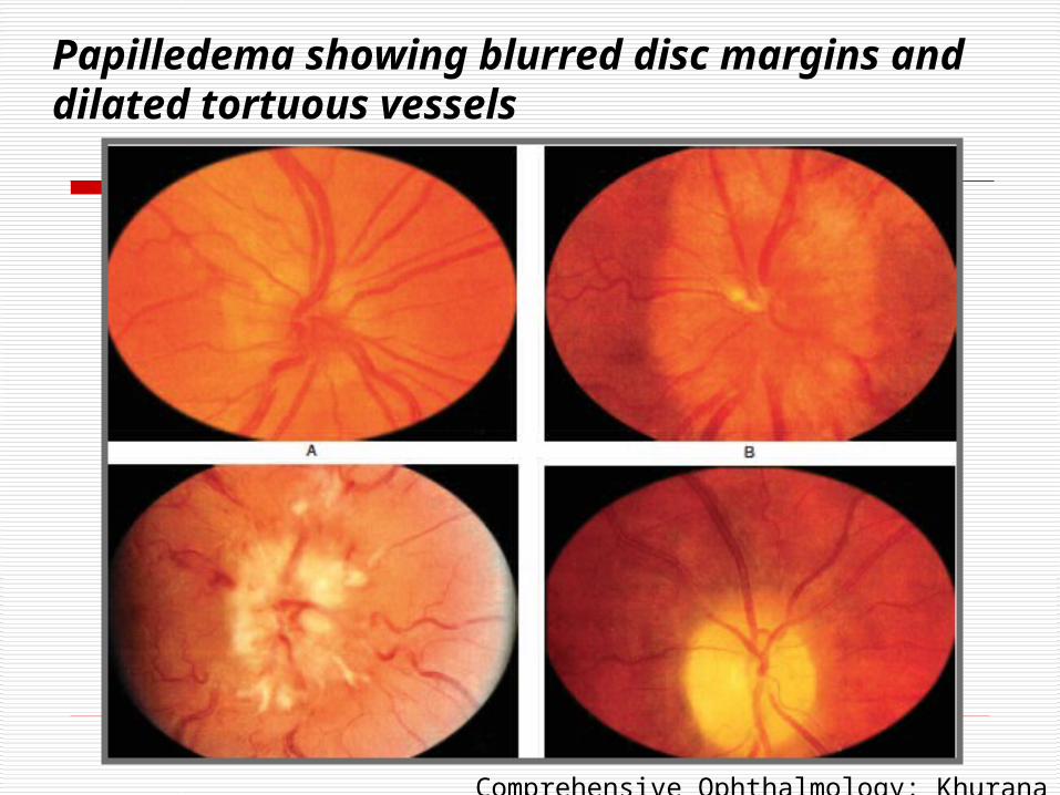
Papilledema showing blurred disc margins and dilated tortuous vessels
Comprehensive Ophthalmology: Khurana
Early papilledema Minimal disc hyperemia with capillary dilation Early opacification of nerve fiber layer (peripapillary
retina loses its superficial linear and curvilinear light reflex and appears red without luster)
Early swelling of disc Absence of venous pulsations Peripapillary retinal nerve fiber layer hemorrhage
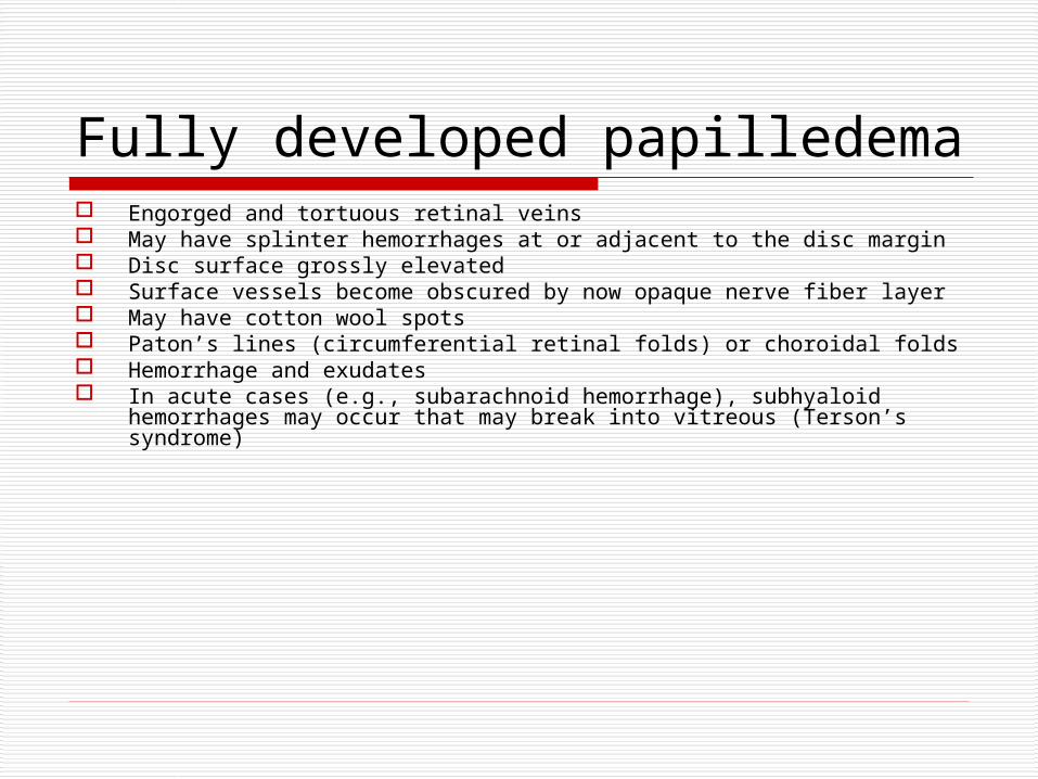
Fully developed papilledema Engorged and tortuous retinal veins May have splinter hemorrhages at or adjacent to the disc margin Disc surface grossly elevated Surface vessels become obscured by now opaque nerve fiber layer May have cotton wool spots Paton’s lines (circumferential retinal folds) or choroidal folds Hemorrhage and exudates In acute cases (e.g., subarachnoid hemorrhage), subhyaloid hemorrhages
may occur that may break into vitreous (Terson’s syndrome)

Frisen Papilledema Grading System – Stage 1 Obscuration of the nasal
border of the disc No elevation of the disc
borders Disruption of the normal
radial nerve fiber layer (NFL) arrangement with grayish opacity accentuating nerve fiber bundles
Normal temporal disc margin
Subtle grayish halo with temporal gap
C-shaped halo with a temporal gap

Frisen Papilledema Grading System – Stage 2
Obscuration of all borders
Elevation of nasal border
Complete peripapillary halo
Halo becomes circumferential

Frisen Papilledema Grading System – Stage 3 Obscuration of all
borders Elevation of all borders Increased diameter of
the optic nerve head Obscuration of one or
more segments of major blood vessels leaving the disc
Peripapillary halo—irregular outer fringe with finger-like extensions
Loss of major vessels as they leave the disc (arrow)
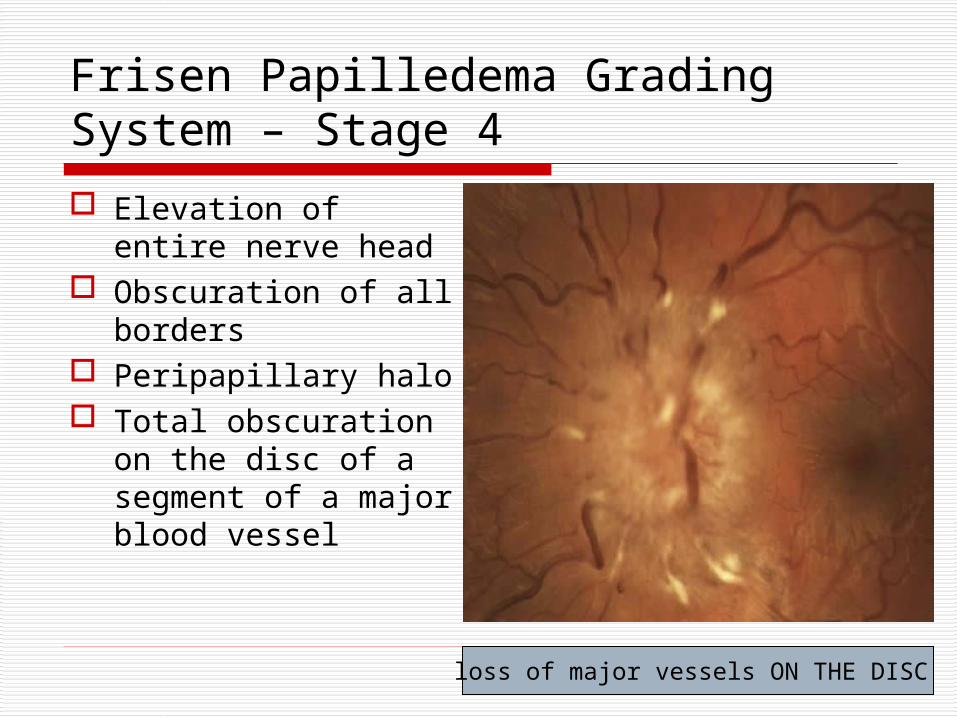
Frisen Papilledema Grading System – Stage 4
Elevation of entire nerve head
Obscuration of all borders
Peripapillary halo Total obscuration
on the disc of a segment of a major blood vessel
loss of major vessels ON THE DISC

Frisen Papilledema Grading System – Stage 5 Dome-shaped
protrusions representing anterior expansion of the optic nerve head
Peripapillary halo is narrow and smoothly demarcated
Total obscuration of a segment of a major blood vessel may or may be present
Obliteration of the optic cup
Grade IV plus partial or total obscuration of all vessels of the disc

Pseudopapilledema
Optic nerve drusen Medullated nerve fiber Hypermetropic disc Congenital anomalous elevation

Optic atrophy - Definition
Optic nerve shrinkage from any process that produce degeneration of axons in the ant.visual (Retinogeniculate) pathway

CLASSIFICATION OF OPTIC ATROPHY
PRIMARY- SECONDARY –
Post- papilloedemic optic atrophy Post-Neuritic optic atrophy Glaucomatous optic atrophy Consecutive optic atrophy

PRIMARY OPTIC ATROPHY Optic nerve fibers
degenerate in an orderly manner and are replaced by columns of glial cells without alteration in the architecture of the optic nerve head
Pale disc Chalky white(full moon
against a dark red sky) Clear margin of
disc/sharply demarcated Normal cup Well seen lamina
cribrosa Normal retinal vessels

Secondary optic atrophy Optic nerve fibers exhibit marked degeneration, with
excessive proliferation of glial tissue The architecture is lost, resulting in indistinct margins.
The disc is grey or dirty grey , looks pale with a greenish tinge
The margins are poorly defined, The lamina cribrosa is obscured due to proliferating
fibroglial tissue. Hyaline bodies (corpora amylacea) or drusen may be
observed. Peripapillary sheathing of arteries as well as tortuous
veins may be observed.

Secondary optic atrophy
OPTIC ATROPHY The Kestenbaum count is the number
of capillaries observed on the optic disc.
The normal count is approximately 10.
In optic atrophy, the number of these capillaries reduces to less than 6; in a hyperemic disc, the count is more than 12


