Gastric antral diaphragm in an adult: Case report and review CASE REPORT a 25-year-old man was transferred from the Philippines to Scott AFB Medical Center in December 1975 for evaluation. Review of his past history indicated that the patient had undergone a vagotomy and Bilroth I anastomosis in November 1974 for duodenal ulcer. Several days post- operatively, the patient developed symptoms of high intestinal obstruction. Atthe second operation, in December 1974, the duodenostomy was markedly narrowed with dense fibrosis around the area of anastomosis. The fibrotic area was resect- ed, and a Bilroth II anastomosis was performed. During the next 12 months, the patient developed postprandial early dumping and intermittent bilious vomiting. Epigastric pain recurred previous to his transfer to the United States. The patient did not regain the 32 pounds he lost during his hospitalization and operations. On admission to Scott AFB Medical Center, the patient appeared thin, anxious, but in no acute distress. Physical examination revealed 2 well-healed paramedian surgical scars and epigastric tenderness. Hemogram, urinalysis, SMA-1B, serum folate, chest x-ray, serum triglycerides, EKG, stool for ova and parasites, and quantitative stool fat were unremarkable. The patient was given a "dumping diet" with 6 feedings daily and propantheline bromide (ProBanthine) 15 mg 4 times daily. Diarrhea and abdominal cramping subsided, but inter- mittent epigastric distress persisted. Upper gastrointestinal radiography was interpreted as showing deformity of the gastric side of the gastrojejunostomy. Endoscopy was per- formed to evaluate this area. The Olympus GIF-D2 was passed without difficulty. The esophagus was normal. The stomach was noted to be diminished in size by approximately 40%. Some erythema and white exudate were seen covering half the dorsal surface of the stoma. The instrument was advanced into the jejunostomy. In the afferent limb of the jejunum, a small, tubular, white structure was seen (Figure 1). Over the next 10 minutes, this object was noted to undulate and contract, rapidly advancing toward the efferent section of Ithe jejunos- tomy. On closer inspection an ascaris was identified, and its locomotion was carefully observed. The biopsy forceps were used to grasp the worm, and the entire instrument was re- The first case of a gastric antral diaphragm in an adult was reported in 1949 by Sames.' A 40-year-old woman with peptic ulcer symptoms for 6 years had a radiographically demon- strated ulcer of the lesser curvature, Also noted was a con- comitant prepyloric diaphragm, 1.5 cm proximal to the pylorus, 3 mm thick with a3 mm opening. Histologically, antral diaphragmatic membranes have been shown to be gastric mucosa on either side with muscularis mucosa and submucosa. 2 ,3 Sokol' first described and photographed the gastroscopic appearance of a mucosal diaphragm in 1965. Since then, 11 'Reprint requests: Teresita javier-Gabriel, MD, Department of Medicine, Southern California Permanente Medical Group, 9985 Sierra Avenue, Fontana, California 92335. 88 moved. The parasite was 23 cm long. Several additional stools were examined for ova, both before and after treatment with piperazine citrate (3 g orally for 2 days). No parasites could be identified. The patient's stomal ulcer was treated with ant- acids, and his epigastric distress subsided. DISCUSSION Details concerning the movement of ascarids were first described by Makidono in 1956. 2 ,3 He used fluoroscopy to study the movement of ascarids in the small intestine, the only habitat of these adult nematodes; 90% of the parasites were in the jejunum while most of the remainder were in the ileum. Since ascarids are unable to move effec- tively outside the human body, in vivo study was necessary. His attempts to compress the abdomen or externally stimulate motion of the parasite during fluoroscopy were unsuccessful. The nematode discovered during this patient's endoscopy was actively motile. The parasite was noted to move by both extending its front and contracting its hind portion. Also locomotion was carried out by coiling its body and extending its front section, Insufflation of air and water into the vicinity of the nematode stimulated withdrawal of the ascarid. Pushing against the organism with the biopsy forceps caused it to try to scurry down the efferent limb of the jejunostomy, Movement was independent of peristalsis. Once the midsection was firmly grasped by the biopsy forceps, all movement ceased. Ascarids have been identified in the past by radiography, by presence of ova in the stool, or when causing surgical emergencies.' The lack of previous endoscopic identification is probably related to the usual inability to examine the jejunum. Patients who have undergone gastrojejunostomies provide an opportunity to see these parasites, With the de- velopment of instruments to examine the small bowel, these parasites will be encountered more frequently. REFERENCES 1. PIGGOTT J, HANS BARGER EA, NEAFIE RC: Human ascariasis. Am JOin Path 53:223, 1970 2. MAKIDONO J: Observations on Ascaris under Fluoroscopy. Am J Trap Med Hyg 5:699, 1956 3. PAUL M: The movement ofthe adult Ascaris fumbricoides. Br J Surg 59:437, 1972 4. SAW HS, SOMAsUNDARAM K, KAMATH R: Hepatic ascariasis. Arch Surg 108:733, 1974 Teresita Javier-Gabriel, MD" Section of Gastroenterology Department of Medicine Southern California Permanente Medical Group and Kaiser Foundation Hospital Fontana, California cases have been diagnosed with gastroscopy as a main or ancillary procedure. Fifty-one cases of gastric antral webs or diaphragms have been reported as of September 1974. 5 - 6 In 12 of these, a complete diaphragm was present, all of which were found in infants less than 2 weeks of age.- The age at diagnosis ranged from infancy to the eighth decade, with the majority beyond age 40. 7 - 10 Most of the previously reported cases were as- sociated with obstructive symptoms and gastric or duodenal ulcer disease, In infants, postprandial vomiting with second- ary failure to thrive and respiratory distress were the presenting symptoms. 7 Diagnosis had been made by radiographic tech- niques, surgery, and, in the past 10 years, by gastroscopy, GASTROINTESTINAL ENDOSCOPY
Transcript
Gastric antral diaphragm in an adult: Case report and review
CASE REPORT a 25-year-old man was transferred from thePhilippines to Scott AFB Medical Center in December 1975for evaluation. Review of his past history indicated that thepatient had undergone a vagotomy and Bilroth I anastomosisin November 1974 for duodenal ulcer. Several days postoperatively, the patient developed symptoms of high intestinalobstruction. Atthe second operation, in December 1974, theduodenostomy was markedly narrowed with dense fibrosisaround the area of anastomosis. The fibrotic area was resected, and a Bilroth II anastomosis was performed. During thenext 12 months, the patient developed postprandial earlydumping and intermittent bilious vomiting. Epigastric painrecurred previous to his transfer to the United States. Thepatient did not regain the 32 pounds he lost during hishospitalization and operations.
On admission to Scott AFB Medical Center, the patientappeared thin, anxious, but in no acute distress. Physicalexamination revealed 2 well-healed paramedian surgicalscars and epigastric tenderness. Hemogram, urinalysis,SMA-1B, serum folate, chest x-ray, serum triglycerides, EKG,stool for ova and parasites, and quantitative stool fat wereunremarkable.
The patient was given a "dumping diet" with 6 feedingsdaily and propantheline bromide (ProBanthine) 15 mg 4 timesdaily. Diarrhea and abdominal cramping subsided, but intermittent epigastric distress persisted. Upper gastrointestinalradiography was interpreted as showing deformity of thegastric side of the gastrojejunostomy. Endoscopy was performed to evaluate this area. The Olympus GIF-D2 was passedwithout difficulty. The esophagus was normal. The stomachwas noted to be diminished in size by approximately 40%.Some erythema and white exudate were seen covering half thedorsal surface of the stoma. The instrument was advanced intothe jejunostomy. In the afferent limb of the jejunum, a small,tubular, white structure was seen (Figure 1). Over the next 10minutes, this object was noted to undulate and contract,rapidly advancing toward the efferent section of Ithe jejunostomy. On closer inspection an ascaris was identified, and itslocomotion was carefully observed. The biopsy forceps wereused to grasp the worm, and the entire instrument was re-
The first case of a gastric antral diaphragm in an adult wasreported in 1949 by Sames.' A 40-year-old woman with pepticulcer symptoms for 6 years had a radiographically demonstrated ulcer of the lesser curvature, Also noted was a concomitant prepyloric diaphragm, 1.5 cm proximal to thepylorus, 3 mm thick with a 3 mm opening. Histologically,antral diaphragmatic membranes have been shown to begastric mucosa on either side with muscularis mucosa andsubmucosa. 2,3
Sokol' first described and photographed the gastroscopicappearance of a mucosal diaphragm in 1965. Since then, 11
'Reprint requests: Teresita javier-Gabriel, MD, Department of Medicine,Southern California Permanente Medical Group, 9985 Sierra Avenue, Fontana,
California 92335.
88
moved. The parasite was 23 cm long. Several additional stoolswere examined for ova, both before and after treatment withpiperazine citrate (3 g orally for 2 days). No parasites could beidentified. The patient's stomal ulcer was treated with antacids, and his epigastric distress subsided.DISCUSSION Details concerning the movement of ascaridswere first described by Makidono in 1956.2,3 He usedfluoroscopy to study the movement of ascarids in the smallintestine, the only habitat of these adult nematodes; 90% ofthe parasites were in the jejunum while most of the remainderwere in the ileum. Since ascarids are unable to move effectively outside the human body, in vivo study was necessary.His attempts to compress the abdomen or externally stimulatemotion of the parasite during fluoroscopy were unsuccessful.
The nematode discovered during this patient's endoscopywas actively motile. The parasite was noted to move by bothextending its front and contracting its hind portion. Alsolocomotion was carried out by coiling its body and extending
its front section, Insufflation of air and water into the vicinity ofthe nematode stimulated withdrawal of the ascarid. Pushingagainst the organism with the biopsy forceps caused it to try toscurry down the efferent limb of the jejunostomy, Movementwas independent of peristalsis. Once the midsection wasfirmly grasped by the biopsy forceps, all movement ceased.
Ascarids have been identified in the past by radiography, bypresence of ova in the stool, or when causing surgicalemergencies.' The lack of previous endoscopic identificationis probably related to the usual inability to examine thejejunum. Patients who have undergone gastrojejunostomiesprovide an opportunity to see these parasites, With the development of instruments to examine the small bowel, theseparasites will be encountered more frequently.
REFERENCES1. PIGGOTT J, HANSBARGER EA, NEAFIE RC: Human ascariasis. Am JOin Path
53:223, 19702. MAKIDONO J: Observations on Ascaris under Fluoroscopy. Am J Trap Med
Hyg 5:699, 19563. PAUL M: The movement ofthe adult Ascaris fumbricoides. Br J Surg 59:437,
Teresita Javier-Gabriel, MD"Section of Gastroenterology
Department of MedicineSouthern California Permanente Medical Group
and Kaiser Foundation HospitalFontana, California
cases have been diagnosed with gastroscopy as a main orancillary procedure.
Fifty-one cases of gastric antral webs or diaphragms havebeen reported as of September 1974.5- 6 In 12 of these, acomplete diaphragm was present, all of which were found ininfants less than 2 weeks of age.- The age at diagnosis rangedfrom infancy to the eighth decade, with the majority beyondage 40.7
- 10 Most of the previously reported cases were associated with obstructive symptoms and gastric or duodenalulcer disease, In infants, postprandial vomiting with secondary failure to thrive and respiratory distress were the presentingsymptoms. 7 Diagnosis had been made by radiographic techniques, surgery, and, in the past 10 years, by gastroscopy,
GASTROINTESTINAL ENDOSCOPY
Figure 1 . A thin, fixed, band-like contraction traverses the prepyloric.antrum.
With improved fiberoptic techniques combined withhypotonic duodenography, non-obstructuve diaphragms arebeing diagnosed with greater precision.CASE REPORT This 41-year-old woman gave a long-standinghistory of intermittent abdominal pains since childhood.There was no associated nausea, vomiting, or heartburn.There were no symptoms of respiratory difficulty, and she hada normal growth pattern. Pains were not of sufficient severityas to interfere with her usual activities.
Since January 1975 she had more frequent symptoms ofabdominal pain with gas, burping, fullness, and a distendedfeeling in the abdomen. There was occasional nausea but novomiting. She could not eat a full meal because of the feelingof distention and aggravation of the pain. In spite of this therehad been no weight loss.
Physical examination revealed a well developed patient(weight, 169 Ibs; height 170 cm). The physical findings wereessentially unremarkable. There was an appendectomy scarbut no abdominal mass, tenderness, distention, or succussionsplash. Laboratory data were within normal limits.
Upper gastrointestinal radiography showed a short, bandlike segment of concentric narrowing at the distal gastricantrum with an intact mucosal pattern. Muscular contractionspassed normally through this area, although the constrictedband never dilated completely. Barium passed freely into theduodenum. Intravenous glucagon promptly halted peristaltic activity, and the contracted area remained unchanged(Figure 1).
Figure 2. A, Gastroscopic picture of gastric antral diaphragm withtiny aperture centrally located on a mound-like area. S, A polyvinylcatheter has been passed through the narrow aperture.
VOLUME 23, NO.2, 1976
At endoscopy, the distal antrum was so narrow that only asmall concentric opening was seen in the middle of a small,mound-like area (Figure 2A). The mucosa appeared normalwith no evidence of ulceration, induration, or biliary reflux.With the passage of peristaltic waves the aperture was obliterated. A repeated endoscopy 1 month later showed the samefindings. A polyvinyl catheter was passed through the biopsychannel of the instrument and was threaded through theaperture of the gastric diaphragm. The initial cannulation wasdifficult, but once it passed through there was no resistance,and the catheter was pushed into the duodenum. Fifty milliliters of water were injected through the catheter, and only afew milliliters regurgitated back through the orifice, indicatingthat there was free flow into the duodenum (Figure 2 B).
At operation, 1.5 cm proximal to a normal pylorus, therewas a gastric antral diaphragm with a 5 mm diameter lumenand a thickness of 5 mm. On palpation the duodenum anddistal antrum were grossly normal without evidence of scarring or ulceration. The antral diaphragm was incised, and apyloroplasty of the Heinecke-Mikulicz type was done.DISCUSSION The occurrence of gastric webs or diaphragmsin infants strongly suggests a congenital origin."'" During thesixth week of fetal development the solid cord of gutepithelium canalizes. Failure of this embryologic process tooccur has been thought to be an etiologic factor.','2 Examination of the diaphragm has shown no fibrosis or inflammatorychanges. Histologically it is usually composed of 2 mucosalsurfaces enclosing a layer of submucosa and muscularismucosa.13 The serosal and muscular coasts are not included inthe formation of the diaphragm. 5 Another theory suggests thatischemic episodes during the embryologic developmentcause these anomalies. ' °
The possibility that this may be an acquired abnormality issuggested by its association with ulcer disease. 3,'0,,,,lJ,14
Concomitant ulceration has been reported in about one-fourthof cases at the time of diagnosis. 8 Rhind 14 suggested that it wasan acquired anomaly produced by scarring from an annularulceration and hypothesized that these were due to healing oflinear circumferential ulcers. Others believe that ulceration issecondary to gastric stasis induced by the web with resultantexcessive antral stimu lation. 10 Ulceration and inflammationmay then further decrease the size of the antral aperture.
In the case reported here there is a strong support for acongenital origin in view of the chronicity of symptoms sincechildhood and the absence of evidence of inflammation,erosion, or scarring. The reason for progression ofthe patient'ssymptoms in recent months can only be conjectured. Someauthors attribute the late onset of symptoms to narrowing ofthe aperture due to recurring erosions or gastritis. l3
·l5 It has
also been postulated that the diaphragm becomes less distensible with aging. 13 Poor mastication by edentulous patientsand decreased gastric peristalsis have been implicated in theonset of symptoms in adult life. 5'13 Initially peristalsis may beable to force the food through the small aperture, but withaging the stomach may lose its tone.'o
Symptoms relate to the size of the aperture. Generally,apertures larger than 1 cm pose no problems, but diaphragmswith narrow openings may result in complaints of pain, vomiting, fullness, epigastric and postprandial discomfort, and evenweight loss.
The radiographic configuration 3.5,'," and endoscopic appearance 4,8,13"5 have been previously described.
89
The treatment of choice is operative. The primary objectiveis to relieve the gastric outlet obstruction and restorepatency.'O Procedures have ranged from simple incision orexcision of the web, antrectomy, partial gastrectomy, incisionwith pyloroplasty.5,','o In the presence of ulcer disease, thedefinitive procedure shou Id be directed toward the cure of theulcer. 2 At operation the recognition of the diaphragm may bedifficult once it is incised as it may retract and merge into thesurrounding mucosa and, thus, be missed. 3
At present there is no established technique by which thisdiaphragm might be incised or dilated through the fiberoptic
REFERENCES1, SAMES CP: Case of partial atresia of pyloric antrum due to mucosal
endoscope. As endoscopists become more proficient in theuse of cautery equipment in lesions of the intestinal tract,anomalies such as these may possibly be corrected byendoscopic procedures. Appropriate instruments may be developed in the future that could be used to dilate or incisewebs or diaphragms without doing a gastrotomy.
ACKNOWLEDGEMENTSThe author wishes to acknowledge the assistance of the staff of the Depart
ment of Radiology (Or. Fortin and Or. Bonney) and the staffof the Department ofSurgery (Or. Umgelter and Dr. Northrup). I also wish 10 thank Dr. Wierzbinskifor reviewing the manuscript and Mrs. Elaine Jacobs for clerical assistance.
8. McBEE jW, NORTH LB: Antral mucosal diaphragm in an adult.Gastrointestinal Endoscopy 16:196, 1970
12. MUNRO AI: Prepyloric mucosal diaphragm. Br} Surg 50:981, 196313. BANKS PA, WAYEjD, WAITMAN A, CORNELL A: Mucosal diaphragm of the
gastric antrum. Gastroenterology 52:1003, 196714. RHINO IA: Pyloric antral mucosal diaphragm. Br Moo } 1:1309, 19651S. KATZ LA: Asymptomatic mucosal diaphragm of gastric antrum: report of a
case. Gastrointestinal Endoscopy 15:106, 1968
Colonoscopic diagnosis of nonspecific ulcer of the colonMichael D. Kurtz, MD*
Gastroenterology ServiceTri-City Hospital
Oceanside, California
Since the first description of a nonspecific ulcer of the colonin 1832' approximately 150 published reports have appeared.In a 50-year period at the Mayo Clinic only 22 patients werereported. ' It has been speculated that this may not be anunusual condition and that many of these ulcers are missedsince the diagnosis has been made only at operation or atpost-mortem examination. If this were a more common problem, one would expect with the advent of colonoscopy thatmany new cases would be found. Isolated rectal ulcers are nolonger considered to be reportable cases; however, recentreferences have included only 1 case of a rectal ulcer2 andnone of isolated ulcers in the proximal colon.CASE REPORT A 73 year-old woman of Russian ancestry wasadmitted in January 1976 with a chief complaint of bright redrectal bleeding for 3 hours. For several days previously she hadbeen having vague upper abdominal discomfort, and 3 daysbefore admission upper gastrointestinal radiography, including small bowel follow-through, was normal. The patientadmitted to the frequent use of aspirin but no other medications.
The abdomen was not distended, and the bowel tones werehyperactive. There was a slight tenderness in the left lowerquadrant. Laboratory studies were unremarkable except forthe hemoglobin of 8.4, hematocrit of 26, and a fasting bloodsugar of 149 mg. (The patient was known to have had adiabetic glucose tolerance test in the past.)
"Reprint requests: Michael D. Kurtz, MD, 3923 Waring Road, Suite A, Oceanside, California 92054.
90
The patient remained afebrile throughout hospitalization.During the first hospital day she continued to bleed heavilyand was transfused with 4 units of packed cells. As soon as hercondition became stable, superior mesenteric angiogram wasperformed and indicated a bleeding site in the distal transversecolon. By the second hospital day her bleeding stopped. Abarium enema examination was normal.
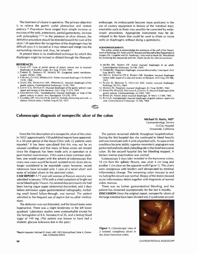
Colonoscopy 5 days later revealed in the transverse colon,10 cm from the splenic flexure, one ulcer 4 cm long andanother 1 cm ulcer on the opposite wall (Figure 1). The ulcerswere serpiginous with borders well demarcated by minimalinflammatory change. The remaining colon mucosa to andincluding the cecum was normal. Biopsy of the lesion showedacute inflammatory debris together with fragments of normalcolon mucosa.
There was no further gastrointestinal bleeding, and thepatient has remained asymptomatic for the last 4 months.DISCUSSION Since the original report, nonspecific ulcers ofthe large intestine have been divided into 3 syndromes accord-