97
General and Systemic Veterinary Pharmacology VPT 311 URVISH MISTRY AAU

General and Systemic Veterinary Pharmacology VPT 311
URVISH MISTRYAAU

1 | U R V I S H M I S T R Y
1 | U R V I S H M I S T R Y
Contents History of Pharmacology ................................................................................................................................................... 8
Ancient medicine ...................................................................................................................................................... 8
Pre‐ Christian era ...................................................................................................................................................... 8
Mediaeval medicine .................................................................................................................................................. 8
Revolts in medicine ................................................................................................................................................... 8
Modern medicine ...................................................................................................................................................... 8
Scope of pharmacology ................................................................................................................................................. 9
Branches of pharmacology ........................................................................................................................................... 9
Terms and definitions ..................................................................................................................................................... 10
Pharmacology related terms ....................................................................................................................................... 10
Drug related terms ...................................................................................................................................................... 10
Dose related terms ...................................................................................................................................................... 11
Sources of drugs .............................................................................................................................................................. 11
Plant sources of Drugs ................................................................................................................................................ 11
Alkaloids .................................................................................................................................................................. 11
Glycosides ............................................................................................................................................................... 12
Oils .......................................................................................................................................................................... 12
Tannins .................................................................................................................................................................... 12
Saponins .................................................................................................................................................................. 12
Resins ...................................................................................................................................................................... 12
Gums ....................................................................................................................................................................... 12
Mineral source ............................................................................................................................................................ 12
Animal source of drugs ............................................................................................................................................... 12
Synthetic sources of drugs .......................................................................................................................................... 13
Microbial source of drugs ........................................................................................................................................... 13
Pharmacokinetics: principles of drug activity ................................................................................................................. 13
Routes of administration ............................................................................................................................................ 13
Oral administration ................................................................................................................................................. 13
Intravenous route ................................................................................................................................................... 14
Subcutaneous and intramuscular routes ................................................................................................................ 15
Other parenteral routes .......................................................................................................................................... 16
Local administration ................................................................................................................................................ 16
Structure of biological membranes ............................................................................................................................... 17
Fluid mosaic models .................................................................................................................................................... 17
Functions of membranes ............................................................................................................................................ 17

2 | U R V I S H M I S T R Y
2 | U R V I S H M I S T R Y
Passage of drugs across membranes .............................................................................................................................. 17
Passive diffusion .......................................................................................................................................................... 17
Carrier mediated transfusion ...................................................................................................................................... 18
Facilitated diffusion ..................................................................................................................................................... 18
Active transport .......................................................................................................................................................... 18
Pinocytosis and phagocytosis ..................................................................................................................................... 19
Filtration ...................................................................................................................................................................... 19
Absorption of drugs ........................................................................................................................................................ 19
Absorption of drugs from GI tract ............................................................................................................................... 20
Role of ionization and lipid solubility in drug absorption ........................................................................................... 20
Henderson and Heselbelch equation ...................................................................................................................... 20
Absorption of drugs after parenteral administration ................................................................................................. 21
Absorption of drugs after inhalation or topical administration .................................................................................. 21
Distribution of drugs ....................................................................................................................................................... 22
Plasma protein binding of drugs ................................................................................................................................. 22
Plasma protein binding ............................................................................................................................................... 23
Tissue storage of drugs ............................................................................................................................................... 23
Barriers to drug distribution ....................................................................................................................................... 23
Biotransformation of drugs/xenobiotics: drug metabolism ........................................................................................... 24
Sequelae of biotransformation reactions ................................................................................................................... 25
Phase‐I Biotransformation ‐ oxidation reactions and others ...................................................................................... 25
Phase I reactions ......................................................................................................................................................... 25
Phase II reactions ‐ synthetic reactions ...................................................................................................................... 26
Enzyme induction and inhibition ................................................................................................................................ 26
Excretion of drugs ........................................................................................................................................................... 27
Renal elimination of the drugs .................................................................................................................................... 27
Hepatic and other routes of administration ............................................................................................................... 29
Pharmacokinetic parameters .......................................................................................................................................... 29
Important pharmacokinetic parameters .................................................................................................................... 30
Models in pharmacokinetics ....................................................................................................................................... 30
Drug accumulation ...................................................................................................................................................... 31
Principles of Drug activity – Pharmacodynamics ............................................................................................................ 31
Modes of drug action .................................................................................................................................................. 31
Enzyme mediated drug action .................................................................................................................................... 31
Receptor mediated drug action .................................................................................................................................. 32
Terms related to drug receptor binding ..................................................................................................................... 32

3 | U R V I S H M I S T R Y
3 | U R V I S H M I S T R Y
Receptors ‐ structure and function ................................................................................................................................. 33
Receptor theories ....................................................................................................................................................... 33
Functions of receptors ................................................................................................................................................ 34
Types of receptors ...................................................................................................................................................... 34
Second messenger systems ........................................................................................................................................ 34
Dose response curve ....................................................................................................................................................... 35
Plotting ........................................................................................................................................................................ 35
Features of dose response curve ................................................................................................................................ 35
Quantal dose response curve ..................................................................................................................................... 35
Factors modifying drug action ........................................................................................................................................ 35
Factors affecting drug action ...................................................................................................................................... 35
Adverse effects of drugs ................................................................................................................................................. 36
Types of adverse effects ............................................................................................................................................. 37
Drug interactions ........................................................................................................................................................ 37
Drug screening and drug discovery ................................................................................................................................. 38
Drug discovery ............................................................................................................................................................ 38
Drug screening ............................................................................................................................................................ 39
Preclinical safety and toxicity testing .......................................................................................................................... 39
Clinical trials ................................................................................................................................................................ 40
Bio prospecting ........................................................................................................................................................... 41
Assay of drugs ............................................................................................................................................................. 42
Biopharmaceuticals and gene therapy ........................................................................................................................... 42
Biopharmaceuticals ..................................................................................................................................................... 42
Gene therapy .............................................................................................................................................................. 42
Gastro – intestinal pharmacology ................................................................................................................................... 43
Sialagogues (sialics) and antisialagogues (antisialics) ................................................................................................. 43
Sialagogues.............................................................................................................................................................. 43
Antisialogogues ....................................................................................................................................................... 43
Stomachics .............................................................................................................................................................. 43
Appetite stimulants ..................................................................................................................................................... 43
Emetics ........................................................................................................................................................................ 44
Antiematics ................................................................................................................................................................. 45
Prokinetics ................................................................................................................................................................... 46
Ulcer management...................................................................................................................................................... 46
Antacids ................................................................................................................................................................... 46
Histamine receptors – H2 antagonist ...................................................................................................................... 47

4 | U R V I S H M I S T R Y
4 | U R V I S H M I S T R Y
Sucralfate ................................................................................................................................................................ 47
Proton pump antagonists ....................................................................................................................................... 47
Cytoprotective drugs ............................................................................................................................................... 48
Digestants and purgatives ............................................................................................................................................... 48
Choleretics and cholagogues ...................................................................................................................................... 48
Purgatives and laxatives .............................................................................................................................................. 48
Classification ........................................................................................................................................................... 49
General mechanism of action ................................................................................................................................. 49
Osmotic purgatives ................................................................................................................................................. 49
Irritant purgatives ................................................................................................................................................... 50
Bulk purgatives ........................................................................................................................................................ 51
Lubricant purgatives ............................................................................................................................................... 52
Choice, indications, combinations, abuse of purgatives ............................................................................................. 52
Antidiarrheal ................................................................................................................................................................... 53
Opioids ........................................................................................................................................................................ 53
Anticholinergics ........................................................................................................................................................... 54
Protectants and adsorbants ........................................................................................................................................ 54
Antisecretory drugs ..................................................................................................................................................... 55
Protectants .................................................................................................................................................................. 55
Rumen pharmacology ..................................................................................................................................................... 55
Oesophageal groove closure ....................................................................................................................................... 55
Ruminotorics ............................................................................................................................................................... 56
Rumen antacids ........................................................................................................................................................... 56
Antizymotics ................................................................................................................................................................ 56
Cardiovascular pharmacology ......................................................................................................................................... 56
Classes of drugs acting on heart ................................................................................................................................. 56
Effects of drugs acting on heart .................................................................................................................................. 57
Congestive heart failure .............................................................................................................................................. 57
Cardiac glycosides ........................................................................................................................................................... 57
Chemistry of cardiac glycosides .................................................................................................................................. 58
Mechanism of action on cardiac glycosides ................................................................................................................ 58
Pharmacological effects of cardiac glycosides ............................................................................................................ 58
Pharamacokinetics ...................................................................................................................................................... 59
Therapeutic uses of cardiac glycosides ....................................................................................................................... 59
Digitalization ............................................................................................................................................................... 59
Adverse reactions of cardiac glycosides ..................................................................................................................... 59
5 | U R V I S H M I S T R Y
5 | U R V I S H M I S T R Y
Treatment of cardiac glycosides toxicity ..................................................................................................................... 60
Precautions and drug interactions .............................................................................................................................. 60
Drug interactions ........................................................................................................................................................ 60
Other drugs used in CHF ............................................................................................................................................. 60
Antiarrhythmic drugs ...................................................................................................................................................... 60
Types of arrhythmia .................................................................................................................................................... 60
Arrhythmia .................................................................................................................................................................. 61
Antiarrhythmic drugs .................................................................................................................................................. 61
Classification ............................................................................................................................................................... 61
Actions of sodium channel blockers ........................................................................................................................... 62
β – Adrenergic blockers .............................................................................................................................................. 63
Actions on potassium currents ................................................................................................................................... 63
Actions of Ca –channel blockers ................................................................................................................................. 63
Clinical uses of antiarrhythmic drugs .......................................................................................................................... 64
Adverse effects of Antiarrhythmic drugs .................................................................................................................... 64
Adverse extra – cardiac effects ................................................................................................................................... 64
Vasodilators and antihypertensive drugs ....................................................................................................................... 65
Direct acting Vasodilators ........................................................................................................................................... 65
Indirectly acting vasodilators ...................................................................................................................................... 66
Clinical uses ................................................................................................................................................................. 66
Vasoconstrictors.......................................................................................................................................................... 66
Clinical uses of vasoconstrictors ................................................................................................................................. 66
Classification of antihypertensive drugs ......................................................................................................................... 66
Antihypertensive drugs ............................................................................................................................................... 67
Hematinic, coagulants and anticoagulants ..................................................................................................................... 68
Haematinics................................................................................................................................................................. 68
Coagulants ................................................................................................................................................................... 69
Anticoagulants ............................................................................................................................................................ 70
In vitro anticoagulants ................................................................................................................................................ 70
Systemic anticoagulants – in vivo ............................................................................................................................... 70
Clinical uses of anticoagulants .................................................................................................................................... 70
Coumarin derivatives .................................................................................................................................................. 70
Heparin antagonist ...................................................................................................................................................... 71
Respiratory pharmacology – expectorants ..................................................................................................................... 71
Expectorants ............................................................................................................................................................... 71
Inhalant expectorants ................................................................................................................................................. 71
6 | U R V I S H M I S T R Y
6 | U R V I S H M I S T R Y
Ingested expectorants ................................................................................................................................................ 71
Mucolytics ................................................................................................................................................................... 71
Decongestants and antitussives ...................................................................................................................................... 72
Decongestants ............................................................................................................................................................. 72
Antitussives ................................................................................................................................................................. 72
Directly acting antitussives ......................................................................................................................................... 72
Centrally acting antitussives ....................................................................................................................................... 73
Bronchodilators ............................................................................................................................................................... 73
Methyl xanthene ......................................................................................................................................................... 74
Sympathomimetic ....................................................................................................................................................... 74
Parasympatholytics – spasmolytics ............................................................................................................................. 75
Corticosteroids ............................................................................................................................................................ 75
NSAIDs ......................................................................................................................................................................... 75
Antihistaminics ............................................................................................................................................................ 75
Mast cell stabilizers ..................................................................................................................................................... 75
Respiratory stimulants ................................................................................................................................................ 76
Diuretics .......................................................................................................................................................................... 76
Physiological diuresis .................................................................................................................................................. 76
Classification of diuretics ............................................................................................................................................ 76
Osmotic diuretics ........................................................................................................................................................ 76
Carbonic anhydrase inhibitors .................................................................................................................................... 77
Loop diuretics .............................................................................................................................................................. 77
Thiazide derivatives .................................................................................................................................................... 78
Potassium sparing diuretic .......................................................................................................................................... 79
Xanthines and mercurial ............................................................................................................................................. 80
Drugs acting on urogenital systems ................................................................................................................................ 80
Alkalization of urine .................................................................................................................................................... 80
Acidification of urine ................................................................................................................................................... 80
Ecbolics ........................................................................................................................................................................ 81
Tocolytics .................................................................................................................................................................... 81
Fluid therapy ............................................................................................................................................................... 81
Choice of fluid ............................................................................................................................................................. 82
Pharmacotheraputics of Hormones ................................................................................................................................ 83
Administration of hormones ....................................................................................................................................... 83
Corticotrophins and related peptides ......................................................................................................................... 83
Insulin .......................................................................................................................................................................... 84

7 | U R V I S H M I S T R Y
7 | U R V I S H M I S T R Y
Pharmacological actions of insulin .............................................................................................................................. 84
Preparations of insulin ................................................................................................................................................ 84
Oral hypoglycemic drugs ............................................................................................................................................. 85
Thyroid hormone ........................................................................................................................................................ 85
Thyroid inhibitors ........................................................................................................................................................ 86
General principles in corticosteroids therapy ............................................................................................................. 86
Anti‐inflammatory and sodium retaining potencies of Glucocorticoids ..................................................................... 87
Reproductive hormones and Vitamins ........................................................................................................................... 88
Hormones influencing reproductive functions ........................................................................................................... 88
Anterior pituitary gonadotropins ................................................................................................................................ 88
Placental gonadotropins ............................................................................................................................................. 89
Estrogens ..................................................................................................................................................................... 89
Progesterone ............................................................................................................................................................... 90
Androgens ................................................................................................................................................................... 90
Anabolic steroids ......................................................................................................................................................... 91
Posterior – pituitary hormones ................................................................................................................................... 91
Pharmacotherapeutics of fat soluble vitamins ........................................................................................................... 91
Pharmacotherapeutics of water soluble vitamins ...................................................................................................... 92
Drugs acting on skin and mucous membranes ............................................................................................................... 93
Counterirritants ........................................................................................................................................................... 93
Penetration enhancers ................................................................................................................................................ 93
Adsorbants .................................................................................................................................................................. 94
Demulcents ................................................................................................................................................................. 94
Emollients .................................................................................................................................................................... 94
Astringents .................................................................................................................................................................. 94
Other agents used on skins ......................................................................................................................................... 94
Bioenhancers, immunostimulants and immunosuppressors ......................................................................................... 95
Bioenhancers............................................................................................................................................................... 95
Immunostimulants ...................................................................................................................................................... 95
Immunosuppressant ................................................................................................................................................... 95

8 | U R V I S H M I S T R Y
8 | U R V I S H M I S T R Y
History of Pharmacology Ancient medicine Pre‐Christian era Mediaeval medicine Revolts in medicine Modern medicine
Ancient medicine Chinese medicine is the earliest and records dated about 2500 B.C. available today give an idea about the
medical knowledge of Chinese. In Chinese medicine, the use of Ephedra or Ma huang as a tonic has been reported. Ayurveda or Indian Medicine is equally ancient. To form the science of life namely Ayurveda, Charaka,
Sushruta and Vagbhata made a compilation of old and new drugs in the cure of diseases. Egyptian medicine is also very ancient. The Ebers Papyrus dated about 1500B.C. gives a collection of drugs
prevalent in Egypt at that time, their classification and their use. Some of the drugs employed now such as, castor oil and pomegranate bark are mentioned in this papyrus.
Pre- Christian era Greek medicine is said to be the origin of modern medicine and therapeutics. Hippocrates in fifth century
B.C. separated medicine from religion and was known as the father of medicine. He laid down certain principles on which modern medicine is built. According to Hippocrates the four
elements of nature namely water, fire, air and earth gave rise to the four humors of the body namely blood, phlegm, yellow bile or urine and black bile. Any imbalance in one or more of these humors inflicted sufferings.
Galen was a famous Greek Physician who practiced in Rome. His name is still used to refer some drugs as galenical drugs. He was the father of polypharmacy.
Galenical drugs are pharmaceuticals compounded by mechanical means, mostly of the vegetable material.
Mediaeval medicine Paracelsus introduced inorganic chemicals like mercury into medicine. He called this ‘Iatro Chemistry’ or
medicinal chemistry. He induced practitioners to use laudanum (an opium preparation), sulphur, iron, copper sulphate, potassium
sulphate, mercurials and tinctures and fluid extract of various plants for treatment of diseases.
Revolts in medicine By the beginning of 19th century the principle of shotgun prescription flourished (Shotgun prescription is
one that contains a number of substances with no therapeutic efficacy. It is a result of ignorant attempt to cure the disease, no matter what may be its nature).
Gregory advocated methods like venesection, leeching emetics and drastic purgatives. Large doses of purgatives were given. The patient either survived or died. This sort of symptomatic treatment was referred to as allopathy meaning 'other suffering'. This term allopathy is now being used to refer modern medicine.
Samuel Hahnemann introduced homeopathy meaning ‘similar suffering’ at the commencement of 19th century. In Greek, “homos” means same and “patheia” means suffering. He was known as the father of homeopathy. Homeopathy introduced by him had two newer principles that ‘like cures like’ and ‘dilution potentiates the action of drugs’.
Modern medicine Buccheim, a professor of Dorpat University who was known as the father of Pharmacology set up the first
laboratory to study pharmacology. He discarded many remedies because rational scientific action or explanation could not be demonstrated in his laboratory.
By the middle of the 19th century, modern medicine had brought to fight disease only one effective weapon ie.immunization against smallpox.

9 | U R V I S H M I S T R Y
9 | U R V I S H M I S T R Y
Later in quick succession came the anaesthetics and antiseptics. In the last quarter, the causative organisms for malaria, plaque, cholera etc. were identified.
Beginning in the 20th century, the fresh wind of synthetic chemistry began to revolutionise the pharmaceutical industry and with it the science of pharmacology.
New synthetic drugs, such as barbiturates and local anaesthetics, began to appear and the era of antimicrobial chemotherapy began with the discovery of arsenical compounds for the treatment of syphilis by Paul Ehrlich in 1909. He was known as the father of chemotherapy.
Further breakthroughs came with the discovery of sulphonamides by Gerhard Domagk in 1935 and the development of penicillin during worldwar II.
The addition of drugs to the therapeutic jungle is growing with rapid pace from the later half of the 20th century.
Scope of pharmacology Pharmacology is the science which involves all aspects of the action of drugs on living system. It is the study
of the therapeutic value and/or potential toxicity of chemical agents on biological systems. It targets every aspect of the mechanisms for the chemical actions of both traditional and novel therapeutic agents.
Important and interrelated areas are: pharmacodynamics and pharmacokinetics. Pharmacodynamics is the study of how drugs act on the body while pharmacokinetics is the study of how the
body acts on drugs. Pharmacodynamic and pharmacokinetic aspects of the action of chemical agents are applicable to all related areas of study, including toxicology and therapeutics.
Toxicology is the study of the adverse or toxic effects of drugs and other chemical agents. It is concerned both with drugs used in the treatment of disease and chemicals that may present household, environmental, or industrial hazards.
Therapeutics focuses on the actions and effects of drugs and other chemical agents with physiological, biochemical, microbiological, immunological, or behavioral factors influencing disease. Each of these areas is closely interwoven with the subject matter and experimental techniques of physiology, biochemistry, cellular biology, microbiology, immunology, genetics, and pathology. The ultimate goal of Pharmacology is to design chemical agents to cure , ameliorate, or preventdisease
Branches of pharmacology Neuropharmacology is the study of neurophysiological or neurobiochemical functions of the nervous system
including the brain, spinal cord, and the nerves that are modified by drug action. Cardiovascular pharmacology concerns the effects of drugs on the heart, the vascular system, and those
parts of the nervous and endocrine systems that participate in regulating cardiovascular function. Molecular pharmacology deals with the biochemical and biophysical characteristics of interactions between
drug molecules and those of the cell. It is molecular biology applied to pharmacology and toxicology . Biochemical pharmacology is the study of action of drugs and drug metabolism, how drugs interact with,
and influences, the physiology of the organism. Behavioral pharmacology studies the effects of drugs on behavior of organism. It includes topics such as the
effects of psychoactive drugs on the phenomena of learning, memory, wakefulness, sleep and the behavioral consequences of experimental intervention in enzyme activity and brain neurotransmitter levels and metabolism.
Endocrine pharmacology is the study of drugs that are either hormones or hormone derivatives, or drugs that may modify the sections of normally secreted hormones.
Clinical pharmacology is the application of pharmacodynamics and pharmacokinetics to patients with diseases, it also includes pharmacogenetic component. Clinical pharmacologists study how drugs work, how they interact with the genome and with other drugs, how their effects can alter the disease process, and how disease can alter their effects. Clinical trial design, the prevention of medication errors, and the optimization of rational prescribing are critical components of clinical pharmacology.
Chemotherapy is the area of pharmacology that deals with drugs used for the treatment of microbial infections and malignancies. Chemotherapeutic agents selectively inhibit the growth of, or kill, the infectious agent or cancer cell without seriously impairing the normal functions of the host.

10 | U R V I S H M I S T R Y
10 | U R V I S H M I S T R Y
Toxicology is the science of adverse effects of chemicals/ drugs on living systems. It also includes problems of drug safety, effects of drug over dosage.
Pharmacy is a separate discipline in the health sciences. It is the profession responsible for the preparation, dispensing and appropriate use of medication, and provides serv ices to achieve optimal therapeutic outcomes.
Terms and definitions
Pharmacology related terms Pharmacology is the science that embraces the knowledge of the history, source, physical and chemical
properties, compounding, biochemical and physiological effects, mechanism of action, absorption, distribution, biotransformation and excretion of drugs. It is also defined as an experimental science dealing with the properties of drugs and their effects on living system.
Pharmacodynamics is the study of the biochemical and physiological effects of drugs and their mechanism of action. It is the response of the organism to the action of a drug in the absence of a disease. Pharmacodynamics is 'what the drug does to the body'.
Pharmacokinetics is the study of the actions of the drugs in the body over a defined period of time. It deals with the absorption, distribution, biotransformation and excretion of the drug. Pharmacokinetics is 'what the body does to the drug'.
Pharmacometrics is the study of the techniques used in the measurement of drug effects to the administered dose of drug.
Pharmacogenetics is the study of genetically determined variations in animals that are revealed by the effect of drugs.
Pharmacogenomics This term describes the use of genetic information to guide the choice of drug therapy on an individual basis. .
Pharmacoepidemiology is the study of drug effects at the population level. It is concerned with the variability of drug effects between individuals in a population and between populations.
Pharmacoeconomics aims to quantify in economic terms the cost and benefit of drugs used therapeutically. Pharmacy is the science that deals with the preparation, formulation, manufacture, standardization,
preservation and dispensing of drugs. The term pharmacy also indicates the place where drugs are dispensed or sold.
Pharmacognosy is the study of the source of drugs. It also deals with the physical and chemical properties of drugs.
Materia medica is an obsolete didactic subject that was concerned with pharmacy, posology, pharmacognosy and indications for therapeutic use of the drug.
Metrology is the study of weights and measures as applied to the preparation and administration of drugs. Chronopharmacology is the sudy of how the effects of drugs vary with biological timing and endogenous
periodicities. Pharmacovigilance is the science and activities relating to the detection, assessment, understanding and
prevention of adverse effects or any other drug‐related problem. Pharmacoepidemiology is the study of the use of and the effects of drugs in large numbers of people.
Drug related terms Drug is any chemical agent except food that is used to promote or safeguard the health of human beings or
animals. It is also defined as any substance or product that is used or intended, to be used to modify or explore physiological systems or pathological states for the benefit of the recipient. The word drug is derived from a French word ‘Drogue’ meaning a dry herb.

11 | U R V I S H M I S T R Y
11 | U R V I S H M I S T R Y
Over the counter drugs are those preparations that can be sold without any prescription because they can be adequately labeled for layman use.
Prescription drugs are drugs that can be used only on the order of a licensed veterinarian/physician/dentist/surgeon based on a prescription. They are also known as legend drugs.
Essential drugs are agents that satisfy the healthcare needs of majority of the population. They should therefore be available at all times in adequate amounts and in appropriate dosage form.
Pro‐drugs are drugs that are inactive or have a low order of activity in the form administered and are metabolised to the active form in the body.
Hard drugs are drugs used for non‐medical purposes that are liable to disable the individual seriously as a functioning member of the society by inducing severe psychological and/or physical dependence. eg. Heroin
Soft drugs are drugs used for non‐medical purposes that are less dependence producing. There may be psychological dependence but not physical dependence, except with heavy dose. eg. Amphetamine.
Nootropic drugs are drugs that affect the intellect. These drugs are claimed to enhance learning, increase brain resistance to stress including hypoxia and stimulate brain metabolism especially in senile patients. eg. Piracetam
Orphan drugs are drugs or biological products useful for diagnosis/treatment/prevention of a rare disease condition for which there is no reasonable expectation that the cost of developing and marketing it will be recovered from the sales of that drug. Eg. Acetylcysteine. These drugs may be life saving for some patients, but are not commercially available.
Placebo is a vehicle for cure by suggestion and is surprisingly often successful though only temporarily. It can be used as a control in scientific evaluation of drugs and to benefit or please a patient not by pharmacological actions but by psychological means (Latin: Placebo – I shall be pleasing or acceptable). Placebo reactor is an individual who report changes of physical and mental state after taking a pharmacologically inert substance.
Dose related terms Dose of the drug is an estimate amount of a drug, that when administered by a particular route to a certain
species is most likely to produce a certain intensity of response. It is the quantity of medication to be administered at one time. Dosage is the determination and regulation of doses.
Posology is the study of the medicine dosages, which varies with the species of animals, the intended effect of the drug and the individual tolerance or susceptibility.
Loading dose is one or a series of doses that may be given at the onset of therapy with the aim of achieving the target concentration rapidly.
Maintenance dose is a series of relatively small doses that follow the loading dose in order to maintain an effective concentration in the bio phase.
Sources of drugs
Plant sources of Drugs Many drugs available from plants are even today used in the treatment of pathological conditions. With the increasing tendency for the use of alternate medicine, this source has gained more importance in
the recent past. The pharmacological activities of plants are attributed to certain active principles in plants. They are
alkaloids, glycosides, fats, oils, tannins, saponins etc.
Alkaloids Alkaloids are nitrogenous substances obtained from various parts of the plant. Alkaloids containing oxygen
are solids and comparatively non volatile (cocaine) while those that do not contain oxygen are liquids and volatile (nicotine, lobeline and coniine).
Alkaloids are insoluble in water while their salts (atropine sulphate, caffeine citrate) are soluble in water. Alkaloids are bitter to taste. They are incompatible with the alkalies, tannic acid and heavy metals. Alkaloids represent the waste products of plant metabolism and their names end with ‘ine’.

12 | U R V I S H M I S T R Y
12 | U R V I S H M I S T R Y
Alkaloids should be administered in small quantities and when given in excess they may produce death without much postmortem changes for diagnosis. (Adrenaline is considered as animal alkaloid).
Glycosides Glycosides are non‐nitrogenous substances obtained from plants. The glycosides on hydrolysis yield two molecules namely a sugar molecule and a ‘genine’ or ‘aglycone’
molecule. Sugar helps in the dissolution of the preparation while the pharmacological action rests with the ‘aglycone’. When the sugar molecule is glucose, the glycoside is known as glucoside. Cardiac glycosides digitalis, strophanthus and squill play a major role in the treatment of congestive cardiac
failure. Cyanogenetic glycoside is one in which hydrocyanic acid is released on glycolysis.
Oils There are two types of oils namely fixed oils and volatile oils. Volatile oils are also known as essential oils. Castor oil, coconut oil etc. are fixed oils while turpentine oil, eucalyptus oil etc. are volatile oils. Fixed oils are obtained by expression while volatile oils are obtained by distillation. (Mineral oils are obtained
from the earth and some ae used pharmacologically. Eg: Liquid paraffin.)
Tannins These are non‐nitrogenous phenol derivatives found especially in leaves and bark. They are astringent in nature and form inky solutions with ferric salts. Catechu a tannic acid is used in the
control of diarrhoea. Eg: Black catechu, Pale catechu
Saponins These are non‐nitrogenous substances resembling glycosides. They are soluble in water and on shaking they give persistent foam. When the saponin is toxic it is known as
sapotoxin. On hydrolysis saponins split into sugar and aglycone (sapogenin). Eg: Fenugreek, Ginseng etc.
Resins These are solid brittle substances formed from terpenes by oxidation. They are insoluble in water. Resin can be oleo resin, gum resin or balsams. Eg: Asafoetida, Camphor, Storax
Gums Gums are dried exudates obtained by incision on stems of various plants. They form a jelly with water. Eg: Gum acacia
Mineral source Metallic and nonmetallic minerals provide various inorganic materials not available from plants or animals.
Mineral sources are used as they occur in nature or can be combined with other ingredients. The drugs that are included in this category include metals and their salts, non metals, metalloids, acids,
alcohols and coaltar drugs etc. Examples are: sodium chloride, copper sulphate, magnesium sulphate, potassium permanganate, etc. They are used in the purified form as drugs.
Animal source of drugs The body fluids or glands of animals are also natural drug sources. The drugs obtained from animal sources include:
hormones, such as insulin

13 | U R V I S H M I S T R Y
13 | U R V I S H M I S T R Y
oils and fats (usually fixed), such as cod‐liver oil enzymes, which are produced by living cells and act as catalysts, such as pancreatin and pepsin vaccines, which include suspensions of killed, modified, or attenuated microorganisms, or antigenic
materials obtained from these.
Synthetic sources of drugs A number of drugs synthesized in the laboratory are used most commonly. Even natural products such as hormones, antimicrobials etc. are also synthesized in the laboratory.
Microbial source of drugs Microbes provide an important source of drugs especially the antibiotics. All the antibiotics used against a
variety of pathogens and also cancer are obtained from fungi, bacteria or actinomycetes. Some systemic drugs like ergot alkaloids (fungal source) are also obtained from microbes.
Eg: Penicillin from Penicillium notatum Streptomycin, Tetracyclines, Chloramphenicol from Streptomyces sp.
Pharmacokinetics: principles of drug activity
Routes of administration Routes of drug administration can be divided into three main classes as enteral, parenteral and topical.
o Enteral administration refers to administration of drugs via the gut. o Parenteral (par – beyond, enteral ‐ intestinal) administration covers intravenous, intramuscular,
subcutaneous, intraperitoneal etc. o Topical application refers to application of drugs on the skin and mucous membrane.
Factors governing the choice of route are: o Physical and chemical properties of the drug (solid/liquid/gas; solubility, stability, pH, irritancy) o Site of desired action – localized or generalized o Rate and extent of absorption of the drug from different routes o Effect of digestive juices and first pass metabolism of the drug o Rapidity with which the response is desired (routine treatment or emergency) o Accuracy of dosage required (intravenous and inhalation need fine tuning of dose) o Condition of the patient (unconscious, vomiting)
Oral administration Dosage forms available for oral use include Liquids: Aqueous solutions, suspension (dispersion of a solid in a liquid) and emulsion (dispersion of a liquid
in liquid) Solids: Powders, tablet, enteric coated tablet, capsule, granules For tablets and capsules the factors that affect the actual amount of the drug absorbed or available include
disintegration, coating, adjuvants used, compression, drug particle size – micronization, amount of the drug, chemical form (salt), physical state (amorphous or crystalline, solvated or anhydrous) and local pH of the absorptive area.
Advantages of oral route Generally the safest route

14 | U R V I S H M I S T R Y
14 | U R V I S H M I S T R Y
Economical Convenient and relatively simple for owner No need for sterile equipment Systemic distribution can be achieved
Disadvantages Absorption may be variable Gastric irritation may cause vomiting Not useful if animal is vomiting Requires cooperation of patient, which is not usually forthcoming ina veterinary patient Drugs may be destroyed by gastric acidity, gut flora, mucosal enzymes and liver enzymes Onset of effect is usually slow Not generally preferred in ruminants since the drug gets diluted in the voluminous ruminal contents When antimicrobials are administered to ruminants orally for a longer duration of time, they may affect the
microbial ecosystem of the rumen Presence of food and other drugs may alter the absorption pattern leading to unpredicatability in the
desired action. Enteric coated preparations
Drugs that are destroyed by the gastric juice or that cause gastric irritation can be administered orally with a coating that prevents dissolution in the acidic gastric contents.
They dissolve once they reach the duodenum and release the active drug. Onset of drug action can be delayed with enteric‐coated tablets.
Sublingual tablets Drugs that are lipid soluble and non‐irritating can be administered sublingually, so that absorption directly
from the oral cavity is achieved when a rapid response is required, particularly when the drug is either unstable at gastric pH or rapidly metabolised by the liver. Eg. Glyceryl trinitrate.
Drugs absorbed by mouth pass directly into the systemic circulation without entering the portal system and so escape the first‐pass metabolism. This type of tablet is useful in the treatment of angina pectoris where the drug enters directly into the systemic circulation and provides immediate effect. Once the required effect has been achieved, the excess tablet can be spit off.
Timed release preparations Timed release preparations are designed to produce slow uniform release and absorption of the drug over a
period of 8 hours or more. They are also known as spansules or timesules. Advantages
o Less frequent administration o Lasts overnight o Drug levels are more constant and do not peak after each administration (less toxic effects) o Good for short‐acting drugs
Disadvantages o Marketed preparations are sometimes not reliable o Dissolution rates may be irregular o Not needed for long acting drugs o Not good for a brief therapeutic effect
Rectal administration This route of administration is useful when the animal is unconscious or vomiting. Rectal absorption is often incomplete and erratic. Drugs can be administered rectally in the form of enema or suppository. Irritant and unpleasant drugs can be administered per rectum. However, rectal inflammation may occur due
to highly irritant drugs.
Intravenous route Parenteral administration
While considering parenteral administration, the points of importance include o Volume to be administered

15 | U R V I S H M I S T R Y
15 | U R V I S H M I S T R Y
o Concentration of the drug o pH o Toxicity o Viscosity o Particle size, if suspension is used o Adjuvant used in the preparation
In general, parenteral administration requires skill of injection and use of sterile equipment. Parenteral preparations are normally used as solutions or suspensions.
Intravenous administration Advantages
o Extremely rapid onset of action o Initial absorption step is bypassed o Drug levels can be controlled more accurately o Suitable for irritant drugs o Suitable for large volumes of drugs
Disadvantages o Most dangerous route as toxicity can easily occur o Drugs must be in aqueous solution o Must be performed slowly o Once injected, drug cannot be retrieved
Sites for venipuncture in different species Cattle, sheep and goat ‐ At any point along the whole length of the jugular vein in the jugular furrow, on the
venterolateral aspect of the neck on either side Horse ‐ External jugular vein in the jugular furrow only in the cranial part of the neck. Pig ‐ Auricular vein Dog ‐ saphenous vein on the medial aspect of the leg or recurrent tarsal vein on the dorsal aspect of the leg.
Subcutaneous and intramuscular routes Subcutaneous administration
This route is useful when slow and continuous absorption is required. The formulation must be isotonic and at physiological pH.
Certain drugs that are irritating can cause severe pain and necrosis. The rate of distribution of the drug is largely dependent on blood flow and hence, the rate of distribution can be slowed by including a vasoconstrictor.
Warmth or vigorous massage will increase distribution. Addition of hyaluronidase can enhance drug dispersion. This enzyme hydrolyses the hyaluronic acid polymers that comprises the intercellular cement and thus facilitates diffusion through the tissues.
Specialised subcutaneous preparations include dermojet and pellet implantation. Dermojet is a process where no needle is used. A high velocity jet of the drug solution is projected from a
microfine orifice using a gun like implement. The solution passes through the superficial layer and gets deposited in the subcutaneous tissue. It is essentially painless and suitable for mass inoculation.
Pellet implantation provides sustained release of the drug over weeks or months. The pellet impregnated with the drug is implanted in the subcutaneous tissue. Sialistic (non biodegradable) and biodegradable implants are used.
Crystalline drug is packed in tubes made of suitable material and implanted under the skin. Constant blood levels can be maintained as the drug is released uniformly over a period of time. If non‐biodegradable implant is used, it should be removed after the specified period of time.
Sites for subcutaneous injection in different species Cattle, sheep and goat ‐ Fold of flank extending from the caudoventral abdominal wall to the craniomedial
aspect of the thigh near stifle joint or loose skin on the lateral aspect of the neck. Horse ‐ Loose skin on the lateral aspect of the neck. Normally subcutaneous injection is not preferred in
horses. Dog ‐ Fold of the flank
16 | U R V I S H M I S T R Y
16 | U R V I S H M I S T R Y
Intramuscular administration Drugs in aqueous solution are rapidly absorbed after intramuscular administration. However, very slow
constant absorption occurs if the drug is administered in oil or suspended in other repository vehicles as depot preparations. It can be used for relatively irritant drugs and such drugs must be administered deep intramuscularly. Intramuscular injections are always painful and large volumes cannot be injected.
A disadvantage of this route is the possibility of improper deposition in nerves, blood vessels, fat or between muscle bundles in connective tissue sheaths. Whenever drugs are administered intramuscularly, it is always advisable to confirm that the needle is not in the blood vessel.
Sites for intramuscular injection in different species: Cattle, sheep and goat – 1. Hind limb a) Gluteal region covered by gluteal muscles and b) posterior aspect of
thigh between the semimembranous and semitendinous. (In sheep and goat (b) is preferred). 2. Neck – In the heavy muscles on the caudodorsal aspect of the neck. Needle can pierce through the
following structures – skin, fascia, trapezius, rhomboideus, splenius and complexus, depending upon the length of the needle and force applied. This site is preferred only in well built animals. Care should be taken to avoid vertebral column and the dorsal branch of the XI cranial nerve.
In indigenous cattle with hump, hump is also preferred to administer intramuscularly. Horse ‐ Neck region and brisket region (pectoral muscles) Dog ‐ Hind limb between the semimembranosus and semitendinosus.
Other parenteral routes Intraperitoneal administration
This route is particularly useful in laboratory animal medicine and neonatal animals and for the administration of large volumes.
There is a very large absorbing area and absorption is rapid. There is a danger of infection and peritoneal adhesions. Thus it is not used routinely. Peritoneal dialysis is becoming more frequently used in small animals with renal failure and renal insufficiency.
Intradermal administration This route is used mainly for diagnostic purposes eg. for Tuberculin and Johnin testing in cattle and also for
hypersensitivity testing before administering some drugs known to induce hypersensitivity. Intrathecal administration
In this route the drug is administered through the membranes enclosing the central nervous system in the lumbar area or into the cisterna magna.
It is occasionally used for radiographic examinations and chemotherapy of central nervous system infections and neoplasms.
Epidural administration
This route is mainly used to anaethetise animals for surgery like parturition in cattle. The drug is administered between the first and second coccygeal vertebrae.
Intraarticular administration This route is used to administer anti‐inflammatory agents into the joint capsule. The other parenteral routes of drug administration rarely used are intra‐ arterial, intramedullary,
intratesticular, intracardiac etc.
Local administration Local administration of drugs refers to external application or application to a localized site such as eye. ear.
mucous membranes etc. Some of the local sites of administration include:
o Skin: Application of drugs on to the skin such as powder, lotion, ointment etc. o Mucous membrane:
Mouth and pharynx: mouthwash, gargle, paint etc. Eye, ear and nose: Drops, spray, irrigation GI tract: non absorbable drugs such as kaolin, antacids, some antibiotics which are intended
to remain within the GI tract and not absorbed

17 | U R V I S H M I S T R Y
17 | U R V I S H M I S T R Y
Bronchi and lungs: aerosol, inhalations Vagina: Pessary, bolus, tablets Rectum: Suppository, enema etc.
o Tissues: Drugs can be administered to some of the tissue but are intended to act only on the site of absorption. eg. Intraauricular, intrathecal
o Arterial: Some drugs are given intraarterially to act on the localized area supplied by the artery. Eg: angiography, anticancer drugs.
Structure of biological membranes
Fluid mosaic models Biological membranes may be viewed as mosaics of functional units composed of lipoprotein complex. Most membranes are composed of a fundamental structure called the unit membrane or plasma
membrane, that is 80 to 100 Aο thick, surrounds single cells and nuclei. Complex barriers such as the intestinal epithelia and the skin are composed of multiples of this functional
structure. The plasma membrane consists of a bilayer of thic lipids with their hydrocarbon chains oriented within to
form a continuous hydrophobic phase and their hydrophilic heads oriented outward. Individual lipid molecules in the bilayer can move laterally, making the membrane fluid, flexibile, electrically
resistant and relatively impermeabe to highly polar molecules. This view of membrane structure is known as the fluid mosaic model.
o Membrane proteins embedded in the bilayer serves as receptors to elicit electrical or chemical signaling pathways and provide selective targets for drug action.
o The proteins are able to freely float through the membrane and some of the intrinsic ones, which extend through the full thickness of the membrane, surround fine aqueous pores. Paracellular spaces or channels also exist between certain epithelial or endothelial cells and other proteins have enzymatic or carrier properties.
o Biological membranes behave as if they were lipoids punctured by aqueous pores and allow drugs and physiological materials cross by passive or carrier mediated process. Which mechanism operates is determined by the physicochemical properties of drugs and the options available in the membrane.
Functions of membranes Membranes perform the following functions:
1. They provide structural stability to the cell 2. They act as barriers restricting the entry of substances inside the cell. 3. They provide vital communication through the entry of messengers for various cellular processes.
Passage of drugs across membranes
Passive diffusion Passive diffusion is a random movement of drug molecules from an area of higher concentration to an area
of lower concentration. When a drug is injected into the body, it passively diffuses from the injection site to areas of lower concentration, eventually reaching a blood capillary and entering the systemic circulation. In this process no cellular energy is expended and no transport carrier protein is involved. Hence the term passive diffusion is used.
Many drugs pass through the biological membranes such as cell membranes by passive diffusion. For a drug to passively diffuse from one side to another, the drug must dissolve in the membrane that is composed of phospholipids, and diffuse down the concentration gradient.
Passive diffusion continues until enough molecules have passed from an area of higher concentration to an area of lower concentration till equilibrium is attained on either side of the membrane. Drug molecules continue to move, however, such that an equal number move into and out of both the areas.

18 | U R V I S H M I S T R Y
18 | U R V I S H M I S T R Y
Passive diffusion is the most important mechanism for majority of drugs. Lipid soluble drugs diffuse by dissolving in the lipoidal matrix of the membrane. The rate of transport being proportional to lipid: water partition coefficient of the drug.
Passive diffusion is mostly dependent upon: o Concentration gradient (greater is the difference in the concentration of the drug on the two sides of
the membrane, faster is its diffusion) o Drug molecular size (smaller molecules move more rapidly than bigger molecules) o Lipophilic nature of the molecule (higher the lipid solubility higher is the diffusion) o Temperature (lower the temperature, slower the diffusion) o Thickness of the membrane (the thicker the membrane the slower the diffusion)
Carrier mediated transfusion In Carrier mediated transport, the drug combines with a carrier present in the membrane and the complex
then translocates from one side of the membrane to the other. The carriers for polar molecules appear to form a hydrophobic coating over the hydrophyllic groups and thus
facilitate passage through the membranes. Substances permitting transit of ions across membranes are called ionophores.
Carrier transport is specific, saturable and competitively inhibited by analogues that utilise the same carrier. Intestinal absorption sometimes depends on carriermediated transport.
Examples of carrier mediated transport are: levodopa is taken up by a carrier that normally transports phenylalanine; Fluorouracil is transported by the system that carries natural pyrimidines; iron is absorbed via a specific carrier on the surface of the mucosal cells in the jejunum and Calcium is absorbed by means of a vitamin D dependent carrier system. This carrier mediated transport is of two types, namely
o facilitated diffusion and o active transport
Facilitated diffusion Facilitated diffusion is a transport mechanism across biological membranes that involve a special “carrier
molecule” in the membrane which facilitates the movement of certain molecules across the membrane. As in passive diffusion, facilitated diffusion involves no energy to move the drug molecules and the direction
of the drug movement is determined by the concentration gradient. In addition, once the equilibrium is attained, the number of drug molecules crossing the membrane in either
direction remains the same. This process proceeds more rapidly than simple diffusion and even translocates non‐diffusible substrates,
but along their concentration gradient. However, the transporter may become saturated and other compounds may compete or inhibit the
transport. Examples of facilitated diffusion are:
o Vitamin B12 absorbed from the gut. o Transport of sugars and amino acids across membranes.
Active transport Like facilitated diffusion, active transport also involves a specialized carrier molecule. However, in active
transport a drug molecule is taken up by a specialized carrier molecule in the membrane and the cell expends energy to move the drug molecules across or to “reset” the carrier molecule for the next transport movement.
Unlike diffusion in which the direction of net drug movement is determined by the concentration gradient, active transport can move drug molecules against the concentration gradient (from areas of lower concentration to areas of higher concentration).

19 | U R V I S H M I S T R Y
19 | U R V I S H M I S T R Y
Glucose entry within cell is facilitated diffusion while passage across gastric mucosa and excretion by proximal renal tubular cells is active transport.
Drugs related to normal metabolites are actively absorbed from the gut by aromatic amino acid transport processes.
Drugs actively transported may potentially reach very high concentrations within cells that they exert a toxic effect eg. aminoglycoside antibiotics.
When the energy production of the cell is disrupted (such as toxicities), active transport of drug molecules across biological membranes may be prevented.
Nonspecific active transport of drugs and their metabolites occurs in renal tubules and hepatic sinusoids separately for organic acids and organic bases. Certain drugs have been found to be actively transported into the brain and choroid plexus also.
Examples for active transport are levodopa crossing the blood brain barrier, secretion some drugs into the bile and secretion of many organic acids and bases by renal tubular cells.
Pinocytosis and phagocytosis Pincytosis (Cell drinking) and phagocytosis (Cell eating) Drug molecules may enter a cell by being physically engulfed by the cell. In both pinocytosis and
phagocytosis, a portion of the cellular membrane surrounds the drug molecule and takes it within the cell. In these processes transport across the cell membrane is facilitated by formation of vesicles. This is an active
process and requires the cell to expend energy. If the engulfed particle is not susceptible to enzyme degradation, it will persist like particles of talc or droplets of liquid paraffin.
Pincytosis and phagocytosis are especially important for movement of large drug molecules such as complex proteins or antibodies that would otherwise be unable to enter a cell or pass intact through a membrane barrier. The absorption of immunoglobulins through the gut mucosa of young calves depends on phagocytosis.
Pinocytosis and phagocytosis are of little interest in pharmacology.
Filtration Filtration is the passage of drugs through aqueous pores in the membrane or through paracellular spaces.
This can be accelerated if hydrodynamic flow of the solvent is occurring under the hydrostatic or osmotic pressure gradient.
Lipid insoluble drugs cross biological membranes by filtration if their molecular size is smaller than the diameter of the pores. Majority of the cells have very small pores and drugs with higher molecular weight will not be able to penetrate. However capillaries (except those in brains) have large pores and most drugs can filter through these pores.
Passage of drugs across capillaries is dependent on the rate of blood flow through them rather than on lipid solubility of the drug or pH of the medium.
Filtration seems to play almost a minor role in drug transfer within the body except for glomerular filtration, removal of drugs from CSF and passage of drugs across hepatic sinusoidal membrane.
Absorption of drugs Absorption is the movement of drug from the site of administration to circulation. Often the entire quantity of administered drug does not reach the systemic circulation. Only the fraction of
drug that was absorbed is available for action. During the process of absorption, the quantity of drug absorbed and also the rate of drug absorbed
determine the drug action. In all routes of administration, except with intravenous administration, the drugs have to cross the biological
membranes for entering the systemic circulation and hence are governed by factors affecting passage of drugs across membranes.

20 | U R V I S H M I S T R Y
20 | U R V I S H M I S T R Y
Absorption of drugs from GI tract Before a drug can be absorbed, it must dissolve in the aqueous contents of the gut. Thus, the actual amount
of the drug present in a dose is only one of the factors that will affect the amount of drug actually absorbed or available.
Factors affecting absorption include o Molecular size of the drug and its concentration o Degree of ionization (depends on the pKa of the drug and pH of the medium) o Lipid solubility of the neutral or nonionized form of the drug. o Chemical or physical interaction with co‐administered preparations and food constituents o Pharmaceutical preparation and dosage form, especially their disintegration rate and dissolution
rate o Gastric motility and secretion as well as gastric emptying o Intestinal motility and secretion as well as intestinal transit time o Fluid volume within the gastrointestinal tract o Osmolality of the intestinal contents o Intestinal blood and lymph flow o Disruption of the functional and structural integrity of the gastric and intestinal epithelium o Drug biotransformation within the intestinal lumen by microflora or within the mucosa by host
enzymes o Volume and surface area of the absorbing surface. Stomach has a relatively small surface area
compared to the duodenum. Hence absorption is more in the duodenum. o Presence of food in the stomach. Normally in full stomach there is a delay in absorption because the
drug gets diluted in the stomach contents.
Role of ionization and lipid solubility in drug absorption Many drugs are weak electrolytes ie. they can ionize to a greater or lesser extent, according to
environmental pH. Usually most molecules are present partly in the ionized and partly in the un‐ionised state.
The degree of ionization influences lipid solubility and, in turn, their absorption, distribution and elimination. The degree to which these drugs are lipid soluble (nonionized, the form in which drugs are able to cross
membranes) is determined by their pKa and the pH of the medium containing the drug. pKa of a drug is the pH at which 50% of the drug is ionized and 50% is non‐ionized. In monogastric animals with a low stomach pH, weak acids such as aspirin with a pKa of 3.5 tend to be better
absorbed from the stomach than the weak bases because of the acidic conditions. Weak bases are poorly absorbed from the stomach because they exist mostly in the ionized state in the
acidic environment of the stomach . Weak bases are better absorbed from the small intestine where the environmental pH is more alkaline.
Henderson and Heselbelch equation Henderson Hasselbalch equation is used to calculate the percent of a drug that exists in ionized form or to
determine the concentration of a drug across the biologic membrane.
Given this formula, if a drug is having a pKa of 4.4, and given the pH of plasma and gastric mucosa are 7.4
and 4.4 respectively, then

21 | U R V I S H M I S T R Y
21 | U R V I S H M I S T R Y
Absorption of drugs after parenteral administration Drugs injected intramuscularly or subcutaneously may readily diffuse through tissue fluid and reach a
capillary to be absorbed. Anything that interferes with diffusion of the drug from the site of administration or alters the blood flow to the injection site can delay absorption of the drug. The major factor that determines the absorption is the blood flow to the muscle.
Aqueous solutions of drugs are usually absorbed from intramuscular injection site within 10‐30 minutes provided the blood flow is unimpaired. Faster or slower absorption is possible, depending on the concentration and lipid solubility of the drug, vascularity of the site, volume of injection, the osmolality of the solution and other pharmaceutical factors.
Absorption of drugs from subcutaneous tissues is influenced by the same factors that determine the rate of absorption from intramuscular sites. Some drugs are absorbed as rapidly from subcutaneous tissues as from muscles, although absorption from injection sites in subcutaneous fat is always significantly delayed.
Increasing the blood supply to the injection site by heating, massage or exercise hastens the rate of absorption. Spreading and absorption of a large fluid volume, which has been injected subcutaneously may be facilitated by including hyaluronidase in the solution.
Absorption of drugs after inhalation or topical administration The volatile and gaseous anaesthetics are the most important group of drugs administered by inhalation.
These substances enter the circulation by diffusion across the alveolar membranes. Since they all have relatively high lipid water partition coefficients and generally are rather small molecules, they equilibrate practically instantaneously with the blood in the alveolar capillaries.
Particles contained in aerosols may be deposited, depending on the size of the droplets, on the mucosal surface of the bronchi or bronchioles, or even in the alveoli.
Absorption of drugs from topical sites of application

22 | U R V I S H M I S T R Y
22 | U R V I S H M I S T R Y
Drugs may be absorbed through the skin following topical application. The intact skin allows the passage of small lipophilic substances, but efficiently retards the diffusion of water soluble molecules in most cases.
Highly lipid soluble preparations may be absorbed in considerable proportions encouraging the use of the topical route especially in veterinary practice. Pour on preparations of anthelmintics is a typical example.
Lipid insoluble drugs generally penetrate the skin slowly in comparison with their rates of absorption through the other body membranes. Absorption of drugs through the skin may be enhanced by inunction or more rarely by iontophoresis if the compound is ionized. Certain solvents like dmethylsulfoxide may facilitate the penetration of drugs through the skin.
Damaged, inflamed or hyperemic skin allows many drugs to penetrate the dermal barrier much more readily. The same principles that govern the absorption of drugs through the skin also apply to the application of topical preparations on the epithelial surfaces.
Many drugs traverse the cornea at rates that are related to their degree of ionization and lipid solubility. Thus organic bases such as atropine, ephedrine and pilocarpine often penetrate quite readily, whereas the highly polar aminoglycoside antibiotics generally penetrate cornea poorly.
Distribution of drugs Drugs taken throughout the body by bloodstream Once absorbed, it gets distributed to other tissues which had no drug
The factors that govern Lipid solubility, pH, pKa etc. Blood flow to the organ Binding and tissue affinity of drugs Circulatory disorders, kidney diseases Competition among drugs for binding sites
Blood flow rate limiting step in distribution Highly perfused organs such as Liver, Kidney, GI tract, Brain, Lungs receive rapidly high levels of drug. Lesser levels are achieved in less perfused organs like muscle, fat tissue.
Plasma protein binding of drugs Drugs tend to become associated with several blood constituents. Many drugs are bound to plasma proteins. It is only the unbound or free fraction of a drug that can diffuse out of capillaries into tissues. Factors affecting plasma protein binding include
o Drug concentration o Number of drug binding sites on the protein o Protein concentration o Lipid solubility ‐ there is a good correlation between this and the binding of penicillins and
tetracyclines o Weak acids like penicillins are bound more extensively than weak bases o Competing molecules o Species variation o Disease
Albumin and other plasma proteins are essentially contained within blood vessels and so the distribution of drugs that are bound is restricted. When more than 70 – 80 % of the drug is bound to plasma protein, it acts as a circulating reservoir for the drug.
When two or more drugs can bind to the same site of the plasma proteins, administration of a second drug may significantly affect the binding of the first drug.
At its therapeutic concentration warfarin is 97.4% bound. If a therapeutic dose of the non‐steroidal anti‐inflammatory drug phenylbutazone is administered, bound warfarin decreases to 92%. Thus free warfarin increases from 2.6 to 8%. This increases the anticoagulant effect of warfarin considerably. It also reduces its half‐life from 18.4 to 9.6 hours since it is also more available for biotransformation and excretion.

23 | U R V I S H M I S T R Y
23 | U R V I S H M I S T R Y
Changes in binding have the greatest effect on the proportion of free drug when the percent bound is high. Reducing the binding from 98% to 96% will double the amount of free drug from 2% to 4% and thus the half‐life of the drug would be reduced much.
The drug protein binding reaction is reversible and obeys the law of mass action. Drug + protein => Drug‐protein complex Acidic drugs are bound primarily to albumin and basic drugs are bound primarily to α1‐ acid glyco protein.
Binding does not prevent a drug from reaching its site of action but it slows the rate at which a drug reaches a concentration sufficient to produce a pharmacologic effect.
Drug protein binding limits glomerular filtration or an elimination process, because bound drug cannot be filtered. However, binding does not limit the elimination of drugs by active secretion or metabolized by the liver, because the fraction of the drug that is free is transported and metabolized.
When free drug concentration is lowered, there is rapid dissociation of the drug – receptor complex to maintain the amount of drug in the free state.
Plasma protein binding Some drugs that bind
To albumin To alpha 1 acid glycoprotein
NSAIDs Barbiturates Phenytoin Penicillins Tetracyclines
Verapamil Lidocaine Bupivacaine Quinidine Prazosin
Drugs within the same group bind variably Eg. Sulfonamides: Sulfamethazine – 30%, Sulfadiazine ‐ 50% Sulfamethoxazole – 60%, Sulfisoxazole ‐ 90%
Tissue storage of drugs Some drugs accumulate in specific tissues Examples are
Heart ‐ Digoxin Liver ‐ Chloroquine, digoxin, cadmium Kidney ‐ digoxin, chloroquine, cadmium Iris ‐ atropine Bone and teeth ‐ tetracycline Adipose tissue ‐ thiopentone, ether, DDT storage in fat long due to poor blood supply Mercury ‐ RBC Arsenic = Nail and hair
These specific storage sites are to be borne in mind when these drugs are administered or in treating their toxicities Redistribution
Occurs in highly fat soluble drugs‐ Initially taken up by brain, heart, kidney Later deposited in less vascular tissues – muscle, fat On redistribution, plasma concentration falls leading to loss of drug action On repeated doses, these sites fill up leading to longer action of the drug.
Barriers to drug distribution Blood brain barrier: (BBB)
A physical / functional barrier which does not permit ionized and non lipid soluble drugs to pass through into the Central Nervous System

24 | U R V I S H M I S T R Y
24 | U R V I S H M I S T R Y
It is mainly formed by the endothelial cells of the CNS blood vessels. Its purpose is to protect the brain from the chemical environment of the rest of the body due to the delicate balance between excitation and inhibition maintained within the CNS.
The brain capillaries do not contain fenestrations (holes). There are more tight junctions in brain capillaries decreasing the rate of diffusion through interstitial spaces. Glial cells ensheath the brain capillaries providing a second set of cell membranes which must be traversed as well as a second intracellular compartment where cellular metabolic processes can transform entering substances
Chemical or enzymatic barrier ‐ MonoAmineOxidase, cholinesterase ‐ This barrier prevents 5 HT, catecholamines, ACh from entry into CNS
Carrier mediated transport – to facilitate exit of drugs Highly lipophilic drugs enter easily because they cross membranes. (Fig. 18) Unionized forms of drugs enter
more easily than ionized forms. Penicillin, streptomycin, gentamicin – do not cross the BBB Chloramphenicol ‐ can cross the BBB
Placenta
Virtually not a barrier Any drug taken by mother is likely to affect the fetus Hence care must be taken while administering drugs to pregnant animals. Only highly ionised drugs and
drugs with low lipid solubility are excluded from passing through the placenta. It is important from the point of view of teratogenicity of drugs
Milk Governed by rules of passage across membranes. The mammary gland epithelium, like the other biological membranes act as a lipid barrier and many drugs
readily diffuse from the plasma into the milk. The pH of milk varies somewhat; but in goats and cows it is generally 6.5 – 6.8 if mastitis is not present.
Weak bases tend to accumulate in milk because the fraction of ionized, nondiffusible drug is higher. Agents delivered by intramammary infusion can diffuse into the plasma to a greater or lesser degree based on pH differences and the pKa of the drug.
In cows Maximum Residue Levels (MRL) in milk has been recommended for many drugs to minimise human exposure risk.
In case of drug admnistration, milk to be discarded upto withdrawal time.
Biotransformation of drugs/xenobiotics: drug metabolism The chemical alteration of drugs in the body
To render nonpolar lipid soluble compounds into polar water soluble compounds to enhance their excretion from the body
Intended to protect body from the foreign substances by making them less toxic and easily excreted Primary site ‐ liver Other important sites are kidney, intestine, lungs and plasma General Pattern of biotransformation ‐ biphasic

25 | U R V I S H M I S T R Y
25 | U R V I S H M I S T R Y
Sequelae of biotransformation reactions Biotransformation reactions can lead to any one of the following:
Inactivation – most drugs are rendered pharmacologically inactive o Example: Many drugs ‐ morphine, chloramphenicol, procaine etc.
Formation of active metabolite – the metabolite formed possesses biological activity o Ciprofloxacin from enrofloxacin o Oxyphenbutazone from Phenylbutazone o Salicylic acid from aspirin
Inactive drug to active drug (Prodrug) An inactive drug gets converted into an active metabolite once it undergoes biotransfomation inside the body
o Example: Hetacillin to ampicillin Prontosil to sulfanilamide
Less active or inactive drug is converted into a more toxic drug (Lethal synthesis) o Example:
Parathion to paraoxon Fluoroacetic acid to fluorocitrate
Phase-I Biotransformation - oxidation reactions and others Although every tissue has some ability to metabolize drugs, the liver is the principal organ of drug
metabolism. Other tissues that display considerable activity include the gastrointestinal tract, the lungs, the skin and the kidneys.
Drug biotransformation in vivo can occur by spontaneous, noncatalysed chemical reactions (Hofmann degradation), which however is very rare.
The vast majority of xenobiotics undergo biotransformation the reactions of which are catalysed by specific cellular enzymes. At the subcellular levels, these enzymes may be located in the endoplasmic reticulum, mitochondria, cytosol, lysosomes or even the nuclear envelope or plasma membrane.
Many drug metabolising enzymes are located in the lipophilic membranes of the endoplasmic reticulum of the liver and other tissues. When these lamellar membranes are isolated, they re‐form into vesicles called microsomes. The rough microsomes tend to be dedicated to protein synthesis and the smooth microsomes are relatively rich in enzymes responsible for oxidative drug metabolism. They contain the important class of enzymes known as the Mixed Function Oxidases or monooxygenases or CYP 450 enzymes(Cytochrome P450 enzyme system)
Microsomal enzymes / mixed function oxidases catalyse oxidative and glucuronide conjugation reactions Requires molecular O2, CYP 450 ( a heme protein), NADPH (reduced nicotinamide adenine dinucleotide
phosphate ) NADPH + CYP 450 (A)+H+ AH2 + NADP
+ AH2 + O2 ‘active oxygen complex’ Drug + ‘active oxygen complex’ oxidized drug + A + H2O
Phase I reactions Reaction Example
Oxidative reactions
N‐ and O‐ dealkylation phenacetin, morphine
Aliphatic and aromatic hydroxylation Phenobarbital, phenytoin
N‐Oxidation Guanethidine, acetaminophen
Sulphoxide formation Chlorpromazine

26 | U R V I S H M I S T R Y
26 | U R V I S H M I S T R Y
Deamination of amine Amphetamine
Desulfuration Thiobarbital, parathion
Hydrolysis of esters and amides Procaine
Reduction
a. Azo reduction Prontosil
b. Nitro reduction hloramphenicol Chloramphenicol
Non‐microsomal reactions Mono Amine Oxidase (for biogenic and other amines) Esterases (for choline and dietary esters) Dehydrogenases (for alcohols and aldehydes)
Phase II reactions - synthetic reactions Conjugation of a functional group to the drug or its metabolite to make it water soluble The presence of –OH, ‐COOH, ‐NH2, ‐SH groups favour formation of conjugates (these groups are added to
(or) exposed in Phase I) Glucuronidation
The most important reaction – most drugs incl. endogenous steroids and bile pigments undergo this reaction.
Glucuronide conjugation takes place in microsomes. The source of glucuronide is UDPGA through glucuronyl transferase. The product is highly soluble and is excreted by urine (if MW<300 – 500) or via bile ( if >500) Beta ‐glucuronidase in intestine may liberate the active drug which is again reabsorbed and undergoes
enterohepatic recirculation which leads to longer duration of action of the drug Sulfate conjugation (non‐microsomal)
Forms ethereal sulfates ‐ eg. Phenols, acetaminophen, morphine and endogenous heparin, chondroitin and some steroids
Acetylation (non microsomal) Acetylates are formed eg. Sulphonamides ‐‐‐ acetyl CoA is the donor Acetylates are poorly soluble in water and lipid and are likely to cause crystalluria
Methylation (non‐microsomal) Adds methyl radicals – The aminoacids methionine and cysteine are the donors eg. Histamine, adrenaline, sex steroids
Glycine conjugation (non‐microsomal) Not a major pathway, eg. Salicylates
Glutathione conjugation Not a major pathway – forms mercapturic acids – important for removal of toxic radicals (superoxides,
epoxides and toxic intermediates) Ribonucleoside / Ribonucleotide synthesis
These reactions are important for the activation of purine and pyrimidine antimetbolites used in cancer therapy
Drug conjugates were once believed to represent terminal inactivation events and as such have been viewed as “true detoxification” reactions.
However, this concept must be modified, since it is now known that certain conjugation reactions may lead to the formation of reactive species responsible for the hepatotoxicity of the drug.
Enzyme induction and inhibition Enzyme inhibition
Some drugs on repeated administration have the ability to “inhibit” cytochrome P‐450 enzyme activity. These drugs cause a reduction in the metabolism of other drugs that are adminstered subsequently.

27 | U R V I S H M I S T R Y
27 | U R V I S H M I S T R Y
Some drugs that act as inhibitors are chloramphenicol, cimetidine, dicumarol, oral contraceptives, ethanol and isoniazid. This is an immediate process due to direct inhibition of the enzyme or competition among drugs for the enzyme site
As a result the concentration of drug increases leading to increased drug effect Enzyme induction or stimulation
Some drugs on repeated administration have the ability to “induce” cytochrome P‐450 by enhancing its rate of synthesis or by reducing its rate of degradation.
Induction results in an acceleration of metabolism and usually in a decrease in the pharmacologic action of the inducer and also of coadministered drugs.
Increase in enzyme concentration due to induction of protein synthesis ( hence called INDUCERS) and takes long time to set in and once the inducing drug is stopped, takes long time to return to original values.
Some drugs that act as inducers are griseofulvin, phenobarbital, phenylbutazone, phenytoin and rifampin. Significance of enzyme induction / inhibition
Inhibitors prolong the activity of co‐administered drugs It also increases the toxic effects of unchanged drugs, if any Drug metabolism varies in smokers and alcoholics ( in human patients) In case of combined administration, alteration of dose may be required
Factors affecting biotransformation Age: Biotransformation is poor in neonates since enzyme systems are not well developed. Biotransformation
is poor in old animals, too Disease: Diseases of the liver‐ hepatitis, cirrhosis etc.. and also diseases causing low blood flow to liver such
as CHF, shock etc. will alter biotransformation Species differences:
o Cats are deficient in glucuronidation (low level of glucuronyl transferase) o Dogs lack acetylation o Pigs have low capacity for sulfation o Genetic differences do exist in human races in metabolizing many drugs ( eg.. metoprolol in man) o Losartan – an antihypertensive – effective in man but less so in dogs since the active metabolite is
not formed in dogs o Rabbits possess atropinase – therefore do not suffer from toxicity due to atropine (in deadly night
shade poisoning) Co‐administration of other drugs – inducer or inhibitor Habits like alcoholism and smoking or industrial pollutants, pesticides may alter metabolism of some drugs
First pass metabolism Refers to the process of metabolism of a drug before it reaches systemic circulation. Normally happens in drugs administered by oral route. Besides oral route, it can occur in topical (skin) and
lungs, too (however at low levels) Effects of first pass metabolism:
o Increased oral dose of the drug o Unpredictable absorption of oral dose variation among individuals in drug action o Oral bioavailability increased, if there is liver disease
Enterohepatic recirculation Some drugs after biotransformation by glucuronide conjugation, undergo biliary excretion. In the large
intestine they are detached from the conjugate, leading to release of free drug which is absorbed again and enters liver. Thus the drug recirculates leading to longer duration of action.
Eg: Phenalpthaelin used as purgative.
Excretion of drugs
Renal elimination of the drugs Routes of elimination
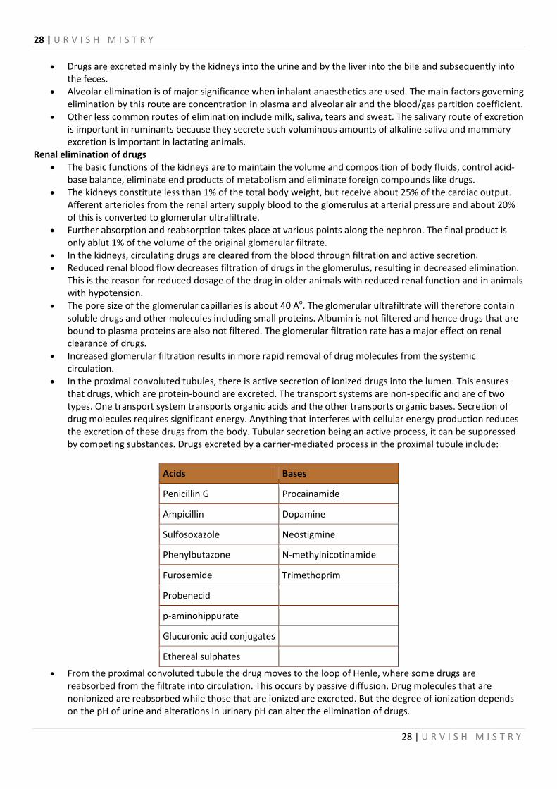
28 | U R V I S H M I S T R Y
28 | U R V I S H M I S T R Y
Drugs are excreted mainly by the kidneys into the urine and by the liver into the bile and subsequently into the feces.
Alveolar elimination is of major significance when inhalant anaesthetics are used. The main factors governing elimination by this route are concentration in plasma and alveolar air and the blood/gas partition coefficient.
Other less common routes of elimination include milk, saliva, tears and sweat. The salivary route of excretion is important in ruminants because they secrete such voluminous amounts of alkaline saliva and mammary excretion is important in lactating animals.
Renal elimination of drugs The basic functions of the kidneys are to maintain the volume and composition of body fluids, control acid‐
base balance, eliminate end products of metabolism and eliminate foreign compounds like drugs. The kidneys constitute less than 1% of the total body weight, but receive about 25% of the cardiac output.
Afferent arterioles from the renal artery supply blood to the glomerulus at arterial pressure and about 20% of this is converted to glomerular ultrafiltrate.
Further absorption and reabsorption takes place at various points along the nephron. The final product is only ablut 1% of the volume of the original glomerular filtrate.
In the kidneys, circulating drugs are cleared from the blood through filtration and active secretion. Reduced renal blood flow decreases filtration of drugs in the glomerulus, resulting in decreased elimination.
This is the reason for reduced dosage of the drug in older animals with reduced renal function and in animals with hypotension.
The pore size of the glomerular capillaries is about 40 Ao. The glomerular ultrafiltrate will therefore contain soluble drugs and other molecules including small proteins. Albumin is not filtered and hence drugs that are bound to plasma proteins are also not filtered. The glomerular filtration rate has a major effect on renal clearance of drugs.
Increased glomerular filtration results in more rapid removal of drug molecules from the systemic circulation.
In the proximal convoluted tubules, there is active secretion of ionized drugs into the lumen. This ensures that drugs, which are protein‐bound are excreted. The transport systems are non‐specific and are of two types. One transport system transports organic acids and the other transports organic bases. Secretion of drug molecules requires significant energy. Anything that interferes with cellular energy production reduces the excretion of these drugs from the body. Tubular secretion being an active process, it can be suppressed by competing substances. Drugs excreted by a carrier‐mediated process in the proximal tubule include:
Acids Bases
Penicillin G Procainamide
Ampicillin Dopamine
Sulfosoxazole Neostigmine
Phenylbutazone N‐methylnicotinamide
Furosemide Trimethoprim
Probenecid
p‐aminohippurate
Glucuronic acid conjugates
Ethereal sulphates
From the proximal convoluted tubule the drug moves to the loop of Henle, where some drugs are reabsorbed from the filtrate into circulation. This occurs by passive diffusion. Drug molecules that are nonionized are reabsorbed while those that are ionized are excreted. But the degree of ionization depends on the pH of urine and alterations in urinary pH can alter the elimination of drugs.

29 | U R V I S H M I S T R Y
29 | U R V I S H M I S T R Y
Highly lipid soluble drugs will be rapidly reabsorbed from the kidney tubule. For this reason excretion of lipid soluble drugs often occur after conversion to more polar metabolite. Even though, many protein bound drugs are actively secreted into the proximal tubule, reabsorption may be favoured especially with drugs that are largely protein bound in plasma.
In canine the urinary pH range from 5.0‐7.0 and in herbivores the urinary pH may range from 7.0‐8.0. Excretion can be enhanced for drugs excreted by the kidneys by altering the pH of the urine. For practical purposes this principle can be applied only to weak acidic or weak basic drugs with pH 5.0‐8.0. Quarternary drugs are polar at all urine pH. They are eliminated rapidly because they cannot be reabsorbed.
Hepatic and other routes of administration Hepatic elimination of drugs
Hepatically eliminated drugs usually move by passive diffusion from the blood into the hepatocyte from where they are secreted into the bile or metabolised first and then secreted into the bile.
The bile then conveys this to the duodenum. In acute liver diseases and in chronic degenerative processes like cirrhosis, the liver’s ability to metabolise and/or eliminate drugs is reduced. Therefore the dose of the drugs eliminated by liver must be reduced in liver disorders in order to prevent drug accumulation in toxic concentrations.
Choleretics or a high fat intake promote bile flow and therefore, biliary excretion and enhance the hepatic secretion of drugs. Broad spectrum antibacterial agents are expected to diminish the hydrolytic action of intestinal flora and thus, may prevent effective enterohepatic cycles.
Large polar molecules (molecular weight >300) are often excreted and are not reabsorbed in the intestine. The ability to excrete polar compounds in bile with molecular weights between 300 and 500 is good in dog,
chicken and rat, moderate in cat and sheep and poor in guinea pig, rabbit, monkey and man. Compounds that undergo biliary excretion
Unchanged drugs and endogenous substances
Compounds excreted as glucuronide conjugates
Erythromycin Chloramphenicol
Clindamycin Trimethoprim
Digitoxin Sulphadimethoxine
Steroid hormones Morhine
Excretion of drugs in milk This route of excretion has both therapeutic and public health importance. The principles of excretion through the mammary gland are similar to those acting in the kidneys, namely
the diffusion of unionised lipid soluble forms of the drugs diffusing through the epithelial cells of the mammary gland.
The pH of plasma and milk are important factors. Since milk is usually more acidic than plasma the basic compounds may be slightly more concentrated and acidic compounds are less concentrated in milk than plasma.
Excretion of drugs is altered in cases of mastitis due to changes in the pH of milk.
Pharmacokinetic parameters Pharmacokinetics The mathematical description of drug concentration in the body The importance of kinetic study:
For arriving at the dosage regimen (dose amount and dosing interval) For understanding the absorption, distribution, metabolism and excretion of drugs For comparison of drug routes For comparison of different formulations (bioequivalence) As an essential part of any drug development study

30 | U R V I S H M I S T R Y
30 | U R V I S H M I S T R Y
For therapeutic drug monitoring
Important pharmacokinetic parameters Most important parameters t1/2, ClB, Vd, AUC Half life (t1/2) – Plasma half life – the time taken for the plasma concentration to be reduced to half its original value
Elimination half life (t1/2 β ) – the half life calculated from the elimination curve (log dose in y axis vs time ) t1/2 b = 0.693 / β ( elimination rate constant) (where β = slope of the curve x 2.303) Longer the half life longer is the duration of action At one t1/2 – 50 % of drug is eliminated At two t1/2 ‐ 75% of drug is eliminated (50 + 50% of remaining 50) At three t1/2 ‐ 87.5% of drug is eliminated At 4 t1/2 ‐ 93.75 % of drug is eliminated
AUC = Area Under the concentration time Curve Calculated by the trapezoidal rule. Important in calculating bioavailability and other parameters
Volume of Distribution (Vd) It is defined as the amount of body fluid containing the drug ‐ an apparent not an accurate value – the drug
to be equally distributed in the whole body fluid. Vd = dose given / conc. of the drug in plasma For a more accurate description,
o Vd area = Dose / β x AUC for i.v. (or) Dose x F / β x AUC for non i.v. Vd is indicative of extent of drug penetration. More distribution – greater Vd Highly protein bound and tissue bound drugs have higher Vd Drugs with higher Vd preferred for treatment in remote sites – skin, bone , muscle, etc.
Clearance – (Cl) Total body clearance (ClB) ‐ An index of efficiency of drug elimination Defined as the volume of body fluid cleared of the drug per unit time For a given drug by the given route, Cl and Vd are characteristic and help in dose determination V d = Cl / β
Models in pharmacokinetics Linear vs Nonlinear drug elimination
If rate of elimination proportional to plasma concentration linear or first order (constant fraction of the drug eliminated per unit time) occurs for most normal kinetic processes. If rate of elimination is constant irrespective of concentration ‐‐‐> nonlinear or zero order reaction (constant
amount of the drug eliminated per unit time) Occurs in toxicity, saturable mechanisms of elimination, At very high dose, reaction changes from first order to zero order Conc. increases disproportionately with increase in dose
Compartmental models A mathematical description of processes based on the number of phases. One compartment or two‐
compartment or multi‐compartment a concept of imaginary division of the body into number of compartments. The compartments do not correlate to any physical or anatomical compartments
A single compartment model consists of only one elimination phase. A two compartment model consists of a rapid distribution phase and a slow elimination phase
Usually, I compartment indicates blood and highly perfused tissues and II compartment indicates peripheral tissues. In rare cases a third compartment corresponding to a deeper tissue component.
Elimination is only from central compartment Most drugs follow two compartment open models with first order processes of absorption and elimination
31 | U R V I S H M I S T R Y
31 | U R V I S H M I S T R Y
Drug accumulation In first order kinetics, on repeated dose within the complete elimination of the previous dose, drug
accumulates in the body. Plateau (steady state) achieved within 4 –5 half lives. The Css is the average of min. and max. of the steady
state or the plateau state. For long term therapy, it is aimed to have the therapeutic conc. as Css Target Cp = Css Dose rate = (target Cp x Cl ) (mg / kg per time)/F In some cases immediate higher dose is to be given (otherwise target Cp will be reached only after 4 – 5 half
lives) In such cases, Loading dose = (target Cp x Vd )/F
Principles of Drug activity – Pharmacodynamics
Modes of drug action Pharmacodynamics ‐ what the drug does to the body? Principles of drug action Drugs do not impart new functions – except gene therapies Basic types of drug actions are:
Stimulation ‐ enhancement of existing level of action o Eg. adrenaline on heart, pilocarpine on salivary gland
Depression ‐ selective diminution of activity of specialized cells o Eg. barbiturates on CNS, Acetylcholine on heart
Irritation ‐ a nonselective, often noxious stimuli. Mild irritation on some cells (skin, connective tissue) stimulate some functions for a beneficial effect.
o Eg. Bitters, topical counterirritants but severe irritation inflammation, necrosis, damage Replacement ‐ of natural substances eg.. hormonal therapy, deficiencies Cytotoxicity ‐ eg. Antibacterials, antivirals, anticancer agents
Mechanisms of drug action Physical action – physical property of the drug causes the main action Eg. mass of the drug ‐ bulk laxatives Adsorption ‐‐ charcoal Osmosis ‐‐ mannitol Radioactivity – 131I ‐ for hyperthyroidism Radioopacity – as contrast media (barum) Chemical action – chemical property of the drug responsible for action Antacids ‐ AlOH2 acidifiers, ‐ NH4Cl alkalinizers, ‐ NaHCO3 oxidizers ‐‐ KmnO4, H2O2 chelating agents – Ca Na EDTA, BAL, Pencillamine
Enzyme mediated drug action Enzymes are ubiquitous biological catalysts – main targets of drug actions Drugs either increase or decrease
enzyme mediated reactions Stimulation is done by endogenous sub. (cofactors) vitamins and minerals Rate of reaction increases
Enzyme induction – microsomal induction by drugs – increased synthesis of proteins Enzyme inhibition – a most common mode of drug action Non specific – metal salts, acids, alkalis inhibit nonspecifically Specific ‐ particular enzyme affected – either competitive or noncompetitive Competitive – competition with normal substrate
Equilibrating type

32 | U R V I S H M I S T R Y
32 | U R V I S H M I S T R Y
Increasing the concentration of normal substrate reverses drug action Eg. sulfonamides and PABA for folate synthetase neostigmine and Ach for cholinesterase
Non‐equilibrating type React with the same site as that of substrate but form strong covalent bonds or have very high affinity than
the normal substrate eg.. organophosphates form covalent bond with cholineesterase methotrexate ‐ 50,000 times more affinity for the enzyme DHFR than DHFAcid
Non‐competitive ‐ reacts with some other site and not on the catalytic site but alters the enzyme loss of catalytic property eg.. aspirin – Cyclooxygenase disulfiram – aldehyde dehydrogenase digoxin ‐ Na+ K+ ATPase
Receptor mediated drug action Most important type of drug action
Receptor is a binding site within biological tissues for either endogenous or exogenous(!!!) substances Macromolecules situated on the surface of the cell or inside the cell Regulate critical functions like enzyme activity, permeability, structural features, etc
Evidence Many drugs show structural specificity minor substitution alters the effect. Stereospecificity of drugs d‐propranolol less active than l‐isomer, l‐noradrenaline – more potent than d‐
noradrenaline antagonism between substances of closely related structures some chemical identity Langley (1878)
proposed the term receptive substance This idea developed by Clark (1920) – propounded the Receptor occupation theory D + R DR E (effect) The postulates of the theory are:
o Intensity of response α fraction of receptors occupied; maximal response when all receptors are occupied ‘all or none’ action of drug on each receptor – each receptor is either fully activated or not at alldrug and receptor have structurally ‘lock and key’ arrangement
Terms related to drug receptor binding Agonists
Agonists are agents that mimic the effects of the endogenous regulatory compound. Agonists may be defined as drugs that possess affinity for a particular kind of a receptor and has the ability
to cause a change in the receptor that gives rise to an observable effect. Their value in clinical practice often rests on their greater capacity to resist degeneration and thus acts for a longer duration than the natural substances they mimic. For this reason, bronchodilatation produced by salbutamol lasts longer than that induced by adrenaline.
The properties exhibited by agonists include affinity, intrinsic activity, maximal efficacy, potency, selectivity and specificity.
Affinity of a ligand (drug or endogenous substance) is a measure of its capacity to bind to the receptor. Affinity may vary greatly among agonists as well as antagonists. Intrinsic activity is a measure of the ability of the agonist‐receptor complex to initiate the observed biological response. A full agonist has an intrinsic activity of 1.
Maximal efficacy reflects the upper limit of the dose‐response relationship without toxic effects being evident. Agonists differ from each other in this regard.
Potency refers to the range of concentrations over which an agonist produces increasing responses. Highly potent drugs produce their effects at lower concentrations; this may impart an advantage to their clinical use, provided the increase in potency is not accompanied by an increase in toxicity.
Agonists in their occupation of receptors exhibit selectivity and specificity. Few agonists are so specific that they interact only with a single subtype of receptor. However, several agonists do show evidences of selectivity for certain subpopulation of receptors.
Full agonists Full agonists are agonists that produce a maximal response by occupying all or a fraction of receptors.
Partial agonists

33 | U R V I S H M I S T R Y
33 | U R V I S H M I S T R Y
Partial agonists are drugs that act both as agonists and antagonists. These drugs in addition to blocking access of the natural agonist to the receptor are capable of a low degree of activation. These agents produce less than a maximal response even when they occupy all of the receptors.
Inverse agonists Some substances produce effects that are specifically opposite to those of agonists and are called as inverse agonists.
Antagonists These are agents that are themselves devoid of any intrinsic activity but cause the effects by inhibition of the
action of an agonist. Drugs that have no activating effect whatsoever on the receptor are termed as pure antagonists.
They are sufficiently similar to the natural agonist to be ‘recognized’ by the receptor and to occupy without activating it, thereby preventing the natural agonist from exerting its effect.
Down regulation When tissues are continuously exposed to an agonist, the number of receptors decreases and this may lead
to a reduction in the number of receptors. This may be a reason for tachyphylaxis. Up regulation
Prolonged contact with an antagonist leads to formation of new receptors and this phenomenon is known as up regulation.
Spare receptors This term refers to the production of a maximal tissue response when only a fraction of the total number of
receptors is occupied. EC50: The concentration of an agonist that produces half maximal effect is known as EC50. IC50: The concentration of an antagonist that produces half maximal inhibition is known as IC50.
Receptors - structure and function
Receptor theories Occupation theories
A.J.Clark propounded the occupation theory which assumes that the extent to which a tissue is dependent on the proportion of its receptor population that has been occupied by a drug and maximal response is reached when all the receptors are occupied. He presumed that each occupied receptor delivered a constant unit of response and that this individual occupancy stimuli was summated in mathematical fashion to give a linearly proportional response. But this theory was not applicable in all situations. In the case of partial agonists a maximal response could not be exhibited even though all the receptors were occupied.
E.J.Ariens, divided the biological response into two separate parameters namely affinity and intrinsic activity. Affinity described the binding of the drug to the receptor and was assumed to be governed by mass action. Intrinsic activity was related to the ability of the drug to induce an effect after binding. The assumptions are the same as those for Clarke’s theory. However, the notion of partial agonism can be accounted for by assuming a lower intrinsic activity for a partial agonist than for a full agonist. Again, a discrepancy between EC50 and KD values and the observations of spare receptors could not be explained.
R.P.Stephenson, assumed that the drug receptor complex provides a stimulus to the tissues and that the stimulus is directly proportional to the fraction of receptors occupied. The response is then related to the stimulus by an unspecified function. The proportionality factor between the fractional occupancy of the receptor population was defined as the efficacy. The efficacy of a drug refers to the action of the drug to produce a response in a given tissue. An additional parameter, intrinsic activity was defined to describe the stimulant activity of the drug itself independent of the tissue. The problem with Stephenson’s model is that the exact link between effect and occupancy remains unclear.
Rate theory Rate theory was developed by W.D.M. Paton and postulates that the biological response is proportional to
the rate at which the drug combines with the receptor – that is, each association of the drug with receptor results in a quantum of excitation. This implies that an agonist must dissociate rapidly from the receptor to enable other successful associations and subsequent generation of quantum of excitation.

34 | U R V I S H M I S T R Y
34 | U R V I S H M I S T R Y
On the other hand, an antagonist is assumed to dissociate slowly to prevent the generation of other quanta of excitation. Thus, dissociation rate constant was considered to be the factor, which determined whether a ligand was an agonist, antagonist or partial agonist. The experimental analysis of drug dissociation rates did not support this model.
Allosteric theories Two allosteric models originally developed to describe enzyme regulation have been proposed. The idea of the allosteric theory is that receptors can exist in a variety of discrete conformational states
differing in their ability of that state to produce a response. Ligands then interact with the receptor in such a way so as to control the conformational state. These
models define precisely the relationship between binding and effect. Receptors sub serve two essential function viz., recognition of the specific ligand molecule and transduction
of signal with a response. Accordingly, the receptor has a ligand binding domain (spatially and energetically suitable for binding the specific ligand) and effector domain, which undergoes a functional conformational change.
Functions of receptors The functions of receptors are
To propagate regulatory signals from outside to within the effector cell when the molecular species carrying the signal cannot itself penetrate the cell membrane
To amplify the signal To integrate various extracellular and intracellular regulatory signals To adapt to short term and long term changes with regulatory milieu and maintain homeostasis.
Types of receptors Type I – Channel linked receptors (ligand‐gated ion channels or inotropic receptor) – These are also known as
ionotropic receptors. These are membrane receptors that are coupled directly to ion channels and are the receptors on which fast neurotransmitters act. Examples include nicotinic acetylcholine receptors and glutamate receptors.
Type II – G‐Protein coupled receptors (metabotropic receptors) – These are also known as metabotropic receptors or 7‐transmembrane spanning receptors. They are membrane receptors that are coupled to intracellular effector systems via a G‐protein. This class includes receptors for many hormones and slow transmitters. Examples include muscarinic acetylcholine receptors and adrenergic receptors.
Type III – Kinase‐linked receptors – These are membrane receptors that incorporate an intracellular protein kinase domain within their structure. They include receptors for insulin, various cytokines and growth factors. Closely related are receptors linked to guanylate cyclase such as the atrial natriuretic factor receptor.
Type IV – Receptors that regulate gene transcription (nuclear recptors) – These are also known as nuclear receptors, though some are actually located in the cytosol rather than the nuclear compartment. They include receptors for steroid hormones and thyroid hormone.
Second messenger systems Many hormones, neurotransmitters, autacoids and drugs act on specific membrane receptors, the
immediate consequence of which is activation of a cytoplasmic component of the receptor, which may be an enzyme such as adenylate cyclase, guanyl cyclase or activation of a transport system or opening of an ion channel.
These cytoplasmic components which carry forward the stimulus from the receptors are known as second messengers. The receptor itself is the first messenger.
Examples of second messengers are cAMP, cGMP, Ca2+, G‐Proteins, IP3, DAG etc. cAMP serves as second messenger for adenosine, opioid, VIP, α2 and β adrenoceptors and H2 receptors. cGMP serves as second messenger for angiotensin receptor. IP3 and DAG serve as second messengers for α1 adrenoceptors, H1 receptors and cholecystokinin. cAMP has varied regulatory effects on cellular functions, for example, energy metabolism, cell division and
cell differentiation, ion‐transport, ion‐channel funtion, smooth muscle contractility etc.

35 | U R V I S H M I S T R Y
35 | U R V I S H M I S T R Y
cGMP has ben identified in cardiac cells, bronchial smooth muscle cells and other tissues. For most effects produced cAMP seems to be stimulatory while cGMP seems to be inhibitory in nature. When the cAMP and cGMP systems are bothe present in a single cell or tissue, they are linked to receptors through which drugs produce opposite effects.
IP3 and DAG are degradation products of membrane phospholipid by the enzyme phospholipase C.
Dose response curve
Plotting As the name indicates this refers to the plotting of the dose vs response, with the dose in X axis and
response in some measurable units in the Y axis. The features of the curve so obtained will provide useful information about the action of the drug or in
comparison among drugs.
Features of dose response curve Plotting of dose response curve
Effect‐ Y axis and Dose – X axis – hyperbola Effect‐ Y axis and Log Dose – X axis – sigmoid curve By taking log dose – a linear relationship in 30 – 70 % range gives a possibility of prediction of response in
this range Response is proportional to an exponential function of the dose Log dose response curves are used to
o display wide range of dose – effect o compare agonists and antagonists
Potency – amount of drug required to elicit a response Relative potency – more meaningful than absolute potency
o Eg. Drug A (potency 10mg) more potent than drug B (100 mg) A 10 times more potent than B – rightward shift of DR curve if less potent Potency need not mean clinical superiority Efficacy – the maximal effect produced by a drug and refers to the peak obtained in the DR curve. Eg. Morphine produces very high analgesia that cannot be obtained with any dose of aspirin Morphine highly efficacious than aspirin
Quantal dose response curve Quantal DR curve due to biological variation ( diff. among individuals in response to same treatment) the
response to drug dose in a population occurs as a frequency distribution. A cumulative freq. Distribution (as % of individuals showing effect plotted against log dose ‐‐‐> yields the
quantal DR curve The dose that produces desired effect in 50% ‐ ED50 The dose that produces death in 50% ‐ LD50 Therapeutic index = LD50 / ED50
Factors modifying drug action
Factors affecting drug action Factors modifying drug action:
Body size: influenzes the conc. achieved at the site of action. Doses are normally given for adults of medium build. In extreme cases – dose to be actually calculated based on per kg dose.
o Body surface area is also an important parameter‐ used in calculation of dose for anticancer drugs o (BSA in sq.m. = K x BW in Grams2/3 x 10 –4 (K= 10 for dogs, 10.1 cats)
Age: drug behavior differs in infants and children. – low GFR,

36 | U R V I S H M I S T R Y
36 | U R V I S H M I S T R Y
o Blood Brain Barrier more permeable, absorption is altered poor metabolizing capacity up to 4 weeks of age
o In the old, poor metabolizing capacity, low PP binding, slower Absorption – dose alteration needed in both cases
Sex: Females smaller in size and may need lower dose. o Pregnancy induced changes – delayed absorption, plasma albumin falls, acid glycoprotein increases,
renal blood flow increases, induction of CYP 450 enzymes – all these affect the kinetics of drug o Besides – possibility of drug affecting fetus or the young ones thro’ milk
Species – changes in sp. in the pattern of metabolism – rabbits resist o Atropine, dogs are poor acetylators, cats deficient in glucuronidation
Race – Blacks need higher atropine to dilate pupil Mongols need lower dose o Fewer reports of aplastic anemia after chloramphenicol use in Asians compared to the West
Diet and plane of nutrition – drug action predictable in good plane of nutrition Genetics – G‐6‐PD deficiency hemolysis with antimalarial agents Malignant hyperthermia after halothane in
some human individuals and pigs Route of administration: Besides kinetics, in some cases route may alter action‐ eg. MgSO4 – orally
purgation, skin – decrease swelling, i/v – CNS and cardiac depression Iodine – antiseptic and antigoitre Environment – Insecticides, tobacco smoke, charcoal broiled meat – induce metabolism.
o Hypnotics taken at night at bed work well. Psychological – beliefs, attitudes, expectations – more in humans
o Placebo (Latin – I shall please) used in trials and to treat otherwise normal patients (hypochondriacs) o Placebos induce endorphins in brain analgesia
Disease states Diseases of GI tract –orally given drugs ‐ altered absorption Liver diseases – Bioavailability of drugs with First pass metabolism Increased – Renal disease affects excretion of drugs In all the above – change of dose needed. Immunocompetence – Normally in very young and very old patients immunity is compromised. So,
bacteriostatic to be replaced by bactericidals Drug interactions – physical or chemical inactivation or incompatibility Food drug interactions – availability of food delays absorption. Ingredients of food may cause specific interactions Pharmacological Interactions – may be beneficial – epinephrine + local anaesthetics preanaesthetics,
antibacterial combinations penicillin + probenecid, ampicillin + sulbactam harmful interactions ‐ aminoglycosides with Neuromuscular
blocking agents Enzyme inhibitors and inducers
Tolerance and Tachyphylaxis – Tolerance is the requirement of higher dose to produce given response – Natural – individual less sensitive to drug (rabbits) Acquired – by repeated use of the drug in an individual who was initially responding – may be due to faster
elimination of the drug on long use or changes in receptor sensitivity (downregulation) Cross tolerance – tolerance among closely related drugs ( morphine and pethidine) Tachyphylaxis ‐ rapid development of tolerance – after only a few doses Seen in ephedrine, tyramine – which deplete stores of catecholamines
Adverse effects of drugs Adverse effects of drugs
Any undesirable or unintended consequence of drug administration may be trivial, serious or fatal All drugs are capable of producing side effects Definition: Any noxious change which is suspected to be due to a drug at normal doses, requires treatment
or decrease in dose or caution in future use of the drug. Not uncommon seen in 10 –25% of the cases. Still drugs are used considering the benefit vs risk

37 | U R V I S H M I S T R Y
37 | U R V I S H M I S T R Y
Predictable ( type I) based on the pharmacological properties of the drug. Side effects, toxic effects and drug withdrawal symptoms
Unpredictable (type II) based on peculiarities of the patient and not on known drug effect. Severity
Minor – no therapy needed Moderate – may need change in drug / or its dosage or prolong hospital stay Severe – potentially life threatening, causes permanent change or requires intensive medical treatment Lethal – contributes to the death
Prevention of adverse effects of drugs Following steps to minimize ADE
o Avoid inappropriate uses o Use correct dose and route of the drug o Consider previous history of reactions, if any o Find out if any other allergic disease existed, and take caution o Rule out harmful interactions among drugs prescribed o Adopt correct drug administration technique o Carry out correct lab. procedures to rule out reactions
Types of adverse effects Side effect ‐ expected effect and unavoidable – eg.. dryness of mouth in atropine treatment Secondary effect – indirect consequences of a primary effect eg.. weakening of host immunity by
corticosteroids flare up of TB Toxic effect – due to overdosage. eg.. Paracetamol induced liver damage, coma by barbiturates, auditory
disturbance in streptomycin Intolerance – appearance of toxic effects at therapeutic doses in some individuals.. eg..only few doses of
carbamazepine ataxia in some patients Idiosyncracy‐‐‐ genetically determined abnormal reactivity to a chemical and also some uncharacteristic
reactions to drugs egg. Barbiturate induced excitement and confusion in some patients. Drug allergy ‐‐‐ immunologically mediated reaction – not related to the action of the drug
( hypersensitivity but not suprsensitivity) – needs a priming dose and the second dose produces violent reactions. drug acts as antigen (hapten) and capable of stimulating the body to produce Antibodies against it.
Type I ‐‐ anaphylactic (Immediate hypersensitivity) – itching , urticaria, asthma etc.. Type II – cytolytic – causes decrease of blood cells and organ damage Type III – Arthus reaction – on vascular endothelium – serum sickness, fever, arthralgia etc.. Type IV – (delayed hypersensitivity) ‐ takes more than 12 h to develop eg.. contact dermatitis, rashes, fever
etc.. Treatment ‐ stopping the offending drug, administration of antihistamines, glucocorticoids, adranaline etc.. Examples of drugs causing hypersensitivity: Penicillin, sulfonamide, tetracycline, salicylates, quinolones, Anti
TB drugs, etc… Photosensitivity ‐ cutaneous reaction due to drug induced sensitization of the skin to UV light or radiation.
Eg.. phenothiazine ( phototoxic), sulfonamide, griseofulvin ( photoallergic) Dependence – psychological – patient feels that he will be alright only if he takes the drug Withdrawal reactions – for non addictive drugs.. eg.. seizures on withdrawal of an antiepileptic drug. Teratogenicity – ability to cause fetal abnormalities eg.. thalidomide, anticancer drugs, tetracyclines. Mutagenicity and carcinogenicity. eg.. anticancer drugs, estrogen, radioactivity, tobacco etc.. Drug induced diseases – (iatrogenic – physician induced)—eg.. ulcer by NSAIDs..
Drug interactions A veterinarian is often posed with the necessity of treating a patient with more than one drug
simultaneously. It is highly probable that one drug might affect the action of the other, the result of which may be beneficial or harmful to the patient. A clinician should possess adequate knowledge about such interactions to avoid any adverse effects.
38 | U R V I S H M I S T R Y
38 | U R V I S H M I S T R Y
Drugs exhibit synergism or antagonism Synergism: It is the cooperation between two drugs where the presence of one drug facilitates or increases
the action of the other. It can be of two types: o Synergism – additive (1+1 = 2) the net effect is only the sum of the individual effects. eg. Aspirin +
paracetamol ( analgesic + antipyretic), ephedrine + theophylline (bronchodilator) o Supraadditive or potentiation (1+1 = 3) net effect is more than the sum of the individual effects o Sulfonamide + trimethoprim ( bacteriostatic becomes bactericidal), Ach + physostigmine ‐‐‐
(inhibition of breakdown) Antagonism: Refers to a decrease or reversal of the pharmacologic response of one drug by another drug.
The drug which antagonizes the action of one drug is called an antagonist. Antagonism is of the following types:
o Antagonism – (1+1 = 0 or < 1) o Can be
physical – based on physical property – adsorption of alkaloids by charcoal chemical – based on chemical property – antacids like NaHCO, chelating agents BAL, EDTA
for heavy metals Physiological or functional
o histamine and adrenaline – on bronchial muscle o Insulin and glucagons – on blood sugar
Receptor antagonism – antagonist binds with the receptor and inhibits generation of response may be competitive or non competitive
Competitive and Non‐competitive Competitive antagonism: The antagonist is chemically similar to the agonistand compete for the same
binding site. The antagonist blocks the effect of the agonist and the antagonistic effect can be overcome by increasing the concentration of the agonist. It is also called surmountable antagonism. Eg: acetylcholine and its antagonist atropine.
Noncompetitive antagonist: The antagonist is not chemically related to the agonist but modifies the binding of the agonist to the receptor by binding to another site. The antagonistic effect cannot be overcome by increasing the concentration of the agonist. It is also called unsurmountable antagonism. Eg: Acetylchloline and decamethonium.
Pharmacokinetic antagonism: where one drug alters the action of the other drug by preventing or reducing any one of the pharmacokinetic processes such as absorption, distribution, biotransformation and excretion.
Eg: Antacids prevent the absorption of other drugs (absorption) Aspirin displaces warfarin from its plasma protein binding site (distribution) Microsomal inducers and inhibitors modulate the metabolism of other drugs (biotransformation) Probenecid antagonizes penicillin excretion by competing for the binding site (excretion)
Drug screening and drug discovery
Drug discovery Most new drugs or drug products are discovered or developed through one of the following methods:
1. Identification or elucidation of a new drug target. 2. Rational drug design based on an understanding of biological mechanisms, drug receptor structure, enzymes
sites etc. 3. Chemical modification of an existing molecule. 4. Screening for biologic activity of natural products, previously discovered chemical entities, and libraries of
peptides, nucleic acids, and other organic molecules. 5. The explosive growth in biotechnology and cloning has paved the way to identify genes to produce peptides
and proteins. Efforts continue to focus on the discovery of new targets and approaches, based on studies with genomics, proteomics, nucleic acids and molecular pharmacology for drug therapy.
6. Combinations of known drugs to obtain additive or synergistic effects or a repositioning of a known drug for a new therapeutic use.

39 | U R V I S H M I S T R Y
39 | U R V I S H M I S T R Y
Drug screening Drug screening refers to a sequence of experiments and characterization using a variety of biologic assays at
the molecular, cellular, organ system, and whole animal levels to define the activity and selectivity of the drug. For example, anti‐ infective drugs may be tested against a variety of infectious organisms some of which are resistant to standard agents.
The molecule will also be studied for a broad array of other actions to establish the mechanism of action and selectivity of the drug. This will reveal any suspected and unsuspected toxic effects.
Occasionally, an unexpected therapeutic action is discovered by chance.. It becomes very important to develop suitable preclinical in vitro and in vivo models to predict drug action.
During drug screening, studies are performed to define the pharmacologic profile of the drug at the molecular, cellular, system, organ, and organism levels.
At the molecular level the drug would be tested for receptor binding activity to target recptors, metabolizing enzymes etc.
At the cellular level, the drug would be studied to determine whether it stimulates or inhibits the cellular function.
At the organ level the pharmacologic activity and selectivity for the organ studied would be studied. Isolated organ studies esp. smooth muscles and other in vitro preparations can be used.
Whole animal studies are generally necessary to determine the effect of the drug on organ systems and disease models. Cardiovascular and renal function studies of all new drugs are generally first performed in normal animals.
Where appropriate, studies on disease models would be performed. Evidence would be collected on duration of action and efficacy following oral and parenteral administration.
If the agent possessed useful activity, it would be further studied for possible adverse effects on other major organ systems, including the respiratory, gastrointestinal, endocrine, and central nervous systems.
These studies might suggest the need for further chemical modification to achieve more desirable pharmacokinetic or pharmacodynamic properties. For example, oral administration studies might show that the drug was poorly absorbed or rapidly metabolized in the liver; modification to improve bioavailability might be indicated.
If the drug was to be administered long‐term, an assessment of tolerance development would be made. For drugs related to or having mechanisms of action similar to those known to cause physical dependence, abuse potential would also be studied. For each major action found, a pharmacologic mechanism would be sought.
The outcome of this screening procedure is called a lead compound, i.e., a leading candidate for a successful new drug.
Preclinical safety and toxicity testing All drugs are toxic at some dose. To correctly define the toxicities of drugs and the therapeutic index
comparing benefits and risks of a new drug is an essential part of the drug development. Most drug candidates fail to reach the market, due to the limitations of toxicity. Lead drugs that survive the initial screening and profiling procedures must be carefully evaluated for
potential risks before and during clinical testing. Although no chemical can be certified as completely "safe" (free of risk), the objective is to estimate the risk
associated with exposure to the drug candidate and to consider this in the context of therapeutic needs and duration of likely drug use.
Toxicity testing is time‐consuming and expensive. Two to six years may be required to collect and analyze data on toxicity and estimates of therapeutic index before the drug can be considered ready for testing in humans.
Large numbers of animals may be needed to obtain valid preclinical data. Owing to the situation arising out of concerns of animal usage, animal testing has become a public issue and some segments of the public attempt to halt all animal testing in the unfounded belief that it has become unnecessary.
Extrapolations of therapeutic index and toxicity data from animals to humans are reasonably predictive for many but not for all toxicities.

40 | U R V I S H M I S T R Y
40 | U R V I S H M I S T R Y
Clinical trials In each of the three formal phases of clinical trials, volunteers or patients must be informed of the
investigational status of the drug as well as the possible risks and must be allowed to decline or to consent to participate and receive the drug.
In phase 1, the effects of the drug as a function of dosage are established in a small number (25‐50) of healthy volunteers. If the drug is expected to have significant toxicity, as is often the case in cancer and AIDS therapy, volunteer patients with the disease are used in phase 1 rather than normal volunteers.
o Phase 1 trials are done to determine whether humans and animals show significantly different responses to the drug and to establish the probable limits of the safe clinical dosage range.
o These trials are nonblind or "open"; that is, both the investigators and the subjects know what is being given. Many predictable toxicities are detected in this phase.
o Pharmacokinetic measurements of absorption, half‐life, and metabolism are often done in phase 1. o Phase 1 studies are usually performed in research centers by specially trained clinical
pharmacologists. In phase 2, the drug is studied in patients with the target disease to determine its efficacy.
o A modest number of patients (100‐200) are studied in detail. A single‐blind design is often used, with an inert placebo medication and an established active drug (positive control) in addition to the investigational agent.
o Phase 2 trials are usually done in special clinical centers (eg, university hospitals). o A broader range of toxicities may be detected in this phase.
In phase 3, the drug is evaluated in much larger numbers of patients with the target disease‐sometimes thousands‐to further establish safety and efficacy.
o Using information gathered in phases 1 and 2, phase 3 trials are designed to minimize errors caused by placebo effects, variable course of the disease, etc. Therefore, double‐blind and crossover techniques are frequently used.
o Phase 3 trials are usually performed in settings similar to those anticipated for the ultimate use of the drug. The investigators are usually specialists in the disease being treated.
o Certain toxic effects, especially those caused by immunologic processes, may become apparent only in phase III.
Phase 4 begins once approval to market a drug has been obtained. o This constitutes monitoring the safety of the new drug under actual conditions of use in large
numbers of patients. o The careful and complete reporting of toxicity by physicians after marketing can reveal some side
effects may after chronic dosing. o Several hundred thousand patients may have to be exposed before the first case is observed of a
toxicity that occurs with an average incidence of 1 in 10,000. o Phase 4 has no fixed duration

41 | U R V I S H M I S T R Y
41 | U R V I S H M I S T R Y
Bio prospecting Refers to the search for useful applications, process or product with commercial value in the biosphere. It is the manipulation of nature’s bounty to find newer substances with economic importance. Many of the drugs in vogue today have come from traditional knowledge on the use of the plants in various
biodiversity regions. After the discovery of penicillin, a systematic search of soil bacteria and microbes have led to the discovery
of large groups of antibiotics, which are very successfully employed in the treatment of many bacterial infections in man and animals.
Scientists are on a constant search into the deepest and farthest corners of the earth for a new product to be used as a drug for many diseases.
Bioprospecting has its own issues and concerns. Often it leads to many new drug discoveries and treatment of diseases but the indigenous people who were instrumental are left to languish with no support and credit.
Successful bioprospecting requires the development of a sustainable model in which a benefit sharing system is evolved among industries, ecosystem and the projected communities.
Cure for many a dreaded diseases must be hidden in the remotest forests, deep oceans and highest mountains and bioprospecting is an instrument to uncover such new knowledge.
When performed in a large‐scale operation, the effort is referred to as mass bioprospecting. Experiences from the mass bioprospecting efforts demonstrate that mass bioprospecting is a complex
process, involving expertise from diverse areas of human endeavors, what is important is the recognition of issues on genetic access, prior informed consent, intellectual property and the sharing of benefits that may arise as a result of the effort.
42 | U R V I S H M I S T R Y
42 | U R V I S H M I S T R Y
Future mass bioprospecting endeavors must take heed of the lessons learned from past and present experiences in the planning for a successful mass bioprospecting venture.
Assay of drugs Assay of drugs refers to the quantitative estimation of a drug / its active component in a formulation. It is carried out for the following purposes:
o To test the purity of a drug preparation. o To compare the potency of a drug with that of a standard. o To assess the efficiency of preparation while purifying the drug from a mixture / natural product.
Assays are of different types Chemical assay
o In this the concentration of drug is determined by chemical methods suah as spectrophotometry, fluorometry, HPLC or mass spectroscopy. These methods use one of the physiochemical properties of the compounds such as absorbance, fluorescence, polarity, charge to mass ratio etc. These are the most commonly used methods.
Bioassay o In this the concentration of biologically active component in a unit quantity of the test drug is
compared to the response produced by the standard or reference drug. Bioassays are more cumbersome but less accurate when compared to chemical assays. Eg: assay of histamine in isolated guinea pig ileum, assay of acetylcholine in frog rectus abdominis muscle and assay of antibiotics using bacteria.
Immunoassay o This is commonly used for the assay of hormones. The method depends on the binding between the
radiolabelled hormone and the corresponding antibody. The method is highly sensitive to detect minute quantities of the hormones in body fluids.
Biopharmaceuticals and gene therapy
Biopharmaceuticals These are therapeutic agents produced through biotechnological means, but not by conventional laboratory
(chemical) synthesis. Biopharmaceuticals have their origin from the advancement in the knowledge of biotechnology and
molecular cell biology. Biopharmaceuticals popularly called as “Designer Proteins” are the promising therapeutic tolls of the future. Examples are functional human peptides such as:
o ADH, oxytocin, GnRH, ACTH, TSH/TRH, Calcitonin, Insulin, Somatostatin, Growth Hormone, Cyclosporin etc., and
o Enzymes / proteins (Streptokinase, Asparaginase, DNAase, glucocerebrosidase, erythropoietin, clotting factors, interferons, Monoclonal Antibodies, Vaccines etc) Daclizumab(Zenopox).
Genetically engineered human IgG (immunosuppressant monoclonal antibody binds specifically to a‐chain of IL2 receptor) used to lower the rate of rejection phenomenon in kidney transplant recipients.
Through DNA recombinant technology, bacteria are induced to produce hormones like insulin and growth hormone and enzymes like L – asparaginase.
Gene therapy It means prevention or treatment of diseases through manipulation of gene function. It is done by insertion
of specific genes (therapeutic genes) exogenously into the animal cells. It involves replacing the defective gene or supplementation to a nonfunctional gene or suppression of an
abnormal gene. Recombination DNA technology forms the basis of synthesis of therapeutic genes. Gene delivery into host is highly complicated and different from that of conventional drug delivery systems. The process involves inserting a therapeutic gene first into a vector (Viral vectors: Retro – or Adenoviruses or
Nonviral Vectors: Liposomes, microspheres and plasmid DNA).
43 | U R V I S H M I S T R Y
43 | U R V I S H M I S T R Y
A vector with the gene is then introduced into the patient through either in vivo or ex vivo means. The in vivo method involves injecting the suspension of the vector having the therapeutic gene intravenously
or into the target tissue/ organ. The ex vivo method involves first inserting the therapeutic gene into stem cells obtained from the patient (
blood or bone marrow) and inject the same into the patient. Gene therapy is currently aimed at treatment of diseases not responding to conventional drug therapy, such
as inherited metabolic diseases through either correction of a deficiency or blockade of an abnormal metabolic reaction.
It is also applied to induce synthesis of a deficient protein in the host’s cells and treatment of a variety of cancers (melanoma, renal carcinoma, lung cancer, cystic fibrosis etc) certain immune deficiencies and infectious diseases ( like HIV).
Gastro – intestinal pharmacology
Sialagogues (sialics) and antisialagogues (antisialics)
Sialagogues Sialogogues are agents that increase the volume of saliva, thereby increasing the appetite and the
digestability of food. Such remedies are used in large animal practice as ‘tonics’. Increased salivation is obtained by administering substances that stimulate taste buds like the vegetable
bitters. Substances with this type of activity include gentian, quassia and nuxvomica. Increase in salivation is also achieved on administration of parasympathomimetics.
Antisialogogues Antisialagogues (antisialics) are agents that decrease the volume of salivary secretions. The antisialagogue effect may be required during surgery of the mouth or after excision of salivary cysts. Parasympatholytic drugs have antisialagogue effect. Atropine or glycopyrrolate are commonly used to
reduce secretions as a premedicant during surgery to reduce salivary and bronchial secretions.
Stomachics Stomachics are agents that increase the tone and function of stomach and increase appetite.
Eg: Bitter stomachics such as ginger, gentian, chirata. These plant based preparations stimulate the taste buds and reflexly stimulate the stomach.
Appetite stimulants Inappetance or anorexia is common in disease states and the resultant malnutrition can delay recovery and
may aggravate the underlying disease. The various ways by which appetite can be improved are:
o Enteral alimentation with liquid supplements is useful in small animals o Small amounts of palatable food should be offered at frequent intervals. Warming the food may
enhance appetite in carnivores o Various drugs are used for the short‐term stimulation of appetite.
Benzodiazepines – e.g., Diazepam, Oxazepam The antiserotonergic effect depresses the satiety center in the hypothalamus. Benzodiazipines are used most frequently in cats and less frequently in horses, dogs
and goats for a short‐term stimulation of appetite after which the effects diminish. Diazepam can be administered orally, intravenously, intramuscularly once or twice
daily, Oxazepam can administered once daily orally. The adverse effects include sedation and ataxia.
Cyproheptadine

44 | U R V I S H M I S T R Y
44 | U R V I S H M I S T R Y
Cyproheptadine acts as a serotonin and a histamine (H1) antagonist and suppresses the satiety center in the hypothalamus.
Cyproheptadine stimulates appetite in cats and in humans, but not in dogs. Adverse effects includes CNS excitement and marked aggressive behaviour in cats.
Glucocorticoids‐e.g., Prednisolone, Dexamethasone Glucocorticoids stimulate appetite due to glucocorticoid‐induced euphoria. In small animals prednisolone or dexamethasone is administered intramuscularly
once daily or every other day where as in large animals it is administered intramuscularly once daily.
Adverse effects of glucocorticoids include immunosuppression and gastric ulceration.
Bitters like plant derived compounds such as nux vomica, chiretta, ginger These are used as salivary stimulants and their effect on appetite is questionable. However, bitters are a component of tonics for appetite stimulation in large animals
in traditional medicine.
Emetics Emetics are agents that induce vomition and are classified as
centrally acting and reflex acting emetics.
The main area in the brain responsible for vomiting is the vomiting center. This area is located in the lateral reticular formation of the medulla.
It receives input from many areas like: o Chemoreceptor trigger zone: This is located in the floor of the fourth ventricle and it picks up
circulating chemicals/toxins in the blood (e.g. bacterial food poisoning toxins, drugs used for chemotherapy) and cause vomiting.
o Vestibular apparatus o Tractus solitarius ‐ vagal afferents from the gut, heart and testes o Direct input from gut (reflex)
The neurotransmitters involved in the emetic response are o On the CTZ: 5HT3, D2 o On the vagal afferents: 5HT3 o In the vomiting center: Muscarinic, H1 receptors
Centrally acting emetics This includes: Apomorphine is a D2 agonist that acts on the CTZ to cause vomiting.
o Apomorphine is contraindicated in cats as it may cause extreme excitement in cat. o Apomorphine can be administered subcutaneously or as a lamella (eye tablet) in the conjunctival
sac. o Since apomorphine depresses the emetic centre, repeated dosing is not recommended when the
initial dosing is ieffective. Xylazine is used as an emetic in cats. Syrup of Ipecacunha is also used as an emetic.
o It acts both centrally and locally (reflexly) as an emetic. o It irritates the gastric mucosa and within 15‐30 minutes after administration emesis is observed. o It produces a toxic metabolite and hence if emesis does not occur after administering the syrup, it
should be removed by administering a gastric lavage. o High concentrations of ipecac are cardiotoxic. o Animals with ipecac overdose may exhibit arrhythmias, hypotension and myocarditis. o Activated charcoal ( a constituent of universal antidote) should not be administered with ipecac as
the charcoal adsorbs the syrup of ipecac and prevents it from irritating the gastric mucosa and in turn producing emesis.
Locally acting emetics

45 | U R V I S H M I S T R Y
45 | U R V I S H M I S T R Y
Warm water Warm saturated sodium chloride solution Sodium chloride crystals placed at the back of the tongue Sodium carbonate as crystals Copper sulphate 50 ml of a 1% solution Zinc sulphate 50 ml as a 1% solution Freshly ground mustard paste Hydrogen peroxide as a 3% solution
Clinical uses of emetics include removal of ingested poison (except when corrosive poisons of petroleum products have been ingested) emptying the stomach contents in emergency surgery Emetics should not be administered in an unconscious patient, weak and debilitated patient and patients
with clinical circumstances like pregnancy, bowel obstructions and hernia.
Antiematics Antiemetics are agents that are used to control emesis Species like the horses, rabbits and rodents are unable to vomit. Protracted vomiting is undesirable in dogs, cats and other species. Vomiting usually occurs secondary to another disease process. If the primary cause is treated then vomiting
disappears. Antiemetics are useful in motion sickness, uremia, liver disease, endotoxemia, canine parvovial
gastroenteritis and in cancer chemotherapy. Use of antiemetics may mask the primary disease. Prolonged vomiting leads to electrolyte and acid base imbalances and dehydration. Antiemetics may also be
locally acting or centrally acting. Centrally acting antiemetics Phenothiazine derivatives
A number of phenothiazine derivatives (chlorpromazine, promethazine, prochlorperazine, trifluopromazine etc.) are classified as broadspectrum antiemetics.
They act by blocking the chemoreceptor trigger zone at low doses and the emetic centre in the brain at higher doses.
Promethazine is a H1 antagonist (classical antihistaminic) which also has antimuscarinic actions. It is very effective at preventing motion sickness (since the vestibular afferents input in the vomiting center which has H1 and muscarinic receptors).
Prochlorperazine is a D2 antagonist and has no antipsychotic effects. It is useful as an antiemetic as well as for dizziness. It has minor anticholinergic effects. Since it blocks D2receptors elsewhere (e.g. substantia nigra), it may cause extrapyramidal effects.
Chlorpromazine, another phenothiazine, can also be used as an antiemetic, although it tends to be sedative. It also has antipsychotic effects.
Hyoscine (scopolamine) is a muscarinic antagonist (M2). It is used as a patch behind the ear for controlling CTZ mediated vomiting.
Metoclopromide is a D2 antagonist which is also a weak 5HT3 antagonist. It increases the motility of the gut in the upper regions (does not cause diarrhoea ‐ only facilitates gastric emptying). It is useful in gastric stasis in a number of species including the cattle and horses. Metoclopromide will help the absorption of drugs because it stimulates gastric emptying. This drug is useful in vomiting associated with vagal afferents (gut disorders, heart, testes, gut irritants ‐ all stimulate the 5HT3 receptor on the vagal afferents) .
Ondansetron is a 5HT3 antagonist and is a potent antiemetic. It is very effective in patients receiving cancer chemotherapy. It can also be used for CTZ nausea.
Locally acting antiemetics H2 antagonists like cimetidine, ranitidine etc. These drugs reduce the acid output and hence decrease the
irritating effects on acid stomach lining. As a consequence, afferent signals to vomiting centres are decreased. Antacids like sodium bicarbonate are also useful.

46 | U R V I S H M I S T R Y
46 | U R V I S H M I S T R Y
Prokinetics Prokinetics are agents that promote gastric motility and thus hasten the process of emptying of the gastric
contents into the intestine, becaues of their ability to evacuate the gastric contents they are of value as antiemetics.
They are also indicated in gastritis, gastric reflux and dyspepsia. They are known to act via 5 HT4 receptors. Eg: Cisapride, metoclopromide, tegeserod etc.
Ulcer management Factors
Factors that play a role in peptic ulcer formation in gastric and duodenal ulcers: o Ulcerogenic factors include
acid hypersecretion pepsin drugs (NSAIDs, corticosteroids, ethanol, chemotherapeutic agents, cigarettes) infection tumors stress ‐ head trauma, shock/sepsis, emotional alteration of protective factors disruption of mucosal integrity and decreased bicarbonate secretion
Symptoms of ulcers are due to a combined effect of hydrochloric acid and pepsin. Acid causes pain but not tissue damage. Pepsin, which is activated in acid environment, digests mucosal, muscular and vascular layers of stomach &
intestines. Gastric (stomach) ulcers are not due to too much acid. Alkaline reflux from intestines may inflame gastric mucosa. Duodenal (intestinal) ulcers are due to increased acid secretion in stomach. When stomach empties into intestines there is too much acid for pancreatic secretions to neutralize and this
acid erodes tissues. Most common causes are H. pylori and NSAIDs as well as gastrin hypersecretion.
Goals of drug therapy for ulcers include neutralize existing gastric acid ‐ antacids inhibit acid secretion ‐ H2 antagonists, proton pump inhibitors treat infection (H. pylori) ‐ antibiotics, bismuth subsalicylate protect ulcer from further damage ‐ sucralfate
Antacids Antacids are agents which neutralize excess acid. Some of the commonly used antacids include sodium bicarbonate, aluminium hydroxide and dihydroxy‐
aluminium, calcium carbonate and magnesium hydroxide. The weak base reacts with the gastric hydrochloric acid to form a salt and water. This reaction causes an increase in the gastric pH and above pH 4, pepsin is inactive. Antacids differ in
o neutralizing capacity ‐ determines dosage o time to onset ‐ slow or rapid o duration of action ‐ long or brief o side effect ‐ constipation vs. diarrhea o potential to produce electrolyte disturbances o convenience ‐ liquid vs. tablet o palatability o cost
Sodium bicarbonate It has a rapid onset of action.

47 | U R V I S H M I S T R Y
47 | U R V I S H M I S T R Y
But it is not an ideal antacid due to the rebound acidity i.e., stimulation of acid production once the pH exceeds 4, due to gastrin secretion.
As sodium bicarbonate is readily absorbed into systemic circulation, it alters systemic pH and produces electrolyte disturbances.
This in turn may lead to edema, hypertension or heart failure. Aluminium hydroxide
The insoluble aluminum chloride that is formed due to the reaction of hydrochloric acid and aluminium hydroxide often causes constipation.
It also binds with tetracycline to inhibit its absorption. Calcium carbonate
The absorption of ~10% of calcium chloride may result in hypercalcemia (muscle weakness, kidney stone formation) and rebound gastric acidity.
Constipation may occur with high doses. Magnesium hydroxide
This causes prolonged neutralizing effect due to slow stomach emptying. Poor absorption of magnesium salts may result in diarrhoea . Combinations of aluminum or calcium containing antacids with magnesium containing antacids can nullify
the adverse effects on bowel function.
Histamine receptors – H2 antagonist Endogenous histamine released from paracrine enterochromaffin‐like cells or mast cells stimulates parietal
H2 receptors. Histamine activates a cyclic AMP pathway, activates H+_ K+‐ATPase and leads to increased H+ in gastric
lumen. H2 receptor antagonists can block this histamine induced secretion and are helpful reducing acid secretion during ulcers.
Examples of H2 receptor antagonists include cimetidine, ranitidine, famotidine, nizatidine etc. In general, these drugs are well tolerated with few side effects (less than 3% with cimetidine and <1% with
other drugs). Cimetidine exhibits antiandrogenic properties leading to decreased sperm count, gynecomastia and also
inhibits hepatic drug metabolism. Inhibition of cytochrome P450 activity by cimetidine leads to potential drug interactions. This feature is not seen with ranitidine, famotidine and nizatidine.
Sucralfate It is a complex of sulfated sucrose and aluminum hydroxide and is known as gastric band aid. Sucralfate polymerizes to a viscous gel at a pH less than 4. It combines with proteins and adheres to ulcer forming a barrier resistant to acid and pepsin and binds to
bile salts which are implicated in ulcer pathogenesis. It precipitates surface proteins at the ulcer base and acts as a physical barrier preventing acid, pepsin and
bile from coming in contact with the ulcer base. It does not decrease gastric acid secretion or neutralize acid or stimulate healing. The drug requires an acidic environment to be more effective. It is as effective as cimetidine or antacids at healing peptic ulcers and is useful in treating gastric reflux. It is a Gastric protective. Use: Sucralfate is used in the treatment of gastric and duodenal ulcers in dogs , cats and foals. Adverse effect: Constipation may occur with long term therapy. It interacts with many drugs and shown to interfere with absorption of tetracyclines, fluoroquinolones,
cimitidine,phenytoin and digoxin.
Proton pump antagonists Proton pump inhibitors are also called as acid blockers. Examples of this group of drugs are omeprazole,
lansoprazole, pantoprazole etc.

48 | U R V I S H M I S T R Y
48 | U R V I S H M I S T R Y
These drugs irreversibly inhibit K+H+‐ATPase proton pump in gastrointestinal parietal cells i.e. inhibits entry of H+ ion entry into lumen.
These drugs also appear to have antimicrobial activity against H. pylori. These drugs break down in the acid environment of the stomach and must be given orally as enteric coated
preparations and they have short plasma half‐lives but are long acting. They inhibit hepatic drug metabolism and are very well tolerated. These drugs heal duodenal ulcers more rapidly than H2 antagonists. They are used primarily for ulcers refractory to other treatments and are also used for reflux esophagitis and
management of gastrinomas.
Cytoprotective drugs Misoprostol, an analogue of prostaglandin (PGE1) analog is used as a cytoprotective drug. Prostaglandins (membrane derived second messengers) have protective actions on gastric mucosa. They increase mucosa formation and increases release of bicarbonate (HCO3
‐). Adverse effects are not common except diarrhoea and abdominal cramping. As this drug may cause abortion, it is not recommended for use in pregnant animals. It is used for short‐term management of peptic ulcers and also to reduce gastrointestinal irritation
associated with NSAID (aspirin‐like drugs) therapy. Drugs that eradicate Helicobacter pylori bacteria
multiple antibiotics (metronidazole + amoxicillin or tetracycline) bismuth subsalicylate Most effective treatment is a combination of multiple antibiotics with H2 antagonist or proton pump
inhibitor for 6 months .
Digestants and purgatives
Choleretics and cholagogues Choleretics are agents that increase the volume of secretion of bile
Bile acids (e.g: cholic acid, chenodeoxycholic acid) o Semisynthetic bile acid derivatives (e.g: dehydrocholic acid) o Sodium salts of bile acids
Mechanism of action o Bile salts stimulate choleresis (bile flow) o Bile salts emulsify lipids, enhancing the digestion of ffats and the absorption of long chain fatty acids.
Uses o Bile salts and acids treat vigorous maldigestion syndromes characterized by steatorrhea (e.g:
pancreatitis and bile salt deficiency). Cholagogues
These are agents that promote the flow of bile from the liver and gall bladder. Eg: licorice, ursodeoxycholic acid.
Purgatives and laxatives Purgatives are drugs which cause marked intensification of intestinal activity and results in the expulsion of
intestinal content from the colon and rectum. The terms aperient and cathartic are also applied to describe the purgatives. A distinction is made according
to the intensity of action. Laxative or aperient: are milder in action, elimination of soft but formed stools Purgatives or cathartic: are stronger action resulting in more fluid evacuation. Laxative and cathartics are used for:
o Relief of acute non dietary constipation o Removal poisons from the gastrointestinal tract o Prevention of tenesmus in advanced pregnancy or prolapse

49 | U R V I S H M I S T R Y
49 | U R V I S H M I S T R Y
o Evacuation of the bowel prior to surgery or radiography Many drugs in low doses act as laxative and in larger doses as purgatives. Type of stools and latency of action of purgatives employed in usually recommended doses
Soft formed feces (take 1‐3 days)
Semi fluid stools (take 6‐8 hours)
Watery evacuation (within 1‐3 hours)
Bulk forming Docusates Liquid paraffin Lactulose
PhenolphthaleinBisacodyl Senna
Saline purgatives Castor oil
Classification Bulk forming agents: Dietary fiber like bran, Psyllium (Plantago), Ispaghula Stool softening agents: Docusates (Dioctyl Sodium sulfosuccinate) Lubricant agent: Liquid paraffin Stimulant (contact) purgative agents:
o Diphenylmethane, Phenolphthalein, Bisacodyl o Anthroquinones (emodins), Senna, Cascara sagrada o Fixed oils: castor oil
Osmotic purgative agents: o Magnesium salts: sulfate (Epsom salt) , hydroxide, oxide o Sodium salts: sulfate (glauber’s salt), phosphate o Sodium potassium tartrate o Sodium phosphate and sodium tartrate o mixtures (Fleet mixtures) o Lactulose
General mechanism of action All purgatives increase the water content of feces by:
o A hydrophilic or osmotic action, retaining water and electrolytes in the intestinal lumen‐ increase volume of colonic content and make it easily propelled.
o Acting on intestinal mucosa to decrease net absorption of water and electrolyte intestinal transit is enhanced indirectly by fluid bulk
o Increasing propulsive motility as primary action‐ allowing less time for absorption of salt and water as a secondary effect
Certain purgatives increase motility through an action on the myenteric plexus. Laxatives modify the fluid dynamics of the mucosal cell and may cause fluid accumulation in gut lumen by
one or more of the following mechanisms: o inhibiting Na+K+ ATPase of villus cells – impairing electrolyte and water adsorption o Stimulating adnylyl cyclase in crypt cells – increasing water and electrolyte secretion o Enhancing prostaglandin synthesis in mucosa which increases secretion o Structural injury to absorbing intestinal mucosal cells.
Osmotic purgatives Osmotic cathartics are nonabsorbable or poorly absorbable salts or polymers that osmotically retain water in
the intestinal lumen. Solutes that are not absorbed in the intestine retain water osmotically and distend the bowel – increase peristalsis indirectly.
All inorganic salts used as osmotic purgatives have similar action differ only in dose, palatability and risk of systemic toxicity.
They have a rapid onset of action that begins in the small intestine. Osmotic cathartics are the cathartics of choice for elimination of poisons. Magnesium sulfate and magnesium oxide are administered orally 5‐15g; bitter in taste Sodium sulfate 10‐15g is administered orally or via a stomach tube as a 6% solution

50 | U R V I S H M I S T R Y
50 | U R V I S H M I S T R Y
Sodium phosphate 6‐12 g Sodium potassium tartrate (Rochelles salt) 8‐15g Polythelene glycol‐electrolyte solutions are administered orally prior to colonoscopy Sodium phosphate and sodium tartrate mixtures are administered rectally to dogs. In cats, they produce
hyperphosphatemia and should not be used. The salts mentioned above are dissolved in 100‐200ml of wter, produce 1‐2 fluid evacuation within 1‐23
hours with mild cramping, casue nearly complete emptying of bowels. Smaller doses have milder laxative action. Magnesium ions release cholecystokinin which may aid purgative action of magnesium salts. Magnesium salts are contraindicated in renal insufficiency, while sodium salts should not be used in patients
suffering form Congestive Heart Failure and other sodium retaining states. Repeated use of saline purgatives can cause fluid and electrolyte imbalance.
Lactulose It is a semisynthetic disaccharide of fructose and lactose which is not digested or absorbed form small
intestine but retains water. It is broken down in the colon by the colonic bacteria to osmotically more active products. Flatulence is common, cramps occur in few cases. Nausea may be produced becsue of unpleasant taste. It is not preferred for constipation. Lactulose causes reduction of blood ammonia by 25‐50% in patients with hepatic encephalopathy. The breakdown products of lactulose are acidic and reduce the pH of stools. Ammonia produced by bacteria in colon in converted to ionized ammonium and is not absorbed.
Irritant purgatives Irritant/Stimulant/contact catharatics: (e.g: castor oil, aloes, senna, cascara sagarada) Irritant cathartics are plant derivatives. They are powerful purgatives and often produce griping. They were thought to irritate the intestinal mucosa and thus stimulate motor activity. It accumulates water and electrolytes in the lumen by altering absorptive and secretory activity of the
mucosal cell. They inhibit Na+K+ ATPase at the basolateral membrane of villous cells‐transport of Na+ and accompanying
water into the interstitium is reduced. Secretion is enhanced by activated cAMP in crypt cells and by increased prostaglandin synthesis. Larger doses of stimulant purgatives can cause exces purgation, fluid and electrolyte imbalance. Hypokalemia can occur on regular use. Routine and long‐term use must be avoided, produces colonic atony. They may reflexly stimulate gravid uterus, hence contraindicated in pregnancy and also in subacute and
chronic intestinal obstruction. Castor oil is a bland vegetable oil obtained from the seeds of Ricinus communis. It is cleaved by pancreatic lipases in the small intestine to yield irritant ricinoleates, which stimulate
peristalsis and reduce fluid absorption.This is hydrolyzed in the ileum by lipase to ricinoleic acid and glycerol. Ricinoleic acid being polar is poorly absorbed.
Aloe, senna and cascara sagrada contain anthroquinone glycosides that are hydrolyzed in the large intestine to yield to irritant anthraquinones emodins Senna is most popularly used.
Unabsorbed in the small intestine, they are passed to the colon where bacteria liberate the active anthrol form which either acts locally or is absorbed into circulation – excreted in bile to act on small intestine.
Thus they take 6‐7 hours to produce action. They are secreted in milk sufficient to cause purgation in the suckling young ones. It is one of the oldest purgatives used. It mainly contains triglycerides of ricinolenic acid which is a polar long chain fatty acid which It was believed to irritate the mucosa and stimulate intestinal contraction. The primary action has now been shown to be decreased intestinal absorption of water and electrolytes, and
enhanced secretion by a detergent like action on the mucosa.
51 | U R V I S H M I S T R Y
51 | U R V I S H M I S T R Y
It causes morphological damage of villus tip and peristalsis is increased. Due to its unpalatibility, frequent cramping and violent action, possibility of dehydration and after
constipation mucosal damage it is no longer favoured. Senna is obtained from leaves and pod of certain Cassia sp, while Cascara sagrada is the powdered bark of
th buck‐thorn tree. Anthraquinones
Anthraquinones stimulate smooth muscle and increase colonic motility. Because they act in the large intestine, their onset of action is slow. Usually given at nighttime which cause a
single, soft but formed evacuation generally occurs in the morning. Cramps and excessive purging occur is some cases. The active principle is believed to act on the myentric plexus to increase peristalsis and decrease
segmentation. They also inhibit salt and water absorption in the colon. Senna anthraquinone has been found to simulate PGE2 in the rat intestine. Regular useage causes colonic atony and mucosal pigmentation. Aloe especially releases the anthraquinone emodin, which has affinity towards the sigmoid flexure of the
horse’s intestine, and produces the cathartic effect. Irritant cathartics are administered orally to relieve acute constipation in small and large animals.
Diphenylmethanes Phenolphthalein is an indicator and is in use as purgative since earlier days. It turns urine pink if alkaline. Bisacodyl is a later addition and more popular. Diphenylmethanes are partly absorbed and reexcreted in bile, entero‐hepatic circulation is more important
in phenolphthatein which can produce protracted action. Bisacodyl is activated in the intestine by deacetylation. Their primary site of action is in the colon in which one or two semiformed motions occur after 6‐8 hours.
Phenolphathalein: 60‐130 mg: to be administered at night time Bisacodyl ( Dulcolax) 5mg, 10mg tablet Adverse effects
These doses of phenolphthalein produce fluid evacuations and cramps. Morphological alterations in the colonic mucosa is likely to occur and makes mucosa more leaky.
Bisacodyl suppository acts by irritating the anal and rectal mucosa leading to reflex increase in motility causing evacuation in 20‐40 minutes. It may cause inflammation and mucosal damage.
Bulk purgatives Bulk laxatives (e.g: methylcellulose, agar, psyllium, wheat bran) Dietary fibres consists of unabsorbable cell wall and other constituents of vegetable food – cellulose, pectins,
glycoproteins and other polysaccharides. Bran is a byproduct of flour industry consists of 40% dietary fibre. It absorbs water in the intestines, swells,
increases water content of feces.‐ softens it and facilitates colonic transit. Osmotically active products may be formed in the colon by bacterial degradation of pectins etc., which act to
retain water. Dietary fibre supports bacterial growth in colon which contribute to the fecal mass. Certain dietary fibres (gums, lignins, pectins) bind bile acids and promote their excretion in feces leading to
enhancement of degradation of cholesterol in liver and thereby plasma LDL cholesteol is lowered. Increased intake of dietary fibres is the most appropriate method for prevention and treatment of functional
constipation. It is the first line approach for most dogs and cats in the treatment of simple constipation. Prolonged intake of bran and other bulk forming agents reduces rectosigmoid intraluminal pressure – relives
symptoms of irritable bowel disease and colonic diverticulosis. It is also useful when tenesmus at stools has to be avoided.
Demerits Unpalatable, 20‐40g/day needs to be ingested. Full effect requires daily administration for at least 3‐4 days.
52 | U R V I S H M I S T R Y
52 | U R V I S H M I S T R Y
It does not soften feces already present in colon or rectum. Flatulence may occur.
Psyllium (Plantago) and Ispaghula Contain natural colloid mucilage, which forms a gelatinous mass by absorbing water, 3‐12 g of refined husk
freshly mixed with water or milk and taken daily acts in 1‐3 days. It should not be swallowed dry (may lead to oesophageal impaction) Ispaghula husk (refined) Plantago husk
Methylcellulose and carboxymethylcellulose are semi‐synthetic colloidal hydrophilic derivatives of cellulose; 4‐6 g/day is satisfactory to produce laxative effect. Mechanism of action
Bulk laxatives contain hydrophilic colloids, which absorb water and increae bulk. Bulk stimulatives large bowel peristalsis Wheat bran is administered orally by adding to the diet. The laxative effect occurs within 1‐3 days. Generous amount of water must be given with all bulk forming agents. It should not be used in patients with gastric ulcerations, adhesions, stenosis and when fecal impaction is a
possibility
Lubricant purgatives Lubricants, mineral oil (liquid petroleum) and white petroleum lubricate and soften feces. Liquid paraffin is a viscous liquid; a mixture of petroleum hydrocarbons. It was introduced as a laxative
earlier. It is pharmacologically inert. When taken for 2‐3 days, it softens stools and is said to lubricate hard scybali by coating them.
Dose: 15‐30 ml /day oil as such or in emulsified form in dog Disadvantages
It will be very bland and unpleasant to swallow because of oily consistency Small passes into the intestinal mucosa, carried into the lymph and may produce foreign body granuloma in
the intestinal submucosa, mesenteric lymph nodes, liver and spleen. Probalility of causing lipid pneumonia because of trickling into lungs while swallowing Carries fat soluble vitamins with it into the stools leading to deficiency Leakage of the oil past anal sphincter may embarrass and stain the floor May interfere with healing in the anoractal region.
Hence advisable for occasional use and that too postoperatively. Surfactants
Docusates is an anionic surfactant that acts in the large bowel to hydrate and soften feces by an emulsifying action.
It causes net water accumulation in the intestinal lumen by an action on the intestinal mucosa. It emulsifies the colonic contents.
Being a detergent, it can disrupt the mucosal barrier and enhance absorption of may nonabsorbable drugs , e.g liquid paraffin – should not be combined with it. Eg: Cellubril, Laxicon as capsules
Dose: 100‐200 mg /day; acts in 1‐3 days. It is a mild laxative; specially indicated when straining as t stools must be avoided.
Cramps and abdominal pain can occur. It is bitter in taste. Liquid preparations may cause nausea and is hepatotoxic on prolonged usage.
Choice, indications, combinations, abuse of purgatives Choice and use of purgatives
Laxatives are important for their harmfulness as they are for their value in medicine. All laxatives are contraindicated in:
o An animal of undiagnosed abdominal pain, colic or vomiting o Organic constipation due to stricture or obstruction in bowel, hypothryroidism, hypercalcemia,
malignancies an ceratin drugs like opioids, sedatives,, anticholinergics etc.,

53 | U R V I S H M I S T R Y
53 | U R V I S H M I S T R Y
Valid indications of laxatives are Functional constipation – Spastic (irritable bowel) and Atonic ( Sluggish bowel) Recumbent animals Tenesmus Bowel surgery, endoscopyu, abdominal X‐ray After anthelmintics treatment Food/drug poisoning.
Combined preparations of purgatives Agarol: Liquid paraffin+Phenolphthalein+ Agar Cremaffin: Milk of magnesia+Liquid paraffin+Phenolphthalein Julax: Bisacodyl+ Casanthrol Laxatin: Sennosides+Docusates Pursennid‐IN: Purified senna extract + docusates.
Purgative abuse Chronic use of purgatives must be discouraged. Once the purgative habit forms, it is difficult to break. Dangers of purgative abuse are:
o Flairing of intestinal pathology, rupture of inflamed bowel o Fluid and electrolyte imbalance, specially hypokalemia o Steatorrhoea, malabsorption syndrome o Protein losing enteropathy o Spastic colitis
Antidiarrheal General considerations
o Acute diarrhea may respond to symptomatic therapy with antidiarrheal drugs, but chronic diarrhea requires a definitive diagnosis and specific therapy.
Oral rehydration therapy represents a significant advance in treating diarrhea in the absence of vomiting. In addition to glucose or aminoacids, or both, these solutions contain sodium chloride, potassium chloride,
sodium bicarbonate, and potassium phosphate. Sodium‐glucose and sodium – aminoacid‐linked absorption by the enterocyte remain intact even in the
presence of moderate damage to the intestinal villi, providing the driving force for water and electrolyte absorption form the lumen to replace fecal losses.
Opiates o Paregoric is a camphorated tincture of opium o Diphenoxylate is a synthetic congener of mepridine. Lomotil is diphenoxylate plus atropine
Opioids Loperamide
Loperamide is a synthetic piperidine opioid It is an opiate analogue with weak anticholinergic property. Because of poor water solubility little is absorbed form the intestine. Entry into brain is negligible. In addition to its opiate like action on motility, it also inhibits secretion. It improves fecal continence by enhancing anal sphincter tone.
Mechanism of action Opiates inhibit ACh release. The resultant increased gastrointestinal rhythmic segmentation and decreased propulsive motility slow the
transit time of luminal contents and increase absorption In addition , opiates directly stimulate absorption of fluid and electrolyte via mu ‐opiate receptors in the CNS
and intestinal mucosa. Codeine

54 | U R V I S H M I S T R Y
54 | U R V I S H M I S T R Y
Antidiarrheal action is primarily attributed to its peripheral action on small intestine and colon. It does not have central effects , buit produce dependance. It should be used only for a short period.
Diphenoxylate It is an opioid chemically related to pethidine: used exclusively as constipating agent. The antidiarrheal action is prominent, but because it is absorbed systemically and crosses blood brain barrier
CNS effects may occur. Atropine is added in a non‐pharmacological dose to discourage abuse. Abuse liability is rated low and overdose will produce disturbing atropinic side effects. Use: Opiates are effective in the symptomatic treatment of acute diarrhea.
Administration Paregoric is administered orally 2‐3 times daily to dogs and cats once daily to calves and foals Diphenoxylate is administered orally 2‐3 times daily to dogs and cats Loperamide is administered orally 1‐2 times daily to dogs and cats Codeine is administered orally 2‐3 times daily to dogs and cats. Adverse effects: bacterial overgrowth in the intestinal lumen of animals with infectious diarrhea may result
from slowed intestinal transit time. In cats, excitatory reactions render opiate use controversial. Paregoric is a camphorated tincture of opium. Diphenoxylate is a synthetic congener of mepridine. Lomotil is diphenoxylate plus atropine.
Anticholinergics Anticholinergic agents
Methscopolamine. Other drugs include aminopentamide, propantheline and isopropamide, which are also effective as antiemetics
Mechanism of action Anticholinergics inhibit propulsive and non propulsive gastrointestinal motility. They also inhibit normal, cholinergically mediated basal secretions of the gastrointestinal tract.
Uses Anticholinergic agents may be used to treat diarrhea: however, they are of questionable benefit in this
capacity because diarrhea is more commonly associated with hypomotility than hypermotility Anticholinergic agents may be used to treat gastrointestinal spasm
Administration Aminopentamide is administered orally, intramuscularly or subcutaneously every 8‐12 hours Propantheline is administered orally every 8 hours Isopropamide is administered orally every 12 hours
Adverse effects Adverse effects of anticholinergics include xerostomia, xerophthalmia, loss of visual accommodation,
tachycardia, urine retention, paralytic ileus and constipation. Anticholinergics are contraindicated in patients with glaucoma.
Protectants and adsorbants Kaolin‐pectin suspensions (20% kaolin (hydrated aluminium silicate) and 1 % pectin ( a polygalacturonic acid
carbohydrate polymer) Bismuth subsalicylate.
Mechanism of action Protectives and adsorbents adsorb toxins and provide a protective coating on inflamed mucosa. Bismuth subsalicylate has an anti ‐ prostaglandin action in addition to its adsorbent properties. Uses: Protectants and adsorbents are used for the symptomatic therapy of acute diarrhea. Adsorbents may
decrease fluidity of feces without actually decreasing fecal water loss, thereby limiting their usefulness. Adminsitration
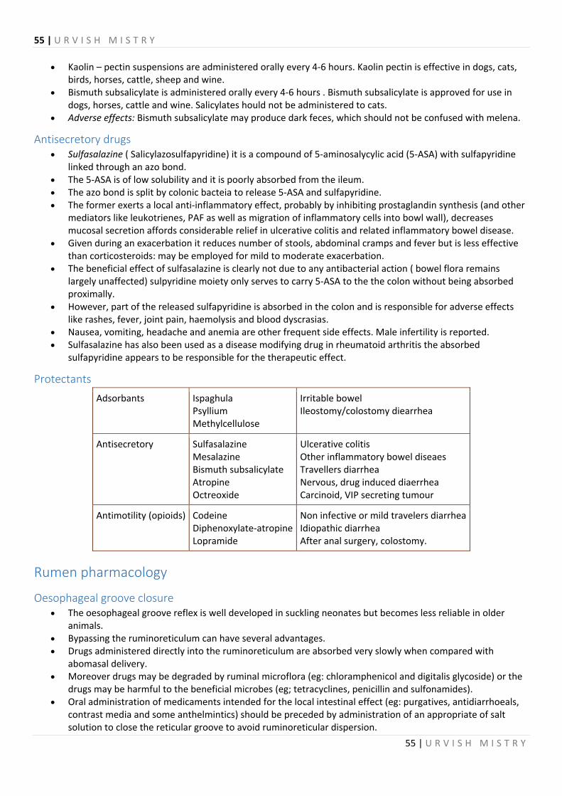
55 | U R V I S H M I S T R Y
55 | U R V I S H M I S T R Y
Kaolin – pectin suspensions are administered orally every 4‐6 hours. Kaolin pectin is effective in dogs, cats, birds, horses, cattle, sheep and wine.
Bismuth subsalicylate is administered orally every 4‐6 hours . Bismuth subsalicylate is approved for use in dogs, horses, cattle and wine. Salicylates hould not be administered to cats.
Adverse effects: Bismuth subsalicylate may produce dark feces, which should not be confused with melena.
Antisecretory drugs Sulfasalazine ( Salicylazosulfapyridine) it is a compound of 5‐aminosalycylic acid (5‐ASA) with sulfapyridine
linked through an azo bond. The 5‐ASA is of low solubility and it is poorly absorbed from the ileum. The azo bond is split by colonic bacteia to release 5‐ASA and sulfapyridine. The former exerts a local anti‐inflammatory effect, probably by inhibiting prostaglandin synthesis (and other
mediators like leukotrienes, PAF as well as migration of inflammatory cells into bowl wall), decreases mucosal secretion affords considerable relief in ulcerative colitis and related inflammatory bowel disease.
Given during an exacerbation it reduces number of stools, abdominal cramps and fever but is less effective than corticosteroids: may be employed for mild to moderate exacerbation.
The beneficial effect of sulfasalazine is clearly not due to any antibacterial action ( bowel flora remains largely unaffected) sulpyridine moiety only serves to carry 5‐ASA to the the colon without being absorbed proximally.
However, part of the released sulfapyridine is absorbed in the colon and is responsible for adverse effects like rashes, fever, joint pain, haemolysis and blood dyscrasias.
Nausea, vomiting, headache and anemia are other frequent side effects. Male infertility is reported. Sulfasalazine has also been used as a disease modifying drug in rheumatoid arthritis the absorbed
sulfapyridine appears to be responsible for the therapeutic effect.
Protectants Adsorbants Ispaghula
Psyllium Methylcellulose
Irritable bowel Ileostomy/colostomy diearrhea
Antisecretory Sulfasalazine Mesalazine Bismuth subsalicylate Atropine Octreoxide
Ulcerative colitis Other inflammatory bowel diseaes Travellers diarrhea Nervous, drug induced diaerrhea Carcinoid, VIP secreting tumour
Antimotility (opioids) Codeine Diphenoxylate‐atropineLopramide
Non infective or mild travelers diarrheaIdiopathic diarrhea After anal surgery, colostomy.
Rumen pharmacology
Oesophageal groove closure The oesophageal groove reflex is well developed in suckling neonates but becomes less reliable in older
animals. Bypassing the ruminoreticulum can have several advantages. Drugs administered directly into the ruminoreticulum are absorbed very slowly when compared with
abomasal delivery. Moreover drugs may be degraded by ruminal microflora (eg: chloramphenicol and digitalis glycoside) or the
drugs may be harmful to the beneficial microbes (eg; tetracyclines, penicillin and sulfonamides). Oral administration of medicaments intended for the local intestinal effect (eg: purgatives, antidiarrhoeals,
contrast media and some anthelmintics) should be preceded by administration of an appropriate of salt solution to close the reticular groove to avoid ruminoreticular dispersion.

56 | U R V I S H M I S T R Y
56 | U R V I S H M I S T R Y
5% copper sulphate, 5% zinc sulphate, 10% sodium bicarbonate or 10% sodium sulphate can be administered at doses of 60ml to cattle to bring about closure of oesophageal groove.
1‐2% copper sulphate is useful in sheep. Onset of reflex response takes about 5‐10seconds and the groove may remain closed for up to 60 seconds.
Suckling is a strong stimulus for this reflex even in adults. It is best to allow sick calves and lambs to drink medicated milk from a nipple to assure abomasal delivery and rapid absorption.
Administration of liquid medication to ruminants orally may result in spontaneous closure of the reticular groove in a certain number of calves which results in an complete or partial rumino reticular by pass.
When stimulated, buccal and pharyngeal receptors activate a vagal reflex that closes the groove withing 2‐5 seconds: effects last for 60 seconds.
Milk, sodium bicarbonate (10% to calves) or copper sulphate (5% to calves, 2% to lambs) but not water may be used to induce esophageal groove closure.
Ruminotorics Ruminotorics are agents and mixtures that promote forestomach function (fermentation and motility). Typically such mixtures consist bitters like nuxvomica to stimulate salivation and perhaps ruminal
contractions, alkalinizing compounds like magnesium oxide to elevate a low ruminal fluid pH, galactogenic substance like glycerol, minerals like cobalt as co‐factors for microbial enzyme function and salts like phosphate as buffers to maintain osmolality. Bitters: (e.g: nux vomica, ginger, capsicum)‐ stimulate salivation which may enhance rumen function,
however, the efficacy of bitters is minimal. Bitters are administered orally. Cholinergics (e.g: neostigmine, bethenachol)‐ transiently increase the frequency, but not the strength , of
contractions in rumen atony. Cholinergics are administered subcutaneously. Opiate antagonists (e.g: naloxone)‐ stimulate extrinsic contractions when administered parenterally. Opiate
antagonists are useful for the treatment of endotoxin‐induced rumen stasis. Rumen fluid transfer ‐ Oral inoculation of viable rumen bacteria and protozoa is the most effective means of
restoring rumen function following correction of the primary cause of stasis
Rumen antacids Rumen antacids (e.g: magnesium oxide, magnesium carbonate, aluminium hydroxide, calcium carbonate, ammonium carbonate)
Antacids are used to treat mild cases of lactic acidiosis resulting form carbohydrate engorgement. These agents are administered orally every 8‐12 hours Systemic alkalosis may result from overdose
especially of magnesium oxide.
Antizymotics Antizymotics are agents that used to contol fermentation in the ruminant forestomach. When orally
administered these agents act by inhibiting microbial growth in the rumen. They are of use in conditions such as bloat where thee is uncontrolled fermentation. Eg: Formalein,
turpentine oil.
Cardiovascular pharmacology
Classes of drugs acting on heart Drugs acting on the heart can be classified into Drugs that directly affect the myocardial cells which includes
o autonomic neurotransmitters and related drugs o cardiac glycosides and other inotropic agents o antidysrhythmic or antiarrhythmic drugs
Drugs that affect the cardiac function indirectly through actions elsewhere in the vascular system calcium antagonists acting both directly on the myocardial cells and indirectly by relaxing the arterioles.
57 | U R V I S H M I S T R Y
57 | U R V I S H M I S T R Y
Autonomic transmitters and related drugs Both sympathetic and parasympathetic systems exert a tonic effect on the heart at rest. The main effects of sympathetic activity on the heart are
o positive inotropic effect (increased force of contraction) o positive chronotropic effect ( increased heart rate) o increased automaticity o repolarisation and restoration of function following generalised cardiac depolarisation o Reduced cardiac efficiency.
The parasympathetic effects include o cardiac slowing and reduced automaticity o decreased force of contraction o inhibition of A‐V conduction
Effects of drugs acting on heart The effect of drugs on the heart may be expressed as
Inotropic effect if it affects the contractility Chronotropic if it affects the rhythmicity Dromotropic if it affects the conductivity Bathmotropic if it affects the excitability Tonotropic if it affects the tonicity.
Congestive heart failure Heart failure is a global term for the physiological state in which cardiac output is insufficient in meeting the
needs of the body and lungs. Often termed "congestive heart failure" or CHF, this is most commonly caused when cardiac output is low and the body becomes congested with fluid.
Common causes of heart failure include : myocardial infarction ischemic heart disease, hypertension, valvular heart disease Cardiomyopathy.
It may also occur due to severe anemia, Gram negative septicemia, beriberi (vitamin B1/thiamine deficiency), thyrotoxicosis, arteriovenous fistulae, arteriovenous malformations.
Cardiac glycosides The main source of cardiac glycosides is from the foxglove family. Their effectiveness in cardiac failure was, described by William Withering in 1775. He published his classic
monograph ‘An account of the foxglove and some of its medicinal uses: with practical remarks on dropsy on used by an old woman of Shorpshireand other diseases’ in 1785.
Initially kidney was thought to be the target organ The cardiac glycosides are unique in that they not only improve the contractility of the myocardium, but also
reduce the heart’s demand for energy and oxygen. These drugs also decease the conduction of certain impulses within the heart and therefore decrease the
heart rate. Source of glycosides
Digitalis purpurea or Purple foxglve (leaf) ‐ Digitoxin, Gitoxin, Gitalin Digitalis lanata or White foxglove (leaf) ‐ Digitoxin, Gitoxin, Digoxin
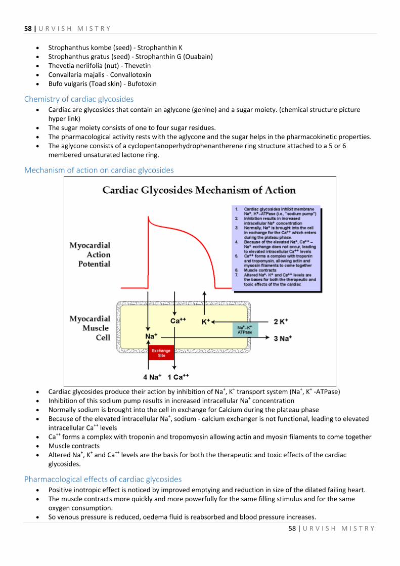
58 | U R V I S H M I S T R Y
58 | U R V I S H M I S T R Y
Strophanthus kombe (seed) ‐ Strophanthin K Strophanthus gratus (seed) ‐ Strophanthin G (Ouabain) Thevetia neriifolia (nut) ‐ Thevetin Convallaria majalis ‐ Convallotoxin Bufo vulgaris (Toad skin) ‐ Bufotoxin
Chemistry of cardiac glycosides Cardiac are glycosides that contain an aglycone (genine) and a sugar moiety. (chemical structure picture
hyper link) The sugar moiety consists of one to four sugar residues. The pharmacological activity rests with the aglycone and the sugar helps in the pharmacokinetic properties. The aglycone consists of a cyclopentanoperhydrophenantherene ring structure attached to a 5 or 6
membered unsaturated lactone ring.
Mechanism of action on cardiac glycosides
Cardiac glycosides produce their action by inhibition of Na+, K+ transport system (Na+, K+ ‐ATPase) Inhibition of this sodium pump results in increased intracellular Na+ concentration Normally sodium is brought into the cell in exchange for Calcium during the plateau phase Because of the elevated intracellular Na+, sodium ‐ calcium exchanger is not functional, leading to elevated
intracellular Ca++ levels Ca++ forms a complex with troponin and tropomyosin allowing actin and myosin filaments to come together Muscle contracts Altered Na+, K+ and Ca++ levels are the basis for both the therapeutic and toxic effects of the cardiac
glycosides.
Pharmacological effects of cardiac glycosides Positive inotropic effect is noticed by improved emptying and reduction in size of the dilated failing heart. The muscle contracts more quickly and more powerfully for the same filling stimulus and for the same
oxygen consumption. So venous pressure is reduced, oedema fluid is reabsorbed and blood pressure increases.

59 | U R V I S H M I S T R Y
59 | U R V I S H M I S T R Y
Negative chronotropic effects by direct stimulation of the vagal centre and by slowing the rate of passage of impulses through the conducting system.
Diuretic effect due to increased blood flow and increased glomerular filtration rate.
Pharamacokinetics Absorption differs among the digoxin preparations. Prepared digitalis causes local irritation and vomiting in pig and dog and for these species digitalis glycoside
should be given. In ruminants, oral administration of glycosides leads to destruction of the drug and hence it is preferable to
give the drug parenterally. Plasma protein binding is high with digitoxin. Excretion of the drug is by the kidneys. Enterohepatic recycling increases the duration of action of the drug. The use of these drugs in dogs and cats is not uncommon. However, the absorption of digoxin from the gastrointestinal tract though adequate may differ somewhat
among animals and can be influenced by feeding times.
Therapeutic uses of cardiac glycosides Congestive heart failure. Arrhythmias by depressing the AV nodal conductivity and increasing refractoriness.
Digitalization Digitalis glycosides have a narrow margin of safety. Initial administration of large dose in divided doses. (24‐48 hrs)
Followed by maintenance dose. Loading dose in dogs
o Slow method‐ 5 equal parts for 48hrs o Rapid method‐ 3 equal parts @ 6hr interval. o Intensive method‐ half dose initially, one fourth after 6hrs and one eighth each after 4 hr intervals o Digitoxin‐ 0.11 – 0.22mg/kg total loading dose
Maintenance dose‐0.011mg/kg @12hr intervals in dogs There is a considerable, individual variation in response to these glycosides.
Adverse reactions of cardiac glycosides Risk of toxicity is greater in patients with advanced heart disease,in high dosage, renal disease, age and
hypothyroidism. Factors that increase myocardial sensitivity to digitalis toxicity include myocardial disease or ischemia,
hypokalemia, high serum calcium and low serum magnesium. Symptoms of toxicity can be grouped as cardiac symptoms and non‐cardiac symptoms. Cardiac symptoms of toxicity
o Sinus bradycardia, o S‐A block, o A‐V block, tachycardia, o premature ventricular contraction
Non‐cardiac symptoms of toxicity gastrointestinal disturbances like anorexia, nausea, vomiting fatigue and muscle weakness CNS effects like confusion, hallucination, restlessness, insomnia, drowsiness and occasionally overt
psychoses Visual effects like hazy vision, difficulty in reading, photophobia, chromatopsia (yellow or green colour
appearance) gynaecomastia (antiadrenergic effect).

60 | U R V I S H M I S T R Y
60 | U R V I S H M I S T R Y
Treatment of cardiac glycosides toxicity Withhold cardiac glycosides Withhold potassium depleting diuretics Administer potassium salts Administer antiarrhythmics if arrhythmias appear to be life threatening
Administer agents like activated charcoal to bind digoxin in the gastrointestinal tract.
Precautions and drug interactions Hypokalemia will lead to increased digitalis toxicity In elderly patients and in patients with renal and severe hepatic disease the toxicity will be higher Thyrotoxicosis reduces the responsiveness to digitalis Myxoedema increases the responsiveness to digitalis Partial A‐V block may be converted to complete A‐V block
Drug interactions Diuretics – potassium depleting diuretics precipitate digitalis toxicity while potassium sparing diuretics
reduce the renal excretion of digitalis Calcium precipitates digitalis toxicity Quinidine reduces binding to proteins and thereby increase the concentration and toxicity of digitalis Verapamil and diltiazem increase plasma concentrations of digoxin Antacids and neomycin reduce absorption of digitalis.
Other drugs used in CHF Phosphodiesterase inhibitors like theophylline. Diuretics like furosemide to increase water and sodium excretion, decrease preload and relieve symptoms of
pulmonary and systemic congestion Vasodilators like hydralazine ACE inhibitors like enalapril, lisinopril etc. Adrenergic alpha1 agonists like prazosin Calcium channel blockers like nifedipine
Antiarrhythmic drugs
Types of arrhythmia Sinus bradycardia ‐ low sinus rate <60 beats/min. Sinus tachycardia ‐ high sinus rate of 100‐180 beats/min as occurs during exercise or other conditions that
lead to increased SA nodal firing rate. Atrial tachycardia ‐ a series of 3 or more consecutive atrial premature beats occurring at a frequency
>100/min; usually due to abnormal focus within the atria and paroxysmal in nature. This type of rhythm includes paroxysmal atrial tachycardia (PAT).
Atrial flutter ‐ sinus rate of 250‐350 beats/min. Atrial fibrillation ‐ uncoordinated atrial depolarizations. Junctional Escape Rhythm ‐ SA node suppression can result in AV node‐generated rhythm of 40‐60 beats/min
(not preceded by p‐wave). AV blocks ‐ a conduction block within the AV node (or occasionally in the bundle of his) that impairs impulse
conduction from the atria to the ventricles. Supraventricular tachycardia (SVT) ‐ usually caused by reentry currents within the atria or between ventricles
and atria producing high heart rates of 140‐250. Ventricular premature beats (VPBs) ‐ caused by ectopic ventricular foci; characterized by widened QRS. Ventricular tachycardia (VT) ‐ high ventricular rate caused by aberrant ventricular automaticity or by
intraventricular reentry; can be sustained or non‐sustained (paroxysmal); characterized by widened QRS; rates of 100 to 200 beats/min; life‐threatening.

61 | U R V I S H M I S T R Y
61 | U R V I S H M I S T R Y
Ventricular flutter ‐ ventricular depolarizations >200/min. Ventricular fibrillation ‐ uncoordinated ventricular depolarizations Ventricular fibril first‐degree AV nodal block ‐ the conduction velocity is slowed so that the P‐R interval is
increased to greater than 0.2 seconds. Can be caused by enhanced vagal tone, digitalis, beta‐blockers, calcium channel blockers, or ischemic damage.
Second‐degree AV nodal block ‐ the conduction velocity is slowed to the point where some impulses from the atria cannot pass through the AV node. this can result in P‐waves that are not followed by ARS complexes. for example, 1 or 2 P‐waves may occur alone before one is followed by a QRS. When the QRS follows the P‐wave, the P‐R interval is increased. In this type of block, the ventricular rhythm will be less than the sinus rhythm.
Third‐degree AV nodal block ‐ conduction through the AV node is completely blocked so that no impulses are able to be transmitted from the atria to the ventricles.
QRS complexes will still occur (escape rhythm), but they will originate from within the AV node, bundle of his, or other ventricular regions. Therefore, QRS complexes will not be preceded by P‐waves.
Furthermore, there will be complete asynchrony between the P‐wave and QRS complexes. Atrial rhythm may be completely normal, but ventricular rhythm will be greatly reduced depending upon the
location of the site generating the ventricular impulse. Ventricular rate typically range from 30 to 40 beats/min. WOLFF PARKINSON SYNDROME – An accessory pathway (Bundle of Kent) that connects the atria and the
ventricles, in addition to the AV node. This accessory pathway does not share the rate‐slowing properties of the AV node, and may conduct electrical activity at a significantly higher rate than the AV node
Arrhythmia Arrythmia is any abnormal pattern of electrical activity in the heart. Normally the waves of depolarization
follow a specific sequence, starting in the S‐A node and ending with the contraction of the ventricles. Sometimes another area of the myocardium or conducting system begins to depolarize out of sequence or more rapidly than the S‐A node, disrupting the normal electric pattern.
Arrhythmias are divided into two general groups: those that result in an increased heart rate and those that result in a decreased heart rate.
Arrhythmias are further subdivided into groups according to the location of the ectopic foci or lesion causing arrhythmia.
A ‘supraventricular arrhythmia’ indicated that the cause of the problem is above the ventricle. A ‘ventricular arrhythmia’ indicates that the problem originates in the ventricles. The abnormal site of depolarization is called an ectopic focus. An ectopic focus in the ventricles may be indicated on the ECG as a single large bizarre wave. Multiple
premature ventricular contractions cause flutter. If the conduction disturbance is severe, the heart has no coordinated contractions and the condition is referred to as ventricular fibrillation.
Antiarrhythmic drugs Antiarrhythmics are agents used to control ectopic foci or reverse disorganized conduction of electrical
impulses through the heart Although research has provided some information regarding the cellular mechanisms of arrhythmias and the
mode of action of antiarrhythmic drugs, the general approach to therapy remains empirical.
Classification Antiarrhythmics or antidysrhythmics are classified as follows.
o Class I drugs act by blocking the sodium channel. They are subdivided into 3 subgroups, IA, IB, and IC, based effects on phase‐0 deploarization and repolarization:
Subclass IA drugs have moderate potency at blocking the sodium channel and also usually prolong repolarization (increase QRS) Eg. quinidne, procainamide, disopyramide
Subclass IB drugs have the lowest potency as sodium channel blockers, produce little if any change in action potential duration, and usually shorten repolarization. Eg. lidocaine, phenytoin
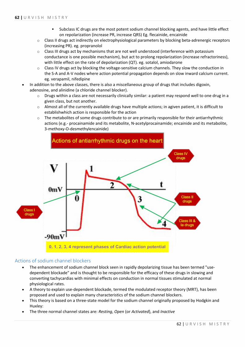
62 | U R V I S H M I S T R Y
62 | U R V I S H M I S T R Y
Subclass IC drugs are the most potent sodium channel blocking agents, and have little effect on repolarization (increase PR, increase QRS) Eg. flecainide, encainide
o Class II drugs act indirectly on electrophysiological parameters by blocking beta‐adrenergic receptors (increasing PR). eg. propranolol
o Class III drugs act by mechanisms that are not well understood (interference with potassium conductance is one possible mechanism), but act to prolong repolarization (increase refractoriness), with little effect on the rate of depolarization (QT). eg. sotalol, amiodarone
o Class IV drugs act by blocking the voltage‐sensitive calcium channels. They slow the conduction in the S‐A and A‐V nodes where action potential propagation depends on slow inward calcium current. eg. verapamil, nifedipine
In addition to the above classes, there is also a miscellaneous group of drugs that includes digoxin, adenosine, and alinidine (a chloride channel blocker).
o Drugs within a class are not necessarily clinically similar: a patient may respond well to one drug in a given class, but not another.
o Almost all of the currently available drugs have multiple actions; in agiven patient, it is difficult to establishwhich action is responsible for the action
o The metabolites of some drugs contribute to or are primarily responsible for their antiarrhythmic actions (e.g.‐ procainamide and its metabolite, N‐acetylprocainamide; encainide and its metabolite, 3‐methoxy‐O‐desmethylencainide)
Actions of sodium channel blockers The enhancement of sodium channel block seen in rapidly depolarizing tissue has been termed "use‐
dependent blockade" and is thought to be responsible for the efficacy of these drugs in slowing and converting tachycardias with minimal effects on conduction in normal tissues stimulated at normal physiological rates.
A theory to explain use‐dependent blockade, termed the modulated receptor theory (MRT), has been proposed and used to explain many characteristics of the sodium channel blockers.
This theory is based on a three‐state model for the sodium channel originally proposed by Hodgkin and Huxley:
The three normal channel states are: Resting, Open (or Activated), and Inactive

63 | U R V I S H M I S T R Y
63 | U R V I S H M I S T R Y
o Under normal resting conditions, the sodium channels are predominantly in the Resting state and are nonconducting.
o When the membrane is depolarized, the sodium channels Open and conduct sodium, resulting in the inward sodium current that makes the major contribution to phase 0 of the action potential
o The inward sodium current rapidly decays as channels move to the Inactive state o The return of the Inactive channel to the Resting state is termed reactivation and is voltage‐ and
time‐dependent. o The theory assumes that sodium channel blocker drugs bind different channel states with different
affinities and that drug binding alters the transition rates between different states. o Drug binding results in transitions to R*, O*, and I* channel states. These "*" states have different
transition rates between states than the normal channel states. o The most clinically useful drugs would have affinity for the Open and/or Inactive state, and thereby
exhibit use‐dependent blockade. o Drugs with high affinity for the Resting state would be toxic.
β – Adrenergic blockers The drugs in this class suppress adrenergic mediated ectopic activity. The members of the group like
propranolol block the beta receptors of the cardiac tissue. It decreases slope of Phase IV depolarization and automaticity in SA node.
Propranolol is useful in treating sinus tachycardia, extrasystoles provoked by exercise or emotion. Sotalol is beta blocker with also Class III activity. Esmolol is a short acting drug for emergency control of ventricular rate
Actions on potassium currents Potassium currents, predominantly the current known as the delayed rectifier, are responsible for
repolarizing the membrane during the action potential. Block of potassium currents during the plateau phase of the action potential is thought to be the mechanism
of action of many drugs that prolong the refractory period. Drugs that are thought to act, at least in part, by inhibiting potassium currents include quinidine, sotalol, N‐
acetylprocainamide, and amiodarone. These drugs tend to have a higher propensity to cause torsades de pointes (a proarrhythmia also referred to
as drug‐induced long QT syndrome). Most of the available drugs that prolong repolarization exhibit negative rate‐dependence, whereas positive
rate‐dependence (i.e.‐ greater efficacy in tachycardias) is the ideal characteristic for drugs used to convert spontaneous tachycardia.
Some drugs used to treat hypertension and angina (nicorandil, pinacidil, and several other drugs) activate or open potassium channels and thereby act to shorten refractoriness of myocardial tissue which leads to arrhythmogenic states in animal models.
Actions of Ca –channel blockers Most clinical ventricular arrhythmias are neither prevented nor suppressed by calcium‐channel blockers Calcium‐channel blockers are useful in treating arrhythmias involving re‐entry within the AV node and in the
rare forms of monomorphic ventricular tachycardia seen in patients with structurally normal myocardium Many of the calcium‐channel blockers also have the ability to block sodium channels, probably due to the
similarity in their amino acid sequences (sequence homology) Calcium‐channel blockade is enhanced by verapamil (and to a lesser extent by diltiazem) as the frequency of
depolarization is increased Diltiazem
For SVTs ,hypertension Dose: Dogs 0.125‐0.35mg/kg i.v, chronic management‐ 0.5‐1.5mg/kg p.o q8h Cats – 0.5‐1mg/kg p.o Ferrets – ( hypertrophic cardiomyopathy)‐ 2‐7.5mg/kg p.o twice daily V erapamil‐
64 | U R V I S H M I S T R Y
64 | U R V I S H M I S T R Y
Dose: Dogs – 0.05mg/kg i.v slowly, repeat every 5 minutes oral‐ 0.5‐2mg/kg.q.8h
Clinical uses of antiarrhythmic drugs Class I drugs: Qunidine and procainamide are seldom used in arrhythmias now because of their side effects.
Disopyramide is used in atrial fibrillation. Lignocaine is used intravenously to treat ventricular tachycardia and prevent ventricular fibrillation. (Preparations containing adrenaline along with lignocaine should not be used).
Class II drugs: These drugs are used in patients recovering from myocardial infarction. They are also used prophylactically against recurrent tachycardia.
Class III drugs: These drugs are useful in supraventricular and ventricular arrhythmias. Class IV drugs: Verapamil is used to prevent recurrence of supraventricular tachycardia, to reduce ventricular
rate in patients with atrial fibrillation. These drugs are dangerous and ineffective in ventricular arrhythmias.
Adverse effects of Antiarrhythmic drugs One of the most common and serious adverse cardiac effects is proarrhythmia (arrhythmia ironically
precipitated by antiarrhythmic therapy) which can occur in 5‐20% of patients treated with Class I and Class III drugs.
Class I drugs (Sodium‐channel blockers): o Proarrhythmic effect o Dose‐dependent negative inotropic effect
Subclass IA (especially quinidine) – Torsades de pointes ("twisting of the points"): o Usually occurs within the first week of therapy o Pre‐existing prolonged QT intervals may be indicator of susceptibility o Potentiated by bradycardia o Often associated with concurrent electrolyte disturbances (hypokalemia, hypomagnesemia)
Subclass IC ‐ Ventricular tachycardia Class II drugs (Beta‐blockers):
o Sinus bradycardia o Atrioventricular block o Depression of left ventricular function
Class III drugs (Potassium‐channel blockers): o Sinus bradycardia o Torsades de pointes
Class IV drugs (Calcium‐channel blockers): o Atrioventricular block o Negative inotropic action
Adverse extra – cardiac effects One of the most common and serious adverse cardiac effects is proarrhythmia (arrhythmia ironically
precipitated by antiarrhythmic therapy) which can occur in 5‐20% of patients treated with Class I and Class III drugs.
Class I drugs (Sodium‐channel blockers): o Proarrhythmic effect o Dose‐dependent negative inotropic effect
Subclass IA (especially quinidine) – Torsades de pointes ("twisting of the points"): o Usually occurs within the first week of therapy o Pre‐existing prolonged QT intervals may be indicator of susceptibility o Potentiated by bradycardia o Often associated with concurrent electrolyte disturbances (hypokalemia, hypomagnesemia)
Subclass IC ‐ Ventricular tachycardia Class II drugs (Beta‐blockers):
o Sinus bradycardia o Atrioventricular block

65 | U R V I S H M I S T R Y
65 | U R V I S H M I S T R Y
o Depression of left ventricular function Class III drugs (Potassium‐channel blockers):
o Sinus bradycardia o Torsades de pointes
Class IV drugs (Calcium‐channel blockers): o Atrioventricular block o Negative inotropic action
Vasodilators and antihypertensive drugs
Direct acting Vasodilators Directly acting vasodilators produce their action mainly by interfering with the availability of calcium for
vasoconstriction. Calcium entry is prevented either by blocking voltage dependent Ca2+ channels or by causing
hyperpolarization. Drugs produce vasodilator activity by altering Ca2+ release from the sarcoplasmic reticulum or reuptake into
it and enzymes that determine Ca2+ sensitivity. These mechanisms are exemplified by calcium antagonists, potassium channel activators and drugs that
influence cytoplasmic concentrations of cyclic neucleotides. Nitrates
Amyl nitrate, isosorbide dinitrate, glyceryl trinitrate ( nitroglycerine) are used as vasodilators. These are available as oral tablets, buccal tablets, sublingual tablets, inhalation preparations, topical patches
(sustained release) and topical ointment. Nitroglycerine also helps to dilate coronary blood vessels. These drugs cause a direct relaxation of arterial and venous smooth muscles by acting as a source of nitric
oxide which is released and causes direct relaxation. Toxicity includes hypotension, methemoglobinaemia and dermatitis.
Calcium channel blockers These drugs cause generalized vasodilatation, though individual agents differ in regional distribution of the
effect by reducing intracellular availability of Calcium ions. Drugs under this category include verapamil, diltiazem, nifedipine, nimodipine and amlodipine.
Potassium channel activators These drugs relax smooth muscles by selectively increasing the membrane permeability to K+. This hyperpolarizes the membrane, switching off voltage dependent Ca2+ channels and inhibiting action
potential generation. Drugs under this category include cromokalin, pinacidil, minoridil and diazoxide.
Agents that act by increasing cyclic nucleotide concentration Cyclase activation
o Many drugs relax vascular smooth muscle by increasing the cellular concentration of either cGMP or cAMP.
o Dopamine has mixed vasodilator and vasoconstrictor actions, but, is used therapeutically to dilate the renal vasculature where it increases the cAMP by activating adenylate cyclase.
Nitroprusside o Is a very powerful vasodilator with little effects outside the vascular system. o It breaks down under physiological conditions to yield nitric oxide, which produces the
vasodilatation effect. o Toxicity to nitroprusside includes excessive vasodilatation and accumulation of thiocyanate. o This drug also has a very short half life and must be administered by intravenous route as a freshly
prepared solution. Natriuretic peptides cause vasodilatation by activating guanylate cyclase. Phosphodiesterase inhibitors like theophylline can also cause vasodilatation.
Vasodilators with unknown mechanism of action

66 | U R V I S H M I S T R Y
66 | U R V I S H M I S T R Y
Hydralazine o Acts mainly on arteries and arterioles causing a fall of blood pressure accompanied by reflex
tachycardia. o This drug produces an increase in the stroke volume and is useful in mitral insufficiency.
Ethanol o Dilates cutaneous blood vessels causing the familiar drunkard’s flush.
Indirectly acting vasodilators This consists of two groups namely
o Those that inhibit sympathetically mediated vasoconstriction. Drugs can inhibit sympathetic pathway at any point from the central nervous system to the peripheral nervous terminal. Prazosin a selective α1 adrenergic antagonist reduces mean arterial pressure.
o those that inhibit rennin‐angiotensin system Renin inhibitors
Several drugs inhibit release of rennin, but this mechanism of action is not of importance. Renin activity inhibitors like enalkiren have been developed. But their clinical use is not appreciable.
ACE inhibitors Captopril was the first ACE inhibitor to be developed. Other ACE inhibitors include enlapril, lisinopril,
ramipril, perindopril and trandolaprl. These drugs are useful in hypertension, cardiac failure, myocardial infarction, diabetic nephropathy and
progressive renal failure. Angiotensin II receptor subtype antagonists
Losartan is a orally active pure antagonist of AT1 receptor. They are being tried in elderly patients with heart failure.
Clinical uses Clinical uses of vasodilators include
Hypertension, Cardiac failure and Shock
Vasoconstrictors α1 adrenergic agonists and drugs that release noradrenaline from the sympathetic nerve terminals or inhibit
its reuptake cause vasoconstriction. Some eicosanoids like thromboxane A and several peptides like endothelin, angiotensin and vasopressin are
also predominantly vasoconstrictors.
Clinical uses of vasoconstrictors Clinical uses of vasoconstrictors include
nasal decongestion co‐administration with local anaesthetics vasopressin may be used to stop bleeding from oesophageal varices in patients with portal hypertension
caused by liver diseases.
Classification of antihypertensive drugs These are agents used to reduce the elvate blood pressure. They can be classified based on the mechanism of action as under:
Agents interfering with sympathetic activity o Alpha adrenergic receptor blockers: Phentolamine, tolazoline, prazosin, o Beta adrenergic bolckers: Propranolol, Atenolol, Metoprolol o Alpha and Beta adrenergic blocker: Labetalol o Agents interfering with sentral sympathetic activity: Clonidine
Vasodilators
67 | U R V I S H M I S T R Y
67 | U R V I S H M I S T R Y
o Nitrates: Amyl nitrate, isosorbide dinitrate, glyceryl trinitrate ( nitroglycerine) are used as vasodilators.
o Calcium channel blockers: Drugs under this category include verapamil, diltiazem, nifedipine, nimodipine and amlodipine.
o Potassium channel activators: Drugs under this category include cromokalin, pinacidil, minoridil and diazoxide.
o Agents that act by increasing cyclic nucleotide concentration: Nitroprusside o Vasodilators with unknown mechanism of action: Hydralazine o Agents acting through inhibition of Angiotensin
Angiotensin Converting enzyme inhibtors: Enalapril, Captopril Angiotensin Receptor blocker: Losartan
Antihypertensive drugs First line antihypertensive drugs
ACE inhibitors AT1 antagonists β ‐adrenergic blockers Calcium channel blockers Diuretics
Diuretics Thiazides and related drugs are drugs of choice in uncomplicated hypertension Thiazides are mild antihypertensives Effective in elderly patients
o They are effective in Isolated systolic hypertension, o low renin hypertension o Obese with volume overload
They are indicated in hypertension complicated by o Heart failure o Risk of Coronary artery disease o Diabetes
Diuretics should be avoided in o Patients with abnormal lipid profile o Pregnancy induced hypertension
ACE Inhibitors/AT1 blockers First choice drug in all grades of essential as well as renovascular hypertension ACE inhibitors should be avoided in
o Bilateral renal artery stenosis o Pregnancy o Hyperkalaemia
Most appropriate antihypertensives in patients with o Diabetes o Nephropathy/chronic kidney disease o Left ventricular hypertrophy o Congestive heart failure o Post Myocardial Infarction o Gout o Dyslipidemia
They appear to be more effective in relatively young patients ACE inhibitors produce persistent cough (during the first two weeks) and dysguesia (loss of taste sensation).
β ‐blockers Mild antihypertensive They are indicated as antihypertensive in patients with
o Stable heart failure

68 | U R V I S H M I S T R Y
68 | U R V I S H M I S T R Y
o Post myocardial infarction o High coronary artery disease risk
Highly suitable for o Patients with coexisting anxiety or tachycardia o Relatively young non‐obese patients o High renin hypertensive patients o Migraine patients o Pregnant individuals
Contraindicated in o Peripheral vascular disease o Pulmonary disease‐asthma, COPD o Cardiac disease‐Conduction defects, decompensated heart failure o Abnormal lipid profile
Calcium Channel blockers Preferred in
o Elderly hypertensive patients who have poor arterial wall compliance o Isolated systolic hypertension o Asthma/COPD patients o Pregnant hypertensive o Diabetes
Prevents recurrent stroke Verapamil and diltiazem should be avoided in
o Congestive heart failure , cardiac conduction defects Dihydropyridines should be avoided in
o Ischaemic heart disease o Post myocardial infarction o Gastroesophageal reflux o Males with prostate enlargement
Hematinic, coagulants and anticoagulants
Haematinics Haematinics are agents which are used in the treatment of anaemia and generally act by stimulation of haemoglobin synthesis of erythropoiesis. Iron
Iron is necessary for hamoglobin formation. It is available in the diet. However often it is supplemented for the treatment of deficiency of iron by giving as oral or parenteral
preparation. Oral preparation should be supplied as ferrous sulphate, gluconate and fumarate. The usual dose is 100 ‐ 300
mg / kg for dogs and 50 ‐ 100 mg / kg for cats. Parenteral preparations can be administered if oral preparations are not feasible. Iron dextran can be given
as a single injection at 2‐3 days of age in neonatal pigs. Copper
Copper is utilized as a cofactor in the synthesis of haemoglobin. Copper deficiency, if suspected is treated by administering copper containing salts such as copper sulphate or copper glycinate.
Cobalt Cobalt is utilized for the synthesis of Vitamin B12. Supplementation is by sulafate or chloride of Cobalt @ 0.2
to 0.5 gm / day for cattle or 0.05 ‐ 0.1 gm / day for sow through feed or oral drenching. Folic acid
Folic acid is also essential for synthesis of DNA and RNA and thus synthesis of RBCs. Anaemia due to folic acid deficiency is called megaloblatic anaemia. Folic acid is also available by diet from yeast, liver, vegetables etc. Supplementation of folic acid can be given as oral supplements @ 2‐ 5 mg / kg / day.

69 | U R V I S H M I S T R Y
69 | U R V I S H M I S T R Y
Vitamin B12 Vitamin B12 is essential for DNA synthesis. Deficiency of Vitamin B12 causes nuclear maturation and division
and causes pernicious anaemia. Supplementation is given by parenteral preparations of cyanocobalamin @ 2‐ 5 µg /kg.
Erythropoietin It is a glycoprotein hormone produced by renal peritubular cells in response to hypoxic signals received in
the kidney. It is essential for normal erythropoiesis. This acts on bone marrow to stimulate erythropoiesis. Erythropoietin is indicated in anaemia due to chronic renal failure. The synthetic form of erythropoietin Epoetin alfa is available and is given subcutaneous injections.
Anabolic steroids These are testosterone like copounds with protein anabolic activity. They increase circulating RBC mass.
They are indicated in chronic nonregenerative anaemias. Nandrolone, Stanozalol are producys that are available to be given by oral route.
Coagulants Coagulants are agents that promote coagulation of blood. Various coagulants used are
Thromboplastin Thrombopalstin (thrombokinase) is produced naturally by platelets and damaged tissue. Commercial thromboplastin is an extract of cattle brain in normal saline. It is applied locally as a haemostatic in capillary oozing, in the treatment of epistaxis.
Thrombin Thrombin is an enzyme, which converts fibrinogen into fibrin. Bovine thrombin is a powder supplied with suitable diluent. It is used in arresting capillary haemorrhage and in conjunction with fibrinogen or fibrin foam for this
purpose. Fibrinogen
Fibrinogen is used in assisting the adhesion of grafts of skin and mucous membrane as a 2% solution. Fibrin
Fibrin is available as fibrin foam in the form of strips of a fine white sponge. These strips are soaked in thrombin solution and placed over the site of haemorrhage or in the cavity.
Coagulation occurs immediately. Oxidised cellulose
It should be applied dry and when it swells forms a brown gelatinous mass which may take about 7 days or more to be absorbed.
Calcium alginate This consists of calcium or a mixture of calcium and sodium alginates. It is an absorbable haemostatic.
Absorbable gelatin sponge This is prepared from gelatin and treated with formaldehyde, heated, whisked into a foam and freeze dried. This sponge is insoluble in water, but can be wetted. Miscellaneous local haemostatics include strong solution of ferric chloride, alum and tannic acid.
o Fresh blood or plasma o Vitamin K
Vitamin K1 (from plants, fat soluble) – Phytonadione Vitamin K2 (produced by bacteria) – Meanquinone Vitamin K3 – (i) Fat soluble – Menadione Water soluble – Menadione sodium bisulfite, Menadione sodium diphosphate
o Miscellaneous Fibrinogen Antihaemophilic factor Tissue extract Adrenochrome monosemicarbazone Rutin

70 | U R V I S H M I S T R Y
70 | U R V I S H M I S T R Y
Ethamsylate
Anticoagulants Anticoagulants are agents that inhibit coagulation of blood either in vitro or in vivo. The in vitro anticoagulants are useful in the collection of blood for laboratory analysis or for blood
transfusion
In vitro anticoagulants Anticoagulants useful for blood collection for laboratory use include
o Sodium oxalate 20% solution at 0.01ml/ml of blood o Sodium citrate 25% solution at 0.01ml/ml of blood o Edetate disodium (EDTA) 2% solution at 0.01ml/ml of blood o Heparin sodium 75 IU/10 ml of blood
Anticoagulants useful for blood collection for transfusion include o Sodium citrate solution at 10 ml/90 ml of blood containing Sodium citrate 2.5 g, sodium chloride 0.9
g and distilled water to make 100 ml o ACD solution – Acid citrate dextrose solution at 15 ml/100 ml of blood containing Sodium citrate 2.5
g, citric acid 0.8 g, dextrose 2.5 g and distilled water to make 100 ml o Heparin sodium at 400 to 600 IU/100 ml of whole blood
In addition to the above agents applying cold at 2 to 5 C or collecting blood in a receptacle having smooth or unwettable walls like silicon coated receptacles can also be used to collect blood without coagulation.
Systemic anticoagulants – in vivo Heparin
Heparin is used both as in vitro and in vivo anticoagulant. Heparin is used parenterally as an anticoagulant. It has direct and almost instantaneous action on the coagulation process. Heparin is present in mast cells along with histamine and serotonin and is prepared commercially from
bovine lung and porcine intestinal mucosa. Heparin is a highly negatively charged mucopolysaccharide having larger molecular size and hence is
administered parenterally. Heparin prolongs the clotting time of blood both in vivo and in vitro. Heparin prevents fibrin formation in the process of coagulation. It increases the activity of antithrombin III. Antithrombin III then inhibits the conversion of prothrombin to thrombin by thromboplastin. Antithrombin III also directly inactivates thrombin in the presence of heparin. Heparin decreases aldosterone secretion, increases the concentration of free thyroxine. It slows wound healing and depresses cell mediated immunity.
Clinical uses of anticoagulants Prophylaxis and treatment of venous thrombosis and pulmonary oedema In atrial fibrillation with embolisation In the diagnosis and treatment of chronic consumptive coagulopathies. As an anticoagulant in blood transfusion For the prevention of clotting in atrial and cardiac surgery For prevention of cerebral thrombosis
Coumarin derivatives Coumarin is normally present in some species of sweet clover and has no anticoagulant activity. The derivatives of coumarin namely dicumarol (bishydroxy coumarin) and warfarin are coumarin derivatives. Warfarin sodium is the drug of choice and is the prototype coumarin derivative anticoagulant. Coumarin derivatives are used as oral anticoagulants. They are vitamin K antagonists. They take about 8 – 12 hours to produce their action.

71 | U R V I S H M I S T R Y
71 | U R V I S H M I S T R Y
They are widely used in secondary prophylactic treatment of venous thrombosis and pulmonary embolism. Aspirin, cimetitidine, phenylbutazone, oxyphenbutazone, cotrimoxazole, metronidazole and disulfiram
increase the response to anticoagulants.
Heparin antagonist Protamine sulphate is a strongly basic low molecular weight protein obtained from the sperm of certain fish. Given intravenously it neutralizes the heparin weight for weight. In the absence of heparin, protamine sulphate itself acts as a weak anticoagulant
Respiratory pharmacology – expectorants
Expectorants Expectorants increase the fluidity and volume of respiratory secretions, and in the early stages of
inflammation relieves pain and tends to reduce slightly the incidence of coughing. The contribution of expectorants to therapeutic success is slight and their main role today is as components
of compound cough remedies for oral administration. Expectorants are classified as inhalant expectorants, ingested expectorants and mucolytic expectorants.
Inhalant expectorants The inhalants are still used alone. These agents either heated or dissolved in steaming water are intended for inhalation in a confined air
space. Animals, at least initially, appear to resent the strong odour and the steam. The whole procedure is troublesome, and, because of the steam‐generating apparatus, potentially
dangerous. Small animals are best simply exposed to steam for repeated short periods or to an aerosol of water
generated by pump and fed into a face mask or a small, enclosed cage. These procedures are effective in chronic respiratory disease and are supported by appropriate
physiotherapy designed to aid the drainage and expulsion of the liquefied exudates. Inhalant expectorants include benzoin, eucalyptus oil and other volatile oils, and wood tars and oils such as
turpentine or one of its refined fractions, like terebene.
Ingested expectorants Systemic expectorants of diverse origins are available. These are all administered by mouth. Some are nauseants and can induce vomiting, but are administered in subemetic doses. Others are absorbed from the gut and are excreted, at least in part, via the bronchial mucosa. Reflex or nauseant expectorants include ipecaacuanha, squill, balsam of tolu and cocillana, all of which are of
vegetable origin. Ipecacuanha contains an emetic alkaloid, emetine and squill contains a glycoside with emetic and
cardiotonic properties. Bronchial secretions are stimulated as a physiological prelude to vomiting. Locally acting, systemically administered expectorants of value in chronic conditions are represented by
sodium iodide and potassium iodide. The iodide is rapidly excreted through the bronchial mucosa and increases its secretory activity. Ammonium chloride is one of the many ammonium salts, which have been used as expectorants despite lack
of agreement as to their mode of action. Guiacol and Glyceryl guiacolate (guaifenesin), a derivative of guiacol obtained from creosote are common
stimulant expectorants in cough medications,
Mucolytics Mucolytics assist in preventing or removing mucus plugs that cause atelectasis and this may progress to
bronchiectasis.Sodium acetylcysteine solution 20% is inhaled in the form of spray or aerosol. The sulphydryl

72 | U R V I S H M I S T R Y
72 | U R V I S H M I S T R Y
group of acetylcysteine breaks disulphide bonds in the glycoproteins in exudates. The drug shares with DMSO the property of scavenging hydroxyl radicals.
It causes mild irritation to the respiratory tract, but its otherwise safe and successful in effecting the rapid liquefaction of mucopurulent materials.
Several minutes of exposure repeated two or three times a day, perhaps with positive pressure, gives the best results.
Acetylcysteine is also used as an intravenous antidote for acetaminophen toxicity in cats. Bromhexine is of value in achieving liquefaction and improved flow characteristics of mucus by increasing its
volume and decreasing its viscidity. It is available in solution or as a powder, alone or mixed with antibacterial agents. Bromhexine brings about an increase in the content of immunboglobulin and oxytetracycline in bronchial
secretions, presumably by increasing membrane permeability. The drug improves lysosomal function and that lysosomal enzymes hydrolyse the mucopolysaccharide fibers
of mucous. The dose rate is 1 mg/kg body weight twice daily in small animals and 0.1 – 0.25 mg/kg body weight in
horses, either orally or parenterally, for 7 days. Dembrexine is of value in treating equines and for its action on the secretory activity of serous glandular cells
in respiratory mucosae. The mucus of modified composition and hence decreased viscosity is secreted. Such mucus is more amenable to mucociliary clearance and cause speedier reduction in coughing and
cessation of mucus production in treated animals. The product can be administered intravenously or by mouth at a dose rate of 0.3‐0.5mg/kg twice daily for 10
days.
Decongestants and antitussives
Decongestants Two major categories of drugs used as decongestants are the H1 antagonists and sympathomimetic drugs
like ephedrine, pseudoephedrine and phenylephrine. The alpha adrenoceptor agonists cause shrinkage of the swollen mucosa of the nasal tract in allergic or viral
rhinitis Examples are oxymetazoline, xylometazoline etc.
Antitussives Cough sedatives or antitussives cause depression of the cough centre and reduce the incidence of coughing. They should not be used when respiratory secretions are copious unless coughing is excessive and causing
exhaustion (this state is uncommon in animals). Antitussives, which diminish the frequency of coughing, are of benefit when coughing is painful, non‐
productive, distressing, exhausting and likely to exacerbate or even cause lung damage. They act by interfering with the cough reflex, either at the level of the sensory endings in the upper
respiratory tract, or at the level of central nervous system. A strong antitussive should not be used with an expectorant because increased fluid needs to be removed by
coughing but the antitussive will reduce coughing. Although some antitussives such as dextromethorphan are used with expectorants, the cough suppression
produced is not sufficient to hinder removal of excess secretions.
Directly acting antitussives Local action is achieved by demulcent; the sweet, syrupy vehicles in which other cough remedies are
dissolved. Demulcents (honey and syrup) coat, protect and soothe inflamed mucosae for a brief period. A more specific action in claimed for benzonatate, an agent that depresses the afferent sensors of the cough
reflex and pulmonary stretch receptors. Local anesthetics can be used to control severe cough.
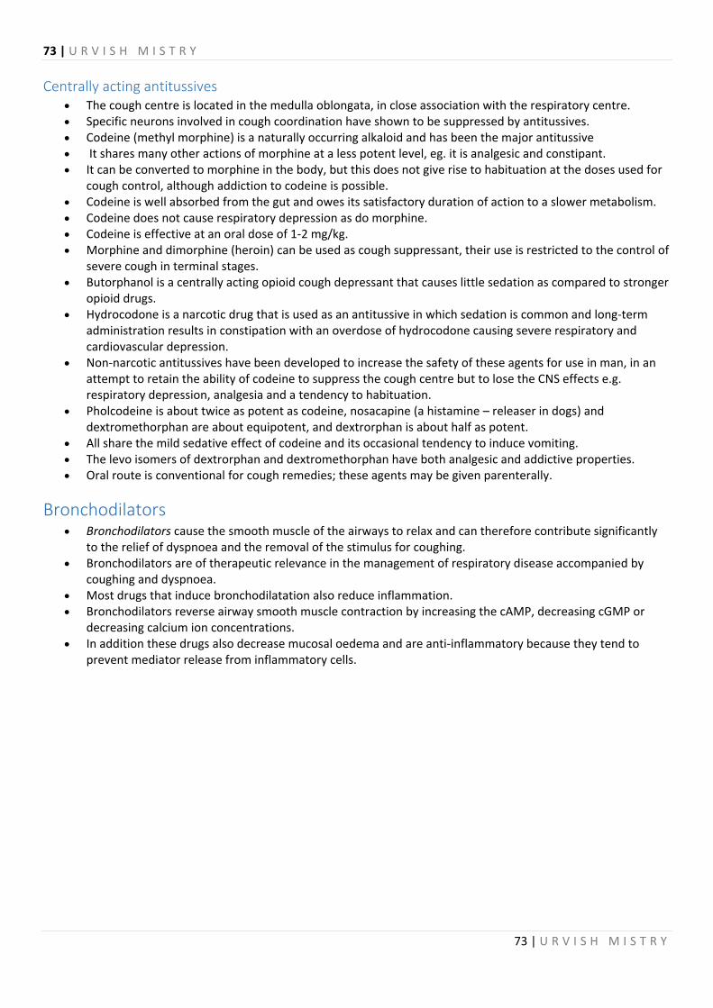
73 | U R V I S H M I S T R Y
73 | U R V I S H M I S T R Y
Centrally acting antitussives The cough centre is located in the medulla oblongata, in close association with the respiratory centre. Specific neurons involved in cough coordination have shown to be suppressed by antitussives. Codeine (methyl morphine) is a naturally occurring alkaloid and has been the major antitussive It shares many other actions of morphine at a less potent level, eg. it is analgesic and constipant. It can be converted to morphine in the body, but this does not give rise to habituation at the doses used for
cough control, although addiction to codeine is possible. Codeine is well absorbed from the gut and owes its satisfactory duration of action to a slower metabolism. Codeine does not cause respiratory depression as do morphine. Codeine is effective at an oral dose of 1‐2 mg/kg. Morphine and dimorphine (heroin) can be used as cough suppressant, their use is restricted to the control of
severe cough in terminal stages. Butorphanol is a centrally acting opioid cough depressant that causes little sedation as compared to stronger
opioid drugs. Hydrocodone is a narcotic drug that is used as an antitussive in which sedation is common and long‐term
administration results in constipation with an overdose of hydrocodone causing severe respiratory and cardiovascular depression.
Non‐narcotic antitussives have been developed to increase the safety of these agents for use in man, in an attempt to retain the ability of codeine to suppress the cough centre but to lose the CNS effects e.g. respiratory depression, analgesia and a tendency to habituation.
Pholcodeine is about twice as potent as codeine, nosacapine (a histamine – releaser in dogs) and dextromethorphan are about equipotent, and dextrorphan is about half as potent.
All share the mild sedative effect of codeine and its occasional tendency to induce vomiting. The levo isomers of dextrorphan and dextromethorphan have both analgesic and addictive properties. Oral route is conventional for cough remedies; these agents may be given parenterally.
Bronchodilators Bronchodilators cause the smooth muscle of the airways to relax and can therefore contribute significantly
to the relief of dyspnoea and the removal of the stimulus for coughing. Bronchodilators are of therapeutic relevance in the management of respiratory disease accompanied by
coughing and dyspnoea. Most drugs that induce bronchodilatation also reduce inflammation. Bronchodilators reverse airway smooth muscle contraction by increasing the cAMP, decreasing cGMP or
decreasing calcium ion concentrations. In addition these drugs also decrease mucosal oedema and are anti‐inflammatory because they tend to
prevent mediator release from inflammatory cells.
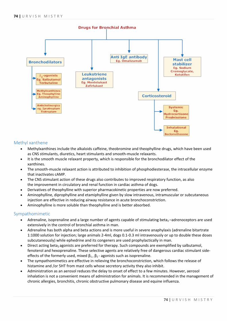
74 | U R V I S H M I S T R Y
74 | U R V I S H M I S T R Y
Methyl xanthene Methylxanthines include the alkaloids caffeine, theobromine and theophylline drugs, which have been used
as CNS stimulants, diuretics, heart stimulants and smooth‐muscle relaxants. It is the smooth muscle relaxant property, which is responsible for the bronchodilator effect of the
xanthines. The smooth‐muscle relaxant action is attributed to inhibition of phosphodiesterase, the intracellular enzyme
that inactivates cAMP. The CNS stimulant action of these drugs also contributes to improved respiratory function, as also
the improvement in circulatory and renal function in cardiac asthma of dogs. Derivatives of theophylline with superior pharmacokinetic properties are now preferred. Aminophylline, diprophylline and etamiphylline given by slow intravenous, intramuscular or subcutaneous
injection are effective in reducing airway resistance in acute bronchoconstriction. Aminophylline is more soluble than theophylline and is better absorbed.
Sympathomimetic Adrenaline, isoprenaline and a large number of agents capable of stimulating beta2 –adrenoceptors are used
extensively in the control of bronchial asthma in man. Adrenaline has both alpha and beta actions and is more useful in severe anaphylaxis (adrenaline bitartrate
1:1000 solution for injection; large animals 2‐4ml, dogs 0.1‐0.3 ml intravenously or up to double these doses subcutaneously) while ephedrine and its congeners are used prophylactically in man.
Direct acting beta2 agonists are preferred for therapy. Such compounds are exemplified by salbutamol, fenoterol and hexoprenaline. These selective agents are relatively free of dangerous cardiac stimulant side‐effects of the formerly used, mixed β1 , β2 ‐ agonists such as isoprenaline.
The sympathomimetics are effective in relieving the bronchoconstriction, which follows the release of histamine and /or 5HT from mast cells whose secretory activity they also inhibit.
Administration as an aerosol reduces the delay to onset of effect to a few minutes. However, aerosol inhalation is not a convenient means of administration for animals. It is recommended in the management of chronic allergies, bronchitis, chronic obstructive pulmonary disease and equine influenza.

75 | U R V I S H M I S T R Y
75 | U R V I S H M I S T R Y
Parasympatholytics – spasmolytics Parasympatholytics such as atropine have been used as a bronchodilator in chronic emphysema in horses. The rationale is that atropine overcomes the muscarinic cholinoceptor‐mediated bronchoconstriction. The
effect is dilation of the larger airways Atropine derivatives e.g. eucatropine, have also long been used in paediatric preparations for whooping
cough. As the sulphate, atropine is routinely injected preoperatively to reduce or eliminate salivary and bronchial
secretions during anesthesia. The dose by subcutaneous injection is 30‐100 m g / kg. Ipratropium a muscarinic – blocking bronchodilator, is used as an aerosol. The reduction in volume, higher viscosity of secretions and decreased mucociliary clearance, can be a
disadvantage.
Corticosteroids The reduction of mucosal edema in bronchi and bronchioles can be achieved by use of glucocorticoids to
suppress the inflammatory response. In man the corticosteroids are used for severe, disabling, bronchodilator‐resistant asthma. These may be administered orally, parenterally or as an aerosol. Their use carries the risks of a reduced ability to gight infections and also the risk of adrenal cortex
suppression if the administration is prolonged. Their effects on the inflammatory response include membrane stabilization, reduced antibody synthesis,
reduced mediator release following antigen‐antibody interactions and lessened fibrosis. These drugs also block uptake 2 and so extend the half‐life of endogenous sympathomimetics. Corticosteroids can be used with advantage to control chronic allergic‐type summer coughs in dogs either by
the oral administration of prednisolone or by the use of injectable depot preparations.
NSAIDs The introduction of NSAIDs with high antiinflammatory potency has made this group beneficial as adjuncts
along with antimicrobials They lack the suppression of the immune response seen with the corticosteroids. Their ability to suppress the production of the chemical mediators of the inflammatory response underlies
their ability to reduce structural damage and functional impairment in the pneumonic lung. Ventilation, gaseous exchange and pulmonary haemodynamics can all be improved by NSAID administration. Flunixin is especially recommended for this purpose.
Antihistaminics The introduction of NSAIDs with high antiinflammatory potency has made this group beneficial as adjuncts
along with antimicrobials They lack the suppression of the immune response seen with the corticosteroids. Their ability to suppress the production of the chemical mediators of the inflammatory response underlies
their ability to reduce structural damage and functional impairment in the pneumonic lung. Ventilation, gaseous exchange and pulmonary haemodynamics can all be improved by NSAID administration. Flunixin is especially recommended for this purpose.
Mast cell stabilizers The drug acts by stabilizing mast‐cell membranes preventing degranulation and the release of chemical
mediators (eg. Histamine and leukotrienes) in response to allergens. A depressant action on chemosensitive ending which trigger bronchoconstriction is also suggested. More recently it has been shown also to prevent the release of tissue‐damaging free radicals of IgE activated
platelets. Cromoglycate is not a bronchodilator and is not effective in the rapid relief of existing bronchoconstriction. It has given sufficient improvement in the respiratory function in horses with chronic obstructive pulmonary
disease.

76 | U R V I S H M I S T R Y
76 | U R V I S H M I S T R Y
Adminsitration is done via a special nebulizer and face – mask, 80 mg once daily for 1‐4 days. The 10‐15 min period of nebulization is generally well accepted by the horse.
It is available as drug for protecting horses with allergic respiratory disease against predictable antigen challenge.
Respiratory stimulants Respiratory stimulants are agents used to improve respiration.
Diuretics Diuretics are agents that increase the salt and water excretion by an action on the kidneys. Diuretics effective for the treatment of edema have been available since the 16th century and mercurous
chloride was known by Paracelsus to be diuretic. In 1930, Swartz discovered that the antimicrobial sulfanilamide could be used to treat edema in patients
with congestive heart failure due to an increase in renal excretion of Na+. Most modern diuretics were developed when side effects of antibacterial drugs were noted, which included
changes in urine composition and output. Except for spironolactone, diuretics were developed empirically, without knowledge of specific transport
pathways in the nephron. They are used mainly in cases of cardiac failure, oedema and hypertension.
Physiological diuresis It is occasionally desirable to provoke diuresis in order to increase urinary flow or volume as in the case of
treatment of cystitis or to prevent crystalluria with sulphonamide therapy, rather than with the intention of reducing body fluid volume.
In such cases increased water intake is sufficient.
Classification of diuretics Based on their action on the kidneys diuretics are classified as
Diuretics acting directly on the cells of the nephron o Agents acting on the ascending loop of Henle – Ethacrynic acid, frusemide o Agents acting on the early distal tubules – Thiazides like chlorthiazide o Agents acting on the Collecting tubules and ducts – Triamterene, amiloride
Diuretics modifying the contents of the filtrate o Osmotic diuretics – Mannitol o Carbonic anhydrase inhibitors – Acetazolamide
Based on their mode of action diuretics are classified as o Osmotic diuretics – Glycerine, mannitol, urea, isosorbide o Inhibitors of carbonic anhydrase – Acetazolamide, dichlorphenamidine o Inhibitors of Na+‐K+‐2Cl‐ symport (High ceiling loop diuretics) – Frusemide, Ethacrynic acid,
Bumetanide o Inhibitors of Na+‐Cl‐ symport – Thiazide and thiazide like drugs o Potassium sparing diuretics
Inhibitors of epithelial sodium channels – Triamterene, amiloride Antagonists of aldosterone – Spironolactone
o Xanthine diuretics – Theophylline It can also be classified as
o Cardiac diuretics ‐ Xanthines o Osmotic diuretics ‐ Mannitol, Isosorbide, Urea o Natriuretics ‐ Mercurials, Thiazides, Acetazolamide
Osmotic diuretics These are inert substances filtered into the glomerulus.

77 | U R V I S H M I S T R Y
77 | U R V I S H M I S T R Y
Their main effect is exerted in the proximal tubule, descending limb of Henle and the collecting duct which are freely permeable to water.
By remaining in the tubular lumen, these agents cause an increase in colloid osmotic pressure in the tubular lumen causing suction of water into the tubular lumen.
There is also reduced water reabsorption due to renal medullary hyperemia. There is impairment of Na+ reabsorption because the diluted solution in the lumen reduces the
Na+ concentration gradient, making it harder for Na+ to be reabsorbed. They are not useful in treating conditions with sodium retention. These drugs are useful in acutely raised intracranial pressure or intraocular pressure. They are also useful in prevention of acute renal failure. Unwanted effects include transient expansion of extracellular fluid volume and hyponatraemia, headache
and vomiting. Mannitol is commonly used as an osmotic diuretic and has to be administered intravenously. Isosorbide has the advantage of being absorbed after oral administration in monogastric species.
Carbonic anhydrase inhibitors Bicarbonates penetrate the luminal membrane only very slowly, and its absorption in the proximal tubule is
heavily dependent on the presence of an enzyme ‘carbonic anhydrase’. This is a zinc containing enzyme whose function is to speed up the attainment of equilibrium of the reaction:
CO2 + H2O –> H2CO3
Role of carbonic anhydrase in diuresis is that it is capable of accelerating this reaction in either direction. This enzyme is found within the proximal tubule cells and in association with their luminal brush border. The activating step in the process of bicarbonate absorption is the secretion of H+ into the tubule lumen,
which is largely responsible for the favourable electrical gradient down which Na+ diffuses into the cell. This H+ is derived from the dissociation of carbonic acid, whose supply is facilitated by the presence of
carbonic anhydrase. The carbonic anhydrase inhibitors interfere with the absorption of bicarbonate from the proximal tubule, by
reducing the supply of H+ to the membrane pump and by reducing the rate of absorption of carbonic acid from the lumen.
Filtered bicarbonate is thus trapped in the urine together with an electrically equal amount of Na+ and an osmotically equivalent amount of water.
These drugs cause an increased excretion of bicarbonate with accompanying sodium, potassium and water excretion resulting in an increased flow of alkaline urine and mild metabolic acidosis.
They are poor diuretics because there is compensatory reabsorption of Na+ is the segments further down the nephron.
These drugs are used in glaucoma and in some types of epilepsy. Their action results in a depletion of extracellular bicarbonate and their effect is self limiting as the blood
bicarbonate level falls. Side effects are drowsiness, numbness and tingling of the face and extremities (due to metabolic acidosis)
and disturbances of vision. This group of drug is rarely used as diuretics. Acetazolamide used as a carbonic anhydrase inhibitor is a derivative of a sulphonamide but does not possess
any antibacterial action. Acetazolamide is a non‐competitive, irreversible inhibitor of carbonic anhydrase.
Loop diuretics Loop diuretics ( High ceiling diuretics) are the most effective diuretics A fairly high proportion of Na+ is also reabsorbed in the loop of Henle (22%). They are inhibitors of Na+, K+ Cl‐ symport Main effect of this group of diuretics is inhibition of chloride pumping throughout the whole length of the
thick ascending limb of Henle, thus paralysing the kidney’s ability to concentrate and to dilute the urine.

78 | U R V I S H M I S T R Y
78 | U R V I S H M I S T R Y
Their effect is from the luminal surface of the loop cells. These drugs are extensively bound to plasma proteins and relatively little is filtered in the glomerulus. They are actively secreted by the organic anion transporters at the site of action. Substances, like probenecid
which block this mechanism , can reduce their efficacy. Examples of Loop diuretics: Frusemide, Bumetanide and Ethacrynic acid. Frusemide and bumetamide are sulphonamide derivatives while ethacrynic acid is not a sulphonamide
derivative. Ethacrynic acid may produce clinical dehydration and acute circulatory collapse as it produces severe
diuresis after administration. Frusemide produces rapid diuresis and hence is useful in emergency cases. It is easier to control the degree of diuresis with this drug by varying the dose rate. There is an increase in the excretion of calcium and magnesium and a decreased excretion of uric acid. The effect on calcium is made use of in the treatment of hypercalcaemia. The loop diuretics are readily absorbed from the gastrointestinal tract and can also be administered
parenterally. Administration
Orally or intravenously at 1‐5 mg/kg in dogs. This can be doubled with successive doses until an effect is obtained. Duration of action is 4‐6 hours. In horses the drug is given at the rate of 0.5 mg/kg to reduce incidence of epistaxis. In cattle given orally at 0.5‐1 mg/kg, 12‐24 hourly. Loop diuretics are useful in patients with salt and water
overload due to acute pulmonary oedema, chronic heart failure, hepatic cirrhosis complicated by ascites, nephrotic syndrome etc.
Hypertension especially if accompanied by renal impairment. Acute treatment of hypercalcaemia. Untoward effects include potassium loss, metabolic alkalosis, depletion of calcium and magnesium,
hypovolaemia and hypotension. Local irritation of the gastronintestinal tract and competition with other drugs like digitalis are noticed. As with other potent diuretics, hyponatraemia can occur, but deafness is an adverse effect peculiar to loop
diuretics. Hence concurrent dosing with aminoglycosides is contraindicated.
Thiazide derivatives Thiazide diuretics group includes chlorthiazide, hydrochlorthiazide, bendrofluazide, cyclopenthiazide. These drugs are more potent than carbonic anhydrase (CA) inhibitors. Most thiazides are weak inhibitors of
CA, but this is not the basis of their action. Site of action of thiazide diuretic is at the distal tubule (hence they are medium acting diuretics) where there
is only a small amount of reabsorption of sodium since most of the Na+ is already absorbed in the earlier parts of the nephron.
In the distal tubule, Na+ is reabsorbed via a Na+ Cl‐ co‐transport. Thiazides inhibit this co‐transport mechanism, thus preventing Na+ from being reabsorbed. Thiazides may also inhibit the Na+ K+ ATPase indirectly. Due to Na+ K+ counter exchange at the collecting tubule, potassium loss may be induced by these drugs
leading to serious hypokalemia. The thiazides are active by mouth as well as by parenteral administration. Their effects are fairly slow in onset. Thiazides are used in the treatment of oedema in cardiac failure. In hepatic cirrhosis, much K+ loss is not
appreciable. Hence thiazides are given in conjunction with K+ tablets or with a K+ sparing diuretic. Thiazides are secreted in the kidney via the organic acid secretary system.
Side effects Hypokalemia Increase in blood sugar (hyperglycemia), especially in diabetics,

79 | U R V I S H M I S T R Y
79 | U R V I S H M I S T R Y
Reduced insulin also causes increased glycogenolysis and reduced glycogenesis. Thiazides may cause a reduced excretion of uric acid, leading to gout. The thiazides are sulphonamides, and
so share cross reactivity with other drugs of this group. Therefore, if a patient is allergic to a sulphonamide, chances are that the patient will also be allergic to
thiazides. Photosensitivity and haemolytic anaemia may occur.
Potassium sparing diuretic There are 2 groups of drugs in this category. The only thing, they have in common is their site of action (late distal tubule and collecting duct). However their mechanism of action is quite different.
o Inhibition of aldosterone ‐ Spiranolactone o Inhibition of the Na+ K+ exchange in the collecting duct ‐ Triamterene, Amiloride
Aldosterone antagonists Aldosterone (a steroid hormone) enters tubular cells and binds to a mineralocorticoid receptor in the
cytoplasm. This hormone receptor complex binds to a response element on the DNA. Binding results in the expression of aldosterone induced proteins leading to activation of silent
Na+ channels/pumps, alteration of the cycling of the channels and pumps, increased expression of channels and pumps and altered permeability of the tight junction (zonula occludens).
All the above effects result in an increase in Na+ conductance (reabsorption) and secretion of K+ (One of the complications of excess aldosterone secretion is hypokalemia).
Spiranolactone binds to the mineralocorticoid receptor and prevents aldosterone from binding. Spiranolactone a true competitive antagonist of aldosterone enhances direct reabsorption of sodium in
more proximal areas of the distal tubules. This drug has a very slow onset of action, which is not dose dependent. It may exert oestrogen like activity It is used in combination with thiazide diuretics. Spiranolactone is not effective in the presence of low levels of aldosterone. It is available only for oral administration. If hyperkalemia occurs as a result of spiranolactone toxicity, thiazide diuretics can be administered.
Triamterene and amiloride These drugs have a limited diuretic efficacy. They act on the collecting tubules and collecting ducts, inhibiting sodium reabsorption and decreasing
potassium excretion. Triamterene inhibits aldosterone specific ATPase. But, is not dependent on aldosterone levels. Amiloride blocks Na+ channels in the luminal membrane affecting Na+ permeability. Both the drugs promote excretion of uric acid. The main importance of these drugs lies in their potassium‐sparing ability. They can be given with potassium‐losing diuretics like thiazides to maintain potassium balance. They are useful in conditions where K+ loss cannot be tolerated (often used in conjunction with thiazides) In hypertension and congestive heart failure ‐ If a person is being treated with digoxin for their heart failure,
loss of K+ can enhance the toxicity of digoxin (cardiac glycoside). If K+ is reduced, the actions of digoxin are enhanced because normally, digoxin must compete with K+ for the
Na+ K+ ATPase. By using the drugs in combination, there is an enhanced diuretic effect and the added benefit of not losing
K+. Spironolactone is used in primary hyperaldosteronism (tumor of the adrenals) or secondary
hyperaldosteronism (cardiac failure, hepatic cirrhosis). Side effects
Hyperkalemia, Metabolic acidosis,

80 | U R V I S H M I S T R Y
80 | U R V I S H M I S T R Y
Gynaecomastia, Impotence, Decreased libido, Hirsuitism and Gastric upsets.
Xanthines and mercurial Xanthines
These drugs stimulate cardiac functions. Their diuretic activity may stem from the resultant increase in renal blood flow and thereby an increase in glomerular filtration rate.
They also exert a direct action on the renal tubule. Renal response is manifested by an increase in levels of sodium and chloride in urine.
There is no significant change in the urine pH and potassium excretion appears to be relatively unaffected. Examples are: Caffeine, Theophylline
Mercurials Diuretic action of mercurials is associated with sulphydryl groups and so they inactivate the sodium
transport mechanism of the nephron. For clinical use, mercurials are obsolete
Drugs acting on urogenital systems
Alkalization of urine Alteration of urine pH Alteration is sometimes desirable as in the case of poisoning and in the excretion of some drugs to prevent side effects. Alkalinisation of urine
Alkalinising agents reduce the irritation of an inflamed urinary tract and discourage the growth of certain organisms like E.coli.
Urine can be made alkaline by sodium bicarbonate intravenous administration or by potassium citrate oral administration.
Sodium or potassium citrate is metabolised to form a stable cation (Na+ or K+) and a labile anion (citrate). Citrate is taken up for use by the TCA. The Na+ or K+ is excreted with the most common anion (HCO3‐. Hence, there will be alkaline urine.
Administration and use To decrease the adverse effects of sulphonamides alkalinisation is useful. Sulphonamides are generally
insoluble in acid urine. They may thus precipitate out and crystallise. Therefore, to increase solubility, the urine is made more alkaline.
To increase the excretion of acidic drugs like Aspirin, salicylates and barbiturates. Alkalinisation of urine increases the antibacterial activity of aminoglycosides in urinary tract infection. Alkalinisation of the urine in usually recommended to prevent the recurrence of urate stones in dogs after
cystotomy. Sodium bicarbonate at 0.5 to 1.0 g is given orally thrice daily.
Acidification of urine Acidification of urine is used as a test for renal tubular acidosis. Acidification helps in the excretion of certain
drugs like amphetamine, phencyclidine, quinine etc. Ammonium chloride, ascorbic acid or calcium chloride can be used to acidify urine. Ammonium chloride (NH4Cl) is metabolised in the liver to NH4
+ and Cl‐. The Cl‐ is excreted along with Na+. The base conserving mechanisms secrete H+ and NH4
+ into the urine. Administration and uses
Acidification increases action of hexamine, penicillins and tetracyclines in the treatment of urinary tract infections.
Acidification increases excretion of basic drugs like Pethidine and amphetamine

81 | U R V I S H M I S T R Y
81 | U R V I S H M I S T R Y
Ecbolics These are agents that bring about increase in uterine contractions. Oxytocin: It is hormone of the posterior pituitary received from the hypothalamus. It has amajor
physiological role in milk let down and labor initiation. o Clinical uses of oxytocin:
For labor induction. Given by slow I/v in dextrose solution. For mIlk letdown . Only in agalactic animals For treatment of uterine inertia and for rapd involution of uterus. To arrest postparturient haemorrhage. For expulsion of retained placenta.
o The dose of oxytocin is 5 ‐ 25 units in bitch, 75 ‐ 100 units in cow / mare and 30 ‐ 50 units in sow / ewe.
Oxytocics: Other agents which perform similar functions are also called oxytocics. Ergot alkaloids: Alkaloids from the fungus Claviveps purpurea viz. ergometrine, ergonovine have prominent
effect on the uterus. Not preferred for induction of labour. Generally indicated for postparturient haemorrhage, rapid involution of uterus, retained placenta.
PGF 2 alpha: Exerts potent stimulant action on myometrium. It is also popularly used for its luteolytic effect.
Tocolytics These are agents that inhibit uterine contractions. They relax uterine smmoth muscle and may be used to
delay labor and to stop threatened abortion. Beta2 agonists: Salbutamol, terbutaline, isoxsuprine Calcium channel blockers: Nifedipine, diltiazem Oxytocin antagonist: Atosiban
Fluid therapy Fluid therapy refers to administration of fluids to compensate the loss of fluids from the body due to a
variety of conditions leading to dehydration. Fluid loss in the body can lead to shock and can become fatal if unattended to.
The purpose of fluid therapy is to correct dehydration or overhydration, elctrolyte imbalance and /or acid ‐base imbalance.
It is also indicated to correct acidosis/alkalosis, treat shock, give parenteral nourishment or even stimulate organ function such as kidney.
Fluid volume and type The type of solution is to be dictated by history, clinical signs and laboratory examination. The volume of fluid is determined the need for maintenance as well as the need for replacement of lost
fluid. For normal maintenance, a thumb rule of 65 ml / kg /24 hr for adults and 130 ml / kg /24 hr for young ones is
calculated. Based on this, a normal mature dog of 20 kg would require 1300 ml for 24hr period. Replacement of fluid loss must be in addition to the maintenance requirement. To calculate fluid loss one must estimate the degree of dehydration. To calculate the fluid for replacement one must calculate the degree of dehydration.
Degree of dehydration
Clinical signs
4% history of fluid loss, leathery skin, mucous membrane still moist and evidence of thirst
6% skin still leathery, when lifted the skin will peak and return to normal slowly, hair coat dull,mucous membrane dry but the tongue still moist

82 | U R V I S H M I S T R Y
82 | U R V I S H M I S T R Y
8% Skin will show lack of pliability and elasticity, the skin loses turgor (when lifted will peak and stay, mucous membrane and tongue dry, eyeballs will be soft and sunken.
12% Signs of circulatory collapse
15% Symptoms of Acute shock
Fluid volume to replace
The volume of fluid to replace loss of fluid is calculate the body weight multiplied by degree of dehydration. For eg., a dog weighing 20 kg with 6% dehydration is given 20 kg x 0.06 ‐ 1200 ml or 1.2 liters. This is apart from the maintenance requirement of 20 x 65 = 1300 ml.
Rates and administration The rate of replacement of fluids should parallel the severity of dehydration. Fluids should be administered
rapidly at first followed by decreasing rates. Most commonly it is agreed that 15 ml / kg / hr is a reasonable rate. In severely dehydrated cases this can go
up to 50 ml / kg /hr. Higher rates can overload the system and may even cause overhydration. Route of administration
The route depends on the type of illness, severity, degree of dehydration, patient condition, time and equipment available.
The most common and overlooked one is the oral or nasogastric. It is the least dangerous, without strict asepsis.
Per rectal administration can also be considered. It may be difficult to retain the fluid especially in the presence of gastrointestinal disease.
The IV route is the most versatile. It is indicated for emergencies, severe cases. It has the advantage of large volume of administration. But maintenance of indwelling catheter, clotting and hematoma and location of vein are some of the drawbacks.
The SC route is handy in cases of small animals where larger volumes need to be give but it is difficult to locate the vein. It is irritating and can be painful.Fluid absorption is more slow.
IP route also suffers form the drawback of easy predisposition to peritonitis.
Choice of fluid Proper selection of fluid begins by determining the type of deficit and choice of fluids‐Colloid, crystalloid or combination of both
Fluid Indications
Isotonic saline (Normal saline, 0.9%)
To correct metabolic acidosis, especially after vomiting
Isotonic dextrose saline (0.18% NaCl + 4.3% Dextrose)
Maintenance therapy, hypertonic dehydration
Darrow’s solution (0.4% NaCl + 0.27% KCl + 0.58% Sodium lactate)
Metabolic acidosis associated with diarrheal dehydration and potassium loss
5% Dextrose Hypertonic dehydration resulting from water deprivation or heat stroke; used in emergency to provide transient alleviation of hypoglycaemia
Compound Sodium Lactate (Hartmann’s solution or Ringer’s lactate)
Metabolic acidosis, Replacement of losses owing to gastrointestinal dysfunction (vomiting, diarrhea), haemorrhage, burns

83 | U R V I S H M I S T R Y
83 | U R V I S H M I S T R Y
Dextrose Saline (0.9% NaCl + 5% Dextrose)
For emergency treatment of dehydration and hypoglycaemia
Sodium bicarbonate (1.3%) Severe Acidosis
Whole blood Replacement for blood loss in treating anaemia
Plasma expanders
Dextran Effective in restoration of blood volume in cases of hemorrhage or shock.
Gelatin colloidal solution Hypovolemic shock, burns, blood loss and diarrhea
Pharmacotheraputics of Hormones
Administration of hormones Administration of hormones falls into two categories:
o Replacement therapy and o Additive therapy
Replacement therapy is the administration of the hormones at physiological levels. For example insulin, thyroxine, vasopressin, gonadotrophins and ACTH are administered at physiological levels.
Additive therapy is the administration of the hormones at pharmacological levels (not usually related to natural metabolic functions). Administration at pharmacological levels may produce side effects or untoward effects. For example androgens and glucocorticoids are administered at pharmacological levels.
Augmentation is the process of increasing duration of action of a hormone by combining the protein hormones with copper, zinc or iron of heme.
Corticotrophins and related peptides Corticotrophin releasing hormone
This hormone stimulates synthesis and secretion of ACTH by pituitary corticotrophs. This hormone appears to exert its effect on ACTH release through both adenylate cyclase and calcium‐
calmodulin signal transduction systems.
Clinical Condition Choice of fluid
Vomiting Normal Saline Dextrose Saline Ringer’s solution
Diarrhea Lactated Ringer’s Darrow’s fluid (In chronic cases with K+ loss)
Bowel obstruction Lactated Ringer’s (+ colloid for shock)
Hemorrhage Whole blood Lactated Ringer’s Colloid
Peritonitis Colloid Lactated Ringer’s
Shock Dextran + Hartmann’s Ringer’s Lactate Gelatin Colloid

84 | U R V I S H M I S T R Y
84 | U R V I S H M I S T R Y
Adrenocorticotrophin ACTH stimulates the secretion of glucocorticoids, mineralocorticoids and adrenal androgens It acts by increasing the activity of the rate limiting agent in steroid synthesis and converts cholesterol to
pregnonolone. ACTH also stimulates adrenal hypertrophy and hyperplasia. Corticotrpin and ACTH are used as diagnostic tools to test adrenocorticotroph and adrenal function
reserve. ACTH can be used for all the purposes for which glucocorticoids are recommended. The advantage of ACTH is that it leads to suppression of pituitary over time while glucocorticoids suppress
the pituitary and adrenal cortex quickly. ACTH gel preparations are derived from pig pituitaries. Both porcine and synthetic ACTH are well absorbed
after intramuscular administration. However, its disadvantage is that it cannot be administered by oral route unlike glucocorticoids.
Insulin Endocrine pancreas
The Beta‐cells of the islets of langerhans secrete the peptide hormone insulin. Insulin is secreted in response to gastrointestinal peptide hormones, plasma concentrations of glucose,
aminoacids or fatty acids and other local (glucagons) or systemic (acetylcholine and beta agonists) chemical messengers or drugs (sulphonylureas).
Diabetes mellitus is a condition due to lack of insulin and is characterized by sweet urine, polydipisia, wasting of tissues, development of ketocidosis, hypersomolar coma and death.
In 1921 Banting and Best extracted the active compound from pancreas that controlled hyperglycemia in diabetic dogs and human.
There are two types of diabetes in human and they are IDDM or TYPE I (Insulin dependent diabetes mellitus) and NIDDM or Type II (Non‐insulin dependent Diabetes mellitus).
In IDDM there is lack of insulin and in NIDDM there is resistance to insulin.
Pharmacological actions of insulin Pharmacological actions
Insulin binds to specific cell membrane located receptors. This facilitates the entry of glucose, aminoacids and potassium ions into most cells. This reverses the hyperglycemia of diabetes mellitus by allowing excess glucose to become intracellular. Inside the cell, protein synthesis is increased and glucose is phosphorylated by ATP and hexokinase and is
used to derive energy or for incorporation into glycogen. The storage of carbohydrate is further aided by the inhibition of the action of the enzymes of glycogenolysis. Protein synthesis is aided by the impairment of gluconeogenesis. Insulin also facilitates fat deposition and strongly inhibits lipolysis. Actions of insulin include
o Increased glucose uptake into muscle and fat o Increased glycogenesis o Decreased glycogenolysis o Decreased gluconeogenesis o Stimulation of growth (anabolic) o Decreased lipolysis o Decreased protein breakdown
Preparations of insulin Insulin was originally derived from the pancreas of either pig of the ox. The standard preparation contains 20, 40 and 80 units of insulin in one milliliter of the injection. The rate of absorption of insulin and its duration of action depends on the physical state (crystalline or
amorphous, proportion of zinc, solution or suspension) and the route of injection of the chosen formulation.

85 | U R V I S H M I S T R Y
85 | U R V I S H M I S T R Y
The complexed solution ie. Protamine zinc insulin and globin of zinc insulin are effective for about 48 hours and 24 hours. But, the plain insulin is effective only for 8 hours and lente insulin is effective for 18‐20 hours duration.
Human insulin (Humulin) is a DNA recombinant derived preparation of insulin. The specialized delivery devices for insulin include insulin syringes, pen devices, inhalation devices, pumps
and implantable preparations. Uses
o Diabetes mellitus in dogs o Acetonemia in cattle (Insulin is a powerful antiketotic agent. It is particularly useful in ketosis that
occurs within the first week of lactation and is non responsive to glucose or glucocorticoids therapy alone)
o Pregnancy toxemia in sheep.
Oral hypoglycemic drugs Sulphonyl ureas
These are agents that were first introduced as oral hypoglycemic agents. There are two groups of sulphonamides namely first and second generation sulphonyl ureas. First generation includes tolbutamide, chlorpropamide, acetohexamide and tolazanide. Second generation includes glibenclamide, glipizide and gliclazide. In the pancreatic beta cells, sulphonyl ureas decrease ATP dependent potassium channels in the plasma
membrane. This results in depolarization and release of insulin. Presence of atleast 30% functional beta cells is essential for the action of sulphonylureas. This group of drugs is often used in combination with metformin. They are bound to plasma proteins, metabolised by the liver and excreted by the kidney. They can cross the placenta (hence not used in pregnancy) and are to be taken before a meal. Side effects include hypoglycemia, GIT effects, rash and increased weight. There is interaction with NSAIDs and sulphonamides (making hypoglycemia more likely). If used with alcohol they will cause severe hypoglycemia.
Biguanides This group includes phenformin and metformin. They cause little or no hypoglycemia in non‐diabetic subjects and do not stimulate pancreatic beta cells. Presence of insulin is essential for their action. They suppress hepatic gluconeogenesis and glucose output from the liver, enhance binding of insulin to its
receptors and stimulate insulin mediated glucose disposal. They increase glucose uptake into skeletal muscle and may decrease the absorption of glucose. This group of drugs have a short half life and are excreted by the kidney. Side effects include hypoglycemia, nausea, vomiting and diarrhea.
Meglitidine analogs Repaglinide lowers blood glucose by stimulating the release of insulin from the pancreas. It achieves this by
closing ATP‐dependent potassium channels in the membrane of the beta cells. This depolarizes the beta cells, opening the cells' calcium channels, and the resulting calcium influx induces insulin secretion. Eg: repaglinide
Thiazolidinediones It works as an insulin sensitizer, by binding to the proxisome proliferator‐activated receptors (PPARs) in fat
cells and making the cells more responsive to insulin. Eg: rosglitazone α glucosidase inhibitor
It is a starch blocker, and inhibits alpha glucosidase, an intestinal enzyme that releases glucose from larger carbohydrates. Eg: acarbose
Thyroid hormone The thyroid gland secretes three main hormones: thyroxine, triiodothyronine and calcitonin.

86 | U R V I S H M I S T R Y
86 | U R V I S H M I S T R Y
Thyroxine and triiodothyronine produced in the thyroid follicles are critically important for the normal growth and development and for the energy metabolism.
Calcitonin produced in the interfollicular cells is involved in the control of plasma calcium. The steps in thyroid hormone synthesis, storage and secretion include:
o uptake of iodine by the follicle cells o oxidation of iodine and iodination of tyrosine residues of thyroglobulin o secretion of thyroid hormone
Thyroid hormone is essential for the normal growth and development. Deficiency results in cretinism. They have a marked effect on the lipid, carbohydrate and protein metabolism. Both T3 and T4 are used to treat thyroid hormone deficiency L thyroxine sodium is used for oral administration. Its absorption is influenced by the presence of food in the stomach, suralfate, iron and calcium. Enzyme inducers like phenytoin, rifampin, carbamazepine increase the metabolism
Thyroid inhibitors These are useful in the treatment of thyrotoxicosis, hyperthyroidism, Grave's disease Inhibit hormone synthesis (Anti thyroid) ‐ Propylthiouracil, methimazole, carbimazole Inhibit iodine trapping – Thiocyanates, perchlorates, nitrates Inhibit hormone release – iodine, iodides of sodium and potassium, organic, iodine Destroy thyroid tissue – Radio active iodine
General principles in corticosteroids therapy 1. Glucocorticoids are only palliative and do not provide complete cure 2. Individual variations occur between patients in their response to glucocorticoids therapy and hence dose
needs to be individualized. 3. Initial dose depends on severity of the disease. Start with a high dose in severe illness and gradually reduce
as clinical signs subside while in mild cases start with the lowest dose and enhance the dose as per need. 4. Prolonged therapy at high doses for several weeks may cause adrenal atrophy. 5. Glucocorticoids even at physiological dosage may result in suppression of Hypothalamo‐pituitary‐adrenal
axis 6. It should preferably be administered locally rather than systemically since low systemic level minimally
interferes with cortical ativity.

87 | U R V I S H M I S T R Y
87 | U R V I S H M I S T R Y
7. Glucocorticoid therapy should not be terminated abruptly since it may induce hypoadrenocorticism. Dosage should be terminated gradually over a period of 2 weeks.
8. Anti‐inflammatory and anti‐allergy therapy o Large proportion of drugs are used in veterinary practice to combat inflammation or allergy o Valuable in the treatment of chronic disease occurring periodically and in the absence of known
causes o Used in the treatment of chronic arthritis, tendonitis, bursitis, conjunctivitis, dermatitis, pruritic
dermatoses, allergic pulmonary disease and allergic gastroenteritis. o May be used for short periods for short periods in anaphylaxis, angioneurotic edema, urticaria and
serum sickness. 9. It should be used with caution in case of infectious disease, since glucocorticoids will promote dissemination
of infectious organism throughout the body resulting in fulminating infectious condition. It is indicated along with antibiotics that are effective in the treatment of underlying infection.
10. Glucocorticoid reduces collagen synthesis and thereby reduces the rate of wound healing. Glucocorticoids are indicated in the treatment of traumatic wound of prepuce of bulls so as to prevent rapid healing that would lead to stricture formation.
11. As a physiological replacement it is used for treatment of Addison’s disease (Chronic adrenal insufficiency) and acute adrenal insufficiency in human beings.
12. Glucocorticoids at high dose induces parturition and can be used to induce parturition in cows, ewes etc. However, since they have teratogenic effects during early pregnancy, it should be used with caution in pregnant animals.
13. Therapy of shock: Early treatment of glucocorticoid improves hemodynamics and enhances survival in hemorrhagic and septic shock but contraindicated in chronic sepsis because of its immunosuppressive effects
14. It can be used to treat depressive states of pet animals and even temperamental dairy cows since it induces euphoria.
15. It depresses immune system. It suppresses B lymphocytes proliferation and T lymphocytes activation. Immune‐mediated thrombocytopenia, autoimmune haemolytic anaemia, collagen disease like systemic lupus erythematosus, polyarteritis nodosa respond to glucocorticoid therapy. For the same reason, it is contraindicated during vaccination
o Corticoids are used in large number of inflammatory ocular disease such as allergic conjunctivitis, iritis, iridocyclitis, keratitis as topical instillation.
o Systemic therapy is useful in the treatment of retinitis, optic neuritis, uveitis. o Contraindicated in corneal ulcers since they slow the process of reepithelialisation of cornea.
16. Corticosteroids can be used along with other immunosuppressants to prevent rejection reaction during organ transplantation and skin allograft.
Anti-inflammatory and sodium retaining potencies of Glucocorticoids Corticosteroid Anti‐inflammatory potency Sodium‐retaining potency
Short –acting (half life ≤ 12 h)
Hydrocortisone 1 1
Cortisone 0.8 0.8
Fludrocortisone 10 125
Intermediate‐acting (half life 12‐36 h)
Prednisone 4 0.8
Prednisolone 5 0.8
Methylprednisolone 5 0.5

88 | U R V I S H M I S T R Y
88 | U R V I S H M I S T R Y
Triamcinolone 5 0
Long‐acting (Half life 36‐72 h)
Paramethasone 10 0
Betamethasone 10 0
Dexamethasone 10 0
Flumethasone 10 0
Reproductive hormones and Vitamins
Hormones influencing reproductive functions Gonadotrophin releasing hormone (GnRH) Effects
Administration of GnRH releases LH and FSH from the anterior pituitary gland. The effects of GnRH on LH/FSH release are dependent on
o the dose and route of administration o the endocrine status of the animal at the time of treatment and o the frequency of injection
Generally the release of LH and FSH are higher following GnRH injection when the animal is under the influence of estrogen and lower in progesterone dominated animals.
Uses Injection of a single large quantity of GnRH during the follicular phase of the estrous cycle stimulates
ovulation 24‐48 hours later. o Injection at the end of a synchronization schedule in order to control the time of ovulation for
scheduling times of insemination o To overcome delayed ovulation in dairy cows, particularly in high yielding post partum cows bred
within 60 days of parturition o Injection of mares during the estrous to time the ovulation and covering in order to maximize the
chances of conception o Injection of cows at the time of insemination to increase conception rate
GnRH stimulates LH and FSH in the male and may have a use in animals with poor semen producing ability. Synthetic GnRH like gonadorelin, buserelin and fertirelin have been produced which stimulate release of
both FSH and LH. Buserelin was developed by substituting two aminoacids in natural GnRH. This modification helped in increasing resistance to enzymatic degradation and an increased plasma
concentration of FSH and LH which persisted for several hours was noticed in mares and cows at therapeutic concentrations.
Anterior pituitary gonadotropins Both LH and FSH are glycoprotein hormones. They are secreted from the gonadotroph cells o f the anterior pituitary gland. They consist of two polypeptide subunits.
Effects The gonadotrophic hormones control the estrous cycle of the female and modulate testicular functions in
the male. In the female, FSH is responsible mainly for growth of the follicle by binding to granulosa cells and
stimulating estradiol production.

89 | U R V I S H M I S T R Y
89 | U R V I S H M I S T R Y
LH is responsible for final maturation and ovulation by binding to the thecal cells of the developing follicle and stimulating the synthesis of androgens.
In the male, LH binds to leydig cells or interstitial cells located in the connective tissue stroma of the testis and stimulate androgen production.
The gonadotrophins are available as a powder which can be easily reconstituted by the addition of sterile water, saline or appropriate buffer.
Once in the liquid form they must be stored at 4 degree centrigrade and not subjected to excessive agitation to prevent denaturation of protein.
Uses The uses of LH are
o to induce ovulation of a mature follicle o to leutinize follicular cysts o to induce follicular growth and ovulation.
The uses of FSH are : o to increase the number of ova shed in the cattle for superovulatory purpose and o to increase the litter size by increasing the ovulation.
Placental gonadotropins The maintenance of the corpus leuteum of pregnancy in some species is favoured by the production of
chorionic gonadotropin (CG) eg. human (hCG) from day 8 in the mare the endomaterial cups produce a hormone (eCG) from day 40 to about day 150 (previously
called pregnant mare serum gonadotropin (PMSG)). hCG contains more of LH than FSH, while eCG contains more of FSH than LH. hCG is mainly leutienising. In female animals this induces ovulation and reinforces the secretion of
progesterone by the ovary. Effects
hCG will bind to LH receptors on the granulos, theca and luteal cells with high affinity and specificity. eCG has LH like activity only in the mare, but in the other species it has mainly FSH like activity. In the mare it only leutinize follicles already present in the ovary but, in the other species it stimulates
follicular growth and ovulation. Serum gonadotrophin has been used to increase the number of developing follicle and ovulation and oestrus
and thus increase the incidence of twins in cattle. Uses
It is used to control the time of ovulation in mares because in these animals ovulation normally occurs towards the end of a long estral period.
It is widely used in the treatment of cystic ovarian disease especially where nymphomania is present and sometimes in the treatment of cyrptorchidism.
In male animals chorionic gonadotropin is used to stimulate the secretion of testosterone by the testes.
Estrogens There are a number of sources of estrogens produced in the body They are produced from the granulose cells of the follicle, the corpus luteum of primates, the placenta the
adrenals and the testes. Oestrogens influence the female sex characteristics such as an increase in the growth, protein synthesis and
fluid secretions of oviduct, uterus, cervix and vagina. They also affect mammary development. They induce the expression of behavioural estrus in the female and libido in the male. Oestrogens also have many effects on general body metabolism including fat distribution and content within
the body, mineral metabolism, bone growth and protein synthesis in may tissues including the muscle. They influence gonadotrophin secretion and at low concentrations have a negative feed back effect on the
hypothalamus, while at high doses, in the absence of progesterone they have a positive feed back effect in females resulting in the preovulatory surge in gonadotrophin output.

90 | U R V I S H M I S T R Y
90 | U R V I S H M I S T R Y
The testes of both the boar and the stallion produce oestrogens in high quantities and pregnant mares produce a wide range of estrogens like equilin, hippulin and equilenin.
Equine male urine and testes contain enormous quantities of estrogens. The natural estrogens include 17 beta ‐ estradiol, estrone and estriol. They have low oral activity. Synthetic steroidal estrogens include simple esters such as estradiol benzoate or valerate. The most common non‐steroidal estrogens, which have a high affinity for the estradiol receptor, are the
stilbenes. They are orally active, highly potent and are not easily metabolized. Some are genotoxic carcinogens and hence their use is prohibited.
Clinical uses Misalliance in the bitch Induction of oestrus and ovulation in anoestrus animals Control of the time of ovulation Control of life‐span of the corpus luteum Treatment of pyometra and mummified foetus Obesity due to hypogonadism in bitches Urinary incontinence in old and spayed bitches Anal oedema in dogs Regression of hypertrophied prostrate glands Caponization of young and old cockerels
Side effects This includes gastrointestinal upsets, feminization in male, sodium retention and oedema. In chronic overdosage genital erythema, irritation, polydipsia and polyuria are noticed.
Progesterone The main function of progesterone is to establish and maintain preganacy, by preparing the uterine
endometrium for implantation and by preventing myometrial uterine contractions during pregnancy. It also acts as a negative feed back hormone during the ovarian cycle suppressing LH and FSH secretion,
oestrus and ovulation. In some species, it primes behavioural centres in the brain and so allows estradiol to induce behavioural
estrus. It is produced mainly from the corpus luetum and the placenta and to a lesser extent from the adrenal gland. In the pregnant animal, the corpus leuteum is maintained for the duration of pregnancy, except in the mare. In some species such as the sow and the goat, the luteal progesterone is required throughout pregnancy: in
other species such as the mare and the ewe, the placental progesterone is the primary source after day 100 in the mare and day 55 in the ewe.
Uses Control of ovulation Induction of ovulation Reduce embryonic loss.
Synthetic progesterones Medroxy progesterone (MAP) – used to control estrus and ovulation in the cyclic ewes and to induce estrus
in anoestrous ewes. It can be used in bitches to suppress the onset of next estrus. Flurogesterone acetate (FGA) – used to control or induce estrous in ewes. Melengesterol acetate (MGA) – used to suppress estrus in heifers Norgestomet – used to control or induce ovulation Proligestone – used to control estrous in bitches.
Androgens Androgens are substances which stimulate the male accessory organs and produce the secondary sex
characteristics. The major androgens of importance are testosterone, dihydrotestosterone and androstenedione.

91 | U R V I S H M I S T R Y
91 | U R V I S H M I S T R Y
Testosterone is converted to: o dihydrotestosterone which has biological activity within testes and o to estradiol within the brain to induce male libido.
Androgens are produced in the leydig cells of the testes and testosterone is also produced in the epididymis of the stallion.
In the male androgens stimulate spermatogenesis, growth, and development and fluid secretions of the testes, secondary male characteristics and muscle development.
In the female, they are the main precursors of estrogen synthesis within the follicle. They also act as part of the negative feed back system to control LH and FSH secretion. In animals, there are only limited uses of androgens to control reproduction. Testosterone has anabolic effects leading to increased protein synthesis, retention of potassium and
phosphorus, increased bone growth, cartilage and other tissues and increased erythropoiesis by secreting erythropoietin.
They are used to treat infertility and impotency with varying success. Androgens are also administered to cows and heifers to use them as detectors of oestrus.
Anabolic steroids Compounds structurally related to testosterone but possessing protein anabolic activity with minimal
androgenic activity are called anabolic steroids. These drugs are used mainly in therapeutically debilitated animals. The metabolic activities include
o Protein anabolism with positive nitrogen balance o Increased muscle mass due to increased protein and glycogen content o High retention of calcium and phosphorus.
Drugs used are nandrolone and stanozalol. Anti‐androgens like delmadinone acetate are used to control hypersexuality in dogs and cats, relieve
prostate hypertrophy, tumours and behavioral problems in dogs and cats.
Posterior – pituitary hormones There are two known posterior pituitary hormones:
o Vasopressin (ADH) and oxytocin. o Posterior pituitary hormones are synthesized in the hypothalamus and transported to the posterior
pituitary, where they are released into circulation. Anti‐diuretic hormone (Vasopresssin)
This hormone regulates water permeability in the distal tubules and collecting duct of the nephron. It is also a vasoconstrictor and influences cardiovascular functions. It is used in the treatment of diabetes insipidus and prophylactically against bleeding in haemophilia.
Oxytocin Oxytocin is produced in neuronal bodies in the supra optic and paraventricular nuclei of the hypothalamus. Oytocin stimulates smooth muscle contractions and also includes PGF2 alpha release which causes luteolysis.
Uses Speeding the expulsion of foetus when uterine inertia is diagnosed Aids in the expulsion of placental debris and promotes involution of the uterus In uterine prolapse injection of oxytocin at several places in the organ reduces the engorgement and
contracts the everted uterus. Thus helps in the replacement more easily Functional agalactia in cows In mastitis in cattle to expel the infected milk before using any antibacterial agents.
Pharmacotherapeutics of fat soluble vitamins Vitamins are essential nutrients present in minute quantities in foods. If there is a deficiency in supply of
vitamins, deficiency diseases occur. In such a scenario, supplementation of vitamins is indicated. Fat soluble vitamins

92 | U R V I S H M I S T R Y
92 | U R V I S H M I S T R Y
Vitamin Deficiency signs Treatment
A Keratinization of epithelium, night blindness, fetal resorption, low egg prduction, poor hatchability
Farm animals: 100 ‐ 200 units /kg / day Poultry: 0.07 ‐ 0.22 gm / kg feed
D Rickets in young and osteomalacia in adults Farm animals: 50 ‐ 100 IU / kg / day Dog: 200 ‐ 400IU / kg / day Chicks ‐ 150 ‐ 300 IU / kg / day
E Muscular dystrophy in young and white muscle disease or stiff lamb disease in cattle
All young stock 40 mg / kg / day PO; 25 mg / kg / day Sc or IM Oral Swine 500 mg; Dog 30 mg /kg and poultry 350 mg / bird/ day
K Delayed clotting, spontanous haemorrhage Menadione / Menaphthone For warfarin pisoning 5 mg / kg IM For Sweet clover poisoning1.2 mg /kg IM For deficiency: Small animals 2 ‐ 10 mg / day PO Large Animals : 100‐400 mg / day PO Poultry 1 ‐2 gm / ton of feed
Pharmacotherapeutics of water soluble vitamins Vitamin Deficiency sign Treatment
B1 Thiamine Bracken fern poisoning Nervous signs
Calf ‐ 5 ‐ 50 mg Sheep 20‐ 200 mg Cat 1‐ 20 mg Dog 5‐50 mg Horse / Ox 100 ‐ 1000 mg PO or IM or SC
B2 Riboflavin Curled toe paralysis in chicken, anaemia, dermatitis, csours in calves, low fertility, eye discharge, photophobia in horse
Pig 5 mg / day Horse 40 mg / day in feed
B3 pyridoxine Acrodynia (dermatitis, hyperkeratinization) in dogs, degeneration of spinal and peripheral nerves
Same as thiamine
Nicotinic acid Pellagra, Black tongue in dog, rough skin, Oral ulcers, diarrhoea, dermatitis
Dog ‐ 5‐10 mg / kgIM Cat 10 ‐ 30 mg / kg IM Calf 25 mg / day SC Pig 0.1 ‐ 0.3 gm SC
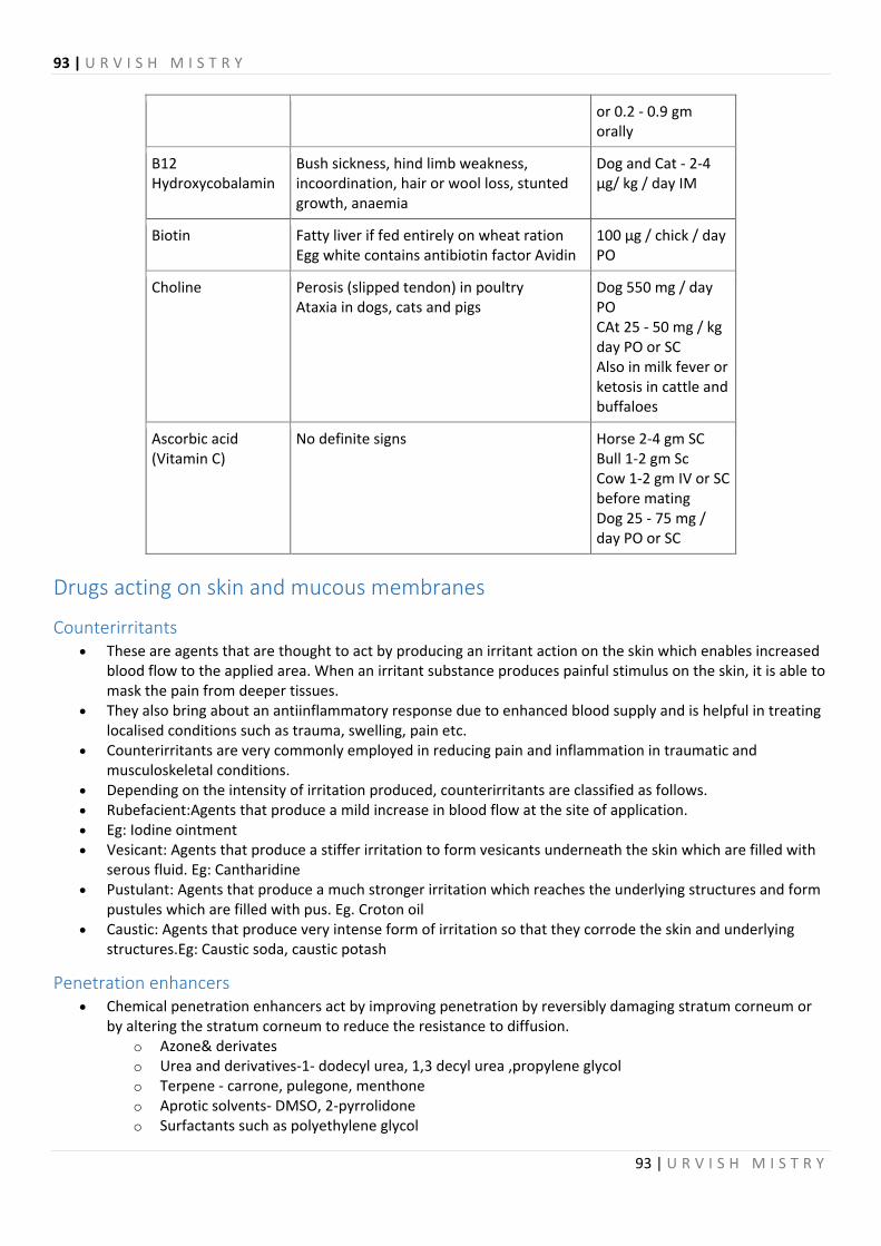
93 | U R V I S H M I S T R Y
93 | U R V I S H M I S T R Y
or 0.2 ‐ 0.9 gm orally
B12 Hydroxycobalamin
Bush sickness, hind limb weakness, incoordination, hair or wool loss, stunted growth, anaemia
Dog and Cat ‐ 2‐4 µg/ kg / day IM
Biotin Fatty liver if fed entirely on wheat ration Egg white contains antibiotin factor Avidin
100 µg / chick / day PO
Choline Perosis (slipped tendon) in poultry Ataxia in dogs, cats and pigs
Dog 550 mg / day PO CAt 25 ‐ 50 mg / kg day PO or SC Also in milk fever or ketosis in cattle and buffaloes
Ascorbic acid (Vitamin C)
No definite signs Horse 2‐4 gm SC Bull 1‐2 gm Sc Cow 1‐2 gm IV or SC before mating Dog 25 ‐ 75 mg / day PO or SC
Drugs acting on skin and mucous membranes
Counterirritants These are agents that are thought to act by producing an irritant action on the skin which enables increased
blood flow to the applied area. When an irritant substance produces painful stimulus on the skin, it is able to mask the pain from deeper tissues.
They also bring about an antiinflammatory response due to enhanced blood supply and is helpful in treating localised conditions such as trauma, swelling, pain etc.
Counterirritants are very commonly employed in reducing pain and inflammation in traumatic and musculoskeletal conditions.
Depending on the intensity of irritation produced, counterirritants are classified as follows. Rubefacient:Agents that produce a mild increase in blood flow at the site of application. Eg: Iodine ointment Vesicant: Agents that produce a stiffer irritation to form vesicants underneath the skin which are filled with
serous fluid. Eg: Cantharidine Pustulant: Agents that produce a much stronger irritation which reaches the underlying structures and form
pustules which are filled with pus. Eg. Croton oil Caustic: Agents that produce very intense form of irritation so that they corrode the skin and underlying
structures.Eg: Caustic soda, caustic potash
Penetration enhancers Chemical penetration enhancers act by improving penetration by reversibly damaging stratum corneum or
by altering the stratum corneum to reduce the resistance to diffusion. o Azone& derivates o Urea and derivatives‐1‐ dodecyl urea, 1,3 decyl urea ,propylene glycol o Terpene ‐ carrone, pulegone, menthone o Aprotic solvents‐ DMSO, 2‐pyrrolidone o Surfactants such as polyethylene glycol

94 | U R V I S H M I S T R Y
94 | U R V I S H M I S T R Y
Suitable use of penetration enhancers would help increase the bioavailability of agents given intradermally.
Adsorbants Adsorbants are substances which can bind noxious and irritant substances. When applied on the skin, they
lessen the irritation and afford protection to the skin. Example:
o Dusting powder o Starch, Zinc oxide, CaCO3 o Boric acid and talc o Collodion o Dimethicone
Demulcents (demulcere ‐ To smooth)
These are inert agents that soothen damages mucosa by preventing contact with air or other irritants. High molecular weight water soluble compounds
o Alleviating irritation with inherent properties to reduce irritation E.g. ‐ Glycerin, gelatin, hydroxyl propyl cellulose, methyl cellulose
Glycerin o Low concentration‐ hydrates skin. However in high concentration, it dehydrates skin
Propylene glycol o Hygroscopic, bacteriostatic, fungistatic
Gum acacia o Colloidal plant carbohydrates that swell in water, forming mucilaginous solutions.
Emollients Emollients are bland oily materials that soften skin Decrease transepidermal water loss and thus helpful in dry skin and cracked skin Examples
o Vegetable oils: Almond oil, Cotton seed oil, Olive oil, Peanut oil, Coco butter o Animal oils: Lard, Lanolin, Spermaceti (from sperm whale) o Petroleum products: Liquid paraffin, paraffin
Astringents Emollients are bland oily materials that soften skin Decrease transepidermal water loss and thus helpful in dry skin and cracked skin Examples
o Vegetable oils: Almond oil, Cotton seed oil, Olive oil, Peanut oil, Coco butter o Animal oils: Lard, Lanolin, Spermaceti (from sperm whale) o Petroleum products: Liquid paraffin, paraffin
Other agents used on skins Besides the above a number of other agents are used on the skin. These drugs will be discussed under appropriate headings.
Keratolytics These agents cause the outer layer of the skin to loosen and shed. They can soften keratin and used in teh
treatment of dandruff and seborrheic dermatitis Eg. Zinc pyrithione, sulfur and salicylic acid Antibacterial Antifungals Corticosteroids Vasodilators Fibrinolytic Antiinflammatory agents
95 | U R V I S H M I S T R Y
95 | U R V I S H M I S T R Y
Vehicles
Bioenhancers, immunostimulants and immunosuppressors
Bioenhancers Bioenhancers are agents that at low doses promote and augment the bioavailability or biological activity of
drugs. Herbal bioenhancers are agents of herbal origin or any phytomolecule, which is capable of enhancing
bioavailability and/or bioefficacy of a specified drug or nutrient at the low dose but it do not show typical pharmacological activity.
The concept of bioenhancers (herbal origin) can be tracked back to Ayurveda system of medicine. 'Trikatu' meaning three acids is an Ayurvedic preparation containing black pepper, long pepper and ginger, which are prescribed routinely for a variety of diseases as part of a multidrug prescription.
Herbal bioenhancers have properties like non‐toxic to humans and animals, effective at low concentrations, easy to formulate and enhance absorption and bioactivity of the drug or nutrient. Herbal bioenhancers are suggested to act through several mechanisms of action. Various mechanisms of action postulated for herbal bioenhancers include
o increase in gastrointestinal blood supply and reduction in hydrochloric acid secretion, o inhibition of gastric emptying time, gastrointestinal transit and intestinal motility, o modifications in GIT epithelial cell membrane permeability, o cholagogous effect, o thermogenic and bioenergetics properties, o Inhibition of drug metabolizing enzymes and suppression of first pass metabolism and stimulation of
gamma glutamyl transpeptidase (GGT) activity which enhances uptake of amino acids. Important Indian Herbal Bioenhancers
Long pepper (Piper longum), Black pepper (Piper nigrum), Ginger (Zingiber officinale), Liquorice (Glycyrrhiza glabra), Black cumin (Cuminum cyminum), Cumin/Caraway (Carum carvi), Garlic (Allium sativum) and Indian Aloe (Aloe vera)
The development of plant‐based bioenhancers is to be targeted for drugs that are poorly bioavailable, given for longer period of time, highly toxic and expensive. Further, research should be carried out to evaluate clinical application of herbal bioenhancers in modern human and veterinary therapeutics.
Immunostimulants These are agents that have been used to enhance an immune response and are helpful as adjunct agents for
therapy. Most common use in dogs is for the treament of chronic recurrent staphylococcal pyoderma. Staphage lysate is a preparation of S. aureus and staphylococcal bacteriophage. It is believed to stimulate T
lymphocytes and activate phagocytic cells. Staphylococcal bacterin toxoid has been used with some success in canine bacterial hypersensitivity. One of the most commonly used immunomodulator in veterinary practice is the anthelmintic levamisole ,
which is advocated at one‐third the anthelmintic dose for immunomodulatory activity for generalised immunity enhancement as is required post vaccination.
Immunosuppressant These are agents known to suppress the immune response on administration. In human beings they are very
commonly used in transplants to prevent graft rejection and in the treatment of autoimmune diseases. In veterinary practice too, they are very useful.

96 | U R V I S H M I S T R Y
96 | U R V I S H M I S T R Y
Glucocorticoids are the most commonly used immunosuppressive agents. It is used for immune mediated skin disorders.
Cyclophosphamide, an alkylating agent, is used in combination with glucocorticoids for severe cases of autoimmnune diseases such as systemic lupus erythematosus, pempighus, rheumatoid arthritis.The side effects include haemorrhagic cystitis, fibrosis of the bladder, bone marrow suppression etc.
Chlorambucil is slower acting and less toxic. It is administered alternately with glucocorticoids. Azathioprine is more commonly used for pemphigus. Cyclosporine which is most commonly used in man for graft rejection, may also be used in animals to
prevent pempighus, lupus etc. It is potentially nephrotoxic, hepatotoxic, toxic to alimentary tract, and bone marrow suppression. It blocks the proliferation of activated T lymphocytes, by inhibiting interleukin‐2, gene activation and RNA transcription. It can be used for Keratoconjunctivitis sicca in dogs.
Dapsone used as an antibacterial for treating leprosy in man, has been found to inhibt chemotaxis, degranulation of mast cells and inhibit IgG, IgA, prostaglandins and also T cell responses. It is also used for canine pemphigus and lupus erythematosus and allows for decrease in the dose of glucocorticoids.


![Veterinary Pharmaceutical Dosage Forms: A Technical Note · chloramphenicol derivatives [1]. Veterinary pharmacology differs from human pharmacology both in the diversity of species](https://static.documents.pub/doc/80x56/60b4d24c513f11331c35e538/veterinary-pharmaceutical-dosage-forms-a-technical-note-chloramphenicol-derivatives.jpg)
