Page 1
1
GENERAL INTRODUCTION
The clinical examination of the mother is important to assess the progress
of pregnancy, but it is not sufficient to get information on the fetal well-being.
When the maternal conditions are compromised, the health of fetuses could
also be affected; on the other hand, a good health status of the mother does not
guarantee that the fetuses are all healthy.
In cases of systemic involvement, as well as for changes in the maternal
homeostasis due to infectious agents, toxic molecules, or hormones and drugs that
may reach embryos or fetuses by transplacental passage, fetal survival could be
negatively affected. During the early stages of gestation embryo resorptions are
often asymptomatic and later, in case of fetal death, bitches may not show clinical
symptoms (Johnston, 1983; England et al., 1990; Yeager et al., 1992; Kutzler et
al., 2003; Root-Kustritz, 2005; Kim et al., 2007; Davidson, 2008; Sridevi, 2013).
For all these reasons, the fetal well-being, i.e. appropriate fetal development
according to gestational age and fetal health, has to be assessed by specific
diagnostic tools.
In human medicine until the 50s, obstetricians put primary emphasis on the
maternal survival and health. The health of the fetus has gained increasing
attention only after 1960 thanks to the technological support that allowed the direct
assessment of the physical fetal conditions “in utero” (Patrick, 1989).
In veterinary medicine during the last decades, the ultrasound examination
of the reproductive system in the bitch has been deeply investigated. Many authors
Page 2
2
have described specific ultrasonographic findings of the canine pregnancy and
nowadays ultrasonography represents the gold standard method for the evaluation
of a pregnant bitch (Johnston, 1983; Poffenbarger & Feeney, 1986; England et al.,
1990; Yeager et al., 1992; Kutzler et al., 2003; Root-Kustritz, 2005; Kim et al.,
2007; Davidson, 2008; Lopate, 2008; Michel et al., 2011; Sridevi, 2013). It
represents the most safe and sensitive method for the pregnancy diagnosis from
the day 25 of gestation (Concannon et al., 1989; England et al., 1990; Yeager et
al., 1992; Miles, 1995). In addition, this technique allows to monitor fetal viability
and development, to recognize the stage of pregnancy, and to estimate the date of
delivery (Johnston, 1983; Poffenbarger & Feeney, 1986; England et al., 1990;
Yeager et al., 1992; Kutzler et al., 2003; Root-Kustritz, 2005; Kim et al., 2007;
Davidson, 2008; Lopate, 2008; Michel et al., 2011; Sridevi, 2013).
Page 3
3
Ultrasonographic evaluation of the fetal development
Many authors have examined the timing of the first ultrasonographic
appearance of certain extra-fetal and fetal structures during the canine pregnancy
(Table 1).
Table 1. The gestational age at first appearance of ultrasonographic features during canine pregnancy
(based on England et al., 1990; Yeager et al., 1992; Boroffka, 2005; Kim & Son, 2007).
Features identified Gestational Age
(days after ovulation)
Conceptus as a 1 - 2 mm uterine vesicle 19-20
Presence of the embryonic mass 22-23
Heartbeat 23-24
Yolk sac membrane 25-27
Bipolar embryo shape 26-28
Allantoic membrane 27-31
Limb bud 27-31
Placenta develops zonary shape 27-29
Head 27-30
Chorionic cavity exceeds the size of the yolk sac 28-30
Anechoic area in head 29-33
Stomach 29-33
Dorsal tubular spinal column 30-36
Urinary bladder 31-35
Collapsing of the elongated yolk sac 31-34
Fetal movement 32-34
Axial skeleton 33-34
Lung hyperechoic vs liver 34-36
Liver hypoechoic vs abdomen 35-38
Trunk diameter exceeds diameter of head 38-40
Trunk diameter exceeds 50% of chorionic cavity diameter 38-42
Crown-rump length exceeds length of placenta 38-42
Kidneys 40-46
Eyes 40-46
4 Cardiac chambers 40-44
Trunk diameter exceeds 50% of uterine outside diameter 46-48
Intestines 58-62
Page 4
4
These information are useful to detect whether the pregnancies progress
regularly (Concannon, 2000; Nyland & Matton 2002). However, the estimation of
gestational age based on the assessment of organ development is unpractical
because it would require a daily ultrasonographic assessment of embryos/fetuses.
Moreover, the first identification of specific anatomical features depends by the
examiner experience and by the resolution of the used equipment (Lenard et al.,
2007). To overcome these limits, the gestational age is commonly estimated by the
ultrasonographic measurements of extra-fetal and fetal structures.
Different extra-fetal parameters can be evaluated during early pregnancy.
From day 20 up to approximately day 40 of gestation, the chorionic cavity (Figure
1) has a typical spherical appearance with well-defined margins and anechoic
content. The internal diameters (inner chorionic cavity, ICC), made at 90° angles
from one side of the trophoblastic decidual reaction to the other, can be easily
recognized and measured (England et al., 1990; Yeager et al., 1992; Luvoni &
Grioni, 2000; Kutzler et al., 2003; Luvoni & Beccaglia, 2006).
In the same gestational period, the outer uterine diameter (OUD) at the
implantation sites, the placental thickness (PT) and length (PL) can also be
estimated (England et al., 1990; Yeager et al., 1992; Luvoni & Grioni, 2000; Son
et al. 2001). However, the ICC measurement is easier to obtain than the OUD, PT
and PL because the uterine wall and the annexes have less defined margins than
ICC (England et al., 1990; Yeager, 1992; Luvoni & Grioni, 2000; Son et al. 2001;
Kutzler et al., 2003; Michel et al., 2011).
Page 5
5
Figure 1. Ultrasonographic measurement of inner chorionic cavity (ICC)
in a bitch (36 days before parturition).
During fetal growth, different biometric parameters can be evaluated
(Lopate, 2008; Michel et al., 2011). In the longitudinal plane, the fetal length
(crown-rump length, CRL), between the most rostral point and the caudal edge of
the fetus, and, in the transverse plane, the body diameter (BD) at the level of the
stomach and liver, can be measured.
When the fetal head can be distinguished from the body, fetal head diameter
is measured as its largest cross-sectional diameter. When parietal bones are
identified in the coronal section of the head, the biparietal diameter (BP, Figure 2)
is the parameter of choice. The deep portion of diencephalo-telencephalic vesicle
(DPTV) can also be visualized in the same scan. The BP is the distance between
the two parietal bones of the skull, and the DPTV, is an ovoid anechoic structure
with well-defined margins symmetrical to the longitudinal fissure separating the
two cerebral hemispheres (Figure 2).
Page 6
6
The major and minor axis of the fetal heart (diameter of fetal heart, HDF)
have also been considered as biometrical parameters and used to evaluate the
gestational age (England et al., 1990; Yeager, 1992; Moriyoshi et al. 1996; Luvoni
& Grioni, 2000; Son et al. 2001; Kutzler et al., 2003; Michel et al., 2011).
During each examination, it is advisable to obtain the average of at least two
measurements of the same extra-fetal or fetal parameter in different fetuses, to
minimize the risk of inconsistent measurements. In the case of singleton
pregnancy, different parameters should be assessed on the same fetus (Luvoni &
Beccaglia 2006), although this could be also helpful in non-singleton pregnancies
(Lopate, 2008; Michel et al., 2011).
Figure 2. Ultrasonographic measurement of biparietal diameter (BP) and deep portion of diencephalo-
telencephalic vesicle (DPTV) in a bitch (20 days before parturition).
Page 7
7
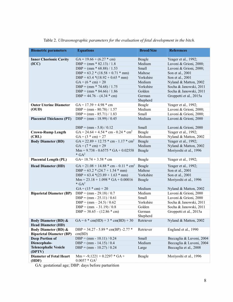
The table 2 summarizes the extra-fetal and fetal ultrasonographic
parameters and the related formulae for the gestational age and/or days before
parturition in the bitch.
Page 8
8
Table 2. Ultrasonographic parameters for the evaluation of fetal development in the bitch.
Biometric parameters Equations Breed/Size References
Inner Chorionic Cavity
(ICC)
GA = 19.66 + (6.27 * cm) Beagle Yeager et al., 1992;
DBP = (mm * 82.13) / 1.8 Medium Luvoni & Grioni, 2000;
DBP = (mm * 68.88) / 1.53 Small Luvoni & Grioni, 2000;
DBP = 63.2 * (18.58 + 0.71 * mm) Maltese Son et al., 2001
DBP = 63.4 *(18.92 + 0.65 * mm) Yorkshire Son et al., 2001
GA = (6 * cm) + 20 Medium Nyland & Matton, 2002
DBP = (mm * 74.68) / 1.75 Yorkshire Socha & Janowski, 2011
DBP = (mm * 84.66) / 1.86 Golden Socha & Janowski, 2011
DBP = 44.76 – (4.34 * cm) German
Shepherd
Groppetti et al., 2015a
Outer Uterine Diameter
(OUD)
GA = 17.39 + 4.98 * cm Beagle Yeager et al., 1992;
DBP = (mm - 80.78) / 1.57 Medium Luvoni & Grioni, 2000;
DBP = (mm - 85.71) / 1.83 Small Luvoni & Grioni, 2000;
Placental Thickness (PT) DBP = (mm - 18.99) / 0.45
Medium Luvoni & Grioni, 2000
DBP = (mm - 5.8) / 0.12 Small Luvoni & Grioni, 2000
Crown-Rump Length
(CRL)
GA = 24.64 + 4.54 * cm - 0.24 * cm2 Beagle Yeager et al., 1992;
GA = (3 * cm) + 27 Medium Nyland & Matton, 2002
Body Diameter (BD) GA = 22.89 + 12.75 * cm - 1.17 * cm2 Beagle Yeager et al., 1992;
GA = (7 * cm) + 29 Medium Nyland & Matton, 2002
Mm = 9.738 - 0.6575 * GA + 0.02558
* GA2
Beagle Moriyoshi et al., 1996
Placental Length (PL) GA= 18.74 + 3.58 * cm Beagle Yeager et al., 1992;
Head Diameter (HD) GA = 21.08 + 14.88 * cm – 0.11 * cm2 Beagle Yeager et al., 1992;
DBP = 63.2 * (24.7 + 1.54 * mm) Maltese Son et al., 2001
DBP = 63.4 *(23.89 + 1.63 * mm) Yorkshire Son et al., 2001
Mm = 23.18 + 1.098 * GA + 0.00016
* GA2
Beagle Moriyoshi et al., 1996
GA = (15 * cm) + 20 Medium Nyland & Matton, 2002
Biparietal Diameter (BP) DBP = (mm - 29.18) / 0.7 Medium Luvoni & Grioni, 2000
DBP = (mm - 25.11) / 0.61 Small Luvoni & Grioni, 2000
DBP = (mm – 24.5) / 0.62 Yorkshire Socha & Janowski, 2011
DBP = (mm - 31.19) / 0.8 Golden Socha & Janowski, 2011
DBP = 38.65 – (12.86 * cm) German
Shepherd
Groppetti et al., 2015a
Body Diameter (BD) &
Head Diameter (HD)
GA = 6 * cm(HD) + 3 * cm(BD) + 30 Retriever Nyland & Matton, 2002
Body Diameter (BD) &
Biparietal Diameter (BP)
DBP = 34.27 - 5.89 * cm(BP) -2.77 *
cm(BD)
Retriever England et al., 1990
Deep Portion of
Diencephalo-
Telencephalic Vesicle
(DPTV)
DBP = (mm - 10.11) / 0.24 Small Beccaglia & Luvoni, 2004
DBP = (mm - 14.15) / 0.4 Medium Beccaglia & Luvoni, 2004
DBP = (mm - 10.27) / 0.24 Large Beccaglia et al., 2008
Diameter of Fetal Heart
(HDF)
Mm = -9,1221 + 0.2297 * GA +
0.0057 * GA2
Beagle Moriyoshi et al., 1996
GA: gestational age; DBP: days before parturition
Page 9
9
The accuracy of this estimation has been studied by different authors
(Luvoni & Grioni, 2000; Kutzler et al., 2003; Beccaglia & Luvoni 2006; Lenard
et al., 2007; Lopate, 2008; Michel et al., 2011; Socha & Janowski, 2014).
The gestational period in which the examination is performed mainly affects
the accuracy of parameters. that resulted higher during early pregnancy, than
afterwards (Yeager et al., 1992; Son et al., 2001; Kutzler et al., 2003; Beccaglia &
Luvoni, 2006; Socha et al., 2012; Socha & Janowski, 2014). According to Kutzler
et al., 2003, the most accurate predictions of parturition date are obtained when
ICC measurements are performed on day 30. The accuracy of BP, that is measured
for an extended period (from week 5 to 9) is highly consistent during the 6th week
of gestation (Beccaglia & Luvoni, 2012).
It has been demonstrated that the fetal sex ratio and the litter size do not
generally influence the accuracy of parturition date prediction (Kutzler et a., 2003;
Beccaglia & Luvoni, 2006), but the BP accuracy is higher in normal litter size than
in small and large litters (Beccaglia & Luvoni, 2006; Groppetti et al., 2015a). This
may be due to the fact that BP measurement might be affected by individual
variability of growth when few fetuses are present, or it may be less accurate when
the overlapping of multiple fetuses in the same ultrasonographic image field
occurs (Beccaglia & Luvoni, 2006).
In dogs, the wide breed variability requires specific reference curves of
biometric values based on different breed sizes (Luvoni & Grioni, 2000; Son et
al., 2001; Kutzler et al., 2003; Luvoni & Beccaglia, 2006; Socha et al., 2012).
Some authors demonstrated that maternal bodyweight affects the accuracy of
parturition date prediction (Kutzler et al., 2003), but ICC and BP are both highly
reliable when size-related specific formulae are applied (Beccaglia & Luvoni,
Page 10
10
2006; Socha et al., 2015). Thus, an equally accurate prediction can be obtained
both in early and late gestation (Beccaglia & Luvoni, 2006).
The fetal development has been deeply investigated in small and medium
dogs, whereas only few information are available for large and giant size bitches
even though they are very well represented in the one hundred most popular canine
breeds (Sverdrup Borge et al., 2011; Tønnessen et al., 2012). For these dogs,
specific formulae for the evaluation of fetal growth are not yet available. Kutzler
and colleagues (2003) suggested the use of a correction factor to adjust the
difference between actual and predicted parturition date obtained with previously
published equations for dogs of smaller size (England et al., 1990; Yeager et al.,
1992). Although the formulae for medium dogs (Luvoni & Grioni, 2000) have
been also used in large and giant breeds (Socha & Janowski, 2014; Socha et al.,
2015), specific equations for these dogs would allow the most accurate prediction
of parturition term (Michel et al. 2011; Socha & Janowski, 2014; Socha et al.,
2015).
Page 11
11
Evaluation of fetal health
Ultrasonography
To assess the fetal health, different fetal and the extra-fetal parameters can
be evaluated during pregnancy by the B-mode ultrasonography and the Echo Color
Doppler examination.
The elective method to assess embryo/fetal viability or to recognize the
interruption of pregnancy, at any time and at any stage of the development of the
conceptuses, is the ultrasonography. In dogs, the embryonic death before the day
35 after ovulation (followed by the complete resorption of the conceptus) is not
recognizable radiologically. With the ultrasound exam, this event is characterized
by a decrease in the volume of the embryo vesicle, an increase echogenicity of the
fluid, an absence of heartbeat, and a distortion and collapse of the embryonic mass
(Konde, 1988; Concannon, 2003). The uterine wall appears moderately
hyperechoic and a small amount of free fluid can be found into the lumen of the
organ (Concannon, 2003).
After the day 35 of pregnancy, fetal death may be associated with a vaginal
discharge, but it can also go unnoticed. In all cases, the ultrasound examination
detects changes in the fluids and the absence of the heartbeat. When an abortion
occurs, after the expulsion of the died puppies, the uterus will show the typical
appearance of post-partum (Concannon, 2003). Only in case of late fetal death, X-
rays can identify skeletal deformities, altered relationships between different
skeletal sites, the cranial bones spaced and the presence of gas around fetal bodies
(Rendano, 1983; Toal et al., 1986; Miles, 1995; Lopate, 2008; Lamm & Makloski,
2012).
Page 12
12
B-mode ultrasonographic exam
The B-mode ultrasound exam provides many important information about
fetal well-being through the analysis of some indicators as the fetal development,
the amount and the echogenicity of fetal fluids, the placental thickness (PT), and
the fetal movements. When one or more of these indicators do not fall within the
normal range, the suffering condition of the fetus is usually defined as "fetal stress"
(Table 3).
Table 3. Parameters to identify fetal stress in the bitch by B-mode ultrasonographic exam.
Parameters References
Fetal development See previous paragraph
Abdominal:Biparietal diameter ratio Zone & Wanke, 2001
Placental thickness Lopate, 2008
Fetal fluids Zone & Wanke, 2001;
Lopate, 2008
Fetal movements Yeager et al., 1992;
Bocking et al., 1985;
Davidson, 2001;
Zone & Wanke, 2001;
England & Russo, 2006
The parameters to evaluate the fetal development in dogs have been
extensively discussed in the previous paragraph (see Tables 1 and 2).
Some authors suggested that an intrauterine growth retardation may be
suspected when the ratio between abdominal and biparietal diameters is less than
2 from day 48 of gestation (Zone & Wanke, 2001). Puppies with a low ratio are at
Page 13
13
risk for early neonatal loss, since the low ratio is usually associated with a 20%
decrease of bodyweight at birth (Zone & Wanke, 2001).
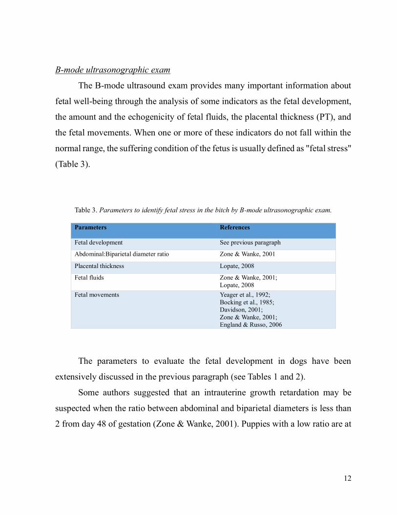
In dogs, the placental thickness (Figure 3) must not exceed 1.2 cm along the
whole pregnancy. Thickening or edema of the placenta indicate diminished ability
of the organ to drain fetal waste fluids properly, alterations or abnormalities of
blood flow, or placentitis (Lopate, 2008).
Figure 3. Ultrasonographic measurement of placental thickness (yellow line).
Along pregnancy, the amount of fetal fluid decreases as the fetus itself
enlarges. Quantitative and qualitative assessment of fetal fluids can be performed
by the ultrasonography. An increase or an abnormal decrease of these fluids may
be due to abnormalities of placental functions with alteration of blood flow and/or
decreased ability to drain the products of the fetal catabolism, or to rupture of the
fetal membranes. A variation in the quality of the fluids, as an increased
Page 14
14
echogenicity can be determined by a hemorrhage with premature detachment of
the placenta or to the passage of meconium (Zone & Wanke, 2001; Lopate, 2008).
Fetuses generally do not survive for long time after the placenta begins to detach,
although the detachment may be partial or complete (Lopate, 2008).
Several studies have analyzed and quantified the fetal movements in
humans and livestock (Dawes et al., 1972; Ruckebusch, 1972; Patrick et al., 1982;
Di Renzo et al., 1994a; Di Renzo et al., 1994b; Baska-Vincze et al., 2014), but
little information are available in the dog (Davidson, 2001). The canine fetal
movements are clearly visible by ultrasound from the day 35 of pregnancy and
more frequent activities are the back bending of the head and the extension of the
limbs (Yeager et al., 1992; Zone & Wanke, 2001; England & Russo, 2006). It has
been suggested that the fetal movements are associated with an increased heart
rate (Bocking et al., 1985; Davidson, 2001).
In human medicine breathing movements of the fetus can be recognized
from the 10th week (De Vries et al., 1982), but in veterinary medicine, the dynamic
of respiratory movements has been described only in the sheep (Dawes et al.,
1972; Boddy et al., 1974; Patrick et al., 1987) and no information are available for
dogs.
Page 15
15
Echo Color Doppler exam
The purpose of the application of the Doppler in the obstetric monitoring of
human and animal species is to evaluate the hemodynamic characteristics of the
fetal-maternal circulation and to identify high-risk pregnancies (Fleischer &
Emerson, 1994; Reed et al., 1996; Nautrup, 1998; Nicolaides et al., 2000;
Bollwein et al., 2002; Dubiel et al., 2003; Bollwein et al., 2004; Di Salvo et al.,
2006; Blanco et al., 2008).
The arterial blood of the mother, rich in oxygen and nutrients, through the
placenta reaches the fetus via the umbilical vein. Some peculiarities, such as, the
venous ductus, that connects the portal vein to the caudal vena cava, the ductus
arteriosus, that combines the pulmonary trunk to the aorta, and the oval foramen
(interatrial) characterize the fetal cardiovascular system that only at birth becomes
as that of the adults (Götze, 1955). Furthermore, during pregnancy, the fetal lungs
do not function and the pulmonary circulation is bypassed.
For the evaluation of fetal stress the parameters/structures described in the
Table 4 are usually examined with the Doppler.
Table 4. Parameters to identify fetal stress in the bitch by Echo Colour Doppler exam.
Parameters References
Utero-placental arteries Nautrup, 1998;
Di Salvo et al., 2006
Umbilical artery and vein Nautrup, 1998;
Di Salvo et al., 2006
Fetal aorta Nautrup, 1998;
Di Salvo et al., 2006
Fetal common carotid artery Nautrup, 1998
Fetal caudal vena cava Di Salvo et al., 2006
Fetal heart rate Lopate, 2008
Page 16
16
In the dog, the low resistance blood flow of the utero-placental arteries is
characterized by a systolic peak, a small diastolic wave and by a relatively high
speed of the end diastole (Nautrup, 1998; Di Salvo et al., 2006). Pulsatility and
resistivity indexes are related to the growth of the placenta (Di Salvo et al., 2006).
In all species, at the umbilical level, the blood flows of the umbilical artery
and vein are simultaneously present. In dogs, the umbilical cord can be identified
from the week 4 of gestation with color Doppler and from the 5th week with two-
dimensional ultrasonography. The blood flow in the umbilical artery is only
systolic until the 6th week of pregnancy, and then the diastolic wave can also be
identified. The umbilical vein is characterized by uniform flow with flat waves
(Nautrup, 1998; Di Salvo et al., 2006).
The fetal aorta is characterized by high flow velocities recorded in two
standard regions: in the thoracic area immediately above the diaphragm and in the
abdominal region before the iliac arteries emerge. In dogs, no significant variation
between the thoracic and abdominal region has been observed and the blood flow
of the aorta until the 6th week of pregnancy is only systolic, whereas in the late
phase of gestation the diastolic peak becomes evident (Nautrup, 1998; Di Salvo et
al., 2006).
In canine fetuses, the common carotid artery can be recognized from the
week 6 of gestation. This vessel is characterized by a quick systolic flow, with
accelerations and decelerations, and by a diastolic flow with flat velocity (Nautrup,
1998). The flow in this vessel is pulsatile: a minor diastolic peak follows the
systolic wave. Sometimes, a third retrograde wave due to atrial contraction can be
observed (Maulik, 1999; Di Salvo et al., 2006).
Page 17
17
Finally, in the bitch, the different degree of patient cooperation and the
physiological respiratory arrhythmia, that determines a lengthening of the diastole,
may influence parameters of the vessel flow (pulsatility index, resistivity index,
ratio between the systolic and diastolic speed) (Nautrup, 1998). Moreover, the
normal ranges of the obstetric Doppler parameters differ depending on the size of
the dog (Blanco et al., 2008).
Fetal Heart Rate
Several authors have investigated maternal and fetal heart rate (MHR and
FHR) features, their relationship and their mutual influence, to define clinical
parameters of maternal and fetal well-being.
During pregnancy, the cardiovascular systems of the mother and the fetuses
closely interact and the adaptation of the maternal system ensures a proper
development of the fetus (Van Leeuwen et al., 2009).
Studies in pregnant women demonstrated an increase of blood volume that
induces a chronic distention of the cardiac ventricles, followed by a positive
inotropic effect and an increased activation of the sinoatrial node (Bader et al.,
1955; Robson et al., 1989; Spatling et al., 1992; Khodiguian et al., 1996). The
concurrent decrease in peripheral resistance ensures a proper spraying of the uterus
to promote an adequate fetal development (Mone et al., 1996; Valensise et al.,
2000).
Also in the pregnant bitch circulating blood volume and stroke volume
increase (Brooks & Keil, 1994a; Brooks & Keil, 1994b; Johnston et al., 2001;
Williams et al., 2007; Abbott, 2010; Blanco et al., 2011). In the late pregnancy,
between 50 and 60 days, both the hypertrophy of the wall and the increase of the
Page 18
18
diameter of the left ventricle are well evident at the echocardiographic exam
(Williams et al., 2007; Abbott, 2010; Blanco et al., 2011). The increased density
of the fetal membranes and the increased number of placental capillaries induce a
significant reduction in the resistance of the uterine artery resulting in a
hypotension that provides an optimal blood supply to the fetus (Nautrup, 1998; Di
Salvo et al., 2006; Blanco et al., 2009). Moreover, along pregnancy characterized
by the predominance of the sympathetic tone, MHR and systolic function increase
until delivery to support placental and fetal requirements (Williams et al., 2007;
Abbott, 2010; Blanco et al., 2011). Therefore, the pregnancy induces some
hemodynamic changes and the MHR tends to be higher to give adequate blood
supply to the fetus (Lucio et al., 2009). However, several factors, regardless of the
pregnant status, such as size, age, temperament, training etc., might influence the
HR and therefore the MHR (Hamlin et al., 1967; Fleischer & Emerson, 1994;
Bodey & Michell, 1996; Hezzell et al., 2013).
The FHR represents the most important parameter to estimate fetal health
both in human and in veterinary medicine (Blanco et al., 2008; Gil et al., 2014).
In the bitch, the embryonic heartbeat can be recognized by the ultrasound exam
from the day 23 after the LH surge as a brilliant flicker in the embryo (Concannon
et al., 1989; Yeager & Concannon, 1990; England et al., 1990; Yeager et al., 1992;
Verstegen et al., 1993).
With the B-mode ultrasound exam the evaluation of the heart anatomy can
be performed along fetal development. With the M-mode or Echo Color Doppler
the cardiovascular function can be evaluated. It has been reported that a healthy
and viable embryo/fetus shows a heart rate of 220-240 bpm (beats per minute). A
value of FHR between 180 and 220 bpm is considered an early sign of fetal
Page 19
19
distress, whereas a frequency less than 140-160 bpm indicates a severe fetal stress
usually due to hypoxia (Verstegen et al., 1993; Davidson, 1998; Zone & Wanke,
2001). Recently, some authors reported that acceleration and deceleration of FHR
in all of the fetuses might be observed in the last hours of gestation and it could
be considered a parameter to identify the approaching delivery (Gil et al., 2014).
In human medicine the FHR is generally detected by the cardiotocography
(CTG) or the echo color Doppler, but the CTG, which simultaneously detects
uterine activity, is considered the gold standard technique for the peri-partum
monitoring (Fischer, 1979).
Some years ago, a cardiotocographic device for bitches (WhelpWise;
Veterinary Perinatal Specialties Inc.; Wheat Ridge, Colorado) was introduced into
the market. The instrument allows to correlate the FHR oscillations and the uterine
contractions to identify uterine dysfunctions or fetal distress (Davidson & Eilts,
2006; Lopate, 2008). In women the application of this method is fully standardized
and commonly used at the end of gestation, whereas in the bitch it raises some
critical issues. In the woman the transducer is secured by a belt on the abdomen
and it is precisely placed on the basis of fetal position. For the monitoring of twins
it is equipped with two Doppler transducers to detect the two individual FHRs.
The problem in the bitch is that a single transducer is placed in the lateral abdomen
of small size dogs or in the back of those of greater size (Davidson & Eilts, 2006).
The use of a single transducer in the presence of multiple fetuses implies that the
recorded signals are disturbed by the interference and the overlapping of fetal heart
beats. In addition, the positioning of the transducer based on the size of the mother,
rather than on the position of the fetus, can affect the results. Therefore, the FHRs
detected in the bitch cannot be considered as accurate as those obtained in the
Page 20
20
woman.
To obtain good Doppler signals and not to create artifacts, it is necessary to
take into account several aspects such as the angles of incidence, the size and depth
of the heart, and the fetal movements. For this reason, the echo color Doppler
represents the more appropriate technique for the FHR evaluation.
In human medicine, some authors investigated the influence of the MHR on
the trend of FHR. The studies in domestic animals are limited to the sheep in
which, during the last quarter of gestation, the FHR follows the maternal circadian
trend. Furthermore, the increase of the frequency of beats in the mother seems to
coincide with fetal movements (Bocking et al., 1985).
The paucity of information regarding the mutual influence of MHR and
FHR in canine pregnancy prompts to investigate their relationship. The FHR alone
may not be sufficient for a reliable evaluation of fetal well-being and influencing
factors have not yet been assessed.
Page 21
21
AIMS
The main goal of a physical examination of a pregnant bitch is the
evaluation of fetal development and fetal and maternal well-being.
As previously reported, the fetal development has been deeply investigated
in small and medium dogs, whereas only few information are available for large
and giant size.
Thus, the aim of this study was to derive the growth curves of ICC and BP
in large and giant size bitches and to evaluate their accuracy. The effects of litter
size and fetal sex ratio on the accuracy of the prediction were also investigated
(Paper 1).
In addition, only few parameters are available to objectively assess the fetal
health during canine pregnancy. Among them, the FHR is generally used, but the
availability of reference values of the ratio FHR/MHR could better contribute to
the evaluation of the fetal health at different gestational ages, than the single FHR
values. For this purpose, the trend of FHR and FHR/MHR ratio in bitches of
different pre-gestational bodyweight was evaluated during pregnancy (Paper 2).
Page 22
22
MATERIALS & METHODS
Growth curves of ICC and BP in large and giant size bitches (Paper 1)
Eight large size (26-40 kg) bitches (Bergamasco Shepherd, Boxer,
Doberman, German Shepherd, and Old English Shepherd) and 9 giant size (>40
kg) bitches (Great Dane, Bernese Mountain Dog, and Newfoundland), aged
between 2 and 8 years presented to the Department for breeding management and
pregnancy evaluation, were included in this study. Informed owner consent was
obtained.
All bitches were healthy at the physical examination. For breeding
management, the day of the ovulation was considered to be when plasma
progesterone concentration ranged between 4-10 ng/ml (Arbeiter, 1993; Lévy &
Fontbonne, 2007), as evaluated using an Enzyme Linked Fluorescent Assay
(MiniVidas, BioMerieux, Marcy l'Etoile, France).
Serial ultrasonographic exams were performed weekly from day 20 after
mating until parturition. Bitches were positioned in lateral recumbency,
transmission gel was applied, and two-dimensional, gray-scale, real-time
ultrasound images were produced using a 7.5 MHz microconvex probe (SonoAce
8800, Medison Co. Ltd., Seoul, Korea). During early pregnancy, inner diameter of
chorionic cavity, and in late pregnancy the biparietal diameter (Figure 4) were
measured. At least three measurements of ICC or BP, according to the gestation
period, were recorded and the mean values were calculated. The time of actual
parturition, the litter size, and the sex of the puppies were reported by the owners.
Page 23
23
Statistical analysis: the relationship between ICC or BP growth and days before
parturition was analyzed by a linear regression model. The growth equations for
both parameters were derived as y=a+bx (y=days before parturition,
x=measurement in mm of ICC or BP, a=intercept coefficient and b=first order
coefficient) and the regression coefficients were analyzed by the Student's T test
(p<0.05, Software Stat Plus 2009).
Figure 4. Ultrasonographic measurement of Biparietal Diameter (BP)
in a bitch 20 days before parturition.
Page 24
24
Accuracy of ICC and BP for the prediction of parturition day in large
and giant size bitches (Paper 1)
To assess the accuracy of the prediction, measurements of ICC and BP were
performed in 65 and 102 ultrasound examinations of large size bitches and in 39
and 52 of giant size bitches with unknown breeding dates.
As previously reported, the prediction was considered accurate when the
difference between actual and predicted parturition date was within ±1 day and ±2
days (Beccaglia & Luvoni, 2006).
To evaluate the effect of litter size on the accuracy, data were grouped for
small (<5 pups), normal (5-9 pups) and large (>9 pups) litters in large and giant
bitches (Beccaglia et al., 2008).
Moreover, predictions within ±1 day and ±2 days were analyzed on the basis
of fetal sex ratio in terms of numerical prevalence (>2) of one gender.
Statistical analysis: data were analyzed by Chi-Square test and the level of
significance was set at p<0.05.
Page 25
25
Assessment of trend of FHR and FHR/MHR ratio in bitches of different
pre-gestational bodyweight during pregnancy (Paper 2)
Seventeen client-owned pregnant bitches of different breeds (Shih-tzu,
Shetland, Jack Russell Terrier, Weimaraner, Boxer, and Great Dane), and pre-
gestational bodyweights (5.8-68 kg) aged between 2 and 7 years presented to the
Department for breeding management and pregnancy evaluation, were included in
this study. Informed owner consent was obtained.
All bitches were healthy at the physical examination and with no history
and signs of cardiac diseases. For breeding management, the day of the ovulation
was considered when plasma progesterone concentrations ranged between 4-10
ng⁄ml (Arbeiter, 1993; Lévy & Fontbonne, 2007), as evaluated using an Enzyme
Linked Fluorescent Assay (MiniVidas, BioMerieux, Marcy l'Etoile, France).
According to owner’s availability, ultrasound examinations were performed
in 5 bitches twice a week from day 21 after ovulation, and in 12 bitches at week 4,
7, and 9 of pregnancy.
Two-dimensional, gray-scale, real-time ultrasound and ecocolordoppler images
were produced using a 7.5 MHz microconvex probe (SonoAce 8800, Medison Co.
Ltd., Seoul, Korea).
The bitches were positioned in lateral recumbency, transmission gel was
applied and MHR was evaluated at the level of the aortic valve for three times (at
the beginning of the examination, after 10 minutes and at the end of the
examination) to reduce and control the stress-effect induced by the restraint
(Figure 5).
Page 26
26
Figure 5. Echo Color Doppler evaluation at the aortic level
of the canine maternal heart rate (MHR).
Fetuses’ normal development was assessed by the measurement of
ultrasonographic extra-fetal and fetal parameters (Luvoni & Grioni, 2000, Alonge
et al., 2015). Fetal heart rates of at least three different fetuses (in litter size >3)
were recorded in each examination (Figure 6).
The owners reported the day of parturition and the neonatal survival. Only
data from uncomplicated pregnancies with no evidence of embryo, fetal or
neonatal loss were included in the statistical analysis.
Page 27
27
Figure 6. Echo Color Doppler evaluation of the fetal heart rate (FHR) during canine pregnancy.
Statistical analysis: a polynomial regression model was adopted to analyze the
relationship between FHR, MHR, FHR/MHR ratio and independent variables
(pre-gestational maternal bodyweight and gestational age, in terms of days from
parturition). Statistical significance was set at p≤0.05 (Software Statistica 7 for
Windows platform).
Page 28
28
RESULTS
Growth curves of ICC and BP in large and giant size bitches (Paper 1)
The regression analysis resulted in a significant relationship between days
before parturition and ICC or BP (p<0.001).
The derived equations for the prediction of parturition day in large and giant
bitches are reported in table 5.
Table 5. Growth curves of ICC and BP in large and giant size bitches.
Parameter Large Bitches Giant Bitches
Equation R2 Coefficient Equation R2 Coefficient
ICC diameter y = (x-105.1)/2.5 0.92 y = (x-88.1)/1.9 0.97
BP diameter y = (x-88.1)/1.9 0.99 y = (x-29)/0.7 0.97
y=days before parturition, x=measurement in mm of ICC or BP,
a=intercept coefficient, b=first order coefficient.
ICC inner chorionic cavity; BP biparietal.
Page 29
29
Accuracy of ICC and BP for the prediction of parturition day in large
and giant size bitches (Paper 1)
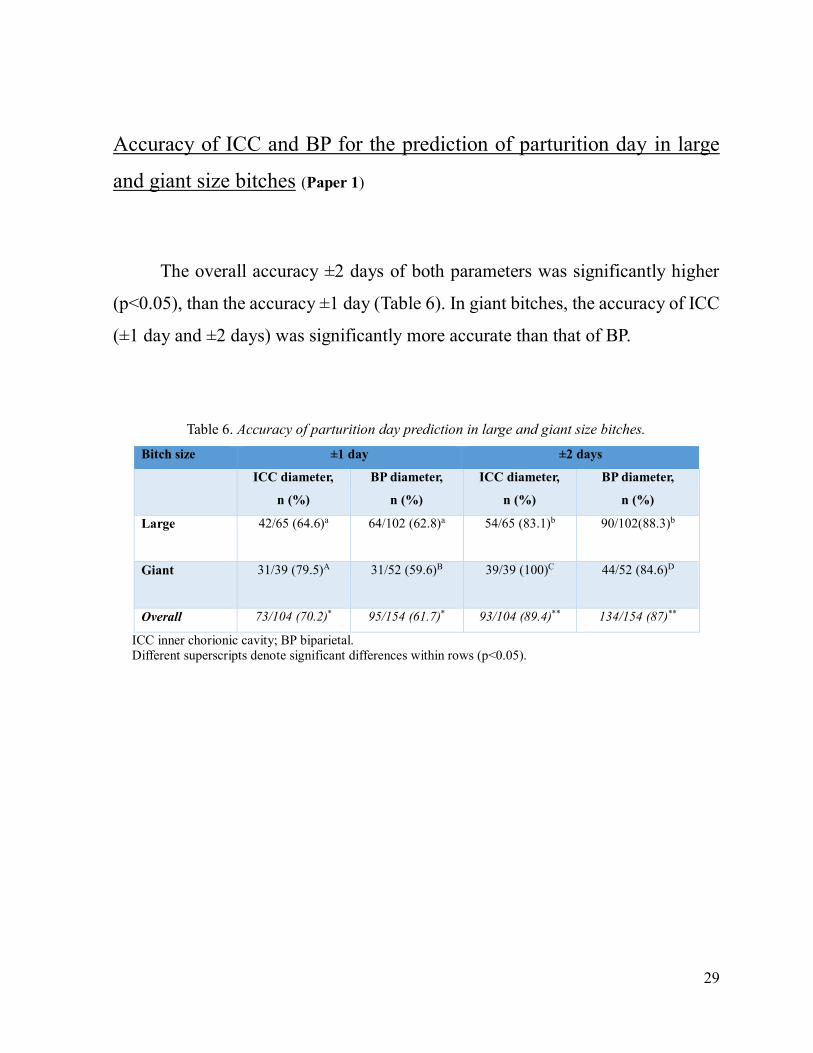
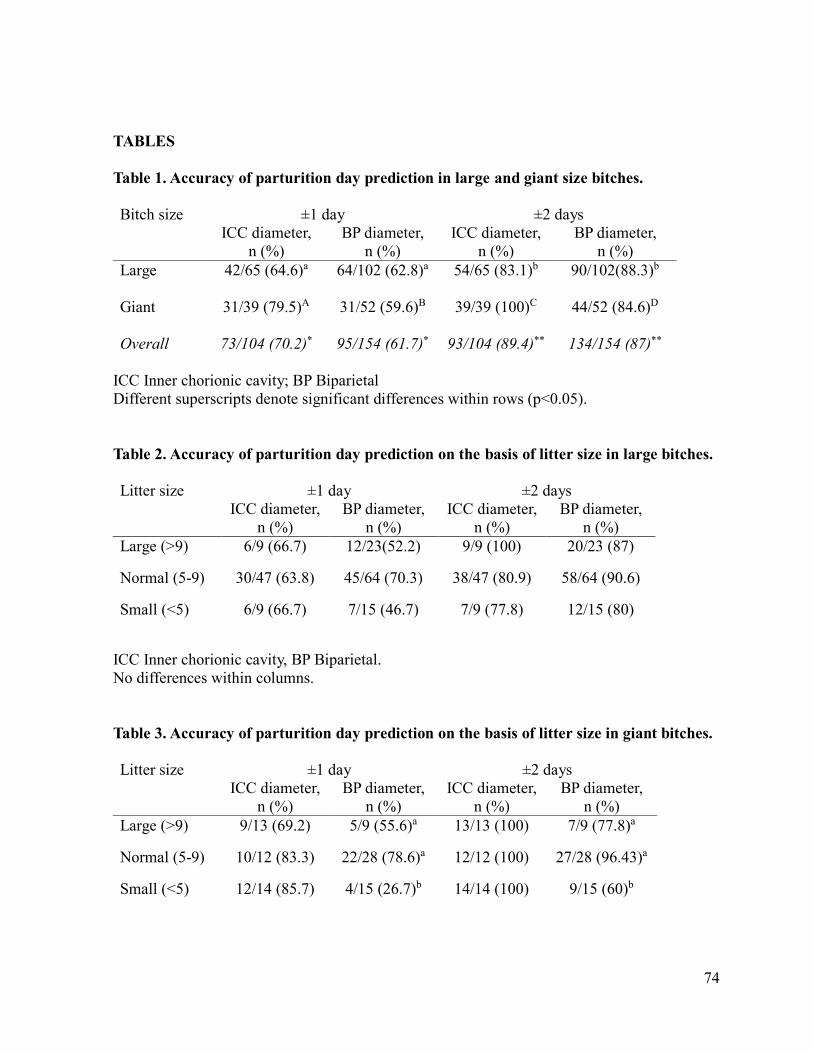
The overall accuracy ±2 days of both parameters was significantly higher
(p<0.05), than the accuracy ±1 day (Table 6). In giant bitches, the accuracy of ICC
(±1 day and ±2 days) was significantly more accurate than that of BP.
Table 6. Accuracy of parturition day prediction in large and giant size bitches.
Bitch size ±1 day ±2 days
ICC diameter,
n (%)
BP diameter,
n (%)
ICC diameter,
n (%)
BP diameter,
n (%)
Large 42/65 (64.6)a
64/102 (62.8)a
54/65 (83.1)b
90/102(88.3)b
Giant 31/39 (79.5)A
31/52 (59.6)B
39/39 (100)C
44/52 (84.6)D
Overall 73/104 (70.2)* 95/154 (61.7)* 93/104 (89.4)** 134/154 (87)**
ICC inner chorionic cavity; BP biparietal.
Different superscripts denote significant differences within rows (p<0.05).
Page 30
30
With regard to litter size, no differences (p>0.05) were observed in large
bitches for both parameters (Table 7).
Table 7. Accuracy of parturition day prediction based on litter size in large bitches.
Litter size ±1 day ±2 days
ICC diameter,
n (%)
BP diameter,
n (%)
ICC diameter,
n (%)
BP diameter,
n (%)
Large (>9) 6/9 (66.7) 12/23(52.2) 9/9 (100) 20/23 (87)
Normal (5-9) 30/47 (63.8) 45/64 (70.3) 38/47 (80.9) 58/64 (90.6)
Small (<5) 6/9 (66.7) 7/15 (46.7) 7/9 (77.8) 12/15 (80)
ICC inner chorionic cavity, BP biparietal. No significant differences within columns.
In giant bitches, only the accuracy of the prediction by BP was significantly
lower (p<0.05) in small, than normal litter size (Table 8).
Table 8. Accuracy of parturition day prediction based on litter size in giant bitches.
Litter size ±1 day ±2 days
ICC diameter,
n (%)
BP diameter,
n (%)
ICC diameter,
n (%)
BP diameter,
n (%)
Large (>9) 9/13 (69.2) 5/9 (55.6)a 13/13 (100) 7/9 (77.8)a
Normal (5-9) 10/12 (83.3) 22/28 (78.6)a 12/12 (100) 27/28 (96.43)a
Small (<5) 12/14 (85.7) 4/15 (26.7)b 14/14 (100) 9/15 (60)b
ICC inner chorionic cavity, BP biparietal.
Different superscripts denote significant differences within columns (p<0.05).
Page 31
31
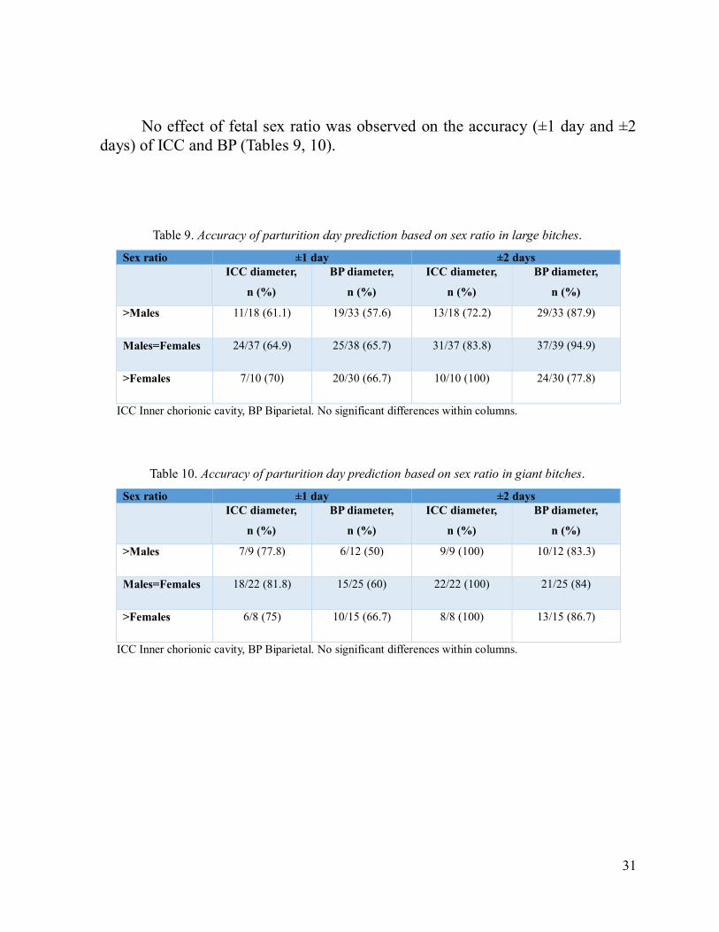
No effect of fetal sex ratio was observed on the accuracy (±1 day and ±2
days) of ICC and BP (Tables 9, 10).
Table 9. Accuracy of parturition day prediction based on sex ratio in large bitches.
Sex ratio ±1 day ±2 days
ICC diameter,
n (%)
BP diameter,
n (%)
ICC diameter,
n (%)
BP diameter,
n (%)
>Males 11/18 (61.1) 19/33 (57.6) 13/18 (72.2) 29/33 (87.9)
Males=Females 24/37 (64.9) 25/38 (65.7) 31/37 (83.8) 37/39 (94.9)
>Females 7/10 (70) 20/30 (66.7) 10/10 (100) 24/30 (77.8)
ICC Inner chorionic cavity, BP Biparietal. No significant differences within columns.
Table 10. Accuracy of parturition day prediction based on sex ratio in giant bitches.
Sex ratio ±1 day ±2 days
ICC diameter,
n (%)
BP diameter,
n (%)
ICC diameter,
n (%)
BP diameter,
n (%)
>Males 7/9 (77.8) 6/12 (50) 9/9 (100) 10/12 (83.3)
Males=Females 18/22 (81.8) 15/25 (60) 22/22 (100) 21/25 (84)
>Females 6/8 (75) 10/15 (66.7) 8/8 (100) 13/15 (86.7)
ICC Inner chorionic cavity, BP Biparietal. No significant differences within columns.
Page 32
32
Assessment of trend of FHR and FHR/MHR ratio in bitches of different
pre-gestational bodyweight during pregnancy (Paper 2)
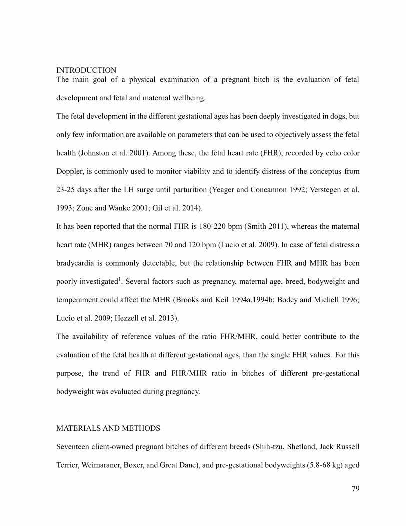
Results include eleven uncomplicated pregnancies of bitches of different
pre-gestational bodyweight (5.8-68 kg).
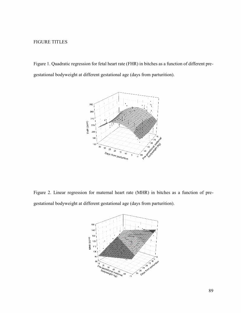
Fetal heart rates fitted a multiple quadratic regression (saddle, Fig. 7), with
significance at p<0.02 of all independent variables. Multiple r was about 0.50, a
mean value, with a low determination coefficient (r2 =0.25).
Figure 7. Quadratic regression for fetal heart rate (FHR) in bitches as a function
of different pre-gestational bodyweight at different gestational age (days from parturition).
Page 33
33
The coefficients of the regression, the significance, and the confidence
intervals are reported in Table 11. An increase of FHR was observed from 35 to
20 days before parturition. After the maximum, the curve followed a decreasing
pattern until parturition. Higher values of FHR were observed in bitches of lowest
and highest bodyweight.
Table 11. Coefficients for the quadratic regression of fetal heart rate (FHR) in bitches
of different pre-gestational bodyweight at different gestational.
FHR
coefficient
p -95% +95%
Intercept 211.6732 0.0001 202.9648 220.3817
Pre-gestational bodyweight -0.6935 0.01 -1.2262 -0.1609
Pre-gestational bodyweight 2 0.0100 0.016 0.0019 0.0182
Days from parturition 3.2479 0.0001 2.3645 4.1312
Days from parturition2 -0.0810 0.0001 -0.1061 -0.0559
Significances and 95% confidence intervals are reported.
Page 34
34
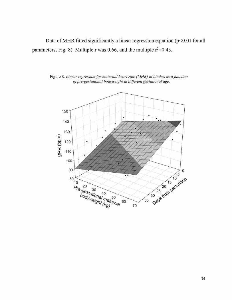
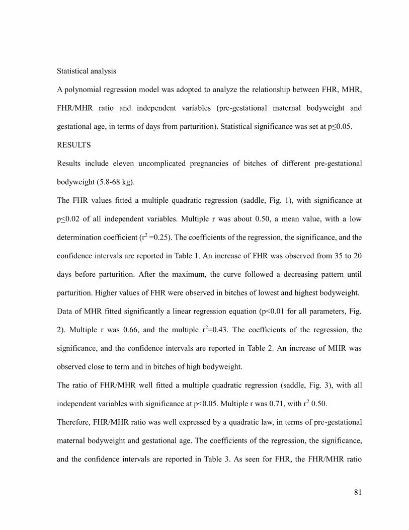
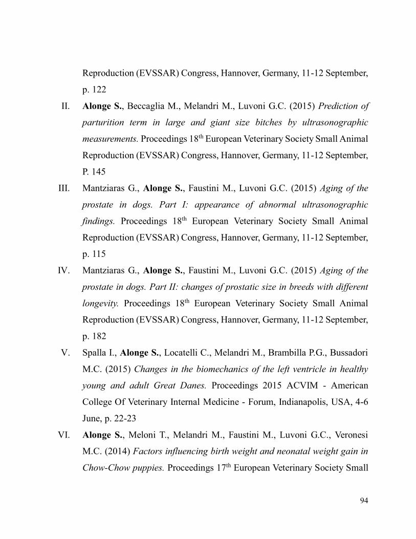
Data of MHR fitted significantly a linear regression equation (p<0.01 for all
parameters, Fig. 8). Multiple r was 0.66, and the multiple r2=0.43.
Figure 8. Linear regression for maternal heart rate (MHR) in bitches as a function
of pre-gestational bodyweight at different gestational age.
Page 35
35
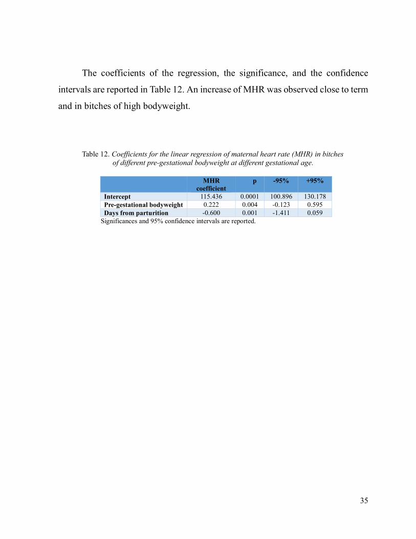
The coefficients of the regression, the significance, and the confidence
intervals are reported in Table 12. An increase of MHR was observed close to term
and in bitches of high bodyweight.
Table 12. Coefficients for the linear regression of maternal heart rate (MHR) in bitches
of different pre-gestational bodyweight at different gestational age.
MHR
coefficient
p -95% +95%
Intercept 115.436 0.0001 100.896 130.178
Pre-gestational bodyweight 0.222 0.004 -0.123 0.595
Days from parturition -0.600 0.001 -1.411 0.059
Significances and 95% confidence intervals are reported.
Page 36
36
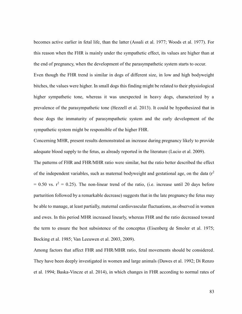
The ratio of FHR/MHR well fitted a multiple quadratic regression (saddle,
Fig. 9), with all independent variables with significance at p<0.05. Multiple r
was 0.71, with r2 0.50.
Figure 9. Polynomial quadratic regression for feto-maternal heart rate (FHR/MHR) ratio in bitches
of different pre-gestational bodyweight at different gestational age.
Page 37
37
Therefore, FHR/MHR ratio was well expressed by a quadratic law, in terms
of pre-gestational maternal bodyweight and gestational age. The coefficients of
the regression, the significance, and the confidence intervals are reported in Table
13. As seen for FHR, the FHR/MHR ratio resulted higher in low and high
bodyweight, and it reached the maximum values at about 20 days before
parturition.
Table 13. Coefficients for the quadratic regression of feto-maternal heart rate (FHR/MHR) ratio in
bitches of different pre-gestational bodyweight at different gestational age.
FHR/MHR
coefficient
p -95% +95%
Intercept 1.828405 0.0001 1.696833 1.959976
Pre-gestational bodyweight -0.013705 0.001 -0.021753 -0.005658
Pre-gestational bodyweight
2
0.000141 0.03 0.000017 0.000264
Days from parturition 0.050715 0.0001 0.037369 0.064061
Days from parturition2 -0.000986 0.0001 -0.001365 -0.000607
Significances and 95% confidence intervals are reported.
The equation derived from the quadratic regression was as follows:
z= 1.8284-0.0137x + 0.00014x2+0.05071y-0.00099y2
where z = FHR/MHR ratio, x = pre-gestational maternal bodyweight (kg), y= days
before parturition.
Page 38
38
GENERAL DISCUSSION
In clinical practice, for the proper management of pregnancy, it is crucial to
make an accurate assessment of the development, viability and health of the fetus
to allow an early detection of complications and to perform an adequate planning
of caesarean section when the pregnancy is considered at risk.
Ultrasonographic evaluation of the fetal development
The lack of specific fetal growth curves in large and giant dogs prompted
this investigation. Data (Paper 1) confirmed that ICC and BP are reliable
indicators of the gestational age, as proved by the coefficients of determination
(r2) greater than 0.9.
The overall accuracy of the prediction (±1 day and ±2 days) that ranged
between 62% and 89% is comparable with the accuracy previously obtained in
small and medium dogs by size-related growth curves (Beccaglia & Luvoni,
2006). The application of specific formulas for giant dogs increased the BP
accuracy compared to what reported in the literature by using non-specific curves
(Socha & Janowski, 2014).
The accuracy at ±2 days of both parameters was significantly higher than
that at ±1 day. This result was foreseeable as, with the extension of the time range,
there is an increase of the probability that the actual and the predicted dates will
fall within the same time range. Anyhow, 2 days between actual and predicted
parturition term might be considered safe and acceptable in clinical practice.
Page 39
39
In large bitches, as in small and medium size dogs (Beccaglia & Luvoni, 2006),
both ICC and BP were equally reliable for the prediction of the delivery day,
whereas in giant dogs the prediction should be preferably performed by ICC whose
accuracy (±1 day and ±2 days) was significantly higher than that of BP.
This result differs from what has been observed in the aforementioned study
in which no differences were found in the accuracy of these parameters in large
and giant dogs when formulas for medium dogs were applied to a small number
of observations (Socha & Janowski, 2014).
The inner chorionic cavity may be less affected than BP by individual
variability of fetal growth during late gestation (Son et al., 2001; Kutzler et al.,
2003; Beccaglia & Luvoni, 2006; Lopate, 2008; Socha & Janowski, 2014) and this
could also explain the effect of litter size on the accuracy of BP measurements.
In small litters of giant dogs, the lower BP accuracy might have been related
to the fetuses overgrowth and/or to the consequent gestation length prolongation
(Gavrilovic et al., 2008). Litter size and duration of gestation are negatively
correlated (Okkens et al., 1993; Okkens et al., 2001) and in giant dogs, where
normal numbers of fetuses is generally higher than in large dogs (Sverdrup Borge
et al., 2011), the effect of the presence of few fetuses could be more evident.
As previously observed in other sizes dogs (Beccaglia & Luvoni, 2006), the
gender did not affect the accuracy of the prediction and this result is further
confirmed by the observation that neonatal sex does not influence newborn
bodyweight (Alonge et al., 2014; Groppetti et al., 2015b).
Page 40
40
Evaluation of fetal health
Fetal heart rate (FHR) is a good indicator of fetal well-being, but its
regulatory mechanisms and variability along pregnancy are still poorly
understood. As previously mentioned, fetal distress is the main cause of FHR
alteration, but other factors such as gestational age and pre-gestational maternal
bodyweight should be taken into account.
Present results (Paper 2) demonstrate that in all bitches the FHR increased
during pregnancy until 20 days before parturition and then a reduction was
observed toward the term. These results are in agreement with those previously
reported in an experimental colony of Beagles where an increase from 214+/-13.3
to 238.2+/-16.1 bpm at day 40 of pregnancy and a decrease to 218+/-6.7 bpm close
to term were observed (Verstegen et al., 1993).
The fetal heart rate trend during pregnancy could be explained by the
dynamics of the circulatory system maturation. Both in human and veterinary
medicine, FHR trend is mainly related to the autonomic nervous system
development and activity (Verdurmen et al., 2013). The neurotransmitters,
noradrenaline and acetylcholine, influence the depolarization of the pacemaker
cells of the heart, directly affecting the heart rate. The cardiac innervation
ontogeny was studied using different techniques and in different species: chicken
embryos, laboratory animals, sheep and dogs and finally humans (Papp, 1988;
Long & Henry, 1992; Rosen & Danilo, 1992). All authors agree on the correlation
between the FHR performance and the functional activity of nervous transmission
during embryonic and fetal development. Sympathetic and parasympathetic
control of circulatory functions mature at different rates during fetal development
and the former becomes active earlier in fetal life, than the latter (Assali et al.,
Page 41
41
1977; Woods et al., 1977). For this reason, when the FHR is mainly under the
sympathetic effect, its values are higher than at the end of pregnancy, when the
development of the parasympathetic system occurs.
Even though the FHR trend is similar in dogs of different size, in low and
high bodyweight bitches, the values were higher. In small dogs this finding might
be related to their physiological higher sympathetic tone, whereas it was
unexpected in heavy dogs, characterized by a prevalence of the parasympathetic
tone (Hezzell et al.m 2013). It could be hypothesized that in these dogs the
immaturity of parasympathetic system and the early development of the
sympathetic system might be responsible of the higher FHR.
Concerning MHR, present results demonstrated an increase during pregnancy
likely to provide adequate blood supply to the fetuses, as already reported in the
literature (Lucio et al., 2009). Since the MHR could have been conditioned by the
stress induced by the restraint and by the typical temperament of breeds, dogs of
different breeds were included in the present study and the average values of three
MHRs recorded during the same exam were considered.
Some authors emphasized the mutual influence between MHR and FHR
during pathological events of the mother (shock or electrolyte imbalances) or
during administration of drugs that cross the placenta (Eisenberg de Smoler et al.,
1975). Therefore, it would seem reasonable to assume that they are also mutual
influenced under physiological conditions.
The relationship between MHR and FHR was evaluated in pregnant women
monitored for 24 hours and the results showed that the lowest and the highest
MHRs corresponded to the FHRs. This was a confirmation of the mutual influence
of the heart rates. The observation in women and sheep that FHR and MHR follow
Page 42
42
a circadian pattern has been considered a further evidence of this influence
(Patrick et al., 1981, 1982; Bocking et al., 1985).
To evaluate the effective correlation between fetal and maternal heart rate
in humans, some authors have included in their studies only pregnancies without
complications, and data were divided into three groups based on the MHR: normal
(71-92 bpm), tachycardic (107-155 bpm) and bradycardic (48-62 bpm). The
comparison of the groups showed that the FHR values remain constant regardless
of the different MHR. Conversely, it has been recently demonstrated that the
maternal and fetal cardiovascular systems, even though strictly connected, are
independent, and the fetus can respond to the maternal circulation oscillations
(Van Leeuwen et al., 2009).
Similarly to FHR, the FHR/MHR ratio has reached the maximum values at
about twenty days before birth and was higher in bitches of small and large
bodyweight. The trends of FHR and FHR/MHR ratio were similar, but the ratio
better described the effect of the independent variables, such as maternal
bodyweight and gestational age, on the data (r2 = 0.50 vs. r2 = 0.25). The non-
linear trend of the ratio, (i.e. increase until 20 days before parturition followed by
a remarkable decrease) suggests that in the late pregnancy the fetus may be able
to manage, at least partially, maternal cardiovascular fluctuations, as observed in
women and ewes. In this period MHR increased linearly, whereas FHR and the
ratio decreased toward the term to ensure the best subsistence of the conceptus
(Eisenberg de Smoler et al., 1975; Bocking et al., 1985; Van Leuwen et al., 2003;
Van Leuwen et al., 2009).
In this study, only data derived from uncomplicated pregnancies were
analyzed, and such strict recruitment had the purpose to exclude any possible
Page 43
43
alteration related to pathological conditions of the fetuses that could influence the
FHRs and therefore their correlation with the MHR.
Among factors that affect FHR and FHR/MHR ratio, fetal movements
should be considered. They have been deeply investigated in women and large
animals (Dawes et al., 1992; Di Renzo et al., 1994a; Baska-Vinze et al., 2014), in
which changes in FHR according to normal rates of movements have been
described. Few information are available in dogs in which temporary accelerations
of FHR may be associated with fetal movements (Davidson, 2003), but normal
rates of fetal activity during different pregnancy periods, have not yet been
defined. Therefore, a potential effect of fetal movements on FHR deserves further
investigations in this species. It remains also to evaluate the potential effect of the
litter size on FHR and on FHR/MHR ratio.
Page 44
44
CONCLUSIONS
In clinical practice, for the proper management of pregnancy, an accurate
assessment of fetal development and health is crucial to achieve an early diagnosis
of complications.
A highly accurate assessment of the fetal development is obtained by the
ultrasonographic fetal biometry. The enormous variety in size among different
canine breeds prompts the use of specific size-related formulae, which ensure an
accurate identification of the gestational age. The ultrasonographic evaluation of
fetal development in large and giant bitches completes and concludes the study of
size-related formulae for clinical application in all different size dogs (Paper 1).
Among the few parameters available in the literature, fetal heart rate is
generally used to objectively evaluate the fetal health during canine pregnancy.
Results of the present study suggest that the maternal pre-gestational bodyweight
and the gestational age influence both FHR and FHR/MHR ratio. The highest
significance of FHR/MHR ratio, compared to FHR, encourages the application of
this ratio to evaluate fetal health. For this reason the obtained equation for
FHR/MHR ratio (z + = 1,8284-0,0137x 0,00014x2 + 0,05071y-0,00099y2 where
z = relationship FHR/MHR, x = maternal weight before pregnancy (Kg), y = days
until parturition), that describes the trend in healthy fetuses, could be helpful in
clinical practice to derive expected values in uncomplicated pregnancies (Paper
2).
Page 45
45
FUTURE PERSPECTIVES
The definition of reliable criteria for fetal monitoring in mammals is needed
to set up therapeutic interventions along pregnancy and to possibly prevent
irreversible damages. The analysis of the recent literature shows the current
attempt to adapt guidelines and knowledges of human medicine and make them
applicable to veterinary obstetrics and gynecology.
Since the '80s in human medicine, for the evaluation of fetal well-being the
fetal biophysical profile (BPP) was introduced. The BPP consists in the ultrasound
evaluation of five fetal variables: breathing movements, body movements, muscle
tone, amniotic fluid index, heart rate reactivity with temporary acceleration in
response to body movements. To each criterion is assigned a score from 0 to 2 and
the normal overall range is set between 8 to 10 points; under 8 points the BPP
might indicate fetal stress, morbidity and perinatal mortality (Manning et al., 1980,
1984, 1985, 1986, 1987; Manning, 1990).
In veterinary medicine the application of BPP has been tested in horses and
cattle.
In the horse, with transabdominal ultrasonography, a BPP has been codified
by six different ultrasound parameters: FHR, fetal aortic diameter, fetal fluid
maximal depth, fetal activity, uteroplacental contact and thickness (Reef et al.,
1995, 1996; Reef, 1998). This profile may give indications of impending birth and
possible complications. Unfortunately, predictive values of the BPP are not as
reliable as in humans and its limited sensitivity and specificity in horses are
primarily due to the choice of the selected parameters. For instance, in clinical
Page 46
46
practice the detection of the aortic diameter and fetal breathing movements is hard
and time consuming (Palmer, 2000; Baska-Vincze et al., 2014).
The application of the BPP in cattle, as described by Reef and colleagues
(1996) in the horse, did not give the expected results. The fetal activity is linked
to the health of the conceptus, but the bovine fetus lives long periods of rest.
During the ultrasound examination, which generally lasts around 10 minutes, body
movements of healthy fetuses can be totally absent (Buczinski et al., 2009).
A different parameter that can be evaluated in cattle is the fetal weight
through the measurement of aortic diameter. In Holstein cows a growth retardation
(Intra Uterine Growth Restriction; IUGR) or the presence of large fetuses
frequently occur; these two conditions can cause complications during delivery
and inauspicious outcomes. However, for some authors, the prediction of the birth
weight of the calf from the diameter of the aorta raises some critical issues
(Buczinski et al., 2007; Baska-Vincze et al., 2014).
In dogs, fetal BPP has not yet been described. As reported above, one of the
main limits of this technique might be represented by the choice of species-
specific parameters to obtain a reliable BPP for the early detection of pregnancy
alterations. Other indicators of fetal development and health, other than ICC or
BP, FHR and FHR/MHR ratio, should be identified and analyzed in bitches.
However, it should not be disregarded that the dog is a polytocic species.
This limits the possibility of an ultrasonographic recognition of individual fetuses.
Only in early pregnancy the embryos might be individually identified, and specific
ultrasonographic indicators could be considered.
It is important to underline that in the same pregnancy suffering/dead and
healthy fetuses could be concurrently present. The decision to perform medical or
Page 47
47
surgical therapy has to be supported by a careful evaluation of risks and benefits
that might be difficult to estimate.
Page 48
48
REFERENCES
Abbott J. A. (2010) The effect of pregnancy on echocardiographic variables in
healthy bitches. Journal of Veterinary Cardiology, 12:123-128
Alonge S., Meloni T., Melandri M., et al. (2014) Factors influencing birth weight
and neonatal weight gain in Chow Chow puppies. Proceedings of the 17th
EVSSAR (European Veterinary Society for Small Animal Reproduction)
Congress, Wroclaw, Poland. p 164
Alonge S., Beccaglia M., Melandri M., et al (2015) Prediction of parturition term
in large and giant size bitches by ultrasonographic measurements. Proceedings of
the 18th EVSSAR (European Veterinary Society for Small Animal Reproduction)
Congress, Hannover, Germany. p 145
Arbeiter K. (1993) Anovulatory ovarian cycles in dogs. Journal of Reproduction
and Fertility, 47S:453-456
Assali N.S., Brinkman C. R., Woods J. R., et al. (1977) Development of
neurohumoral control of fetal, neonatal, and adult cardiovascular functions.
American Journal of Obstetrics and Gynecology, 129:748-759
Bader R.A., Bader M.E., Rose D.J., et al. (1955) Hemodynamics at rest and during
exercise in normal pregnancy as studies by cardiac catheterization. Journal of
Clinical Investigation, 34:1524-1536
Baska-Vincze B., Baska F., Szenci O. (2014) Transabdominal ultrasonographic
evaluation of fetal well-being in the late-term mare and cow. Acta Veterinaria
Hungarica, 62:439-451
Beccaglia M., Luvoni G.C. (2004) Ultrasonographic study during pregnancy of
the growth of an encephalic portion in the canine foetus. Veterinary Research
Communication, 28:161-164
Beccaglia M., Luvoni G.C. (2006) Comparison of the accuracy of two
Page 49
49
ultrasonographic measurements in predicting the parturition date in the bitch.
Journal of Small Animal Practice, 47:670–673
Beccaglia M., Faustini M., Luvoni G.C. (2008) Ultrasonographic study of deep
portion of diencephalo-telencephalic vescicle for the determination of gestational
age of the canine foetus. Reproduction in Domestic Animal, 43:367-370
Beccaglia M., Luvoni G.C. (2012) Prediction of Parturition in Dogs and Cats:
Accuracy at Different Gestational Ages. Reproduction in Domestic Animals,
47:194-196
Blanco P.G., Arias D.O., Gobello C. (2008) Doppler ultrasound in canine
pregnancy. Journal of Ultrasound in Medicine, 27:1745-1750
Blanco P.G., Arias D., Rube A., et al. (2009) An experimental model to study
resistance index and systolic/diastolic ratio uterine arteries in adverse canine
pregnancy outcome. Reproduction in Domestic Animals, 44:164-166
Blanco P.G., Tórtora M., Rodriguez R., et al. (2011) Ultrasonographic assessment
of maternal cardiac function and peripheral circulation during normal gestation
in dogs. The Veterinary Journal, 190:154-159
Bocking A.D., Harding R., Wickham P.J. (1985) Relationship between
accelerations and deceleration in heart rate and skeletal muscle activity in sheep.
Journal of Developmental Physiology, 7:47-54
Boddy K., Dawes G.S., Fisher R., et al. (1974) Foetal respiratory movements,
electrocortical and cardiovascular responces to hypoxaemia and hypercapnia in
sheep. The Journal of Physiology, 243:599-618
Bodey A.R., Michell A.R. (1996) Epidemiological study of blood pressure in
domestic dogs. The Journal of Small Animal Practice, 37:116-125
Bollwein H., Baumgartner U., Stolla R. (2002) Transrectal Doppler sonography
of uterine blood flow in cows during pregnancy. Theriogenology, 57:2053-2061
Bollwein H., Weber F., Woschée I., et al. (2004) Transrectal Doppler sonography
of uterine and umbilical blood flow during early pregnancy in mares.
Theriogenology, 60:597-605
Page 50
50
Boroffka S.A. (2005) Ultrasonographic evalutation of pre- and postnatal
development of the eyes in beagles. Veterinary Radiology and Ultrasound, 46:72-
79
Brooks V.L., Keil L.C. (1994a) Changes in the baroreflex during pregnancy in
conscious dogs: heart rate and hormonal responses. Endocrinology, 135:1894-
1901
Brooks V.L., Keil L.C. (1994b) Hemorrage decreases arterial pressure sooner in
pregnant compared with nonpregnant dogs: role of baroreflex. American Journal
of Physiology, 266:1610-1619
Buczinski S., Fecteau G., Lefebvre R.C., et al. (2007) Fetal well-being assessment
in bovine near-term gestations: current knowledge and future perspectives arising
from comparative medicine. The Canadian Veterinary Journal, 48:178-183
Buczinski S., Fecteau G., Comeau G., et al. (2009) Ultrasonographic fetal well-
being assessment, neonatal and post-partum finding a preliminary study on 10
fetuses and calves. The Canadian Veterinary Journal, 50: 261-269
Concannon P.W., McCann J.P., Temple M. (1989) Biology and endocrinology of
ovulation, pregnancy and parturition in the dog. Journal of Reproduction and
Fertility, 39S:3-25
Concannon P.W. (2000) Canine pregnancy: predicting parturition and timing
events of gestation. International Veterinary Information Service; www.ivis.org.
Concannon P.W. (2003) Ultrasound imaging of reproductive tract of the bitch.
International Veterinary Information Service; www.ivis.org.
Davidson A.P. (1998) Uterine monitoring during pregnancy. Proceedings of the
Annual Meeting of the Society for Theriogenology, Baltimore, USA. p 123-125
Davidson A.P. (2001) Uterine and fetal monitoring in the bitch. The Veterinary
Clinics of North America. Small Animal Practice, 31:305-313
Davidson A.P. (2003) Obstetrical monitoring in dogs. Veterinary Medicine, 6:508-
516
Page 51
51
Davidson A.P., Eilts B. (2006) Advanced Small Animal Reproductive Techniques.
Journal of the American Animal Hospital Association, 42:10-17
Davidson A.P. (2008) Dystocia management. Current Veterinary Therapy,
13:992-998
Dawes G.S., Fox H.E., Leduc B.M. (1972) Respiratory movements and rapid eye
movements sleep in the foetal lamb. The Journal of Physiology, 220:119-143
Dawes G.S., Moulden M., Redman C.W. (1992) Short-term fetal heart rate
variation, decelerations, and umbilical flow velocity waveforms before labor.
Obstetrics and Gynecology, 80:673-678
DeVries J.I.P., Visser G.H.A., Prechtl H.F.R. (1982) The emergence of fetal
behaviour. Early human Development, 7:301-322
Di Renzo G.C., Copray F.J.A., O’Herlihy C., et al. (1994a) Maternal-fetal
surveillance within the European community. In: Van Gejin HP, Copray FA (Eds).
A critical appraisal of fetal surveillance. Amsterdam: Elsevier Science, p 11-15.
Di Renzo G.C., Luzi G., Caserta G., et al. (1994b) The role of telemetry in
perinatal monitoring. Journal of perinatal medicine, 22:517-522
Di Salvo P., Bocci F., Zelli R., et al. (2006) Doppler evalutation of maternal and
fetal vessels during normal gestation in the bitch. Research in Veterinary Science,
81:382-388
Dubiel M., Breborowicz G.H., Gudmundsson S. (2003) Evalutation of fetal
circulation redistribution in pregnancies with absent or reversed diastolic flow in
umebelical artery. Early Human Development, 71:149-156
Eisenberg de Smoler P., Smith H.C., Karchmer S. (1975) Correlation of fetal heart
rate, maternal heart rate, and age of pregnancy. American Journal of Obstetrics
and Gynaecology, 121:62-65
England G.C.W., Allen W.E., Porter D.J. (1990) Studies on canine pregnancy
using B-mode ultrasound: develompent of the conceptus and determination of
gestational age. Journal of Small Animal Practice, 31:324–329
England G.C.W., Russo M. (2006) Ultrasonographic characteristic of early
Page 52
52
pregnancy failure in bitches. Theriogenology, 66:1694–1698
Fischer W.M. (1979) Fondamenti e valore clinico della cardiotocografia. Testo
Atlante di cardiotocografia. Piccin Editore, Milano, Italia.
Fleischer A.C., Emerson D.S. (1994) Ecografia Doppler a colori in Ostetricia e
Ginecologia. Salerno, Momento Medico s.r.l., Salerno, Italia. p 1-15
Gavrilovic B.B., Andersson K., Linde Forsberg C. (2008) Reproductive patterns
in the domestic dog--a retrospective study of the Drever breed. Theriogenology
70:783-794
Gil E.M., Garcia D.A., Giannico A.T., et al. (2014) Canine fetal heart rate: do
accelerations or decelerations predict the parturition day in bitches?
Theriogenology. 82, 933-941
Götze R. (1955) La gravidanza normale nella sua evoluzione terminale. In:
Manuale di Ostetricia, Richter G., Götze R.(Eds). Edizione italiana di Borrelli G.,
Casa editrice Palmiero, Chieti
Groppetti D., Vegetti F., Bronzo V., et al. (2015a) Breed-specific fetal biometry
and factors affecting the prediction of whelping date in the German shepherd dog.
Animal Reproduction Science 152:117-122
Groppetti D., Ravasio G., Bronzo V., et al. (2015b) The role of birth weight on
litter size and mortality within 24h of life in purebred dogs: What aspects are
involved? Animal Reproduction Science 163:112-119
Hamlin R.L. Olsen I., Smith C. R., et al. (1967) Clinical relevancy of heart rate
in the dog. Journal of the American Veterinary Medical Association, 151:30-63
Hezzell M.J., Humm K., Dennis S.G., et al. (2013) Relationships between heart
rate and age, bodyweight and breed in 10849 dogs. Journal of Small Animal
Practice, 54:318-323
Johnston S.D. (1983) Prenatal indicators of puppy viability at term. Compendium
and continuing education for practice veterinary 5:1113-1125
Page 53
53
Johnston S.D., Root Kustritz M.V., Olson P.N. (2001) Canine pregnancy. In:
Canine and Feline Theriogenology; Johnston S.D., Root Kustritz M.V., Olson
P.N(Eds), W.B. Saunders Company, Toronto, p 66-104
Khodiguian N., Jaque-Fortunato S., Wiswell R.A., et al. (1996) A comparison of
cross-sectional and longitudinal methods of assessing the influence of pregnancy
on cardiac function during exercise. Seminars in Perinatology, 20:232-241
Kim B., Son C. (2007) Time of initial detection of fetal and extra-fetal structures
by ultrasonographic examination in Miniature Schnauzer bitches. Journal of
Veterinary Medical Science, 8:289–93.
Kim Y., Travis A.J., Meyers-Wallen V.N., et al. (2007) Parturition prediction and
timing of canine pregnancy. Theriogenology, 68:1177-1182
Konde L. (1988) Diagnostic ultrasound in canine pregnancy and uterine disease.
Theriogenology, 17:247-249
Kutzler M.A., Yeager, A.E., Mohammed, H.O., et al. (2003) Accuracy of canine
parturition date prediction using fetal measurement obtained by ultrasonography.
Theriogenology 60:1309–1317
Lamm C., Makloski C.L. (2012) Current advance in gestation and parturition in
cats and dogs. The Veterinary Clinics of North America. Small Animal Practice,
42:445-456
Lévy X., Fontbonne A. (2007) Determining the optimal time of mating in bitches:
particularities. Revista Brasileira de Reproduo Animal, 31:128-134
Long W.A., Henry G.W. (1992) Autonomic and central neuroregulation of fetal
cardiovascula function. In: Fetal and neonatal physiology, W.B. Saunders
Company, Toronto, Canada. p 629-645
Lopate C. (2008) Estimation of gestational age and assessment of canine fetal
maturation using radiology and ultrasonography: a review. Theriogenology
70:397–402
Lucio C.F., Silva L.C.G., Rodrigues J.A., et al. (2009) Peripartum haemodynamic
status of bitches with normal birth or dystocia. Reproduction in Domestic
Animals, 44:133-136
Page 54
54
Luvoni G.C., Grioni A. (2000) Determination of gestational age in medium and
small size bitches using ultrasonographic fetal measurements. Journal of Small
Animal Practice, 41:292-294
Luvoni G.C., Beccaglia M. (2006) The prediction of parturition date in canine
pregnancy. Reproduction in Domestic Animals, 41:27–32
Lenard ZM, Hopper BJ, Lester NV, et al. (2007) Accuracy of prediction of canine
litter size and gestational age with ultrasound. Australian Veterinary Journal, 85:
222–225
Manning F.A., Platt L.D., Sipos L. (1980) Antenatal fetal evalutation.
Development of a fetal biophysical profile. American Journal of Obstetrics and
Gynecology, 136:787-795
Manning F.A., Lange I.R., Morrison I., et al. (1984) Fetal biophysical profile
score and the nonstress test: a comparative trial. Obstetrics and Gynaecology,
64:326-331
Manning F.A., Morrison I., Lange I.R., et al. (1985) Fetal assessment based on
fetal biophysical profile scoring: experience in 12,620 referred high-risk
pregnancies. American Journal of Obstetrics and Gynecology, 151:343-350
Manning F.A., Harman C.R., Lange I.R., et al. (1986) Fetal assessment by
biophysical profile scoring: 1985 update. European Journal of Obstetrics,
Gynecology and Reproductive Biology, 21:331-339
Manning F.A., Morrison I., Lange I.R., et al. (1987) Fetal biophysical profile
scoring: selective use of the nonstress test. American Journal of Obstetrics and
Gynecology, 156:709-712
Manning F.A. (1990) The use of sonography in the evalutation of the high-risk
pregnancy. The Radiology Clinics of North America, 28:205-216
Maulik D. (1999) Eco Doppler in ostetricia e ginecologia. Mulik D. (Eds) CIC
Edizioni Internazionali, Roma, Italia
Michel E., Spörri M., Ohlerth S., et al. (2011) Prediction of parturition date in the
bitch and queen. Reproduction in Domestic Animals, 46:926–932
Page 55
55
Miles K. (1995) Imaging pregnant dogs and cats. Compendium Continuing
Education Practice Veterinary, 17:1217-1226
Mone S. M., Sanders S. P., Colan S. D. (1996) Control mechanisms for
physiological hypertrophy of pregnancy. Circulation, 94:667-672
Moriyoshi M., Waki Y., Nakao T., et al. (1996) Observation of the growth of
beagle embryo and fetus by ultrasonography. The Journal of Veterinary Medical
Science, 58:443-445
Nautrup C.P. (1998) Doppler ultrasonography of canine maternal and fetal
arteries during normal gestation. Journal of Reproduction and Fertility, 112:301-
314
Nicolaides K.H., Rizzo G., Hecher K. (2000) Placental and Fetal Doppler. The
Parthenon Publishing Group, Casterton Hall Carnforth Lancs (UK), p. 35-66
Nyland T.G, Matton J.S. (2002) Ovaries and uterus. In: Small animal diagnostic
ultrasound. WB Saunders Co. p. 231–49
Okkens A., Hekerman T., de Vogel J., et al. (1993) Influence of litter size and
breed on variation in length of gestation in the dog. The Veterinary Quarterly
15,160-161
Okkens, A., Tenissen, J., Van Osch, W., et al. (2001) Influence of litter size and
breed on the duration of gestation in dogs. Journal of Reproduction and Fertility
57S:193-197
Palmer J. (2000) Fetal monitoring. Proceedings of the Equine Symposium and
Annual Conference, San Antonio, USA. p. 39-43
Papp J.G. (1988) Autonomic rasponses and neurohumoral control in the human
early antenatal heart. Basic Research in Cardiology, 83:2-9
Patrick J., Campbell K., Carmichael L., et al. (1981) Daily relationship between
fetal and maternal herat rates at 38 to 40 weeks of pregnancy. Canandian Medical
Association Journal, 124:1177-1178
Page 56
56
Patrick J., Campbell K., Carmichael L. (1982) Influence of maternal heart rate
and gross fetal body movements on the daily pattern of fetal heart rate near term.
American Journal of Obstetrics and Gynecology, 144:533-538
Patrick J., Challis J.R.G., Cross J. (1987) The relationship between fetal breathing
movements and prostaglandin F2 during ACTH-induced labour in sheep. Journal
of Developmental Physiology, 9:287-294
Patrick J. (1989) The physiological basis for fetal assessment. Seminars in
Perinatology, 5:403-408
Poffenbarger E., Feeney D.A. (1986) Use of gray-scale ultrasonography in the
diagnosis of reproductive disease in the bitch. Journal of the American Veterinary
Medical Association, 189:90-95
Reed K.L., Chaffin D.L., Anderson C.F. (1996) Umbelical Venous Doppler
Velocity pulsations and inferior Vena Cava Pressure Elevations in Fetal lambs.
Obstetrics and Gynaecology, 8:617-620
Reef V.B., Vaala W.E., Worth L.T. et al. (1995) Ultrasonographic evaluation of
the fetus and intrauterine environment in healthy mares during late gestation.
Veterinary Radiology and Ultrasound, 36:533–541
Reef V.B., Vaala W.E., Worth L.T., et al. (1996) Ultrasonographic assessment of
fetal well-being during late gestation: development of an equine biophysical
profile. Equine Veterinary Journal, 28:200-208
Reef V.B. (1998) Equine diagnostic ultrasound. W.B. Saunders Company,
Philadelphia. p. 560
Rendano V.T. (1983) Radiographic evalutation of fetal development in the bitch
and fetal death in the bitch and queen. Current Veterinary Therapy, 8:947-952
Robson S.C., Hunter S., Boys R., et al. (1989) Serial study of factors influencing
changes in cardiac output during human pregnancy. American Journal of
Obstetrics and Gynecology, 256:1060-1065
Root-Kustritz M.V. (2005) Pregnancy diagnosis and abnormalities of pregnancy
in the dog. Theriogenology, 64:755-765
Page 57
57
Rosen M.R., Danilo P.J. (1992) Developmental electrophysiology of the heart.
Fetal and neonatal physiology, W.B. Saunders Company, Philadelphia, 1992, vol.
1, p. 656-665
Ruckebusch Y. (1972) Development of sleep and wakefulness in the foetal lamb.
Clinical Neurophysiology, 32:119-128
Socha P., Rudowska M., Janowski T. (2012) Effectiveness of determining the
parturition date in bitches using the ultrasonographic fetometry as compared to
hormonal and cytological methods. Polish Journal of Veterinary Science, 3:447–
53
Socha P., Janowski T. (2011) Predicting the parturition date in Yorkshire Terrier
and Golden Retriever bitches using ultrasonographic fetometry. Bulletin of the
Veterinary Institute in Pulawy, 55:71–5
.Socha P., Janowski T. (2014) Predicting the parturition date in bitches of different
body weight by ultrasonographic measurements of inner chorionic cavity diameter
and biparietal diameter. Reproduction in Domestic Animals, 49:292–296
Socha P., Janowski T., Bancerz-Kisiel A. (2015) Ultrasonographic fetometry
formulas of inner chorionic cavity diameter and biparietal diameter for medium-
sized dogs can be used in giant breeds. Theriogenology, 84:779–783
Son C., Jeong K., Kim J., et al. (2001) Establishment of prediction table of
parturition day with ultrasonography in small pet dogs. The Journal of Veterinary
Medical Science, 7:715–721
Spatling L., Fallenstein F., Huck A. (1992) The variabililty of cardiopulmonary
adaptation to pregnancy at rest and during exercise. British Journal of Obstetrics
and Gynaecology, 99:1-40
Sridevi P. (2013) Ultrasonographic diagnosis and monitoring of pregnancy in the
bitch, a review. Journal of Veterinary Science, 44:1-7
Sverdrup Borge K., Tønnessen R., Nødtvedt A., et al. (2011) Litter size at birth in
purebred dogs—A retrospective study of 224 breeds. Theriogenology 75:911–919
Page 58
58
Toal R.L., Walker M.A., Henry G.A. (1986) A comparison of real-time
ultrasound, palpation and radiography in pregnancy detection and litter size
determinantion in the bitch. Veterinary Radiology, 27:102-108
Tønnessen R., Sverdrup Borge K., Nødtvedt A., et al. (2012) Canine perinatal
mortality: A cohort study of 224 breeds. Theriogenology 77:1788–1801
Valensise H., Novelli G.P., Vasapollo B., et al. (2000) Maternal cardiac systolic
and diastolic function: relationship with uteroplacental resistances. Ultrasound in
Obstetrics and Gynecology, 15:487-497
Van Leeuwen P, Geue D, Lange S, et al. (2003) Is there evidence of fetal-maternal
heart rate synchronization? BMC Physiol 3:2
Van Leeuwen P., Geue D., Thiel M., et al. (2009) Influence of paced maternal
breathing on fetal-maternal heart rate coordination. Proceedings of the National
Academy of Sciences, 106:13661-13666
Verdurmen K.M.,Renckens J., Van Laar J.O., et al. (2013) The influence of
corticosteroids on fetal heart rate variability: a systematic review of the literature.
Obstetrics and Gynecology Survey, 68:811-824
Verstegen J.P., Silvia L.D.M., Onclin K., et al. (1993) Echocardiographic study
of heart rate in dog and cat fetuses in utero. Journal of Reproduction and Fertility.
47S:174-180
Williams J.C., Rincon-Skinner T., Sun D., et al. (2007) Role of nitric oxide in the
coupling of myocardial oxygen consumption and coronary vascular dynamics
during pregnancy in the dog. American Journal of Physiology, 293:2479-2486
Woods J.R., Dandavino A., Murayama K., et al. (1977) Autonomic control of
cardiovascular functions during neonatal development and in adult sheep.
Circulation Research, 40:401-407
Yeager A.E., Concannon P.W. (1990) Association between the preovulatory
luteinizing hormone surge and the early ultrasonographic detection of pregnancy
and fetal heartbeats in Beagle dogs. Theriogenology 34:655-665
Yeager A.E., Mohammed H.O., Meyers-Wallen V., et al. (1992) Ultrasonographic
appearance of the uterus, placenta, fetus and fetal membranes throughout
Page 59
59
accurately timed pregnancy in Beagles. American Journal of Veterinary Research
3:342–351
Zone M.A., Wanke M.M. (2001) Diagnosis of canine fetal health by
ultrasonography. Journal of Reproduction and Fertility 57S:215-219
Page 60
60
SUMMARY
Nowadays ultrasonography represents the gold standard method for the
evaluation of canine fetal development and health.
In dogs, the gestational age is estimated by the ultrasonographic
measurements of extra-fetal and fetal structures. The fetal development has been
deeply investigated in small and medium dogs, whereas only few information are
available for large and giant size bitches, even though they are very popular canine
breeds.
The measurements of the inner chorionic cavity (ICC) in early pregnancy,
and the biparietal diameter (BP) in late pregnancy are commonly used in clinical
practice. Both ICC and BP are highly reliable when size-related specific formulae
are applied, thus, specific equations for large and giant dogs would allow the most
accurate prediction of parturition term.
Only few parameters are available to objectively assess the fetal health
during canine pregnancy. Among them, the fetal heart rate (FHR) is generally
used, but the relationship between FHR and maternal heart rate (MHR) has been
poorly investigated. The availability of reference values of the ratio FHR/MHR,
could better contribute to the evaluation of the fetal health at different gestational
ages, than the single FHR values.
The aims of this study were 1) to derive the growth curves of extra-fetal and
fetal structures (ICC and BP) in large and giant size bitches and to evaluate their
accuracy (Paper 1); 2) to evaluate the trend of FHR and of the ratio FHR/MHR in
bitches of different pre-gestational bodyweight (Paper 2).
Page 61
61
Present results showed a significant relationship between days before
parturition and ICC or BP in large and giant size bitches. The overall accuracy ±2
days of both parameters was significantly higher than the accuracy ±1 day. Only
in giant bitches, the BP accuracy of the prediction was significantly lower in small
than normal litter size. As previously observed in other sizes dogs, the gender did
not affect the accuracy of the prediction.
The second study demonstrated that both FHR and FHR/MHR significantly
fitted a multiple quadratic regression for all independent variables. They both
resulted higher in low and high bodyweight, and reached the maximum values at
about 20 days before parturition. Maternal pre-gestational bodyweight and the
gestational age influenced both FHR and FHR/MHR.
The highest significance of FHR/MHR, compared to FHR, encourages the
application of this ratio, to evaluate fetal health. The derived equation for
FHR/MHR ratio, that describes the trend in healthy fetuses, could be helpful in
clinical practice to derive expected values in uncomplicated pregnancies.
Page 62
62
PAPER 1
PREDICTION OF PARTURITION TERM IN LARGE AND GIANT
SIZE BITCHES BY ULTRASONOGRAPHIC MEASUREMENTS
(submitted to Journal of Small Animal Practice)
Page 63
63
PREDICTION OF PARTURITION TERM
IN LARGE AND GIANT SIZE BITCHES BY ULTRASONOGRAPHIC MEASUREMENTS
Salvatore ALONGE, DVM1, Michela BECCAGLIA, DVM, PhD, DECAR2, Monica
MELANDRI, DVM1, Gaia Cecilia LUVONI, DVM, PhD, DECAR1
1Dipartimento di Scienze Veterinarie per la Salute, la Produzione Animale e la Sicurezza
Alimentare, Università degli Studi di Milano, Via Celoria, 10 – 20133 Milano Italy;
2Ambulatorio Veterinario “Beccaglia”, Lissone (MI), Italy.
e-mail: [email protected] , [email protected] ,
[email protected] , [email protected]
Corresponding Author:
Gaia Cecilia Luvoni
Department of Health, Animal Science and Food Safety
University of Milan.
Via Celoria 10, 20133 Milan, Italy.
Phone +39 02.503.18147
Fax +39 02.503.18148
e-mail [email protected]
Author contributions
SA and GCL contributed to design the study, collect and analyze the data, and draft the paper.
MB and MM contributed to collect the data. All authors have approved the final version.
Conflict of interest
None of the authors of this article has a financial or personal relationship with other people or
organizations that could inappropriately influence or bias the content of the paper.
Page 64
64
STRUCTURED SUMMARY
Objectives. To derive the growth curves of the inner chorionic cavity (ICC) and the fetal
biparietal diameter (BP) in large and giant size bitches by ultrasonographic measurements. To
evaluate their accuracy in the prediction of parturition and the effect of litter size and fetal sex
ratio.
Methods. Ultrasonographic examinations were performed in 8 large (26-40kg) and 9 giant
(>40kg) pregnant bitches. The relationship between ICC or BP growth and days to parturition
was analyzed by linear regression and the equations derived from growth curves were applied
to predict the day of delivery. The accuracy of the prediction (±1 day and ±2 days) and the effect
of litter size and sex ratio were assessed in large and giant bitches with unknown breeding dates.
Results. The results showed a significant relationship between days before parturition and ICC
or BP. The overall accuracy at ±2 days was higher, than at ±1 day. In giant bitches, the accuracy
of the prediction by BP was significantly lower in small, than normal litter size. No effect of
fetal sex ratio was observed.
Clinical significance. Large and giant dogs are well represented in canine population and
specific equations to predict the day of parturition might be useful in clinical practice.
Running head: ultrasonographic fetal biometry in large and giant bitches
Keywords: dog, ultrasonography, fetal biometry.
Page 65
65
INTRODUCTION
Ultrasonographic fetal biometry is commonly applied for the assessment of fetal growth along
pregnancy. In medium and small size dogs, the fetal development has been deeply investigated,
and the measurement of extra-fetal and fetal structures has allowed the prediction of parturition
day through equations generated from the growth curves of selected parameters (see reviews:
Luvoni and Beccaglia 2006; Kim et al. 2007; Lopate 2008).
In the one hundred most popular canine breeds (Sverdrup Borge et al. 2011; Tønnessen et al.
2012), large and giant size bitches are very well represented. For these dogs, specific formulas
for the evaluation of fetal growth are not yet available. Kutzler and colleagues (2003) suggested
the use of a correction factor to adjust the difference between actual and predicted parturition
date obtained with previously published equations for dogs of smaller size (England et al. 1990;
Yeager et al. 1992).
Although the formulas for medium dogs (Luvoni and Grioni, 2000) have been also used in large
and giant breeds (Socha and Janowski 2014; Socha et al. 2015), specific equations for these
dogs would allow the most accurate prediction of parturition term (Michel et al. 2011; Socha
and Janowski 2014; Socha et al. 2015).
Among several ultrasonographic parameters, the inner diameter of chorionic cavity (ICC) and
the biparietal diameter (BP) provided a highly accurate prediction in small and medium size
dogs (Luvoni and Grioni 2000; Kutzler et al. 2003; Beccaglia and Luvoni 2006; Socha and
Janowski 2014). Therefore, the aim of this study was to derive the growth curves of ICC and
BP in large and giant size bitches and to evaluate their accuracy. The effects of litter size and
fetal sex ratio on the accuracy of the prediction were also investigated.
Page 66
66
MATERIALS AND METHODS
Growth curves of ICC and BP in large and giant size bitches.
Eight large size (26-40 kg) bitches (Bergamasco Shepherd, Boxer, Doberman, German
Shepherd, and Old English Shepherd) and 9 giant size (>40 kg) bitches (Great Dane, Bernese
Mountain Dog, and Newfoundland), aged between 2 and 8 years presented to the Department
for breeding management and pregnancy evaluation, were included in this study. Informed
owner consent was obtained.
All bitches were healthy at the physical examination. For breeding management, the day of the
ovulation was considered to be when plasma progesterone concentration ranged between 4-10
ng/ml (Arbeiter 1993; Lévy and Fontbonne 2007), as evaluated using an Enzyme Linked
Fluorescent Assay (MiniVidas, BioMerieux, Marcy l'Etoile, France). Serial ultrasonographic
exams were performed weekly from day 20 after mating until parturition. Bitches were
positioned in lateral recumbency, transmission gel was applied, and two-dimensional, gray-
scale, real-time ultrasound images were produced using a 7.5 MHz microconvex probe
(SonoAce 8800, Medison Co. Ltd., Seoul, Korea). During early pregnancy, inner diameter of
chorionic cavity, and in late pregnancy the biparietal diameter were measured. At least three
measurements of ICC or BP, according to the gestation period, were recorded and the mean
values were calculated. The time of actual parturition, the litter size, and the sex of the puppies
were reported by the owners.
Statistical analysis: the relationship between ICC or BP growth and days before parturition was
analyzed by a linear regression model. The growth equations for both parameters were derived
Page 67
67
as y=a+bx (y=days before parturition, x=measurement in mm of ICC or BP, a=intercept
coefficient and b=first order coefficient) and the regression coefficients were analyzed by the
Student's T test (p<0.05). Data analysis was performed with the software Stat Plus 2009.
Accuracy of the ICC and BP for the prediction of parturition day in large and giant size bitches.
To assess the accuracy of the prediction, measurements of ICC and BP were performed in 65
and 102 ultrasound examinations of large size bitches and in 39 and 52 of giant size bitches with
unknown breeding dates. As previously reported, the prediction was considered accurate when
the difference between actual and predicted parturition date was within ±1 day and ±2 days
(Beccaglia and Luvoni, 2006). To evaluate the effect of litter size on the accuracy, data were
grouped for small (<5 pups), normal (5-9 pups) and large (>9 pups) litters in large and giant
bitches (Beccaglia et al., 2008). Moreover, predictions within ±1 day and ±2 days were analyzed
on the basis of fetal sex ratio in terms of numerical prevalence (>2) of one gender.
Statistical analysis: data were analyzed by Chi-Square test and the level of significance was set
at p<0.05.
RESULTS
Growth curves of ICC and BP in large and giant size bitches.
The regression analysis resulted in a significant relationship between days before parturition and
ICC or BP (p<0.001). The derived equations for the prediction of parturition day were:
in large size bitches ICC: y = (x-105.1)/2.5 (r2=0.92), and BP: y = (x-30)/0.8 (r2=0.99);
in giant size bitches ICC: y = (x-88.1)/1.9 (r2=0.97), and BP: y = (x-29)/0.7 (r2=0.97).
Page 68
68
Accuracy of the ICC and BP for the prediction of parturition day in large and giant size bitches.
The overall accuracy ±2 days of both parameters was significantly higher (p<0.05), than the
accuracy ±1 day (Table 1). In giant bitches, the accuracy of ICC (±1 day and ±2 days) was
significantly more accurate than that of BP.
With regard to litter size, no differences (p>0.05) were observed in large bitches for both
parameters (Table 2). In giant bitches, only the accuracy of the prediction by BP was
significantly lower (p<0.05) in small, than normal litter size (Table 3).
No effect of fetal sex ratio was observed on the accuracy (±1 day and ±2 days) of ICC and BP
(Table 4, 5).
DISCUSSION
In bitches, a highly accurate prediction of the parturition day is crucial for the proper breeding
management. The lack of specific fetal growth curves in large and giant dogs, prompted this
investigation. Data showed that ICC and BP are reliable indicators of the gestational age, as
proved by the coefficients of determination (r2) greater than 0.9.
The overall accuracy of the prediction (±1 day and ±2 days) that ranged between 62% and 89%
is comparable with the accuracy previously obtained in small and medium dogs by size-related
growth curves (Beccaglia and Luvoni 2006). The application of specific formulas for giant dogs
increased the BP accuracy compared to what reported in the literature by using non-specific
curves (Socha and Janowski 2014).
The accuracy at ±2 days of both parameters was significantly higher than that at ±1 day. This
result was foreseeable as, with the extension of the time range, there is an increase of the
Page 69
69
probability that the actual and the predicted dates will fall within the same time range. Anyhow,
2 days between actual and predicted parturition term might be considered safe and acceptable
in clinical practice.
In large bitches, as in small and medium size dogs (Beccaglia and Luvoni 2006), both ICC and
BP were equally reliable for the prediction of the delivery day, whereas in giant dogs the
prediction should be preferably performed by ICC whose accuracy (±1 day and ±2 days) was
significantly higher than that of BP.
This result differs from what has been observed in the aforementioned study in which no
differences were found in the accuracy of these parameters in large and giant dogs when
formulas for medium dogs were applied to a small number of observations (Socha and Janowski
2014).
The inner chorion cavity may be less affected than BP by individual variability of fetal growth
during late gestation (Son et al. 2001; Kutzler et al. 2003; Beccaglia and Luvoni 2006; Lopate
2008; Socha and Janowski 2014) and this could also explain the effect of litter size on the
accuracy of BP measurements.
In small litters of giant dogs, the lower BP accuracy might have been related to the fetuses
overgrowth and/or to the consequent gestation length prolongation (Gavrilovic et al. 2008).
Litter size and duration of gestation are negatively correlated (Okkens et al. 1993; Okkens et al.
2001) and in giant dogs, where normal numbers of fetuses is generally higher than in large dogs
(Sverdrup Borge et al. 2011), the effect of the presence of few fetuses could be more evident.
Page 70
70
As previously observed in other sizes dogs (Beccaglia and Luvoni 2006), the gender did not
affect the accuracy of the prediction and this result is further confirmed by the observation that
neonatal sex does not influence newborn bodyweight (Alonge et al. 2014; Groppetti et al. 2015).
In clinical practice, when there is a history of dystocia, the elective cesarean section should be
planned. A prediction of the term as much accurate as possible is needed to identify the proper
time of surgery and to prevent neonatal loss due to fetal immaturity caused by a pre-term
cesarean section. In case of mismating or when the maternal health is threatened, the evaluation
of gestational age is also required to apply the more safe and efficient protocol for pregnancy
interruption.
A highly accurate prediction of the gestational term might be obtained by a careful estrus
monitoring, but often the information about estrous cycle are unavailable and the
ultrasonographic fetal biometry is a valid alternative. The enormous variety in size among
different canine breeds prompts the use of specific size-related formulas which ensure an
accurate identification of the gestational age.
Page 71
71
REFERENCES
Alonge, S., Meloni, T., Melandri, M., et al. (2014) Factors influencing birth weight and
neonatal weight gain in Chow Chow puppies. Proceedings of the European Veterinary Society
for Small Animal Reproduction. September 26, Wroclaw, Poland. p 164
Arbeiter, K. (1993) Anovulatory ovarian cycles in dogs. Journal of Reproduction and Fertility
47S, 453-456
Beccaglia, M., Luvoni, G.C. (2006) Comparison of the accuracy of two ultrasonographic
measurements in predicting the parturition date in the bitch. Journal of Small Animal Practice
47, 670–673
Beccaglia, M., Faustini, M., Luvoni, G.C. (2008) Ultrasonographic study of deep portion of
diencephalo-telencephalic vesicle for the determination of gestational age of the canine fetus.
Reproduction in Domestic Animals 43, 367-370
England, G.C.W., Allen, W.E., Porter, D.J. (1990) Studies on canine pregnancy using B-mode
ultrasound: develompent of the conceptus and determination of gestational age. Journal of
Small Animal Practice 31, 324–329
Gavrilovic, B.B., Andersson, K., Linde Forsberg, C. (2008) Reproductive patterns in the
domestic dog--a retrospective study of the Drever breed. Theriogenology 70, 783-794
Groppetti, D., Ravasio, G., Bronzo, V., et al. (2015) The role of birth weight on litter size and
mortality within 24h of life in purebred dogs: What aspects are involved? Animal
Reproduction Science 163, 112-119.
Kim, Y., Travis, A.J., Meyers-Wallen, W.N. (2007) Parturition prediction and timing of canine
pregnancy. Theriogenology 68, 1177–1182
Page 72
72
Kutzler, M.A., Yeager, A.E., Mohammed, H.O., et al. (2003) Accuracy of canine parturition
date prediction using fetal measurement obtained by ultrasonography. Theriogenology 60,
1309–1317
Lévy, X., Fontbonne, A. (2007) Determining the optimal time of mating in bitches:
particularities. Revista Brasileira de Reproduo Animal 31, 128-134
Lopate, C. (2008) Estimation of gestational age and assessment of canine fetal maturation
using radiology and ultrasonography: a review. Theriogenology 70, 397–402
Luvoni, G.C., Beccaglia, M. (2006) The prediction of parturition date in canine pregnancy.
Reproduction in Domestic Animals 41, 27–32
Luvoni, G.C., Grioni, A. (2000) Determination of gestational age in medium and small size
bitches using ultrasonographic fetal measurements. Journal of Small Animal Practice 41, 292–
294
Michel, E., Spörri, M., Ohlerth, S., et al. (2011) Prediction of parturition date in the bitch and
queen. Reproduction in Domestic Animals 46, 926–932
Okkens, A., Hekerman, T., de Vogel, J., et al. (1993) Influence of litter size and breed on
variation in length of gestation in the dog. The Veterinary Quarterly 15,160-161
Okkens, A., Tenissen, J., Van Osch, W., et al. (2001) Influence of litter size and breed on the
duration of gestation in dogs. Journal of Reproduction and Fertility 57s, 193-197
Socha, P., Janowski, T. (2014) Predicting the parturition date in bitches of different body
weight by ultrasonographic measurements of inner chorionic cavity diameter and biparietal
diameter. Reproduction in Domestic Animals 49, 292–296
Page 73
73
Socha, P., Janowski, T., Bancerz-Kisiel, A. (2015) Ultrasonographic fetometry formulas of
inner chorionic cavity diameter and biparietal diameter for medium-sized dogs can be used in
giant breeds. Theriogenology 84, 779–783
Son, C., Jeong, K., Kim, J., et al. (2001) Establishment of prediction table of parturition day
with ultrasonography in small pet dogs. The Journal of Veterinary Medical Science 7, 715–721
Sverdrup Borge, K., Tønnessen, R., Nødtvedt, A., et al. (2011) Litter size at birth in purebred
dogs—A retrospective study of 224 breeds. Theriogenology 75, 911–919
Tønnessen, R., Sverdrup Borge, K., Nødtvedt, A., et al. (2012) Canine perinatal mortality: A
cohort study of 224 breeds. Theriogenology 77, 1788–1801
Yeager, A.E., Mohammed, H.O., Meyers-Wallen, V., et al. (1992) Ultrasonographic
appearance of the uterus, placenta, fetus and fetal membranes throughout accurately timed
pregnancy in Beagles. American Journal of Veterinary Research 3, 342–351
Page 74
74
TABLES
Table 1. Accuracy of parturition day prediction in large and giant size bitches.
Bitch size ±1 day ±2 days
ICC diameter,
n (%)
BP diameter,
n (%)
ICC diameter,
n (%)
BP diameter,
n (%)
Large 42/65 (64.6)a
64/102 (62.8)a
54/65 (83.1)b
90/102(88.3)b
Giant 31/39 (79.5)A
31/52 (59.6)B
39/39 (100)C
44/52 (84.6)D
Overall 73/104 (70.2)* 95/154 (61.7)* 93/104 (89.4)** 134/154 (87)**
ICC Inner chorionic cavity; BP Biparietal
Different superscripts denote significant differences within rows (p<0.05).
Table 2. Accuracy of parturition day prediction on the basis of litter size in large bitches.
Litter size ±1 day ±2 days
ICC diameter,
n (%)
BP diameter,
n (%)
ICC diameter,
n (%)
BP diameter,
n (%)
Large (>9) 6/9 (66.7) 12/23(52.2) 9/9 (100) 20/23 (87)
Normal (5-9) 30/47 (63.8) 45/64 (70.3) 38/47 (80.9) 58/64 (90.6)
Small (<5) 6/9 (66.7) 7/15 (46.7) 7/9 (77.8) 12/15 (80)
ICC Inner chorionic cavity, BP Biparietal.
No differences within columns.
Table 3. Accuracy of parturition day prediction on the basis of litter size in giant bitches.
Litter size ±1 day ±2 days
ICC diameter,
n (%)
BP diameter,
n (%)
ICC diameter,
n (%)
BP diameter,
n (%)
Large (>9) 9/13 (69.2) 5/9 (55.6)a 13/13 (100) 7/9 (77.8)a
Normal (5-9) 10/12 (83.3) 22/28 (78.6)a 12/12 (100) 27/28 (96.43)a
Small (<5) 12/14 (85.7) 4/15 (26.7)b 14/14 (100) 9/15 (60)b
Page 75
75
ICC Inner chorionic cavity, BP Biparietal.
Different superscripts denote significant differences within columns (p<0.05).
Table 4. Accuracy of parturition day prediction on the basis of sex ratio in large bitches.
Sex ratio ±1 day ±2 days
ICC diameter,
n (%)
BP diameter,
n (%)
ICC diameter,
n (%)
BP diameter,
n (%)
>Males 11/18 (61.1) 19/33 (57.6) 13/18 (72.2) 29/33 (87.9)
Males=Females 24/37 (64.9) 25/38 (65.7) 31/37 (83.8) 37/39 (94.9)
>Females 7/10 (70) 20/30 (66.7) 10/10 (100) 24/30 (77.8)
ICC Inner chorionic cavity, BP Biparietal.
No significant differences within columns.
Table 5. Accuracy of parturition day prediction on the basis of sex ratio in giant bitches.
Sex ratio ±1 day ±2 days
ICC diameter,
n (%)
BP diameter,
n (%)
ICC diameter,
n (%)
BP diameter,
n (%)
>Males 7/9 (77.8) 6/12 (50) 9/9 (100) 10/12 (83.3)
Males=Females 18/22 (81.8) 15/25 (60) 22/22 (100) 21/25 (84)
>Females 6/8 (75) 10/15 (66.7) 8/8 (100) 13/15 (86.7)
ICC Inner chorionic cavity, BP Biparietal.
No differences within columns.
Page 76
76
PAPER 2
FETO-MATERNAL HEART RATE RATIO
IN PREGNANT BITCHES:
EFFECT OF GESTATIONAL AGE AND MATERNAL SIZE
(submitted to Reproduction in Domestic Animals)
Page 77
77
Università degli Studi di Milano, via Celoria 10, 20133 Milano, Italy
FETO-MATERNAL HEART RATE RATIO IN PREGNANT BITCHES: EFFECT OF
GESTATIONAL AGE AND MATERNAL SIZE.
Salvatore ALONGE1, Marta MAURI1, Massimo FAUSTINI 2, Gaia Cecilia LUVONI1
1Department of Health, Animal Science and Food Safety, 2Department of Veterinary Science
and Public Health.
Page 78
78
ABSTRACT
Few information are available on parameters that can be used to objectively assess the fetal
health during canine pregnancy. To identify a reliable parameter for the evaluation of fetal
wellbeing, the effect of pre-gestational maternal bodyweight and gestational age on fetal heart
rate (FHR) and on feto-maternal heart rate ratio (FHR/MHR) was investigated. Seventeen client-
owned pregnant bitches of different pre-gestational maternal bodyweight were examined by
serial echo color Doppler. Only data from 11 uncomplicated pregnancies were included in the
statistical analysis. The relationship between FHR, and FHR/MHR, and independent variables
was analyzed by polynomial regression (p≤0.05). The FHR and the FHR/MHR significantly
fitted a multiple quadratic regression for all independent variables. They both increased from 35
to 20 days before parturition, then a decreasing pattern followed. Higher values of both
parameters were observed in bitches of lowest and highest bodyweight.
Patterns of FHR and FHR/MHR were similar, but the ratio better describes the effect of the
independent variables on the data. Thus, the highest significance of FHR/MHR compared to
FHR alone, encourages the application of this ratio to evaluate fetal wellbeing. The equation
derived by the regression analysis of FHR/MHR could be applied in clinical practice to obtain
its expected values in healthy pregnancies.
ABRIDGED TITLE
Feto-maternal heart rate ratio in canine pregnancy.
KEY WORDS
dog, pregnancy, heart rate, fetal health
Page 79
79
INTRODUCTION
The main goal of a physical examination of a pregnant bitch is the evaluation of fetal
development and fetal and maternal wellbeing.
The fetal development in the different gestational ages has been deeply investigated in dogs, but
only few information are available on parameters that can be used to objectively assess the fetal
health (Johnston et al. 2001). Among these, the fetal heart rate (FHR), recorded by echo color
Doppler, is commonly used to monitor viability and to identify distress of the conceptus from
23-25 days after the LH surge until parturition (Yeager and Concannon 1992; Verstegen et al.
1993; Zone and Wanke 2001; Gil et al. 2014).
It has been reported that the normal FHR is 180-220 bpm (Smith 2011), whereas the maternal
heart rate (MHR) ranges between 70 and 120 bpm (Lucio et al. 2009). In case of fetal distress a
bradycardia is commonly detectable, but the relationship between FHR and MHR has been
poorly investigated1. Several factors such as pregnancy, maternal age, breed, bodyweight and
temperament could affect the MHR (Brooks and Keil 1994a,1994b; Bodey and Michell 1996;
Lucio et al. 2009; Hezzell et al. 2013).
The availability of reference values of the ratio FHR/MHR, could better contribute to the
evaluation of the fetal health at different gestational ages, than the single FHR values. For this
purpose, the trend of FHR and FHR/MHR ratio in bitches of different pre-gestational
bodyweight was evaluated during pregnancy.
MATERIALS AND METHODS
Seventeen client-owned pregnant bitches of different breeds (Shih-tzu, Shetland, Jack Russell
Terrier, Weimaraner, Boxer, and Great Dane), and pre-gestational bodyweights (5.8-68 kg) aged
Page 80
80
between 2 and 7 years presented to the Department for breeding management and pregnancy
evaluation, were included in this study. Informed owner consent was obtained.
All bitches were healthy at the physical examination and with no history and signs of cardiac
diseases. For breeding management, the day of the ovulation was considered to be when plasma
progesterone concentrations ranged between 4-10 ng ⁄ml (Arbeiter 1993; Levy and Fontbonne
2007), as evaluated using an Enzyme Linked Fluorescent Assay1.
According to owner’s availability, ultrasound examinations were performed in five bitches twice
a week from day 21 after ovulation, and in 12 bitches at week 4, 7, and 9 of pregnancy.
Two-dimensional, gray-scale, real-time ultrasound and echo color Doppler images were
produced using a 7.5 MHz microconvex probe2. The bitches were positioned in lateral
recumbency, transmission gel was applied and MHR was evaluated at the level of the aortic
valve for three times (at the beginning of the examination, after 10 minutes and at the end of the
examination) to reduce and control the stress-effect induced by the restraint. Fetuses’ normal
development was assessed by the measurement of ultrasonographic extra-fetal and fetal
parameters (Luvoni and Grioni 2000; Alonge et al. 2015).
The FHR values of at least three different fetuses (in litter size >3) were recorded in each
examination.
The day of parturition and the neonatal survival were reported by the owners. Only data from
uncomplicated pregnancies with no evidence of embryo, fetal or neonatal loss were included in
the statistical analysis.
1
VIDAS®, Biomerieux, Marcy l’Etoile, France 2 SonoAce 8800, Medison Co. Ltd., Seoul, Korea
Page 81
81
Statistical analysis
A polynomial regression model was adopted to analyze the relationship between FHR, MHR,
FHR/MHR ratio and independent variables (pre-gestational maternal bodyweight and
gestational age, in terms of days from parturition). Statistical significance was set at p≤0.05.
RESULTS
Results include eleven uncomplicated pregnancies of bitches of different pre-gestational
bodyweight (5.8-68 kg).
The FHR values fitted a multiple quadratic regression (saddle, Fig. 1), with significance at
p<0.02 of all independent variables. Multiple r was about 0.50, a mean value, with a low
determination coefficient (r2 =0.25). The coefficients of the regression, the significance, and the
confidence intervals are reported in Table 1. An increase of FHR was observed from 35 to 20
days before parturition. After the maximum, the curve followed a decreasing pattern until
parturition. Higher values of FHR were observed in bitches of lowest and highest bodyweight.
Data of MHR fitted significantly a linear regression equation (p<0.01 for all parameters, Fig.
2). Multiple r was 0.66, and the multiple r2=0.43. The coefficients of the regression, the
significance, and the confidence intervals are reported in Table 2. An increase of MHR was
observed close to term and in bitches of high bodyweight.
The ratio of FHR/MHR well fitted a multiple quadratic regression (saddle, Fig. 3), with all
independent variables with significance at p<0.05. Multiple r was 0.71, with r2 0.50.
Therefore, FHR/MHR ratio was well expressed by a quadratic law, in terms of pre-gestational
maternal bodyweight and gestational age. The coefficients of the regression, the significance,
and the confidence intervals are reported in Table 3. As seen for FHR, the FHR/MHR ratio
Page 82
82
resulted higher in low and high bodyweight, and it reached the maximum values at about 20
days before parturition.
The equation derived from the quadratic regression was as follows:
z= 1.8284-0.0137x + 0.00014x2+0.05071y-0.00099y2,
where z = FHR/MHR ratio, x = pre-gestational maternal bodyweight (kg), y= days before
parturition.
DISCUSSION
The FHR is a good indicator of fetal wellbeing, but its regulatory mechanisms and variability
along pregnancy are still poorly understood. As previously mentioned, fetal distress is the main
cause of FHR alteration, but other factors such as gestational age and pre-gestational maternal
bodyweight should be taken into account.
Present results demonstrate that in all bitches the FHR increased during pregnancy until 20 days
before parturition and then a reduction was observed toward the term. These results are in
agreement with those previously reported in an experimental colony of Beagles where an
increase from 214+/-13.3 to 238.2+/-16.1 bpm at day 40 of pregnancy and a decrease to 218+/-
6.7 bpm close to term were observed (Verstegen et al. 1993).
Such condition could be explained by the dynamics of the circulatory system maturation. Both
in human and in veterinary medicine FHR trend is mainly related to the autonomic nervous
system development and activity (Verdurmen et al. 2013). Sympathetic and parasympathetic
control of circulatory functions mature at different rates during fetal development and the former
Page 83
83
becomes active earlier in fetal life, than the latter (Assali et al. 1977; Woods et al. 1977). For
this reason when the FHR is mainly under the sympathetic effect, its values are higher than at
the end of pregnancy, when the development of the parasympathetic system starts to occur.
Even though the FHR trend is similar in dogs of different size, in low and high bodyweight
bitches, the values were higher. In small dogs this finding might be related to their physiological
higher sympathetic tone, whereas it was unexpected in heavy dogs, characterized by a
prevalence of the parasympathetic tone (Hezzell et al. 2013). It could be hypothesized that in
these dogs the immaturity of parasympathetic system and the early development of the
sympathetic system might be responsible of the higher FHR.
Concerning MHR, present results demonstrated an increase during pregnancy likely to provide
adequate blood supply to the fetus, as already reported in the literature (Lucio et al. 2009).
The patterns of FHR and FHR/MHR ratio were similar, but the ratio better described the effect
of the independent variables, such as maternal bodyweight and gestational age, on the data (r2
= 0.50 vs. r2 = 0.25). The non-linear trend of the ratio, (i.e. increase until 20 days before
parturition followed by a remarkable decrease) suggests that in the late pregnancy the fetus may
be able to manage, at least partially, maternal cardiovascular fluctuations, as observed in women
and ewes. In this period MHR increased linearly, whereas FHR and the ratio decreased toward
the term to ensure the best subsistence of the conceptus (Eisenberg de Smoler et al. 1975;
Bocking et al. 1985; Van Leeuwen et al. 2003, 2009).
Among factors that affect FHR and FHR/MHR ratio, fetal movements should be considered.
They have been deeply investigated in women and large animals (Dawes et al. 1992; Di Renzo
et al. 1994; Baska-Vincze et al. 2014), in which changes in FHR according to normal rates of
Page 84
84
movements have been described. Few information are available in dogs in which temporary
accelerations of FHR may be associated with fetal movements (Davidson 2003), but normal
rates of fetal activity during different pregnancy periods, have not yet been defined. Therefore,
a potential effect of fetal movements on FHR deserves further investigations in this species. It
remains also to evaluate the potential effect of the litter size on FHR and on FHR/MHR ratio.
In clinical practice, for the proper management of pregnancy, an accurate assessment of fetal
viability and health is crucial to achieve an early diagnosis of complications.
In puppies, a neonatal loss less than 8% is considered acceptable (Tønnessen et al. 2012).
However, to not negatively affect the results of this study, only data from pregnancies with no
evidence of embryo, fetal or neonatal loss were included in the data analysis to prevent to record
any abnormal FHR.
This strict recruitment ensures a satisfying sensibility and specificity of the results that suggest
that the maternal pre-gestational bodyweight and the gestational age influence both FHR and
FHR/MHR ratio. The highest significance of FHR/MHR ratio, compared to FHR, encourages
the application of this ratio to evaluate fetal wellbeing. For this reason the obtained equation for
FHR/MHR ratio, that describes the trend in healthy fetuses, could be helpful in clinical practice
to derive expected values in uncomplicated pregnancies.
CONFLICT OF INTEREST
None of the authors of this article has a financial or personal relationship with other people or
organizations that could inappropriately influence or bias the content of the paper.
Page 85
85
AUTHORS CONTRIBUTION
SA, MM, MF and GCL contributed to design the study, collect and analyze the data, and
draft the paper. All authors have approved the final version.
CORRESPONDING AUTHOR:
Dr. Gaia Cecilia LUVONI
Department of Health, Animal Science and Food Safety
University of Milan.
Via Celoria 10, 20133 Milan, Italy.
Phone +39 02.503.18147
Fax +39 02.503.18148
e-mail [email protected]
REFERENCES
Alonge S, Beccaglia M, Melandri M, Luvoni GC, 2015: Prediction of parturition term in large
and giant size bitches by ultrasonographic measurements. In: Schäfer-Somi S, Podhajsky E,
Günzel-Apel AR, Hagman R (eds.), Proc 18th Ann Congr EVSSAR, Hannover (Germany), 11-
12 September 2015, p. 145.
Arbeiter K, 1993: Anovulatory ovarian cycles in dogs. J Reprod Fertil 47 Suppl, 453-456.
Assali NS, Brinkman CR, Woods JR, Dandavino A, Nuwayhid B, 1977: Development of
neurohumoral control of fetal, neonatal, and adult cardiovascular functions. Am J Obstet
Gynecol 129, 748-759.
Baska-Vincze B, Baska F, Szenci O, 2014: Transabdominal ultrasonographic evaluation of fetal
well-being in the late-term mare and cow. Acta Vet Hung 62, 439-451.
Page 86
86
Bocking AD, Harding R, Wickham PJ, 1985: Relationship between accelerations and
deceleration in heart rate and skeletal muscle activity in sheep. J Dev Physiol 7, 47-54.
Bodey AR, Michell AR, 1996: Epidemiological study of blood pressure in domestic dogs. J
Small Anim Pract 37, 116-125.
Brooks VL, Keil LC, 1994a: Hemorrage decreases arterial pressure sooner in pregnant
compared with nonpregnant dogs: role of baroreflex. Am J Physiol 266, 1610-1619.
Brooks VL, Keil LC, 1994b: Changes in the baroreflex during pregnancy in conscious dogs:
heart rate and hormonal responses. Endocrinology 135, 1894-1901.
Davidson A, 2003: Obstetrical monitoring in dogs. Vet Med 6, 508-516.
Dawes GS, Moulden M, Redman CW, 1992: Short-term fetal heart rate variation, decelerations,
and umbilical flow velocity waveforms before labor. Obstet Gynecol 80, 673-678.
Di Renzo GC, Copray FJA, O’Herlihy C, Van Geijn HP, 1994: Maternal-fetal surveillance
within the European community. In: Van Gejin HP, Copray FA (eds), A critical appraisal of fetal
surveillance. Elsevier Science Amsterdam, pp. 11-15.
Eisenberg de Smoler P, Smith HC, Karchmer S, 1975: Correlation of fetal heart rate, maternal
heart rate, and age of pregnancy. Am J Obstet Gynecol 121, 62-65.
Gil E.M., Garcia D.A., Giannico A.T., et al. (2014) Canine fetal heart rate: do accelerations or
decelerations predict the parturition day in bitches? Theriogenology. 82, 933-941.
Hezzell MJ, Humm K, Dennis SG, Agee L, Boswood A, 2013: Relationship between heart rate
and age, bodyweight and breed in 10849 dogs. J Small Anim Pract 54, 318-324.
Page 87
87
Johnston SD, Root Kustritz MV, Olson PNS, 2001: Canine pregnancy. In: Johnston SD, Root
Kustritz MV, Olson PNS (eds), Canine and feline theriogenology. WB Saunders Co
Philadelphia, pp. 66-104.
Lévy X, Fontbonne A, 2007: Determining the optimal time of mating in bitches: particularities.
Rev Bras Reprod Anim 31, 128-134.
Lucio CF, Silva LCG, Rodrigues JA, Veiga GAL, Vannucchi CI, 2009: Peripartum
haemodynamic status of bitches with normal birth or dystocia. Reprod Domest Anim 44, 133-
136.
Luvoni GC, Grioni A, 2000: Determination of gestational age in medium and small size bitches
using ultrasonographic fetal measurements. J Small Anim Pract 41, 292-294.
Smith FO, 2011: Prenatal care of the bitch and queen. In: Peterson ME, Kutzler MA (eds), Small
animal pediatrics. Elsevier Saunders St. Louis, pp. 1-10.
Tønnessen R, Borge KS, Nødtvedt A, Indrebø A, 2012: Canine perinatal mortality: a cohort
study of 224 breeds. Theriogenology 77, 1788-1801.
Van Leeuwen P, Geue D, Lange S, Cysarz D, Bettermann H, Grönemeyer D, 2003: Is there
evidence of fetal-maternal heart rate synchronization? BMC Physiol 3, 2.
Van Leeuwen P, Geue D, Thiel M, Cysarz D, Lange S, Romano MC, Wessel N, Kurths J,
Grönemeyer D, 2009: Influence of paced maternal breathing on fetal-maternal heart rate
coordination. Heart-rate characteristics and effects of maternal respiratory sinus arrhythmia, in
Proceedings. Natl Acad Sci 106, 13661-13666.
Verdurmen K, Renckens, J, van Laar J, Oei SG, 2013: The influence of corticosteroids on fetal
heart rate variability: a systematic review of the literature. Obstet Gynecol Surv 68, 811-824.
Page 88
88
Verstegen JP, Silva LD, Onclin K, Donnay I, 1993: Echocardiographic study of heart rate in dog
and cat fetuses in utero. J Reprod Fertil 47 Suppl, 75-80.
Woods JR, Dandavino A, Murayama K, Brinkman CR, Assali NS, 1977: Autonomic control of
cardiovascular functions during neonatal development and in adult sheep. Circulation Res 40,
401-407.
Yeager AE, Concannon PW, 1990: Association between the preovulatory luteinizing hormone
surge and the early ultrasonographic detection of pregnancy and fetal heartbeats in Beagle dogs.
Theriogenology 34, 655-665.
Zone MA, Wanke MM, 2001: Diagnosis of canine fetal health by ultrasonography. J Reprod
Fertil 57 Suppl, 215-219.
Page 89
89
FIGURE TITLES
Figure 1. Quadratic regression for fetal heart rate (FHR) in bitches as a function of different pre-
gestational bodyweight at different gestational age (days from parturition).
Figure 2. Linear regression for maternal heart rate (MHR) in bitches as a function of pre-
gestational bodyweight at different gestational age (days from parturition).
Page 90
90
Figure 3. Polynomial quadratic regression for feto-maternal heart rate (FHR/MHR) ratio in
bitches of different pre-gestational bodyweight at different gestational age (days from
parturition).
Page 91
91
TABLE TITLES
Table 1. Coefficients for the quadratic regression of FHR in bitches of different pre-gestational
bodyweight at different gestational age (days from parturition). Significances and 95%
confidence intervals are reported.
FHR coefficient p -95% +95%
Intercept 211.6732 0.0001 202.9648 220.3817
Pre-gestational bodyweight
-0.6935 0.01 -1.2262 -0.1609
Pre-gestational bodyweight 2
0.0100 0.016 0.0019 0.0182
Days from parturition 3.2479 0.0001 2.3645 4.1312
Days from parturition2 -0.0810 0.0001 -0.1061 -0.0559
Table 2. Coefficients for the linear regression of MHR in bitches of different pre-gestational
bodyweight at different gestational age (days from parturition). Significances and 95%
confidence intervals are reported.
MHR coefficient p -95% +95%
Intercept 115.436 0.0001 100.896 130.178
Pre-gestational bodyweight
0.222 0.004 -0.123 0.595
Days from parturition -0.600 0.001 -1.411 0.059
Page 92
92
Table 3. Coefficients for the quadratic regression of FHR/MHR ratio in bitches of different
pre-gestational bodyweight at different gestational age (days from parturition). Significances
and 95% confidence intervals are reported.
FHR/MHR coefficient
p -95% +95%
Intercept 1.828405 0.0001 1.696833 1.959976
Pre-gestational bodyweight
-0.013705 0.001 -0.021753
-0.005658
Pre-gestational bodyweight 2
0.000141 0.03 0.000017 0.000264
Days from parturition 0.050715 0.0001 0.037369 0.064061
Days from parturition2 -0.000986 0.0001 -0.001365
-0.000607
Page 93
93
OTHER PUBBLICATIONS AND CO-AUTORSHIP
International peer-reviewed journals:
1. Alonge S., Romussi S, Grieco V., Luvoni G.C. (2015) Congenital
abnormality of the vagina complicated by hemato-pyocolpos in a 1-year
labrador retriever. Reproduction in Domestic Animals, 50:514-516
2. Caloni F., Cortinovis C., Rivolta M., Alonge S., Davanzo F. (2013) Plant
poisoning in domestic animals: epidemiological data from an italian survey
(2000-2011). Veterinary Record, 172:580-583
Submitted to International peer-reviewed journals:
Beccaglia M., Alonge S., Trovò C., Luvoni G.C. Determination of
gestational time and prediction of parturition: An update. Reproduction in
Domestic Animals.
International congress proceedings:
I. Alonge S., Faustini M., Luvoni G.C. (2015) Fetal and maternal heart rate
ratio in pregnant bitches: effect of gestational age and maternal
bodyweight. Proceedings 18th European Veterinary Society Small Animal
Page 94
94
Reproduction (EVSSAR) Congress, Hannover, Germany, 11-12 September,
p. 122
II. Alonge S., Beccaglia M., Melandri M., Luvoni G.C. (2015) Prediction of
parturition term in large and giant size bitches by ultrasonographic
measurements. Proceedings 18th European Veterinary Society Small Animal
Reproduction (EVSSAR) Congress, Hannover, Germany, 11-12 September,
P. 145
III. Mantziaras G., Alonge S., Faustini M., Luvoni G.C. (2015) Aging of the
prostate in dogs. Part I: appearance of abnormal ultrasonographic
findings. Proceedings 18th European Veterinary Society Small Animal
Reproduction (EVSSAR) Congress, Hannover, Germany, 11-12 September,
p. 115
IV. Mantziaras G., Alonge S., Faustini M., Luvoni G.C. (2015) Aging of the
prostate in dogs. Part II: changes of prostatic size in breeds with different
longevity. Proceedings 18th European Veterinary Society Small Animal
Reproduction (EVSSAR) Congress, Hannover, Germany, 11-12 September,
p. 182
V. Spalla I., Alonge S., Locatelli C., Melandri M., Brambilla P.G., Bussadori
M.C. (2015) Changes in the biomechanics of the left ventricle in healthy
young and adult Great Danes. Proceedings 2015 ACVIM - American
College Of Veterinary Internal Medicine - Forum, Indianapolis, USA, 4-6
June, p. 22-23
VI. Alonge S., Meloni T., Melandri M., Faustini M., Luvoni G.C., Veronesi
M.C. (2014) Factors influencing birth weight and neonatal weight gain in
Chow-Chow puppies. Proceedings 17th European Veterinary Society Small
Page 95
95
Animal Reproduction (EVSSAR) Congress, Wroclaw, Poland, 26
September, p. 164
VII. Alonge S., Romussi S., Grieco V., Luvoni G.C. (2014) Congenital
abnormality of the vagina complicated by hemato-pyocolpos in a 1-year
Labrador Retriever. Proceedings 17th European Veterinary Society Small
Animal Reproduction (EVSSAR) Congress, Wroclaw, Poland, 26
September, p. 158
VIII. Mantziaras G., Alonge S., Luvoni G.C. (2014) Ultrasonographic study of
age-related changes on the size of prostate and testicles in healthy German
Shepherd dogs. Proceedings 17th European Veterinary Society Small
Animal Reproduction (EVSSAR) Congress, Wroclaw, Poland, 26
September, p. 150
IX. Mantziaras G., Alonge S., Luvoni G.C. (2014) Effect of
hyperadrenocorticism on ultrasonographic findings in prostate and
testicles of aged dogs. Proceedings 17th European Veterinary Society Small
Animal Reproduction (EVSSAR) Congress, Wroclaw, Poland, 26
September, p. 194
X. Alonge S., Melandri M., Meloni T., Morselli M.G., Luvoni G.C. (2013)
Could a single p4 determination predict the days for successful breedings
in bitches? Proceedings 16th European Veterinary Society Small Animal
Reproduction (EVSSAR) Congress, Toulouse, France, 5-6 July, p 135
XI. Meloni T., Alonge S., Martino P.A., Veronesi M.C. (2013) Whelping
management in a bitch with vaginal ß-haemolytic streptococci. Proceedings
16th European Veterinary Society Small Animal Reproduction (EVSSAR)
Congress, p 154 Toulouse, France, 5-6 July, p. 154